Project Title: AI for heart failure identification based on ECG
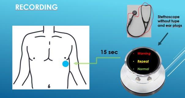
Description: Hospitalization for worsening heart failure is associated with a 30% mortality and an average price of 100.000 DKK. Currently, home care monitoring is limited to daily weight and symptom scoring, which are coarse and unprecise measures. The best and most common way of detecting worsening in heart failure is from pulmonary auscultation of rales with a stethoscope. Until today it has not been practical as it requires a doctor. We have used the recent development in AI detection algorithms and developed an automated stethoscope. Using an electronic stethoscope which is cable of recording and sending pulmonary sounds, the patient can perform auscultation on the left chest, and the sound file is subsequently transferred and analyzed using an AI-algorithm.
We have trained the AI-algorithm on sound files available on online databases and applied the algorithm on our files from the patients and the normal volunteers. The algorithm has a sensitivity of 85% of detecting pulmonary congestion and a specificity of 60%. These results indicate that it may be possible to prevent 8 out of 10 hospitalizations. We are now investigating the predictive power of the electrocardiogram (ECG).
In this Master project, the student will work with optimizing an AI algorithm to predict heart failure from the ECG. The project will involve AI-programing, data analysis, and sound analysis.
Required qualifications: Experience with machine learning, ECG, signal processing.
Responsible institution/department: DTU Health Technology and cardiological research unit at AmagerHvidovre Hospital.
Contact information:
Associate professor, MD, Jens Dahlgaard Hove, E-mail: Jens.Dahlgaard.Hove@regionh.dk
Professor, Sadasivan Puthusserypady, E-mail: sapu@dtu.dk
PhD Student, Xiaopeng Mao, E-mail: xiama@dtu.dk
Allowed no of students per report: 1
KU and/or DTU supervisor: DTU Professor Sadasivan Puthusserypady
Project Title: AI stethoscope for heart failure
Description: We have an electronic stethoscope which is cable of recording and sending pulmonary sounds. We have sampled sounds from patients hospitalized with acute heart failure and normal volunteers. We have trained an AI-algorithm on sound files available on online databases and applied the algorithm on our files from the patients and the normal volunteers. The algorithm has a sensitivity of 85% of detecting pulmonary congestion and a specificity of 60%. These results indicate that it may be possible to prevent 8 out of 10 hospitalizations. We have since sampled more sounds file to further improve the algorithm. Next step is now to conduct a small proof-of-concept study to verify that the system is working in the home-care setting of heart failure patients. We will conduct such a trial of 10 patients and are looking for a master student to perform the analysis and validation of these real-world data in close cooperation with the hospital staff in charge of the treatment of the patients. The project is the first of its kind to apply an electronic stethoscope with subsequent AI analysis for home care monitoring. In the initial proof-ofconcept study, we will provide the device for 10 patients and investigate quality of the sound files and the AI performance.
Required qualifications: Experience with medical data processing, machine learning, and acoustic signal processing.
Responsible institution/department: DTU Health Technology and cardiological research unit at AmagerHvidovre Hospital.
Contact information:
Associate professor, MD, Jens Dahlgaard Hove, E-mail: Jens.Dahlgaard.Hove@regionh.dk
Professor, Sadasivan Puthusserypady, E-mail: sapu@dtu.dk
PhD Student, Xiaopeng Mao, E-mail: xiama@dtu.dk
Allowed no of students per report: 1
KU and/or DTU supervisor: DTU Professor Sadasivan Puthusserypady
AI-Guided Discovery and Validation of Peptide Binders for Cancer and Immunological Targets in Imaging and Therapy
Recent advances in AI-based protein modeling, particularly AlphaFold-derived architectures, have revolutionized the way peptide–protein interactions can be predicted and designed in silico. This project aims to leverage such technologies to discover, optimize, and validate de novo peptide binders against selected oncological or immunologically relevant targets. The ultimate goal is to develop novel peptidebased radiopharmaceuticals for molecular imaging and targeted radionuclide therapy
The project combines computational peptide design with preclinical translational assessment. The computational component will focus on leveraging different AlphaFold-based AI tools such as EvoBind, BindCraft, and PepMLM to identify short, high-affinity peptides capable of specifically recognizing cell proteins. The most promising peptide candidates will be synthesized, validated for target binding in vitro, and tested in vivo to assess their feasibility as peptide-based agents for molecular imaging and radionuclide therapy (see workflow below)

The project offers flexibility, enabling the student to focus on the parts they find most interesting, such as computational modeling, in vitro studies, or in vivo testing, while still understanding the full workflow.
Required qualifications:
We are looking for enthusiastic students who enjoy being in a lab environment, are willing to learn, and have a can-do attitude toward problem-solving. Knowledge within protein structure and computational biology and basic coding skills to learn to use pre-existing AI tools. FELASA animal certification is mandatory for all projects involving animal work.
Responsible institution/department: Cluster for Molecular Imaging, Department of Biomedical Sciences & Department of Clinical Physiology and Nuclear Medicine, University of Copenhagen and Rigshospitalet.
Contact information:
Danna Kamstrup Sell Cand. Pharm., PhD, Postdoc danna@sund.ku.dk
Allowed no of students per report: 1-2
KU and/or DTU supervisor: Professor Andreas Kjær, MD, DSc, PhD
Project Title: Annotation of non-human DNA reads in cell-free DNA samples from the Copenhagen Pregnancy Loss Cohort
Description:
This project investigates non-human DNA fragments found in cell-free DNA (cfDNA) sequencing data from 3,000 women in the Copenhagen Pregnancy Loss Study (COPL). The samples were collected at the time of pregnancy loss, with the initial goal of determining whether each loss was euploid or aneuploid. However, a small fraction of the cfDNA remains unaligned, representing potential DNA from microbial or viral commensals or, importantly, pathogens
The objective of this project is to identify and characterize the origin of these unaligned reads and to determine the contribution of pathogen-associated signatures in euploid vs. aneuploid pregnancy loss This work has high clinical relevance for understanding the potential infectious component in the pathology of pregnancy loss.
The project will proceed in four steps:
1. Read identification and preprocessing: Isolate and quality-filter unmapped reads to ensure reliable downstream analysis.
2. Sequence classification: Explore different read classifiers (such as sequence alignment and k-merbased approaches) and databases (microbial, viral). cfDNA reads are typically short, which poses unique challenges for standard metagenomic pipelines
3. Validation: Apply decontamination tools, calculate expected coverage based on abundance and compare with actual genome coverage, and verify the robustness of organism detections
4. Clinical correlation Compare microbial and viral profiles between euploid and aneuploid pregnancy losses and cross-reference findings with existing serological data (pathogen-specific antibody tests) to validate genomic results and explore correlations with the patient immune response.
During the project, you will also gain experience working with the Danish Supercomputer for AI, Gefion.
Required qualifications: Metagenomics (for example 23260 Applied methods in metagenomics or similar). Experience with alignment or k-mer-based tools. Introductory statistics.
Responsible institution/department: Department of Health Technology, Technical University of Denmark and Department of Gynecology and Obstetrics, Hvidovre Hospital
Contact information: David Westergaard (David.westergaard@region.dk) or Pernille Neve Myers (pernille.neve.myers@regionh.dk)
Allowed no of students per report: 1-2
KU and/or DTU supervisor: Associate Professor David Westergaard (DTU)
Project Title: Automated organ segmentation in preclinical MRI
Description: Radioligand Therapy (RLT) is a therapeutic approach that delivers radiation directly to diseased tissue by attaching radionuclides to targeting molecules. Unlike conventional, external radiation therapy, which affects larger tissue regions, RLT relies on molecular targeting to provide highly localized treatment. The clinical potential of this has sparked major academic and commercial interest, with several radiopharmaceuticals already approved and many others under development.1
The effectiveness and safety of RLT depend on accurate dosimetry, which quantifies how radiation is distributed between tumors and healthy organs. Dosimetry requires molecular imaging modalities such as PET or SPECT to track radioligand distribution, combined with anatomical imaging for reference. Manual annotation of imaging data for this purpose is highly time-consuming, making automated segmentation an essential component of modern dosimetry pipelines. Within the Cluster for Molecular Imaging (CMI), a CT-based organ segmentation model already reduces annotation time from nearly an hour to just a few minutes while maintaining high accuracy.
CT provides reliable anatomical information but has limited soft-tissue contrast. MRI offers much better visualization of soft tissues and therefore has the potential to improve organ segmentation quality in preclinical studies. However, annotated MRI datasets are scarce, and models trained on CT do not transfer easily to MRI.
This project will address these challenges by developing an organ segmentation framework for preclinical MRI. The student(s) will curate and annotate an MRI dataset, adapt the existing CT-based nnU-Net model2 through transfer learning, and explore strategies such as cross-modality fine-tuning, contrastive representation learning, or synthetic data generation3. Performance will be evaluated in terms of segmentation accuracy, robustness across imaging conditions, and usability within CMI’s automated dosimetry pipeline. The results will clarify the role of MRI as a complementary or alternative modality to CT for automated organ segmentation in RLT research.
Preferred qualifications/courses:
• 02456 – Deep learning
• 02502 – Image analysis
• General programming skills in Python and PyTorch
Responsible institution/department:
Cluster for Molecular Imaging, Department of Biomedical Sciences & Department of Clinical Physiology and Nuclear Medicine, University of Copenhagen and Rigshospitalet
Contact information: Andreas Clemmensen, andreas.clemmensen@sund.ku.dk
Allowed no of students per report: 1-2
KU and/or DTU supervisor: Professor Andreas Kjær, MD, DSc, PhD
References:
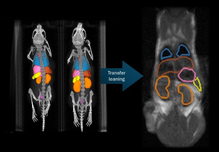
LEFT: AUTOMATICALLY SEGMENTED ADULT MICE USING THE CURRENT CMI PIPELINE FOR CT
RIGHT: MANUALLY SEGMENTED ADULT MOUSE T1-WEIGHTED MRI - SCAN
1 Sgouros, G., Bodei, L., McDevitt, M. R., & Nedrow, J. R. (2020). Radiopharmaceutical therapy in cancer: clinical advances and challenges. Nat Rev Drug Discov, 19(9), 589-608. https://doi.org/10.1038/s41573-020-0073-9
2 Fabian Isensee et al., “nnU-Net: A Self-Configuring Method for Deep Learning-Based Biomedical Image Segmentation,” Nature Methods 18, no. 2 (February 1, 2021): 203–211, https://doi.org/10.1038/s41592-020-01008-z
3 Luca Vellini et al., “A Deep Learning Algorithm to Generate Synthetic Computed Tomography Images for Brain Treatments from 0.35 T Magnetic Resonance Imaging,” Physics and Imaging in Radiation Oncology 33 (2025): 100708, accessed October 1, 2025, https://linkinghub.elsevier.com/re-
Project Title: Automated Sleep Profiling for Neurodegenerative Disease Using Sleep Signals and Machine Learning
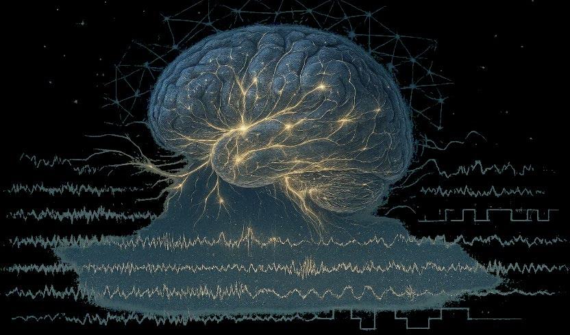
Description: Sleep is a sensitive marker of brain health, and disturbances in sleep architecture, muscle activity, and autonomic regulation often precede the clinical onset of neurodegenerative diseases such as Parkinson’s disease Polysomnography (PSG) provides a unique window into these early pathophysiological changes, but large-scale and automated analyses are required to translate such insights into clinical and epidemiological applications.
In this project, the student will build a comprehensive computational pipeline for automatic sleep analysis and disease profiling. Using existing algorithms, the student will integrate and fine-tune methods into a unified framework for large-scale analysis of PSG data. The goal is to extract a rich set of sleep -related biomarkers and use interpretable machine learning models to identify patterns associated with neurodegenerative disease risk.
Ultimately, the developed pipeline will be applied to nationwide PSG and linked to patient health outcomes from national registries hosted at Danmarks Statistik.
Required qualifications: The student should have a background in biomedical engineering, computer science, or related disciplines, with prior experience in Python programming, signal processing, and machine learning.
Responsible institution/department: DTU Health Tech
Contact information: andbri@dtu.dk
Allowed no of students per report (1-4): 1 - 4
KU and/or DTU supervisor:
Andreas Brink-Kjær, PhD – Assistant Professor, DTU Health Tech.
Umaer Hanif, PhD – Postdoc, Rigshospitalet
Poul Jennum, MD, PhD – Chief Physician, Rigshospitalet
Project Title: Automatic Detection of Central Hypersomnias Using Sleep Signals and Machine Learning
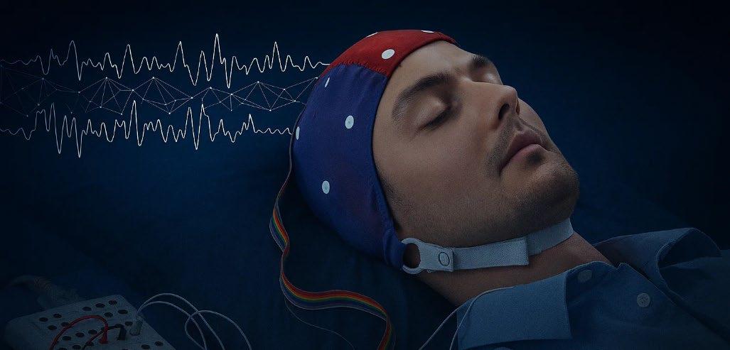
Description: Central hypersomnias, including narcolepsy type 1, are characterized by excessive daytime sleepiness and abnormal transitions between sleep and wakefulness. These disorders remain challenging to diagnose, as they require laborintensive clinical tests such as polysomnography (PSG), the Multiple Sleep Latency Test (MSLT), and cerebrospinal fluid measurements. Automating aspects of this diagnostic process could enable earlier, more consistent, and scalable detection of hypersomnias.
In this project, the student will develop and evaluate machine learning approaches for the automatic detection of central hypersomnias. Using large-scale PSG and MSLT recordings provided by the Danish Center for Sleep Medicine at Rigshospitalet, the student will employ probabilistic sleep staging algorithms to characterize sleep architecture and identify abnormal patterns such as sleep - onset REM periods and mixed-state phenomena. These features will then be used to derive quantitative biomarkers and interpretable models that distinguish hypersomnia patients from controls and other sleep disorder groups.
Required qualifications: The student should have a background in biomedical engineering, computer science, or related disciplines, with prior experience in Python programming, signal processing, and machine learning.
Responsible institution/department: DTU Health Tech
Contact information: andbri@dtu.dk
Allowed no of students per report: 1 - 2
KU and/or DTU supervisor:
Andreas Brink-Kjær, PhD – Assistant Professor, DTU Health Tech.
Umaer Hanif, PhD – Postdoc, Rigshospitalet
Poul Jennum, MD, PhD – Chief Physician, Rigshospitalet
Brain spirals and their modulation by psilocybin
A recent theory suggests that brain activity can be understood through the emergence of and interactions between brain spirals in a two-dimensional flattened version of the unihemispheric human cortex, similar to how fluid dynamics are modelled via dynamical systems (see Figure below). These spirals are detected by analyzing the oscillatory motion of functional MRI data. This idea not only suggests that brain activity in each region is oscillatory but also that the speed and directionality of this oscillatory motion is spatially constrained by the closeness to nearby spiral centers.
At the Neurobiology research unit (NRU), we are initially intrigued by (and slightly skeptical of) this idea. We want to understand in further detail the (spatiotemporal) constraints of the method. If we deem the method trustworthy, it could fundamentally change the way we think about how brain activity spreads along the cortex. The student(s) should:
• Implement brain spiral detection on fMRI data from healthy controls
• Investigate how the number and behavior of brain spirals change as a function of spatial and temporal constraints (filters) on the model
• Analyze how brain spirals are affected by the psychedelic drug psilocybin.
Due to the scale of the project, it is likely mostly suitable for master’s students.
Main supervisor: Anders S Olsen, NRU, anders.s.olsen@nru.dk
Official supervisor: Prof. Morten Mørup, DTU, mmor@dtu.dk
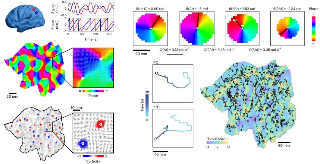
Brain spiral detection – figure modified from Xu et al., 2023, Nature Human Behavior: “Interacting spiral wave patterns underlie complex brain dynamics and are related to cognitive processing”
Project Title:
Co-development of UX for Spasticity Treatment after Acquired Brain Injury
Description:
Spasticity is a common consequence of acquired brain injury (e.g. stroke or traumatic brain injury) and requires lifelong, cross-sectoral follow-up. Patients often face cognitive, coordination, and visual challenges that make standard app interfaces unusable.
This project aims to co-design and test a user-friendly app interface that:
• Supports therapists and physicians in documenting spasticity treatments
• Enables patient-centered goal setting and follow-up
• Facilitates communication between sectors (hospital, municipality, therapists, etc.)
The project focuses on UX design and user involvement rather than technical app development. Methods include literature review, interviews, co-design workshops with patients/relatives, prototyping in Figma, and user testing with validated tools. Expected outcomes are an interactive UX prototype, design principles for apps targeting cognitive and visual impairments, and a thesis report.
Required qualifications:
• Interest in UX and human-centered design
• Familiarity with design tools (e.g. Figma) is an advantage
• Background in health sciences, design, or related fields
• Danish language skills are useful but not strictly required
Responsible institution/department: Movement Disorder and Pain Research Center, Rigshospitalet Glostrup
Contact information: trine.hoermann.thomsen@regionh.dk
Bo.biering-soerensen@regionh.dk
Allowed no of students per report (1–4): 2-4
KU and/or DTU supervisor:
Assistant Professor Trine Hørmann Thomsen, Digital Health, Department of Health and Technology, and Department of Brain and Spinal Cord Injuries, Rigshospitalet
Project Title: Conditional VAEs for Multi-Omics Batch Effect
Removal
Description: Our group focusses on Data Science and bioinformatics for women’s health. In the Copenhagen Pregnancy Loss study (COPL) data of over 3000 participants were collected to decipher the pathways and mechanisms underlying pregnancy loss. This rich dataset includes multiple layers of omics data. Multi-omics studies which integrate diverse data types like genomics, transcriptomics, and proteomics provide a holistic view of biological systems. However, these datasets are highly susceptible to batch effects, i.e. technical variation introduced by differences in experimental conditions rather than true biological differences . Each omics layer (e.g., metabolomics, proteomics) is often processed on different machines, at different times, or in different labs, introducing significant technical noise. This noise dominates true biological signals, preventing effective integration and reliable downstream analysis. Conditional Variational Autoencoders (cVAEs) offer a powerful deep learning solution: they can learn complex, non-linear relationships across omics layers while explicitly being guided to discard the nuisance batch variation.
The project utilizes a cVAE framework to harmonize and integrate multi-omics datasets by isolating and removing batch effects.
• Integrated Model Design: Develop a multi-modal cVAE architecture that takes all omics layers as input. The model's encoder must be conditioned on the batch labels (e.g., source lab, sequencing date) associated with the data. This configuration forces the model to learn a shared lower dimensional representation of the data, a so-called latent space common to all omics types but independent of the technical batch.
• Decoupling Biological and Technical Variation: Employ a robust training strategy, often incorporating an adversarial or regularization component. The goal is to generate a clean, unified latent representation (Zbio) where the features related to biological variation (e.g., disease state) are retained, but batch-specific noise is minimized.
• Data Harmonization and Validation: Generate the batch-corrected multi-omics data by decoding the batch-independent latent representations (Zbio), conditioning the decoder on a single, uniform target batch state. Validate the correction by integrating the omics layers in the corrected decoded space (e.g., using multi-omics factor analysis) and visually confirming that technical clustering is eliminated while biological clustering is preserved.
Required qualifications: Machine learning, deep learning, ‘omics analysis, intermediate statistics
Responsible institution/department: Department of Health Technology, Technical University of Denmark and Department of Gynecology and Obstetrics, Hvidovre Hospital
Contact information: David Westergaard (David.westergaard@region.dk) and Luisa Schäfer (luisa.schaefer@regionh.dk)
Allowed no of students per report: 1-2
KU and/or DTU supervisor: Associate Professor David Westergaard (DTU)
Project Title: Deep Learning for Uterine Pathology Detection
Description:
Our group focusses on Data Science and bioinformatics for women’s health. In the Copenhagen Pregnancy Loss study (COPL), data from over 3000 participants were collected to decipher the pathways and mechanisms underlying pregnancy loss. This rich dataset includes thousands of 3D ultrasound scans of the uterus, providing a unique opportunity to apply advanced computational approaches
Specifically, AI for Pathology Detection in 3D Ultrasound Scans is a transformative area of research that uses deep learning models to process such volumetric ultrasound data, aiming to enhance diagnostic accuracy, reduce operator variability, and improve clinical workflow.
Traditional 2D ultrasound is highly operator-dependent, with diagnostic accuracy often limited by the sonographer's skill and the inherently noisy, real-time nature of the images (e.g., speckle noise and acoustic shadows). While 3D ultrasound offers richer spatial information (volumetric context) compared to 2D images, the sheer size and complexity of these 3D datasets make manual examination time-consuming and prone to human error. AI, particularly Deep Learning (DL) models like Convolutional Neural Networks, is being developed to overcome these limitations by automating the analysis of this complex, volumetric data.
Key tasks and applications include:
1. Volumetric Data Processing and Network Architecture: Develop a machine learning model tailored for pathology detection in ultrasound volumes. Implement comprehensive preprocessing workflows including voxel intensity and volume size standardization as well as geometric transformations for data augmentation. Generate modular, well-documented code that supports reproducible model training and inference on volumetric medical imaging data.
2. Training Strategy and Hyperparameter Optimization: : Establish a robust learning scheme that balances model complexity with available training data. Systematically optimize critical hyperparameters including learning rate, batch size, network depth, and regularization strength to maximize classification performance while maintaining computational efficiency. Employ validation strategies to prevent overfitting and to assess model stability and generalization capacity
3. Evaluate model performance using standard classification metrics (accuracy, sensitivity, specificity, AUC-ROC) and validate diagnostic reliability across independent test cohorts. Implement explainable AI techniques to visualize which anatomical regions drive the model's predictions, thereby enabling clinical interpretation and building trust in automated diagnostic decisions through transparent, interpretable outputs. Quantitatively compare model performance against human expert annotations using inter-rater agreement metrics, establishing whether automated detection meets or exceeds clinical standards for uterine pathology identification.
Required qualifications: Machine learning, Deep learning, Image analysis
Responsible institution/department: Department of Health Technology, Technical University of Denmark and Department of Gynecology and Obstetrics, Hvidovre Hospital
Contact information: David Westergaard (David.westergaard@region.dk) and Luisa Schäfer (luisa.schaefer@regionh.dk)
Allowed no of students per report: 1-2
KU and/or DTU supervisor: Associate Professor David Westergaard (DTU)
Project Title: EEG-to-text AI technology
Description: According to the WHO, approximately 1.3 billion people worldwide are affected by significant disabilities, such as Locked-in Syndrome (LIS), Cerebral Palsy (CP), Amyotrophic Lateral Sclerosis (ALS), dysarthria, etc. Despite having intact intelligence, these health issues can restrict the patient’s ability to move and communicate, thus hindering their capacity to engage in everyday activities. This project aims to develop a brain-to-text model a system that translates patterns of brain activity directly into written language. Electroencephalography (EEG) measures tiny electrical signals from the scalp that reflect how the brain processes language. By learning to interpret these signals, an EEG-to-text model could one day help people with speech impairments communicate more easily or provide new tools for studying how the brain understands and produces language.
To train and evaluate the model, we use two complementary datasets. Zurich Cognitive Language Processing Corpus (ZuCo) 1.0 records EEG and eye-tracking data from participants as they read English sentences naturally. It provides rich information about how the brain responds to language during real reading. Chinese Imagined Speech Corpus (Chisco), on the other hand, focuses on imagined speech in Chinese. Participants silently recall or imagine sentences, and their EEG is recorded during this internal speech process. Chisco contains over 20,000 phrases across 39 semantic categories.
Your task in this project is to understand and implement a state-of-the-art classification or language decoding model applied on the ZuCo dataset. The final product will be an improved model that can be applied to the Chisco dataset.
Required qualifications: You should have great experience with EEG modelling and machine learning for working with this project.
Responsible institution/department: DTU Health Technology
Contact information:
Professor, Sadasivan Puthusserypady, E-mail: sapu@dtu.dk
PhD Student, Xiaopeng Mao, E-mail: xiama@dtu.dk
Allowed no of students per report: 1
KU and/or DTU supervisor: DTU Professor Sadasivan Puthusserypady
Project Title: Functional Characterization of Microbial β-
Glucuronidase in the Menstrual Cycle and Pregnancy
Description:
The human microbiome plays a crucial role in regulating hormone metabolism and reproductive physiology. One key microbial enzyme, β-glucuronidase (GUS), can cleave glucuronidated (inative) estogens to release their active form. Estrogens are produced in the ovaries, metabolized and conjugated in the liver, and excreted into the bile. In the gut, microbial GUS enzymes can reactivate these conjugated estrogens, which are then reabsorbed via enterohepatic circulation. The gut microbiome is therefore central to maintaining estrogen bioavailability. Together, these systems form the estrabolome.
The Department of Gynecology and Obstetrics at Hvidovre Hospital is conducting a large-scale study to map changes in the estrabolome across two key reproductive states: the menstrual cycle and pregnancy This Master's project contributes to that effort through the bioinformatics analysis of deep shotgun sequencing data from gut and vaginal microbiomes collected from patients at the hospital. The student will:
1. Identify active vs. inactive GUS: Use MSA (Multiple Sequence Alignment) on known active GUS sequences to pinpoint specific motifs that distinguish functionally active (estrogen-releasing) from inactive enzymes.
2. Microbiome Screening: Develop specialized models (HMMER or refined BLAST) based on the active motifs to accurately identify and quantify the abundance of these active GUS genes in the patient's gut and vaginal microbiome sequencing data.
3. Correlation: Statistically correlate the measured abundance of active GUS genes with the patient's actual, measured circulating estrogen levels across different phases of the menstrual cycle and pregnancy.
Required qualifications: Metagenomics (23260 Applied methods in metagenomics or similar), molecular evolution techniques (such as Multiple sequence alignment (MSA), Hidden Markov Models for protein domain detection (HMMER), and phylogenetic trees), and introductory statistics are also recommended.
Responsible institution/department: Department of Health Technology, Technical University of Denmark and Department of Gynecology and Obstetrics, Hvidovre Hospital
Contact information: David Westergaard (David.westergaard@region.dk) or Pernille Neve Myers (pernille.neve.myers@regionh.dk)
Allowed no of students per report: 1-2
KU and/or DTU supervisor: Associate Professor David Westergaard (DTU)
Project Title: Genomic Architecture of Placental Health
Project objectives
The placenta is a vital organ in pregnancy and placental dysfunction is a feature of many pregnancy complications. There is therefore a need for a deeper understanding of the pathological changes that can occur in the placenta such as reduced oxygen access, inflammation, and delayed maturation of the placental structures. Macro- and microscopic changes can be surveyed manually by pathological specialists; however, automated classification and phenotyping tools exist. Furthermore, the genetic architecture underlying placental pathology remains incompletely characterized, presenting an opportunity to investigate the genomic determinants of placental health through genome-wide association studies (GWAS).
In this project, you will get firsthand experience working with the implementation, testing, and validation of pre-trained model for deriving quantitative imaging phenotypes from digitized placental biopsies. You will perform GWAS on the derived phenotypes to elucidate the biology underlying the different features. If time permits, you will investigate the relation between imaging-derived phenotypes, genetic variants, and complications in pregnancy
During the project, you will:
• Set the project in context of the current literature on placental cellular pathology.
• Implement, test, and validate the current state-of-the-art models for analysis of placental microscopic imaging. If relevant, improve performance through further fine-tuning and/or optimization of the pipeline.
• Identify different cell and tissue types in H&E-stained placental biopsies, as well as pathogenic features.
• Describe the study cohort, conduct subgroup analysis, and identify phenotypes of interest.
• Conduct genome-wide association studies (GWAS) of imaging-derived placental phenotypes using linear or logistic regression models adjusted for relevant covariates including gestational age, maternal age, and principal components of genetic ancestry.
• Compare and critically analyze the generated results in the context of scientific findings within the field. Identify the utility of the findings from a clinical perspective.
Allowed no of students per report: 1-2
Supervisors: Postdoctoral researcher Agnete Troen Lundgaard & Associate Professor Karina Banasik (DTU)
Contact information: Karina Banasik (karina.banasik@region.dk) or Agnete Troen Lundgaard (agnete.troen.lundgaard@regionh.dk)
Project Title: Heart rate and heart rate variability effects in the relation to acoustic exposure and listening effort
Description:
This project explores the potential of heart rate (HR) and heart rate variability (HRV) as physiological indicators in the context of everyday listening. Using wearable technology and ecological momentary assessment (EMA), the study will examine how individuals experience and respond to different acoustic environments. The specific focus can be on quiet vs. noisy environments, changes during the day, hearing status, or investigations during lab-based speech-in-noise tasks, and can be adapted to include additional wristband metrics. The project can be further defined depending on the student’s interests.
Required qualifications:
We are looking for one or more students interested in collection and analysis of HR/HRV data within the hearing field.
Responsible institution/department:
The study is a collaboration between DTU hearing systems, the Ear, Nose, Throat & Audiology department at Rigshospitalet, and Eriksholm Research Centre
Contact information:
Maaike Van Eeckhoutte: mcvee@dtu.dk
Jeppe Høy Konvalinka Christensen: jych@eriksholm.com
Allowed no of students per report: 1-2
KU and/or DTU supervisor:
Maaike Van Eeckhoutte (DTU Hearing Systems and Copenhagen Hearing and Balance Centre Rigshospitalet)
How obstructive sleep apnea impacts cardiac hemodynamics –using clinical medical imaging in a large animal model
Description:
Obstructive sleep apnea leads to heart failure and arrhythmia in patients. The underlying mechanisms are not completely understood. Nevertheless, stretching of the myocardial tissue and altered hemodynamics of the heart – especially upon release of the apnea – are considered to play a role. This study consists of image processing of computer tomography (CT) scans (dynamics, multi-slide covering the entire heart) in living pigs subjected to an obstructive respiratory event, thus mimicking obstructive sleep apnea in patients. This truly translational project will provide evidence of how the heart’s mechanics are altered by sleep apnea. We envisage to develop a semi-automatized analytical tool for volume estimation (ventricles) in pig hearts – which does not currently exist.
Required qualifications:
Independent and talented student(s) with excellent knowledge in human biology and image processing, motivated to work with real-life data and translational science. If interested, please send CV, study record incl. grades, and short motivational letter mentioning a bit about yourself and your future professional ambitions. This project involves a lot of data processing and is therefore most suitable for project +20 ECTS points.
Responsible institution/department:
Department of Biomedical Sciences (BMI), KU
Contact information:
Lisa A. Gottlieb gottlieb@sund.ku.dk
Allowed no of students per report: 1-2
KU and/or DTU supervisor:
Dr. Lisa A. Gottlieb, BMI, KU
DTU supervisor TBA.
How the heart chambers crosstalk – an electrocardiographic study in patients with prior myocardial infarction
Description:
After a myocardial infarction in the ventricles, ventricular arrhythmias often occur, and the underlying mechanisms have been studied. Still, atrial arrhythmias also manifest after a myocardial infarction, and the mechanisms here are yet to be explored. We will use long-term (min. 24 hours) EKG recordings from patients with prior myocardial infarction to study the interplay between the atria and ventricles. Our results will pave the path for a new way of thinking arrhythmias in the heart.
Required qualifications:
Independent and talented student(s) with excellent knowledge in human biology and signal processing, motivated to work with real-life data If interested, please send CV, study record incl. grades, and short motivational letter mentioning a bit about yourself and your future professional ambitions. This project involves a lot of data processing and is therefore most suitable for project +20 ECTS points.
Responsible institution/department:
Department of Biomedical Sciences (BMI), KU
Contact information:
Lisa A. Gottlieb gottlieb@sund.ku.dk
Allowed no of students per report: 1-2
KU and/or DTU supervisor:
Dr. Lisa A. Gottlieb, BMI, KU
Rigshospitalet supervisor (TBA)
DTU supervisor TBA.
Impact of Obstructive Sleep Apnea on Cardiac Electrophysiology and
Cardiac Disease Development in a Danish Patient Cohort
Description:
Obstructive sleep apnea associates with heart failure and arrhythmia in patients. Still, the patho(physiological) processes of how apnea leads to disease are unknown. In this project, we will analyze polysomnography data (EKG, oxygen saturation, blood pressure recordings during an entire night) from patients with and without obstructive sleep apnea and thereby establish biological patterns during apnea associated with arrhythmia formation.
Required qualifications:
Independent and talented student(s) with excellent knowledge in human biology and signal processing, motivated to work with real-life data with the potential to improve diagnosis in patients with obstructive sleep apnea. If interested, please send CV, study record incl. grades, and short motivational letter mentioning a bit about yourself and your future professional ambitions. This project involves a lot of data processing and is therefore most suitable for project +20 ECTS points.
Responsible institution/department:
Department of Biomedical Sciences (BMI), KU
Contact information:
Lisa A. Gottlieb gottlieb@sund.ku.dk
Allowed no of students per report: 1-2
KU and/or DTU supervisor:
Dr. Lisa A. Gottlieb, BMI, KU
Dr. Andreas Brink-Kjær, DTU
Prof. Poul Jennum, Rigshospitalet
Investigating the effect of common artifacts on the frequency spectrum and phase of fMRI data.
Functional MRI (fMRI) is an imaging modality that measures brain activity on slow timescales, typically with wavelengths between 128s and ~5s (frequencies 0.008Hz – 0.2Hz), and an acquisition is typically 5-15min duration. A question often of interest in fMRI analysis is whether inferred brain function, be it the activity in a particular brain network or the difference between healthy controls and those suffering from brain disease or the effect of a particular drug, is specific for frequencies or frequency bands. However, the low sampling rate (fs~=0.5Hz) means that artifacts, particularly motion, often obscure large parts (i.e., proportionally many samples) of an acquisition. These artifacts are not easily described by sine waves, and the Fourier spectrum will therefore be affected over multiple frequency components.
This project aims to describe exactly how common heterogeneous fMRI artifacts (motion in particular) affect 1) the frequency spectrum, and, if time permits, 2) the phase, where the data is constrained to a narrow frequency band. The project should analyze data where synthetic artifacts are generated and added to motion-free fMRI to investigate the effect on the spectrum. Secondly, the project should analyze fMRI data from a psilocybin experiment, where 28 subjects were scanned at multiple time-points during a “magic mushroom” trip. This trip naturally induces motion in some subjects, where the intensity and occurrence of motion correlate with the intensity of the psychedelic trip. Therefore, we wish to disentangle motion effects from drug effects when investigating the Fourier spectrum and phase.
Main supervisor: Anders S. Olsen, postdoc at Neurobiology Research Unit (NRU), Copenhagen university hospital Rigshospitalet. anders.s.olsen@nru.dk
Official supervisor: Either Cyril R. Pernet or Gitte M. Knudsen, also at NRU.
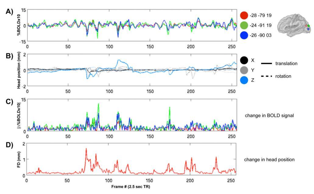
Figure taken from “Spurious but systematic correlations in functional connectivity MRI networks arise from subject motion”, JD Power et al, Neuroimage, 2010 and shows typical effects of motion on the fMRI time-series. In this project, we will investigate the effects on the fMRI spectrum.
Title: Machine learning graph representations for 12-lead ECG
MSc project
Introduction
Electrocardiograms (ECGs) are a cornerstone of cardiovascular care and are widely used for diagnostics in primary or emergency care. Typically, ten electrodes are attached at different points of the human skin, resulting in 12 so -called “leads” which are signals offering different “perspectives” on the heart. However, most ECG processing methods – from traditional signal processing to deep learning approaches treat them as multidimensional time series without considering the spatial and physiological relationships between leads.
Task
In this project, we will explore the representation of 12-lead ECG signals as graphs, where nodes represent individual leads and/or time segments and edges represent the relationship among them. Initial works have already been proposed; however, it is still unclear which representation combines spatial and temporal information “optimally”.
Goals and environment
The goal of this project is to find a graph representation balancing spatial and temporal information and to use it for typical ECG -related tasks, e.g. classification. We will compare the performance of this novel representation with state- of-the-art methods on different, relevant validation datasets. Working in our labs at DTU Lyngby (B345) is possible and encouraged. Additionally, clinical partners from cardiology are available and provide their knowledge to validate our findings.
Requirements
If you have interest in this project, you should have a background in working with medical data, ideally signals, and ideally experience with Python libraries for medical data science. After the project, you will have gained profound knowledge of ECGs and their interpretation, graphs structures, and machine learning techniques.
Supervision
Nicolai
Spicher (Ph.D.)
Associate Professor Department of Health Technology, DTU
nicsp@dtu.dk
Project Title:
Mapping microglia in Multiple System Atrophy – whole slide image analysis of human hemispheres
Description:
The overarching aim of the proposed project is to understand the role of neuroinflammation in relation to pathological disease progression in multiple system atrophy (MSA). Specifically, we will test the hypothesis that activation of microglia precedes the deposition of alpha-synuclein in glial cytoplasmic inclusions (GCIs) and neuronal loss.
The objective of the study is to tailor an automated image analysis algorithm to map the spatial distribution of neuroinflammation, GCIs and neuronal densities in series of whole hemisphere sections stained for alpha-synuclein and activated microglia. We will do this in our bespoke cohort of 12 MSA patients [ref ref ref] for which we have detailed clinical information and stereological cell counting to support the 2D densities on which we will build out disease progression atlases.
Building a model for the pathological disease progression may help to identify novel biomarkers by which we may track and trace disease progression and treatment effects in vivo
Required qualifications:
1. Students from Biomedical Engineering (Medicin og Teknologi) or related fields
2. Programming experience (preferably in Matlab and/or Python)
3. Introductory knowledge of image analysis (e.g., through course 02515 or similar)
Responsible institution/department:
Dept. of Health Technology
Contact information:
Sanne Simone Kaalund, sansim@dtu.dk
Allowed no of students per report: 1-2
KU and/or DTU supervisor:
Sanne Simone Kaalund, Technical University Hospital, DTU Health Tech
Method development for Magnetic Resonance Imaging (MRI)
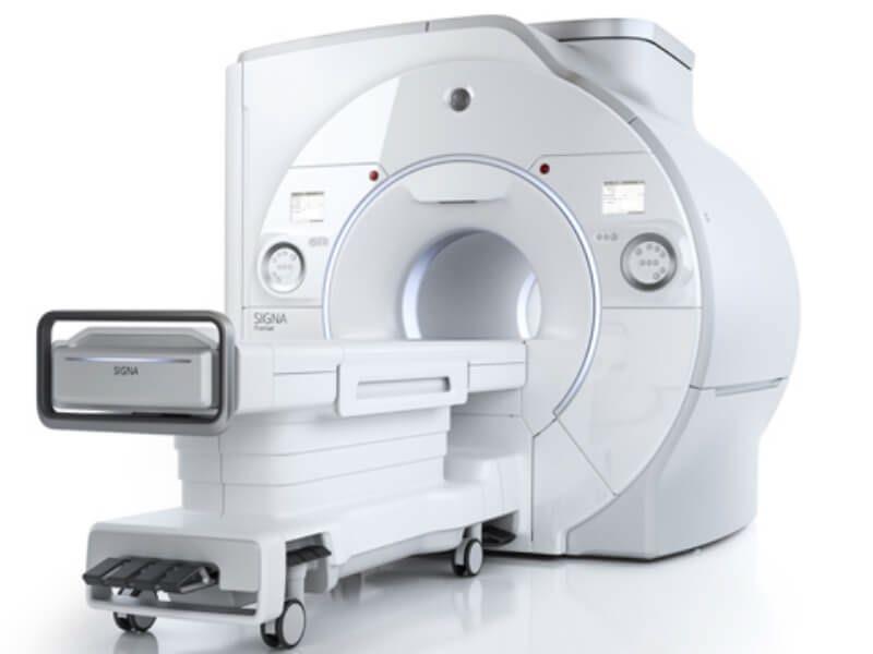
Description: Magnetic Resonance Imaging and Spectroscopy techniques (MRI/MRS) are challenging, but also safe and extremely flexible providing non-invasive and detailed tissue characterization. Magnetic fields of typically 1.5 to 3 tesla are used for humans. The state-of-the-art 3 tesla human scanner at DTU is ready for experimentation, for example, and a 7 tesla human scanner is available at Hvidovre Hospital as a result of a national collaboration involving DRCMR, DTU and other partners.
New innovations give more possibilities, but also challenges that need to be overcome. There are hundreds of magnetic resonance imaging and spectroscopy techniques and more are constantly being developed, refined, validated and employed for clinical or research uses at Danish MRI sites operating at different field strengths. Students with relevant skills are invited to express interest, so project options can be discussed. There are possibilities for projects that are oriented toward physics, electronics, method development, statistics, medical applications, brain function and more.
Required qualifications: Different competences are of interest, and projects matching your background can likely be proposed. It is an advantage to have one or preferably more of the courses 22481, 22485, 22506, 22507 or 22508 (see https://www.cmr.healthtech.dtu.dk/education/mr-courses-and-their-connection). MRI can be quite challenging, and some projects are therefore only suited for MSc students.
Responsible institution/department: DTU Health Tech / DTU Compute and/or Danish Research Centre for MR, DRCMR, http://www.cmr.healthtech.dtu.dk , http://www.compute.dtu.dk, http://drcmr.dk/
Contact information: Lars G. Hanson, lghan@dtu.dk or people mentioned below.
Allowed no of students per report: 1-4
KU and/or DTU supervisor: For example Axel Thielscher (neurophysics), Mathilde Hauge Lerche (brain metabolism), Henrik Lundell (microstructure & ultra-highfield MRI), Kristoffer Hougaard Madsen (machine learning), Tim Dyrby and Marco Pizzolato (microstructure), Vitaliy Zhurbenko (coil technology) or Lars G. Hanson (measurement design). See web for their mail addresses and interests (only examples are given).
BSc/MSc project for students in Biomedical Engineering and/or Quantitative Biology and Disease Modelling, DTU/KU
Project Title:
Optical coherence tomography-derived biomarkers for diagnosis and monitoring of diseases
Description: (n.a. if confidential)
This project comprises a range of possible projects working with optical coherence tomography. Optical coherence tomography is a non-invasive optical imaging modality that provides volumetric images of tissue microstructure without the need for contrast agents. OCT is most commonly used in ophthalmology, however, our research group aims to expand the use of OCT to other diagnostics, including in dermatology and oncology. The project can be more engineering focused, image processing, or application focused depending on the student. Some opportunities could include:
-Quantitative analysis of microvasculature using OCT
-Analyzing optical attenuation from OCT images in bone
-OCT imaging and analysis of tumor spheroids and co-cultures
-Identifying biomarkers of small fiber neuropathy using OCT
-Design and fabrication of fiber optic catheters for OCT
-Building and characterizing OCT systems
-Acquiring and analyzing OCT data from in vitro samples, animal models, and human patients
Required qualifications:
Project description can be adjusted to reflect the background and interest of individual students
Responsible institution/department:
DTU Health Tech
Contact information:
Gavrielle Untracht: greun@dtu.dk
Allowed no of students per report: No preference – the project can be adjusted to reflect the number of students
KU and/or DTU supervisor:
Gavrielle Untracht and Peter Andersen
Project Title: Optimizing inference speed of 3D U-Net–based organ segmentation for automated dosimetry in molecular imaging
Description: Radioligand Therapy (RLT) delivers radiation directly to diseased tissue by attaching radio nuclides to targeted molecules. While the concept is not new, recent technological breakthroughs and the approval of novel therapeutics have sparked renewed interest, with major pharmaceutical companies investing heavily in the development of new radiopharmaceuticals.1
Molecular imaging methods such as positron emission tomography (PET) and single-photon emission computed tomography (SPECT) are used to track radioligand distribution between healthy organs and diseased tissue. However, manual segmentation of images is labor-intensive and error-prone, which is why we have developed a pipeline for automated quantification of these scans. A key component of this pipeline is a semantic segmentation model based on the nnU-Net framework2, which reduces annotation time from approximately 45 minutes to just 5 minutes per scan while achieving human-level accuracy.
The project aims to further optimize this model for inference speed by exploring improvements in network architecture3 as well as pre- and post-processing strategies. The student(s) will be responsible for implementing and benchmarking optimization techniques, conducting controlled experiments, and analyzing the trade-offs between computational efficiency, segmentation accuracy, and robustness. For this project, students will have access to a large, internally developed dataset of native mouse and rat computed tomography (CT) scans with corresponding organ annotations. This unpublished dataset ensures that the project can focus on methodological and computational aspects, while also providing opportunities for novel research contributions.
Preferred qualifications/courses:
• 02456 – Deep learning
• 02502 – Image analysis
• General programming skills in Python and PyTorch
Responsible institution/department:
Cluster for Molecular Imaging, Department of Biomedical Sciences & Department of Clinical Physiology and Nuclear Medicine, University of Copenhagen and Rigshospitalet
Contact information: Andreas Clemmensen, andreas.clemmensen@sund.ku.dk
Allowed no of students per report: 1-2

PREVIOUS STUDY ON THE TRADE-OFFS IN SEGMENTATION PERFORMANCE AND SEGMENTATION MODEL ARCHITECTURE ON AN INTERNAL DATASET OF RAT AND MOUSE NATIVE CT SCANS
KU and/or DTU supervisor: Professor Andreas Kjær, MD, DSc, PhD
References:
1 Sgouros, G., Bodei, L., McDevitt, M. R., & Nedrow, J. R. (2020). Radiopharmaceutical therapy in cancer: clinical advances and challenges. Nat Rev Drug Discov, 19(9), 589-608. https://doi.org/10.1038/s41573-020-0073-9
2 Fabian Isensee et al., “nnU-Net: A Self-Configuring Method for Deep Learning-Based Biomedical Image Segmentation,” Nature Methods 18, no. 2 (February 1, 2021): 203–211, https://doi.org/10.1038/s41592-020-01008-z
3 Minyoung Park et al., “ES-UNet: Efficient 3D Medical Image Segmentation with Enhanced Skip Connections in 3D UNet,” BMC Medical Imaging 25, no. 1 (August 13, 2025): 327, accessed October 10, 2025, https://doi.org/10.1186/s12880-025-01857-0.
Project Title: Paratonia in Parkinsons Disease
Description:
Paratonia is an inability to relax muscles during passive assessment (such as when a clinician is moving the arm for a patient). It has two subtypes: resisting passive movement (gegenhalten or oppositional paratonia) and involuntarily assisting in the movement (mitgehen or facilitory paratonia).
Paratonia has mainly been linked to dementia, but clinical observation has shown that many Parkinson patients also develop paratonia. Furthermore, paratonia may be a precursor to dystonia (involuntary muscle spasms/contractions). The prevalence and characteristics of paratonia in Parkinsons Disease are unknown, and there is no standardized objective method to quantify paratonia. We want to develop a method to systematically and objectively measure paratonia presence and severity, to inform treatment decisions and assess impact on quality of life.
Required qualifications:
Interest in muscle/movement measurements (IMUs, EMG, etc).
Responsible institution/department:
Rigshospitalet Glostrup, Department of Neurology, Movement Disorder Clinic
Contact information:
Trine.hoermann.thomsen@regionh.dk
Allowed no of students per report (1-4):
Up to 4.
KU and/or DTU supervisor:
Trine Hørmann Thomsen, Assistant Professor, Department of Health Technology, DTU/TUH, and Department of Brain and Spinal Cord injuries, and Movement disorder Clinic, Rigshospitalet
Passive mechanics of heart tissue – understanding how stretching can be prevented
Description:
The heart tissue contracts and relaxes upon every heartbeat. Mechanical stretching of the heart tissue is important to ensure adequate contraction – but stretching also disturb the heart’s electrical impulses, thereby causes arrhythmias. This project will develop a methodological pipeline for investigating passive mechanics in heart tissue from pigs and humans as well as test mechanics of various disease states. This is a real hands-on project where the student will work in the lab, in close collaborations with other scientists, incl. veterinarians, as well as developing a valuable tool to be used in the future.
Required qualifications:
Independent and talented student(s) with excellent knowledge in human biology and mechanical engineering, motivated to work with real-life data and translational science. If interested, please send CV, study record incl. grades, and short motivational letter mentioning a bit about yourself and your future professional ambitions. This project involves a lot of data processing and is therefore most suitable for project +20 ECTS points.
Responsible institution/department:
Department of Biomedical Sciences (BMI), KU
Contact information:
Lisa A. Gottlieb gottlieb@sund.ku.dk
Allowed no of students per report: 1-2
KU and/or DTU supervisor:
Dr. Lisa A. Gottlieb, BMI, KU
DTU supervisor TBA.
Project Title: Postmortem MR imaging of the human brain - mapping disease related changes
Description:
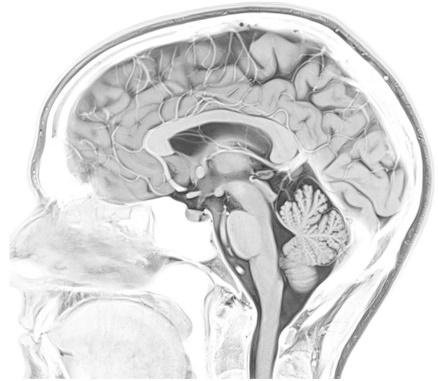
With this project we wish to optimize an MR imaging sequence for 3T scanning of postmortem human brain hemispheres and develop an image analysis pipeline for parcellation of brain regions with the aim to quantify regional volumes and surfaces.
Postmortem scanning of human brain tissue is an important tool to screen for disease related changes to regional volumes or microstructures of the brain. Further, it significantly enhances the value of downstream histological or molecular studies, as it allows for co-registrering results from small fields of view < 1 cm2 with the 3D context from which they were sampled. The Allen Brain atlas is a prime example of how combining MRI and histology may accelerate our understanding of the human brain. We want to be able to do that for mapping neurodegenerative diseases. However, tissue contrasts changes when tissues are fixated In this project, you will work on tailoring the imaging parameters/sequences and the data analysis methods to achieve accurate registration and segmentation of the postmortem MRI data.
Required qualifications:
Knowledge and interest in MRI acquisition and/or image analysis, for example via courses
22506 Medical Magnetic Resonance Imaging
22525 Medical Image Analysis
Responsible institution/department: Dept. of Health Technology
Contact information:
Axel Thielscher (axthi@dtu.dk)
Oula Puonti (oupa@dtu.dk)
Sanne Simone Kaalund (sanne.simone.kaalund@regionh.dk)
Allowed no of students per report: 2
KU and/or DTU supervisors: Axel Thielscher, Oula Puonti, Sanne Simone Kaalund
Project Title:
Predicting Corticosteroid Treatment Response in Asthma Using Gene Expression and Clinical Data
Description:
Corticosteroids are the most widely used therapy for asthma, but many patients show incomplete or no response. This project investigates whether transcriptomic data, especially inflammation-related gene expression, can help predict which patients will benefit from treatment.
Students will analyze longitudinal RNA-sequencing data from asthma biobanks and GEO, where patients were sampled at baseline and after corticosteroid treatment. By integrating transcriptomic profiles of inflammatory pathways with clinical data (FEV₁, severity scores, eosinophil counts), students will test whether molecular signatures can predict treatment response. This provides both a biological and clinical perspective, making the project highly relevant for future personalized medicine.
Aim: To identify transcriptomic and inflammatory signatures linked to corticosteroid responsiveness and integrate them with clinical markers of asthma severity.
Required qualifications:
Basic knowledge in biomedical sciences, respiratory physiology and R/Python programming
Responsible institution/department: Department of Biomedical Sciences and Rigshospitalet, University of Copenhagen.
Contact information:
Associate Prof. Henrik El ALI (henrik.elali@sund.ku.dk) and Prof. Jann Mortensen (jann.mortensen@regionh.dk)
Allowed no of students per report: 1-4
KU and/or DTU supervisor: Henrik El ALI and Jann Mortensen
Project for BSc and MSc students in Biomedical Engineering (MedTek) and Quantitative Biology and Disease Modelling (KBS)
Project Title:
Quantifying Cranial Base and Head-Shape Change in Pediatric MRI using Black Bone & 3D T1: A Comparative, Registration, and (Optionally) AI-Assisted Study
Description:
Join a cutting-edge project combining medical imaging, engineering, and AI to track how children’s skulls develop over time. Using 3D MRI (Black Bone and 3D T1), you’ll compare modalities, place cranial landmarks, and apply non-rigid registration to fuse images for clear visualization. Explore optional AI-assisted landmark detection to speed up analyses. Your work will quantify cranial base and head-shape changes using deformation maps, distances and angles. Implement the pipeline in 3D Slicer with Python and produce reproducible visualizations. Deliverables include an automated workflow, interactive cranial-change maps, and a report. Make an impact at the intersection of engineering and pediatric medicine!
Required qualifications:
• Comfortable with Python; prior 3D image analysis experience an advantage, but not a requirement.
• Interest in medical image registration/morphometrics; willingness to interact with clinicians.
Allowed no of students per report: 2. It is also possible that 2 students share the project but write separate reports.
Responsible institution/department:
Project is to be carried out at the 3D-Lab (3D Craniofacial Image Research Laboratory) located at School of Dentistry, Nørre alle 20, 2200 Copenhagen N.
KU and/or DTU supervisor:
Associate Professor Vedrana Andersen Dahl (DTU Compute) (main supervisor), Associate Professor Nuno Hermann, Pediatric Dentistry and Clinical Genetics, University of Copenhagen (co-supervisor), and Research Engineer Tron Darvann, 3D Craniofacial Image Research Laboratory (KU, Righshospitalet and DTU) (main day-to-day supervision).
Contact information:
Tron Darvann, trd@sund.ku.dk
Please find some more details about the project below.
Half-year Student Project Proposal
Title
Head Shape & Cranial Base Change
Quantifying Cranial Base and Head-Shape Change in Pediatric MRI using Black Bone & 3D T1: A Comparative, Registration, and (Optionally) AI-Assisted Study
Host & Study Direction
Biomedical Engineering (MedTek) and Quantitative Biology and Disease Modelling (KBS) Department of Applied Mathematics and Computer Science (DTU Compute) and 3D Craniofacial Image Research Laboratory (3D-Lab) (KU, Rigshospitalet and DTU).
Duration
6 months, full time
Motivation
Head shape and cranial base morphology are clinically relevant in several pediatric conditions. Modern MRI protocols (Black Bone and 3D T1) can visualize skull sutures and cranial base with minimal radiation, yet multi-center variability and motion can complicate consistent measurement. A standardized, semi-automated pipeline for longitudinal change could directly support clinical decision-making.
Data Available
• Pilot adults: 15 paired scans (Black Bone + 3D T1) from multiple centers/scanners.
• Pediatric longitudinal set (Black Bone + 3D T1): 9 patients × 2 time points (≈18 scans) currently available; ongoing accrual from three centers with standardized protocol. By January 2026, expect ≥2× current pediatric volume (conservative estimate based on center commitments).
• All data de-identified and stored under institutional governance.
Primary Aims
1. Modalities comparison: Which cranial landmarks are best identified on Black Bone vs 3D T1?
2. Robust co-visualization: Non-rigidly register Black Bone ↔ 3D T1 to combine complementary contrast for landmarking/segmentation.
3. Longitudinal change: Quantify cranial base and global head-shape change over ~1 year using:
o Manual landmarks (with inter/intra-rater reliability),
o Deformable registration–derived morphometry (Jacobian maps, pointwise displacement), and/or
o AI-assisted landmark suggestion or atlas-to-subject propagation (optional stretch goal, see Track B below).
Key Landmarks (tentative)
Cranial base: Nasion (Na), Sella (S), Basion (Ba), Opisthion (Op), Porion (Po), Orbitale (Or); vault/face: Glabella (G), Lambda (L), Euryon (Eu), Gonion (Go). Final set to be agreed with clinical team. Define angles (e.g., Na–S–Ba), linear distances, and Procrustes-based shape metrics.
Methods & Workflow
M0. Data curation & QC
• Harmonize DICOM → NIfTI; record scanner/protocol metadata.
• Motion/noise QC; create inclusion flags; document artifacts.
M1. Manual landmarking baseline
• Place landmarks in 3D Slicer on both modalities.
• Compute inter-/intra-rater ICCs and Bland–Altman plots.
• Landmark visibility scoring per modality (Likert 1–5) + time budget per case.
M2. Registration & co-visualization
• Rigid/affine: Landmark Registration / Fiducial Registration Wizard.
• Deformable: SlicerElastix and/or ANTs (through 3D Slicer extensions). Tune presets for skull/base; evaluate overlap (e.g., Dice of bone masks) and target registration error (TRE) at landmarks.
• Output: fused views, propagated landmarks, and deformation fields.
M3. Longitudinal change metrics
• Same-modality T0→T1 registration per patient; compute Jacobian determinant maps and regional volume change in cranial base ROIs.
• Landmark-based longitudinal metrics: Δ distances/angles; Procrustes alignment and PCA for shape trajectories.
M4. Optional AI Track (choose A or B)
• Track A Atlas/registration-assisted automation (low risk): Build a study-specific template; propagate landmarks via deformable registration; optionally refine with SlicerMorph tools (e.g., ALPACA-style correspondence on surface models). Evaluate vs manual ground truth.
• Track B Learning-based landmark suggestion (medium risk): Train a compact 3D CNN/heatmap regressor or use transfer learning to predict landmark probability maps on Black Bone & T1. Use active learning: student corrects suggestions to iteratively improve. Report mean radial error and success detection rate.
Evaluation Plan
• Accuracy: landmark error (mm), angular/linear Δ, TRE at independent landmarks.
• Reliability: ICC(2,1), within-subject SD.
• Registration quality: Dice/Jaccard on bone masks; bending energy/regularity; visual QA.
• Longitudinal sensitivity: effect sizes for Δ over one year; minimal detectable change (MDC) from test–retest where available.
• Runtime & usability: per-case time, failure modes, and user feedback.
Expected Deliverables (by Month 6)
1. Reproducible pipeline (scripts + Slicer scene templates) for:
o Landmarking, registration, and metric extraction,
o Batch processing & QC reports.
2. Benchmark report: Black Bone vs 3D T1 visibility + measurement precision.
3. Longitudinal analysis: cohort-level tables/figures of cranial base change.
4. (If AI track) Trained prototype model + validation on held-out cases.
5. Documentation: step-by-step guide for clinicians; short video demo.
6. Manuscript draft suitable for a workshop/journal or conference abstract.
Tools
• 3D Slicer (Markups, Landmark/Fiducial Registration, Transforms).
• Deformable registration via SlicerElastix and/or ANTs extensions.
• SlicerMorph for morphometrics and (optionally) automated correspondence.
• Python (Slicer Python, NumPy, SimpleITK), R (geomorph) for Procrustes/PCA.
Timeline (indicative)
• Weeks 1–2: Data governance, set-up, QC protocol, finalize landmark set; pilot 3–5 cases.
• Weeks 3–4: Manual landmarking reliability study; first modality comparison.
• Weeks 5–7: Registration experiments (rigid→deformable), pick best recipe; automate batch.
• Weeks 8–10: Longitudinal pipeline; Jacobian/shape metrics; preliminary cohort plots.
• Weeks 11–14: Choose Track A or B; implement and validate.
• Weeks 15–16: Stress-test on latest incoming cases; write-up; clinician demo & feedback.
Student Profile
• Comfortable with Python; prior 3D Slicer experience an advantage, but not a requirement
• Interest in medical image registration/morphometrics; willingness to interact with clinicians.
Supervision & Collaboration
Project is to be carried out with a full-time work load at the 3D-Lab (3D Craniofacial Image Research Laboratory) located at School of Dentistry, Nørre alle 20, 2200 Copenhagen N.
Supervisors: Associate Professor Vedrana Andersen Dahl (DTU Compute) (main supervisor), Associate Professor Nuno Hermann, Pediatric Dentistry and Clinical Genetics, University of Copenhagen (cosupervisor), and Research Engineer Tron Darvann, 3D Craniofacial Image Research Laboratory (main day-to-day supervision).
Project Title: Pharmacokinetics of oral glucose intake
Description:
Positron Emission Tomography is a highly sensitive technique for visualizing metabolism or receptorbased tracers in-vivo. Tracers are typically injected in the blood stream which transports the molecules through the circulatory system. Glucose is, however, normally taken in orally through diet and subsequently exchanged in the intestines before entering the blood stream. The dynamics and time development of oral glucose is obviously very different and will have different rate constants compared to intravenous injected [18F]FDG resulting in different kinetics and model requirements With novel high resolution, high sensitivity scanners whole-body exam can be recorded with high time resolution enabling exploration of the time course of tracer uptake after injection. This is essential to understand
In this project you will work on the data from oral administered [18F]FDG and compare to intravenously injected [18F]FDG and develop models for kinetic analysis of the data.
Required qualifications:
22485 Medical Imaging Systems (preferred)
KU005 Modelling of physiological systems (preferred)
General programming skills in Python
Responsible institution/department:
Department of Clinical Physiology and Nuclear Medicine, Copenhagen University Hospital –Rigshospitalet, Copenhagen, Denmark
Contact information:
Thomas Lund Andersen, thomas.lund.andersen@regionh.dk
Allowed no of students per report (1-4): 1-2
KU and/or DTU supervisor: Thomas Lund Andersen
Project Title: Ultra-high time resolution PET data
Description:
Positron Emission Tomography is a highly sensitive technique for visualizing metabolism or receptorbased tracers in-vivo. With the recent introduction high sensitivity PET scanners time resolution of down to 1 s. or lower can be achieved, enabling visualization of physiological effects that has previously been unobtainable. However, images with such high time resolution suffers from low SNR where current methods struggle hampering the use of the data for new and exploratory analysis.
In this project ultra-high time resolution PET data reconstruction methods and methodologies such as Monte Carlo simulation and Complementary reconstructions will be investigated and characterized to enable high time-resolution images. Several datasets ranging across different tracers and injected activities are available for testing and characterization.
Required qualifications:
22485 Medical Imaging Systems (preferred)
KU005 Modelling of physiological systems (preferred) General programming skills in either MatLab or Python
Responsible institution/department:
Department of Clinical Physiology and Nuclear Medicine, Copenhagen University Hospital –Rigshospitalet, Copenhagen, Denmark
Contact information:
Thomas Lund Andersen, thomas.lund.andersen@regionh.dk
Allowed no of students per report (1-4): 1-2
KU and/or DTU supervisor: Thomas Lund Andersen / Ulrich Lindberg
Project Title: Quantifying the Specificity of MLBased ProPET for Small Lesion Detection

Description
The Machine Learning based ProPET methodology has demonstrated increased sensitivity, better contrast, and reduced noise in PET imaging. However, enhanced sensitivity could potentially lead to false-positive lesion detection, particularly for small structures.
This project will quantify the specificity of ML-based ProPET for detecting small high-activity regions simulating cancer. The student will insert synthetic lesions of varying sizes (3-15 mm) and activity levels (SUV 2-8) into healthy patient PET data, apply standard reconstruction and ML-based ProPET, and quantify specificity and false-positive rates.
Key metrics include specificity/sensitivity as function of lesion size, false-positive rate in known healthy regions, and ROC curves compared to standard methods.
Required qualifications:
22485 Medical Imaging Systems (preferred)
KU005 Modelling of physiological systems (preferred)
General programming skills in Python
Responsible institution/department:
Department of Clinical Physiology and Nuclear Medicine, Copenhagen University Hospital –Rigshospitalet, Copenhagen, Denmark
Contact information:
Thomas Lund Andersen, thomas.lund.andersen@regionh.dk
Allowed no of students per report (1-4): 1-2
KU and/or DTU supervisor: Thomas Lund Andersen / Thomas Mejer Hansen
References
Hansen, T. M., & Vendelbo, M. (2023). Probabilistic analysis of PET images using informed prior models and machine learning. Journal of Nuclear Medicine, 64(supplement 1), P848.
Hansen, T. M., Mosegaard, K., Holm, S., Andersen, F. L., Fischer, B. M., & Hansen, A. E. (2023). Probabilistic deconvolution of PET images using informed priors. Frontiers in Nuclear Medicine, 2, 1028928.
Project Title: Ultra-high time resolution PET data
Description:
Positron Emission Tomography is a highly sensitive technique for visualizing metabolism or receptorbased tracers in-vivo. With the recent introduction high sensitivity PET scanners time resolution of down to 1 s. or lower can be achieved, enabling visualization of physiological effects that has previously been unobtainable. However, images with such high time resolution suffers from low SNR where current methods struggle hampering the use of the data for new and exploratory analysis.
In this project ultra-high time resolution PET data reconstruction methods and methodologies such as Monte Carlo simulation and Complementary reconstructions will be investigated and characterized to enable high time-resolution images. Several datasets ranging across different tracers and injected activities are available for testing and characterization.
Required qualifications:
22485 Medical Imaging Systems (preferred)
KU005 Modelling of physiological systems (preferred) General programming skills in either MatLab or Python
Responsible institution/department:
Department of Clinical Physiology and Nuclear Medicine, Copenhagen University Hospital –Rigshospitalet, Copenhagen, Denmark
Contact information:
Thomas Lund Andersen, thomas.lund.andersen@regionh.dk
Allowed no of students per report (1-4): 1-2
KU and/or DTU supervisor: Thomas Lund Andersen / Ulrich Lindberg
Project Title: The effect of vasodilators on renal perfusion modelling
Description:
Positron Emission Tomography (PET) is a highly sensitive technique for visualizing metabolism or receptor-based tracers in-vivo. Perfusion, that is the passage of fluid from the circulatory system to an organ or tissue, can be quantitatively measured with PET using a radioactive isotope of oxygen bound in water, i.e. [15O]-H2O. The measurement across typically 3 minutes where the tracer uptake and washout are visualized and subsequently modelled. Historically, the methodology has been applied primarily in the brain, but recent advances of PET instrumentation has enabled perfusion calculations in other organs as well. In this respect the kidneys have attracted interest due to their central function of controlling, among others, body fluids, acid-base balance and electrolyte concentrations.
In this project you will investigate the effect of a vasodilator (acetazolamide) on renal perfusion modelling. Several datasets with acetazolamide challenge are available for testing and characterization.
Required qualifications:
22485 Medical Imaging Systems (preferred)
KU005 Modelling of physiological systems (preferred)
General programming skills in Python
Responsible institution/department:
Department of Clinical Physiology and Nuclear Medicine, Copenhagen University Hospital –Rigshospitalet, Copenhagen, Denmark
Contact information:
Thomas Lund Andersen, thomas.lund.andersen@regionh.dk
Allowed no of students per report (1-4): 1-2
KU and/or DTU supervisor: Thomas Lund Andersen
Project Title: Connectome perfusion correlation from long axial field of view data
Description:
Positron Emission Tomography (PET) is a highly sensitive technique for visualizing metabolism or receptor-based tracers in-vivo. Perfusion, that is the passage of fluid from the circulatory system to an organ or tissue, can be quantitatively measured with PET using a radioactive isotope of oxygen bound in water, i.e. [15O]-H2O. The measurement across typically 3 minutes where the tracer uptake and washout are visualized and subsequently modelled. Historically, the methodology has been applied primarily in the brain due to the traditional short FOV coverage of PET scanners. However, recent advantages in PET detector technology and hardware have increased the FOV from approximately 25 cm. to 106 cm. covering all vital organs. This enables perfusion calculations not only in the brain but also in other organs enabling correlation of perfusion in multiple organs simultaneously to examine connections between them.
In this project you will investigate the correlation of vital organ perfusion to characterize perfusion patterns in both disease and healthy controls
Required qualifications:
22485 Medical Imaging Systems (preferred)
KU005 Modelling of physiological systems (preferred)
General programming skills in Python
Responsible institution/department:
Department of Clinical Physiology and Nuclear Medicine, Copenhagen University Hospital –Rigshospitalet, Copenhagen, Denmark
Contact information:
Thomas Lund Andersen, thomas.lund.andersen@regionh.dk
Allowed no of students per report (1-4): 1-2
KU and/or DTU supervisor: Thomas Lund Andersen
Project Title: Validation of Machine Learning-Based ProPET for Low-Dose / Sparse-Data PET Imaging
Description: The recently developed ProPET methodology uses machine learning to perform probabilistic PET image analysis with informed prior models (Hansen et al., 2023). Hansen and Vendelbo (2023) proposed a neural network approach that directly estimates posterior distribution features orders of magnitude faster than traditional sampling methods, making clinical implementation feasible.
However, ProPET has only been tested on full-dose, full-data acquisitions. This project will evaluate the ML-based ProPET methodology under clinically relevant low-dose and reduced-data conditions. The student will apply the ML-based ProPET algorithm to clinical or preclinical PET and test
• Low-dose scenarios
• Reduced-data scenarios
Performance will be compared against standard reconstruction methods and full-dose/full-data reference images.
Required qualifications:
22485 Medical Imaging Systems (preferred)
KU005 Modelling of physiological systems (preferred)
General programming skills in Python
Responsible institution/department:
Department of Clinical Physiology and Nuclear Medicine, Copenhagen University Hospital –Rigshospitalet, Copenhagen, Denmark
Contact information:
Thomas Lund Andersen, thomas.lund.andersen@regionh.dk
Allowed no of students per report (1-4): 1-2
KU and/or DTU supervisor: Thomas Lund Andersen / Thomas Mejer Hansen
References
Hansen, T. M., & Vendelbo, M. (2023). Probabilistic analysis of PET images using informed prior models and machine learning. Journal of Nuclear Medicine, 64(supplement 1), P848.
Hansen, T. M., Mosegaard, K., Holm, S., Andersen, F. L., Fischer, B. M., & Hansen, A. E. (2023). Probabilistic deconvolution of PET images using informed priors. Frontiers in Nuclear Medicine, 2, 1028928.
Project Title: Fractional tissue perfusion
Description:
Positron Emission Tomography (PET) is a highly sensitive technique for visualizing metabolism or receptor-based tracers in-vivo. Perfusion, that is the passage of fluid from the circulatory system to an organ or tissue, can be quantitatively measured with PET using a radioactive isotope of oxygen bound in water, i.e. [15O]-H2O. The measurement across typically 3 minutes due to the short half-life 15O of two minutes. With new high-sensitive scanners the measurement time can now be expanded to and beyond 12 minutes. Such measurements offer a unique insight into the physiology of more slowly perfused tissue which can be measured on longer time scales. Longer measurement times also open for more advanced model to estimate the perfusion components of the underlying tissue.
In this project novel models for perfusion imaging calculation should be investigated, implemented, and characterized tested on long time scale [15O]-H2O scans.
Required qualifications:
22485 Medical Imaging Systems (preferred)
KU005 Modelling of physiological systems (preferred)
General programming skills in Python
Responsible institution/department:
Department of Clinical Physiology and Nuclear Medicine, Copenhagen University Hospital –Rigshospitalet, Copenhagen, Denmark
Contact information:
Thomas Lund Andersen, thomas.lund.andersen@regionh.dk
Allowed no of students per report (1-4): 1-2
KU and/or DTU supervisor: Thomas Lund Andersen / Ulrich Lindberg
KU and/or DTU supervisor:
Thomas Lund Andersen
Project Title: Effect of proton therapy treatments with PET
Description:
Proton therapy can be used to treat certain brain tumours. Unfortunately, some patients develop reactive changes in the brain that can be difficult to distinguish from tumour recurrence on MRI. FET PET examinations measure the metabolic activity in these changes, which is often lower than in tumour recurrence. This project aims to investigate whether kinetic studies of FET uptake can characterize the reactive changes more precisely and hence help aid diagnosis of tumour recurrence.
In this project you will work on the dynamic data acquired with the aminoacid tracer, [18F]FET to characterize and differentiate tumour reoccurrence and reactive changes due to treatment.
Required qualifications:
22485 Medical Imaging Systems (preferred)
KU005 Modelling of physiological systems (preferred)
General programming skills in Python
Responsible institution/department:
Department of Clinical Physiology and Nuclear Medicine, Copenhagen University Hospital –Rigshospitalet, Copenhagen, Denmark
Contact information: Thomas Lund Andersen, thomas.lund.andersen@regionh.dk
Allowed no of students per report (1-4): 1-2
KU and/or DTU supervisor: Thomas Lund Andersen / Karine Madsen
Project Title: Arterial Input Function estimation from carotid arteries
Description:
Accurate pharmacokinetic modelling relies on a correct arterial input function (AIF). For Long-Axial Field of View (LAFOV) scanners the AIF is extracted from the descending Aorta. With Short-Axial field of view (SAFOV) scanners the aorta is not in the imaging field when scanning the brain. The AIF is therefore often extracted from the carotid arteries However, this impose a problem due to partial volume effects lowering the recovery coefficient ultimately losing quantification.
This project seeks to investigate if a correct AIF can be determined from the carotid arteries by using the AIF found in the aorta as ground truth.
Required qualifications:
22485 Medical Imaging Systems (preferred)
KU005 Modelling of physiological systems (preferred)
General programming skills in either MATLAB or Python
Responsible institution/department:
Department of Clinical Physiology and Nuclear Medicine, Copenhagen University Hospital –Rigshospitalet, Copenhagen, Denmark
Contact information:
Ulrich Lindberg, ulrich.lindberg@regionh.dk
Allowed no of students per report (1-4): 1-2
KU and/or DTU supervisor: Thomas Lund Andersen / Ulrich Lindberg


Targetedjob
Engineering a Life-Scale Programmable Gut Bioreactor: Developing Electronic Control Systems for Advanced Microbiome Research
BactolifeA/S
Publishedon8April2025
Contract
Project
Location KøbenhavnØ
Startdate
September2025
Salary
Informationnotprovided
Remoteworking
Partial
EngineeringaLife-ScaleProgrammableGutBioreactor:DevelopingElectronic ControlSystemsforAdvancedMicrobiomeResearch
Projectstart:September2025
Duration:6months(30ECTS)
Location:BactolifefacilitieswithregularsupervisionfromDTU
Thisprojectrepresentsanoutstandingopportunitytoapplyelectricalandbiomedical
engineering principles to create a novel research platform that could significantly advance our understanding of the human microbiome and gut physiology
Humangutmicrobiomeresearchisoneofthefastestgrowingareasinbiomedical scienceworldwide,yetprogressishamperedbythelimitationsofcurrent
experimentalmodels Existinginvitrosystemsaretypicallyoversimplifiedandfailto replicatethecomplexityandscaleofthehumanintestinalenvironment.
ThisambitiousMaster'sthesisprojectaimstodevelopanopen-source,life-scale programmablegutbioreactorthatcanauthenticallysimulatehumanintestinal conditions.Real-timedatacollectionthroughintegratedsensorsystemsiscrucialfor understandingthedynamicintestinalenvironment—acapabilitycurrentsystems largelylack.Thisprojectcouldfundamentallytransformhowmicrobiomeresearchis conductedglobally.
ProjectPartners
ThisisacollaborativeprojectbetweentheTechnicalUniversityofDenmark(DTU)and Bactolife,abiotechcompanyleadingresearchinmicrobiomescienceanddeveloper ofBindingProteintechnology.
TheprojectwillbeformallysupervisedbyAssociateProfessorMartinDufvafromDTU WorkwillbeprimarilyconductedatBactolife'sfacilities,wheretheyhaveallnecessary equipmentfordevelopment,testing,andvalidationintheirmolecularlaboratories.
ProjectDescription
Theprimaryfocusofthisthesisisontheelectricalengineeringaspectsofcreating whatwillbethelargestprogrammablegutbioreactorplatformeverdeveloped.This isanopen-endedprojectwherethestudent'sownconsiderationsandideaswillbe takenintoaccount,withnecessarymaterialsandcomponentsacquiredbasedon theevolvingdesignrequirements.Thestudentwilldeveloptheembeddedcontrol systems,sensorintegrationnetworks,anduserinterfaceneededtosimulatethe physicalandbiologicalconditionsofthehumanintestineatanatomicallyrelevant dimensions,somethingneverbeforeachievedatthisscale.
Thestudentwilldeveloptheelectronicsystemsforaplanarbioreactorprototype, focusingoncreatingarobust,well-documentedsystemthatcanbecomeavaluable toolformicrobiomeresearch.
Thecoreengineeringchallengesinclude:
1.SensorIntegrationandSignalProcessing:
Sourcingandimplementationofamulti-modalsensorarray(pH,temperature, oxygen,TEER)forreal-timedataacquisition
Developmentofdigitalsignalprocessingalgorithmsfornoisereductionand accuratemeasurementinabiologicallyactiveenvironment
1.EmbeddedControlSystemDevelopment:
Programmingmicrocontrollers(Arduino/RaspberryPi)toimplementclosed-loop controlofactuatorsandfluidsystems
DevelopingPWMcontrolofperistalticmotorstoachievepreciseflowrate regulationandpressuregradients
CreatingPIDfeedbacksystemstomaintaintargetparametersdespitebiological variationandsystemdynamics
1 IoTandInterfaceDevelopment:
Designingainterfaceforsystemmonitoringandcontrol
Implementingdatatransmissionprotocolsforreal-timevisualizationand experimentalparameteradjustment
Creatingrobustdataloggingsystemswithtimestampsynchronizationfor experimentalreproducibility
1 SystemIntegrationandElectronicValidation:
Developinghardware/softwareintegrationtestingprotocolstovalidatesystem performance
Implementingfaultdetectionandrecoverymechanismstoensurecontinuous operationduringlong-termexperiments
Creatingcomprehensivedocumentationandschematicsforfuturereplication oftheopen-sourceplatform
CandidateProfile
WeareseekingahighlymotivatedMaster'sstudentwithabackgroundin:
ElectricalEngineering
BiomedicalEngineering
AutomationandControlEngineering
Orrelatedtechnicalfieldswithstrongelectronicscomponent
Theidealcandidatewillhavecompletedcourseworkinembeddedsystems,control theory,orsensortechnologies.Whilespecifictechnicalskillscanbedevelopedduring theproject,familiaritywithArduino/RaspberryPiplatforms,sensors,orbiological systemswouldbeadvantageous.
Thisprojectoffersauniqueopportunitytodevelopexpertiseacrosselectronics, programming,andbiologicalintegrationwhilecreatingapotentiallyfield-changing researchtool.
HowtoApply
Interestedstudentsshouldcontactbothmax@bactolife.comanddufva@dtu.dkwith:
1.Youracademictranscript
2.Abriefstatementofinterestintheproject
3 Ashortdescriptionofapreviousprojectyouhavecompletedthatalignswith thiswork
Project Title: Semi-supervised learning for increasing training data size
Description: Radioligand Therapy (RLT) relies on accurate organ segmentation to perform reliable dosimetry. Deep learning models trained in a supervised fashion already play an important role in Cluster for Molecular Imaging (CMI’s) automated dosimetry pipeline. However, supervised training requires extensive manual annotation, which limits the amount of data that can realistically be used for model development. This creates a trade-off: smaller, carefully curated datasets enable high-quality supervision, but they do not fully capture the variability present in larger imaging collections.
CMI has access to a vast archive of preclinical CT scans, including both native and contrast-enhanced studies. Many of these datasets lack detailed annotations, which makes them challenging to use for conventional supervised training. At the same time, they represent an untapped opportunity for improving model generalization and robustness.
This project will investigate whether semi- and un-supervised learning strategies can unlock the potential of these large, sparsely annotated datasets. Student(s) will implement and benchmark approaches such as self-supervised pretraining, consistency regularization, pseudo-labeling, or generative modeling. The central research questions are: 1) whether such strategies can achieve better overall segmentation performance and generalization than supervised training on smaller curated datasets, and 2) how annotation effort and dataset size trade off against each other in practice.
The outcome of the project will provide valuable insights into the scalability of organ segmentation models and clarify the potential of leveraging very large but less curated datasets in preclinical imaging research.
Preferred qualifications/courses:
• 02456 – Deep learning
• 02502 – Image analysis
• General programming skills in Python and PyTorch
Responsible institution/department:
Cluster for Molecular Imaging, Department of Biomedical Sciences & Department of Clinical Physiology and Nuclear Medicine, University of Copenhagen and Rigshospitalet
Contact information: Andreas Clemmensen, andreas.clemmensen@sund.ku.dk
Allowed no of students per report: 1-2
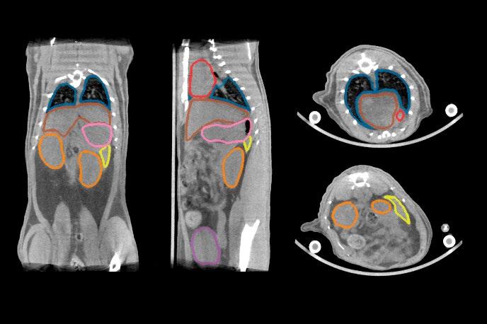
KU and/or DTU supervisor: Professor Andreas Kjær, MD, DSc, PhD AUTOMATICALLY
References:
1 Sgouros, G., Bodei, L., McDevitt, M. R., & Nedrow, J. R. (2020). Radiopharmaceutical therapy in cancer: clinical advances and challenges. Nat Rev Drug Discov, 19(9), 589-608. https://doi.org/10.1038/s41573-020-0073-9
2 Fabian Isensee et al., “nnU-Net: A Self-Configuring Method for Deep Learning-Based Biomedical Image Segmentation,” Nature Methods 18, no. 2 (February 1, 2021): 203–211, https://doi.org/10.1038/s41592-020-01008-z.
3 Luca Vellini et al., “A Deep Learning Algorithm to Generate Synthetic Computed Tomography Images for Brain Treatments from 0.35 T Magnetic Resonance Imaging,” Physics and Imaging in Radiation Oncology 33 (2025): 100708, accessed October 1, 2025, https://linkinghub.elsevier.com/retrieve/pii/S2405631625000132.
Project Title: Speech-level regulation for improving conversations
Description:
Lombard speech is the concept of altering one’s voice to make speech more audible in noisy situations, but importantly, also to signal to conversation partners that you are having issues hearing and participating in the conversation. In a number of studies focusing on conversations, we observe that when unaided, in both quiet and noise, hearing impaired (HI) talkers speak louder, which is taken as a sign of them indicating that they require their conversation partner to speak up in order to easily participate in the conversation (Petersen, 2024; Petersen et al., 2022; Petersen & Parker, 2024) In quiet, providing personalized amplification through hearing aids to the HI person cause all speakers, the HI and their normal-hearing (NH) conversation partner(s), to speak softer.
Previously, we have observed that in a high level of background noise, providing hearing-aid noise reduction to the one HI talker, causes them to reduce their speech level. However, this results in the two NH conversation partners not being able to hear what the HI person says and affect their ability to participate in the conversation (Petersen, 2024) In other words: Hearing-aid signal processing might improve the conversation from the HI person’s point-of-view, causing them to lower their speech level, however the overall conversation suffers because the NH conversation partners are now struggling to hear.
In the current project, we wish to explore whether it is possible to incorporate a mechanism in the hearing-aid processing which allows the HI interlocutor to experience an improvement in SNR through directional sound processing, while at the same time preventing them from reducing their speech level The goal of the project is to implement and test different sound processing schemes that could allow the HI person to experience the benefit of reduced noise levels through hearing-aid signal processing, while at the same time avoid that they reduce their own speech level
The core aspects of the project are:
• (10%) Literature study on possible mechanisms through which voice level can be regulated without changing the SNR experienced by the speaker.
• (30%) Develop a Matlab (or similar) real-time processing test framework to allow testing the identified mechanisms. This will be done in close collaboration with signal processing experts at WSA.
• (30%) Conduct conversations experiment with the real-time processing framework to investigate the effect of the proposed method
• (30%) Present the methods and findings in a written thesis
Required qualifications: Matlab programming skills, understanding of basic signal processing techniques, ideally courses 22003 and/or 22007
Responsible institution/department: DTU Hearing Systems
Contact information: Eline Borch Petersen (eline.petersen @WSA.com). The work will be done at the WS Audiology headquarter in Lynge in the scientific department ORCA labs (www.orca-labs.net).
Allowed no of students per report (1-4): 2
KU and/or DTU supervisor:
Petersen, E. B. (2024). Investigating conversational dynamics in triads : Effects of noise , hearing impairment , and hearing aids. Frontiers in Psychology, 15, 1289637. https://doi.org/10.3389/fpsyg.2024.1289637
Petersen, E. B., MacDonald, E. N., & Sørensen, A. (2022). The Effects of Hearing Aid Amplification and Noise on Conversational Dynamics between NormalHearing and Hearing-Impaired Talkers. Trends in Hearing, 26, 1–18. https://doi.org/10.1177/23312165221103340
Petersen, E. B., & Parker, D. (2024). Speak Up: How Hearing Loss and the Lack of Hearing Aids Affect Conversations in Quiet. Journal of Speech, Language, and Hearing Research, 1–12. https://doi.org/10.1044/2024_jslhr-23-00667
Project Title: Stereological Investigation of Type-I Interferon
Signalling in Alzheimer’s
Description:
Disease Pathogenesis
Alzheimer’s disease (AD) is driven by amyloid-β (Aβ) and tau pathology, with inflammation playing a key role in neurodegeneration. Type-I interferons (IFN-I) have emerged as potent mediators of neuroinflammation, driving tau aggregation and neurotoxicity. This project will investigate whether IFN-I signalling contributes to neuronal loss and glial dysfunction in AD, in collaboration with Ella Lacey, a PhD student (McEwan Lab) at the University of Cambridge. Using a novel AD mouse model lacking the IFN-I receptor (IFNAR), the student will use unbiased stereological techniques to quantify neuronal, microglial, and astrocytic populations across key brain regions. Stereology is a set of methods build on statistical and mathematical models applied to thin microscope sections for quantification of 3D objects. By comparing AD mice with and without IFN-I signalling, this work will determine whether specific cell types or regions exhibit heightened IFN-I-linked vulnerability. Findings will clarify the inflammatory mechanisms connecting Aβ and tau pathology, advancing understanding of neurodegenerative progression in AD.
The experimental work will take place at Centre for Neuroscience and Stereology, Bispebjerg Hospital.
Required qualifications:
Experience with molecular and/or histological laboratory work
Experience with microscopy and basic knowledge of the different brain cell types.
Responsible institution/department:
Dept. of Health Technology
Contact information:
Sanne Simone Kaalund, sanne.simone.kaalund@regionh.dk, phone 38636298
Allowed no of students per report: 1
KU and/or DTU supervisor:
Sanne Simone Kaalund
Synthetic blood perfusion imaging using deep learning
Dual-energy computed tomography (DECT) provides novel reconstructions in addition to traditional attenuation coefficients and has recently become clinically available in radiotherapy (RT). Contrast enhanced (CE) DECT offer material decomposition where the iodine concentration (IC) can be isolated in each voxel. The IC map can visualize the perfused blood volume presented e.g. within tumors (see Figure 1A and C). DECT or single energy CT (SECT) is acquired in the planning phase of RT and cone-beam CT (CBCT) is acquired daily in the delivery of RT over 5-6 weeks. For retrospective analysis, however, RT patients were not scanned with DECT why it is not possible to easily extract the IC map. Instead, it is likely that CE SECT and first fraction CBCT exist for the patients. Using deformable registration and CE vs. non-CE the voxel characteristics within the gross tumor volume (GTV), the project will focus on algorithm development of extracting IC maps similar to those derived from DECT, see Figure 1B and D Modelling is planned to center around AI-based convolution neural network architectures such as U-NET or GAN which could be trained on corresponding CE-SECT and non-CE CBCT as inputs, and CE-DECT derived IC maps as output to produce synthetic IC maps through supervised learning More simple algorithm development can also be included.
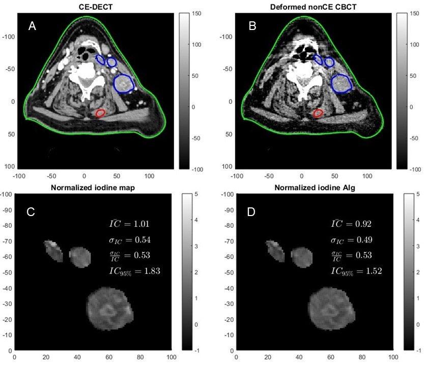
Figure 1. Preliminary data illustrating a simple iodine extraction algorithm. A: contrast enhanced DECT. B: Corresponding non-contrast enhanced CBCT at first fraction deformably registered to the DECT. C: DECT derived GTV iodine map with corresponding metrics normalized to the median. D: Algorithm derived GTV iodine map and metrics. Green = body. Blue = gross tumor and lymph node volume (GTV). Red = neck muscle.
Required qualifications:
Mandatory programming skills in Matlab, Python or similar. Knowledge about medical images, image registration and convolution neural networks is not required but an advantage.
Responsible institution/department:
Herlev and Gentofte Hospital, Department of Oncology, Radiotherapy Research Unit, Borgmester IB Juuls Vej 7, 2730 Herlev.
Contact information:
Jens M. Edmund, PhD. Medical physicist jens.edmund@regionh.dk Tlf.: 38682648
Allowed no of students per report: 1-2
KU and/or DTU supervisor:
Claus Behrens (DTU/TUH)
Project Title: The effects of stimulus parameters on auditoryand visually-evoked functional near-infrared spectroscopy (fNIRS) responses
Description:
With functional near-infrared spectroscopy (fNIRS), changes in blood oxygen are measured using nearinfrared light generated by LEDs or lasers. In the last couple of years, fNIRS has gained attention for investigating human hearing, as it runs silently and does not interfere with the patients’ hearing devices. This has potential for future clinical practice for hearing-impaired patients.
Variability in hearing outcomes has been linked to neuroplastic changes in the brain caused by hearing impairment, such as cross-modal plasticity. In our current experiments, participants are exposed to visualonly, audio-only, and audiovisual speech stimuli, and auditory and visual cortices, as well as language-related areas are investigated. However, some stimulus parameters may affect the responses such as speech intelligibility and loudness.
We are therefore looking for students who can disentangle the effects of either speech intelligibility or loudness/background noise. The current setup can be modified and the project can be further defined depending on the student’s interests.
Required qualifications:
We are looking for one or more students interested in fNIRS data collection and in signal processing using the Python fNIRS analysis toolbox. The lab facilities to collect the data are located at Rigshospitalet.
Responsible institution/department:
The study is a collaboration between DTU hearing systems and the Ear, Nose, Throat & Audiology department at Rigshospitalet. The fNIRS setup is located at Rigshospitalet. The analyses can be performed at any computer, meetings can take place either at DTU Hearing Systems in Lyngby or at Rigshospitalet in Copenhagen.
Contact information:
Maaike Van Eeckhoutte: mcvee@dtu.dk
Beatriz Tomas da Costa: bede@dtu.dk
Allowed no of students per report: 1-2
KU and/or DTU supervisor:
Maaike Van Eeckhoutte (DTU Hearing Systems and Copenhagen Hearing and Balance Centre Rigshospitalet)
Project Title: 3P project – To Promote, Prevent and Predict: A multi-centre, randomized controlled Trial
Description:
This multicentre, randomized, controlled study evaluates a new digital model for outpatient follow-up of people with Parkinson’s disease (PD). The intervention, called the 3P model (Prevent, Promote, Predict), combines a decision-support algorithm with structured nurse-led follow-up to improve monitoring, triage, and treatment planning. The algorithm uses validated questionnaires and data from a wearable movement sensor (STAT-ON) to generate individualized care plans, which are reviewed and adjusted by a specially trained 3P nurse. The aim is to provide earlier interventions, reduce acute deterioration, and optimize use of healthcare resources.
A total of 360 patients with moderate PD will be recruited from neurology clinics in Sweden and Denmark. Participants must have access to a smartphone or tablet and be able to engage in home monitoring and telehealth consultations. After informed consent, patients will be randomized into three groups: (1) 3P follow-up with an external nurse, (2) 3P follow-up with an in-house nurse, or (3) standard care. Follow-up will last one year, during which all health contacts, hospital admissions, medication changes, and device data will be recorded.
The primary objective is to assess patient satisfaction and health-related quality of life (PDQ-8) compared with standard care. Secondary outcomes include health-related quality of life (EQ-5D), number and length of hospital admissions, acute care visits, medication changes, OFF-time and dyskinesias (via wearable device), staff workload, and health economic measures. Patient, caregiver, and staff experiences will also be explored through qualitative interviews.
Required qualifications: Skills within ML (supervised and non-supervised), interface modulation and Human-Computer Interaction (HCI) are suggestions. More specific related to the project it could be:
Wearable data processing (develop or refine pipelines to clean and analyze motion sensor (STATon) data for OFF-time/dyskinesia detection, or create dashboards or scripts ( in Python/R/Matlab) to visualize patient motor patterns and compare to clinical outcomes.
• Algorithm validation (Test the sensitivity/specificity of the 3P algorithm’s prioritization by simulating patient scenarios, and quantify how nurse overrides of the algorithm affect predicted care priorities.
• App/Interface prototype and digital workflow (help design a user-friendly mobile or web interface for questionnaire completion, reminders, or video calls. Additionally, integrate wearable data displays for patients and clinicians in collaboration with patients and caregivers + health care professionals.
Responsible institution/department: Movement disorder Clinic, Rigshospitalet- Glostrup, Valdemar Hansens Vej 7, 2600 Glostrup
Contact information: Trine Hørmann Thomsen, PhD, Senior Researcher
Email: trine.hoermann.thomsen@regionh.dk
Allowed no of students per report (1-4): 2
KU and/or DTU supervisor: Trine Hørmann Thomsen, Assistant Professor, Department of Health Technology, DTU/TUH, and Department of Brain and Spinal Cord injuries, and Movement disorder Clinic, Rigshospitalet
BSc/MSc project for students in Biomedical Engineering and/or Quantitative Biology and Disease Modelling, DTU/KU
Project Title:
Two-photon microscopy for diagnosis and monitoring of diseases
Description: (n.a. if confidential)
This project comprises a range of possible projects working on two-photon fluorescence microscopy. The project can be more engineering focused, image processing, or application focused depending on the student. Some opportunities could include:
-Investigation of metabolic biomarkers using 2D and 3D cultured cells and cancer models
-Beam shaping through multi-core and multi-mode optical fibers
-Dynamic beam shaping for light-sheet microscopy with exotic beams
-Imaging and analysis of cultured skin or colon samples with two-photon light sheet microscopy
-Imaging and analysis of autofluorescence and/or exogenous biomarkers with two-photon microscopy
-Analysis of laser damage to cells using two-photon microscopy
-Comparison of benchtop and fiber probe-based two-photon imaging systems
-Applications of two-photon fluorescence imaging in neuroscience
Required qualifications:
Project description can be adjusted to reflect the background and interest of individual students
Responsible institution/department:
DTU Health Tech
Contact information:
Henry Sansom: hgosa@dtu.dk
Peter Andersen: peta@dtu.dk
Allowed no of students per report: No preference – the project can be adjusted to reflect the number of students
KU and/or DTU supervisor:
Henry Sansom and Peter Andersen
Project Title: Uncovering Asthma Endotypes: Integrating Transcriptomics and Clinical Lung Function Data
Description: (n.a. if confidential)
Asthma is not one disease but many biological subtypes, often called endotypes. Standard lung function tests such as FEV₁ give important information but cannot fully explain why some patients have mild disease while others develop severe, treatment-resistant asthma.
In this project, students will work with publicly available transcriptomic datasets from biobanks and the Gene Expression Omnibus (GEO). Transcriptomics (RNA-sequencing) makes it possible to measure thousands of genes at once, including genes involved in inflammation, such as those regulating eosinophils and Th2 pathways. By combining these molecular data with clinical measures of lung function and inflammatory markers, students will use clustering to identify biologically meaningful subgroups (endotypes) of asthma.
Aim: To uncover asthma endotypes using transcriptomic data, and explore how they differ in lung function and inflammatory profiles.
Required qualifications:
Basic knowledge in biomedical sciences, respiratory physiology and R/Python programming.
Responsible institution/department:
Department of Biomedical Sciences and Righshospitalet, University of Copenhagen
Contact information:
Associate Prof. Henrik El ALI (henrik.elali@sund.ku.dk) and Prof. Jann Mortensen (jann.mortensen@regionh.dk)
Allowed no of students per report: 1-4
KU and/or DTU supervisor: Henrik El ALI and Jann Mortensen
Project Title: Unified tumor and organ segmentation model
Description: Radioligand Therapy (RLT) is an emerging therapeutic approach that delivers radiation directly to diseased tissue by attaching radionuclides to targeted molecules. Its success depends critically on accurate dosimetry, which requires detailed knowledge of how radioligands distribute between tumors and healthy organs. Manual annotation of imaging data for dosimetry is highly time-consuming, making automated image segmentation a key enabling technology.
At the Cluster for Molecular Imaging (CMI), two separate deep learning models are currently in use: a standalone model for subcutaneous tumor segmentation in mice2, and an organ segmentation model applicable to both mice and rats. Both models perform well individually, but it remains an open question whether segmentation of tumors and organs can be integrated into a single unified machine learning model.
This project will explore the trade-offs between maintaining separate task-specific models versus developing a joint multi-task segmentation model. A central challenge is that the tumor and organ datasets have been developed independently and are not fully compatible. The student(s) will therefore investigate strategies such as 1) creating a combined, fully annotated dataset, or 2) applying weak supervision and sparse labeling methods to enable joint training without complete annotation overlap3.
The project will involve implementing and benchmarking both approaches, with evaluation based on segmentation accuracy, robustness across species (mice and rats), computational efficiency, and usability within CMI’s automated imaging pipeline. The outcome will clarify whether a unified tumor-and-organ segmentation model offers practical benefits for automated dosimetry in RLT research.
Preferred qualifications/courses:
• 02456 – Deep learning
• 02502 – Image analysis
• General programming skills in Python and PyTorch
Responsible institution/department:
Cluster for Molecular Imaging, Department of Biomedical Sciences & Department of Clinical Physiology and Nuclear Medicine, University of Copenhagen and Rigshospitalet
Contact information: Andreas Clemmensen, andreas.clemmensen@sund.ku.dk
Allowed no of students per report: 1-2
KU and/or DTU supervisor: Professor Andreas Kjær, MD, DSc, PhD
References:
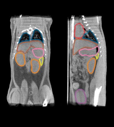
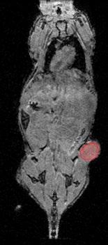
1 Sgouros, G., Bodei, L., McDevitt, M. R., & Nedrow, J. R. (2020). Radiopharmaceutical therapy in cancer: clinical advances and challenges. Nat Rev Drug Discov, 19(9), 589-608. https://doi.org/10.1038/s41573-020-0073-9
2 Jensen, Malte, Andreas Clemmensen, Jacob Gorm Hansen, Julie Van Krimpen Mortensen, Emil N. Christensen, Andreas Kjaer, and Rasmus Sejersten Ripa. “3D Whole Body Preclinical Micro-CT Database of Subcutaneous Tumors in Mice with Annotations from 3 Annotators.” Scientific Data 11, no. 1 (September 19, 2024): 1021. Accessed January 2, 2025. https://www.nature.com/articles/s41597-024-03814-y
3 Feng Gao et al., “Segmentation Only Uses Sparse Annotations: Unified Weakly and Semi-Supervised Learning in Medical Images,” Medical Image Analysis 80 (2022): 102515, accessed September 30, 2025, https://linkinghub.elsevier.com/retrieve/pii/S1361841522001621.
Project Title: Validate automated segmentations with ex-vivo biodistribution
Description: Radioligand Therapy (RLT) requires precise dosimetry to ensure therapeutic effectiveness while keeping organ doses within safe limits1. Automated segmentation methods have recently been introduced at the Cluster for Molecular Imaging (CMI) to accelerate dosimetry estimation and reduce the subjectivity of manual annotation. These approaches achieve high apparent accuracy compared to human raters, but it remains unclear whether they provide a more reliable basis for dosimetric calculations when compared against true radioligand uptake.
To address this question, this project will compare automated and manual organ segmentations against ground-truth uptake measurements obtained from ex vivo organ isolation. Student(s) will design and carry out the study, which includes performing segmentations, processing imaging data, and analyzing discrepancies between predicted and measured uptake values.
The goal of the project is to determine whether automated segmentation improves dosimetric accuracy relative to manual annotation and to identify organ- or modality-specific limitations. The results will provide important evidence for the validation and further development of automated imaging pipelines in preclinical RLT research.
Preferred qualifications/courses:
• KU181 - Radioactive Isotopes and Ionizing Radiation
• 02502 – Image analysis
• General programming skills in Python and PyTorch
Responsible institution/department:
Cluster for Molecular Imaging, Department of Biomedical Sciences & Department of Clinical Physiology and Nuclear Medicine, University of Copenhagen and Rigshospitalet
Contact information: Andreas Clemmensen, andreas.clemmensen@sund.ku.dk
Allowed no of students per report: 1-2
KU and/or DTU supervisor: Professor Andreas Kjær, MD, DSc, PhD
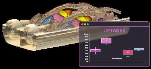
References:
1 Sgouros, G., Bodei, L., McDevitt, M. R., & Nedrow, J. R. (2020). Radiopharmaceutical therapy in cancer: clinical advances and challenges. Nat Rev Drug Discov, 19(9), 589-608. https://doi.org/10.1038/s41573-020-0073-9
2 Jensen, Malte, Andreas Clemmensen, Jacob Gorm Hansen, Julie Van Krimpen Mortensen, Emil N. Christensen, Andreas Kjaer, and Rasmus Sejersten Ripa. “3D Whole Body Preclinical Micro-CT Database of Subcutaneous Tumors in Mice with Annotations from 3 Annotators.” Scientific Data 11, no. 1 (September 19, 2024): 1021. Accessed January 2, 2025. https://www.nature.com/articles/s41597-024-03814-y.
3 Deeley, M A, A Chen, R Datteri, J Noble, A Cmelak, E Donnelly, A Malcolm, et al. “Comparison of Manual and Automatic Segmentation Methods for Brain Structures in the Presence of Space-Occupying Lesions: A Multi-Expert Study.” Physics in medicine and biology 56, no. 14 (July 21, 2011): 4557–4577. Accessed October 3, 2025. https://www.ncbi.nlm.nih.gov/pmc/articles/PMC3153124/.
