Merrilee Williams - Chairperson MerrileeWilliams44@gmail.com
Jessica Southall - Secretary and Hepatology Specialty Group secretaryofnzgnc@gmail.com
Kirsten Arnold - Treasurer and IBD sub group treasurerofnzgnc@gmail.com
Karen Kempin - Committee Member and NE sub specialty Karen.kempin@huttvalleydhb.org.nz
Justin Augustine - Committee Member justin.augustine@wcdhb.health.nz
Gino Borromeo - Committee Member
Nicola Caine - Committee Member and Hepatology
Fiona Willams - Committee Member and Enteral Feeding
Julia Anderson - Professional Nurse Advisor NZNO Julia.Anderson@nzno.org.nz
Life
• Sandra Burton
• Karen Gower
• Sherry
From the Chairperson
NZNO NZGNC October 2024 Chair Report
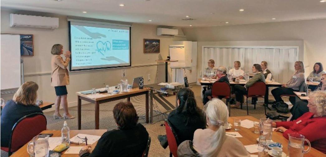
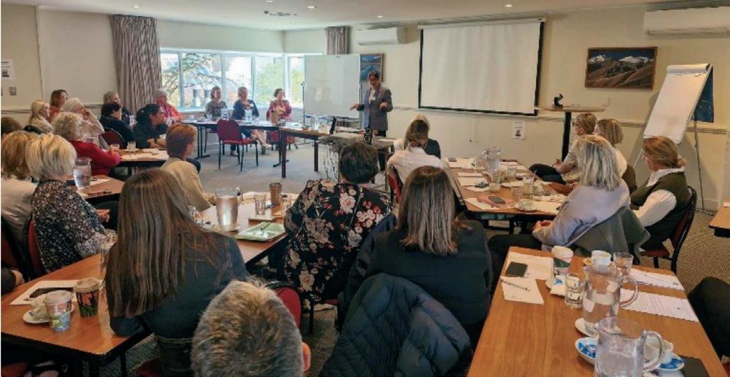
Welcome to your October edition of the Tube Journal. Brought to you by the NZgNC committee, as well as our valued sponsors. We are excited to share with you a number of educational contributions from the successful recipients of our Education Fund in the first half of the year.
Thank you for the time you have put into these, and thanks also to committee member, Gino Borromeo, for your editorial expertise. We feel sure that you will find valuable learning within this journal. Spring has sprung, and the warmth that comes longer, lighter days brings fresh energy. Planning for the end of year projects including conference, is well underway, and you will be starting to think of your conference dinner costume I’m sure. Your nurse convenors have an excellent line up of speakers for you to learn from and I am grateful for their hard work.
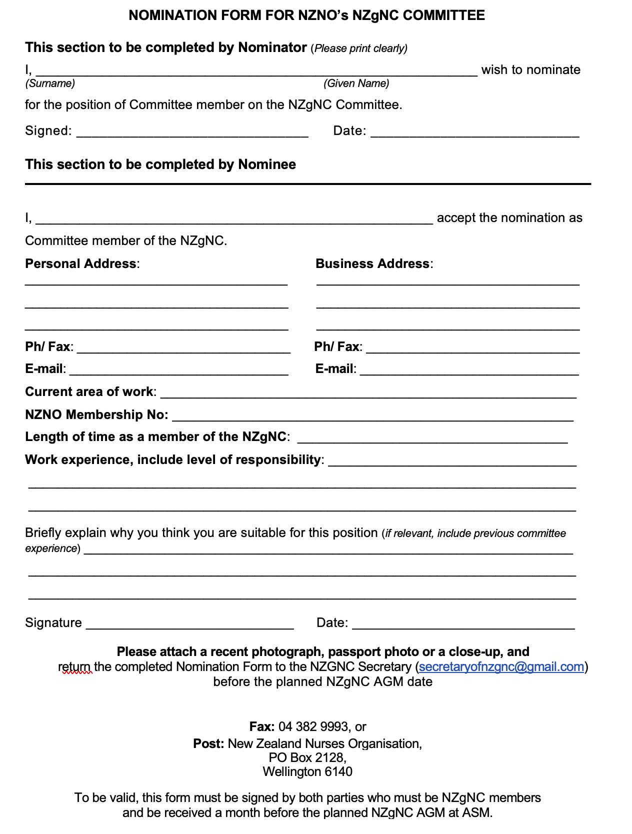
We have some openings on the committee this year including key positions such as treasurer and secretary, and committee, so please get in touch if you are interested in this opportunity. These key roles come with lots of support from the committee, as well as NZNO and their finance team, so do not be worried that it is a very complex undertaking. I have included the nomination form in the Tube as well, please forward this to us before the AGM which will be held at conference in November. You do not need to be at conference to be considered for a nominated position, but please let us know in advance.
Please get in touch with us if you have any questions, or are interested in knowing more. NZGNC Secretary (secretaryofnzgnc@gmail.com).
I will be stepping down from my position on the committee this year after several years in various roles. I will also be stepping away from Gastroenterology as a specialty, after many years of enjoying the abundant opportunities to grow and develop my skills, and contribute to patient care and nursing development. I will look forward to seeing you at conference to hand over the Chairperson role, and tear up the dance floor one last time. Thank you to everyone I have worked alongside in a wide range of capacities over my gastroenterology career, we have had some great times!
Merrilee Williams Your Chairperson NZGNC Secretary secretaryofnzgnc@gmail.com
Notification of NZNO Gastroenterology Nurses College Annual General Meeting,
and call for Remits (proposals to change the NZgNC College Rules)
Thursday 28th November 2024, 12:45pm-13:45pm.
Claudlands Event Centre, Hamilton.
Please send any remits or nominations for committee positions to the secretary before the 30th October 2024. secretaryofnzgnc@gmail.com. Reports will be available on the NZgNC website prior to the AGM for members to read in preparation.
Annual Scientific Meeting 2024 https://www.gastroconference.co.nz/ 27-29 November, Claudelands, Hamilton. Registration and abstracts submission- is now open. This year you will see an increase to the nurse’s registration fee from $550 to $650. The time has come to increase the nurse’s registration, as there has been significant increases to overall running costs over the last few years. To ensure ongoing viability of this excellent conference, we must pass on some of these costs. There has not had an increase for many years, and I am sure you will agree that the value is still excellent.
ANZGITA
Registrations for 2025 programmes are open now https://www.anzgita.org/ Australia and New Zealand Gastroenterology International Training Association’s (ANZGITA) mission is to support improved health in the people of developing Asia-Pacific nations by enhancing the standards of practice of gastroenterology, and build capacity to treat digestive diseases. Annual or biannual training programs are run in Fiji at the World Gastroenterology Organisation (WGO) Training Centre and in Solomon Islands, Samoa, Tonga, TimorLeste and Nepal. Over 100 doctors and more than 90 nurses in the Pacific region alone have attended as trainees on programs that have run for over 10 years.
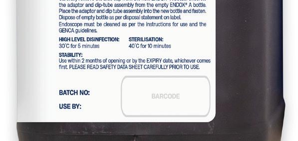
AS5369 Standard for reprocessing of reusable medical devices
After the update of AS/NZS4187:2014 last year, the replacement standard AS 5369:2023 became an Australia only standard. However, it has now been endorsed by Ministry of Health as a standard that we feel meets our requirements in New Zealand as well. As such, the NZgNC and the Sterile Services College also endorses this standard. Please refer to the webpage to acquire a copy of this standard for your services.
Please note, as with all standards, there is a cost to downloading it, however it is essential that this is available on site for guidance. NB- the AS/NZS 4187:2014 has now been withdrawn and is no longer recognised.
AS 5369:2023 Reprocessing of reusable medical devices and other devices in health and non-health related facilities | Standards Australia Store
Nurse Endoscopists News
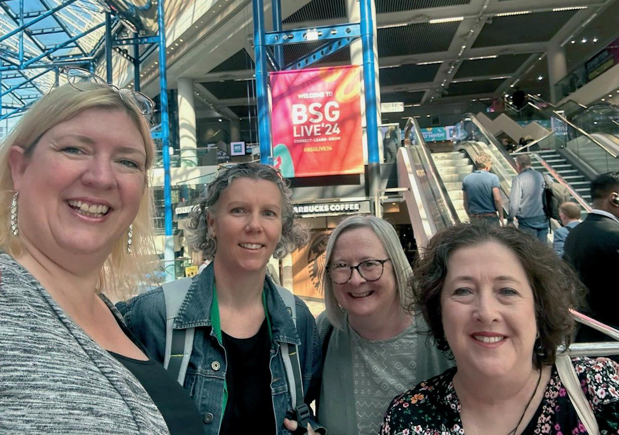
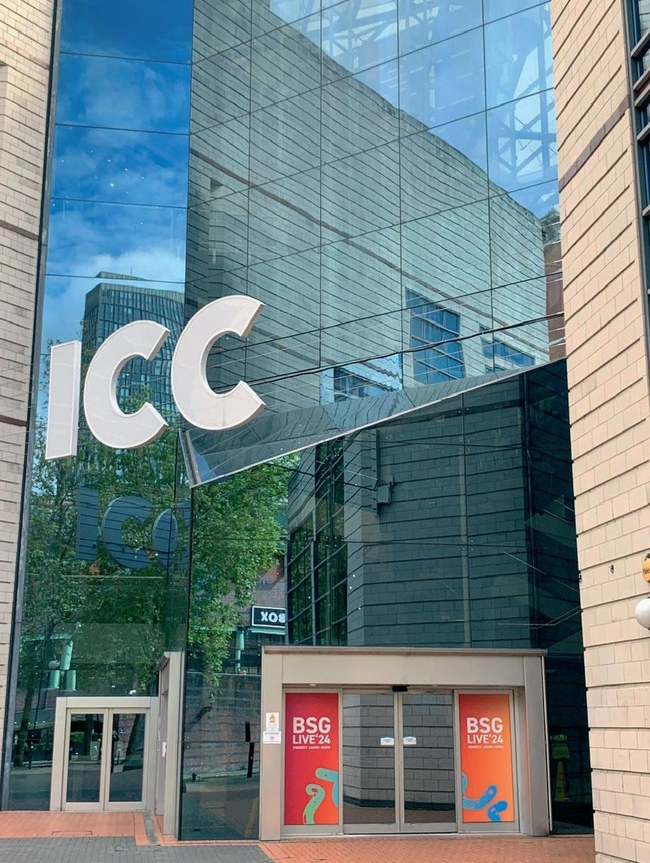
NZ Nurse Endoscopists have started the year with education in mind. Nicola Griffiths, Sofia Krylova-Smith and myself all made it to Sydney for SIES in March and took away a lot of great learning. Karen Kempin and myself were also very privileged to attend BSG Live in Birmingham, UK, in June.
Thank you very much to NZgNC for supporting us both with education grants. As well as attending the conference, we reconnected with Helen Livett and Sharon Powell, the two clinical endoscopists from the UK who attended and spoke at our Annual Scientific Meeting last year. They also very kindly to showed us around their training facilities, which was an amazing opportunity to see how others work and train.
Lastly, creation of this group has resulted in NZ Nurse Endoscopists being a much more connected group.
Our quarterly NZNEA meetings have seen great attendance and included many interesting discussions.
Our May meeting had a fabulous 30-minute presentation from Dr Aditya Sheth, Advanced Gastroenterology Trainee, on Radiology in Gastroenterology, and we have commenced planning for our yearly NE study day. We hopefully have one or two new recruits in the wings, including Michelle Harman (Mid Central DHB) who has commenced endoscopy training. Go Michelle!
If you are interested in joining NZNEA, or just attending a meeting to ask questions or listen, please see our page on the NZgNC website, or email us at nzneassoc@ gmail.com.
Tania Waylen Co-chair NZNEA
ARE YOU PASSIONATE ABOUT ENDOSCOPY? DO YOU CARE ABOUT QUALITY IMPROVEMENT?
An exciting opportunity has become available for the right person to join the National Endoscopy Quality Improvement Programme (NEQIP) as the nurse lead for New Zealand. You will work closely with the clinical lead and coordinator and have strong links with the senior advisor from Health New Zealand, Te Whatu Ora.
The role requires an endoscopy subject matter expert with strong leadership skills and good organisational attributes to ensure delivery of strategic programme objectives. You must be forward thinking and have a collaborative approach to supporting patient centred endoscopy quality and service improvements using the national framework of quality standards. Please send any enquiries and expressions of interest to Neqip@cdhb.health.nz
NEQIP: Striving to facilitate safe, patient-focused endoscopy services that are efficient, accountable and sustainable
Appropriate Endoscopy
HOLLY WEALE Associate clinical nurse manager, gastro-investigative endoscopy day unit. Infection prevention and control CNS, Waitaha, Canterbury. holly.weale@cdhb.health.nz
In June this year, I was lucky enough to attend the British Society of Gastroenterology (BSG) conference in Birmingham. Amongst the many aspects of endoscopy covered, one session stood out for me which focussed on appropriate endoscopy. I have a keen interested in green endoscopy and within this space there is a lot of discussion around the avoidance of procedures that are not clinically necessary. Endoscopy has been shown to be a high producer of waste and CO2 emissions (Cunha Neves et al., 2023; Donnelly, 2022) and one of the best ways to reduce this is to not perform the procedure if it is not required (Park & Cha, 2023).
While working for the National Endoscopy Quality Improvement Programme (NEQIP) as the nurse lead, one of the standards used within the New Zealand global rating scale (NZGRS) was appropriateness: making sure the right procedure, of the best quality, is being performed for the right patient at the right time (NEQIP, 2024). In 2019, the Ministry of Health published very clear guidance about referring patients in NZ for colonoscopy or computer tomography colonography (CTC) which states the referrer must consider whether the patient will benefit from the procedure. Following on from this, in 2020, the Endoscopy Guidance Group New Zealand (EGGNZ) developed standards for endoscopy unit service and facilities stating that referral forms must have sufficient clinical information to permit prioritising of the appropriateness of procedures against guidelines.
However, despite the many guidelines and standards, patients are still being referred inappropriately. Jain (2023) demonstrates studies have shown that up to 50% of referrals for endoscopy may be unnecessary and that despite established referral guidelines many patients are referred for endoscopy when other options may be available. The NZGRS (NEQIP, 2024) and American society for gastrointestinal endoscopy (ASGE, 2012) advocate working closely with primary referrers to ensure appropriate referrals with sufficient information are sent, and sharing information so that all practitioners are aware of the alternatives to endoscopy such as colon capsule, stool tests and other non-endoscopic procedures.
There is no single solution to this ongoing issue, but there are areas where more focus is needed. These include increased referral education, better informed patients, consistent triaging, and knowledgeable booking teams.
According to Clutten (2018), referral education is a modern challenge within primary care. She explores how GPs can best be supported to keep up to date with the latest referral guidelines and demonstrates that studies have shown GPs prefer face to face learning. This has led to the development of short group-courses, one of which includes the role of the faecal calprotectin testing as one of its ‘hot-topics’ (Medcast, 2024). This idea is echoed by Tzartzas et al. (2019) who state that training opportunities are needed to address the way referrals are completed. As well
as increasing engagement, face to face learning is preferable to simply disseminating clinical guidelines as, shown in a qualitative study by Le et al. (2015), the approach to implementing any textbased guidance can vary greatly between practices. One other key measure to ensure referrers are up to date and engaged, could be through establishing interest: an example of which could be collaboration in the writing and reviewing of the guidelines between primary and secondary care. The NZGRS requires that referral guidelines are available to all clinicians involved and reviewed yearly by all parties.
Not only must the referrers consider whether the patient will benefit from the procedure, they must also ensure the patient is properly informed. The Ministry of Health (2019) states the patient must be informed about the procedure and be willing to undergo the procedure before the referral is made. This correlates with the informed consent journey that starts with the General Practitioner and continues until the procedure date. Donnelly (2022) reiterates this, highlighting the need for clear conversations with patients regarding expectations about any future procedures from the first date of the referral. At the conference in Birmingham Dr Rutter (Chair of the United Kingdom Joint Advisory Group) talked about the provision of patient information, both verbal and written, being pivotal in patients being prepared for the procedure. This means both mentally and physically. Patients that understand what the procedure will entail and have a well-prepared bowel could reduce the need for a repeated procedure.
Once a referral has been made, careful and efficient triage is needed to ensure that patients who are in most need of endoscopy have access in the timeliest manner (Innes et al., 2018). The appropriate triage of endoscopy referrals is also essential in reducing waiting lists (Narayanan et al. poster presentation). Donnelly (2022) campaigns in the UK for implementing stricter triaging that follows evidence and guidelines. However, to perform effective triage clinicians must be given time. Innes et al. (2018) state that triaging is time consuming and when conducted by gastroenterologists reduces capacity for endoscopy. This raises the question about whether nurses would be better placed to triage (depending on local policy guidelines). Some hospitals in New Zealand do have nurse triaging and a trial, presented at the annual scientific meeting, demonstrated a very small difference between the triaging opinions and thus the outcomes for patients (Narayanan et al.). Anecdotally (in Waitaha, Canterbury), the introduction of nurse led telephone preassessment for the National Bowel Screening Programme and for patients undergoing colonoscopy has acted as a secondary triage to ensure the patient is having the most appropriate procedure and has shown improvement in the reduction of Did Not Attend (DNA) rates and cancellations.
The importance of scheduling of procedures can sometimes be overlooked, but it is a critical part of how well an endoscopy unit runs. Having a booking team that values patients time and understands the need to reduce waste will provide a good quality service by making the most of available capacity. It is also vital that the booking team understand the areas of the service that can increase productivity such as booking the right patient on the right endoscopists list, thus making efficient use of endoscopists skillsets. The NHS improving quality project (2015) states that giving staff the time to perform accurate scheduling supports good patient outcomes and that good scheduling is essential to ensure endoscopy units achieve benefits for patients, staff and the organisation.
In conclusion, it needs to be understood that gastrointestinal investigative (GI) endoscopy is ultimately a limited resource with significant direct and indirect costs (Innes et al., 2018). Making sure the procedure is required and is completed in a timely and efficient manner, is the responsibility of the referring clinician, the triaging team, the booking team, and of course the endoscopist undertaking the procedure. Following the guidelines and creating a best practice environment will ultimately lead to appropriate procedures, the best outcomes for the patient, and a greener endoscopy.
My thanks to the NZNO gastrointestinal nursing college (NZgNC) for their generous funding to make it possible for me to attend this very informative conference, and share the knowledge gained.
References
• American Society for Gastrointestinal Endoscopy. (2012). Appropriate use of GI endoscopy. Gastrointestinal endoscopy, 75(6), 1127-1131
• Clutton. K. (2018). How can GPs stay up to date with the rapid changes in medical knowledge? https://medcast.com.au
• Cunha Neves, J., Roseira, J., Queirós, P., Sousa, H., Pellino, G., & Cunha, M. (2023).
• Targeted intervention to achieve waste reduction in gastrointestinal endoscopy. Gut, 72(2), 306–313. https://doi. org/10.1136/gutjnl-2022-327005
• Donnelly, L. (2022). Green endoscopy: Practical implementation. Frontline Gastroenterology, 13(e1), e7–e12. https://doi.org/10.1136/flgastro-2022-102116
• Endoscopy Guidance Group New Zealand. (2020). Endoscopy unit service and facility standards for New Zealand.
• Innes, S., Wong, J., McPhedran, D., De Guzman, G., Broome, K., Slim, D., & Sandford, R. (2018). Agreement of triage decisions between gastroenterologists and nurses in a hospital endoscopy unit. Clinical and experimental gastroenterology, 11, 339-403
• Le, J. Hansen., H., Riisgaard, H., Lykkegaard, J., Nexoe, J., Bro, F., & Sondergaard, J. (2015). How GPs implement clinical guidelines in everyday clinical practice – a qualitative study. Family Practice, 32(6), 681-685
• Ministry of Health. (2019). Referral criteria for direct access outpatient colonoscopy or computed tomography colonography. Wellington: Ministry of Health.
• Narayanan,V., Gibbons, E., Lynn-Ryan, S., Shahzad, C., Hussain, A., Fenn, J., Hall, B., Kelly, O., Smyth, C., & Farrell, R. (nd). Endoscopy nurse triage significantly reduces urgent, nonurgent and surveillance endoscopies with significant interrater agreement with consultant gastroenterologists after 1 year. Poster presentation
• Park, S., & Cha, J. (2023). Gastrointestinal endoscopy’s carbon footprint. Clinical Endoscopy, 56(3), 263–267. https://doi. org/10.5946/ce.2023.003
• Tzartzas, K., Oberhauser, P., Marion-Veyron, R., Bourquin, C., Senn, N., & Stiefel, F. (2019). General practitioners referring patients to specialists in tertiary healthcare: a qualitative study. BMC family practice. 20:165. https://doi.org/10.1186/ s12875-019-1053-1
Tania Waylen and Karen Kempin attended the live event in Birmingham in June and have these reports to share about their experiences...
TANIA WAYLEN MNURS Co-chair NZNEA Nurse Practitioner/Nurse Endoscopist, Health New Zealand/Te Whatu Ora, Bay of Plenty. Tania.Waylen@bopdhb.govt.nz
It was a huge privilege to be able to attend BSG Live 2024 in Birmingham (17-20 June), and amazing to be able to reconnect with Helen Livett and Sharon Powell, the two Clinical Endoscopists who presented at both the NZ Nurse Endoscopists Study Day and Annual Scientific Meeting last year.
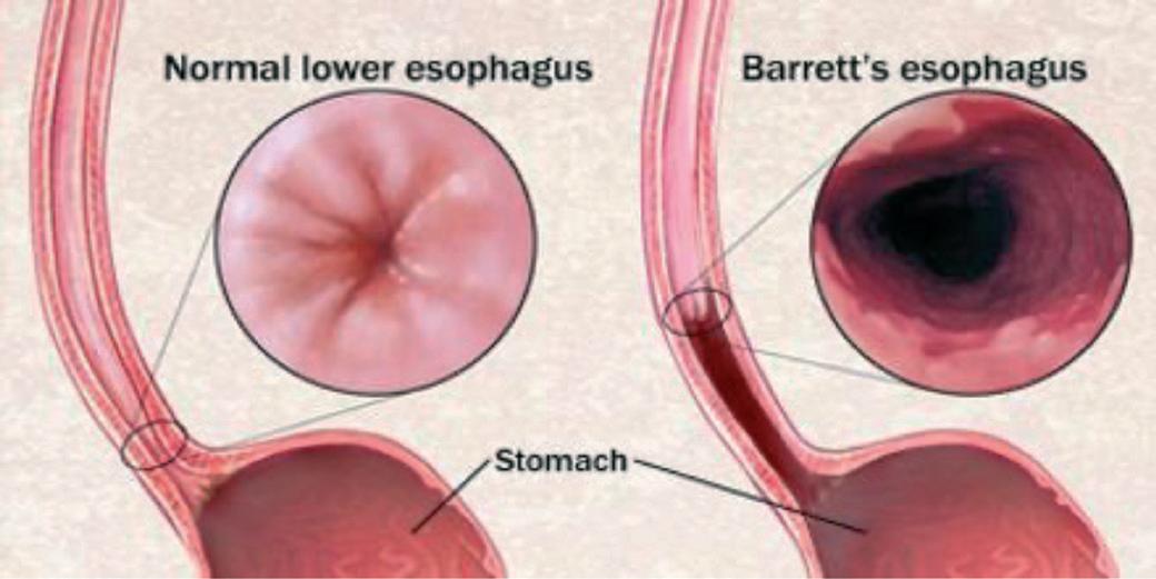
BSG Live was a great event, with plenary sessions all day on day 1, then anywhere from one to seven rooms to choose from on the other 3 days. All rooms had fantastic presenters and topics, catering to all levels of practice, which made it difficult to choose what to attend at times. For me, it is always great to brush up on basic polypectomy knowledge and skills, and I am also developing an interest in Barrett’s oesophagus so found myself drawn to many of the oesophageal talks.
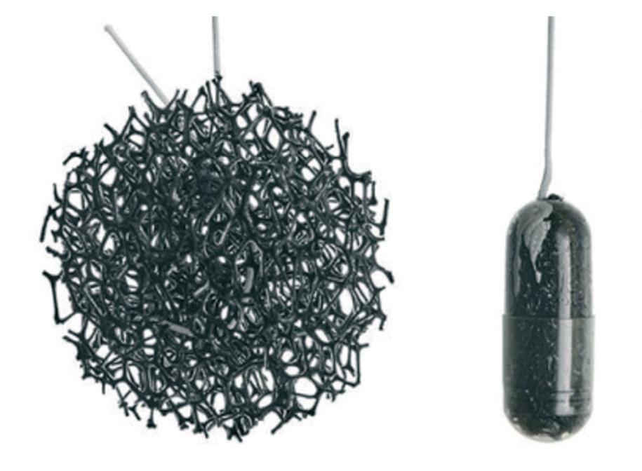
Of particular interest, was a Cyted Health stand in the trade hall displaying EndoSign. EndoSign is a brand of capsule sponge and is a simple way to test for Barrett’s oesophagus, and is utilised throughout the UK. A capsule sponge is a small, soft, spherical, polyester sponge, that is compressed into a small gelatin capsule, and attached to a string. When swallowed with a sip of water, the capsule advances into the stomach where it dissolves over 5 minutes, exposing the small sponge, with the string still accessible externally. After 5 minutes, the string is then pulled, bringing the sponge up the oesophagus and out the mouth. As it advances up the oesophagus, the sponge gently brushes against the entire of the oesophagus, collecting cells which are then analysed at a laboratory and examined for Barrett’s changes, dysplasia and malignancy.
Capsule sponges are marketed as a screening tool for patients with reflux, and those who test positive for Barrett’s changes proceed to gastroscopy. During the covid pandemic, NHS Trusts throughout the UK trialled and subsequently implemented capsule sponge testing as a triage tool to fast-track treatment for patients with signs of oesophageal cancer, appropriately schedule those at medium to low risk for surveillance, and discharge those with no concerning features back to their GP. Audits found it significantly reduced the demand on endoscopy services, reduced waiting times for endoscopy, and improved overall detection rates of pathology.
The BEST 2 Study (Ross-Innes et al, 2015), a systematic review estimating the diagnostic accuracy of Cytosponge (another brand of capsule sponge), reported a sensitivity of 90% and specificity of 92% for detecting Barrett’s oesophagus in patients with chronic reflux. Another study found a false positive rate of 7.6%. Comparatively, there is a false positive rate of 2%–9% for colorectal screening and 6%–15% for cervical cancer screening (Reflux UK, 2024). In the NHS England, Cytosponge triage of patients with chronic reflux who were referred to secondary care for an endoscopy, resulted in an estimated 78% of patients being removed from the endoscopy waiting list (Scottish Health Technologies Group Assessment, 2023).
Capsule sponge testing is advantageous in that it samples the entire oesophagus, whereas endoscopy only gains quadrantic +/- targeted biopsies, it is quick, it avoids endoscopy for some patients, and requires no sedation. It is not designed to replace endoscopy which remains the gold standard investigation for upper GI conditions, however as shown in the NHS, is a useful
Continued over page
Helen Livett, Tania Waylen, Karen Kempin, Sharon Powell.
tool for screening and triaging patients for endoscopy, hopefully resulting in a reduction in demand on endoscopy services.
Discussion with the EndoSign product reps found the main disadvantage of implementing this in New Zealand at the moment, is that samples would need to be sent overseas for processing, and results obtained from an online data reporting site. Hopefully laboratory processing will be available here in New Zealand in the future. Also, current guidelines for surveilling and managing Barrett’s do not include use of capsule sponges so work in this area will need to be undertaken.
Risks and adverse events associated with capsule sponge use are minimal. The safety of the capsule sponge device has been evaluated in a systematic review of 2672 procedures done across four different studies in the UK, the USA, and Australia. In this review, 2334 (97%) of 2418 patients swallowed the device successfully and there were only two adverse events associated with the device - one was a detachment, and one was a selflimiting pharyngeal bleed (Januszewicz et al, 2019). Should detachment occur, endoscopic retrieval is recommended. Another RCT, the BEST 3 Study (Fitzgerald et al, 2020) found 4% of people experienced a sore throat after the procedure. This was the most common side effect.
Capsule sponge testing is potentially an exciting venture for gastroenterology nurses as a nurse run clinic would be a great service that could have a significant impact on timeliness of assessment and treatment for patients with Barrett’s or suspected Barrett’s, reduction in cost of endoscopy services, reduction in demand on services, and hence reduction in endoscopy waiting times.
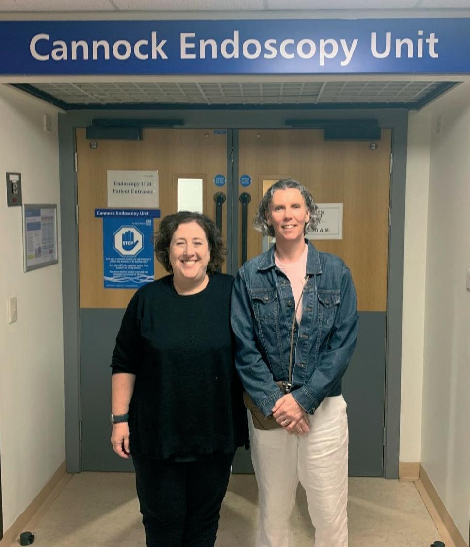
In addition to attending the conference, Karen Kempin and I were honoured to be shown around Cannok Endoscopy Unit where Helen and Sharon work and given the opportunity to see their department in action. They have amazing training opportunities for their Endoscopy Nurses and Clinical Endoscopists and set a great example for us to strive for here in New Zealand.
I would like to thank the Gastroenterology Nurses College for the Education Scholarship which certainly reduced the financial stress on this amazing trip.
References.
• Fitzgerald, RC, di Pietro, M., O’Donovan, M., Maroni, R., Muldrew, B., Debiram-Beecham, I.,…Sasieni, P. (2020). Barrett’s oESophagus Trial 3 (BEST3): Cytosponge-trefoil factor 3 versus usual care to identify Barrett’s oesophagus in a primary care setting: a multicentre, pragmatic, randomised controlled trial. Lancet; 396: 333–44. Cytosponge-trefoil factor 3 versus usual care to identify Barrett’s oesophagus in a primary care setting: a multicentre, pragmatic, randomised controlled trial.
Sharon Powel and Tania Waylen.
• Januszewicz, W., Tan, W.K., & Lehovsky, K., Debiram-Beecham, I., Nuckcheddy, T., Moist, S.,… Fitzgerald, R. (2019). Safety and acceptability of esophageal cytosponge cell collection device in a pooled analysis of data from individual patients. Clin Gastroenterol Hepatol, 17 (56.e1): 647. doi: 10.1016/j. cgh.2018.07.043.
• Reflux UK. (2024). Cytosponge™ · RefluxUK
• Ross-Innes, CS, Debiram-Beecham, I., O’Donovan, M., Walker, E., Varghese, s., Lao-Sirieix, P.,… Fitzgerald, R; BEST2 Study Group. (2015). Evaluation of a minimally invasive cell sampling device coupled with assessment of trefoil factor 3 expression for diagnosing Barrett’s esophagus: a multicenter case-control study. PLoS Med, 12(1). doi:10.1371/journal. pmed.1001780. PMID: 25634542; PMCID: PMC4310596.
• Scottish Health Technologies Group Assessment. (2023). Capsule sponge technologies for the detection of Barrett’s oesophagus and early stage oesophgeal cancers. Scottish Health Technologies Group & Health Improvement Scotland.
KAREN KEMPIN Nurse Practitioner/Nurse Endoscopist
Hutt Valley Health Region
Karen.kempin@huttvalleydhb.org.nz • 022 039 0632
With support from the NZ Gastroenterology Nurses College I had the privilege of attending this amazing annual conference in Birmingham, England, along with Tania Waylen from BOP. At the NZ ASM 2023 in Rotorua two nurse endoscopists Helen Livett and Sharon Powell were the invited international speakers and we were very happy to meet up with them in Birmingham, which is very close to their respective NHS trust facilities. Opportunities presented themselves to socialise with their team and also visit the Cannock Chase Hospital endoscopy unit to observe training lists being provided by JAG accredited nurse trainers.
BSG 2024 Live is the biggest gastroenterology conference I have attended, spread over 4 days with 5 sessions running from 8am to 6pm plus lunch time trade sponsored lectures, with up to 7 streams running in any given session. In contrast Australian Gastroenterology Week is 3 days, 3 sessions a day and around 4 streams in any session.
The biggest difficulty was picking one presentation to attend, with lots of moving between rooms to catch my selected program. With around 2000 registrations over the 4 days I thought it would be too busy and I would feel lost, but as in gastroenterology everywhere, everyone was welcoming and happy to be together with teams mixing in and friends catching up. They were also excited to meet the travellers from New Zealand, although the person who mentioned they have a gift from a relative of a stuffed koala holding a New Zealand flag seemed a bit geographically challenged.
My focus was on luminal and endoscopy skills development so I joined the gastroenterology and surgical trainee sessions in upper and lower endoscopy at every opportunity, plus sessions on management of bolus and GI bleeding and recognition and management of non-malignant luminal pathology. I felt I had a deficit in the colonoscopy surveillance requirements for IBD patients and attended a whole session on this topic. Wednesday and Thursday featured dedicated nurse stream, both for endoscopy nurses and nurse endoscopists, and featured presentations from some of the UK pioneers of GI specialty nurse roles in IBD, hepatology, nutrition and nurse endoscopist.
The Masterclass presentations given by the best in their fields gave information and advice on how to manage acute and chronic disease applying best practice and up to date guidelines. Advice on how they specifically apply guidelines and communicate with and treat patients was insightful and well received. I also had a
chance to do a bit of hero worshipping, attending talks given by Dr Rehan Haidry, Dr Alessandro Repici, Professor Matt Rutter and Dr Helen Griffiths. These are names I recognise from many journal papers and textbook chapters.
Some of the sessions I missed included management of cyclical vomiting, sustainability, endoscopy in liver disease, oesophageal neoplasia, nursing free papers and interesting endoscopy cases, so I have some work ahead catching up on the recorded sessions through the BSG LIVE! app.
I also noticed that the title Nurse Endoscopist has been abandoned in favour of Clinical Endoscopist, which seems to apply to anyone who is not a gastroenterologist or surgeon endoscopist. UK has a few physician assistants trained for gastrointestinal endoscopy, so this term includes them.
Continued over page
One of the most thought provoking sessions I attended was Nursing in Gastroenterology and Endoscopy – A Different Approach. The session featured speakers on the topics of clinical endoscopy in Europe, being a GI expert witness for coroner and court cases and consideration to the care of transgender and gender diverse patients.
Our hearts all went out to Ute Pfeifer, the international speaker from Germany, who outlined her stalled attempts to be the first and maybe only nurse endoscopist in Germany. She has fought hard and even learned a new language (Dutch) to get endoscopy training and has faced multiple rejections and still has hope that she will be acknowledged and the role allowed to expand. She was also in tears when she realised she was in a room of supportive and understanding colleagues that could feel her frustration and potentially offer some new directions to get to her goal.
Julian Layhe NP and Clinical Endoscopist presented the research results from Trans Lives Survey 2021 Health Care Experience. As a nurse who has gender transitioned and identifies as him/they, he was able to offer some insight into the disparity in access and treatment for transgender patients (people who identify as a different gender to the one they were assigned at birth, are gender fluid or identify as no gender) compared to cisgender patients (people who identify as the same gender they were assigned at birth).
Some of the points made is his presentation made me think deeply about gender. Consider that almost no new-borns have a DNA test at birth, and gender is assigned by a doctor/midwife/ health care professional after a cursory examination of external genitalia. Once this is documented it is very difficult to go back/ change and might not reflect the persons felt gender as they grow up.
Scary statistics from Julian included 70% of respondents having experienced a degree of transphobia while accessing healthcare (verbal, body language, delays in care, etc). 57% have avoided going to their GP when unwell out of fear they will be biased against and a shocking 14% have been refused care (were chased away) by a GP practice. Julian also showed that around 13% of UK residents identify as transgender, meaning many people have experienced terrible incidents when they needed care. If a person is transgender and also a person of colour, non UK ethnicity or disabled these incidents can happen at twice the rate of white transgender people.
It is the attitude of health care staff that often discourages transgender people, with some being unconsciously discriminatory through their lack of understanding/acceptance of self-identified gender (the gender the person relates themselves to). This can be demonstrated by using incorrect gender titles, using the person’s dead name rather than their chosen name or ignoring/avoiding uncomfortable sections of patient assessment/ care planning related to gender affirming hormone treatment or gender affirming surgery. In many cases the staff want to do the right thing but are embarrassed or ashamed that they don’t know how to talk safely to the patient about their health in relation to gender transition.
Of course, there are instances of overt transphobia including refusal of care which is not acceptable, no excuses. Any reported or witnessed prejudiced words or actions must be addressed in the same way as sex, racial, religious, etc discrimination. Quick action to stop the behaviour, reprimand, behaviour management and employment repercussions for staff who display ongoing discriminatory actions are steps to stamp out poor behaviour.
In New Zealand the Professional Association for Transgender Health Aotearoa (PATHA) found in a secondary school survey that 1.2% of adolescents identify as transgender, with a further 2.5% uncertain about their assigned gender (Oliphant, et.al. 2018). Stats NZ (2021) shows 4.4% of NZ adult population identifying as LGBT+, with a 0.4% subset indicating they are transgender. The higher number of adolescents compared to adults reflects society’s slowly changing positive acknowledgment and acceptance of a gender fluid population, meaning individuals feel safer “coming out”.
Julian concluded his presentation with some useful advice on how endoscopy staff and facilities can create a safe space for transgender patients to get equitable care.
Staff need familiarisation with transgender concepts and care, including correct/legal health information recording and gender neutral language to be used when communicating with any patient i.e stop using Mr, Mrs, Miss and just use their name. Staff will sometimes say/ask nothing and just makes assumptions or ignore parts of a patient assessment because they are afraid to be embarrassed or cause offense.
Facilities need to give staff an opportunity to have a team or one on one discussion with a diversity champion where they can ask questions without fear of judgement or ridicule and learn
strategies to manage their own discomfort and provide safe health care to transgender patients.
Specific examples of not to be missed information include finding out about hysterectomy/ pregnancy with a transgender male as part of a pre-assessment for colonoscopy, same as we would discuss these points with a cis female.
Gender affirming surgery should be discussed with transgender females to determine if bowel tissue has been used for a vaginoplasty if IBD surveillance is planned. Gender affirming hormone supplements should be recorded the same as any other medications as these can impact on midazolam and fentanyl breakdown with potential for toxicity and can displace warfarin leading to higher INR.
Clinicians must be careful to collect/record only relevant information specifically needed for the procedure planned but omitting information like this from an assessment can lead to problems/complications that are avoidable.
Facilities need to adopt gender diverse practice by taking gender specific descriptions/titles out of patient information/records, check unit policies and procedures are not biased against transgender patients and undertake a patient journey from a transgender patient’s point of view to check their privacy and safety is sustained.
Te Whatu Ora has a Transgender Service that provides similar advice and guidelines for NZ health professionals, as well as help for transgender patients seeking gender affirming surgery. Numbers of patients who identify as transgender will increase as society becomes more accepting and young people age and start to need our gastroenterology services. We should be getting ahead of the issue in New Zealand to avoid the disparity seen in the UK.
BSG LIVE! 2025 is in Glasgow, Scotland on 23-26 June, so if you are thinking of going to a big (but not massive) international conference, it is one to keep in mind, particularly with the assistance of the NZGNC Education Fund. I managed to fit in about 10 days of UK sight-seeing post-conference, which was lovely and would encourage you to think about attending this conference. Even the jet lag wasn’t too bad!?
References
• Oliphant, JE, Veale, J, Macdonald, J, Carroll, R, Johnson, R, Harte, M, Stephenson, C, Bullock, J (2018)
• Guidelines for Gender Affirming Healthcare for Gender Diverse and Transgender Children, Young People and Adults in Aotearoa New Zealand. Transgender Health Research Lab, University of Waikato.
• Stats NZ (2021) Report LGBT+ population of Aotearoa: Year ended June 2020. https://www.stats.govt.nz/reports/lgbtplus-population-of-aotearoa-year-ended-june-2020
Faecal Microbiota Transplantation
KIRSTY BLACK Bsc, RN
Kirsty.black@cdhb.health.nz
• 027 697 8826
Faecal Microbiota Transplantation
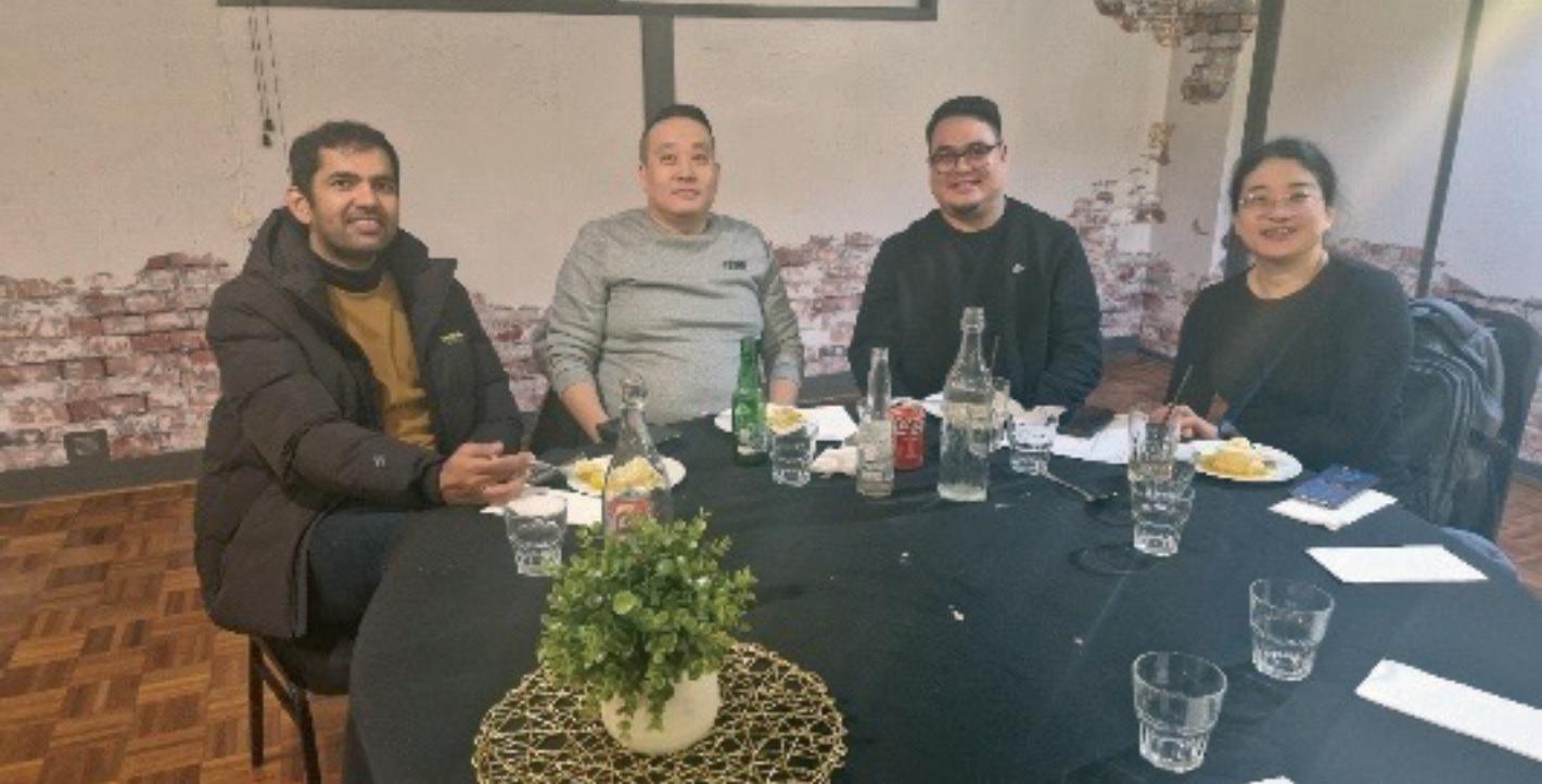
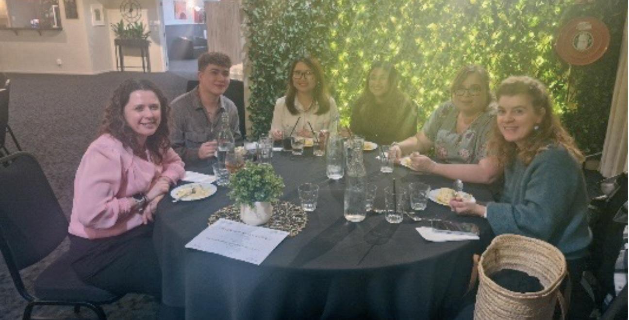
In March, I had the privilege of attending the Sydney International Endoscopy Symposium (SIES), a remarkable event that offered a wealth of knowledge and networking opportunities. I am grateful to the New Zealand Gastroenterology Nurses College for their generous support, which allowed five nurses from the Christchurch Hospitals Gastroenterology department to participate. This experience not only broadened our skill sets but also facilitated the exchange of valuable insights and the establishment of new connections within the gastroenterology community.
Every year this conference is hosted by the Westmead hospital, a private endoscopy center providing state of the art gastrointestinal, biliary and respiratory interventions. Broadcasting from both the city based venues and the hospitals operating suites, we were exposed to live endoscopy and various presentations by renowned local and international health professionals. These speakers were nothing short of exceptional but I would like to emphasize my particular interest on Faecal Microbiota Transplantation (FMT), presented by Associate Professor David Van der Poorten, a Gastroenterologist and Hepatologist from Sydney Norwest Gastroenterology.
What is FMT?
FMT can be described as the infusion of distal faecal material from one healthy donor to a recipients gastrointestinal tract, to reestablish healthy intestinal flora (Borody & Ramrakha, 2024). Faecal matter from potential donors is rigorously screened, anaerobically processed and then immediately frozen to -80degrees for up to 24months until the recipient requires the transplant (Van der Poorten, 2024). Prior to freezing, the stool is sieved and mixed with saline to ensure fibre and other unwanted debris are not transferred to the recipient. Once the transplant is needed, the stool is thawed and then warmed. Delivery of the FMT varies among institutions but includes administration via oral capsule, nasogastric/nasojejunal tube or colonoscopy (Van der Poorten, 2024).Evidence suggests that lower gastrointestinal administration via colonoscopy or retention enema has a higher efficacy that other routes however, clinical circumstances and available options strongly influence administration (Borody & Ramrakha, 2024)
Gut microbiota and FMT
The gastrointestinal tract harbors a complex community of microorganisms and as many as 1200 bacterial species which largely live symbiotically within the gut. There are many beneficial
roles mediated by the gut microbiota including vitamin synthesis, metabolism of bile and hormones, and fermentation of dietary carbohydrates. Development and maturation of the immune system is also influenced by the microbiota through interactions with gut epithelium (Borody & Ramrakha, 2024).
The term microbiota refers to the bacteria, microeukaryotes and viruses that coexist within the body. However, factors including diet, medication use, and an individual’s genome can significantly alter the composition of the microbiota and disrupt homeostasis (Thursby & Juge, 2017).
Dysbiosis is an imbalance of the gut microbiome which is characterized by the abundance and resilience of excess pathogens (Van der Poorten, 2024). Dysbiosis is strongly associated with many inflammatory diseases and infections including Cholelithiasis, inflammatory bowel diseases (IBD), and in particular, Clostridium Difficile infections (CDI) (Thursby & Juge, 2017).
Although not widely utilized within New Zealand, the use of FMT is growing worldwide, particularly within Australia, where they have seen some significant results. FMT has become an effective treatment for Clostridium Difficile infections (CDI), with recent studies from Westmead Hospital indicating clearance rates as high as 94 percent. (Van der Poorten, 2024). Another randomised trial by Kao et al (2017) found 96.2 percent of their 116 patients achieved prevention of recurrent CDI (Kao, et al., 2017).
The therapeutic potential of FMT in patients with IBD, encompassing both Crohn’s and Ulcerative Colitis (UC), is still being widely investigated. Studies have shown higher variability in patient response and most suggest that success is dependent on the microbial diversity of the stool donor. Success was first achieved in 1989 when the first clinical trials took place. One male subject with refractory UC achieved remission for 6 months following a retention enema with healthy donor stool (Wilson, Vatanan, Cutfield, & O’sullivan, 2019).
Since then many trials have been conducted with moderate success. A study conducted by Paramsothy et al. (2017), investigated the effectiveness of FMT in UC over an 8-week trial period. Participants were administered either an FMT or placebo for five days of the week, for eight weeks. Results showed that of the participants who received FMT, 27 percent achieved remission (Paramsothy, et al., 2017).
The “Super- Pooer”!
Genetics and environmental factors influence each individuals microbiome and these fluctuate throughout an individuals lifetime. Within Australia, donors undergo rigorous screening including health questionnaires, blood and stool testing and clinical interviews. Approximately ten percent of those who apply will be selected to donate(Wilson, Vatanan, Cutfield, & O’sullivan, 2019) and for good reason.
The term “super-pooer” can be demonstrated in the study, previously discussed, by Paramsothy et al.(2017). The participants who received FMT had varied mixes of stool from multiple donors. Subsequent analysis of the differing stool batches discovered that one stool donor exhibited a “super-donor” effect. Specifically, patients who received FMT batches from the super donor, exhibited higher remission rates of UC( 37 vs, 18% respectively). Donor recipient matching may also be considered in the future, where donors who are known to be enriched in taxa associated with the metabolic pathway that needs to be restored in the recipient, can be identified and paired (Wilson, Vatanan, Cutfield, & O’sullivan, 2019).
Conclusion
The evolving research on FMT emphasizes the importance of donor selection, screening processes, and delivery methods for successful outcomes. However, the varied results from different trials reinforces that FMT is still in its infancy. Further exploration, funding and standardization of therapy worldwide would help reduce variability in patient response to FMT
Overall, the symposium provided a deep dive into the intricate world of gut microbiota and its impact on human health, shedding light on the groundbreaking potential of FMT and inspiring further research and collaborations in the field of gastroenterology.
References
• Borody, T. J., & Ramrakha, S. (2024, March 19). Faecal microbiota transplant for treatment of Clostridioides difficile infection. Retrieved from Up to Date : https://www.uptodate.com/ contents/fecal-microbiota-transplantation-for-treatmentof-clostridioides-difficile-infection
• Kao, D., Roach, B., Silva, M., Beck, P., Rioux, K., Kaplan, G. G., . . . Louie, T. (2017, November 28). Effect of Oral Capsuale-vs Colonoscopy-Delivered Faecal Microbiota Transplantation on Recurrent Clostridium Difficile Infection. JAMA, 318(20), 19851993. doi:10.10001/jama.2017.17077
• Kim, K. O., & Gluck, M. (2019, March). Faecal Microbiota Transplantation: An update on clinical practise. Clinical Endoscopy, 52(2), 137-143. doi:10.5946/ce.2019.009
• Paramsothy, S., Kamm, M. A., Kaakoush, N. O., Walsh, A. J., Bogarede, J. v., Samuel, D., . . . Borody, T. J. (2017, March). Multi intensive faecal microbiota transplantation for active ulcerative colitis: a randomised placebo-controlled trial. Lancet, 25(389), 1218-1228. doi:10.1016/s0140-6736(17)30182-4
• Thursby, E., & Juge, N. (2017, june 1). Introduction to the Human Gut Microbiota. BioChem Journal, 474(11), 1823-1836. doi: 10.1042/BCJ20160510
• Van der Poorten, D. (2024). Faecal Microbiota Transplantation. Sydney International Endoscopy Symposium. Sydney: David Van der Poortan.
• Wilson, B. C., Vatanan, T., Cutfield, W. S., & O’sullivan, J. (2019). The Super Donor phenomenon in Faecal Microbiota Transplantation. Front. Cell. Infect. Microbial, 9, 33-89. doi:10.3389/fcimb.2019.00002
2024 Auckland Regional Endoscopy
Nurses Study Day and Dinner Lecture: A Growing Tradition of Learning and Networking
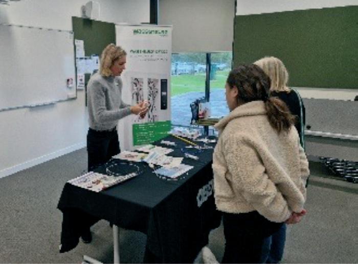
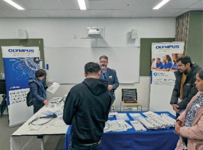
The 2024 endoscopy nurses study day in the Auckland region marked another successful gathering of endoscopy nurses from both the public and private health sectors of Auckland. The event took place on the 17th of September hosted by Te Whatu OraWaitemata. A number of interesting endoscopy topics were on the agenda alongside its enthusiastic speakers.
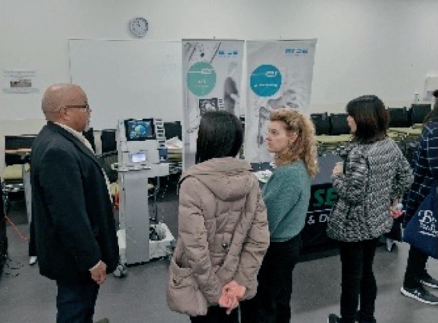
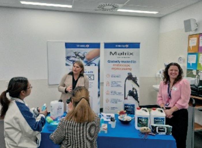
Building on the momentum from last year, this year’s event was different and superb because of the generous support of Boston Scientific, the Gold Sponsor for the day. In addition to that, a host of Bronze Sponsors, including Obex, Medtronic, Bioserve, Whiteley Medical, Alimetry, Olympus, and Endoventure, contributed to the event’s success. The host, as well as the attendees, were very grateful to the benefactors. The occasion would not have been possible without their backing and contribution. The study day was emceed by the Clinical Nurse Educator of Te Whatu Ora –Waitemata, Gino Borromeo.
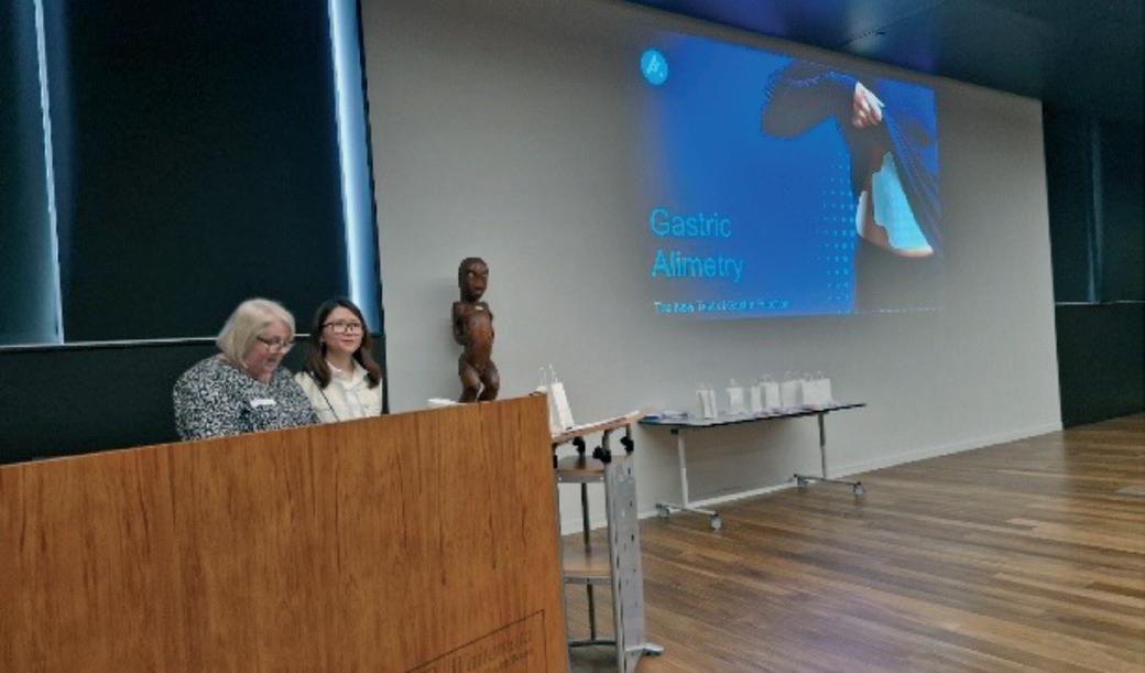
The day was filled with insightful presentations on cutting-edge topics in endoscopy. Dr Marius Van Rijnsoever kicked off the session with a talk on Endoscopic Ultrasonography (EUS) Tips for Nurses, offering practical guidance that was well-received by all in attendance. Endoscopy nurses can apply the tips learned into their current practice e.g. anticipating the correct echoendoscope, or understanding basic echogenicity. Teresa Nguyen and Kim Parsons then presented on Gastric Alimetry, providing a detailed overview of this innovative approach and its implications in guiding medical treatment. This is the first Body Surface Gastric Mapping service in the Auckland region. This specialty of the service was long overdue and it wasn’t without barriers. It needed passionate doctors and nurses to initiate and kick-off this innovation.
Endoscopic Submucosal Dissection (ESD) was expertly covered by Dr Cameron Schauer, who shared his in-depth expertise and passion for this advanced technique, highlighting its impact on curing cancer through endoscopy. According to him, early detection is always better that cure. Otherwise, patients end up six feet underground.
GINO BORROMEO AND JONATHON REALISTA
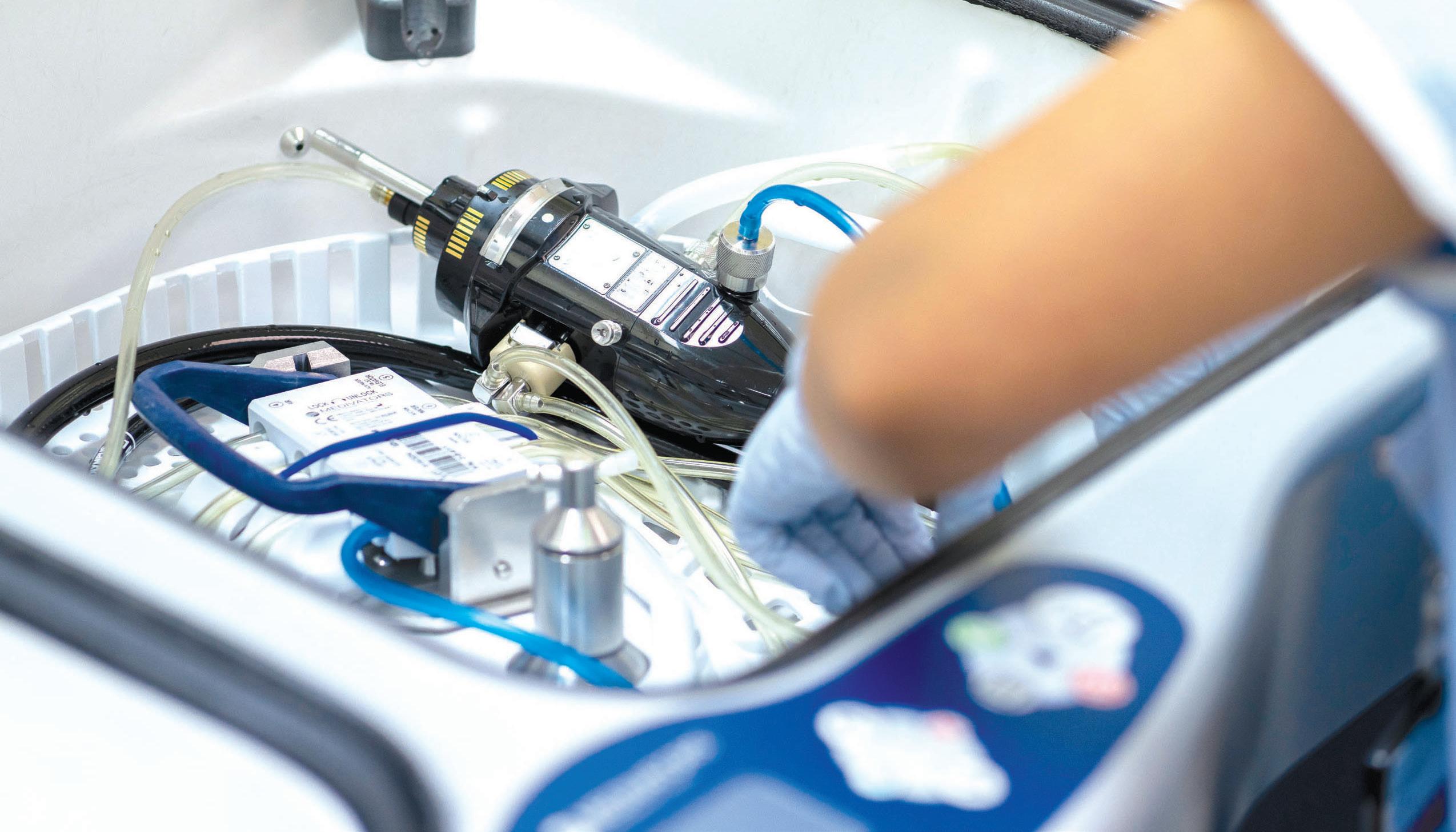
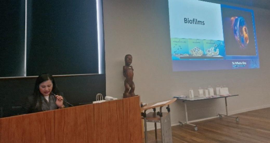
Julie Branzuela presented an important update on GENCA Scope Reprocessing. This focused on the latest guidelines and best practices for endoscope reprocessing. Her talk highlighted the prevention of biofilm formation and ultimately ensuring patient safety. A video was also shown to the attendees to reinforce their knowledge in endoscope cleaning and reprocessing and not just from the slides presented. The takeaway message for the abovementioned topic is to clean it (endoscopes). Clean it. And clean it.
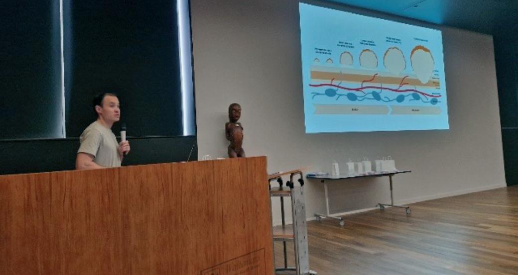
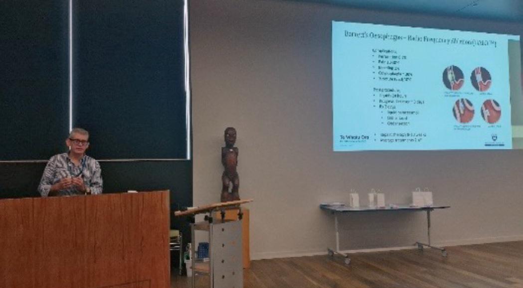
This is then followed by Dr Russell Walmsley’s session on Barrett’s Oesophagus & Radiofrequency Ablation (RFA), which provided crucial insights into managing this condition. He is currently developing a guideline for Waitemata in diagnosing and managing Barrett’s oesophagus. This is a condition that should not be taken lightly. Based from his presentation, the annual risk of developing oesophageal adenocarcinoma in Barrett’s oesophagus with High Grade Dysplasia is 6-19% worldwide.
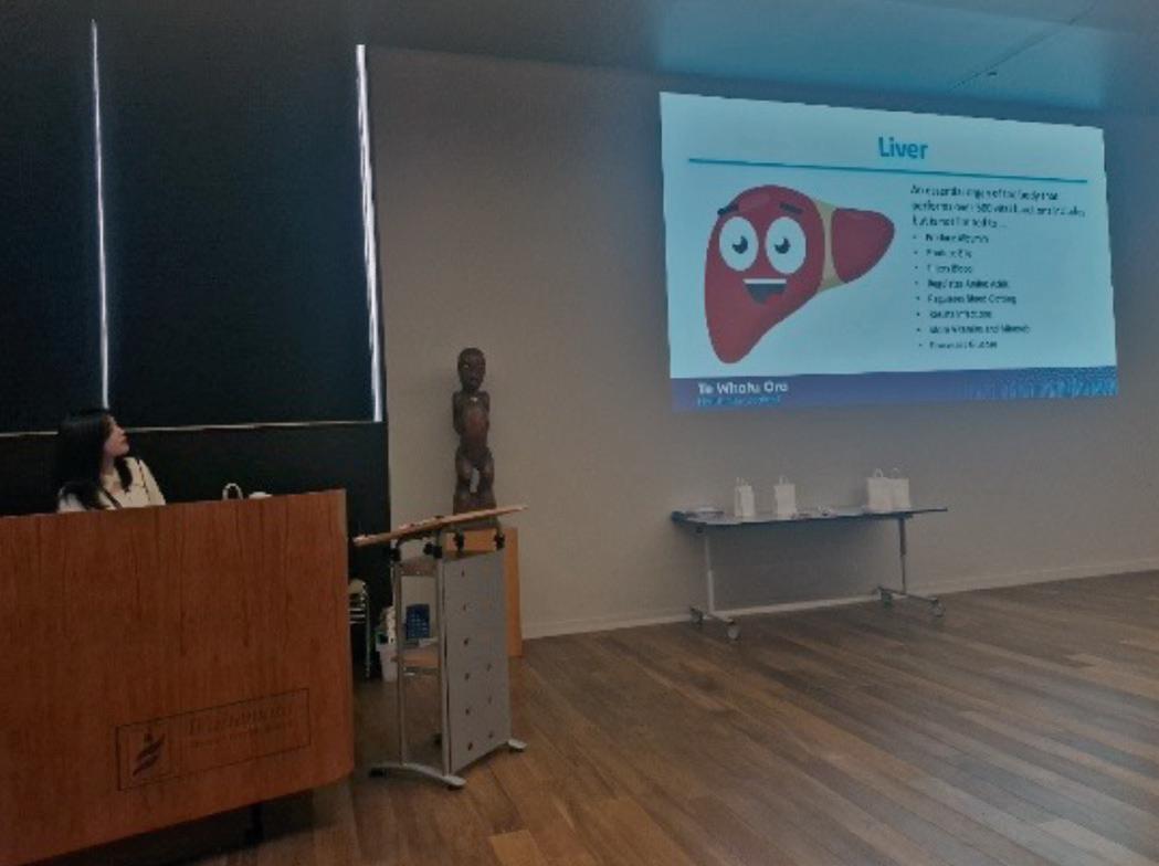
After that, Erae Kim’s discussion on Liver Cirrhosis, Portal Hypertension, and the ELLA Danis Stent was another highlight. The pathophysiology and management of the two conditions were presented during the talk with emphasis on the importance of nursing care in these complex, and sometimes urgent cases. She also imparted to the audience the correlation of the Ella Danis stent to patients with bleeding oesophageal varices. Dr Nathan Atkinson rounded up the presentation with an engaging talk on Reflux & ARMA , offering new perspectives on these common issues. According to Nathan, almost everyone has reflux but the majority is asymptomatic. He also discussed oesophageal manometry and its relation to reflux and ARMA.
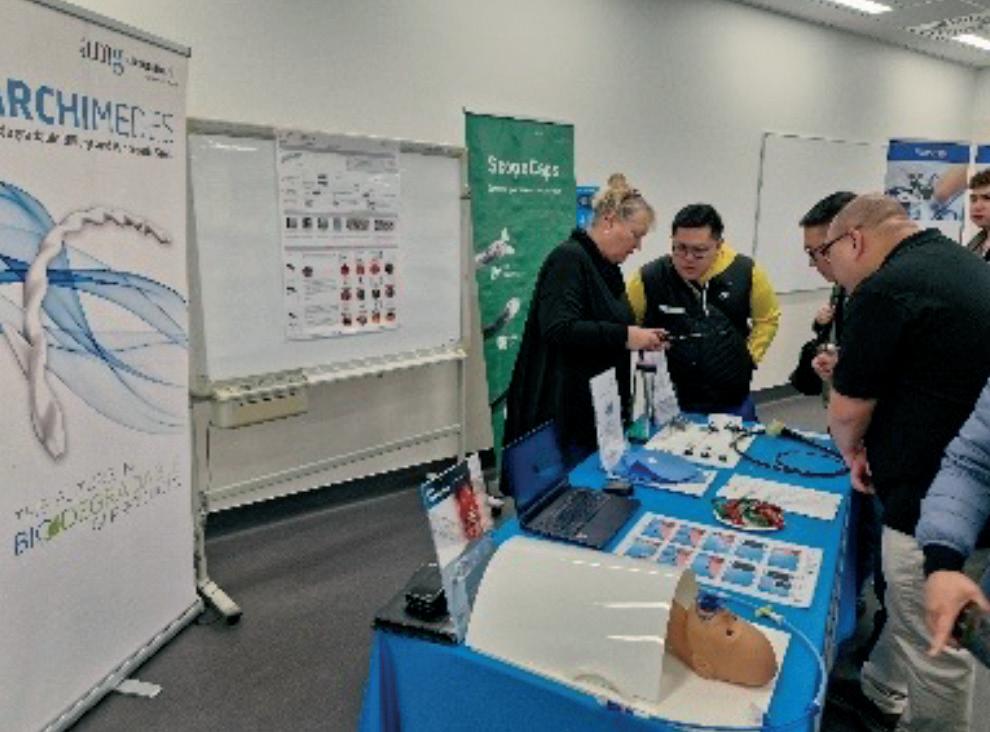
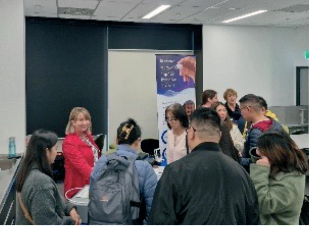
Apart from the talks the supporting medical companies showcased their equipment and products. Skills station were seen everywhere in the auditorium for a hands-on experience by the participants.
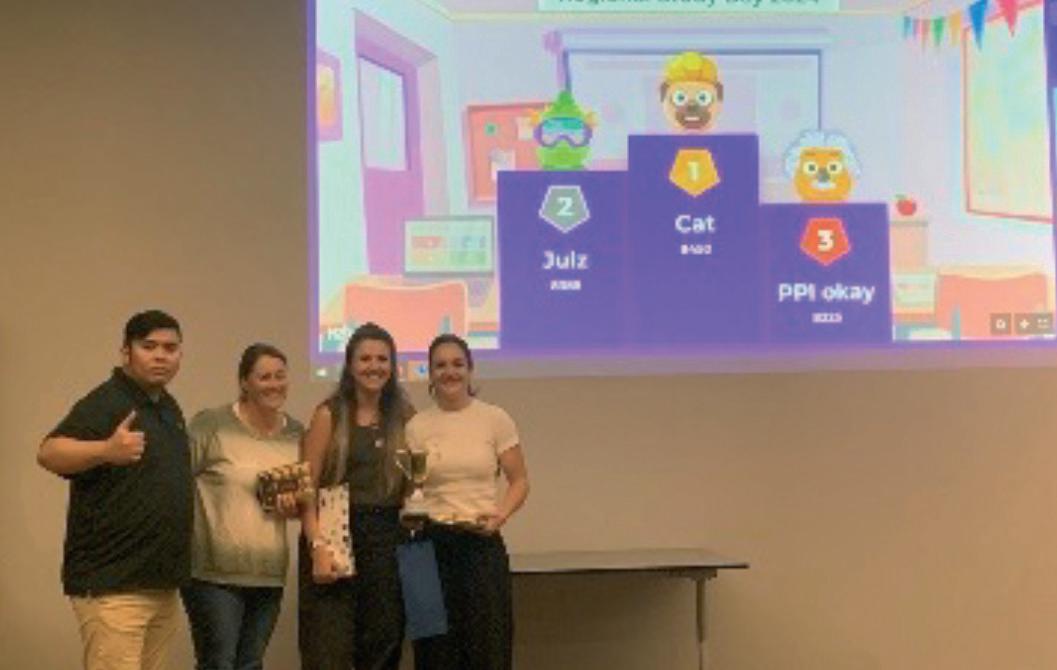
The day concluded with the highly anticipated Kahoot Showdown (online-based, multiple choice quiz), a lively competition that has
Continued over page
quickly become a beloved tradition. This year, the team from Waitemata Endoscopy (Private) dominated the game, sweeping all the top prizes and securing 1st, 2nd, and 3rd place.
In the evening, we hosted our very first dinner lecture, a rare and exciting opportunity for nurses. This is something typically reserved for doctors, but thanks to the support of Boston Scientific, we were able to provide this special session. The speaker for the night was Dr Sam Seleq, a gastroenterologist trained in a wide array of endoscopic techniques. He delivered a captivating talk on Hepatobiliary Pathology, with a focus on Cholangioscopy and the evolving techniques that are reshaping patient care and treatment outcomes. Emphasizing that ERCP is now solely an interventional procedure and is no longer used for the diagnostic purposes.
Beyond the educational content, the event provided ample opportunities for socialising and reconnecting with colleagues. The enthusiastic participation and positive feedback have inspired us to consider making this a yearly event, ensuring that the tradition of learning, sharing, and growing together continue for years to come.
ENDOSCOPY CONSUMABLES
Select & bundle consumables
Stock on-site or in your department Collect when you need to use
Acknowledging and Treating Fatigue for Patients Living with Inflammatory Bowel Disease
DONNA HOWE Dip Registered Comprehensive Nurse, PGDip Health Science
In my experience, fatigue is one of the most common issues identified when patients living with inflammatory bowel disease (IBD) are reviewed in clinic.
This isn’t any surprise when fatigue affects 80% of those with active IBD disease and 50% of those with inactive IBD disease (Borren, van der Woude & Ananthakrishnan, 2019). As a clinical nurse specialist (CNS), when a patient reports fatigue, my next step is to assess disease activity.
Assessing patient symptoms using disease activity scores, either Harvey Bradshaw index for Crohn’s disease (Harvey, n.d), or Simple Clinical Colitis Activity Index for Ulcerative colitis (ECCO, n.d) then confirming with the patient that actual medication doses are reflective of what is prescribed, and lastly obtaining objective data are all routine clinical tasks. This includes blood and stool samples to measure inflammatory markers particularly CRP and faecal calprotectin levels.
Other clues gleaned from the complete blood count include haemoglobin, platelet, neutrophil and mean cell volume levels and by adding a haemolytic screen, I can identify anaemia, iron or vitamin B12 deficiency. It is also useful to check thyroid function, another potential cause of fatigue and vitamin D levels, although studies into vitamin D deficiency being associated with fatigue are not conclusive (Borren, van der Woude & Ananthakrishnan, 2019).
If these results are within range indicating quiescent or disease remission, I then delve into sleep hygiene, exercise, diet, medications and the administration timing as well as mental health status using the quick PHQ-2 score (Pfizer, 1999) as all of these can contribute to fatigue. Often picking one or two goals or interventions to focus on out of these assessment findings validates the issue for the patient and empowers them to trial interventions that may assist with their fatigue, but clinically appropriate follow-up to assess their response whether it be positive, or negative is important, and must be prioritised into the CNS’s workload.
My interest in the topic of fatigue was re-ignited at the GENCA conference that I recently attended in Brisbane, where Palle Bager from Denmark spoke about his recent study on the use of thiamine to treat fatigue. I returned to my practice intrigued and revisited the Crohn’s and Colitis UK website to review their patientfocused document on fatigue, a document that I refer patients to regularly. This document has been amended in September
2023 (Crohn’s and Colitis UK, 2022), with the “Inflammatory Bowel Disease – Fatigue (IBD-F) self-assessment scale” embedded into the updated document. Whilst this questionnaire hasn’t been widely tested and needs further assessment for validity and stability, other fatigue scales have been (Borren, van der Woude & Ananthakrishnan, 2019). Nevertheless, it is a tool that could be used to validate this patient issue as well as appoint a score that could be used to measure intervention response. Behavioural modification has historically been the focus of treatment for fatigue (Borren, van der Woude & Ananthakrishnan, 2019).
The TARIF clinical trial by Bager and associates (Bager et al. 2021) investigating the use of thiamine therapy to improve chronic fatigue for patients in IBD remission, used only a small cohort of patients (n=60), 40 IBD patients that had fatigue and 20 control IBD patients without fatigue. It is known that thiamine (Vitamin B1) is essential for the metabolism of carbohydrates and the production of mitochondrial adenosine triphosphate (ATP), with both processes promoting energy levels for the body. The trial used high doses of oral thiamine hydrochloride administered over four weeks.
This reduced fatigue levels in patients with quiescent IBD and chronic fatigue by ≥3 points on the IBD-F scale in 26 out of 40 participants who were in the fatigued group. (Bager et al. 2021). Chronic fatigue was defined using the IBD-F self-
assessment scale, and participants had to have a score above 12 points, (Czuber-Dochan et al. 2014). A reduction of ≥3 points is defined as being a clinically significant improvement (Bager et al. 2023).
However, the study did raise questions that need further investigation, such as flavin mononucleotide (FMN) which was found at lower levels in the study group with chronic fatigue compared to the control group of non-fatigued as well as levels of riboflavin were decreased in the group of 26 participants that responded to high doses of thiamine. Therefore, further research around the supplementation of Vitamin B2 in conjunction with Vitamin B1 (thiamine) is needed (Bager et al. 2023).
In summary, further study is needed before thiamine can become a routine treatment for fatigue. In the meantime, l will continue to assess patients using disease activity scores, evaluation of medication to ensure these are being used correctly, laboratory monitoring for deficiencies and inflammatory markers, and continue to educate patients on healthy diet and lifestyle options. Mental health monitoring will continue with the use of the PHQ-2 score, the intention is that this data will support a business case for a psychologist to support our Gastroenterology service in the future.
My role as CNS is to support those living with IBD in Northland, to improve their quality of life, fulfil their study and employment obligations and improve their experience when they interact with the health system. It is important as healthcare workers, to recognise the limitations of resources available in our workplaces and prioritise care that is specific to managing the disease that we specialise in. Protection, participation and partnership are a twoway process, otherwise, we will end up suffering from fatigue ourselves.
References
• Bager, P., Hvas, C.L., Hansen, M.M., Ueland, P., Dahlerup, J.F., (2023). B-vitamins, related vitamers, and metabolites in patients with quiescent inflammatory bowel disease and chronic fatigue treated with high dose oral thiamine. Molecular Medicine, 29:143. https://doi.org/10.1186/s10020023-00741-3
• Bager, P., Hvas, C.L., Rud, C.L., Dahlerup, J.F., (2021) Randomised clinical trial: high-dose oral thiamine versus placebo for chronic fatigue in patients with quiescent inflammatory bowel disease. Ailment Pharmacol Ther; 53:79-86.
• Borren, N.Z., van der Woude, C.J., Ananthadrishnan, A.N., (2019) Chapter 2 Fatigue in IBD: epidemiology, pathophysiology and management. Nature Reviews Gastroenterology & Hepatology, Apr;16(4)247-259.
• Czuber-Dochan, W., Norton, C., Bassett, P., Berliner, S., Bredin, F., Davell, M., et al. (2014) Development and psychometric testing of inflammatory bowel disease fatigue (IBD-F) patient self-assessment scale. J Crohn’s Colitis; 8:1398-406
• ECCO - (European Crohn’s and Colitis Organisation), (n.d) Simple clinical colitis activity index (SCCAI) retrieved from https://www.e-guide.ecco-ibd.eu/resources/calculator/ simple-clinical-colitis-activity-index-sccai 21/6/24
• Harvey, F., (n.d), Harvey Bradshaw Index (HBI) for Crohn’s disease retrieved from https://www.mdcalc.com/ calc/10069/harvey-bradshaw-index-hbi-crohns-disease 21/6/24
• Pfizer, (1999), The patient health questionaire-2 (PHQ-2)overview retrieved from https://www.mcstap.com/docs/ tool_phq2.pdf 21/6/24
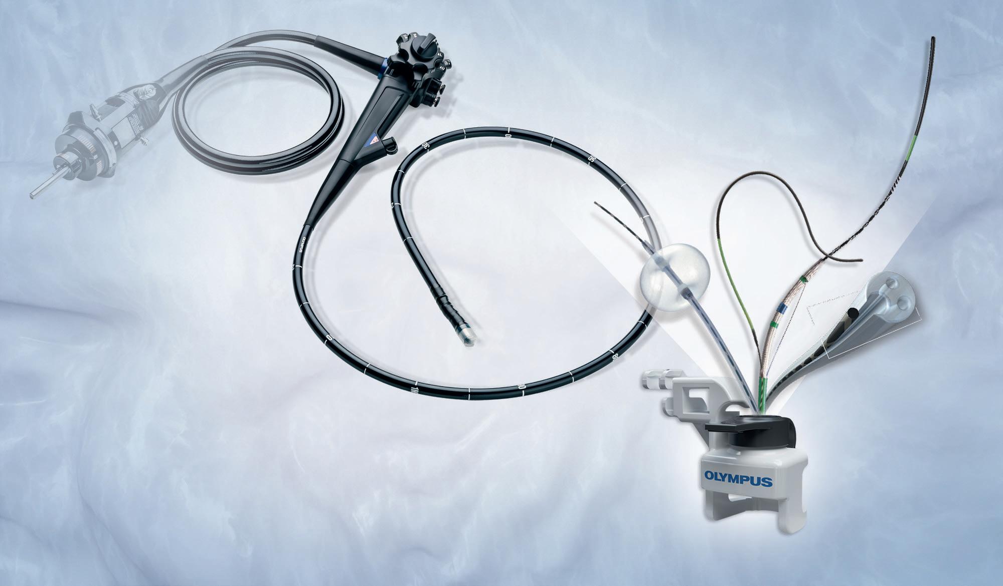
Per-Oral Cholangiopancreatoscopy
JONATHAN REALISTA
Associate Charge Nurse Manager, Endoscopy- North Shore Hospital, Health NZ
Introduction:
Per-oral cholangioscopy (POC) is the direct visualisation of the bile ducts endoscopically through the mouth, hence, called peroral. This endoscopic procedure can achieve a diagnostic and/ or a therapeutic function. This supersedes the constraints that a regular endoscopic retrograde cholangiopancreatography (ERCP) provides. As early as the 1950s, a literature by Roca, et. Al. (1950, as cited in Gopakumar and Sharma, 2023) narrated the utilisation of such a procedure (cholangiopancreatoscopy). Nevertheless, at that time, to find stones the cholangiopancreatoscopy procedure was done during a surgical operation called common bile duct exploration. The per-oral method of cholangioscopy and pancreatoscopy is the most conventional practice. The other types of cholangioscopy are intra-operative as narrated by Kumanduri, S., et. Al. (2016), and percutaneous (making a cut in the skin) by Ponchon, T., et. al. (1996). However, percutaneous transhepatic cholangioscopy (PTC) is invasive and unwieldy according to Oh, et al. (2007, as cited in Lee, et. Al., 2024).
Per-Oral Cholangiopancreatoscopy Techniques:
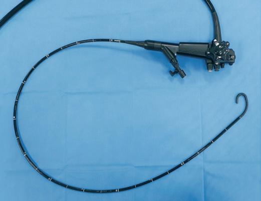
1. Dual-Operator Per-Oral Cholangioscopy
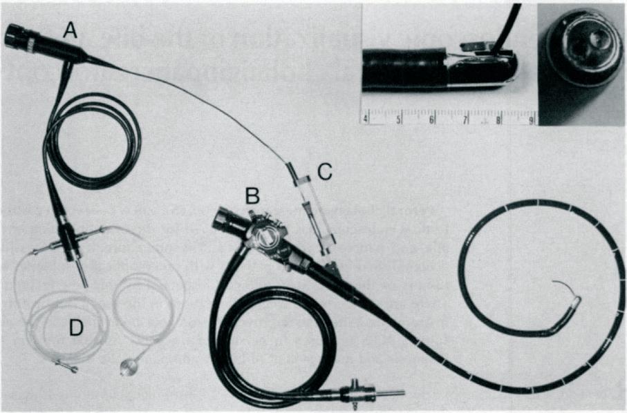
Originally, two endoscopists were required to perform the per-oral cholangiopancreatoscopy. One endoscopist controlled the duodenoscope while the other operated the cholangioscope. This method was known as “mother-daughter” per-oral cholangiopancreatoscopy. These cholangioscopes were reusable. (Nakajima, M., et al., 1978)
Note. Instruments for peroral cholangiopancreatoscopy: A, supplemental cholangioscope (type B); B, master duodenoscope; C, subscope thurster; D, pressure-infusion apparatus for irrigation. From “Direct endoscopic visualization of the bile and pancreatic duct systems by peroral cholangiopancreatoscopy (PCPS),” by Nakajima, et. Al., 1978, Gastrointestinal Endoscopy, 24(4), 141145–145. https://doi.org/10.1016/S0016-5107(78)73488-7
2.
Single-Operator Per-Oral Cholangioscopy
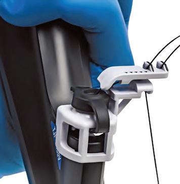
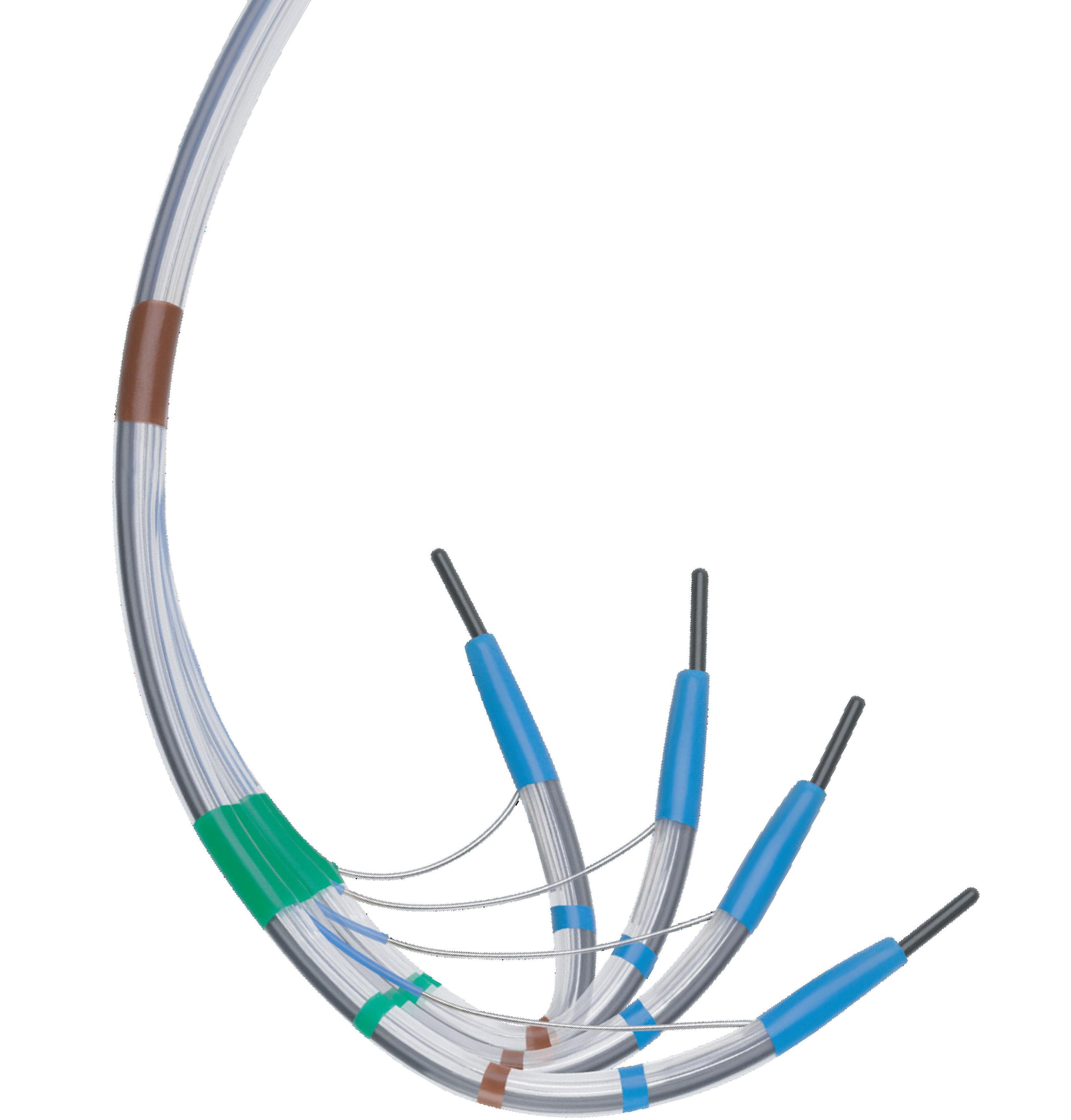
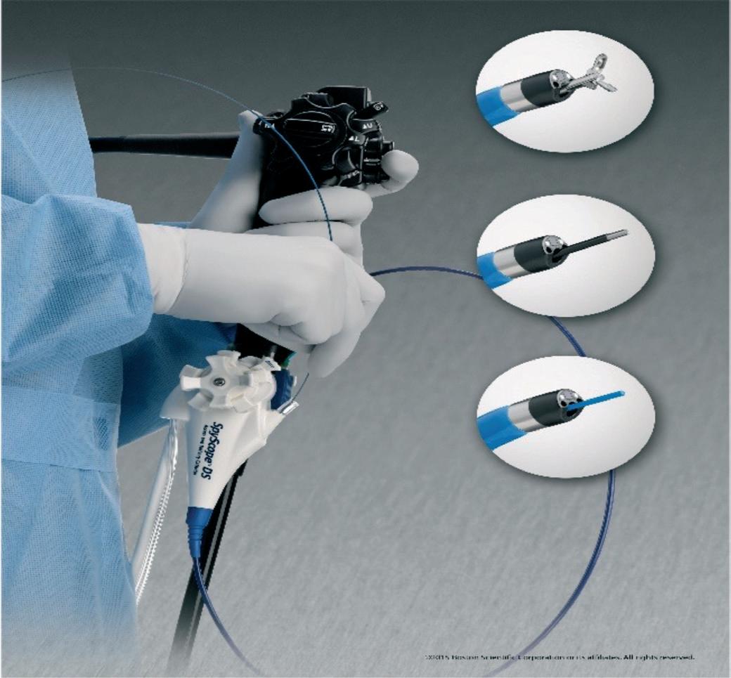
Moving forward, the development of a single operator cholangioscopy emerged removing the necessity of the second operator endoscopist. This was championed by Boston Scientific with their SpyGlass™™ innovation in 2007. Moreover, the SpyScope™™ Delivery System is comprised of two elements, namely: 1. SpyScope™ (cholangioscope) which is now singleuse and 2. SpyGlass™ digital controller (processor). Through the years, the SpyGlass™ system continued to develop a better version than its predecessor.
In 2019 the SpyScope™ DS II was launched. One of its innovations was a resolution 2.5 times higher than the previous SpyScope™ DS. The system and setup are much smaller; therefore, it can seamlessly be integrated into the endoscopy workstation (Gupakumar and Sharma, 2023)
Note. SpyGlass™ DS II with the SpyScope™ DS II delivery and access catheter. From “Role of peroral cholangioscopy and pancreatoscopy in the diagnosis and treatment of biliary and pancreatic disease: past, present, and future,” by Gopakumar and Sharma, 2023, Frontiers in Gastroenterology, 2. https:// doi.org/10.3389/fgstr.2023.1201045. Copyright 2015 by Boston Scientific Corporation or its affiliates
Figure 1: Instruments for PCPS
Figure 2: SpyGlass™ DS II with the SpyScope DS II delivery and access catheter.
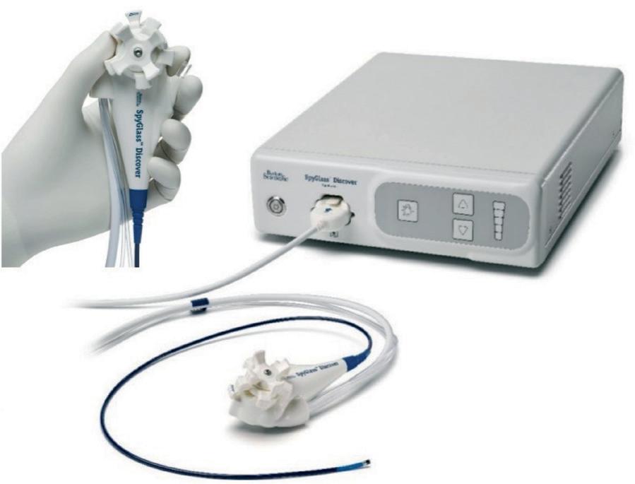
Note. SpyGlass™ Discover system. From “Role of peroral cholangioscopy and pancreatoscopy in the diagnosis and treatment of biliary and pancreatic disease: past, present, and future,” by Gopakumar and Sharma, 2023, Frontiers in Gastroenterology, 2. https://doi.org/10.3389/ fgstr.2023.1201045. Copyright 2015 by Boston Scientific Corporation or its affiliates
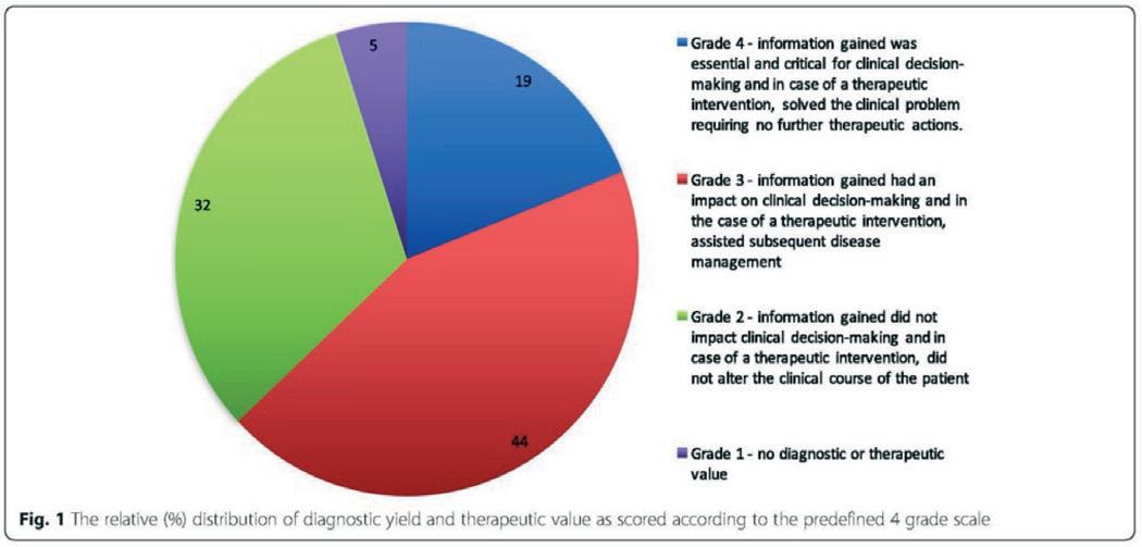
A publication by Reuterwall, M., et al (2018) highlighted the importance of a single-operator, ERCP-assisted, per-oral cholangiopancreatoscopy. The study involved 365 procedures. The results were assessed utilizing a predetermined scoring mark and at the same time taking into consideration the procedure’s therapeutic benefit and diagnostic outcome.
Figure 4: The relative (%) distribution of diagnostic yield and therapeutic value as scored according to the predefined 4 grade scale.
Note. The relative (%) distribution of diagnostic yield and therapeutic value as scored according to the predefined 4 grade scale. From “The clinical value of ERCP-guided cholangiopancreatoscopy using a single-operator system,” by Reuterwall, et. Al., 2018, BMC Gastroenterology, 19(1), 1–7. https://doi.org/10.1186/s12876-019-0953-9
3. Direct Per-Oral Cholangioscopy
This technique operates by using a slim forward viewing endoscope rather than the side viewing duodenoscope. The scope is inserted until the duodenum, then steered accordingly until a direct vision of the ampulla of Vater is achieved. However, this procedure necessitates a bigger sphincterotomy with or without balloon dilation as narrated by Urakami (1977, as cited in Gupakumar and Sharma, 2023). Moreover, this method wasn’t widely used because of the challenges it presented. One, manoeuvring into the sphincter of Oddi just to be able to cannulate the biliary or pancreatic duct while utilizing earlier models of the slim gastroscope. Two, image resolution at that time were not the best (Parsi, 2014). But with the launch of the new high-definition, narrow-band imaging (NBI) capable, and ultra-slim gastroscopes, the direct per-oral cholangioscopy started to gain traction (Sondhi and Law, 2020).
Note. The 3rd-generation multibending ultraslim endoscope. (A) Overall view of the multibending endoscope (CHF-Y0010), which has two bending points at the distal portion of the shaft. (B) The endoscope is equipped with additional control lever for second bending point and two accessory channels of 2.2- and 1.0-mm diameter in the control section. From, “Usefulness of Direct Peroral Cholangioscopy Using a Multibending Ultraslim Endoscope for the Management of Intrahepatic Bile Duct Lesions (with Videos),” by Lee, et. Al., 2024, Gut and Liver, 18(2), 358–364. https://doi.org/10.5009/gnl230163
Figure 5 (A)
Figure 5 (B)
Figure 3: SpyGlass™ Discover system.
A prototype gastroscope made by Olympus (CHF – Y0010) is called the multibending ultraslim endoscope. This is a forward viewing scope. Its latest innovation is the extra two-directional bending section at the distal end of the insertion tube (Shaft), with a 200° upward angulation and 100° downward angulation. It is also outfitted with two accessory channels so that the insufflation and suctioning functions can happen simultaneously. The shaft’s length is about 1.2 meters with a diameter of 7.0mm and the distal end is 4.9mm as shown in Fig 5
A study was published in February 2024 about the effectivity of the new multibending ultraslim endoscope. Twenty-two patients had direct per-oral cholangioscopy utilizing the multibending ultraslim endoscope for intra-hepatic duct (IHD) lesions. The lesions are based on results of previous imaging or cholangiopancreatography. The main objective was a resounding success which was defined as an effective manoeuvre to the IHD and the aimed lesions. Effective free-hand intubation is when the multibending scope is traversed to the target lesion within 15 minutes with neither accessory being used. Procedure time was calculated from ampullary cannulation up until the withdrawal
Table 2. Peroral Cholangioscopy Outcomes for Intrahepatic Duct Lesions
Outcome Value (n=22)
Technical success of peroral cholangioscopy, No. (%) 21 (95.5)
Free-hand insertion 20
Intraductal balloon assistance
1
Total procedure time, median (range), min 29 (9–79)
Adverse events, No. (%)
0
of the multibending scope from the bile duct. A seven-day post procedure follow up with the patients was done. On top of that, procedure-related events like cholangitis, perforation, etc., were outlined based on the American Society for Gastrointestinal Endoscopy criteria (Lee, et. Al., 2024).
The article concluded that the IHD can be viewed directly with the assistance of the prototype multibending ultraslim gastroscope. In the future, this can be a vital procedure and equipment for diagnosing and therapeutic management of certain patients (Lee, et. Al., 2024).
Indications for Cholangioscopy:
• Difficult biliary duct stones
- These are usually stones that presented a challenge upon extraction under an ERCP procedure. These stones are either large (more than 15mm in size), impacted, many stones, shape of stone, intrahepatic location, existence of bile duct stricture distal to the stone, and an altered biliary anatomy (Ayoub, et. Al., 2018).
• Ductal clearance
- This interpretation is normally done via occlusion cholangiogram after the common bile duct (CBD) stones are removed. There are a few publications narrating the presence of residual CBD stones post ERCP-assisted stone extraction. One of the reasons is due to lithotripsy. These studies concluded that POC is an integral procedure in evaluating ductal clearance and managing residual stones (Lee, et. Al., 2012; Anderloni, et. Al., 2019; Yang, et. Al., 2019; Lee, et. Al., 2022).
• Intermediate biliary strictures
- These are strictures without a definite diagnosis after cross-sectional imaging with/without non-diagnostic brush cytology results on ERCP. Research recommended accurate diagnosis of intermediate biliary strictures to avoid unnecessary biliary surgeries which carries with it higher risk of complications. POC assists in evaluating the intermediate biliary strictures by permitting a direct view of the biliary tract, including but not limited to, thorough visual assessment of the mucosa and allowing targeted biopsies (Sato, et. Al., 2022; Nur, et. Aal., 2022).
• Direct visualisation
- According to Angsuwatcharakon, et. Al. (2022) because of the innovations of the single operator cholangioscopes e.g. image enhancements and quality, visual impression can result to better sensitivity than biopsy, however, comes with a suboptimal specificity. Moreover, it has is technical difficulties specially with distal biliary strictures.
- If image enhancing innovations such as NBI can be supplemented with cholangioscopy, this can provide an outstanding means for assessing intermediate biliary strictures.
• Cholangioscopy – directed targeted biopsies.
- Despite the outstanding sensitivity of the direct visualisation, it has a low specificity, therefore, the gold-standard is still tissue biopsy. With the advancements of cholangioscopy and cholangioscopes, so too with its accessories e.g. Boston Scientific’s SpyBite™ Forceps®, this has enhanced the diagnostic yield and accuracy directed targeted biopsies (Gupakumar and Sharma, 2023).
• Diagnosis and staging and therapeutic application for cholangiocarcinoma.
- Cholangiocarcinoma is rare, nevertheless, it is the most common type of cancer developing from the biliary tree. Its statistics is 0.3 – 6.0 in every 100,000 persons (Geraghty, 2021). For patients that are potential candidates for curative treatment of cholangiocarcinoma percutaneous or EUSguided biopsies of primary lesions are opposed due to
likelihood of tumour spreading or seeding (Hattori, et. Al., 2011). POC with is direct visualisation capability, can be applied to analyse and define the scope of the biliary tree association with the cholangiocarcinoma.
- Studies has already been made using POC as therapeutic alternative to manage unresectable extrahepatic cholangiocarcinoma. These are namely: POC-directed radiofrequency ablation (RFA) and POC-directed photodynamic therapy (PDT). In addition, most of the figures regarding RFA and PDT are derived from ERCP-guided treatment. Nonetheless, despite having only a handful articles about the role of POC in RFA and PDT, findings are encouraging. To better recognize the role and potential advantage of the POC-directed RFA and PDT in managing unresectable cholangiocarcinoma, in-depth research plus head-to-head tests should materialize (Gupakumar and Sharma, 2023).
• Selective biliary cannulation
- Traversing a guidewire past strictures can be challenging. This often results in a failed traditional guidewire placement through ERCP. However, a study by Bokemeyer, et. Al. (2019) concluded that POC-assisted guidewire insertion has an exceptional technical success rate, thus, averting the more invasive procedures e.g. PTC and/or EUS-guided biliary drainage.
Indications for Pancreatoscopy:
• Pancreatoscopy-guided lithotripsy
- According to Kaura, et. Al. (2019) conventional ERCP success rate of treating pancreatic duct (PD) stones sits at around 50% despite with expert’s hands. Howell et al. (1999) first reported about Peroral pancreatoscopy-guided (POPS-guided) lithotripsy. A meta-analysis about per-oral pancreatoscopyguided lithotripsy for the endoscopic management of pancreatolithiasis pointed a high technical and clinical success paired with a low complication data. This procedure can be a promising inclusion to the present methods of managing complicated pancreatolithiasis (Guzman-Calderon, et. Al., 2021).
• Intraductal papillary mucinous neoplasm
- 20%–30% of pancreatic cancer are intraductal papillary mucinous neoplasms (IPMNs). They are macroscopic lesions and a sign of pancreatic cancer with a variable possibility of changing into a malignant one. With the current guidelines in the assessment and management of IPMNs, cholangioscopy is not yet part of the standard diagnostic algorithm. Nonetheless, cholangioscopy will be taken into consideration and will be a vital part of guidelines considering the rapid pace of innovations in technology partnered with accrued experience (Nakamura, et. Al., 2017).
• Pancreatic duct strictures
- Assessment and treatment of PD strictures is far more demanding than biliary duct strictures. Considering its intricacies, its management is also limited to specialists that deals with pancreatic endotherapy (Maranaki, 2023). Causes for PD strictures are classified into two, namely: benign condition and malignant. Benign aetiology ranges from chronic pancreatitis to recurring acute pancreatitis, surgical complications, pseudocyst, and trauma. Management of PD strictures also depend on the cause and if a person is asymptomatic or symptomatic. If of benign aetiology and without symptoms, malignancy is to be ruled out then the stricture is left alone. If a person is displaying symptoms, pancreatic sphincterotomy is done, then, dilatation of the stricture and/or placement of pancreatic stent. Similar with POC, targeted biopsy can also be done under direct visualisation with POPS (Dawod and Kahaleh, 2018). If NBI can be added with POPS, it can foster enhancements on its diagnostic accurateness in gauging PD strictures and this is based on the publications of Miura, et. Al. (2010) and Itoi, et. Al. (2007).
Preparation for cholangioscopy – North Shore Hospital
• Equipment
1 Duodenoscope (otherwise discuss any other scope and equipment preference with the doctor)
2 Boston Scientific RX locking device and biopsy cap
3 SpyScope™ DS II Catheter – 3.3 mm outer diameter, 1.2 mm accessory channel & 230cm in length
Features include:
• CMOS chip with an increased resolution (2.5x)
• Adjusted lighting design reduces light flare
• better lighting in the corners of the video
• enhancement of visibility down the lumen (ensure that an extra box of SpyScope™ is available in case the first one malfunctions)
4 Electrohydraulic Lithotripsy (EHL) probe
5 SpyBite™ biopsy forceps
6 SpyGlass™ retrieval basket
7 SpyGlass™ retrieval snare
8 Autotome RX 44 cannulating sphincterotome (.035 in) - loaded with a 450cm long guidewire
9 Omnipaque 240 mg I/mL or Omnipaque 300 mg I/mL (50 mL bottle) and labels
10 5 mL luer lock syringes
11 Sodium Chloride (warmed) 1 L with endogator tubing – This is used for irrigation since the conductive properties of saline allows for optimum functioning of the EHL probe.
12 Suction tubing – Cut the yellow tip off; the tubing will be attached on the SpyScope™ for continuous suction
13 SpyGlass™ DS Digital Controller & Autolith Touch EHL Generator (machines are in room 1 stack)
14 X-ray equipment (Radiology staff should be notified at least 5-10 minutes prior procedure)
15 Indomethacin 100 mg suppository
Continued over page
1
• Setup Interventional Techniques Official Journal of the Society of American Gastrointestinal and Endoscopic Surgeons (SAGES) and European Association for Endoscopic Surgery (EAES), 33(3), 731–737. https://doi.org/10.1007/s00464-0186334-6
Duodenoscope setup: Plug-in the scope into the Olympus machine. Puncture the biopsy cap of the locking device using a blunt needle then place it on the biopsy channel port of the scope. Attach the locking device against the side of the biopsy channel port (Ensure that the locking device’s angle head rests above the biopsy cap and the white rubber inside the biopsy cap hasn’t dislodged) then place the strap around the scope and secure it using the small knob on the locking device.
2 SpyScope™ DS II catheter setup: Fix the Sodium Chloride (warmed) 1 L with endogator tubing into the water irrigation pump and prime. Also prepare additional suction tubing; cut the yellow tip off. KEEP THE SPYSCOPE™ BOX UNOPENED UNTIL DR IS READY TO USE IT.
3 SpyGlass™ DS Digital Controller & Autolith Touch EHL Generator setup: Check that the SpyGlass™ cable is connected at the back of the Olympus machine (See ORANGE marking). Then, turn on the machines and put them on standby mode.
4 Prepare the sphincterotome:
1. Flush the 450cm long guidewire with water for irrigation (this will activate the 5cm hydrophilic tip of the jagwire), then load it in the autotome RX 44.
2. Draw up 5mL Omnipaque contrast using a 5 mL luer lock syringe. Label the syringe. Ensure to prepare at least 5 syringes.
3. Prime the loaded sphincterotome RX 44 using the drawn up 5 mL Omnipaque 240 mg I/mL
5 Put the ERBE’s Nessy Omega Plate on the patient (ensure to check for any metalware from the patient beforehand). Never put an ERBE’s Nessy Omega Plate on a site where there is a metalware implant to avoid the risk of burning in the surrounding tissue when using diathermy.
6 ERBE settings according to doctor’s instructions. Otherwise, use the ERCP settings on the ERBE machine.
Note: Each case will differ so discuss the plan with the doctor prior the start of procedure
References:
• Adler, D. G., Baron, T. H., Davila, R. E., Egan, J., Hirota, W. K., Leighton, J. A., Qureshi, W., Rajan, E., Zuckerman, M. J., Fanelli, R., Wheeler-Harbaugh, J., & Faigel, D. O. (2005). ASGE guideline: the role of ERCP in diseases of the biliary tract and the pancreas. Gastrointestinal Endoscopy, 62(1), 1–8. https:// doi.org/10.1016/j.gie.2005.04.015
• Anderloni, A., Auriemma, F., Fugazza, A., Maselli, R., Carrara, S., D, A. F., Troncone, E., Maia, L., Belletrutti, P. J., & Repici, A. (2019). Direct peroral cholangioscopy in the management of difficult biliary stones: A new tool to confirm common bile duct clearance. results of a preliminary study. Journal of Gastrointestinal and Liver Diseases, 28(1), 89-94–94. https:// doi.org/10.15403/jgld.2014.1121.281.bil
• Angsuwatcharakon, P., Kulpatcharapong, S., Moon, J. H., Ramchandani, M., Lau, J., Isayama, H., Seo, D. W., Maydeo, A., Wang, H.-P., Nakai, Y., Ratanachu-ek, T., Bapaye, A., Hu, B., Devereaux, B., Ponnudurai, R., Khor, C., Kongkam, P., Pausawasdi, N., Ridtitid, W., … Rerknimitr, R. (2022). Consensus guidelines on the role of cholangioscopy to diagnose indeterminate biliary stricture. HPB, 24(1), 17–29. https://doi. org/10.1016/j.hpb.2021.05.005
• Ayoub, F., Yang, D., & Draganov, P. V. (2018). Cholangioscopy in the digital era. Translational Gastroenterology and Hepatology, 3, 82. https://doi.org/10.21037/tgh.2018.10.08
• Bokemeyer, A., Gross, D., Brückner, M., Nowacki, T., Bettenworth, D., Schmidt, H., Heinzow, H., Kabar, I., Ullerich, H., & Lenze, F. (2019). Digital single-operator cholangioscopy: a useful tool for selective guidewire placements across complex biliary strictures. Surgical Endoscopy: And Other
• Chathadi, K. V., & Chen, Y. K. (2009). New kid on the block: development of a partially disposable system for cholangioscopy. Gastrointestinal endoscopy clinics of North America, 19(4), 545–555. https://doi.org/10.1016/j. giec.2009.06.001
• Chen Y. K. (2007). Preclinical characterization of the SpyGlass™ peroral cholangiopancreatoscopy system for direct access, visualization, and biopsy. Gastrointestinal endoscopy, 65(2), 303–311. https://doi.org/10.1016/j.gie.2006.07.048
• Chen, Y. K., & Pleskow, D. K. (2007). SpyGlass™ single-operator peroral cholangiopancreatoscopy system for the diagnosis and therapy of bile-duct disorders: a clinical feasibility study (with video). Gastrointestinal endoscopy, 65(6), 832–841. https://doi.org/10.1016/j.gie.2007.01.025
• Draganov, P. V., Lin, T., Chauhan, S., Wagh, M. S., Hou, W., & Forsmark, C. E. (2011). Prospective evaluation of the clinical utility of ERCP-guided cholangiopancreatoscopy with a new direct visualization system. Gastrointestinal Endoscopy, 73(5), 971–979. https://doi.org/10.1016/j.gie.2011.01.003
• Enad Dawod, & Michel Kahaleh. (2018). Management of Benign and Malignant Pancreatic Duct Strictures. Clinical Endoscopy, 51(2), 156–160. https://doi.org/10.5946/ ce.2017.085
• Geraghty J. (2021). Endoscopic considerations for the management of cholangiocarcinoma. Liver Cancer Int., 2: 96–101. doi:10.1002/lci2.40
• Gopakumar, H., & Sharma, N.R. (2023). Role of peroral cholangioscopy and pancreatoscopy in the diagnosis and treatment of biliary and pancreatic disease: past, present, and future. Frontiers in Gastroenterology, 2. https://doi. org/10.3389/fgstr.2023.1201045
• Guzmán, C. E., Martinez, M. B., Casellas, J. A., & Aparicio, J. R. (2021). Per-oral pancreatoscopy-guided lithotripsy for the endoscopic management of pancreatolithiasis: A systematic review and meta-analysis. Journal of Digestive Diseases, 22(10), 572–581. https://doi.org/10.1111/1751-2980.13041
• Hattori, M., Nagino, M., Ebata, T., Kato, K., Okada, K., & Shimoyama, Y. (2011). Prospective study of biliary cytology in suspected perihilar cholangiocarcinoma. British Journal of Surgery, 98(5), 704-709–709. https://doi.org/10.1002/ bjs.7412
• Howell, D. A., Dy, R. M., Hanson, B. L., Nezhad, S. F., & Broaddus, S. B. (1999). Endoscopic treatment of pancreatic duct stones using a 10F pancreatoscope and electrohydraulic lithotripsy. Gastrointestinal Endoscopy, 50(6), 829–833. https://doi.org/10.1016/S0016-5107(99)70168-9
• Itoi, T., Sofuni, A., Itokawa, F., Kurihara, T., Tsuchiya, T., Ishii, K., Tsuji, S., Ikeuchi, N., Arisaka, Y., & Moriyasu, F. (2007). Initial experience of peroral pancreatoscopy combined with narrowband imaging in the diagnosis of intraductal papillary mucinous neoplasms of the pancreas (with videos). Gastrointestinal Endoscopy, 66(4), 793–797. https://doi.org/10.1016/j. gie.2007.03.1096
• Kaura, T., Willingham, F. F., & Chawla, S. (2019). Role of pancreatoscopy in management of pancreatic disease: A systematic review. World Journal of Gastrointestinal Endoscopy, 11(2), 155–167. https://doi.org/10.4253/wjge.v11. i2.155
• Komanduri, S., Thosani, N., Abu Dayyeh, B. K., Aslanian, H. R., Enestvedt, B. K., Manfredi, M., Maple, J. T., Navaneethan, U., Pannala, R., Parsi, M. A., Smith, Z. L., Sullivan, S. A., & Banerjee, S. (2016). Cholangiopancreatoscopy. Gastrointestinal Endoscopy, 84(2), 209–221. https://doi.org/10.1016/j. gie.2016.03.013
• Lee, T. H., Moon, J. H., Lee, Y. N., Yoo, H. W., Yang, J. K., Cha, S.-W., Cho, Y. D., & Park, S-H. (2022). A Preliminary Study on the Efficacy of Single-Operator Cholangioscopy with a New Basket for Residual Stone Retrieval After Mechanical Lithotripsy. Digestive Diseases and Sciences, 67(6), 2571–2576. https://doi.org/10.1007/s10620-021-07068-1
• Lee, W. M., Moon, J. H., Lee, Y. N., Min, C. W., Shin, I. S., Myeong, J. H., Kim, H. K., Yang, J. K., & Lee, T. H. (2024). Usefulness of Direct Peroral Cholangioscopy Using a Multibending Ultraslim Endoscope for the Management of Intrahepatic Bile Duct Lesions (with Videos). Gut and Liver, 18(1). https://doi. org/10.5009/gnl230163
• Lee, Y. N., Moon, J. H., Lee, T. H., Choi, H. J., Itoi, T., Beyna, T., & Neuhaus, H. (2020). Prospective randomized trial of a new multibending versus conventional ultra-slim endoscope for peroral cholangioscopy without device or endoscope assistance (with video). Gastrointestinal Endoscopy, 91(1), 92–101. https://doi.org/10.1016/j.gie.2019.08.007
• Lee, Y.N., Moon, J.H., Choi, H.J., Min, S.K., Kim, H.I., Lee, T.H., et al. Direct peroral cholangioscopy using an ultraslim upper endoscope for management of residual stones after mechanical lithotripsy for retained common bile duct stones. Endoscopy (2012) 44:819–24. https://doi.org/10.1055/s-0032-1309880
• Maranki, J. (2023). Top tips for pancreatic duct strictures. Gastrointestinal Endoscopy, 97(6), 1144–1147. https://doi.org/10.1016/j.gie.2023.01.003
• Miura, T., Igarashi, Y., Okano, N., Miki, K. and Okubo, Y. (2010), ENDOSCOPIC DIAGNOSIS OF INTRADUCTAL PAPILLARYMUCINOUS NEOPLASM OF THE PANCREAS BY MEANS OF PERORAL PANCREATOSCOPY USING A SMALL-DIAMETER VIDEOSCOPE AND NARROW-BAND IMAGING. Digestive Endoscopy, 22: 119-123. https://doi.org/10.1111/j.14431661.2010.00926.x
• Moon, J. H., Terheggen, G., Choi, H. J., & Neuhaus, H. (2013). Peroral Cholangioscopy: Diagnostic and Therapeutic Applications. Gastroenterology, 144(2), 276–282. https://doi. org/10.1053/j.gastro.2012.10.045
• Nakajima, M., Akasaka, Y., Yamaguchi, K., Fujimoto, S., & Kawai, K. (1978). Direct endoscopic visualization of the bile and pancreatic duct systems by peroral cholangiopancreatoscopy (PCPS). Gastrointestinal Endoscopy, 24(4), 141-145–145. https:// doi.org/10.1016/S0016-5107(78)73488-7
• Nakamura, M., Miyasaka, Y., Sadakari, Y., Date, K., & Ohtsuka, T. (2017). Comparison of guidelines for intraductal papillary mucinous neoplasm: What is the next step beyond the current guidelines? ANNALS OF GASTROENTEROLOGICAL SURGERY, 1(2), 90–98. https://doi.org/10.1002/ags3.12012
• Nur, A.M., Salim, M., Boerner, S., Li, S., Law, C.C.Y., Edwards, L., Ryan, K., & James, P.D. (2022). High diagnostic yield of endoscopic retrograde cholangiopancreatography brush cytology for indeterminate strictures. Journal of the Canadian Association of Gastroenterology, 5:234–239. https://doi. org/10.1093/jcag/gwac011
• Parsi, M.A. Direct peroral cholangioscopy. World J Gastrointest Endosc 2014; 6(1): 1-5 [PMID: 24527174 DOI: 10.4253/wjge.v6.i1.1
• Reuterwall, M., Lubbe, J., Enochsson, L., Lundell, L., Konradsson, M., Swahn, F., Del Chiaro, M., Löhr, M., & Arnelo, U. (2019). The clinical value of ERCP-guided cholangiopancreatoscopy using a single-operator system. BMC Gastroenterology, 19(1), 1–7. https://doi.org/10.1186/s12876-019-0953-9
• Sato, T., Nakai, Y., Fujishiro, M. (2022). Current endoscopic approaches to biliary strictures. Current Opinion in Gastroenterology, 38:450–460. https://doi.org/10.1097/ mog.0000000000000857
• Sondhi, A.R., & Law, R. (2020). A new era in direct peroral cholangioscopy: bending over backward to overcome limitations of the past. Gastrointestinal Endoscopy (2020) 91:102–103. https://doi.org/10.1016/j.gie.2019.09.018
• Te Whatu Ora Health New Zealand (2022). ERCP SpyGlass™ –Gastroenterology (Adults). Te Whatu Ora – Waitemata. http:// staffnet/QualityDocs/Quality%20Documentation/S4%20 Medicine%20and%20Health%20of%20Older%20People/ Gastroenterology/[P]%20ERCP%20SpyGlass™%20Aug22. pdf
• Wehbi, M., Dacha, S., Massaad, J., Obideen, K., Roberts, K. E. (2022, December 2). Cholangioscopy. https://emedicine. medscape.com/article/1891395-overview?form=fpf
• Yang, J.-J., Liu, X.-C., Chen, X.-Q., Zhang, Q.-Y., & Liu, T.-R. (2019). Clinical value of DPOC for detecting and removing residual common bile duct stones (video). BMC Gastroenterology, 19(1). https://doi.org/10.1186/s12876-0191045-6
Optimal Management for Patients with Barrett’s Oesophagus and Dysplasia
VICTORIA HARKESS
Registered Nurse, Gastroenterology Department, Te Whatu Ora – Waitaha Canterbury Victoria.Harkess@CDHB.health.nz
In December 2023, I was privileged to be granted funding by the New Zealand Gastroenterology Nurses College (NZgNC), to attend the annual scientific meeting, which was held at the Rotorua energy events centre. There was a vast selection of interesting topics discussed by the speakers. I was particularly interested in learning more about Barrett’s oesophagus. Including how it is diagnosed, and the treatment of oesophageal dysplasia and lesions.
Barrett’s oesophagus (BO) is a condition often caused by chronic gastro-oesophageal reflux disease (GORD). It occurs when intestinal-like epithelium replaces the normal squamous epithelium of the distal oesophagus (Wani et al., 2018). It is thought to be as many as 5%-12% of patients that have known chronic GORD (5 years or more), will have BO mucosal changes present (Shaheen et al., 2022). Other risks related to BO include having a hiatus hernia, high body mass index, being >50 years of age, and/or male. BO poses an increased risk of oesophageal adenocarcinoma, therefore following guidelines for diagnosis and treatment is crucial (Maione et al., 2022).
Diagnosis
According to Stawinski, Dziadkowiec, Kuo, Echavarria and Saligram (2023) a more accurate diagnosis of BO consists of a minimum of eight biopsy samples be taken, beginning at the z line. The suspected BO changes would generally be salmon coloured, proximal from the gastrointestinal junction, and at least 1cm in length to require biopsy samples to be taken (Shaheen et al., 2022). If changes are evident, samples taken are then graded accordingly by epithelial changes in dysplasia, with the highest grade being full submucosal invasion with carcinoma.
More recent diagnostic advances include artificial intelligence (AI), chromoendoscopy, virtual chromoendoscopy and confocal endomicroscopy (Maione et al., 2022). If the early stages of BO is evident, screening programmes are an effective way to monitor patients that have BO (Stawinski et al., 2023). Patients should also be prescribed a high dose proton pump inhibitor (PPI) to help reduce symptoms of GORD, which some research shows, may reduce the risk of abnormal tissue growth (Shaheen et al., 2022).
Recently, studies and trials have been conducted in the United Kingdom and the United States to investigate the efficacy of using sponge-type devices to detect BO. This technique involves administering a lignocaine throat spray and asking the patient to swallow a collection device, which is attached to a string. Cytology samples are then collected, and the device is removed orally. These devices show promising advances, in minimising the amount of BO surveillance endoscopic procedures a patient may require (Shaheen et al., 2022).
Maione (2022) states that utilising methylene blue, otherwise known as chromoendoscopy has a 95% detection rate of oesophagus dysplasia in patients with known BO. Alternatively, virtual chromoendoscopy uses a narrow band imaging light to detect mucosal dysplasia, this has benefits of ensuring a shorter procedure time and is cost-effective. Shaheen (2022) asserts there are benefits of utilising both white light and chromoendoscopy together, as the endoscopist is spending more time looking for changes, such as subtle lesions. Recent research suggests, AI could be a useful tool for detecting upper gastrointestinal lesions, to assist with specific sampling while assessing patients that have BO (Stawinski et al., 2023). Confocal laser endomicroscopy, is when a contrast agent called fluorescein is injected into the tissue and a blue laser light can then illuminate areas to ensure targeted biopsy samples. This type of in vivo technology is not widely utilised to date (Shaheen et al., 2022).
Treatment
If a superficial lesion is detected and contained within the oesophageal mucosa, endoscopy therapy is the preferred treatment rather than the patient undergoing a surgical option, such as an oesophagectomy (Terheggen et al., 2017). According to Shaheen et al, the survival rate is indifferent between surgical and endoscopy eradication, however the complications surgically have a greater risk for the patient, such as anastomotic leaks, prolonged hospital stays and infection (Shaheen et al. 2022). The risk of complications post endoscopic eradication,
compared to esophagectomy is significantly less. The potential risks endoscopically include perforation, a stricture and bleeding (Wani et al., 2018).
For low grade dysplasia and some high grade dysplasia (precancerous changes in oesophageal mucosa), radiofrequency ablation (RFA), is frequently utilised to treat targeted areas. RFA is where thermal energy is used to promote normal mucosal regeneration by causing necrosis of the abnormal tissue (Maione et al., 2022). Stawinski (2023) adds that 3-4 treatments of RFA are generally required for successful eradication of oesophageal dysplasia and can be utilised alongside resection techniques if a lesion is present.
A routinely used eradication method of oesophageal lesions is endoscopic mucosal resection (EMR), it involves the removal of a specific lesion, this is a diagnostic/staging approach. However, if the margins are found to be clear of abnormal mucosa, the superficial lesion resection would be considered a curative treatment (Stawinski et al., 2023). Another possible treatment identified for eradicating oesophageal dysplasia, is the use of thermal ablation, by spraying liquid nitrogen or a similar substance to freeze tissue and let it slowly thaw thus promoting ischemia. This technique is called cryotherapy (Shaheen et al., 2022).
An endoscopic treatment initially designed for gastric lesions known as Endoscopic submucosal dissection (ESD), is now recognised as a useful method for eradication of some oesophageal dysplasia changes. Some studies suggest ESD has a higher success rate (58.8%) of complete resection in one session, compared to EMR (11.8%). Further collected data also shows a significantly higher cure rate and lower recurrence with ESD (Stawinski et al., 2023). Maione (2022) suggests that flat lesions may not be suitable for ESD, and that with a greater risk of complications, a high level of experience is required for these more time-consuming procedures. Terheggen (2016), states that complete en-block resection (entirety of a lesion) with ESD may have a more precise eradication outcome then EMR, as piecemeal resection with EMR could potentially exclude areas of mucosal dysplasia.
Conclusion
Barrett’s Oesophagus is a complex disease process and is a known precursor to dysplastic and neoplastic changes in the oesophagus. Best practice diagnosis, surveillance and treatment is critical to ensure optimal outcomes for patients and is continuing to evolve globally within the gastrointestinal field.
References
• Assignment point [Image], Retrieved from https:// assignmentpoint.com/barretts-esophagus-causes-andtreatment/
• Maione, F., Chini. A., Maione, R., Manigrasso, M., Marello, A., Cassese, G., … De Palma, G. (2022). Endoscopic Diagnosis and Management of Barrett’s Esophagus with Low-Grade Dysplasia. Diagnostics 2022, 1295(12), 1-3, 6. https://doi. org/10.3390/diagnostics12051295
• Shaheen, N., Falk, G., Lyer, P., Souza, R., Yadlapati, R., Sauer, B., & Wani, S. (2022). Diagnosis and Management of Barrett’s Esophagus: An Updated ACG Guideline. The American Journal of gastroenterology, (117), 559,564,566-567,573-578. https:// doi.org/10.14309/ajg.0000000000001680
• Stawinski, P., Dziadkowiec, K., Kuo, L., Echavarria, J., & Saligram, S. (2023). Barrett’s Esophagus: An Updated Review. Diagnostics 2023,13 (321), 2-3, 8-11. https://doi.org/10.3390/ diagnostics13020321
• Terheggen, G., Horn, E., Vieth, M., Helmut, G., Enderle, M., Neugebauer, A., … Neuhaus, H. (2016). A randomised trial of endoscopic submucosal dissection versus endoscopic mucosal resection for early Barrett’s neoplasia. Gut 2017, (66), 783-784. doi:10.1136/gutjnl-2015-310126
• Wani, S., Qumseya, B., Sultan, S., Agrawal, D., Chandrasekhara, V., Harnke. B., … Dewitt, J. (2018). Endoscopic eradication therapy for patients with Barrett’s esophagus-associated dysplasia and intramucosal cancer. American Society for Gastrointestinal Endoscopy, 87(4), 907, 918. https://doi. org/10.101016/j.gie.2017.10.011
NEW ZEALAND NURSES ORGANISATION
GASTROENTEROLOGY NURSES
COLLEGE
Minutes of the Annual General Meeting of the NZNO Gastroenterology Nurses College held at the ASM, Rotorua on Thursday 7th December 2023, 12:45pm - 13:45pm
Present: Committee: Merrilee Williams (Chair), Holly Weale (secretary), Kirsten Arnold (treasurer), Jessica Southall, Karen Kempin, Justin Augustine
Apologies: Marian O’Connor
Present via Zoom: Julia Anderson (PNA), Sihaam Bagus, Kerrin Bennett, Gino Borromeo, Nicola Caine, Levi Carroll, Jeszell Clemente, Donna Cotter, Ainsley Cunliffe, Elissa Dominguez, Susan Gale, Karen Gower, Victoria Harkess, Yongying He,
1. Welcome and Introduction of Committee
Merrilee Williams opened the meeting.
1.1 Roll Call
1.2 Apologies
1.3 Confirmation of Quorum: Confirmed at over 5% (46 present)
1.4 Confirmation of last Gastroenterology Section meeting Minutes
Moved: The minutes of the Gastroenterology meeting held in 28 November 2022 to be confirmed.
Moved: Michelle Harman
Seconded: Chrissy Rees CARRIED
1.5 Matters Arising from 2022 AGM minutes- No matters arising. 2. Correspondence
Honorary members nomination forms received for: Karen Gower, Sherry Sharp, Sandra Burton
Financial for previous financial year
by Kristen Arnold (treasurer)
Move any accounts for payment
Moved: Kirsten Arnold
Seconded: Donna Howe CARRIED
3.2 Present prospective budget for financial year
Moved: Kirsten Arnold
Seconded: Karen Gower CARRIED
Report
4.1 Chairperson’s Report: Summarised by Merrilee Williams – full report available on website.
Moved: Merrilee Williams
Seconded: Holly Weale CARRIED
4.2 PNA Report:
Summarised by Julia Anderson over zoom – full report available on website.
Amendment: Consultation in 2023 re: RN scope of practice.
Moved: Julia Anderson
Seconded: Merrilee Williams
CARRIED
4.3 Other committee reports
IBD specialty sub group – Summarised by Kirsten Arnold – full report available on website.
Hepatology specialty sub group – Summarised by Jessica Southall – full report available on website.
Nurse endoscopists specialty sub group – Summarised by Karen Kempin – full report available on website.
5. Remits
Summarised by Holly Weale on behalf of the committee – full remit available on website.
Guidance for the development and maintenance of a NZgNC specialty sub group.
CARRIED
6. NZNO Reports
Nil.
7. General Business
Question from Karen Gower about the finances – clarified by treasurer Kirsten Arnold.
Donna Howe – recently attended NZNO stop work meetings. Asked the question ‘should we submit a joint claim to improve the situation for senior nurses and RN prescribers’ from the NZgNC members? As the scope of nursing is changing and we are given more responsibility. Julia Anderson (PNA) responded saying we should all complete the ‘what do we want?’ survey coming from the NZNO and write a submission stating what our concerns are and what we would like to move forward. There is a senior nursing group within the NZNO.
8. National Committee Election Results
A farewell to three committee members: Marian O’Connor, Kiran Joseph, Holly Weale (outgoing secretary)
Three applications to join the committee (all accepted): Fiona Williams, Gino Borromeo, Nicola Caine
9. Other Business
No other business raised.
The meeting concluded at 13:30 pm when Merrilee Williams closed. The next meeting will be held in 2024 in Hamilton.
Chairperson:
Date:
NZ Gastro Nurses Leader’s Day
in Christchurch 2024
On the 20th of September, the New Zealand Gastro Nurses’ College (NZgNC) held its annual Leaders Day at the Commodore Hotel in Christchurch, led by Chairperson Merrilee Williams. The event was packed with insightful sessions and a discussion about key challenges in gastroenterology nursing.
Key Sessions and Themes
The day opened with Jai Chung, a PhD candidate, with a presentation on Occupational Distress in Nursing, focusing on moral distress, moral injury, and burnout. In addition, she also mentioned about strategies and what to do during these difficult situations. Next, Karen Kempin, a Nurse Endoscopist in Te Whatu Ora Capital, Coast, and Hutt Valley, and NZgNC committee member, discussed Generational Changes in the Workplace, examining how different generations interact, whether as older patients or younger staff members, and how understanding these dynamics can improve team cohesion and patient care.
Suzanne Rolls, a Professional Nursing Advisor (PNA) at the NZNO, discussed how NZNO can help nurses in this challenging healthcare environment. She shared eye-opening legislative nursing case studies, showing how legislation impacts the day-to-day responsibilities of nurses as well as the morale and reputation of healthcare teams. Kerri Nuku (Kaiwhakahaere for NZNO), followed with a behind-the-scenes updates on NZNO, on the advocacy efforts being undertaken for nurses, particularly in these challenging times.
In the afternoon, Rachel Marshall, Nurse Coordinator from the Nursing Workforce Development Team at Health NZ, presented a study on internationally qualified nurses (IQNs) in New Zealand.
She highlighted the statistics on IQNs entering and leaving the country, revealing that New Zealand retains fewer IQNs compared to Australia, prompting a discussion about possible reasons for this trend.
Sandy Richardson, a Nurse Researcher, spoke about the power of storytelling in nursing, explaining how storytelling shapes emotions and cognition. She stressed that even as handovers become more electronic, face-to-face oral interactions remain essential for building human connections in healthcare.
Dr Mimoza Soldatovic, a consultant Clinical Psychologist, closed the session by exploring how stress and anxiety can manifest as gastrointestinal symptoms. She explained the pathophysiology behind these symptoms and the extra time such patients need from nurses. Dr Mimoza emphasized finding the right balance in patient care, where setting boundaries while remaining grounded is the most therapeutic approach.
End-of-Day Discussion
The event wrapped up with a lively discussion from the different charge nurses. Attendees considered the critical question: When is enough enough? Discussing how to prioritise mental health amidst overwhelming demands. The group explored strategies for handling staff shortages, outsourcing, and insourcing procedures, with nursing leaders offering support and practical solutions to these ongoing challenges.
Overall, the 2024 Nurse Leaders Day was a tremendous success, providing a platform to address critical issues in nursing, enhance professional development, and strengthen the support network within the gastroenterology nursing community.
APPLICATION FORM NZGNC EDUCATION GRANT
To be eligible for consideration of an education grant, you must have been a member of NZGNC for 12 months prior to applying. Consideration will be given to each application on its individual merits, however there is an annual budgeted limit to support our membership’s professional development.
For full Post Graduate Paper funding: Priority will be given to first time applicants who meet the application criteria. If you are successful in your application for full funding, you will not be eligible to reapply for further funding for 24 months.
Please read all questions carefully and answer all sections fully, including endorsement from your manager. Failure to provide adequate information as requested may affect your application grant.
Grant funds can be used for any aspect of education requirements, including contributing to costs of papers, accommodation for meetings/conferences, and long-distance travel to meetings/conferences. Awarded grants will be made once Tube article has been received from the applicant.
Applications will close at 5pm on advertised dates twice yearly. Late applications will not be accepted. All Grant decisions are the decisions of the Committee and are final.
Full Name:
Home Address:
Place of Employment:
Work Address:
Email Address:
Phone Number:
NZNO Membership Number:
Applications close 1st March and 1st September each year.
The maximum amount of funding allocated:
National Education – NZD $800.00 (reimbursed)
International Education – NZD $1,800.00 (reimbursed)
Post Graduate Education – up to 100% of the course fee
Please complete & email this application to: secretaryofnzgnc@gmail.com
APPLICATION FORM NZGNC EDUCATION GRANT
Please ensure you answer all questions marked with a * You must complete the mandatory questions. It is helpful for you to supply as much information as possible to support your application for the education grant.
1 * Are you a financial member of NZNO and have been a member of NZGNC for more than 12 months? YES NO
2
3
4
* Please indicate the number of years you have been a NZGNC member (must be more than 12mths membership period) ________ YEARS
* Have you ever presented at a National or Local meeting? If yes, please include evidence of your presentation/s with your application
* How many NZGNC National meetings have you attended in the last 2 years? If yes, please include evidence of your attendance at these meetings with your application
5 Have you held an official NZGNC Term of Office? If yes, what position was held? _____________________________ And for how many years? _________
6 Have you contributed to The Tube? If yes, please provide details of the edition your contribution was in:
If you are an endoscopy nurse, have you completed the GENCA web-based Endoscope Reprocessing Training package?
Please include Certificate of completion with your application
* Have you received funding from NZGNC in the last 2 years? If yes, please complete: Month, Year & amount granted:
Please include evidence of previous NZGNC grant/s received with your application
* Please indicate the grant you are applying for. One grant is given per applicant; however, you can apply for more than one education event by rating your preference 1 – 4
1 Choose an item.
2 Choose an item.
3 Choose an item.
4 Choose an item.
10
11
Educational study – please provide the course name and date of completion:
* If the education event for which you are seeking funding is not undertaken, do you agree to notify NZGNC in writing and that the money received by you from the Gastroenterology Education fund will be returned?
* Do you agree to submit a final report from the education event to the committee and the Editor of ‘The Tube’ within 6 weeks of the planned education event and as outlined in the application approval letter?
IMPORTANT: By agreeing to this, it should be understood that this report will comply with the guidelines for “writing for publication in The Tube”. This report should have approximately 1000 words.
NO
APPLICATION FORM NZGNC EDUCATION GRANT
Please ensure you answer all questions marked with a * You must complete the mandatory questions. It is helpful for you to supply as much information as possible to support your application for the education grant.
* Education event you require the grant for: Please include a copy of the programme Programme not published
Education date/s:
* Are you likely to receive any other funding for this education? If yes, who from & how much?
* Give a brief description of the purpose of attending this education:
* State the benefits you perceive as a consequence of attending the Education event (For yourself, colleagues, nursing/health services)
Any additional comments, in support of your application:
Applicant’s signature: Date:
APPLICATION FORM NZGNC EDUCATION GRANT
*Manager Endorsement of Application - this section must be completed
Do you support this application for Education Grant from NZGNC YES NO
Applicant’s PDRP Level:
How is the applicant attending this education event going to benefit your department?
Name: (please print) Signature: Date:
APPLICATION FORM NZGNC EDUCATION GRANT
INFORMATION FOR APPLICANTS:
PURPOSE:
To provide financial assistance for New Zealand Registered, Health Care Professionals to attend Education within New Zealand or Overseas, relevant to the Gastroenterology Nursing profession.
CONDITIONS FOR APPLICATION:
The applicant:
1. Is a Health Care Professional working within Gastroenterology in New Zealand.
2. Is a member of NZNO and has been a member of the Gastroenterology Nurses’ College for a minimum of twelve (12) months.
3. Has not received full PG paper funding in year previous.
4. Is undertaking the education within a year of award being made.
5. Will complete a report and have it published in ‘The Tube’ once education completed.
6. Understands that failure to submit their article within 6 weeks of completing the event for which funding is approved, will make this application null and void. Grant will be paid to applicant on receipt of article.
7. You must advise the NZgNC committee if you do not successfully complete the academic criteria required to pass. You may be required to repay the funding grant should there not be a reasonable consideration to explain why you have not met the academic the requirements.
THE APPLICANT IS ASSESSED ON:
1. Professional activities.
2. Relevance of planned travel to practice.
3. Benefits for nursing service especially for Gastroenterology.
4. Nursing background.
5. Presentation of application.
6. Amount of funding requested equitably fits within the annual budgeted amount for NZgNC education fund. If there are multiple applicants from one site, we may choose to discuss a set amount for that site to support the applicants.
APPLICATION FORM NZGNC EDUCATION GRANT
TO COMPLY WITH THE PRIVACY CODE: ALL INFORMATION REGARDING YOUR APPLICATION WILL BE CONFIDENTIAL TO THE NZNO GASTROENTEROLOGY NURSES’ COLLEGE NATIONAL COMMITTEE AND THE JUDGES.
TUBE Writing Guidelines for Authors
The Tube is the official journal of the NZgNC (New Zealand Gastroenterology Nurses’ College), and is published biannually. We welcome articles that will be of interest to nurses working in Gastroenterology and related. Our aim is to publish a high quality, professional and educational journal for nurses working within the specialty of Gastroenterology.
All manuscripts received by the editor will be acknowledged, however, reports, area news or letters to the Editor will not. If you have not received confirmation of receipt within six weeks, please contact the Editor.
Suggestions for articles include:
• Recommendations for nursing practice based on current global trends/literature
• Overview of learning achieved through post graduate paper, or conference attendance
• Review of literary article relevant to best practice
• Case study relevant to specialty
• Education for nurses based on sub specialty topic
EDITORIAL REVIEW/ACCEPTANCE
Articles submitted to The Tube are currently reviewed at a minimum by the editor and co-editor. The review will assess the accuracy of fact, clarity of presentation, use of references and relevance to practice of gastroenterology nursing. The editor/ co-editor may also request a committee member review any article, particularly if the article is a sub-specialty of gastroenterology nursing and the committee member area of special interest/ work.
All articles which are being considered for publication may be reviewed and returned to the author with suggestions for revisions and improvement. The author will be provided with a deadline in which to provide the revised article in order to comply with publication schedule.
The Editor’s decision to publish or reject an article is final. You are welcome to email or phone the Editor to discuss your article should it not be accepted for publication.
STRUCTURE OF ARTICLE FOR SUBMISSION
The submission should include the following information:
Title Page
• Title of the Paper (20 word max)
• Author(s) name(s) in full
• Qualifications, current position, details of other relevant achievements, and affiliations of author(s)
• Address, contact telephone numbers, email address of the author(s)
• Conflict of interest and / or financial disclosure related to the article or related matter
Body of article
• Title at top of first page
• The body of work should be clearly written in an academic style of writing, and organised with headings/sub-headings (where appropriate)
• Pages numbered consecutively
• Tables, figures (if applicable) should be referred to in the body of the manuscript
• References (APA 6th Edition)
• Written authorisation(s) to publish identifiable person(s)/institutions and copyright materials
Word limit is approximately 1000 words. For the purposes of publication all articles should be formatted in Calibri, font size 10. All work should be saved as MS-Word (.docx) or text only (.txt) files.
All articles must be fully referenced where appropriate (APA 6th Ed)
Authors should keep an original copy of their article.
APPLICATION FORM NZGNC EDUCATION GRANT
SUBMISSION
Articles should be submitted to the editor at NZGNC Secretary secretaryofnzgnc@gmail.com.
If submission of your article is as a requirement of a NZgNC Education/Travel Grant, please ensure you submit within the required 6 week timeframe of your funding application.
REQUEST FURTHER INFORMATION
For advice or clarification on any of the above matters please contact the NZGNC Secretary secretaryofnzgnc@gmail.com
COLLEGE COMMITTEE MEMBERS’ REPORTS:
The aim of such reports is to inform the national College membership of the business and activities of the College during the last quarter.
These reports should include such activities as:
• College meetings/teleconferences (date and venue)
• Decisions arising from these meetings/teleconferences (can be focused on the minutes of these meetings)
• Plans/development the College is involved in/hopes to develop
• Any external meetings committee members have attended relating to the business of the College, e.g. meetings with NZNO professional nursing adviser/professional services manager
• Any contributions to national NZNO business, e.g. contribution to any submissions/ national guideline development
These should be a maximum of 600 words and contain people’s correct names and titles.
CASE STUDY/CLINICAL PRACTICE ARTICLE:
• Outline the nature of the treatment/procedure/product that forms the basis of the case study
• Provide information on the patient: age, sex, history, any other pertinent clinical/social/cultural aspects. Avoid using information, which would clearly identify the patient.
• Tell readers what is new, interesting, different, pioneering, about this treatment/procedure/product
• Outline the actual treatment/procedure or how product works
• Report on the patient’s/client’s response/recovery/
• Tell readers what you have learnt through your involvement with this treatment/procedure/product
• Outline any implications/meaning it may have for gastroenterology nurses’ practice
• Provide references to support the article.
The NZgNC is the national voice for gastroenterology nursing practice in NZ, and works collaboratively with the NZSG, and international societies such as GENCA and BSG. We represent nurses working in endoscopy, Hepatology, Inflammatory Bowel Diseases, Enteral Feeding, and Nurse Endoscopists
We have 2 epublications that are sent to our membership of nearly 500 nurses
The TUBE e-magazine is the official journal of the NZNO Gastroenterology Nurses College and is published twice yearly. It features scholarly articles on all aspects of gastroenterological nursing including current research, education opportunities, reports from conferences, case studies, new medications and products, infection control and updates in clinical practice.
The PANUI newsletter is new for 2023 and is published 6 x per year . It is designed to be a snapshot update for our membership to print and place in their workplaces to keep teams up to date with education and learning opportunities coming up, clinical practice news, and general gastroenterology focussed news.
We welcome sponsor contributions to nursing education for Gastroenterology Nurses through advertising in our publications.
ADVERTSING RATES 2024
PRICING
The Tube:
Full A4 Page - $550 inc GST
Half A4 Page- $275 inc GST
The Panui:
Live link logo- $150 inc GST
Sponsor corner- $200 inc GST
SIZE AND DETAILS
The Tube:
Full A4 Page - 210 x 297mm
Half A4 Page- 180 x 120mm
The Panui:
Live link logo- to sponsor
education content
Sponsor corner- 80 x 80mm with live link to sponsor www
Acceptance of advertising requests is subject to approval by the NZgNC committee, and required by the deadline for publishing. Please complete the booking request form, and email to NZGNC Secretary secretaryofnzgnc@gmail.com Spaces are limited for each publication.
Content deadlines for publication inclusions:
The TubeFebruary Edition
The TubeOctober Edition
The PanuiJanuary, March, May, July, September, December
Due date- February 3rd
Due date- September 3rd
Due dates: 1st of each month prior. Eg, 1st December, 1st February
Artwork:
Please include 3mm bleed
Resolution at least 300dpi at Full Size
Submit PDF or JPEG
Complete advertising request form and send to secretaryofnzgnc@gmail.com
Request for advertising form:
Sponsor Name:
Contact name and email:
Publication requested : Tube:
Edition: February October
Panui:
Edition: January March May July September December
Content requested:
Panui: Live link
Web address you want live link to go to if applicable:
Thank you for your support of the Tube and Panui - and the education it provides our nurses. Once published, we will send copy of publication, and invoice to the contact person you have provided.
Every Adenoma Counts
Increased Optical Clarity:
Increase visibility with the optical technology offered with the X1 System.
Increased Visibility: EndoCuff VisionTM is clinically proven to increase the mucosal surface observed and can increase ADR by up to 17%3
Increased Detection: Endo-Aid AI technology can increase ADR by 10%4.
Association of ADR and Risk of Colorectal Cancer:
• 26% of Adenomas are missed1
• 1% ADR = -3% of missed interval
Colorectal Cancer +- 5% of Fatal Interval Colorectal Cancer2
Increase Your ADR!
With the combination of increased optical clarity, increased observable mucosal surface, and increased detection with Endo-Aid AI technology you can increase your ADR.