INSIDE
Acute variceal bleed insights from Gastroenterology Conference 2023
What is ‘Green Endoscopy’ and how do we achieve it in NZ?
Cholestatic Liver Disease: Primary Biliary Cholangitis (PBS); Part 1
Volume 50 Issue 1 March 2024

Acute variceal bleed insights from Gastroenterology Conference 2023
What is ‘Green Endoscopy’ and how do we achieve it in NZ?
Cholestatic Liver Disease: Primary Biliary Cholangitis (PBS); Part 1
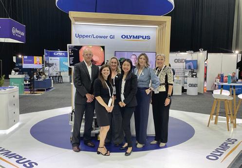
Wrapping up what has been a busy year, Rotorua played host to the NZSG and NZgNC Annual Scientific Meeting.
Wrapping up what has been a busy year, Rotorua played host to the NZSG and NZgNC Annual Scientific Meeting.
A jam-packed programme this year with local and international speakers focused on best practices and new techniques in Endoscopy. As usual the annual conference dinner was a huge hit, and we’d like to take the opportunity to congratulate all the award winners.
A jam-packed programme this year with local and international speakers focused on best practices and new techniques in Endoscopy. As usual the annual conference dinner was a huge hit, and we’d like to take the opportunity to congratulate all the award winners.
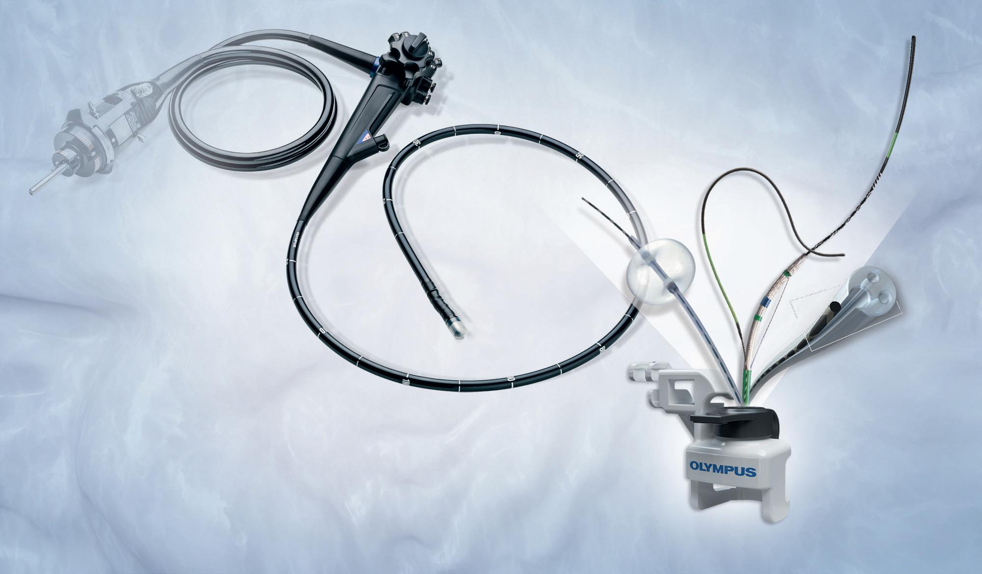
On show at the Olympus exhibitors’ booth, was the EVIS X1 platform and ENDO-AID – Artificial Intelligence technology which aids Polyp Detection. As well as our latest mid-range EUS Processor, EU-ME3, with enhanced imaging and innovative ultrasound technologies.

On show at the Olympus exhibitors’ booth, was the EVIS X1 platform and ENDO-AID – Artificial Intelligence technology which aids Polyp Detection. As well as our latest mid-range EUS Processor, EU-ME3, with enhanced imaging and innovative ultrasound technologies.
There was lots of interest in our consumable range but none more so than EndoCuff Vision, a mechanically enhanced colonoscopy device which is designed to maintain the viewable mucosa during endoscopic therapy. This Endotherapy device compliments ENDO-AID AI and is proven to increase the mucosal surface observed and can increase the Adenoma Detection Rate (ADR) by up to 17%*
There was lots of interest in our consumable range but none more so than EndoCuff Vision, a mechanically enhanced colonoscopy device which is designed to maintain the viewable mucosa during endoscopic therapy. This Endotherapy device compliments ENDO-AID AI and is proven to increase the mucosal surface observed and can increase the Adenoma Detection Rate (ADR) by up to 17%*
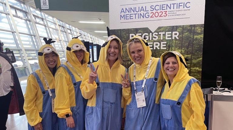
In addition, Olympus was a proud sponsor of the Nurses Hands-On workshop for over 30 Endoscopy Nurses. Attendees had the opportunity to get some valuable handson experience with Olympus Endoscopes using a simulated colon model and Endotherapy products including Polyloop, EndoCuff Vision, Distal Attachments and Loop Cutters.
The Olympus Continuum team is dedicated to educating the specialists of tomorrow. Olympus Continuum offers courses and webinars for reprocessing and endoscope training, specific topics include infection control, reducing the risk of cross contamination, and other adverse events related to the reprocessing of flexible endoscopes and surgical instruments. Getting started is easy—simply visit the website, locate your regional LMS site, and register for Olympus Continuum.
The Olympus Continuum team the specialists of tomorrow. courses and webinars for training, specific topics reducing the risk of cross adverse events related to endoscopes and surgical easy—simply visit the website, site, and register for Olympus
Upcoming Olympus Continuum Courses:
Upcoming Olympus Continuum
Endoscope Reprocessing Specialist Training (ERST)
In addition, Olympus was a proud sponsor of the Nurses Hands-On workshop for over 30 Endoscopy Nurses. Attendees had the opportunity to get some valuable handson experience with Olympus Endoscopes using a simulated colon model and Endotherapy products including Polyloop, EndoCuff Vision, Distal Attachments and Loop Cutters.
Olympus was proud to once again be the exclusive Platinum sponsor. An opportunity we take each year to show our support for the remarkable contribution of Endoscopy Healthcare Professionals in New Zealand. This event provides us with a valuable platform to connect with our customers to share knowledge and experiences and we are grateful for the opportunity to participate.
Endoscope Reprocessing Specialist
Olympus AKL - Thursday 15 Feb | 8:30am - 3:00pm NZST
Olympus AKL - Thursday 15
Endoscopic Retrograde Cholangiopancreatography Course (ERCP)
Endoscopic Retrograde Cholangiopancreatography
Course (ERCP)
Olympus AKL- Wednesday 6 Mar | 1.15pm - 4.30pm NZST
Olympus AKL- Wednesday
Endoscope Reprocessing Specialist Training (ERST)
Endoscope Reprocessing Specialist
Olympus AKL - Thursday 14 Mar | 8:30am - 3:00pm NZST
Olympus was proud to once again be the exclusive Platinum sponsor. An opportunity we take each year to show our support for the remarkable contribution of Endoscopy Healthcare Professionals in New Zealand. This event provides us with a valuable platform to connect with our customers to share knowledge and experiences and we are grateful for the opportunity to participate.
Giddy Up for 2024. We are ready for a Hoe Down in H-Town! See you there!
Olympus AKL - Thursday 14
Endoscope Reprocessing Specialist Training (ERST)
Endoscope Reprocessing Specialist
Olympus CHCH - Friday 5 Apr | 8:30am - 3:00pm NZST
Olympus CHCH - Friday 5 Apr
Endoscope Reprocessing Specialist Training (ERST)
Endoscope Reprocessing Specialist
Olympus WLG - Wednesday 24 Apr | 8:30am - 3:00pm
NZST
Olympus WLG - Wednesday
NZST
Endoscope Reprocessing Specialist Training (ERST)
Endoscope Reprocessing Specialist
Olympus AKL - Wednesday 14 Aug | 8:30am - 3:00pm
Giddy Up for 2024. We are ready for a Hoe Down in H-Town! See you there!
NZST
References: *Floer et al. PLoS One. 2014 Dec 3;9(12):e114267.
References: *Floer et al. PLoS One. 2014 Dec 3;9(12):e114267.
Olympus AKL - Wednesday
NZST
Endoscope Reprocessing Specialist Training (ERST)
Endoscope Reprocessing Specialist
Olympus BOP - Tuesday 27 Aug | 8:30am - 3:00pm NZST
Olympus BOP - Tuesday 27
Endoscope Reprocessing Specialist Training (ERST)
Endoscope Reprocessing Specialist
Olympus WLG - Wednesday 11 Sep | 8:30am - 3:00pm
Olympus WLG - Wednesday
NZST
Endoscope Reprocessing Train The Trainer (TTT)
Endoscope Reprocessing Train
Olympus WLG - Thursday 12 Sep | 10:00am - 6:00pm NZST
Olympus WLG - Thursday 12
Endoscope Reprocessing Specialist Training (ERST)
Endoscope Reprocessing Specialist
Olympus AKL - Thursday 19 Sep | 8:30am - 3:00pm NZST
Olympus AKL - Thursday 19
Endoscope Reprocessing Specialist Training (ERST)
Endoscope Reprocessing Specialist
Olympus CHCH - Thursday 17 Oct | 8:30am - 3:00pm NZST
Olympus CHCH - Thursday
Endoscope Reprocessing Train The Trainer (TTT)
Endoscope Reprocessing Train
Olympus CHCH - Friday 18 Oct | 10:00am - 6:00pm NZST
Olympus CHCH - Friday 18
Endoscope Reprocessing Specialist Training (ERST)
Endoscope Reprocessing Specialist
Olympus PMR - Wednesday 30 Oct | 8:30am - 3:00pm
Olympus PMR - Wednesday
NZST
https://learn-nz.olympus.co.nz
To book, visit: https://learn-nz.olympus.co.nz
Committee
Chairperson
Merrilee Williams merrilee.williams@southerndhb.govt.nz
Secretary
Jessica Southall secretaryofnzgnc@gmail.com
Treasurer
Kirsten Arnold treasurerofnzgnc@gmail.com
Committee Members
Merrilee Williams - Chairperson merrilee.williams@southerndhb.govt.nz
Jessica Southall - Secretary and Hepatology Specialty Group secretaryofnzgnc@gmail.com
Kirsten Arnold - Treasurer and IBD sub group treasurerofnzgnc@gmail.com
Karen Kempin - Committee Member and NE sub
Karen.kempin@huttvalleydhb.org.nz


Welcome to the Tube journal for the first quarter of 2024- I hope you will enjoy reading and learning from the articles that are included in this edition as much as I have. Thank you to those who have submitted these to share.
The committee have met for our first meeting of the year, and we will have our first face to face meeting in April, in Wellington. At this meeting, we will set some goals to achieve for the coming term, so if you have any ideas that we can help work towards as your college committee, please feel welcome to get in touch.
A couple of major documents that underpin our practice have been under review for the past couple of years. This includes the PG09 Guideline on Procedural Sedation 2023, and the AS/NZS 4187 Reprocessing of reusable medical devices in health service organisations 2019. These works have now been completed and will soon be available to update your protocols in your practice areas.
We have been able to have input into the final version of the PG09 along with our NZ colleagues of the NZ Society of Gastroenterology, and the College of Anaesthetists, however, we were unable to have input into the 4187. The 4187 will now be an Australian only standard, so we are working with Standards NZ to understand how we adopt these standards to the NZ context, and then adopt them as our guidelines for practice.
The sustainability group continue to meet, and share practical ideas to be able to improve our enormous clinical waste stream in endoscopy. We all have a role to play in this area, so sharing our successes
and ideas will be the way to get started. You can follow the progress of the group here https://nzsg. org.nz/news-and-events/article/PepJere/wastemanagement-in-endoscopy.
Work is underway within NZNO and Te Whatu Ora to assess and create the new Senior Nurses pay scale in recognition of those who work in advanced practice, and leadership roles. This is important work, and it is crucial that it leads to successful negotiation of improved pay rates. We need to encourage more nurses to advance their practice into senior nurse’s roles, as well as retain those already working so hard in their existing positions; this will be an important step forward.
The Inflammatory Bowel Diseases nurses group have been in contact with NZNO to be involved in this work, and have collected data relating to their specialty nurses workforce, and work load to share with the NZNO senior nurses working group.
I’d like to wish all those undertaking post graduate study this year all the best. It’s a real test of your time management, determination and drive to improve your own clinical practice. Your hard work will see growth in your knowledge and skills, as well as your critical thinking and approach to your patient care. You’ve got this!
Now please go forward and enjoy this edition of the Tube journal, please get in touch with the committee if you have any questions.
Merrilee Williams Your Chairperson NZGNC Secretary secretaryofnzgnc@gmail.comPrieto BN,
PGDipHScGastroenterology Department RN Te Whatu Ora – Hawke’s Bay Jondavid.prieto@hbdhb.govt.nz
The Annual Scientific Meeting 2023, held at Energy Events Centre, Rotorua, provided a valuable opportunity to enhance my knowledge and skills in the field of gastroenterology. The conference featured knowledgeable speakers who shared insights into various topics, it gave me the opportunity to listen to the topics I was very interested in, including Endoscopic Mucosal Resection (EMR), Endoscopic Submucosal Dissection (ESD), Barrett’s oesophagus and squamous cell carcinoma (SCC), oesophageal stenting, and notably, portal hypertensive bleeding.
Portal hypertension is defined as an increased pressure within the portal vein, it is a complication arising from liver cirrhosis (Iwakiri & Trebicka, 2021). It poses the risk of lifethreatening acute variceal bleeding with an overall mortality rate of 12-20% (Khan et al., 2022). Varices are detected in 50% of cirrhosis patients (Maruyama & Yokosuka, 2012). Once varices manifest, there is a notable 20% overall incidence of variceal bleeding within a span of two years (Baignes et al., 2019).
The conventional management for treatment includes volume resuscitation, haemodynamic stabilisation, administration of vasoactive drugs such as Terlipressin, Somatostatin or analogs, and prophylactic administration of antibiotics. Together with gastroscopy and variceal band ligation (Khan et al., 2022). Over the years the mortality rate has significantly decreased because of the implementation of these interventional managements (Garcia-Tsao, 2016).
Endoscopic variceal band ligation is the primary therapeutic modality for oesophageal bleeding, its effectiveness can be limited in cases of massive bleeding. If the band ligation fails, rescue therapies such as the Sengstaken-Blakemore tube (SBT) and the SX-Danis stent can be used (Baignes et al., 2019).
The SBT is employed for uncontrolled oesophageal and gastric variceal bleeding, demonstrating a 90% bleeding control rate. However, it has significant complications such as oesophageal perforation, aspiration, and rebleeding (Currais et al., 2023).
The SX-Danis stent may also be utilised for bleeding control for refractory variceal bleed, boasting higher efficacy and fewer complications compared to the SBT. However, it
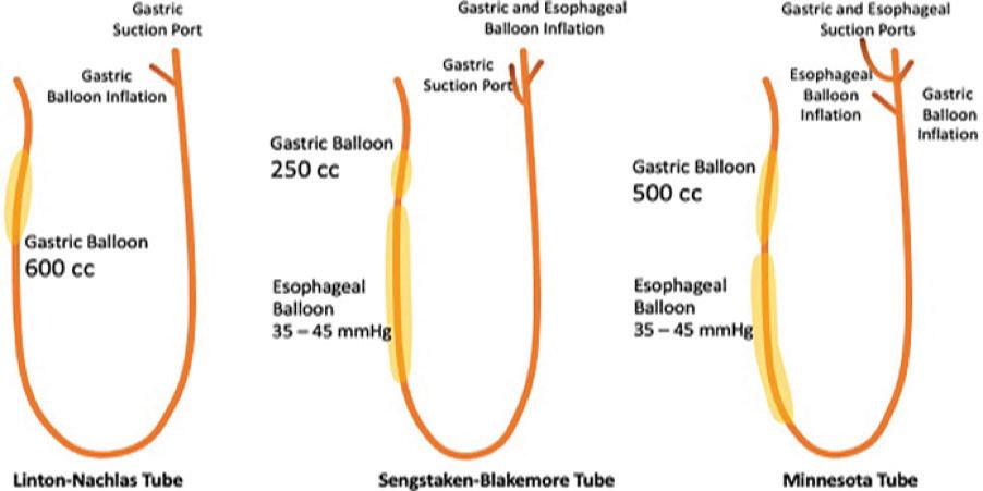
is not suitable for gastric variceal bleeding (Currais et al., 2023). The SX-Danis stent is a removable, fully covered self-expandable metal stent (SEMS), which proves very effective for bleeding from oesophageal varices. It has a 100% technical success rate, 87.5% bleeding control, and is relatively easy to use for refractory variceal bleeding (Khan et al., 2022). Additionally, its placement does not require endoscopic and X-ray control. It can be left in place for 7-14 days but then it must be removed to prevent oesophageal ulcerations.
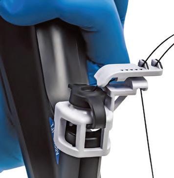
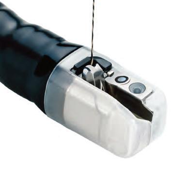
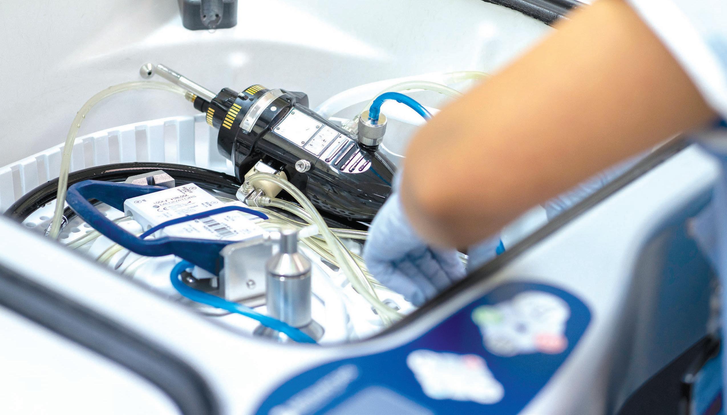
After introduction of the insertion system into the upper gastrointestinal tract, the system is fixed against the mouth guard by removing the blue lock.
The remainder of the system is advanced until the white lock meets the sheath handle. The gastric balloon is inflated with 100 mL to 120 mL of air through the balloon port using a large-volume syringe. Traction is applied to the whole delivery system, anchoring it against the gastroesophageal junction and allowing appropriate positioning. To deploy the stent, the white lock is removed and while holding the distal end of the delivery system still, the sheath handle is pulled back until it reaches the distal extend of the delivery system. This deploys the stent and also deflates the gastric balloon. The stent can be deployed without a need for direct endoscopic of fluoroscopic guidance. The balloon port valve is unscrewed and removed to ensure the gastric balloon is deflated before removal of the delivery device. The SEMS has gold marked loops at the distal and proximal ends that can be used to

reposition or remove it, as well as radiopaque markers to confirm position on radiography. (Source: ELLA http:// ellacs.cz/en/danis-stent (Leaflet – Danis Stent)
In summary, the SX-Danis stent is a great alternative to the SBT to control oesophageal variceal bleed and serve as a bridge to definitive therapies like Trans-jugular intrahepatic portosystemic shunt (TIPS). The SX-Danis has a high technical success rate, a lower complication rate, and is relatively easy to use. This makes it a preferable choice for nurses less familiar with the complexities of the SBT. As nurses, the stress associated with setting up and assisting during SBT placement necessitates continuous education. Refreshing ourselves through videos and manuals is crucial, and having a designated container for variceal bleeding would be beneficial for urgent situations. Notably, our unit in Hawke’s Bay currently lacks the SX-Danis stent, highlighting the importance of having it readily available.
References:
• Baiges, A., Hernández-Gea, V., Cárdenas, A., & GarcíaPagán, J. C. (2019). Portal hypertensive bleeding. Clinical Gastrointestinal Endoscopy. https://doi.org/10.1016/ b978-0-323-41509-5.00015-3
• Currais, P., Nunes, G., Patita, M., Coimbra, É., & Fonseca, J. (2021). SX-Ella Danis-stent for refractory acute esophageal variceal bleeding. GE - Portuguese Journal of Gastroenterology, 30(2), 162–165. https://doi. org/10.1159/000520273
• Garcia-Tsao, G. (2016). Current management of the
complications of cirrhosis and portal hypertension: Variceal hemorrhage, ascites, and spontaneous bacterial peritonitis. Digestive Diseases, 34(4), 382–386. https:// doi.org/10.1159/000444551
• Iwakiri, Y., & Trebicka, J. (2021). Portal hypertension in cirrhosis: Pathophysiological mechanisms and therapy. JHEP Reports, 3(4), 100316. https://doi. org/10.1016/j.jhepr.2021.100316
• Khan, S., Gilhotra, R., Jiang, C. D., Rowbotham, D., Chong, A., Majumdar, A., White, C., Huelsen, A., Brooker, J., O’Beirne, J., Schauer, C., Efthymiou, M., Vaughan, R., & Chandran, S. (2022). The role of a novel self-expanding metal stent in variceal bleeding: A multicenter Australian and New Zealand experience. Endoscopy International Open, 10(03). https://doi.org/ 10.1055/a-1729-0104
• Maruyama, H., & Yokosuka, O. (2012). Pathophysiology of portal hypertension and esophageal varices. International Journal of Hepatology, 2012, 1–7. https:// doi.org/10.1155/2012/895787
• Tafoya, L. A., McGee, J. C., Kaisler, S., Gottula, A. L., Lauria, M. J., & Braude, D. A. (2023). Management of acute upper gastrointestinal bleeding in Critical Care Transport. Air Medical Journal, 42(2), 110–118. https:// doi.org/10.1016/j.amj.2022.12.006


Date: Friday 12th April 2024
Location: CHRISTCHURCH
Time: 8.30am to 4.00pm
Date: Friday 26th July 2024
Location: AUCKLAND
Time: 8.30am to 4.00pm
Date: Friday 9th August 2024
Location: WELLINGTON
Time: 8.30am to 4.00pm
This is a foundation program that serves as an introduction to the reprocessing of flexible endoscopes incorporating infection control, structure and function, water filtration and workplace health and safety.
Registrations:
Can be made via the GENCA website or contact the GENCA office via email.
Simply visit the GENCA calendar to book your place at: https://www.genca.org/education/find-an-event/ A limited number of practical skills assessments are available following each of the Fundamentals workshops.
Please complete the application form and submit to GENCA via fax on 1300 799 439 or via email to fundamentals@genca. org. Individual times for assessments will be sent via email once the registrations are processed.
Andrea Dixon, Endoscopy Services Manager, Ormiston Surgical & Endoscopy Ltd
Holly Weale, Registered Nurse, Waitaha Canterbury.
Climate change has emerged as the most significant global threat of the 21st century, posing an existential risk to the world directly impacting human health by increasing diseases and deaths –but are we listening? Healthcare has contributed greatly to its development, and some countries have taken steps to address this crisis through decarbonisation, such as the NHS UK which aims to achieve net carbon zero by 2040 (greener.nhs, 2020). In Aotearoa, the transition to Te Whatu Ora signalled progress towards developing a national healthcare sustainability plan.
What does this mean for nurses working in endoscopy in NZ?
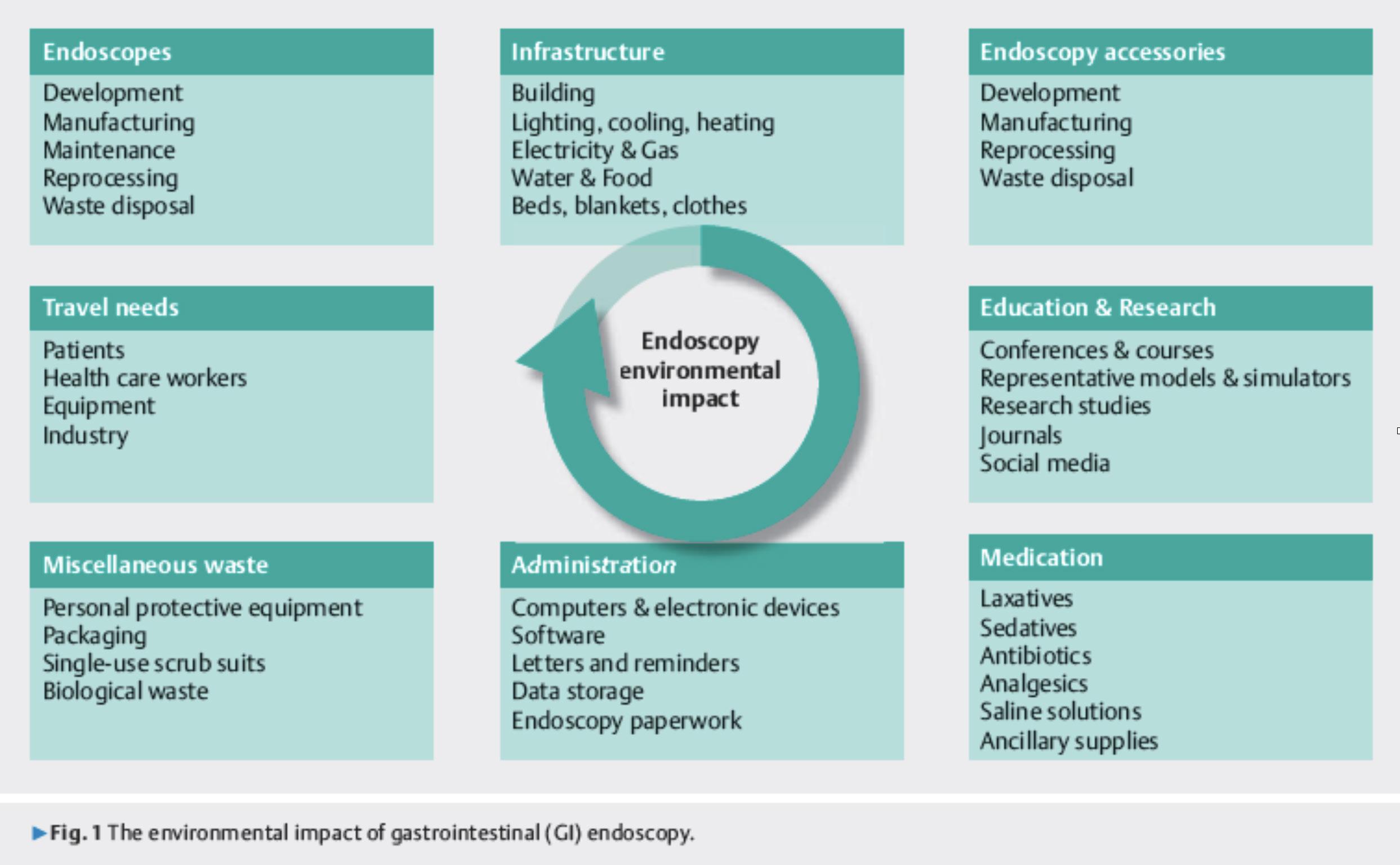
GI endoscopy has been recorded as the third highest waste generating procedure in healthcare facilities across the world. This is because of a high throughput of patients, multiple nonrenewable waste streams, a resource heavy decontamination process and multiple hospital visits for some patients that includes travel (Cunha Neves et al., 2023; Donnelly, 2022; Park & Cha, 2023). The environmental impact of GI endoscopy can be seen in the diagram below:
In 2022, the NZSG established the Sustainability Working Group with a mission to promote sustainable practice in gastroenterology and endoscopy. The group conducted a survey among NZSG and NZgNC members at the end of 2022, receiving 40 responses. While the number of respondents was low, they represented a diverse range of healthcare professionals, including public and private consultants, trainee registrars, endoscopy nurses, and representatives from various subspecialties with a wide variety of clinical experience post-qualification. An overwhelming majority (80%) expressed concern about climate change. Surprisingly, only 19% reported having received previous education on sustainability practices, despite the majority expressing a desire for further training. Additionally, 79% believed that the NZSG and NZgNC should prioritise environmental sustainability in gastroenterology and endoscopy (John, 2023).
Multiple frameworks have been developed demonstrating ways to reduce the carbon footprint in endoscopy units, including simple solutions such as powering down the unit when not in use and double-sided printing of patient reports (Sebastian et
al., 2023). However, the major environmental impact caused by endoscopy procedures is the large volume of waste generatedsuch as plastic, packaging, and personal protective equipment (PPE) - and in the last decade there has been greater awareness of the environmental impacts caused by single use items (Agrawal et al., 2017; Desai et al., 2023).
Recycling seems a logical first step for units to start their green journey however, studies have shown that a low proportion of waste in endoscopy units is recycled (Azouz et al., 2019; Namburar et al., 2022; Petre et al., 2019).
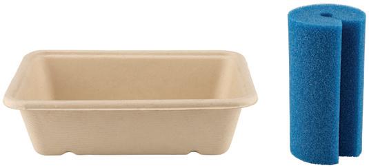
Gayam (2020) estimates that only 0.3% of this waste is recyclable, and because recycle bins are not available in procedure rooms, virtually all waste from endoscopy procedures is going to end up in landfill. The move away from single use consumables such as plastic bowls, pottles and gowns, and replacing these with compostable alternatives is a relatively simple thing to implement with numerous compostable and biodegradable products now available.
On its own this is not going to have the desired effect of decreasing landfill and greenhouse gas (GHG) emissions as biodegradable products will break down eventually in landfill, but it will still take a long time due to it being an anaerobic environment with no dirt, microbes, fungi or bacteria (Earth Talk, 2023). A University of Arizona looked at landfill contents and discovered recognisable food items such as grapes and hotdogs and even a 25-year-old newspaper, the same will be happening with all the biodegradable rubbish per endoscopy day we are sending to landfill.
This is where commercial composting comes into play with most major centres now having collections and commercial composting facilities available.
Biodegradable waste breaks down within weeks and ends up as a fine futile soil rather than filling landfills. The process is cleaner, faster, produces less GHG emissions than landfills and never fills up. It involves high temperatures to kill pathogens and bacteria but also provides an aerobic environment with organic matter and microbes which breaks down waste at high speeds.
Implementing this can be more challenging than adding a recycling bin to a room however in the long run is much more beneficial. It means a change in the way waste management on the hospital site is conducted with different bins next to landfill and recycling bins, however this doesn’t mean needing more space due to the significant decrease in landfill waste that will occur. It means more than just the biodegradable pots and gowns but all food waste and paper towels which take up a lot of area in landfill bins.
It takes more effort to implement commercial composting, getting hospital facilities management on board, sourcing
appropriate bins for within the department and education, education, education for staff to ensure correct items are directed to the right bins. However once implemented and effectively managed it could quickly lead to a reduction in endoscopy waste going to landfill.
We need to start somewhere – the buzz words are the 3 ‘R’s of a greener endoscopy – reduce, reuse and recycle. The ESGE offers tips for how to achieve these three, but also discusses two more ‘R’s in research and rethink. The term research in this case is affiliated with refuse – we should not be performing unnecessary GI endoscopic procedures and should be looking at the environmental impact non-endoscopic diagnostic alternatives could provide (ESGE, 2023).
It is estimated that 20-30% of endoscopies may be unnecessary (Frazzoni et al., 2021). In many talks about sustainability and green endoscopy the last slide is always that the largest impact on carbon emissions we can make is a reduction in GI endoscopic procedures – good triaging and not doing unnecessary procedures (Sebastian et al., 2023).
If an endoscopic procedure is deemed necessary, rationalising and limiting the number of biopsies / tissue samples sent for histopathology is urged, this can be done without compromising diagnosis and disease outcome (Ueda et al., 2023).
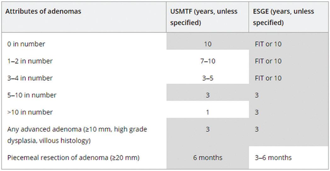
For example, a biopsy of the terminal ilium to confirm intubation is not required (EGGNZ, 2017). Ueda et al. (2023) also suggests the ‘diagnose and leave’ and ‘resect and discard’ strategies when diagnosing diminutive polyps (<5mm). Each sample in a pot sent to the laboratory increases GHG emissions for that procedure. Another idea is to follow up to date guidelines for polyp surveillance recommendations (as per the ESGE table below) which will help decrease the number of screening endoscopies performed each year.
These are aspirational ideas to aim for but tomorrow what can you do in your endoscopy unit? A good way to start the introduction of ‘green endoscopy’ to your unit is the creation of a ‘green team’, a key theme in the literature (Azouz et al., 2019; Petre et al., 2019; Pohl, 2023; Sebatian et al., 2023; Wormer et al., 2013), and the first step towards staff buy-in. The green team is created with likeminded individuals, who can assess the attitudes of other staff, create a shared understanding of the meaning green, and provide information about waste segregation and sustainable principles (Azouz et al., 2019; Donnelly, 2022; Petre et al., 2019; Pohl, 2023; Sebatian et al., 2023).
And what can we focus on today, let’s start by raising awareness amongst our teams, start with small steps such as using reusable cups, printing double sided and only when necessary, and sending reports to GPs electronically rather than a paper version (Donelly, 2022). Thiel (2018) urges us to look at electricity usage stating that lighting and endoscopy processors in procedure rooms accounts for 10-30% of environmental impact (cited in Gayam, 2020).
A simple step wold be to turn off monitors at the end of the day, switch off lighting between lists or extended breaks and at the end of the day, and even change to LED bulbs which use 60% less energy and will pay for themselves in approximately 2 months. This article is the start of the conversation, lets work with the NZSG and the NZgNC and share our knowledge and resources. Let’s as a specialty lobby industry to look at packaging requirements and say no to excess plastic. Get involved by contacting Daryl McGann or the authors of this articles to join together and save our planet. DarylM@macmurray.co.nz andrea.dixon@ormistonhospital.co.nz holly.weale@cdhb.health.nz
• Agrawal, D., Shoup, V., Montgomery, A., Wosik, J., & Rockey, D. C. (2017). Disposal of endoscopic accessories after use: Do we know and do we care? Gastroenterology Nursing, 40(1), 13–18. https://doi.org/10.1097/SGA.0000000000000280
• Azouz, S., Boyll, P., Swanson, M., Castel, N., Maffi, T., & Rebecca, A. M. (2019). Managing barriers to recycling in the operating room. The American Journal of Surgery, 217(4), 634–638. https://doi.org/10.1016/j.amjsurg.2018.06.020
• Cunha Neves, J. A., Roseira, J., Queirós, P., Sousa, H. T., Pellino, G., & Cunha, M. F. (2023). Targeted intervention to achieve waste reduction in gastrointestinal endoscopy. Gut, 72(2), 306–313. https://doi.org/10.1136/gutjnl-2022-327005
• Desai, M., Campbell, C., Srinivasan, S., Higbee, A., Patel, H., Radadiya, D., & Sharma, P. (2023). A detailed and prospective analysis of the environmental impact of waste generation and energy consumption from GI procedures. Gastrointestinal Endoscopy, 97(6), AB848. https://doi.org/10.1016/j. gie.2023.04.1374
• De Santiago, E., Dinis-Ribeiro.M., Pohl, H., Agrawal, D., Arvanitakis, M., Baddeley, R., Bak, E., Bhandari, P., Bretthauer, M., Burga, P., Donnelly, L., Eickhoff, A., Hayee, B., Kaminski, M., Karlović, K., Lorenzo-Zúñiga, V., Pellisé, M., Pioche, M., Siau, K., Siersema, P., Stableforth, W., Tham, T., Triantafyllou K., Tingali, A., Veitch, A., Voiosu, A., Webster, G., Vienne, A., Beilenhoff, A., Bisschops, R., Hassan, C., Gralnek, I. & Messmann, H. (2022). Reducing the environmental footprint of gastrointestinal endoscopy: European Society of Gastrointestinal Endoscopy (ESGE) and European Society of Gastroenterology and Endoscopy Nurses and Associates (ESGENA) Position Statement. Endoscopy. 54(08): 797-826. doi: 10.1055/a-1859-3726
• Donnelly, L. (2022). Green endoscopy: Practical implementation. Frontline Gastroenterology, 13(e1), e7–e12. https://doi.org/10.1136/flgastro-2022-102116
• Earth Talk. (2023). Do Biodegradable Items Degrade in Landfills? Retrieved from: thoughtco.com/do-biodegradable-itemsreally-break-down-1204144
• ESGE. (2023). The 5 R’s of Greener Endoscopy: individual tips. https://www.esge.com/green-endoscopy-working-group
• EGGNZ. (2017). Endoscopy Standards for Individual Colonoscopists Performing Bowel Cancer Screening in New Zealand. https://eggnz.endoscopyquality.co.nz/assets/ Uploads/EGGNZ-Endoscopy-Standards-for-IndividualColonoscopists-Performing-Bowel-Cancer-Screening-inNew-Zealand-Final-v3.0.pdf
• Frazzoni, L., La Marca, M. & Radaelli, F. (2021). Systematic review with meta-analysis: the appropriateness of colonoscopy increases the probability of relevant findings and cancer while reducing unnecessary exams. Alimentry Pharmacology and Therapeutics. 53: 22–32. https://onlinelibrary.wiley.com/ doi/10.1111/apt.16144
• Gayam, S. (2020). Environmental impact of endoscopy: “scope” of the problem. American Journal Gastroenterology, 115: 1931–1932
• Greener.nhs (2020). Delivering a “Net Zero’ National health Service. Retrieved from greener.nhs@nhs.net:https:// www.england.nhs.uk/greenernhs/wp-content/uploads/ sites/51/2020/10/delivering-a-net-zero-national-healthservice.pdf
• John, R. (2023). Establishing a Sustainability Team and a Sustainability Lead within your endoscopy unit. NZSG panui: https://nzsg.org.nz/news-and-events/
• Namburar, S., Von Renteln, D., Damianos, J., Bradish, L., Barrett, J., Aguilera-Fish, A., Cushman-Roisin, B., & Pohl, H. (2022). Estimating the environmental impact of disposable endoscopic equipment and endoscopes. Gut, 71(7), 1326–1331. https://doi.org/10.1136/gutjnl-2021-324729
• Park, S. B., & Cha, J. M. (2023). Gastrointestinal endoscopy’s carbon footprint. Clinical Endoscopy, 56(3), 263–267. https:// doi.org/10.5946/ce.2023.003
• Petre, M.-A., Bahrey, L., Levine, M., Van Rensburg, A., Crawford, M., & Matava, C. (2019). A national survey on attitudes and barriers on recycling and environmental sustainability efforts among Canadian anesthesiologists: An opportunity for knowledge translation. Canadian Journal of Anesthesia/ Journal Canadien d’anesthésie, 66(3), 272–286. https://doi. org/10.1007/s12630-018-01273-9
• Pohl, H. (2023). Transitioning to sustainable care and green endoscopy. Gastroenterology and Hepatology, 19(4), 233–236.
• Sebastian, S., Dhar, A., Baddeley, R., Donnelly, L., Haddock, R., Arasaradnam, R., Coulter, A., Disney, B., Griffiths, H., Healey, C., Hillson, R., Steinbach, I., Marshall, S., Rajendran, A., Rochford, A., Thomas-Gibson, S., Siddhi, S., Stableforth, W., Wesley, E., … Hayee, B. (2023). Green endoscopy: British Society of Gastroenterology (BSG), Joint Accreditation Group (JAG) and Centre for Sustainable Health (CSH) joint consensus on practical measures for environmental sustainability in endoscopy. Gut, 72, 12–26.
• Ueda, T., Li, J., Ho, S., Singh, R. & Uedo, N. (2023). Precision endoscopy in the era of climate change and sustainability. Journal of Gastroenterology and Hepatology. 39(1), 18-27. https://doi.org/10.1111/jgh.16383
Susan Gale, Hepatology CNS, MHSc (Nursing)
Gastroenterology Unit, Dunedin Hospital susan.gale@southerndhb.govt.nz 027 54 222 65
The role of the hepatology nurse continues to evolve and expand, reflecting the ever-changing landscape of healthcare and the population it serves. With this has come the inclusion in nurseled clinics of patients with cholestatic liver disease. Formally a domain of our medical colleagues, nurses are becoming more involved in the follow-up and monitoring of this patient group.
Primary Biliary Cholangitis (PBC) and Primary Sclerosing Cholangitis (PSC) are two such chronic cholestatic liver diseases that significantly impact patients’ quality of life (Dyson et al, 2016) This article aims to provide a basic overview of PBC, including signs and symptoms, diagnostic approaches, and nursing care strategies based on current best practices and international guidelines.
Primary Biliary Cholangitis (PBC)
PBC is an inflammatory, autoimmune liver disease characterized by the progressive destruction of intrahepatic bile ducts (EASL, 2017). This condition is relatively uncommon and tends to affect middle-aged women (Bianchi et al, 2012). Common symptoms include fatigue, cholestatic pruritus, jaundice, sicca complex and abdominal discomfort (Mells et al, 2013). Cholestasis, a characteristic of PBC, affects lipids so patients may also have xanthoma (deposits of yellowish cholesterol-rich material under the skin) and xanthelasma (xanthoma on the eyelids) along with elevated cholesterol (Longo et al, 2002). Up to 25% of patients
with PBC may also have Raynaud’s phenomenon (Watt et al, 2004). However, some patients are asymptomatic and only identified when routine blood tests show abnormal liver function tests (LFT), warranting further investigation.
Diagnostic criteria include elevated alkaline phosphatase (ALP) levels, the presence of circulating antimitochondrial antibodies at a titre >1:40 (AMA), PBC -specific antinuclear antibody (ANA) reactivity and cholestasis (EASL, 2017).
Whilst the trigger for PBC is not well understood, it is thought to be due to a combination of genetic risk factors and environmental triggers, for example: smoking, nail polish, urinary tract infections and hormone replacement therapy (Prince et al, 2010; Selmi et al, 2004). Injury to the bile ducts within the liver, caused by a complex interaction between the immune system and biliary pathway, causes a cycle of cholestasis, ductopenia and fibrosis (Juran &, Lazaridis , 2014; Bianchi et al, 2014).
Advanced cases may lead to end-stage biliary cirrhosis and liver failure emphasizing the importance of early diagnosis and intervention (EASL, 2017). Because PBC does not change the morphology (size, shape and structure) of the liver, imaging is only useful to rule out liver neoplasms, extrahepatic causes of cholestasis and identify signs of advanced liver disease (EASL, 2017). Liver biopsy is required when PBC-specific antibodies are absent and/or autoimmune hepatitis (AIH) or metabolic dysfunction-associated steatotic liver disease (MASLD) are present (EASL, 2017).
Management of PBC involves prescribing ursodeoxycholic acid (UDCA); a naturally occurring bile acid (American Association for the Study of Liver Diseases [AASLD] 2018). Optimal dosage is 13-15mg/kg/day, given either as a single dose or in divided doses (EASL, 2017). It has a high safety profile with minimal side effects (EASL, 2017). The response to this first -line therapy after six months (Zhang et al, 2013) determines if the disease is a ‘high or low risk’ disease (EASL, 2017), along with age of onset, gender, stage at presentation and biomarkers both pre/post UDCA (Trivedi et al, 2016).
Those with an inadequate response or intolerance of UCDA, may be prescribed Obeticholic acid (OCA) as well. The biomarkers ALP and bilirubin have been deemed internationally, as the most important parameters when evaluating the response to UDCA (Lammers et al, 2014). Fibrates (lipid-lowering medications) are
off-label alternatives that may be considered for patients with PBC that have responded inadequately to UCDA (AASLD, 2018).
Addressing symptoms like pruritus in PBC is paramount. Uncontrolled pruritus can lead to sleep disturbances and depression. Nursing interventions may include advising on cool baths, moisturizing with emollients and oatmeal extract to improve dry and inflamed skin (EASL, 2017), distraction therapy and pharmacological agents like bile sequestrants (cholestyramine), rifampicin, (a second-line agent), alternatively antihistamines, SSRIs or opiate antagonists (Kremer et al, 2012). Care around potential drug interactions is also necessary.
Management of fatigue, however, is more difficult as there are many potential variables. Developing healthy coping strategies and avoiding social isolation can be beneficial, provided alternative causes of fatigue have been addressed (EASL, 2017).
Sicca complex is characterised by dry mucous membranes: dry eyes (keratoconjunctivitis sicca)/or mouth (xerostomia), dysphagia or vaginal dryness, can be eased with artificial tears; stimulation of saliva production with sugar-free gum, mouthwashes, or saliva substitutes; and vaginal moisturisers. Pharmacological therapies may also be beneficial. Advice on oral hygiene is useful to prevent dental caries. Nurses must be cognizant of the emotional toll this chronic disease has on their patients and offer psychological support and referrals to appropriate resources, such as support groups or mental health professionals, when necessary or requested. Liver transplantation is effective for the treatment of debilitating pruritus unresponsive to medical treatment.
The goal of treatment is to manage symptoms and prevent endstage liver disease (EASL, 2017). Regular assessments are essential to monitor disease progression. Assessment should include liver function tests 3-6 monthly, annual TSH, Vitamin A,D,E and PT if bilirubin >2.0mg/dl, and a baseline bone mineral density scan, then 2 yearly. Symptom evaluation, and nutritional status is also important. In PBC, monitoring pruritus severity and response to treatment is crucial. Additionally, assessing for complications such as portal hypertension or hepatocellular carcinoma in advanced cases.
Monitoring of PBC progression with vibration-controlled transient elastography (Fibroscan) is also recommended (EASL, 2017). Corpechot et al (2012) suggest that a liver stiffness measurement (LSM) greater than 9.6 kPa is associated with 5 times the risk of liver decompensation, liver transplantation or death. Hepatocellular surveillance is recommended for those with cirrhosis.
Educating patients on the importance of medication adherence, potential side effects, and the need for routine monitoring will empower patients with information about their condition, including triggers, symptom management, and the importance of follow-up appointments.
PBC is a complex cholestatic liver disease that requires comprehensive nursing care tailored to each individual patient. Early diagnosis, symptom management, and regular monitoring are crucial for improving patients’ quality of life and long-term outcomes. Nurses play a vital role in educating, supporting, and empowering patients to manage this chronic condition effectively, ultimately enhancing their overall well-being and quality of life.
• Bianchi, I., Carbone, M., Lleo, A., & Invernizzi, P. (2014). Genetics and epigenetics of primary biliary cirrhosis. Seminars in liver disease, 34(3), 255–264. https://doi.org/10.1055/s-0034-1383725
• Bianchi, I., Lleo, A., Gershwin, M. E., & Invernizzi, P. (2012). The X chromosome and immune associated genes. Journal of autoimmunity, 38(2-3), J187–J192. https://doi.org/10.1016/j.jaut.2011.11.012
• Corpechot, C., Carrat, F., Poujol-Robert, A., Gaouar, F., Wendum, D., Chazouillères, O., & Poupon, R. (2012). Noninvasive elastography-based assessment of liver fibrosis progression and prognosis in primary biliary cirrhosis. Hepatology (Baltimore, Md.), 56(1), 198–208. https://doi.org/10.1002/hep.25599
• Dyson, J. K., Wilkinson, N., Jopson, L., Mells, G., Bathgate, A., Heneghan, M. A., Neuberger, J., Hirschfield, G. M., Ducker, S. J., UK-PBC Consortium, Sandford, R., Alexander, G., Stocken, D., & Jones, D. E. (2016). The interrelationship of symptom severity and quality of life in 2055 patients with primary biliary cholangitis. Alimentary pharmacology & therapeutics, 44(10), 1039–1050. https://doi.org/10.1111/apt.13794
• European Association for the Study of the Liver. Electronic address: easloffice@easloffice.eu, & European Association for the Study of the Liver (2017). EASL Clinical
Continued over page
Practice Guidelines: The diagnosis and management of patients with primary biliary cholangitis. Journal of hepatology, 67(1), 145–172. https://doi.org/10.1016/j. jhep.2017.03.022
• Juran, B. D., & Lazaridis, K. N. (2014). Environmental factors in primary biliary cirrhosis. Seminars in liver disease, 34(3), 265–272. https://doi.org/10.1055/s-0034-1383726
• Kremer, A. E., van Dijk, R., Leckie, P., Schaap, F. G., Kuiper, E. M., Mettang, T., Reiners, K. S., Raap, U., van Buuren, H. R., van Erpecum, K. J., Davies, N. A., Rust, C., Engert, A., Jalan, R., Oude Elferink, R. P., & Beuers, U. (2012). Serum autotaxin is increased in pruritus of cholestasis, but not of other origin, and responds to therapeutic interventions. Hepatology (Baltimore, Md.), 56(4), 1391–1400. https://doi.org/10.1002/hep.25748
• Lammers, W. J., van Buuren, H. R., Hirschfield, G. M., Janssen, H. L., Invernizzi, P., Mason, A. L., Ponsioen, C. Y., Floreani, A., Corpechot, C., Mayo, M. J., Battezzati, P. M., Parés, A., Nevens, F., Burroughs, A. K., Kowdley, K. V., Trivedi, P. J., Kumagi, T., Cheung, A., Lleo, A., Imam, M. H., … Global PBC Study Group (2014). Levels of alkaline phosphatase and bilirubin are surrogate end points of outcomes of patients with primary biliary cirrhosis: an international follow-up study. Gastroenterology, 147(6), 1338–e15. https://doi.org/10.1053/j.gastro.2014.08.029
• Lindor, K. D., Bowlus, C. L., Boyer, J., Levy, C., & Mayo, M. (2019). Primary Biliary Cholangitis: 2018 Practice Guidance from the American Association for the Study of Liver Diseases. Hepatology (Baltimore, Md.), 69(1), 394–419. https://doi.org/10.1002/hep.30145
• Longo, M., Crosignani, A., Battezzati, P. M., Squarcia Giussani, C., Invernizzi, P., Zuin, M., & Podda, M. (2002). Hyperlipidaemic state and cardiovascular risk in primary biliary cirrhosis. Gut, 51(2), 265–269. https://doi. org/10.1136/gut.51.2.265
• Mells, G. F., Pells, G., Newton, J. L., Bathgate, A. J., Burroughs, A. K., Heneghan, M. A., Neuberger, J. M., Day, D. B., Ducker, S. J., Sandford, R. N., Alexander, G. J., Jones,
D. E., & UK-PBC Consortium (2013). Impact of primary biliary cirrhosis on perceived quality of life: the UK-PBC national study. Hepatology (Baltimore, Md.), 58(1), 273–283. https://doi.org/10.1002/hep.26365
• Prince, M. I., Ducker, S. J., & James, O. F. (2010). Casecontrol studies of risk factors for primary biliary cirrhosis in two United Kingdom populations. Gut, 59(4), 508–512. https://doi.org/10.1136/gut.2009.184218
• Selmi, C., Mayo, M. J., Bach, N., Ishibashi, H., Invernizzi, P., Gish, R. G., Gordon, S. C., Wright, H. I., Zweiban, B., Podda, M., & Gershwin, M. E. (2004). Primary biliary cirrhosis in monozygotic and dizygotic twins: genetics, epigenetics, and environment. Gastroenterology, 127(2), 485–492. https://doi.org/10.1053/j.gastro.2004.05.005
• Trivedi, P. J., Corpechot, C., Pares, A., & Hirschfield, G. M. (2016). Risk stratification in autoimmune cholestatic liver diseases: Opportunities for clinicians and trialists. Hepatology (Baltimore, Md.), 63(2), 644–659. https://doi.org/10.1002/hep.28128
• Watt, F. E., James, O. F., & Jones, D. E. (2004). Patterns of autoimmunity in primary biliary cirrhosis patients and their families: a population-based cohort study. QJM : monthly journal of the Association of Physicians, 97(7), 397–406. https://doi.org/10.1093/qjmed/hch078
• Zhang, L., Shi, T., Shi, X., Wang, L., Yang, Y., Liu, B., Gao, L., Shuai, Z., Kong, F., Chen, H., Han, , Han, S., Fei, Y., Cui, Q., Wang, Q., Shen, M., Xu, D., Zheng, W., Li, Y., Zhang, W., Zhang, F. (2013). Early biochemical response to ursodeoxycholic acid and long-term prognosis of primary biliary cirrhosis: results of a 14-year cohort study. Hepatology (Baltimore, Md.), 58(1), 264–272. https://doi.org/10.1002/hep.26322
A highlight at the end of 2023 was the annual New Zealand Gastroenterology Scientific Meeting held in December. The day prior to Conference, the HZ hepatology Nurses Group Committee met to progress the key Knowledge and Skills Framework. The next step is to have it reviewed by the Society of Gastroenterology which is due to happen in the first quarter of this year.
The conference had some good hepatology content. One of the keynote speakers was Dr Guadalupe Garcia-Tsao, Professor of Medicine at Yale University School of Medicine.
Her research focuses on cirrhosis, portal hypertension and related complications, having authored over two hundred original research publications in addition to several society guidelines and position papers in the field.
She has also served as the President of the American Association for the Study of Liver Diseases in 2012 and many of us will have seen her name on journal papers over the years. It was a real honor to be able to attend the conference and her presentations were very engaging.
She reinforced the new guidelines for assessment of risk of clinically significant portal hypertension that we have adopted over the last year. It felt good to know that we are managing our patients in line with international best practice guidelines.
Dr Akilesh Swaminathan presented research on Metabolic Associated Steatotic Liver Disease (MASLD) which is increasingly relevant to our practice as hepatology nurses. Many will have worked with Akhilesh and appreciate his ability to convey technical information in a way that holds your attention. In summary cardio-metabolic risk is being under-treated in patients with MASLD and this is something we need to raise awareness about with our patients and their primary healthcare providers. This year we are hoping to support more nurses to attend educational forums relevant to hepatology nursing and will be sharing more about this over the coming months. One of our committee members will also be attending the Genca National Conference in May as a trans-Tasman speaker.
She will be discussing hepatology nursing in New Zealand/ Aotearoa with a focus on the evolution of our Knowledge and Skills Framework, and innovations in Hepatology Nursing here. This will be a great opportunity to strengthen our relationship with Australian hepatology nurses.
Please email Bridget Faire for any membership queries: nzhepng.secretary@gmail.com
Registered Nurse Gastroenterology, Capsule Endoscopy Nurse, Mortality and Morbidly Meeting Nurse Lead, North Shore Hospital, Te Whatu Ora - Waitemata
124 Shakespeare Road, Takapuna, Auckland 0622, 02102820872, Vincent.borromeo@waitematadhb.govt.nz
No financial disclosure.
Patient MK, a 45-year-old male, presented in hospital for melaena. His haemoglobin dropped to 105 g/L, (130-175) and Ferritin 90 ug/L (N20-450). The patient had an Oesophagogastroduodenoscopy (OGD) and a colonoscopy, which both tests did not show any source of bleeding. This is considered as an obscure GI bleed (OGIB). OGIB is defined as bleeding from an unknown aetiology after negative endoscopy evaluations including OGD and colonoscopy with endoscopiv evaluation of the terminal ileum. (Awatie, et al., 2022). A capsule endoscopy was then ordered as the next step in his investigations. Capsule Endoscopy is the procedure of choice of OGIB investigation due to its effectiveness and non-invasiveness (Tanabe, 2016).
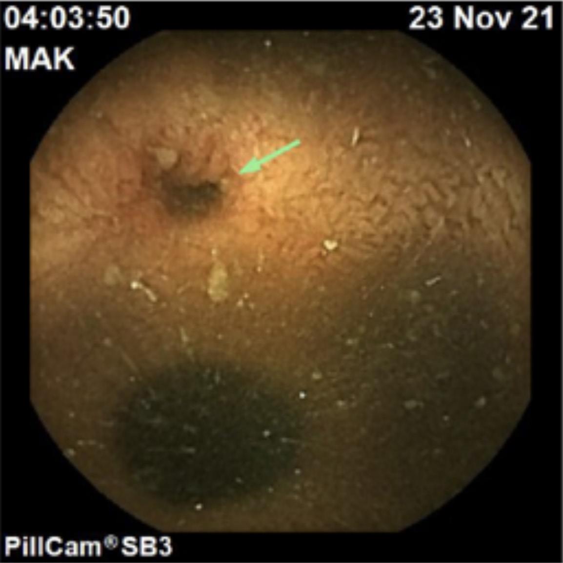
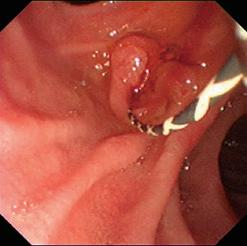
The patient then had a capsule endoscopy that showed an inflamed diverticulum in the distal small bowel (Figure 1). SB progress at 98%.The lead gastroenterologist, Ali Jafer, had a strong suspicion that the patient might have a bleeding Meckel’s diverticulum. He called the patient and informed him of the situation. Later on, the patient had surgery and was confirmed that it was indeed, Meckel’s Diverticulum.
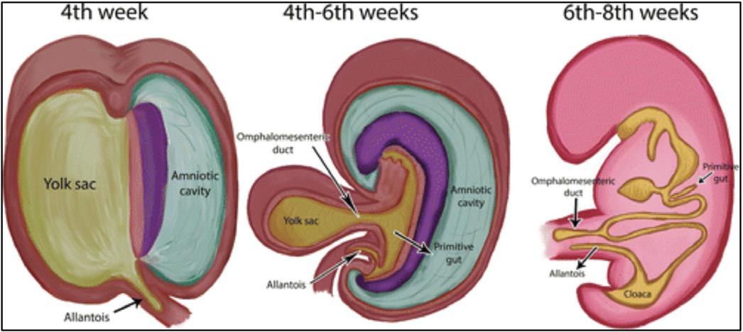
This is defined as a Congenital Malformation due to persistence of the congenital vitello-intestinal duct in which there is a failure of regression of the Omphalomesenteric duct during embryonic development. Ectopic gastric tissue can be present in the diverticulum. Majority of patients are asymptomatic. This is mostly diagnosed during surgery. Symptoms include obstruction or obstructive symptoms, GI bleeding, and inflammatory symptoms with or without perforation. (Sagar, et al., 2006).
The Research and Knowledge Centre of Te Whatu Ora - Waitemata provided the data used in the analysis. The criteria are that patients must be diagnosed with having Meckel’s diverticulum post op or during discharge. 151 cases have been forwarded to me. I have omitted 4 cases as I could not find the findings of Meckel’s diverticulum on their clinical portal.
Demographics.
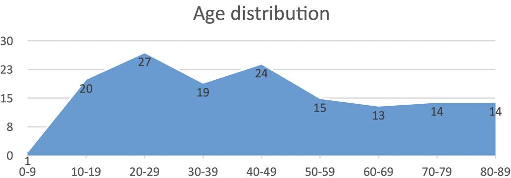
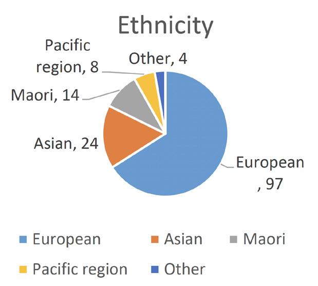
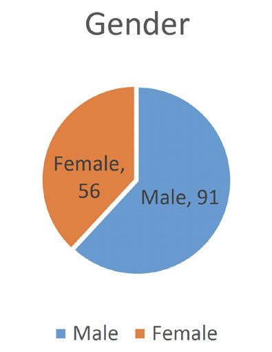
I conducted a manual count of the data, revealing gender, ethnicity, and age distribution. There were 91 male patients and 56 female patients. The predominant ethnic group affected by Meckel’s diverticulum is European, comprising 66% of the cases (97 cases). Asian individuals account for 16% (24 cases), followed by Maori at 9.4% (14 cases), the Pacific Region at 5.4% (8 cases), and other ethnicities at 3.2% (4 cases). Age distribution is as follows 0-9 = 1, 10-19 = 20, 20-29 = 27, 30-39 = 19, 40-49 = 24, 50-59 = 15, 60-69 = 13, 70-79 = 14, 80-89 = 14.
Of the 147 Meckel’s cases, 121 of them have been diagnosed during a laparotomy. This is the list of surgeries done that they found Meckel’s.
• Diagnostic/Explore laparotomy
• Lap appendectomy
• Whipple’s
• Lap abdominoperineal resection
• Lap cholecystectomy
• Ileocolic resection
• Excision of mesenteric abscess
• Small bowel resection
• Resection to fix a perf
• Reversal of Hartman’s
• Hernia repair
• LSCS
• Right hemicolectomy
• Duodenal switch
• Panproctocolectomy + formation of ileostomy
• Panproctolectomy + ileocolonic resection
• Laparoscopy, proceed to laparotomy
For the remaining cases, 22 of them were confirmed in a CT scan, 2 cases were diagnosed via Gastric Meckel’s scintigram, 1 case diagnosed by ultrasound, and 1 other case diagnosed by capsule endoscopy.
Over the past two decades, a total of 148 reported cases of Meckel’s diverticulum have been observed, with a predominant impact on male patients of New Zealand European ethnicity. The age distribution reveals peaks in the 20-29 and 40-49 age groups, indicating its prevalence among both younger and middle-aged individuals. Notably, 82% of the cases are diagnosed through laparotomy, enabling prompt identification and removal upon visual confirmation, while others come to light incidentally or during investigations for unexplained bleeding.
Throughout 20 years of diagnosing Meckel’s diverticulum, its visibility in a capsule endoscopy was a unique occurrence, highlighting its exceptional rarity in such diagnostic procedures and making that particular case especially remarkable. This study aims to contribute valuable insights into the clinical characteristics of Meckel’s diverticulum within our population, enhancing our understanding of this condition’s impact in New Zealand.
References:
• Awadie, H., Zoabi, A., & Gralnek, I. M. (2022). Obscure overt gastrointestinal bleeding: A Review. Polish Archives of Internal Medicine, 132(5). https://doi.org/10.20452/pamw.16253
• Tanabe, S. (2016). Diagnosis of obscure gastrointestinal bleeding. Clinical Endoscopy, 49(6), 539–541. https://doi. org/10.5946/ce.2016.004
• Sagar, J., Kumar, V., & Shah, D. (2006). Meckel’s Diverticulum: A Systematic Review. Journal of the Royal Society of Medicine, 99(10), 501–505. https://doi.org/10.1177/014107680609901011
Committee: Marian O’Connor (Chair), Kirsten Arnold (Co-chair), Nideen Visesio (Education lead), Donna Howe (Secretary)
Report Date: February 2024 Prepared By: Kirsten Arnold Reporting Period: Oct 2023 – Feb 2024
• The annual NZIBDNG education day is being planned for October and will be held in Auckland. Likely dates are 4-5 October (two half day sessions with an educational and networking dinner) but still TBC. Venue also TBC.
• Carly Bramley is now living and working on the Gold Coast and is the acting chair of IBDNA.
• Nideen Visesio has replaced Carly Bramley as the education lead for this group. She and Carly will continue to work together to provide a valuable link for NZ nurses with the IBNA in Australia.
• Marian continues as a board director of GENCA and encourages you to avail of the reduced membership for kiwi’s which provides access to online education and the national conference (planned for Brisbane in May 2024).
• GENCA IBD Foundation school dates are likely to be announced in March – details will be emailed to the group.
• The NZIBDNG Focus group has written to David Wait of the NZNO bargaining team to highlight issues that are facing IBD nurses. He is leading the group who are negotiating the senior nurse MECA with TWO and we have requested formal input into this process. David has acknowledged our letter and will provide an update following the negotiating group’s next meeting, which is occurring in the last week of February.
• We want to collaborate with NZSG, CCNZ, and other interested stakeholders to address the issues with recruitment, and more importantly, retention of IBD nurses. CCNZ are keen to help and we are discussing a nationwide petition that can be delivered to parliament. Merrilee Williams is the chair of the NZgNC and she raised our concerns/issues at the recent NZgNC executive
committee meeting in Wellington. She was able to talk to the committee and representatives of Pharmac who were present, about the workload issues which have been further complicated by management of the new biologics.
• The focus group are collecting all workload data from units for the first quarter of 2024 (January to March). This information is vital for our efforts to highlight the lack of resource and FTE that impacts on our IBD nursing workforce in NZ.
• Please, if you haven’t already started collecting workload data (specifically patient helpline contacts - phone/email/text/ face to face) then email our secretary Donna Howe who can provide you with the template for capturing data. Once you have collected the data for the first quarter, please pass that on to Donna. Her email is Donna.Howe@northlanddhb.org.nz
• This group continues to meet sporadically. Estella Johns, the current chair of the group, was able to discuss IBD medications with Pharmac representatives at the recent NZSG exec meeting. Special authority criteria and improving access to dose escalation were discussed. The group is pursuing funding for Upadacitinib, specifically for the group of patients in whom all funded biological therapies have failed to induce remission. Pharmac has asked for more information about the number of patients in this situation.
• The NZIBDNG committee continues to meet every 4-6 weeks virtually (via zoom), on your behalf and welcome contact from any new IBD nurse with any issues that you think need addressing
The New Zealand Nurse Endoscopists Association (NZNEA) was formed and officially named in November last year, and a friend of co-chair Sofia Krylova-Smith very kindly donated their time to design our logo.
Currently, there are seven practicing Nurse Endoscopists (NE’s) in NZ, with an additional one not currently practicing, and sadly another one just resigning from her role to pursue a career in primary care. While we are very sad to see Jacqui Fletcher leave her NE career behind, we wish her all the best in her endeavors in primary care. Also, a huge congratulations to Nicola Griffiths on passing her Nurse Practitioner exams. Our NE team now has five Nurse Practitioners and two Clinical Nurse Specialists.
December 2023 also saw our very first Nurse Endoscopists Study Day, which we plan on being an annual event. It was nicely timed for the day prior to the Annual Scientific Meeting and had a great turn out with six NE’s and one NE trainee attending.
We were very privileged to have Helen Livett & Sharon Powell, two amazing NE’s from the UK (called Clinical Endoscopists in the UK) to provide four hours of teaching on both endoscopy and non-endoscopy skills, including EMR technique, breaking bad news, and lesion recognition. We were also lucky enough to be able to spend time picking their brains on developing the NE role and training NE’s, as Helen and Sharon are both senior clinical endoscopist trainers.
I would like to take this opportunity to thank Helen and Sharon for travelling all the way from the UK for such a very short amount of time, in order to support the progression of NE’s in NZ. It was a great connection to make and I am sure will be invaluable in the future.
I would also like to say a big thank-you to Fresenius Kabi for their financial support in getting Helen and Sharon to NZ, and also to Pharmaco for sponsoring our afternoon tea at our meeting.
In the short time we have been formed, we have named our subgroup, developed a logo, appointed a small committee, and are in the process of designing a page for the NZgNC website to support Nurse Endoscopists and budding Nurse Endoscopist Trainee’s in New Zealand.
A date is set for our next NZNEA zoom meeting, which we plan to have every quarter, and will be discussing issues such as how to train and support NE’s in the future, and how to recruit more support to the role throughout the country.
We hope the formation of this group will strengthen the role of the Nurse Endoscopist in the New Zealand Health System and in future see the role grow and develop to its full potential.
Tania Waylen Co-chair NZNEATo be eligible for consideration of an education grant, you must have been a member of NZGNC for 12 months prior to applying. Consideration will be given to each application on its individual merits, however there is an annual budgeted limit to support our membership’s professional development.
For full Post Graduate Paper funding: Priority will be given to first time applicants who meet the application criteria. If you are successful in your application for full funding, you will not be eligible to reapply for further funding for 24 months.
Please read all questions carefully and answer all sections fully, including endorsement from your manager. Failure to provide adequate information as requested may affect your application grant.
Grant funds can be used for any aspect of education requirements, including contributing to costs of papers, accommodation for meetings/conferences, and long-distance travel to meetings/conferences. Awarded grants will be made once Tube article has been received from the applicant.
Applications will close at 5pm on advertised dates twice yearly. Late applications will not be accepted. All Grant decisions are the decisions of the Committee and are final.
Full Name: Home Address: Place of Employment:
Work Address: Email Address: Phone Number:
NZNO Membership Number:
Applications close 1st March and 1st September each year.
The maximum amount of funding allocated:
National Education – NZD $800.00 (reimbursed)
International Education – NZD $1,800.00 (reimbursed)
Post Graduate Education – up to 100% of the course fee
Please complete & email this application to: secretaryofnzgnc@gmail.com
Please ensure you answer all questions marked with a * You must complete the mandatory questions. It is helpful for you to supply as much information as possible to support your application for the education grant.
1 * Are you a financial member of NZNO and have been a member of NZGNC for more than 12 months? YES NO
2 * Please indicate the number of years you have been a NZGNC member (must be more than 12mths membership period) ________ YEARS
3 * Have you ever presented at a National or Local meeting? If yes, please include evidence of your presentation/s with your application YES NO
4 * How many NZGNC National meetings have you attended in the last 2 years? If yes, please include evidence of your attendance at these meetings with your application 0 1 2
5 Have you held an official NZGNC Term of Office? If yes, what position was held? _____________________________ And for how many years? _________
6 Have you contributed to The Tube? If yes, please provide details of the edition your contribution was in:
7 If you are an endoscopy nurse, have you completed the GENCA web-based Endoscope Reprocessing Training package?
https://www.genca.org/education/endoscope-reprocessing-modules/
Please include Certificate of completion with your application YES NO
* Have you received funding from NZGNC in the last 2 years? If yes, please complete: Month, Year & amount granted:
8
9
Please include evidence of previous NZGNC grant/s received with your application YES NO
* Please indicate the grant you are applying for. One grant is given per applicant; however, you can apply for more than one education event by rating your preference 1 – 4
1 Choose an item.
2 Choose an item.
3 Choose an item.
4 Choose an item.
10
11
Educational study – please provide the course name and date of completion:
* If the education event for which you are seeking funding is not undertaken, do you agree to notify NZGNC in writing and that the money received by you from the Gastroenterology Education fund will be returned?
* Do you agree to submit a final report from the education event to the committee and the Editor of ‘The Tube’ within 6 weeks of the planned education event and as outlined in the application approval letter?
IMPORTANT: By agreeing to this, it should be understood that this report will comply with the guidelines for “writing for publication in The Tube”. This report should have approximately 1000 words.
YES NO
YES NO
Please ensure you answer all questions marked with a * You must complete the mandatory questions. It is helpful for you to supply as much information as possible to support your application for the education grant.
*
*
*
Any
*Manager Endorsement of Application - this section must be completed
Do you support this application for Education Grant from NZGNC
Applicant’s PDRP Level:
How is the applicant attending this education event going to benefit your department?
Name: (please print)
PURPOSE:
Signature:
YES NO
Date:
To provide financial assistance for New Zealand Registered, Health Care Professionals to attend Education within New Zealand or Overseas, relevant to the Gastroenterology Nursing profession.
CONDITIONS FOR APPLICATION:
The applicant:
1. Is a Health Care Professional working within Gastroenterology in New Zealand.
2. Is a member of NZNO and has been a member of the Gastroenterology Nurses’ College for a minimum of twelve (12) months.
3. Has not received full PG paper funding in year previous.
4. Is undertaking the education within a year of award being made.
5. Will complete a report and have it published in ‘The Tube’ once education completed.
6. Understands that failure to submit their article within 6 weeks of completing the event for which funding is approved, will make this application null and void. Grant will be paid to applicant on receipt of article.
7. You must advise the NZgNC committee if you do not successfully complete the academic criteria required to pass. You may be required to repay the funding grant should there not be a reasonable consideration to explain why you have not met the academic the requirements.
THE APPLICANT IS ASSESSED ON:
1. Professional activities.
2. Relevance of planned travel to practice.
3. Benefits for nursing service especially for Gastroenterology.
4. Nursing background.
5. Presentation of application.
6. Amount of funding requested equitably fits within the annual budgeted amount for NZgNC education fund
TO COMPLY WITH THE PRIVACY CODE: ALL INFORMATION REGARDING YOUR APPLICATION WILL BE CONFIDENTIAL TO THE NZNO GASTROENTEROLOGY NURSES’ COLLEGE NATIONAL COMMITTEE AND THE JUDGES.
The Tube is the official journal of the NZgNC (New Zealand Gastroenterology Nurses’ College), and is published quarterly. We welcome articles that will be of interest to nurses working in Gastroenterology and related. Our aim is to publish a high quality, professional and educational journal for nurses working within the specialty of Gastroenterology.
All manuscripts received by the editor will be acknowledged, however, reports, area news or letters to the Editor will not. If you have not received confirmation of receipt within six weeks, please contact the Editor.
Suggestions for articles include:
• Recommendations for nursing practice based on current global trends/literature
• Overview of learning achieved through post graduate paper, or conference attendance
• Review of literary article relevant to best practice
• Case study relevant to specialty
• Education for nurses based on sub specialty topic
Articles submitted to The Tube are currently reviewed at a minimum by the editor and co-editor. The review will assess the accuracy of fact, clarity of presentation, use of references and relevance to practice of gastroenterology nursing. The editor/coeditor may also request a committee member review any article, particularly if the article is a sub-specialty of gastroenterology nursing and the committee member area of special interest/ work.
All articles which are being considered for publication may be reviewed and returned to the author with suggestions for revisions and improvement. The author will be provided with a deadline in which to provide the revised article in order to comply with publication schedule.
The Editor’s decision to publish or reject an article is final. You are welcome to email or phone the Editor to discuss your article should it not be accepted for publication.
The submission should include the following information:
Title Page
• Title of the Paper (20 word max)
• Author(s) name(s) in full
• Qualifications, current position, details of other relevant achievements, and affiliations of author(s)
• Address, contact telephone numbers, email address of the author(s)
• Conflict of interest and / or financial disclosure related to the article or related matter
Body of article
• Title at top of first page
• The body of work should be clearly written in an academic style of writing, and organised with headings/sub-headings (where appropriate)
• Pages numbered consecutively
• Tables, figures (if applicable) should be referred to in the body of the manuscript
• References (APA 6th Edition)
• Written authorisation(s) to publish identifiable person(s)/ institutions and copyright materials
Word limit is approximately 1000 words. For the purposes of publication all articles should be formatted in Calibri, font size 10. All work should be saved as MS-Word (.docx) or text only (.txt) files.
All articles must be fully referenced where appropriate (APA 6th Ed)
Authors should keep an original copy of their article.
Articles should be submitted to the editor at NZGNC Secretary secretaryofnzgnc@gmail.com .
If submission of your article is as a requirement of a NZgNC Education/Travel Grant, please ensure you submit within the required 6 week timeframe of your funding application.
For advice or clarification on any of the above matters please contact the NZGNC Secretary secretaryofnzgnc@gmail.com
The aim of such reports is to inform the national College membership of the business and activities of the College during the last quarter.
These reports should include such activities as:
• College meetings/teleconferences (date and venue)
• Decisions arising from these meetings/teleconferences (can be focused on the minutes of these meetings)
• Plans/development the College is involved in/hopes to develop
• Any external meetings committee members have attended relating to the business of the College, e.g. meetings with NZNO professional nursing adviser/professional services manager
• Any contributions to national NZNO business, e.g. contribution to any submissions/ national guideline development
These should be a maximum of 600 words and contain people’s correct names and titles.
• Outline the nature of the treatment/procedure/product that forms the basis of the case study
• Provide information on the patient: age, sex, history, any other pertinent clinical/social/cultural aspects. Avoid using information, which would clearly identify the patient.
• Tell readers what is new, interesting, different, pioneering, about this treatment/procedure/product
• Outline the actual treatment/procedure or how product works
• Report on the patient’s/client’s response/recovery/
• Tell readers what you have learnt through your involvement with this treatment/procedure/product
• Outline any implications/meaning it may have for gastroenterology nurses’ practice
• Provide references to support the article.
ASHBURTON HOSPITAL
Operating Theatre Endoscopy Unit
Private Bag 801, Ashburton
Ph: 03 308 4149
CNC: Rachel.McEwan@cdhb.health.nz
BELVERDALE HOSPITAL
Endoscopy
5 Campbell Street, Wanganui
Ph: 06 348 1182
Donna Plumridge manager@belverdale.co.nz
BRAEMAR HOSPITAL
Endoscopy Unit
PO Box 972, Hamilton Ph: 07 839 1899 nicolaw@braemarhospital.co.nz stuartm@braemarhospital.co.nz
CHARITY HOSPITAL
Endoscopy, 349 Harewood Road, Christchurch
Ph: 03 360 2266
Anita Tuck: anita@ccht.org.nz
CLUTHA HOSPITAL
Endoscopy Unit
PO Box 46, Balclutha
Ph: 03 418 0500
DUNSTAN HOSPITAL
Outpatients Dept.
Ph: 03 449 2878
merrilee.williams@southerndhb.govt.nz
GILLIES HOSPITAL
160 Gillies Ave, Epsom Auckland 1023
Ph: 09 925 4000
OR Mgr: Carol Burnside
GREENLANE MEDICAL SPECIALISTS
Ph: (09) 930 6108, F: (09) 930 6109
Building A, Ground Floor, 93 Ascot Ave, Greenlane, Auckland 1051
Ph: (09) 930 6108, F: (09) 930 6109 micah@glms.co.nz
HUTT HOSPITAL
Endoscopy Unit
Private Bag 31 907
Lower Hutt, Wellington Ph: 04 566 6999
Marie.press@huttvalleydhb.org.nz
AUCKLAND HOSPITAL
Gastroenterology Department Level 6
Private Bag 92024, Grafton, Auckland
Ph: 09 307 4949 ext: 125570
CNM: cristinag@adhb.govt.nz
BIDWELL HOSPITAL
Endoscopy Unit
53 Elizabeth Street, Timaru 7910
Ph: 03 687 1230
Operating Theatre Bidwill Trust Hospital
Ph: 03 687 1230 ext 225 DD 03 687 1245
Rachel Pilgrim: theatre@bidwilltrusthospital.co.nz
Carol Campbell: clinicalleader@bidwillhospital.co.nz
BRIGHTSIDE
3 Brightside Road
Epsom, Auckland 1023
Ph: 09 925 4200
Theatre Mgr: John Drinkwater
CHELSEA HOSPITAL
Endoscopy Unit
189 Cobden Street, Gisborne
Ph: 06 867 2237
CREST HOSPITALS
Endoscopy Unit
21 Carroll Street, Palmerston North 4410 PO Box 1622, Palmerston North 4440
Ph: 06 356 5180
OT Mgr: Susie Wright Ward Day Stay Mgr: Pete Baur
ENDOSCOPY AUCKLAND
Endoscopy Unit
148 Gillies Ave, Epsom, Auckland
Ph: 09 623 2020
Sue Valentine: sue@endoscopyak.co.nz
GISBORNE/ TAIRAWHITI
421 Ormond Road, Gisborne 4010
Endoscopy Clinical Nurse Co-ordinator:
Sue Egan-Cunningham
Ph: 06 869 0500 ext. 8320 or 021 346 024
Susanne.egan-cunningham@tdh.org.nz
GREY HOSPITAL (Te Nikau)
Theatre & Endoscopy
PO Box 387, Greymouth 7840
Ph: 03 768 0499
w.stuart@westcoastdhb.health.nz
INTUS DIGESTIVE AND COLORECTAL CARE
Milford Chambers, St Georges Medical Centre
249 Papanui Road, Christchurch Ph: 03 977 5977
nurses@intus.co.nz
AUCKLAND SURGICAL CENTRE
9 St Marks Road, Remuera Auckland 1050
Theatre Mgr: Tracy McConnochie
BOWEN HOSPITAL
Gastroenterology Department
98 Churchill Downs, Wellington
Ph: 04 479 8261 endo@bowen.co.nz
CAMBRIDGE SPECIALIST CENTRE
21 Hamilton Road, Cambridge 3434 edeere@cambridgespecialists.co.nz afyers@cambridgespecilaists.co.nz
CHRISTCHURCH HOSPITAL
Gastroenterology Department
PO Box 4710, Christchurch Ph: 03 364 0925 Gendy.bradford@cdhb.health.nz
DUNEDIN HOSPITAL
Gastroenterology Department
Dunedin Hospital Internal Extn: 59326, DDI: 03 470 9326
CNM: Merrilee.Williams@southerndhb.govt.nz
ACNM: Genevieve.Cowley@southerndhb.govt.nz
FORTÉ HEALTH
Christchurch coisey.voldoire@fortehealth.co.nz
GRACE HOSPITAL
Endoscopy Room
281 Cheyne Road, Tauranga 3112 PO Box 2320, Tauranga 3140 sarahha@gracehospital.co.nz endoscopy@gracehospital.co.nz
HAWKE’S BAY REG. HOSPITAL
Operating Theatre
Endoscopy Unit, Private Bag, Hastings Ph: 06 878 8109
Kerrin.bennett@hbdhb.govt.nz
INTUS WANAKA & QUEENSTOWN Kate.lawrence@intus.co.nz
KAITAIA HOSPITAL
Day Stay Unit
Redan Road, Kaitaia
Ph: 09 408 0010
suzie.walker@northlanddhb.org.nz
KEW HOSPITAL
Endoscopy Unit Invercargill
Ph: 03 218 1949
Mel.kurman@southerndhb.govt.nz
MERCY HOSPITAL
Manaaki by Mercy
72 Newington Ave Maori Hill, Dunedin
Ph: 03 464 0107
Paula.sharp@mercyhospital.org.nz
MIDDLEMORE HOSPITAL
Gastroenterology Department
Private Bag 93311
Otahuhu, Auckland Ph: 09 276 0039 harun.riza@middlemore.co.nz linda.jamieson@middlemore.co.nz
SHORE SURGERY
181 Shakespeare Road Milford, Auckland 0602
Ph: 09 486 0113
office@shoresurgery.co.nz Ingrid.goodenough@shoresurgery.co.nz
NORTH SHORE HOSPITAL
Endoscopy Department
Private Bag 93503
Takapuna, Auckland Ph: 021 192 9825
Kate.Grigg@waitematadhb.govt.nz
PALMERSTON NORTH HOSPITAL
(Mid Central Health)
Gastroenterology Department
PO Box 2056, Palmerston North Ph: 06 350 8665
Ben Duff – Charge Nurse gastro@midcentral.co.nz
CNS: michelle.harman@midcentraldhb.govt.nz
RUTHERFORD HEALTHCARE
132 Collingwood Street, Nelson 7010
Ph: 03 548 8156
Andrew@rutherfordhealthcare.co.nz
Lynda@rutherfordhealthcare.co.nz
SOUTHERN CROSS HOSPITALS
Operating Theatre
131 Bealey Ave, Christchurch
Ph: 03 379 4433
Wasana.burgess@southerncrosshospitals.co.nz
KENEPURU HOSPITAL
Outpatients Department Endoscopy Unit PO Box 50215, Porirua, Wellington
Ph: 04 237 0179
CNM: jane.bilik@ccdhb.org.nz
ACNM: cel.aligno@ccdhb.org.nz
LAKES DISTRICT HOSPITAL
Endoscopy Unit
20 Douglas Street
Frankton, Queenstown
Ph: 03 441 0015
Lindsay.jackson@sdhb.govt.nz
MERCY ASCOT HOSPITAL
Mercy Endoscopy North Shore
46 Taharoto Road, Takapuna, Auckland
Ph: 09 486 4346
Raewyn.paviour@mercyascot.co.nz
ROTORUA HOSPITAL
Operating Theatre Endoscopy Unit
Private Bag 3023, Rotorua
Ph: 07 349 9860
Chrissy.rees@lakesdhb.govt.nz
NELSON HOSPITAL
Endoscopy Unit
Medical Outpatient Department
PO Box 18, Nelson
Ph: 03 546 1833
endoscopytriagenurse@nmdhb.govt.nz
ORMISTON HOSPITAL
125 Ormiston Road, Flat Bush, Howick Manakau City 2016 PO Box 38921, Manakau City, Auckland 2145 Ph: 09 250 1157 ext: 5818
suzannec@ormistonhospital.co.nz
RODNEY SURGICAL CENTRE
Ph: 09 425 1190
Shelley Scott – Gen. Mgr gm@rodneysurgicalcentre.co.nz
SOUTHERN CROSS HOSPITALS
Operating Theatre
108 Deveron Street Invercargill
Ph: 03 214 4269
Hilda.Toole@southerncrosshospitals.co.nz
SOUTHERN CROSS HOSPITALS
Operating Theatre
58 Otonga Road, Rotorua 3015
Ph: 07 348 8156
Chris.Mott@southerncrossqe.co.nz
KENSINGTON HOSPITAL Endoscopy Unit
12 Kensington Ave, Whangarei
Ph: 09 437 9080 tshumaemelda@yahoo.com emaldat@kensingtonhospital.co.nz
MANAKAU HEALTH PARK SUPERCLINIC Joanne.pawley@middlemore.co.nz
MANAKAU STREET HOSPITAL
Operating Theatre Endoscopy Unit 36 Manuka Street, Nelson, 7010
Karen.tijsen@manukastreet.org.nz
MERCY ASCOT HOSPITAL ENDOSCOPY
100 Mountain Road, Epsom PO Box 9911
Newmarket, Auckland Ph: 09 623 5725
Jennifer Hussong
Jennifer.hussong@mercyascot.co.nz
ROYSTON HOSPITAL
Endoscopy Unit
325 Prospect Road, Hastings
Ph: 06 873 1111
Anna.Harland@royston.co.nz
NORTH HARBOUR CAMPUS
232 Wairau Road
Glenfield, North Shore 0745
Ph: 09 925 4400
Theatre Mgr: Avril Astrop
ORMISTON HOSPITAL
Level 3, 125 Ormiston Road, Botany Junction, Auckland
Andrea.Dixon@ormistonhospital.co.nz
THE RUTHERFORD CLINIC
Level 1, 2 Connolly Street Lower Hutt
Ph: 04 903 2900
Kate.broome@rutherfordclinic.co.nz
Sarah.denton@rutherfordclinic.co.nz
SOUTHERN CROSS HOSPITALS
Operating Theatre
21 Von Tempsky Street, Hamilton 3216
Ph: 07 838 1059
Evelyn.McMorran@southerncrosshospitals.co.nz
SOUTHERN CROSS HOSPITALS
Specialist Centre
90 Hanson Street, Newtown, Wellington 6021
Ph: 04 910 2160
Ropeti.Taito@schl.co.nz
SOUTHERN ENDOSCOPY CENTRE
Level 1, 21 Caledonian Road, St Albans, Christchurch 8014
Ph: 03 968 9800
Tamsin.dunn@southernendoscopy.co.nz
TARANAKI BASE HOSPITAL
Endoscopy Unit
Private Bag 2016, New Plymouth 7861
Ph: 06 753 7777
Liana.herbert@tdhb.org.nz
TE KUITI HOSPITAL
Day Surgery Endoscopy Unit
Alisa Street, Te Kuiti
Ph: 07 878 7333
TIMARU HOSPITAL
Endoscopy
Private Bag 911, Timaru Ph: 03 684 4000 endoscopy@scdhb.health.nz jwellford@scdhb.health.nz
WAIRARAPA HOSPITAL
Operating Theatre Endoscopy Unit PO Box 96, Masterton
Ph. 06 946 9800
Janette.sigvertsen@wairarapa.dhb.org
WAITEMATA ENDOSCOPY
232 Wairau Road
Glenfield, Auckland 0627
Ph: 09 925 4449
Sihaam@waitemataendoscopy.co.nz
WELLINGTON HOSPITAL
Endoscopy Unit
Private Bag 7902, Newtown, Wellington
Ph: 04 385 5999
Nurse Manager: Jane.Bilik@ccdhb.org.nz
ACNM: cel.aligno@ccdhb.org.nz
STARSHIP HOSPITAL
Operating Theatre Endoscopy Unit
Private Bag 92 024
Grafton, Auckland
Ph: 09 839 0000
TAUPO HOSPITAL
Endoscopy Unit, Lakes DHB PO Box 841, Taupo
Ph: 07 376 1000
Chrissy.rees@lakesdhb.govt.nz
THAMES HOSPITAL ENDOSCOPY UNIT
Day Stay Unit L3, PO Box 707, Thames 3540
Ph: 07 868 6550
TOKOROA HOSPITAL
Endoscopy Unit
Maraetai Road, Tokoroa Ph: 07 886 7239
WAIRAU HOSPITAL
Endoscopy Suite PO Box 46, Blenheim
Ph: 03 578 4099 ext: 8836/8724
Pravidha.david@nmdhb.govt.nz
Bronwyn Lane: Bronwyn.Lane@nmdhb.govt.nz
WAKEFIELD HOSPITAL
Gastroenterology Department
Private Bag 7909
Newtown, Wellington
Ph: 04 381 8110 ext: 5914
Marie.verschoor@wakefield.co.nz
WHAKATANE HOSPITAL
Operating Theatre Endoscopy
PO Box 241, Whakatane
Ph: 07 307 8999
Irene Whitehead: Irene.whitehead@bopdhb.govt.nz
Mary Honan: Mary.Honan@bopdhb.govt.nz
St GEORGES HOSPITAL
St Georges Day Surgery, Endoscopy
249 Papanui Road, Christchurch
Ph: 03 355 6563
Sheryll.Williamson@stgeorges.org.nz
TAURANGA HOSPITAL
Endoscopy
Private Bag 12024, Tauranga
Ph: 07 579 8603
david.oconnor@bopdhb.govt.nz
THE MACMURRAY CENTRE
3 MacMurray Road Remuera, Auckland 1050
Ph: 09 550 1080 iritb@macmurray.co.nz
WAIKATO HOSPITAL
Gastroenterology Unit PO Box 934, Hamilton Ph: 021 549 675 sarah.cook@waikatodhb.health.nz amanda.desouza@waikatodhb.health.nz
WAITAKERE HOSPITAL
Day Surgery Unit Endoscopy Unit Lincoln Road, Henderson, Auckland
Ph: 021 192 9825
Kate.Grigg@waitematadhb.govt.nz endoscopyWTK@waitematadhb.govt.nz
WHANGANUI HOSPITAL
Theatre Services Endoscopy Unit
Private Bag 3003, Wanganui
Ph: 06 348 1235 endoscopynurse@wdhb.org.nz
WHANGAREI HOSPITAL Endoscopy Unit
Maunu Road, Whangarei
Ph: 09 430 4101 ext: 8772
Diandra.Brass@northlanddhb.org.nz
The NZgNC is the national voice for gastroenterology nursing practice in NZ, and works collaboratively with the NZSG, and international societies such as GENCA and BSG. We represent nurses working in endoscopy, Hepatology, Inflammatory Bowel Diseases, Enteral Feeding, and Nurse Endoscopists.
We have 2 e-publications that are sent to our membership of nearly 500 nurses
The TUBE e-magazine is the official journal of the NZNO Gastroenterology Nurses College and is published twice yearly. It features scholarly articles on all aspects of gastroenterological nursing including current research, education opportunities, reports from conferences, case studies, new medications and products, infection control and updates in clinical practice.
The PANUI newsletter is new for 2023 and is published 6 x per year. It is designed to be a snapshot update for our membership to print and place in their workplaces to keep teams up to date with education and learning opportunities coming up, clinical practice news, and general gastroenterology focussed news.
We welcome sponsor contributions to nursing education for Gastroenterology Nurses through advertising in our publications
A4
Content deadlines for publication inclusions
February Edition Due: 3rd Feb
October Edition Due: 3rd Sept
Artwork
January, March, May, July, September, December Due on the 1st of each month prior ie. Due 1st Dec for Jan edition
Please include 3mm bleed • Resolution at least 300dpi at full size • Submit PDF or JPG
Acceptance of advertising requests is subject to approval by the NZgNC committee, and required by the deadline for publishing.
Please complete the booking request form, and email to NZGNC Secretary secretaryofnzgnc@gmail.com
Spaces are limited for each publication.
Complete the advertising request form and send to: secretaryofnzgnc@gmail.com