
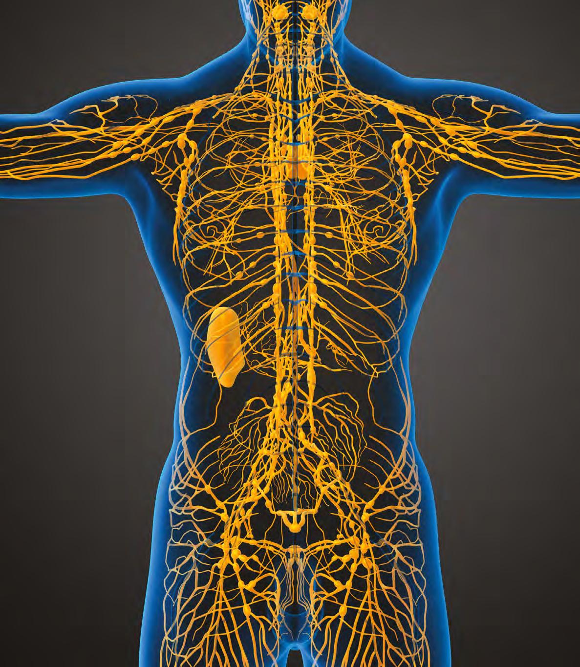


Professional guide to compression garment selection for the trunk and upper limb


S3 Foreword
Christine Moffatt
S4 Introduction
Brandy McKeown and Suzie Ehmann
S5 A natomy, pathophysiology and assessment of upper‑body lymphoedema
Sandi Davis, Suzie Ehmann, Brandy McKeown, Neil Piller, Joseph Dayan, Hiroo Suami, Justine C Whitaker and Karen J Bock
S19 The science of compression textiles and garments for upper‑body lymphoedema
Suzie Ehmann, Brandy McKeown, Sandi Davis and Karen J Bock
S30 Categorising compression garments and accessories for upper‑body lymphoedema
Brandy McKeown, Suzie Ehmann, Sandi Davis, Karen J Bock and Naomi Dolgoy
Editorial lead: Benjamin Snakefield
Associate publisher: Tracy Cowan
Head of projects: Camila Fronzo
Designer: VeeSun Ho
Managing director: Rob Yates rob.yates@markallengroup.com
CEO: Ben Allen
Published by MA Healthcare Ltd
St Jude’s Church, Dulwich Road, London, SE24 0PB, UK +44 (0)20 7738 6726 w ww.markallengroup.com
Produced by Mark Allen Medical Communications w ww.mamedcomms.com
S36 Using the STRIDE algorithm for compression selection in upper‑body lymphoedema
Suzie Ehmann, Brandy McKeown, Sandi Davis, Karen J Bock, Justine Whitaker and Naomi Dolgoy
S49 Case studies of the STRIDE algorithm for compression selection in upper‑body lymphoedema
Suzie Ehmann, Brandy McKeown, Sue Lawrence, Stephanie Moore, Mariam Aldashti and Sandi Davis
S55 Delphi study on the STRIDE algorithm for compression selection in upper‑body lymphoedema
Karen J Bock, Suzie Ehmann, Naomi Dolgoy, Sandi Davis, Brandy McKeown, Justine Whitaker and Elizabeth Anderson
© MA Healthcare Ltd 2025
All rights reserved. No reproduction, transmission or copying of this publication is allowed without written permission. No part of this publication may be reproduced, stored in a retrieval system, or transmitted in any form or by any means, mechanical, electronic, photocopying, recording, or otherwise, without the prior written permission of MA Healthcare or in accordance with the relevant copyright legislation.
Sponsored by Haddenham Healthcare and the US Medical Compression Alliance (USMCA) This sponsorship supported production costs only. Haddenham and USMCA had no involvement in the development, review or selection of content, and none of the authors received financial compensation for their contributions.
STRIDE ™ is a trademark of the International Lymphedema & Wound Training Institute
Christine Moffatt
It is increasingly recognised that lymphoedema has been neglected as an important global disease. Compression therapy is a cornerstone of lymphoedema management, but the science of compression has lacked rigour and depth. The second iteration of the STRIDE algorithm presented in this document is an important step in addressing the lack of international attention and research on compression in lymphoedema, especially in the upper body.
Compression therapy is a transformative treatment that should be available to everyone with lymphoedema, not only those with financial means. However, many people living with lymphoedema have limited options for compression. The International Lymphoedema Framework (ILF) has found that compression products are often unavailable through public health systems, not only in low‑income countries, such as Uganda and India, but also in some high‑income countries in Europe and Asia. In the US, it took decades of lobbying for the 2024 Lymphedema Treatment Act to ensure Medicare coverage for lymphoedema compression. Therefore, healthcare professionals and industry must strive together to increase the range and affordability of available compression products. Epidemiological, clinical and genetic studies continue to illuminate the complexity of lymphoedema. However, ILF research has found that compression therapy is awarded little scientific capital or value when compared with other treatments, such as drug therapy. This poor understanding of a complex area doubtlessly reflects the low global profile of lymphoedema.
Christine Moffatt CBE, Professor of Clinical Nursing in Skin Integrity, Nottingham University Hospital, UK

There remains a pressing need for continued research to contribute to the growing body of evidence in these areas, including the effects of compression on micro‑ and macro‑circulation. Designing and conducting trials on compression in lymphoedema will require interdisciplinary collaboration, commitment and innovation to overcome barriers of cost and complexity due to a heterogenous patient population. Striving for this collective aspiration will ensure that the future of compression therapy moves beyond a reliance on expert consensus to a substantial body of robust scientific evidence.
The second iteration of the STRIDE algorithm not only provides clinicians with a structured framework for informed decision‑making and a comprehensive guide to the evolving field of lymphoedema treatment. It should also be a rich resource for device manufacturers and researchers to advance compression science, innovate with new products and shape the next generation of compression therapy.
Declaration of interest: Sponsored by Haddenham Healthcare and the US Medical Compression Alliance (USMCA).
Brandy McKeown and Suzie Ehmann
This document presents a second iteration of the STRIDE algorithm for selection of compression therapy in lymphoedema. Building on the original 2019 algorithm, which was focussed on the lower limb, this second iteration expands the remit of STRIDE to cover the upper limbs, breast and trunk. This Journal of Wound Care special issue brings together a wealth of knowledge on upper body lymphoedema, compression therapy and the STRIDE algorithm, presented in six articles on the following areas:
1 Anatomy, pathophysiology and assessment of upper body lymphoedema1
2 The science of compression textiles and garments for upper‑body lymphoedema2
3 Categorising compression devices, garments and accessories for upper body lymphoedema3
4 Using the STRIDE algorithm for compression selection in upper body lymphoedema4
5 Case studies of the STRIDE algorithm for compression selection in upper body lymphoedema5
6 Delphi study on the STRIDE algorithm for compression selection in upper body lymphoedema.6
This document represents the consensus of clinical experts from multiple countries, unified in their commitment to advancing compression therapy through evidence‑based practice and international collaboration. Statements have been supported with references to published evidence where possible, with unreferenced claims being founded on the expert consensus of the authors.
The STRIDE algorithm prioritises individualised patient‑centred care, tailored to the multiple needs of specific patients. As such, STRIDE accounts for the complexity seen in clinical practice and aims to avoid simplistic one‑size‑fits‑all methods. At the core of this complexity‑informed approach, STRIDE moves compression selection beyond a traditional focus on pressure dosage alone. Instead, it incorporates other textile properties, such as stiffness, containment, graduation and fatigue. It also takes account of how compression aligns with anatomical variations in oedema distribution, tissue texture and body shape specific to a patient’s clinical presentation. This nuanced approach addresses the intricate anatomy of the upper limb, breast and trunk, where diverse shapes and tissue textures present complex clinical challenges that require specialised solutions.
The STRIDE algorithm also emphasises the dynamic nature of compression and the need for treatment to evolve alongside the patient’s needs. Lymphoedema is not static, and compression requirements shift based on changes in tissue texture, swelling
Brandy McKeown, OTR/L, CLT L ANA, CLWT, Lymphedema Therapist, International Lymphedema and Wound Training Institute and the Lymphedema Center, USA

Suzie Ehmann, DPT, PhD, CWS, CLT L ANA, CLWT, Lymphedema Therapist, McLeod Health Seacoast, Little River, SC, USA

patterns and other aspects of clinical presentation. This is particularly critical for the upper limbs and trunk, where anatomical diversity demands adaptable solutions.
The STRIDE algorithm is intended to provide clinicians with a precise, structured and evidence‑based framework for clinical decision‑making. Selecting the most appropriate and effective compression options should optimise clinical outcomes in upper‑body lymphoedema, helping control swelling and other symptoms and improve patients’ quality of life.
Declaration of interest: Sponsored by Haddenham Healthcare and the US Medical Compression Alliance (USMCA); Brandy McKeown has been a paid consultant or speaker for HMP Global/Wound Source, L&R, LymphaPress, Juzo and Sigvaris; Suzie Ehmann has been a paid consultant or speaker for L&R, Urgo, OVIK, Compression Dynamics and Medline
References
1. Davis S, Ehmann S, McKeown B et al. Anatomy, pathophysiology and assessment of upper‑body lymphoedema. Br J Nurs. 2025; 34(S10B):S5–S18
2. Ehmann S, McKeown B, Davis S, Bock KJ. The science of compression textiles and garments for upper b ody lymphoedema. J Wound Care. 2025; 34(S11C):S19–S29
3. McKeown B, Ehmann S, Davis S, Bock KJ, Dolgoy N. Categorising compression devices, garments and accessories for upper b ody lymphoedema. J Wound Care. 2025; 34(S11C):S30–S35
4. Ehmann S, McKeown B, Davis S et al. Using the STRIDE algorithm for compression selection in upper b ody lymphoedema. J Wound Care. 2025; 34(S11C):S36–S48
5. Ehmann S, McKeown B, Lawrence S et al. Case studies of the STRIDE algorithm for compression selection in upper b ody lymphoedema. J Wound Care. 2025; 34(S11C):S49–S54
6. Bock KJ, Ehmann S, Dolgoy N et al. Delphi study on the STRIDE algorithm for compression selection in upper b ody lymphoedema. J Wound Care. 2025; 34(S11C):S55–S59
Sandi Davis, Suzie Ehmann, Brandy McKeown, Neil Piller, Joseph Dayan, Hiroo Suami, Justine C Whitaker and Karen J Bock
Abstract
This article reviews the lymphatic system’s anatomy and physiology, as well as the etiology of lymphoedema affecting the upper limbs, breast and trunk. It presents evidence-based strategies for assessment, including history-taking, physical exams and clinical tests to guide treatment planning. The importance of selecting personalised compression garments is emphasised. Legislative impacts—such as the US 2024 Lymphedema Treatment Act—and global variability in compression therapy funding are explored, along with nuanced approaches to assessment, staging and diagnostic criteria.
Keywords: Lymphoedema | Lymphatic anatomy | Compression therapy | STRIDE framework | ICG lymphography | Lymphatic drainage | Pathophysiology | Breast and trunk lymphoedema | Comorbidities | Assessment and diagnosis
Lymphoedema is a chronic condition resulting from the accumulation of lymphatic fluid. A nuanced understanding of lymphatic anatomy, physiology and pathophysiology is essential to advancing the care of people with primary and secondary lymphoedema, especially the targeting and sequencing of treatment. This makes the etiology and assessment of lymphoedema fundamental to the STRIDE algorithm for compression selection.
The lymphatic system is a delicate and ubiquitous network of vessels that efficiently transports lymph and its contents without a central pump, relying instead on intrinsic and extrinsic pumping mechanisms that revolve around the structural and functional unit called the lymphangion.1
The process of collecting extracellular fluid and its contents starts at the level of the initial lymphatic capillaries located in the skin (Figure 1). These initial lymphatics can accept large amounts of fluids, metabolic wastes, bacteria and a range of larger molecules, mainly proteins, that blood capillaries cannot.1,2 Lymphatic vessels, referred to as pre‑collectors, connect these initial lymph capillaries to the afferent lymphatic collectors, acting as a major highway in which lymph and its contents travel from the epi‑fascial layers of the tissues to the lymph nodes.2 Even when lymph crosses the deep fascia and drains into the deeper subfascial lymphatic system, it ends up in the lymph nodes, which are crucial for filtering and processing. The lymph nodes filter roughly 8 litres of lymph each day. Half of that lymph is drained through the thoracic duct, whereas the remaining lymph is absorbed by lymphovenous shunts, known as lymph node microvessels.3 The efferent lymphatic vessels transport the residual lymph, which has undergone filtration and purification within the lymph nodes, onward to the larger lymphatic ducts, including the thoracic duct and right lymphatic duct.4 These major ducts provide an essential connection to the
venous system by emptying the lymph at or near the junction where the jugular vein meets the subclavian vein, commonly referred to as the jugular–subclavian junction.2 At this juncture, the purified lymph is reintegrated into the circulatory system, becoming part of the bloodstream and contributing to overall fluid balance in the body (Figure 2).4
The physiology of filtration and purification involves the lymphatic tissue in lymph nodes filtering and recycling lymph fluid while supporting immune defence. When necessary, the cells within the lymph nodes will attack, destroy and remove waste.
Much understanding of detailed lymphatic anatomy is historically based on the work of Mascagni and Sappey, later expanded by Leduc, using cadaver specimens, as described by Shinaoka et al..5 The collective work of these pioneering lymphologists has afforded an expansive view of drainage pathways, describing anatomical division of the lymphatic
Sandi Davis, PT, DPT, CLT L ANA, CLWT, President, Davis Care Physical Therapy, New York City, NY, USA
Suzie Ehmann, DPT, PhD, CWS, CLT L ANA, CLWT, Lymphedema Therapist, McLeod Health Seacoast, Little River, SC, USA
Brandy McKeown, OTR/L, CLT L ANA, CLWT, Lymphedema Therapist, International Lymphedema and Wound Training Institute and the Lymphedema Center, USA
Neil Piller, MD, Lymphologist and Director, Flinders University, Adelaide, Australia
Joseph Dayan, MD, MBA, Plastic and Reconstructive Surgeon, Institute for Advanced Reconstruction, USA
Hiroo Suami, MD, PhD, Associate Professor, Macquarie University, Sydney, Australia
Justine C Whitaker, MSc, RN, PhD(c), Director & Nurse Consultant, Northern Lymphology, and Senior Lecturer, University of Central Lancashire, Preston, UK
Karen J Bock , PT, PhD, CWS, CLT L ANA, Assistant Professor, Rockhurst University, Kansas City, MO, USA
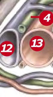
The lymphatics are illustrated in green. The initial lymphatic vessels (1) drain through the fascial layer via the precollectors (2), which then lead to the lymph collectors (3) that run parallel to the deep veins (12) and deep arteries (13) and flow to the lymph nodes. Reprinted with permission from Jobst.

system into superficial lymphatic territories, serviced and drained by specific lymph collectors and separated by watersheds.6
Suami introduced the concept of ‘lymphosomes’, where specific areas of the limb are mapped out and drain into specific lymph nodes (Figure 3).7 More recently, with the introduction of
indocyanine green (ICG) lymphangiography, it is possible to observe the active function of the superficial lymphatic system, as opposed to static cadaver dissection.8 This cutting‑edge technology has unlocked a dynamic view of the upper limbs, trunk and breast, deepening understanding of lymphatic flow and drainage pathways.9
Figure
The right thoracic trunk collects lymph from the right upper limb, head and neck, draining into the right lymphatic duct. The left thoracic trunk, as part of the thoracic duct, drains lymph from the left upper limb, the left side of the trunk and both lower limbs.
Left internal jugular vein
Drained by right lymphatic duct
Thoracic duct
Cisterna chyli
Thoracic duct drains into subclavian vein
Drained by thoracic duct
Granoff et al. observed three main lymph collectors in the normal upper limb: the median pathway along the volar forearm, the radial lymphatics and the ulnar lymphatics (Figure 4).10 The researchers concluded that, although individual lymphatic channels appear as single pathways on ICG, they represent the convergence of numerous tributaries within a lymphosome.10 Upper‑arm connections vary; the median channel consistently connects to the medial upper arm, draining to the lateral axillary lymph nodes, whereas radial and ulnar connections depend on their forearm course, with dorsal pathways often linking to the lateral upper arm, draining to subclavicular lymph nodes.10 On occasion, drainage occurs along a lymphatic collector traversing the deltopectoral groove, into the supraclavicular lymph nodes, avoiding the axillary lymph nodes entirely, particularly when these nodes are removed due to cancer treatments.7
As with other parts of the superficial lymphatic system, drainage from the trunk plays a crucial role in maintaining fluid balance and immune surveillance.2 This region’s drainage system has many variants, but some generalities can be applied.2,8
The drainage patterns of the trunk are as follows:11,12
▪ Anterior thoracic to external mammary (axillary) nodes
▪ Posterior thoracic to scapular (subscapular) nodes
▪ Lateral thoracic to axillary lymph nodes.
The drainage patterns of the breast quadrants are as follows:11,12
▪ Lateral to axillary nodes
▪ Medial to internal mammary (parasternal) nodes
▪ Upper quadrants to axillary and internal mammary lymph nodes
▪ Lower quadrants to axillary and parasternal nodes.
From these nodes, efferent lymph vessels converge into larger lymphatic trunks, ultimately reaching the right or left jugular/ subclavian veins.
The lymphatic drainage patterns of the breast and trunk are structurally distinct from and more complex than those of the limbs. The breast and trunk pathways have a more intricate and interconnected architecture than previously appreciated, with each quadrant potentially draining to a distinct sentinel node.13,14
This structural complexity is compounded by regional variability in lymphatic drainage, as well as the fragile histological structure of lymphatic capillaries, characterised by thin walls
The lymphatic territories are demarcated according to their corresponding lymphatic basins.
Lateral inguinal Popliteal
and microporous structure. Therefore, lymphoedema of the breast and trunk demands tailored therapeutic strategies to ensure effective lymphatic flow while minimising unintended fluid congestion.13‑16
Breast drainage patterns have implications for surgical intervention and compression strategy. Superficial drainage of the breast flows laterally to the sentinel node in the lateral axilla.11 This node serves as a primary drainage point for both the anterior trunk and the upper arm.11
Notably, superficial lymphatics lack direct connections to deep lymphatics in the anterior trunk, except through the lateral axilla.11 This anatomy is particularly relevant when considering sentinel‑node dissection. While the procedure is often viewed as lymphatic‑sparing due to the removal of a single node, disruption of the sentinel node, which manages drainage for both the breast and upper limb, can significantly impair lymphatic flow.11 Recent findings underscore the complexity of breast lymphatic drainage. Giammarile et al. described it as multidirectional yet predominately routed to the ipsilateral axilla.17 With a fraction
of lymph (≈3%) draining to the intercostal, interpectoral, peri‑clavicular, perimammary, contralateral breast or even abdominal nodes, there is a need to anticipate collateral flow disruptions.17
In a 2020 study by Aldrich et al., ICG lymphography was performed on 20 participants, 10 with breast lymphoedema, with the following findings:12
▪ All healthy controls exhibited linear ICG flow toward the ipsilateral axilla with no dermal backflow
▪ Among those with breast lymphoedema, only 40% maintained primary axillary drainage
▪ Importantly, 90% demonstrated compensatory pathways, including parasternal (6/10), contralateral axilla (4/10), intercostal (3/10) and clavicular (2/10) drainage routes. These findings underscore the importance of early clinical assessment and individualised compression planning following breast surgery, particularly for patients at risk of breast or upper‑limb lymphoedema. Pre‑ and post‑operative evaluations allow for patient education and timely intervention. Understanding collateral lymphatic pathways can guide garment design and help avoid over‑compression of newly recruited drainage routes.
Side-by-side images of cadaver A (left) and cadaver B (right) illustrate the lymphatic territories of each sentinel node, colour-coded to match. In cadaver A, lymphatics were traced distally from each node, revealing distinct territories without overlap, interconnected pathways within each region and an orange territory capable of draining into either LN1 or LN3. In cadaver B, lymphatic drainage patterns were traced to align with radiographic findings. Sentinel nodes (LN1 and LN2) exhibit discrete territories, with LN1 forming a dominant pathway that connects proximally to LN3 and LN4. Veins were injected to highlight vascular structures within the mapped regions.
Breast and trunk lymphoedema pose assessment challenges due to anatomical variations and inconsistencies in lymphatic measuring and mapping techniques. Unlike limb lymphatics, breast drainage lacks direct connections to deeper lymphatic structures, making compression crucial for redirecting flow and mitigating fibrotic progression, especially after sentinel‑node removal. Preventative compression is equally, if not more, important in these regions.11,18
Lymph transport relies on two synergistic mechanisms: intrinsic and extrinsic pumping. Intrinsic pumping occurs in lymphatic collectors, which are segmented by one‑way valves that prevent backflow and ensure unidirectional flow.19,20 Contractions are triggered by myogenic responses when vessel‑wall tension exceeds a threshold, and they are modulated by nitric oxide (NO), which reduces tone and enhances flow. Lymphatic pressure remains lower than venous pressure, unless active contraction is occurring.20,21
Extrinsic pumping supports intrinsic lymphatic transport through skeletal muscle movement, respiration and passive forces, such as arm elevation, trunk rotation, walking or manual lymphatic drainage.20,21 These external compressions mobilise lymph within the non‑contractile initial lymphatics and help overcome downstream resistance.21 Lymphangions typically contract 6–10 times per minute at rest, with rates increasing up to tenfold during activity, positional changes or compression garment use.21 Compression applications can enhance these external forces, optimising lymphatic flow and promoting oedema reduction.22 However, if not appropriately selected, applied or worn, compression may obstruct flow or impair collateral drainage.22 These considerations highlight the need for anatomically tailored compression strategies.
Together, the intrinsic and extrinsic pumping mechanisms maintain efficient lymphatic transport; ensure lymph moves unidirectionally and against gravity; and support essential physiological functions, such as fluid balance and immune response. This dynamic interplay illuminates the adaptability and robustness of the lymphatic system.
Etiology
Lymphoedema arises from two primary aetiologies: congenital (primary) and acquired (secondary). Primary lymphoedema involves structural abnormalities, such as aplasia, hyperplasia, hypoplasia or valvular defects, often linked to genetic mutations such as FOXC2 or SOX18.23 Secondary lymphoedema results from external causes, including trauma, filariasis or cancer treatments (i.e., lymph node dissection and radiation), that damage lymphatic vessels.24
Clinical understanding of lymphoedema in the breast and upper limbs is largely shaped by cases involving axillary lymph‑node dissection and/or radiation, most commonly in the context of breast‑cancer treatment.25‑28 While lymphoedema is often attributed to disruption of afferent and efferent lymphatic pathways near excised nodes, immune activation also plays a critical role.²³ After tissue injury, axillary lymph nodes may initiate an amplified inflammatory response instead of a controlled immune reaction, leading to fluid buildup and tissue remodelling.
Nores et al. found that CD4+ T cells exacerbate lymphatic dysfunction post‑injury by increasing fibrosis and peri‑lymphatic inflammation, while inhibiting collateral lymphatic formation.25 This inflammatory shift leads to extracellular fat deposition, fibrosis, chronic inflammation, valvular insufficiency and lymphatic pump failure.25 Ultimately, functional pathways diminish, and lymphatic load surpasses system capacity.26
Lymphoedema, whether primary or secondary, varies based on location of functional lymphatics, comorbidities and tissue changes such as post‑radiation fibrosis. These fibrotic changes may hinder lymphatic regeneration and redirection of flow, complicating bypass of damaged regions. Beyond lymph‑node removal, lymphoedema frequently follows radiation and taxane‑based chemotherapy, both common in breast cancer treatment.29‑31 While radiation causes tissue and lymphatic scarring, the mechanism by which taxane‑based therapy impairs lymphatic function remains unclear.
Increased breast cancer‑related lymphoedema (BCRL) incidence has been reported in patients with taxane‑induced peripheral neuropathy, suggesting a potential link.31 ICG lymphangiography has shown abnormal lymphatic patterns and reduced contractility in patients after taxane therapy, even before axillary dissection or radiation.30 These findings denote that lymphoedema is not merely a plumbing issue but also involves significant immunologic factors.
Though BCRL dominates research, lymphatic dysfunction can also stem from treatments for melanoma, lymphoma and osteosarcoma. Non‑oncologic causes include orthopaedic injuries and surgical procedures, soft‑tissue trauma, burns, neurological‑dependent oedema (e.g., spinal cord injury, cerebrovascular incidents), vascular issues (e.g., dysfunctional dialysis ports, deep vein thrombosis), post implantation of cardiac devices, infections, vasculitis and intravenous drug use.
Ultimately, lymphatic dysfunction of the upper extremities, breast and/or trunk lymphatics signals impaired tissue health. Visible oedema often reflects lymphatic‑system insufficiency, calling for timely and personalised interventions. Coexisting conditions may further complicate management, necessitating a comprehensive, multidisciplinary approach.
Lymphatic function is shaped by innate lymphatic sufficiency and compounded by coexisting comorbidities that increase lymphatic load. Factors such as a genetic predisposition, obesity, cancer therapies, infections and cardiovascular or metabolic conditions can accelerate lymphoedema onset and progression. A range of coexisting conditions can compromise lymphatic function and tissue homeostasis, necessitating a comprehensive clinical approach. These include metabolic and structural factors that disrupt fluid balance, immune response and mechanical lymph transport.
Obesity is a significant contributor to lymphoedema, with obesity‑induced lymphoedema (OIL) common at body mass index (BMI) >40 and nearly universal at BMI >60.32‑34 In OIL, peri‑lymphatic inflammation disrupts lymphatic endothelial cell
(LEC) gene expression, impairs pump efficiency and alters lipid metabolism—creating a feedback loop of fat deposition and lymphatic dysfunction.33,35 Cytokine‑driven inflammation and poor vessel remodelling worsen drainage.33,36
In patients with cancer, pre‑operative BMI ≥30 triples the risk of lymphoedema compared with BMI ≤25.36 Weight fluctuations post‑treatment doubles this risk.37 Obesity also complicates surgery and intensifies recovery, reinforcing the need for early monitoring, weight‑loss strategies and anti‑inflammatory interventions.33,36,37
Lipoedema is a condition that involves symmetrical fat buildup in the arms and legs but sparing the hands and feet. It predominantly affects women, with an unclear etiology.38 It worsens primarily with obesity rather than inherent progression.39 Diagnosis requires a second cardinal symptom, often pain.38,39 Proper diagnosis differentiates lipoedema from lipohypertrophy, idiopathic oedema or cellulite, each requiring distinct treatments.38,39
In stages 3 and 4, lymphatic impairment emerges, particularly in patients with high BMI.38 Compression garments, lymphatic drainage and weight management are critical.38
Reduced motion, whether due to trauma, surgery, stroke or neurological impairment, limits intrinsic and extrinsic lymph transport.40,41 Oedema creates a cycle of pain and inactivity, worsening fluid retention.40,42,43 Poor positioning, compression misuse and cardiovascular dysfunction in patients who are critically ill can further strain drainage.40,42,44
Management includes elevation, light exercise and customised garments.40‑43 For stroke‑related oedema, passive range of movement (ROM), compression and active contraction help preserve lymphatic and venous return.41 Breast oedema benefits from compression bras and early mobilisation.45
Nutrition
Sodium promotes water retention and increases arterial pressure, impairing lymphatic flow.46,47 Processed foods can amplify systemic inflammation.46 Potassium, magnesium and vitamin B6 counter these effects by reducing retention, enhancing circulation and regulating inflammatory mediators.46 Weight loss is vital, although lipoedema adiposity often resists conventional methods.47 Hypocaloric diets paired with supplements (e.g., green tea catechins, caffeine, whey protein) and exercise may improve fat metabolism and lymphatic function.46 Tailored plans, such as Mediterranean or ketogenic diets, can complement compression and physiotherapy, although more research is needed.46
Metastatic disease
Cancer often spreads via lymphatics, disrupting node and vessel integrity.48 Metastatic deposits create obstructions, while tumour‑induced lymphangiogenesis further alters drainage.48 Vascular endothelial growth factor (VEGF)‑C/D signalling supports vessel proliferation but facilitates cancer dissemination.48,49
Lymphoedema incidence varies, with 6–70% in patients with breast cancer50,51 and 4–15% in upper‑limb melanoma.52 High‑burden axillary metastasis—seen in over one‑third of patients post‑neoadjuvant therapy—not only signals treatment resistance but also compounds the risk of lymphoedema by increasing nodal disruption and surgical morbidity.53
Diabetes exacerbates lymphoedema through chronic inflammation, vascular impairment and immune dysfunction. Persistent hyperglycaemia and insulin resistance damage LECs, triggering oxidative stress, disrupting lymphangiogenesis and increasing vessel permeability.54 These effects impair lymph drainage and elevate infection risk.54,55
Individuals with type 2 diabetes are at increased risk for early‑onset lymphoedema due to leaky lymphatics.55 Patients with diabetes and breast cancer undergoing mastectomy or extensive dissection often experience delayed wound healing from tissue hypoxia and poor vascular supply, both linked to higher BCRL rates.56 Chronic inflammation from both conditions amplifies lymphatic damage and worsens disease progression.55,56
In response to infection, lymph nodes recruit macrophages and neutrophils to capture bacteria and trigger an immune responses.57 Fewer nodes or impaired transport reduce this response, increasing susceptibility to infections.26,58 Bacterial invasion can scar lymphatic tissue and aggravate lymphoedema.57 Symptoms include warmth, redness, swelling, pain, fever and chills.59
Timely antibiotic treatment is vital to control spread and prevent complications.59 Patients with advanced arm lymphoedema are at heightened risk for cellulitis, making prevention of lymphoedema progression crucial.58,60
Radiation impairs lymphatics by depleting lymphocytes and inducing fibrosis in lymph nodes.61 This elevates intranodal pressure and disrupts filtration, fostering lymphoedema development.61 Axillary lymph‑node dissection increases the risk of BCRL by over threefold, and, when combined with regional lymph node radiation, the risk approaches fourfold compared with sentinel‑node biopsy alone.62 Radiation alone contributes modestly to this risk.62 Tangential photon radiation poses a higher lymphoedema risk than electron beam therapy, with additional risks tied to total dose, overlapping fields and posterior axillary boost.61
Lymphoedema is a clinical diagnosis, determined through systematic evaluation that differentiates it from other causes of oedema. A thorough understanding of the underlying etiology enables clinicians to conduct an in‑depth examination, combining clinical tests, physical assessments and detailed patient history to confirm lymphoedema presence and stage. Patient‑reported symptoms are the first diagnostic clue in lymphoedema evaluation. Common complaints include arm, breast or trunk size changes, as well as heaviness, numbness,
redness and aching pain.63 Family history can support diagnosis, especially in suspected primary lymphoedema. If hereditary patterns are evident, genetic testing and specialty referral may be warranted.
A comprehensive clinical evaluation of a patient with lymphoedema should follow the Subjective, Objective, Assessment and Plan (SOAP) format, ensuring a systematic approach to diagnosis and management. The history review serves as the foundation, including medical history, surgical history and clinical tests previously performed to assess lymphatic function, vascular status and comorbid conditions. Being part of a collaborative network of healthcare professionals can be helpful in pairing information obtained from these tests and measures to give a baseline as to why the lymph system may be failing, as well as to guide clinical assessment and treatment planning.
Lymphoedema impacts multiple systems, and so, to guide diagnosis and care, assessment must go beyond limb‑volume measurements to encompass ROM, sensation, BMI, vascular health and genetics. Accurate diagnosis involves more than confirming lymphatic insufficiency, it requires assessment of concurrent pathology, such as increased capillary filtration. Often, ultrafiltration and lymphatic overload coexist, perpetuating chronic oedema.
Assessment of a patient’s lifestyle, activity level, environmental context and ability to perform (potentially instrumental) activities of daily living are important for effective compression therapy. Likewise, body habitus and tissue response factors, including refill time, positional variation and oedema fluctuation, guide compression choices suited to the size, shape and severity of swelling. Compression therapy must also align with lymphatic, vascular, neural and metabolic status for safety and effectiveness. Likewise, comorbidities such as Raynaud’s disease may require lower‑pressure garments.
Several imaging modalities are used to evaluate lymphatic function, each method assessing distinct aspects of lymphatic health, including flow dynamics, vessel integrity and inflammation markers (Table 1).27,64‑67
Tissue‑assessment tools can help confirm lymphatic insufficiency and characterise oedema. Further research is needed to validate these tools for truncal and breast lymphoedema, aiming to enhance diagnostic precision and management. Together, these findings inform compression strategies and diagnostic confidence when evaluating tissue integrity and fluid retention.
Originally described in 1976 for lower limbs, Stemmer’s sign is positive when skin over a toe cannot be pinched to determine skin lift, indicating fibrotic change.68 It has also been adapted for the upper limb by assessing skin over the hand’s metatarsophalangeal joint.69 With 92% sensitivity compared to lymphoscintigraphy, this modified method is reliable for limb assessment but not applicable for truncal swelling, which lacks digit‑based evaluation points.
The Bjork bowtie test can be performed anywhere on the body and is useful for assessing truncal or breast oedema. This tactile test involves pinching and rolling skin between the thumb and index finger, noting quality of tissue texture and thickness. Healthy tissue lifts easily, feels smooth and forms wrinkles resembling a bowtie.70,71 A positive result reflects compressed, unyielding skin and absent wrinkling, suggesting fibrosis from chronic lymphatic inflammation (Figure 5).70
The pitting scale is used to gauge tissue fluid dynamics. It is assessed via thumb sustained pressure applied with the thumb pad, noting how long the indentation takes to rebound. There are four severity grades, ranging from grade 1+ to grade 4+.72,73 Rebound time helps determine lymphatic function and appropriate compression levels (Table 2).70,74 Breast assessments may rely on visual cues (e.g., bra imprints) due to limited skin pinchability. Early‑stage lymphoedema shows pronounced pitting, while advanced cases with fibrosis may require extended pressure of 10–60 seconds to elicit indentation.73 Refill times exceeding 30 seconds indicate lymphatic compromise.70
Evaluating tissue texture requires a hands‑on approach, involving gently palpating the limb, breast or trunk to assess skin feel and pliability, alongside documentation of visual appearance. The subjective variability of this assessment can be minimised by using consistent terminology for tissue texture, such as that described in the STRIDE framework (FIgure 6).70 A shared glossary supports standardised documentation and clearer communication within a clinical setting.
Volumetric tools can help quantify limb and trunk oedema, supporting consistent diagnosis, monitoring and treatment evaluation.
Circumferential measurement is a cost‑effective, accessible method to track limb size and shape. Lymphoedema is indicated when the affected limb exceeds 10% of the unaffected side.75 Key metrics include limb volume, excess volume, percentage excess and proximal–distal ratios.76 Breast and trunk measurements are challenging due to tissue variability and lack of a comparable side. Baseline measurement around the trunk and axilla help with monitoring changes.77 Taking baseline circumferential measurements around the trunk and axilla are necessary to track fluid and volume fluctuations and reductions throughout care.


Negative Bjork bowtie test

Positive Bjork bowtie test

Alternate method for the Bjork bowtie test
Table 1. Imaging modalities for assessing lymphatic dysfunction27,64-66
Modality
Ultrasound
High-frequency sound waves to visualise lymphatic structures
Magnetic resonance lymphangiography (MRL)
Heavily T2-weighted sequences or contrastenhanced techniques
Dynamic contrast‑ e nhanced MRL
Contrast injected into lymph nodes, with scans tracking lymphatic flow
Intranodal computed tomography (CT) lymphangiography
Water-soluble contrast injected into lymph nodes and tracked via CT
Contrast e nhanced ultrasound lymphos ‑ cintigraphy
Radioactive tracers injected and tracked via gamma serial images
Single p hoton e mis sion CT with CT (SPECT‑ C T)
More detailed visualisation for the lymphatic system and how the tracer moves
Near infrared fluorescent lymphatic imaging (NIRFLI)
Protein-binding indocyanine dye injected and illuminated with near-infrared light
Function Key findings
Structural assessment of lymphatic vessels and surrounding tissues
Three-dimensional mapping of lymphatic pathways from head to toe
Functional and anatomical assessment
High-resolution imaging of central lymphatics
Functional imaging of lymphatic flow
CT provides anatomic detail to help pinpoint lymph node and structural abnormalities
Real-time visualisation of functional superficial lymphatics
• Impact of venous dysfunction
• Lymphatic obstruction
• Suitability of vessels for lymphovenous anastomosis
• T issue thickening or fibrosis
• V isualisation of altered flow dynamics of lymphatic vessels
• V isualisation of tumours
• A bnormalities
• A dipose hypertrophy
• Lymphatic leaks
• Lymphatic congestion
• Mapping of the thoracic duct
• Lymphatic leaks
• Mapping of the sentinel lymph node
• Lymphoedema diagnosis
• Higher spatial resolution for lymph-node mapping
• Improved differentiation of normal and abnormal lymphatic structures
• Enhanced accuracy in complex anatomical regions, such as axilla
• Lymphatic function
• G uide for surgical interventions
Fluid displacement involves limb immersion in water to measure displaced volume. This method lacks localisation and poses hygiene concerns, limiting routine use.27
Bioelectrical impedance, the movement of electrical currents through the body, can be measured to assess fluid and tissue composition. Bioelectrical impedance analysis measures tissue resistance using a single wavelength to assess extracellular fluid, expressed as a lymphoedema index (L‑Dex). It is useful for early‑stage lymphoedema (stages 0–1).78 Without pre‑operative data, BCRL is indicated by an L‑Dex over 6.5,79 while, when pre‑operative data is available, BCRL is indicated by a 10‑point
AdvantagesDisadvantages
• Non-invasiveness
• W ide accessibility
• E xcellent spatial resolution
• A bsence of radiation exposure
• High resolution
• D ynamic flow data
• Ready availability
• E xcellent spatial resolution
• Minimal invasiveness
• W ide availability
• Improved accuracy compared with lymphos cintig raphy alone
• High resolution
• D ynamic imaging
rise from baseline.75,80
• O perator dependence
• L imited ability to visualise deeper lymphatics (23.5 mm for 48 MHz; 10 mm for 70 MHz)
• High cost
• Need for contrast injection for enhanced imaging
• Need for specialised setup
• Need for contrast injection
• R adiation exposure
• T iming challenges for imaging
• L imited spatial resolution
• Static image
• R adiation exposure
• Pain
• High cost
• L imited availability
• Potential for false positives or misinterpretations leading to misdiagnosis
• Inability to obtain serial images
• L imited penetration depth (<1 cm)
• Need for specialised equipment
• L ack of approval for all applications in US
Bioelectrical impedance spectroscopy, using multiple wavelengths, is a non‑invasive measurement tool for oedema assessment that evaluates limb volume by measuring how easily a low‑level electrical current passes through tissue— higher resistance suggests more extracellular fluid, which indicates oedema.
Grade Pit depth
Rebound time
Grade 12 mm (barely detectable) Immediate
Grade 24 mm
Grade 36 mm
≤20 seconds
≤20 seconds
Grade 48 mm >20 seconds
Figure 6. STRIDE descriptions of tissue texture70
Watery
Soft, pliable feeling, non-fibrotic, easily pitting, quickly rebounding, negative Stemmer sign and Bjork bowtie test
Doughy
Putty-like, somewhat fibrotic, deeply pitting, rebounding after 30 seconds, potentially positive Stemmer sign or Bjork bowtie test
Woody
Hard feeling, severely fibrotic, non-pitting, positive Stemmer sign and Bjork bow-tie test
Fatty
Spongy or squishy feeling, may indicate healthy or abnormal fat (i.e., lipoedema)
Fragile
Thin, delicate, inelastic skin, impaired, prone to breaks, fissures, lipomas, cysts, blebs or blisters (common in older adults)
Optoelectronic plethysmography creates three‑dimensional models of limb volume, visualising oedema patterns and tracking progression on both segmental and total levels.81
The tissue dielectric constant (TDC) is a non‑invasive tool that measures local water content in breast tissue to a depth of 2.5 mm via a 300 MHz probe.82 TDC is cost‑effective for early clinical detection of fluid retention.83 This tool is used to assess site‑specific swelling; it is effective for forearm lymphoedema at a 1:26 ratio,83 and it has shown promise in assessing breast lymphoedema, with a threshold of 1.4, although further validation is needed.83,84
Tonometry assesses the skin’s resistance to applied pressure, serving as an indirect measure of fibrosis and treatment response.18,84 It is quick and portable, with a high interrater reliability,85 and it can be used to guide compression resistance levels.84,86 Normative data is limited, and tissue may soften or re‑harden.18,84,86 A digital tonometry device is known as an indurometer.86
Lymphoedema can impair ROM, alter sensation and disrupt movement patterns, as well as affect vascular and metabolic health, ultimately reducing mobility and quality of life. A comprehensive physical assessment is essential to establish a baseline, guide treatment and monitor progression.
Swelling from lymphoedema can limit joint mobility, causing stiffness, discomfort and functional limitations. Contributing factors include fluid accumulation, fibrosis and reduced tissue elasticity. Goniometry quantifies joint angles and tracks ROM over time. Separate assessments of active and passive pain‑free
ROM can differentiate whether restriction is the result of swelling or a mechanical cause. Early use of stretching, manual therapy and movement‑based interventions can help preserve joint function and prevent complications.
A thorough neurological evaluation, including sensation and deep‑tendon reflexes, is crucial for ruling out nerve involvement and metabolic causes of sensory deficits. Chemotherapy‑induced neuropathy increases the risk of complications in patients wearing compression garments on limbs with reduced sensation.87 Light‑touch and two‑point discrimination testing along dermatomes can reveal sensory deficits. Nerve palsies, in either an oedematous or a non‑oedematous limb, may hinder adherence and the ability to don and doff compression garments. Chemotherapy‑induced neuropathy affects 28% of patients, with 67% reporting post‑chemotherapy numbness and tingling.88 Proper sensory assessment ensures safe and effective compression therapy.
Assessing ROM, flexibility and activity levels helps guide lymphoedema treatment choices, as limits in movement can lead to reduced independence. Steady‑state exercise boosts lymphatic flow over rest by two‑to‑three times.89 Regular exercise reduces lymphoedema flare‑ups, making movement essential.90 Donning and doffing of compression garments requires upper‑limb strength and coordination, highlighting the importance of rehabilitation focussed on early mobility.
Obesity can cause or worsen lymphoedema. BMI and waist–height ratio are reliable indicators of healthy weight.91 In a study of 138 patients, people with a BMI of 30 or more were 3.6 times more likely to develop upper‑limb lymphoedema within 30 months of surgery.92,93 Maintaining a healthy weight helps prevent and manage lymphoedema, reinforcing the importance of dietary and lifestyle modifications.
Upper‑limb vascular assessment may be recommended in cases of lymphoedema affecting the arm, breast or trunk. Radiation therapy may alter arterial perfusion, occasionally causing brachial‑artery narrowing.61,62,94 Increased arterial flow has been reported after breast cancer treatment.22,61 Angiography helps identify abnormal arterial flow. Venous outflow should also be assessed to rule out tumour compression or deep‑vein thrombosis (DVT). DVT monitoring is critical, as vascular issues can worsen swelling and function.95 Duplex ultrasound is key for diagnosis.95 A multidisciplinary vascular team is advised. Vascular status should inform tailored compression therapy. Compression, which supports circulation and lymphatic function, should enhance outflow without compromising arterial inflow.
Emerging research highlights the role of inflammation and genetics in lymphoedema development. Laboratory tests can detect markers including CD8+ T‑cells, macrophages and neutrophils, along with pro‑inflammatory cytokines such as
Table 3. International Society of Lymphology lymphoedema staging system24,70,99
Stage Description
Stage 0 (subclinical)
Stage 1 (reversible)
Stage 2 (spontaneously irreversible)
Stage 3 (elephantiasis)
Known or undiagnosed lymphatic dysfunction with no physical signs of oedema
Oedema that reduces with elevation to a similar size to an unaffected limb (often confused with other causes of oedema)
Pitting oedema with positive Stemmer’s sign and that does not reduce with elevation
Pitting and non-pitting oedema that shows notable skin changes and does not reduce with elevation
TNF‑a, VEGF‑C, and LTB4.96 Genetic mutations linked to primary lymphoedema include FLT4 (Milroy), GJC2 (Meige), FoxC2 (lymphoedema distichiasis) and SOX18 (hypotrichosis lymphoedema telangiectasia).97,98 ICG lymphography reveals stage 0 dysfunction in limbs that appear unaffected, showing disease progression.98 Primary lymphoedema may be systemic, not limited to one limb, emphasising the need for early detection and comprehensive evaluation.98
Lymphoedema should be staged to guide treatment decisions. There are several staging systems for lymphoedema, the most widely accepted being from the International Society of Lymphology (Table 3).24,99 Staging should guide selection of appropriate treatment:24,70,99
The severity of lymphoedema can also be assessed with a volumetric scale determined by the percentage difference in volume between an affected and an unaffected limb (Table 4).24,100 While volumetric scales remain a common reference for lymphoedema severity, they reflect only one dimension of the condition. Current clinical understanding emphasises that tissue texture changes, such as fibrosis, induration and dermal thickening, may precede or occur independently of measurable volume differences. Therefore, there is a growing need for a multidimensional severity scale that incorporates both quantitative volume and qualitative tissue characteristics, including:
▪ Palpable fibrosis
▪ Pitting behaviour
▪ Skin integrity
▪ Responsiveness to compression
▪ Functional impact.
Such a scale would better reflect the complexity of lymphatic dysfunction, especially in oncology‑related presentations, and guide more tailored therapeutic strategies.
Recent discoveries in lymphatic anatomy detailed in this article expand understanding of how the lymphatic system interacts with systemic health. Furthermore, Levick and Michel’s 2010 revision of Starling’s equilibrium reshaped the understanding of fluid dynamics.101 Mortimer and Rockson noted that this
Typical treatment
Preventative therapy is critical at an early stage, especially after radiation or axillary lymph-node dissection
Manual lymphatic drainage, compression garments, exercise and skin care potentially able to halt or revert progression
In advanced cases, multilayer bandaging, customised devices, pump therapy and manual lymphatic drainage can help maintain function
Severe disease may require high-grade compression, surgical options and intensive rehabilitation
Table 4. Volumetric lymphoedema scale24,100
Severity
Volume difference
Mild <20%
Moderate 20–40%
Severe >40%
updated model supports the assertion that chronic oedema exists along a continuum of lymphatic dysfunction.102 Since the lymphatic system is solely responsible for clearing interstitial fluid, persistent swelling warrants a thorough evaluation of each individual clinical presentation.
Despite advancements in lymphatic research, clinical awareness and screening remain limited across medical education and practice. Rockson noted that most medical students receive minimal training on the lymphatic system.103 In fact, a review of 110 US medical schools found an average of only 45 minutes devoted to lymphatic disease.103 This inadequate exposure leads to patients being prescribed pumps or sleeves without referral to lymphoedema specialists—who are essential for targeted management.
A limited understanding of lymphatic anatomy and function often leads to a reactive approach, with intervention beginning only after signs and symptoms emerge. Preoperative lymphatic screening—across oncologic, vascular and orthopaedic procedures—remains uncommon, even though growing evidence suggests that identifying lymphatic vulnerability before insult could reduce post‑surgical complications and support improved recovery.
Specialists ensure precise garment fitting based on refill rates and mobility limitations, optimising therapeutic outcomes. They also educate patients on proper pump use alongside complete decongestive therapy, which includes compression, exercise, manual lymphatic drainage and skin care. Strengthening lymphoedema education and screening protocols is vital for improving long‑term outcomes. These specialists are often trained through accredited institutions, academic programmes or recognised certification pathways, ensuring they possess the clinical expertise needed to assess lymphatic function, tailor interventions and guide patients through complex care decisions.
Insurance coverage and funding disparities remain significant barriers to compression therapy access. In the US, the January 2024 Lymphedema Treatment Act has improved access by mandating Medicare coverage for compression garments. However, care is still restricted by gaps in affordability, coverage limits and prior‑authorisation requirements.
Globally, access challenges persist, particularly in countries with socialised healthcare. For example, the NHS in the UK offers limited funding for compression therapy, often requiring restrictive criteria or extended wait times. Similar constraints are seen in Canada and parts of Europe, where formularies may not include advance compression products.
Thorough documentation of medical necessity, including objective findings, staging and previous interventions, is essential for clinicians to advocate effectively for coverage, reduce delays and improve equitable access to evidence‑based treatment. When coverage is denied or delayed, patients are often unable to obtain medically necessary compression, resulting in unmanaged oedema, increased risk of complications and higher long‑term healthcare costs. These gaps in access not only compromise clinical outcomes but also place significant financial and emotional strain on patients and care systems.104‑106
Disparities in insurance coverage continue to limit access to compression therapy, creating financial hurdles for patients. Although the US Lymphedema Treatment Act has expanded Medicare benefits, many private insurers and international systems still fall short. Broader policy reform is urgently needed to ensure equitable access to care.
The shortage of trained clinicians in compression fitting and lymphatic evaluation compounds the issue—leading to inappropriate garment use and ineffective therapy. Expanding training programmes and certification pathways are essential to equip providers with the skills needed for precision‑based treatment.
Application of the STRIDE framework should be built on an understanding of the complexities of lymphatic anatomy and the pathophysiology of lymphoedema. Therefore, targeted clinician education initiatives will be critical in transforming care delivery. Awareness efforts should also underscore the role of comorbidities in lymphatic dysfunction, as well as the importance of timely referrals and multidisciplinary collaboration. Expanding clinical research into lymphatic anatomy should also drive innovation in compression science.
Thorough and accurate assessment is essential for the personalised, targeted and physiologically aligned compression therapy supported by the STRIDE framework. Lymphoedema diagnosis care continues to advance, with advanced imaging modalities such as ICG lymphography, lymphoscintigraphy and MRI lymphangiography providing critical information regarding lymphatic dysfunction. Emerging technologies such as TDC or BIA analysis offer promise for early detection by quantifying localised oedema and tissue change.83 However, gaps persist in
precision, and utility is often limited by referral delays, insurance constraint and cost barriers. Expanding research and standardising use across clinical settings will be crucial in making lymphatic evaluation more inclusive and actionable. Early‑stage diagnostic tools, refined assessment models and enhancing diagnostic accessibility should improve outcomes and promote equity across healthcare systems, advancing lymphoedema management toward truly individualised, evidence‑informed interventions.
Declaration of interest: Sponsored by Haddenham Healthcare and the US Medical Compression Alliance (USMCA); Sandi Davis has been a paid consultant for Koya Medical; Suzie Ehmann has been a paid consultant or speaker for L&R, Urgo, OVIK, Compression Dynamics and Medline; Brandy McKeown has been a paid consultant or speaker for HMP global/Wound Source, L&R, LymphaPress, Juzo and Sigvaris; Justine C Whitaker has been a paid consultant or speaker for Medi and Jobst; Karen J Bock has been a paid consultant for Pure Medical
1. Brenner E. [Anatomy and physiology of the lymph system: what clinicians should know]. Radiologie (Heidelb). 2025; 65(5):301–306. https://doi.org/10.1007/s00117 025 01432 2
2 Földi M, Földi E, Strößenreuther C, Kubik S. Földi’s textbook of lymphology: for physicians and lymphedema therapists. Amsterdam: Elsevier Health Sciences: 2012
3. Lampejo AO, Jo M, Murfee WL, Breslin JW. The microvascular l ymphatic interface and tissue homeostasis: critical questions that challenge current understanding. J Vasc Res. 2022; 59(6):327–342
4. Breslin JW, Yang Y, Scallan JP, Sweat RS, Adderley SP, Murfee WL. Lymphatic vessel network structure and physiology. Compr Physiol. 2018; 9(1):207–299. https://doi.org/10.1002/cphy.c180015
5. Shinaoka A, Koshimune S, Suami H et al. Lower limb lymphatic drainage pathways and lymph nodes: a CT lymphangiography cadaver study. Radiology. 2020; 294(1):223–229
6. Suami H, Taylor GI, Pan WR. The lymphatic territories of the upper limb: anatomical study and clinical implications. Plast Reconstr Surg. 2007; 119(6):1813–1822. https://doi.org/10.1097/01. prs.0000246516.64780.61
7. Suami H, Scaglioni MF. Anatomy of the lymphatic system and the lymphosome concept with reference to lymphedema. Semin Plast Surg. 2018; 32(1):5–11. https://doi.org/10.1055/s 0 038–1635118
8. Suami H, Kato S. Anatomy of the lymphatic system and its structural disorders in lymphoedema. In: Lee B B , Rockson SG, Bergan J (eds). Lymphedema: a concise compendium of theory and practice. New York (NY): Springer; 2018: 57–78
9. Suami H, Pan W R , Mann GB, Taylor GI. The lymphatic anatomy of the breast and its implications for sentinel lymph node biopsy: a human cadaver study. Ann Surg Oncol. 2008; 15(3):863–871. https:// doi.org/10.1245/s10434 0 07 9709 9
10. Granoff MD, Pardo JA, Johnson AR et al. Superficial and functional lymphatic anatomy of the upper extremity. Plast Reconstr Surg. 2022; 150(4):900–907. https://doi.org/10.1097/ prs.0000000000009555
11. Suami H, O’Neill JK, Pan WR, Taylor GI. Superficial lymphatic system of the upper torso: preliminary radiographic results in human cadavers. Plast Reconstr Surg. 2008; 121(4):1231–1239. https://doi.org/10.1097/01.prs.0000302511.21140.36
12. Aldrich MB, Rasmussen JC, Fife CE, Shaitelman SF, Sevick Muraca EM. The development and treatment of lymphatic dysfunction in cancer patients and survivors. Cancers (Basel). 2020; 12(8). https:// doi.org/10.3390/cancers12082280
13. Suami H, Pan WR, Mann GB, Taylor GI. The lymphatic anatomy of the breast and its implications for sentinel lymph node biopsy: a human cadaver study. Ann Surg Oncol. 2008; 15(3):863–871. https:// doi.org/10.1245/s10434 0 07 9709 9
14 Carati C, Gannon B, Piller N. Anatomy and physiology in relation to compression of the upper limb and thorax. J Lymphoedema. 2010; 5:58–67
15. Hutton R. Developing new practices for managing breast and chest lymphoedema. Br J Community Nurs. 2024; 29(S10):S20–S24. https://doi.org/10.12968/bjcn.2024.0109
16. Mehta M, Sarrami S, Moroni E, Fishman J, De La Cruz C. Alternative lymphatic drainage pathways in the trunk following oncologic therapy. Ann Plast Surg. 2024; 92(4S2):S258–S261. https:// doi.org/10.1097/sap.0000000000003861
17. Giammarile F, Vidal Sicart S, Paez D et al. Sentinel lymph node methods in breast cancer. Semin Nucl Med. 2022; 52(5):551–560. https://doi.org/10.1053/j.semnuclmed.2022.01.006
18. Piller NB, Clodius L. The use of a tissue tonometer as a diagnostic aid in extremity lymphoedema: a determination of its conservative treatment with benzo p yrones. Lymphology. 1976; 9(4):127–132
19. Moore JE, Bertram CD. Lymphatic system flows. Annu Rev Fluid Mech. 2018; 50:459–482. https://doi.org/10.1146/annurev fl uid-122316–045259
20. Bertram CD, Macaskill C, Davis MJ, Moore JE. Contraction of collecting lymphatics: organization of pressure dependent rate for multiple lymphangions. Biomech Model Mechanobiol. 2018; 17(5):1513–1532. https://doi.org/10.1007/s10237 018 10 42 7
21. Zawieja DC. Contractile physiology of lymphatics. Lymphat Res Biol. 2009; 7(2):87–96. https://doi.org/10.1089/lrb.2009.0007
22. Carati C, Gannon B, Piller N. Anatomy and physiology in relation to compression of the upper limb and thorax. J Lymphoedema. 2010; 5(1):58–67
23. Carson Sibley R, Crow Petree D, Ferguson H. Primary lymphedema. 2024. https://rarediseases.org/rare diseases/ hereditary l ymphedema/ (accessed 20 August 2025)
24. International Society of Lymphology. The diagnosis and treatment of peripheral lymphedema: 2020 consensus document of the International Society of Lymphology (revised edition). Lymphology. 2023; 56(4):133–151
25. García Nores GD, Ly CL, Cuzzone DA et al. CD4+ T cells are activated in regional lymph nodes and migrate to skin to initiate lymphedema. Nat Com. 2018; 9(1):1970. https://doi.org/10.1038/ s41467–018–04418 y
2 6. Yuan Y, Arcucci V, Levy SM, Achen MG. Modulation of immunity by lymphatic dysfunction in lymphedema. Front Immunol. 2019; 10:76. https://doi.org/10.3389/fimmu.2019.00076
27. Pappalardo M, Starnoni M, Franceschini G, Baccarani A, De Santis G. Breast cancer related lymphedema: recent updates on diagnosis, severity and available treatments. J Pers Med. 2021; 11(5). https://doi.org/10.3390/jpm11050402
28. Hara Y, Otsubo R, Shinohara S et al. Lymphedema after axillary lymph node dissection in breast cancer: prevalence and risk factors a single center retrospective study. Lymphat Res Biol. 2022; 20(6):600–606. https://doi.org/10.1089/lrb.2021.0033
29. Swaroop MN, Ferguson CM, Horick NK et al. Impact of adjuvant taxane b ased chemotherapy on development of breast cancer related lymphedema: results from a large prospective cohort. Breast Cancer Res Treat. 2015; 151(2):393–403. https://doi. org/10.1007/s10549 015 3 408 1
3 0. Johnson AR, Granoff MD, Lee BT, Padera TP, Bouta EM, Singhal D. The impact of taxane b ased chemotherapy on the lymphatic system. Ann Plast Surg. 2019; 82(4S3):S173–S178. https://doi. org/10.1097/sap.0000000000001884
31. Tokumoto H, Akita S, Nakamura R, Yamamoto N, Kubota Y, Mitsukawa N. Investigation of the association between breast cancer-related lymphedema and the side effects of taxane-based chemotherapy using indocyanine green lymphography. Lymphat Res Biol. 2022; 20(6):612–617. https://doi.org/10.1089/lrb.2021.0065
32. Greene AK, Zurakowski D, Goss JA. Body mass index and lymphedema morbidity: comparison of obese versus normal weight patients. Plast Reconstr Surg. 2020; 146(2):402–407. https://doi. org/10.1097/prs.0000000000007021
33. Sudduth CL, Greene AK. Lymphedema and obesity. Cold Spring Harb Perspect Med. 2022; 12(5). https://doi.org/10.1101/ cshperspect.a041176
34. Williamson K, Nimegeer A, Lean M. Rising prevalence of BMI≥ 40 kg/m2: a high d emand epidemic needing better documentation. Obesi Rev. 2020; 21(4):e12986
35. Westcott GP, Rosen ED. Crosstalk between adipose and lymphatics in health and disease. Endocrinology. 2022; 163(1):bqab224
36. Mehrara BJ, Greene AK. Lymphedema and obesity: is there a link? Plast Reconstr Surg. 2014; 134(1):154e 160e. https://doi. org/10.1097/prs.0000000000000268
37. Jammallo LS, Miller CL, Singer M et al. Impact of body mass index and weight fluctuation on lymphedema risk in patients treated for breast cancer. Breast Cancer Res Treat. 2013; 142(1):59–67. https:// doi.org/10.1007/s10549 013 2715 7
3 8. Faerber G, Cornely M, Daubert C et al. S2k guideline lipedema. Journal der Deutschen Dermatologischen Gesellschaft. 2024; 22(9):1303–1315. https://doi.org/https://doi.org/10.1111/ddg.15513
39. Herbst KL. Subcutaneous adipose tissue diseases: dercum disease, lipedema, familial multiple lipomatosis, and madelung disease. In: Feingold KR, Ahmed SF, Anawalt B et al (eds). Endotext. South Dartmouth (MA): MDText; 2000
40. Wickline A, Cole W, Melin M, Ehmann S, Aviles F, Bradt J. Mitigating the post o perative swelling tsunami in total knee arthroplasty: a call to action. J Orthopaed Exper Innov. 2023; 4(2). https://doi.org/10.60118/001c.77444
41. Todhunter B rown A, Sellers CE, Baer GD et al. Physical rehabilitation approaches for the recovery of function and mobility following stroke. Cochrane Database Syst Rev. 2025; 2(2):Cd001920. https://doi.org/10.1002/14651858.CD001920.pub4
42. Peters SE, Jha B, Ross M. Rehabilitation following surgery for flexor tendon injuries of the hand. Cochrane Database Syst Rev. 2021; 1(1):Cd012479. https://doi.org/10.1002/14651858.CD012479. pub2
43. Saha S, Sur M, Ray Chaudhuri G, Agarwal S. Effects of mirror therapy on oedema, pain and functional activities in patients with poststroke shoulder hand syndrome: a randomized controlled trial. Physiother Res Int. 2021; 26(3):e1902. https://doi.org/10.1002/ pri.1902
44. Ahmadinejad M, Razban F, Jahani Y, Heravi F. Limb edema in critically ill patients: comparing intermittent compression and elevation. Int Wound J. 2022; 19(5):1085–1091. https://doi. org/10.1111/iwj.13704
45. Verbelen H, Tjalma W, Dombrecht D, Gebruers N. Breast edema, from diagnosis to treatment: state of the art. Arch Physiother. 2021; 11(1):1–10. https://doi.org/10.1186/s40945 021 0 0103 4
4 6. Cavezzi A, Ambrosini L, Campana F, Mosti G. Lymphedema and nutrition: a review. Vein Lymphat. 2019; 8:24–29
47. Bonetti G, Herbst KL, Dhuli K et al. Dietary supplements for lipedema. J Prev Med Hyg. 2022; 63(2S3):169–173. https://doi. org/10.15167/2421 4248/jpmh2022.63.2S3.2758
48. Padera TP, Meijer EF, Munn LL. The lymphatic system in disease processes and cancer progression. Annu Rev Biomed Eng. 2016; 18:125–158. https://doi.org/10.1146/annurev b ioeng 112315–031200
49. Lee C, Kim MJ, Kumar A, Lee HW, Yang Y, Kim Y. Vascular endothelial growth factor signaling in health and disease: from molecular mechanisms to therapeutic perspectives. Signal Transduct Target Ther. 2025; 10(1):170. https://doi.org/10.1038/ s41392 025 02249 0
50. Armer JM. The problem of post b reast cancer lymphedema: impact and measurement issues. Cancer Invest. 2005; 23(1):76–83
51. Choi JE, Chang MC. Management of lymphedema is really a matter in patients with breast cancer. World J Clin Cases. 2024; 12(15):2482–2486. https://doi.org/10.12998/wjcc.v12.i15.2482
52. Arié A, Yamamoto T. Lymphedema secondary to melanoma treatments: diagnosis, evaluation, and treatments. Glob Health Med. 2020; 2(4):227–234. https://doi.org/10.35772/ghm.2020.01022
53. Gentile D, Canzian J, Barbieri E, Sagona A, Di Maria Grimaldi S, Tinterri C. Predictors of high b urden residual axillary disease after neoadjuvant therapy in breast cancer. Cancers (Basel). 2025; 17(10). https://doi.org/10.3390/cancers17101596
54. Jiang X, Tian W, Nicolls MR, Rockson SG. The lymphatic system in obesity, insulin resistance, and cardiovascular diseases. Front Physiol. 2019; 10:1402. https://doi.org/10.3389/fphys.2019.01402
55. Wang M, He Y, He Q, Di F, Zou K, Wang W, Sun X. Comparison of clinical characteristics and disease burden between early an d late o nset type 2 diabetes patients: a population b ased cohort study. BMC Public Health. 2023; 23(1):2411. https://doi.org/10.1186/ s12889 023 17 280 5
5 6. Rifkin WJ, Kantar RS, Cammarata MJ et al. Impact of diabetes on 30 day complications in mastectomy and implant b ased breast reconstruction. J Surg Res. 2019; 235:148–159. https://doi. org/10.1016/j.jss.2018.09.063
57. Burian EA, Franks PJ, Borman P et al. Factors associated with cellulitis in lymphoedema of the arm an international cross s ectional study (LIMPRINT). BMC Infect Dis. 2024; 24(1):102. https:// doi.org/10.1186/s12879 023 0 8839 z
58. Engin O, Sahin E, Saribay E, Dilek B, Akalin E. Risk factors for developing upper limb cellulitis after breast cancer treatment. Lymphology. 2022; 55(2):77–83
59. Vignes S, Poizeau F, Dupuy A. Cellulitis risk factors for patients with primary or secondary lymphedema. J Vasc Surg Venous Lymphat Disord. 2022; 10(1):179–185. https://doi.org/10.1016/j. jvsv.2021.04.009
60. Keeley V, Mortimer P, Gordon K. British Lymphology Society guidelines on the management of cellulitis in lymphoedema. 2022. https://www.thebls.com/public/uploads/documents/ document 75091530863967.pdf (accessed 20 August 2025)
61. Allam O, Park KE, Chandler L, Mozaffari MA, Ahmad M, Lu X, Alperovich M. The impact of radiation on lymphedema: a review of the literature. Gland Surg. 2020; 9(2):596–602. https://doi. org/10.21037/gs.2020.03.20
62. Naoum GE, Roberts S, Brunelle CL et al. Quantifying the impact of axillary surgery and nodal irradiation on breast cancer related lymphedema and local tumor control: long term results from a prospective screening trial. J Clin Oncol. 2020; 38(29):3430–3438. https://doi.org/10.1200/jco.20.00459
63. Armer JM, Ballman KV, McCall L et al. Lymphedema symptoms and limb measurement changes in breast cancer survivors treated with neoadjuvant chemotherapy and axillary dissection: results of American College of Surgeons Oncology Group (ACOSOG) Z1071 (Alliance) substudy. Support Care Cancer. 2019; 27(2):495–503. https://doi.org/10.1007/s00520 018 4334 7
6 4. Kitayama S, Maegawa J, Matsubara S, Kobayashi S, Mikami T, Hirotomi K, Kagimoto S. Real-time direct evidence of the superficial lymphatic drainage effect of intermittent pneumatic compression treatment for lower limb lymphedema. Lymphat Res Biol. 2017; 15(1):77–86. https://doi.org/10.1089/lrb.2016.0031
65. Polomska AK, Proulx ST. Imaging technology of the lymphatic system. Adv Drug Deliv Rev. 2021; 170:294–311. https://doi. org/10.1016/j.addr.2020.08.013
66. Nagy BI, Mohos B, Tzou CJ. Imaging modalities for evaluating lymphedema. Medicina (Kaunas). 2023; 59(11). https://doi. org/10.3390/medicina59112016
67. Kitayama S. Diagnosis and treatments of limb lymphedema: review. Ann Vasc Dis. 2024; 17(2):114–119. https://doi.org/10.3400/ avd.ra.24 0 0011
68. Stemmer R. [A clinical symptom for the early and differential diagnosis of lymphedema]. Vasa. 1976; 5(3):261–262
69. Goss JA, Greene AK. Sensitivity and specificity of the Stemmer sign for lymphedema: a clinical lymphoscintigraphic study. Plast Reconstr Surg Glob Open. 2019; 7(6):e2295. https://doi.org/10.1097/ gox.0000000000002295
70. Bjork R, Ehmann S. S.T.R.I.D.E. Professional guide to compression garment selection for the lower extremity. J Wound Care. 2019; 28(S6a):1–44. https://doi.org/10.12968/jowc.2019.28. Sup6a.S1
71. Bjork R, Hettrick H. Lymphedema: new concepts in diagnosis and treatment. Current Dermatology Reports. 2019; 8:190–198
72. Calzon ME, Blebea J, Pittman C. Quantitative measurement of pitting edema with a novel edema ruler. J Vasc Surg Case Innov Technique. 2024; 10(1). https://doi.org/10.1016/j.jvscit.2023.101373
73. Sanderson J, Tuttle N, Box R, Reul Hirche H, Laakso EL. Pitting is not only a measure of oedema presence: using high f requency ultrasound to guide pitting test standardisation for assessment of lymphoedema. Diagnostics (Basel). 2024; 14(15). https://doi. org/10.3390/diagnostics14151645
74. Hogan MA, Estridge S, Zygmont D, Davenport J. Prentice Hall nursing reviews & rationales: medical surgical nursing. London: Pearson; 2007
75. Kassamani YW, Brunelle CL, Gillespie TC, Bernstein MC, Bucci LK, Nassif T, Taghian AG. Diagnostic criteria for breast cancer related lymphedema of the upper extremity: the need for universal agreement. Ann Surg Oncol. 2022; 29(2):989–1002. https://doi. org/10.1245/s10434 021 10 645 3
76. Williams A. Measuring change in limb volume to evaluate lymphoedema treatment outcome. EWMA Journal. 2015; 15(1):27–32
77. Kassamani YW, Brunelle CL, Gillespie TC, Bernstein MC, Bucci LK, Nassif T, Taghian AG. Diagnostic criteria for breast cancer related lymphedema of the upper extremity: the need for universal agreement. Annals of Surgical Oncology. 2022:1–14
78. Liu S, Zhao Q, Ren X et al. Determination of bioelectrical impedance thresholds for early detection of breast cancer related lymphedema. Int J Med Sci. 2021; 18(13):2990–2996. https://doi. org/10.7150/ijms.53812
79. Borman P, Yaman A, Doğan L et al. The comparative frequency of breast cancer related lymphedema determined by bioimpedance spectroscopy and circumferential measurements. Eur J Breast Health. 2022; 18(2):148–154. https://doi.org/10.4274/ejbh. galenos.2022.2021 9 2
8 0. Levenhagen K, Davies C, Perdomo M, Ryans K, Gilchrist L. Diagnosis of upper quadrant lymphedema secondary to cancer: clinical practice guideline from the oncology section of the american physical therapy association. Phys Ther. 2017; 97(7):729–745. https://doi.org/10.1093/ptj/pzx050
81. Farina G, Galli M, Borsari L, Aliverti A, Paraskevopoulos IT, LoMauro A. Limb volume measurements: a comparison of circumferential techniques and optoelectronic systems against water displacement. Bioengineering (Basel). 2024; 11(4). https://doi. org/10.3390/bioengineering11040382
82. Koehler LA, Mayrovitz HN. Tissue dielectric constant measures in women with and without clinical trunk lymphedema following breast cancer surgery: a 78 w eek longitudinal study. Phys Ther. 2020; 100(8):1384–1392. https://doi.org/10.1093/ptj/pzaa080
83. Riches K, Cheung KL, Keeley V. Improving the assessment and diagnosis of breast lymphedema after treatment for breast cancer. Cancers (Basel). 2023; 15(6). https://doi.org/10.3390/ cancers15061758
84. Vargo M, Aldrich M, Donahue P, Iker E, Koelmeyer L, Crescenzi R, Cheville A. Current diagnostic and quantitative techniques in the field of lymphedema management: a critical review. Med Oncol. 2024; 41(10):241. https://doi.org/10.1007/s12032 024 02472 9
8 5. Pallotta O, McEwen M, Tilley S et al. A new way to assess superficial changs to lymphoedema. J Lymphoedema. 2011; 6(2):34–41
86. Vanderstelt S, Pallotta OJ, McEwen M, Ullah S, Burrow L, Piller N. Indurometer vs. tonometer: is the indurometer currently able to replace and improve upon the tonometer? Lymphat Res Biol. 2015; 13(2):131–136. https://doi.org/10.1089/lrb.2014.0016
87. Borman P. Lymphedema diagnosis, treatment, and follow up from the view point of physical medicine and rehabilitation specialists. Turk J Phys Med Rehab. 2018; 64(3):179
88. Burgess J, Ferdousi M, Gosal D et al. Chemotherapy induced peripheral neuropathy: epidemiology, pathomechanisms and treatment. Oncol Ther. 2021; 9(2):385–450. https://doi.org/10.1007/ s40487 021 0 0168 y
8 9. Lane K, Worsley D, McKenzie D. Exercise and the lymphatic system: implications for breast c ancer survivors. Sport Med. 2005; 35(6):461–471. https://doi.org/10.2165/00007256–200535060–00001
90. Dittus KL, Lakoski SG, Savage PD et al. Exercise b ased oncology rehabilitation: leveraging the cardiac rehabilitation model. J Cardiopulmon Rehabili Prev. 2015; 35(2):130–139
91. Tewari A, Kumar G, Maheshwari A, Tewari V, Tewari J. Comparative evaluation of waist to h eight ratio and BMI in predicting adverse cardiovascular outcome in people with diabetes: a systematic review. Cureus. 2023; 15(5):e38801. https://doi. org/10.7759/cureus.38801
92. Ridner SH, Dietrich MS, Stewart BR, Armer JM. Body mass index and breast cancer treatment related lymphedema. Support Care Cancer. 2011; 19(6):853–857. https://doi.org/10.1007/s00520 011 10 89 9
9 3. Mehrara BJ, Greene AK. Lymphedema and obesity: is there a link? Plastic and reconstructive surgery. 2014; 134(1):154e 160e
94. Zhang F, Wang Y, Xu W et al. Dosimetric evaluation of different intensity m odulated radiotherapy techniques for breast cancer after conservative surgery. Technol Cancer Res Treat. 2015; 14(5):515–523. https://doi.org/10.1177/1533034614551873
95. Koethe Y, Bochnakova T, Kaufman CS. Upper extremity deep venous thrombosis: etiologies, diagnosis, and updates in therapeutic strategies. Semin Intervent Radiol. 2022; 39(5):475–482. https://doi.org/10.1055/s 0 042 1757937
96. Bowman C, Rockson SG. The role of inflammation in lymphedema: a narrative review of pathogenesis and opportunities for therapeutic intervention. Int J Mol Sci. 2024; 25(7). https://doi. org/10.3390/ijms25073907
97. Senger JB, Kadle RL, Skoracki RJ. Current concepts in the management of primary lymphedema. Medicina (Kaunas). 2023; 59(5). https://doi.org/10.3390/medicina59050894
98. Chen WF, Jou C, Pandey SK, Lo SL. Primary lymphedema: anatomically isolated or a pervasive systemic disorder? Plast Reconstr Surg Glob Open. 2024; 12(12):e6328. https://doi. org/10.1097/gox.0000000000006328
99. Douglass J, Kelly H ope L. Comparison of staging systems to assess lymphedema caused by cancer therapies, lymphatic filariasis, and podoconiosis. Lymphatic Res Biol. 2019; 17(5):550–556
100. Greene AK, Goss JA. Diagnosis and staging of lymphedema. Sem Plast Surg. 2018; 32(01):12–16
101. Levick JR, Michel CC. Microvascular fluid exchange and the revised Starling principle. Cardiovasc Res. 2010; 87(2):198–210. https://doi.org/10.1093/cvr/cvq062
102. Mortimer PS, Rockson SG. New developments in clinical aspects of lymphatic disease. J Clin Invest. 2014; 124(3):915–921. https://doi.org/10.1172/jci71608
103. Rockson SG. Lymphatic medicine: paradoxically and unnecessarily ignored. Lymphat Res Biol. 2017; 15(4):315–316. https://doi.org/10.1089/lrb.2017.29033.sr
104. De PK. Impacts of insurance expansion on health cost, health access, and health behaviors: evidence from the medicaid expansion in the US. Int J Health Econ Manage. 2021; 21(4):495–510. https://doi.org/10.1007/s10754 021 0 9306 5
105. Weiss R. Cost of a lymphedema treatment mandate - 16 years of experience in the Commonwealth of Virginia. Health Econ Rev. 2022; 12(1):40. https://doi.org/10.1186/s13561 022 0 0388 6
106. Lynn JV, Hespe GE, Akhter MF, David CM, Kung TA, Myers PL. Cross s ectional analysis of insurance coverage for lymphedema treatments in the United States. JAMA Surg. 2023; 158(9):920–926. https://doi.org/10.1001/jamasurg.2023.2017
Suzie Ehmann, Brandy McKeown, Sandi Davis and Karen J Bock
Abstract
Compression therapy is a cornerstone in the management of upper-body lymphoedema. Compression helps reduce oedema, restore function and improve tissue integrity, contributing to enhanced quality of life and more efficient use of healthcare resources. To achieve this, compression garments must be appropriately designed, selected and applied to meet diverse patient needs. Therapeutic effect is determined by the garment’s textile properties, including resting pressure, stiffness and gradient, as well as containment, fatigue and moisture wicking. This article synthesises the evidence for how these properties impact management of lymphoedema in the upper limb, breast and trunk, as well as making recommendations for future research and innovation.
Keywords: Compression therapy | Upper limb lymphoedema | Breast and trunk oedema | Textile properties | Interface pressure | Stiffness and containment | Pressure distribution | STRIDE framework | Fibrosis and tissue remodelling | Patient adherence and comfort
Lymphoedema affecting the upper limb, breast and trunk can cause pain, limit function and risk infection, significantly impairing quality of life and increasing healthcare resource use.1 3 The cornerstone of lymphoedema management is compression therapy, with well established benefits including volume reduction, symptom relief, improved tissue composition and enhanced overall wellbeing.2,4 10 However, compression therapy for upper body presentations, compared to lower limb applications, remains underrepresented in research and clinical guidance.
This article synthesises current evidence to explore how compression influences lymphatic physiology and soft‑tissue architecture in the upper body. It examines the interplay of textile properties, such as interface pressure, stiffness, containment and gradient, with anatomical variation and pathophysiological changes, particularly in complex presentations involving fibrosis, post‑radiation effects, asymmetry or obesity. These interrelated properties form the foundation of a structured framework for compression selection, supporting individualised treatment decisions and advancing therapeutic precision.
By identifying clinical challenges, research priorities and emerging innovations, this review aims to strengthen the scientific basis for upper‑body compression therapy and support its evolution toward more personalised, accessible and effective care.
Suzie Ehmann, DPT, PhD, CWS, CLT L ANA, CLWT, Lymphedema
Therapist, McLeod Health Seacoast, Little River, SC, USA
Brandy McKeown, OTR/L, CLT L ANA, CLWT, Lymphedema
Therapist, International Lymphedema and Wound Training Institute and the Lymphedema Center, USA
Sandi Davis, PT, DPT, CLT L ANA, CLWT, President, Davis Care Physical Therapy, New York City, NY, USA
Karen J Bock, PT, PhD, CWS, CLT L ANA, Assistant Professor, Rockhurst University, Kansas City, MO, USA
Compression therapy has similar physiological effects whether it is used in the lower limb, upper limb, breast or trunk. Compression works by elevating interstitial tissue pressure to enhance vascular and lymphatic pumping associated with muscle contraction, promoting lymphatic fluid reabsorption and softening fibrosclerotic tissue. It has been linked to improvements in lymphatic function, lymph‑packet velocity and tissue mobility, as well as haemodynamic parameters including arterial perfusion.11‑13
In healthy upper limbs, compression has been shown to achieve a mean lymphatic occlusion pressure of 86 mmHg, beyond which lymphatic flow may be impaired.14 Moroever, increasing interface pressure beyond 44–58 mmHg has been associated with reduced patient comfort without additional volume reduction.15 Similarly, it has been found that while interface pressure and initial limb volume were predictive of volume loss during decongestive therapy, elevated pressures did not consistently yield superior outcomes and were linked to lower patient tolerance.15‑17 These findings underscore the importance of appropriate dosing to balance therapeutic efficacy with patient comfort.
Beyond fluid dynamics, compression exerts mechanical forces that influence cellular behaviour within the interstitium. Mechanical deformation of soft tissue can modulate fibroblast activity, reduce inflammatory signalling and promote extracellular matrix remodelling—particularly in fibrosclerotic or irradiated regions.18,19 These mechanobiological effects contribute to improved tissue pliability, reduced fibrosis and restoration of lymphatic architecture. Lymphoedema is not solely a fluid imbalance; it involves progressive tissue texture changes driven by chronic inflammation and impaired lymphatic clearance.20‑23 Understanding the anatomical and physiological distinctions of the upper limb, breast and trunk, including the challenges of achieving adequate pressure in regions such as the axilla, is essential to optimising compression therapy and guiding future research.24
The anatomy and physiology of the lymphatic system of the upper limbs, breast and trunk provide the foundation for compression therapy in managing lymphoedema. Compression enhances lymphatic drainage, microcirculatory function and tissue homeostasis, reducing swelling and promoting fluid transport.12,25,26. Early application of compression therapy, particularly in stage 0 lymphoedema, may improve microvascular flow and minimise inflammatory changes before oedema becomes clinically evident.27 A study has provided evidence of a 42–68% increase in arterial flow in lymphoedematous limbs, supporting the idea that vascular changes may precede visible swelling and that early compression therapy could be beneficial.28
Compression therapy plays a vital role in preventing and managing breast cancer‑related lymphoedema (BCRL), which can manifest across the upper limbs and trunk. Use of compression in BCRL is associated with 2.85‑times greater patient satisfaction.29 Prophylactic use of well‑fitted compression garments has been shown to reduce and delay onset of BCRL in people at high risk, particularly those who have undergone axillary lymph‑node dissection.7,30‑33 However, circular‑knit armsleeves (20–24 mmHg) did not outperform conservative management in patients with mild oedema following axillary clearance in BCRL.34 Compression therapy for BCRL should be used consistently and integrated as part of a broader management strategy, including prospective surveillance and timely interventions.
Compression therapy is delivered using specialist bandages or garments designed to exert controlled pressure on the affected area. While some generic or non‑medical‑grade garments may offer temporary symptomatic relief, such as improved comfort in radiation‑induced breast lymphoedema,35 they lack the precision, durability and biomechanical consistency required for therapeutic dosing. Specialised compression garments are engineered to medical‑grade specifications, incorporating calibrated yarn tension, fibre resilience and structural integrity to ensure sustained efficacy. The therapeutic applicability of a compression garment is determined by its textile composition. Dynamic textile properties—including interface pressure, dosage, graduation, stiffness, containment, fatigue and moisture wicking—directly influence tissue biomechanics and lymphatic fluid propulsion. Ideally, these properties should be harmonised to modulate lymphatic stimulation, optimise drainage and maintain pressure distribution across complex anatomical regions.
The force exerted on a limb or other surface by a garment’s elastic recoil is known as the interface pressure, measured in millimeters of mercury (mmHg).36 Interface pressure directly impacts tissue interstitial pressure and fluid exchange, making it a key determinant of haemodynamic efficacy.37,38 Interface
pressure is measured with a sensor placed under the garment at a specific location. Separate interface pressures are measured when the body is at static rest (resting pressure) and during dynamic movement or posture changes (working pressure).
The interface pressures achieved by a compression garment are influenced by several factors. These include design features, such as the strength, elasticity, thickness and other properties of the specific yarns or other textiles used in its construction, as well as the tension applied to the yarns by different knitting patterns and techniques.37,39 Real‑life interface pressures are also influenced by limb size, limb shape and tissue composition. Variations in tissue characteristics and mechanics may explain variable outcomes from the same textiles.40‑47 For example, compression outcomes can be influenced by body mass index, and people with obesity may require non‑standard garments to accommodate their limb contours and deliver sufficient therapeutic interface pressures.34 Likewise, actual interface pressures are influenced by wearer activity and position. For example, a study of orthotic garments in children with cerebral palsy showed variation in interface pressures across different activities and body segments.40 This highlights the need for garments designed to adapt to user movement and selected to match their likely activity patterns and specific needs.
The expected range of resting pressure exerted by a garment or bandage is known as dosage. Dosage is generally prescribed in categorical ranges matched to disease severity.48 While multiple dosage classification systems exist for the lower limb, only two are commonly referenced for the upper limb, breast and trunk. One is the German RAL Standard (GZ387:2008), which defines therapeutic classes with the 'HOSY' method of laboratory testing using a mechanical limb to stretch the fabric and records pressure at key points, helping ensure garments meet medical‑grade standards.39 The other is US system, which lacks a formal testing standard. The second iteration of the STRIDE model offers a clinically intuitive functional classification of pressure dosage as either low, moderate or high, translating to respective RAL and US classes (Table 1). For example, a presentation requiring low pressure could be managed with either an RAL class I (15–21 mmHg) garment or a US class 15–20 mmHg garment. In STRIDE, these dosage classes are only one of many factors that should guide precise, individualised garment selection.
Dosing for upper‑body compression based on these classifications is extrapolated from lower‑limb standards and may not reflect the anatomical and biomechanical complexity of the upper limb or trunk. Prescription practices remain highly variable, often guided by clinical severity rather than standardised dosing protocols. Despite widespread clinical use, dosage parameters remain a grey zone, with limited guidance on site‑specific pressure targets.49 However, there are published
Table 1. Lymphoedema compression dosage recommendations39
II (23–32 mmHg)21–30 mmHg
Ill (34–46 mmHg)31–40 mmHg
studies that demonstrate the haemodynamic potential of upper‑body compression across diverse populations and garment types—including circular‑knit sleeves, flat‑knit garments, and post‑surgical chest garments. These studies offer preliminary guidance on upper‑end dosage ranges, but they fall short of establishing a definitive optimal dose for upper‑body lymphoedema. Selection of a therapeutic dosage must be individualised and evidence‑informed, considering factors such as garment construction, interface pressure, anatomical site and patient tolerance.
Selection of the most appropriate compression dosage for upper‑limb, breast or trunk lymphoedema can be informed by published research. Most evidence has focussed on the physiological effects of pressure ranges, including lymphatic stimulation, symptom relief and volume reduction (Table 2).12,13,31,32,50‑52 Transferable studies on compression bandages have also investigated the impact of compression dosage on patient tolerance (Table 3).7,8,15,17,39,48,53,54 These findings underscore the need for individualised dosing strategies that balance efficacy, tolerance and garment wearability.
Several studies have demonstrated that low‑to‑moderate compression pressures (15–32 mmHg) can achieve meaningful outcomes in BCRL, often with greater patient comfort. For example, medium‑pressure sleeves (20–25 mmHg) have been
shown to reduce and delay arm swelling at 1 year.32 Low‑pressure sleeves (15–21 mmHg) were found to prevent progression in 84% of patients with mild BCRL.7 Other studies have also reported positive outcomes using garments in the 15–32 mmHg range, with adherence influenced by comfort and perceived efficacy.31,49
Comparative trials suggest that higher pressures do not necessarily yield superior volume reduction and may compromise adherence. Low‑pressure bandages (20–30 mmHg) have been found to be equally effective as high‑pressure bandages (44–58 mmHg) in reducing arm volume, but are better tolerated.15 Similarly, intermediate pressures (31–40 mmHg) have been observed to achieve comparable reductions in arm circumference to higher pressures (41–60 mmHg), with improved comfort.48 Comfort remains a critical factor influencing patient adherence. Patients are more likely to continue using garments they perceived as effective and comfortable.49 However, another study demonstrated that high‑pressure bandages (45–55 mmHg) produced faster reductions in subcutaneous tissue thickness and symptom relief, without compromising comfort or sleep quality. Notable outcomes included an 82% reduction in erythema, 75% decrease in pain, 76% improvement in skin dryness and 97.8% of patients reporting enhanced sleep quality. These rapid improvements were most evident during the initial phase of therapy.48
Healthy menCustom circular-knit sleeve Low (13–23 mmHg)
Post-mastectomy and radiotherapy patients
Women with breast/chest wall edema
BCRL patients post MLD
Women with BCRL
Women with mild BCRL at risk of progression
Women at high risk of BCRL
BCRL patients
BCRL in maintenance phase
Women at high risk of BCRL
Chest garment Low (18.98 mmHg)
Custom vest Medium (23–32 mmHg)
Flat-knit sleeve Medium (23–32 mmHg)
Circular knit sleeve Low (15 mmHg)
Circular-knit sleeve Low (11.2 ± 2.8 mmHG)
Controlled lab study
Double arterial inflow and flow reserve over 3 hours with no discomfort
Observational study Alleviated pain and reduced trunk oedema; key to managing truncal BCRL
Prospective pilot study
Pre/post-intervention study
2-year longitudinal study
Prospective randomised controlled trial
Circular knit sleeve Medium (20–25 mmHg) 1-year prospective study
Circular-knit sleeve Medium (23–32 mmHg)
Observational study
Night- t ime sleeve Low (CCL 1) RCT
Circular-knit sleeve No dosage stated RCT
Subclinical BCRLCircular-knit sleeve Medium (20–30 mmHg)
Prospective observational cohort study
Use of compression reduced breast/chest wall pain and swelling, QoL improved
Sustained 57% increase in lymph-packet velocity
Significantly reduced oedema and improved QoL with physical activity
Early use of compression prevented progression, reduced oedema and improved tissue water content
Reduced and delayed onset of arm swelling without compromising QoL
Reduced symptoms and improved skin condition and QoL
Greater arm reduction with combined daytime and night- t ime compression
Compression sleeves significantly reduced arm swelling at 1 year without affecting quality of life
Subclinical lymphedema reversed with short-term daily wear
Note: BCRL=breast c ancer related lymphoedema; MLD=manual lymphatic drainage; QoL=quality of life
Bochmann et al., 200513
HansdorferKorzon et al., 201651
Gregorowitsch et al., 2020 8
Lopera et al., 201712
Ochalek et al., 201950
Blom et al., 20227
Paramanandam et al., 2022 32
Dietrich et al., 2023 31
McNeeley et al. 202 24
O’Rourke, 2022 33
Stout Gergich et al., 2008 56
Table 3.
Population
BCRL patients
Inelastic bandages
20–30 mmHg vs 44–58 mmHg
General clinical guidance n/a 15–21 mmHg, 23–32 mmHg, 34–46 mmHg
Upper-arm lymphoedema patients
BCRL patients
Breast-cancer survivors (+/- lymphedema)
Mixed lymphoedema population
Inelastic bandages
Bandages vs circular-knit sleeves
Short-stretch bandages
Bandages
>30 mmHg
20–30 mmHg
31–40 vs 41–60 mmHg
20–30 mmHg vs 45–55 mmHg
Comparative study
Prescriptive framework
Observational study
Comparative study
Comparative study
Comparative study
Equal 24-hour volume reduction; lower pressure better tolerated Damstra and Partsch, 200915
Dosage often matched to severity (mild, moderate or severe lymphoedema)
Higher pressure found to be counterproductive
Bandages had better volume reduction but worse upper-limb function
Similar circumference reduction; better tolerance at lower pressure
Faster symptom relief and tissue improvement at higher pressure without discomfort
Note: BCRL=breast c ancer related lymphoedema; ILF=International Lymphedema Framework
Clinical pearl: Higher pressures tend to increase the therapeutic effect of compression garments, but they can also be less comfortable, harder to tolerate and more difficult to don and doff. To avoid compromising adherence, the necessary therapeutic effect may be achievable at a lower pressure dosage by modifying other textile characteristics.
Compression garments that appear to deliver uniform pressure across their surface area in vitro often produce variable interface pressures when applied to the body in vivo. These local pressure variations can have significant clinical implications, not only for lymphatic function, but also for venous and arterial circulation. Garments may exert disproportionately high pressures in areas of smaller diameter, altered tissue composition or fibrosis. This can result in focal pressure zones that impair microvascular flow, disrupt lymphatic drainage and, in some cases, lead to device‑related pressure injury or superficial tissue necrosis. 53,57,58 Pressure‑mapping studies have revealed that standard configuration garments for the upper trunk, including bras, often concentrate pressure at the lower cup and underband, while inadvertently creating high‑pressure zones over the shoulder and lateral chest wall—regions critical to lymphatic flow and oedema management.5,24,59
Compression garments for the upper limb and trunk should not aim to deliver uniform interface pressure across all anatomical regions. Instead, compression should aim to distribute pressure in a way that strategically aligns with the patient’s specific lymphatic anatomy, drainage patterns and therapeutic needs— reinforcing the importance of individualised garment selection to optimise outcomes.
For the upper limb, this typically involves graduated (or tapered) pressure profiles, with higher pressure distally and lower pressure proximally to promote fluid movement out of the
ILF, 200939
Partsch et al., 201117
King et al., 2012 54
Karafa et al., 2018 48
Duygu-Yildiz et al., 202353
limb and counteract hydrostatic forces from upright posture and limb dependency (Figure 1).36,37,39 Pressure distribution can be measured by mapping resting pressures at multiple anatomical sites, such as from the proximal to distal ends of the arm. However, dosage classifications for upper‑limb garments are typically measured only at the wrist, which may not reflect pressure distribution across the full sleeve or adjacent anatomical regions. In practice, some individuals may require only a sleeve without a glove or gauntlet, while others benefit from full distal coverage to address hand or forearm involvement. There is currently no documented standard for gradient profiles that specify recommended pressure increments. As a result, compression graduation varies considerably—even among garments from the same manufacturer—introducing variability in lymphatic stimulation, tissue‑pressure distribution and overall therapeutic efficacy.37,60,61 One assessment of five compression armsleeves reported inconsistent compression gradients, with a median interface pressure range of 22–32 mmHg, regardless of stated dosage class.61 This underscores the need for more precise and consistent testing and reporting of graduation to ensure uniform efficacy and alignment with haemodynamic objectives.
To optimise lymphatic drainage, compression garments should be precision‑engineered to integrate controlled distribution of pressure across multiple aspects of textile composition and structural design. For example, targeted inlay yarn tension applied during knitting can reinforce areas requiring higher pressure.37,39 Likewise, warp or flat‑knit construction tend to offer more controlled pressure distribution than circular‑knit fabrics, promoting consistent pressure without localised constriction.37,62 Garments with a flat‑seam or seamless construction can eliminate bulky seams and pressure points to minimise localised tissue compression and microvascular compromise.37 Moreover, focal pads placed beneath garments can increase local interface pressure for patient‑specific needs, such as fibrosis, asymmetry or regional oedema variability.63
Some compression garments and focal pads incorporate textured textiles engineered with lymphatic alternating pressure profiles (LAPPs) (Figure 2).60,64 LAPPs distribute compression as an adaptive dynamic interface, rather than a static force. This can stimulate dermal lymphatics, modulate fluid movement in synchrony with natural lymphatic propulsion and address fibrosis.60 LAPPs can be achieved with microchannel fabrics that enhance pressure distribution, fluid propulsion, fibrosis reduction and moisture control.60,65 Likewise, textured textiles with adjustable segmented zones can respond to changing oedema patterns and maintain consistent pressure without fluid trapping.66‑68
These features work together to deliver therapeutic pressure, accommodate anatomical variability and support physiological lymphatic flow. These innovations reflect a shift toward garments that actively support lymphatic physiology, offering tailored solutions for complex anatomical presentations.
Stiffness refers to a textile’s resistance to stretch during swelling, muscle contraction or movement. It influences how garments respond to posture changes, tissue recoil and lymphatic refill timing. Stiffness can be quantified as a static stiffness index (SSI)—the increase in interface pressure (mmHg) per 1 cm of
limb expansion. It can be measured in vitro using hysteresis curves or in vivo by comparing resting and working pressures.69
Compression garments within the same dosage class can vary significantly in stiffness.70 Yet stiffness is rarely reported on packaging or in published research, limiting clinicians’ ability to match garment properties to patient needs. This is especially problematic in upper‑limb, trunk and breast applications, where tissue composition and lymphatic architecture vary widely.
A standardised descriptive framework for stiffness could help clinicians assess garment responsiveness and containment potential, bridging the gap between textile engineering and clinical decision‑making. To support this, the second iteration of the STRIDE model classifies stiffness as elastic, medium stiffness or stiff, which correspond to the alternative terminology of light, regular or firm.
Stiffness can directly influence therapeutic outcomes. Stiffer textiles exert higher working pressures relative to their dosage, while maintaining relatively low resting pressures—a combination that enhances lymphatic stimulation during activity while preserving comfort and reducing risk of excessive compression.69 This dynamic responsiveness allows garments to adapt to movement, posture changes and lymphatic refill timing, improving drainage efficiency and protecting fragile lymphatic channels.60
Stiffer textiles exert higher working pressures relative to their dosage, enhancing lymphatic stimulation and haemodynamic velocity during activity.69 They also maintain lower resting pressures, preserving comfort and reducing risk of excessive compression.69 This dynamic responsiveness can improve oedema containment and drainage efficiency, particularly in patients with obesity, adiposity or fibrotic tissue, where underlying resistance is greater and containment demands are higher.60,71,72
However, stiffness must be carefully matched to anatomical and functional needs. Excessive rigidity may obstruct lymphatic transport, especially in regions with limited deep lymphatic connections, such as the trunk, and in impaired dexterity in areas requiring fine motor control, such as the hand.24,73,74
Stiffness is influenced by multiple textile factors, including yarn thickness, modulus, knit density and fabrication tension. Flat‑knit garments tend to be stiffer than circular‑knit equivalents due to coarser construction and fewer needles per inch. This makes them effective for bridging skin folds and supporting distorted anatomy.39,60,71,75 However, stiffness can also be achieved through layering, reinforced zones, or adjustable wrap systems—many of which offer high‑modulus performance and customisable containment.44,60,71
Importantly, stiffness and dosage are related but not interchangeable. Dosage refers to the magnitude of pressure delivered, while stiffness governs how that pressure is sustained, resisted and redistributed across the tissue interface. Both must be considered in garment selection to ensure therapeutic efficacy, comfort and patient adherence.
Higher stiffness is not always preferable. High‑pressure (30–40 mmHg) breathable, elastic compression garments significantly increase haemodynamic velocity, rivalling more complex multilayer bandage systems while offering greater comfort and long‑term wearability.11 This highlights the potential of well‑engineered garments to achieve therapeutic goals traditionally associated with bandaging, but with improved adherence and ease of use. Elastic textiles that allow controlled expansion and contraction can mimic physiological tissue movement, supporting extrinsic muscle‑driven lymph transport without excessive rigidity.76 In breast and trunk oedema, lower‑pressure, lower‑stiffness garments may be required to avoid obstructing lymphatic flow in areas lacking deep drainage pathways.24,77 In these regions, LAPP‑engineered textured textiles can play a critical role in bridging anatomical folds, redistributing pressure across irregular contours and stimulating dermal lymphatics without compromising comfort or mobility. These designs offer targeted support in areas prone to fluid trapping, fibrosis or skin shear, enhancing containment while respecting the multidirectional nature of truncal lymphatic drainage.
Clinical pearl: By amplifying working pressure and containment, stiffer textiles can increase the therapeutic effect of compression without the discomfort associated with increasing the pressure dosage.
Containment refers to a compression garment’s ability to resist outward expansion and actively shape the underlying tissue. It encompasses both dosage and stiffness, functioning not only to compress oedema but also to structurally support and stabilise
the affected area. A well‑contained limb reflects the garment’s ability to shape the tissue, rather than being stretched or distorted by it. This creates a controlled environment for fluid drainage, preserves anatomical integrity and prevents textile deformation or cording that could worsen swelling. Containment is often linked to stiffer fabrics,59 while more elastic textiles may offer lower containment.60 However, containment is not formally quantified in standard compression protocols, and its impact on tissue remodelling and wound healing remains underexplored. Advancements in textile engineering may allow for more precise containment strategies tailored to anatomical and clinical needs.
Clinical pearl: An upper limb that creases at the elbow or wrist and shows distal fullness in the hand may require a stiffer or anatomically reinforced armsleeve to better contain tissue distortion.
Wearing, washing and drying a compression garment all apply stretch and stress to the material. Over months of use, these repeated processes can result in cumulative damage that gradually reduces the interface pressures exerted, known as textile fatigue.78 Should exerted pressures fall below the optimum dosage for lymphatic stimulation, textile fatigue can negatively impact therapeutic outcomes from compression and thus compromise tissue management.
This risk can be minimised with garments that are designed and proven to sustain interface pressures throughout the day and across months of wear. Textile fatigue can be qualitatively assessed by measuring resting pressures at the same place at different times to plot a temporal pressure gradient over a period of wear. Compression garments vary in their rate of textile fatigue, independent of dosage or knitting design.79 Best practice for manufacturers is to conduct both internal and external laboratory tests to guarantee that their compression garments can continue to exert resting pressures within their stated dosage range over their expected lifespan. Although used for quality control, this important measure of efficacy is often overlooked in compression studies.
Textile fatigue may occur even within a compression garment’s expected lifespan. If a patient reports that their garment feels significantly easier to apply or experiences persistent swelling despite routine wear, replacement is warranted to restore therapeutic pressure. Some degree of textile fatigue is inevitable, and all compression garments should be replaced regularly to maintain clinical efficacy. For daytime garments, this lifespan is typically 4–6 months; for night‑time garments, it is 6–12 months. To minimise the impact of fatigue and ensure consistent therapeutic pressure, patients should have access to at least three garments per body region every 6 months. This rotation allows for rest between washes, preserves garment integrity and aligns with hygiene standards. Regular replacement is not just best practice—it is a clinical necessity to sustain lymphatic stimulation and prevent treatment regression.
Clinical pearl: People with lymphoedema must receive three compression garments for each affected site, and these must be replaced every 4–6 months—or earlier if fatigue is suspected.
Compression garments with moisture‑wicking properties play a critical role in maintaining skin integrity, especially in patients with compromised lymphatic clearance who are prone to maceration, fungal infections and cellulitis.80,81 This is particularly important in regions with skin folds or anatomical traps, such as the axilla, inframammary fold and abdominal pannus, where moisture accumulation and friction are common. Compression garments worn on the trunk or breast may also elevate core temperature, further increasing perspiration and skin vulnerability. Moisture‑wicking textiles help regulate microclimate, reduce thermal burden and actively prevent skin breakdown. Their use should be prioritised in garment design for upper‑quadrant lymphoedema, truncal swelling and patients with obesity or limited mobility.
Clinical pearl: Heat retention can irritate the skin and limit adherence. Some garments, such as hook and loop adjustable wraps or dense fabrics, may trap more heat.
However, thermal burden can be reduced by alternating between high containment garments with breathable, moisture wicking textiles on non therapy days.
There is a need for more targeted research on how therapeutic outcomes in lymphoedema are impacted by textile properties, such as interface pressure (dosage and distribution), stiffness, containment and moisture wicking. These textile properties can be mapped with scientific modelling techniques, as well as tested with both in vitro and in vivo methods (Box 1). Further evaluation of textile properties would help drive technological innovation and optimise the mechanical construction of compression products, supporting evidence‑based and patient‑centred design.
However, there is a lack of uniform protocols for practices and devices for measuring and reporting textile properties.36,37,82 This lack of uniformity in measurement significantly limits
Box 1. Testing modalities for compression garments
In vitro
Conducted in a controlled laboratory setting, outside of a living organism
Modalities
• A rmsleeve dynamometer (e.g., HOSY, HATRA and Zwick machines)
Process
• Wrist section of garment (C1 point) is secured and transversely stretched
• G arment’s resistance to applied force is measured
Output data
• H ysteresis curve of inward forces exerted on limb across a range of limb circumferences (e.g., a 20–30 mmHg garment exerts 20 mmHg at smallest limb girth and 30 mmHg at largest)
Purpose
• Confirmation of dosage accuracy
• Q uality control for armsleeve compression dosage over a period of wear
Gaps in standards
• A bsence of reported manufacturer methods for assessing compression dosage for the breast or chest-wall garments
In vivo
Conducted in a living organism, focusing on real-word biological conditions
Modalities
• Pneumatic pressure sensor (captures changes in air pressure in a bladder under garment)
• Piezoresistive pressure sensor (captures electrical resistance change in response to mechanical pressure under garment)
Process
• Pressure sensor is applied under compression garment, directly in contact with skin
• Pressure measurements are registered at various timepoints (e.g., on initial application, on changes in position or activity or over a period of wear)
Output data
• O bjective pressure (real-time measurement of sub-garment pressure values at specific points)
• G raphical tracing (visual representation of pressure variation over time or during activity)
• Pressure mapping (geographical depiction of pressure distribution across sensor surface)
Purpose
• Verification of therapeutic effectiveness and durability (ability to produce and maintain stated dosage in real-world conditions)
• E valuation of pressure distribution (how compression is applied and maintained across anatomical points)
• A ssessment of user impact to improve design (effects on specific presentations that inform opportunities to optimise functionality)
Gaps in standards
• L ack of standard protocols for measuring interphase pressure or regional performance
• D ifferences in sensor placement can lead to data variation
• A bsence of long-term testing
• Impact of patient-specific factors (e.g., tissue texture or body composition)
• Impact of textile-specific factors (e.g., pressure gradient or profile)
generalisation across commercial and clinical applications. Thus, there is a need for more standardised and validated dynamic testing methods, supported by efficacy‑based studies and sensor technology, to improve accuracy and consistency in compression measurements and to support broader adoption.
Understanding the material properties of a compression garment is one key factor in determining its therapeutic effectiveness. There is also a need for literature assessing clinical outcomes, such as oedema volume reduction assessed via bioimpedance spectroscopy or tissue dielectric constant. Understanding the therapeutic impact of compression can also be enriched by secondary endpoints, such as quality of life, functional performance and tissue quality. These outcomes have been increasingly recognised in the literature as essential to evaluating treatment efficacy beyond volume reduction.31,35,37,53,60,61 For example, in a study examining the intensive phase of treatment, compression bandaging, compared to low‑pressure compression sleeves, was associated with faster and greater volume reductions but worse functional outcomes, underscoring the importance of balancing mechanical efficacy with patient‑centred metrics.83
The haemodynamic and biophysical effects of compression have been extensively studied in lower‑limb lymphoedema, with evidence for benefits including reduced swelling, improved lymphatic flow and enhanced limb function.27,84‑89 In contrast, research on compression and textile properties for the upper limb, breast and trunk remains limited.2,4,5,7,9,32 This gap in data often leaves clinicians and patients relying on anecdotal evidence and trial‑and‑error approaches. Therefore, further investigation of compression applied to the upper body is essential.
Meanwhile, studies of compression in the upper body tend to focus on a single compression modality and a single clinical indication (typically BCRL). A wider scope would broaden the clinical applicability of this research.
Manufacturers should be transparent about the textile properties of compression products, including resting pressure, stiffness and fatigue. The absence of defined dosage profiles can leave clinicians and patients relying on anecdotal strategies and trial‑and‑error care. For example, commercially available armsleeves have demonstrated wide variability in delivered pressure, with a range of 15–60 mmHg.82 Performance data should be reported by more than textile dosage in vitro. Garments within the same compression class and even the same product line can produce markedly different pressure profiles.46,61 This inconsistency highlights the need for standardised performance reporting and clearer categorisation of textile dosage. Beyond in vitro textile specifications, in vivo measurement of interface pressure distribution is essential, particularly the distal‑to‑proximal gradient. In stiffer circular knit garments, this gradient tends to be narrower, resulting in more uniform pressure delivery throughout the sleeve.64,90
Consistent measurement and clear labelling would make it possible to compare efficacy across products. This would empower both clinicians and patients to make informed choices and select solutions that align with individual anatomy and treatment goals, helping ensure compression prescriptions are precise and equitable across diverse patient populations. Greater standardisation and transparency would not only
benefit clinical decision‑making and therapeutic optimisation but also aid systemic accountability, product development and accessibility.
There is a growing need for research on the financial impact of compression therapy in lymphoedema. Long‑term management can be associated with significant financial toxicity, particularly for upper‑body involvement. Out‑of‑pocket costs for compression garments, therapy sessions and adjunct treatments often increase with disease severity, creating substantial economic barriers for many patients.91 In addition to financial strain, upper‑body lymphoedema may carry a heightened psychological burden due to its visibility. Compared with lower‑limb swelling, which can often be concealed under clothing, swelling of the hand, arm, breast or trunk is more difficult to hide and may contribute to emotional distress and social stigma. Personalised, patient‑centred compression therapy and long‑term disease management should align with socioeconomic considerations as well as anatomical needs.
Comprehensive cost–benefit analyses of compression use could guide reimbursement strategies, support more equitable treatment access and call attention to the need for policy reform.
Emerging technologies offer promising avenues to transform compression therapy—bridging existing gaps and optimising clinical outcomes. Sensor‑integrated garments could enable real‑time monitoring of interface pressures, ensuring consistent and individualised dosing aligned with therapeutic goals. Smart textiles embedded with pressure sensors, temperature modulators or moisture‑management features may significantly enhance patient comfort and adherence, especially during long‑term wear.
Advancements in three‑dimensional printing and custom garment fabrication could pave the way for highly personalised solutions tailored to anatomical contours and clinical presentations. Research into dynamic compression systems— which adjust pressure in response to movement or fluid shifts—may further improve lymphatic flow and oedema control.
At the molecular level, future studies exploring how compression influences inflammatory pathways, fibrosis remodelling and microcirculation could uncover novel therapeutic mechanisms. Innovations in eco‑friendly, cost‑effective textile materials may not only reduce environmental impact but could also help alleviate the financial burden associated with chronic lymphoedema management. By integrating these advancements with standardised measurement protocols and clinical decision frameworks, next‑generation compression therapies could enhance accessibility, precision and patient‑centred care—marking a pivotal step forward in lymphoedema science. To ensure meaningful impact, these innovations must be validated through clinical research and integrated into evidence‑based care pathways. Co‑design with patients and clinicians will be essential to optimise usability, comfort and adherence. Interdisciplinary collaboration—spanning textile science, engineering, clinical practice and patient advocacy—will be key to translating technological promise into real‑world outcomes.
Compression therapy is key to managing lymphoedema of the upper limb, breast and trunk, with well‑documented benefits. However, considerable gaps remain in understanding of the optimal textile properties and garment design for particular presentations and physiological characteristics.
Therapeutic efficacy depends on precise garment design that is attentive to pressure distribution, region‑specific lymphatic architecture and tissue pathophysiology, among other anatomical nuances and considerations. This may be particularly important in cases involving fibrosis, post‑radiation changes, asymmetry or obesity. Garment selection should be tailored to specific therapeutic goals to ensure compression therapy meets clinical needs. Compression therapy should also balance lymphatic stimulation with patient comfort and convenience to optimise adherence and outcomes.
Advances in textile engineering and garment design—such as higher containment textiles focal padding, adaptive fabrics and zonal pressure modulation—hold promise for improving therapeutic precision and patient experience. However, translation of evidence into practice requires standardised protocols, not only for pressure measurement, but also for mapping how pressure is distributed across the entire surface area of the garment. Capturing regional gradients and dynamic pressure profiles is essential to understanding dosage beyond static, single‑point metrics. Transparency in classification and reporting will be key to aligning innovation with clinical outcomes.
Incorporating these textile innovations into clinical practice requires more than product availability; it demands a framework for matching garment properties to individual patient presentations. The STRIDE algorithm for compression selection supports this individualised approach by emphasising consistent measurement and transparent reporting of textile properties, such as interface pressure, stiffness, containment and gradient.60 This algorithmic approach enables clinicians to compare compression options more effectively and offers manufacturers a structured way to position their product portfolios. Establishing shared terminology and standardised reporting will lay the foundation for ongoing research, clinical decision‑making and innovation—ensuring compression therapy continues to evolve toward precision, accessibility and patient‑centred care.60
Declaration of interest: Sponsored by Haddenham Healthcare and the US Medical Compression Alliance (USMCA); Suzie Ehmann has been a paid consultant or speaker for L&R, Urgo, OVIK, Compression Dynamics and Medline; Brandy McKeown has been a paid consultant or speaker for HMP global/Wound Source, L&R, LymphaPress, Juzo and Sigvaris; Sandi Davis has been a paid consultant for Koya Medical; Karen J Bock has been a paid consultant for Pure Medical
References
1. Burian EA, Franks PJ, Borman P et al. Factors associated with cellulitis in lymphoedema of the arm an international cross s ectional study (LIMPRINT). BMC Infect Dis. 2024; 24(1):102. https:// doi.org/10.1186/s12879 023 0 8839 z
2. Li JX, Gao J, Song JY, Li HP, Yang WJ, Tong DT, Zou Y. Compression therapy for the patients with breast cancer: a meta a nalysis of randomized controlled trials. Cancer Nurs. 2022; 45(4):E736 e745.
https://doi.org/10.1097/ncc.0000000000001005
3. Kopec JA, Colangelo LH, Land SR et al. Relationship between arm morbidity and patient reported outcomes following surgery in women with node n egative breast cancer: NSABP protocol B 32. J Support Oncol. 2013; 11(1):22–30. https://doi.org/10.1016/j. suponc.2012.05.003
4. McNeely ML, Shallwani SM, Al Onazi MM, Lurie F. The effect of compression therapies and therapeutic modalities on lymphedema secondary to cancer: a rapid review and evidence map. Med Oncol. 2024; 41(11):288. https://doi.org/10.1007/s12032 024 02447 w
5 Hutton R. Developing new practices for managing breast and chest lymphoedema. Br J Community Nurs. 2024; 29(S10):S20–S24. https://doi.org/10.12968/bjcn.2024.0109
6. Donahue PMC, MacKenzie A, Filipovic A, Koelmeyer L. Advances in the prevention and treatment of breast cancer related lymphedema. Breast Cancer Res Treat. 2023; 200(1):1–14. https:// doi.org/10.1007/s10549 023 0 6947 7
7. Blom KY, Johansson KI, Nilsson W ikmar LB, Brogårdh CB. Early intervention with compression garments prevents progression in mild breast cancer related arm lymphedema: a randomized controlled trial. Acta Oncologica. 2022; 61(7):897–905. https://doi.or g/10.1080/0284186X.2022.2081932
8. Gregorowitsch ML, Van den Bongard D, Batenburg MCT et al. Compression vest treatment for symptomatic breast edema in women treated for breast cancer: a pilot study. Lymphat Res Biol. 2020; 18(1):56–63. https://doi.org/10.1089/lrb.2018.0067
9. Davies C, Levenhagen K, Ryans K, Perdomo M, Gilchrist L. Interventions for breast cancer related lymphedema: clinical practice guideline from the Academy of Oncologic Physical Therapy of APTA. Phys Ther. 2020; 100(7):1163–1179. https://doi. org/10.1093/ptj/pzaa087
10. Iuchi T, Dai M, Sanada H, Okuwa M, Nakatani T, Sugama J. Associations between the treatments and outcomes of patients with upper and lower lymphoedema in Japan: a cross s ectional observational study. Int J Nurs Stud. 2015; 52(5):913–919. https:// doi.org/10.1016/j.ijnurstu.2015.01.011
11. Rezende MS, Marsengo AL, de Jesus Guirro RR, de Oliveira Guirro EC. Blood flow velocity in brachial and subclavian vessels immediately after compressive procedures for treatment of postcancer therapy lymphedema in breast cancer: a randomized blind clinical trial. Lymphat Res Biol. 2017; 15(1):23–31. https://doi. org/10.1089/lrb.2016.0022
12. Lopera C, Worsley PR, Bader DL, Fenlon D. Investigating the short-term effects of manual lymphatic drainage and compression garment therapies on lymphatic function using near infrared imaging. Lymphat Res Biol. 2017; 15(3):235–240. https://doi. org/10.1089/lrb.2017.0001
13. Bochmann RP, Seibel W, Haase E, Hietschold V, Rödel H, Deussen A. External compression increases forearm perfusion. J Applied Physiol. 2005; 99(6):2337–2344. https://doi.org/10.1152/ japplphysiol.00965.2004
14. Belgrado JP, Vandermeeren L, Vankerckhove S, Valsamis JB, Malloizel D elaunay J, Moraine JJ, Liebens F. Near infrared fluorescence lymphatic imaging to reconsider occlusion pressure of superficial lymphatic collectors in upper extremities of healthy volunteers. Lymphat Res Biol. 2016; 14(2):70–77. https://doi. org/10.1089/lrb.2015.0040
15. Damstra RJ, Partsch H. Compression therapy in breast cancer related lymphedema: a randomized, controlled comparative study of relation between volume and interface pressure changes. J Vasc Surg. 2009; 49(5):1256–1263. https://doi.org/10.1016/j. jvs.2008.12.018
16. Moffatt CJ, Burian E, Karlsmark T et al. Factors predicting limb volume reduction using compression bandaging within decongestive lymphatic therapy in lymphedema: a multicountry prospective study. Lymphat Res Biol. 2021; 19(5):412–422. https:// doi.org/10.1089/lrb.2021.0060
17. Partsch H, Damstra R, Mosti G. Dose finding for an optimal compression pressure to reduce chronic edema of the extremities. Int Angiol. 2011; 30(6):527–533
18. Tiskratok W, Chuinsiri N, Limraksasin P, Kyawsoewin M, Jitprasertwong P. Extracellular matrix stiffness: mechanotransduction and mechanobiological response driven strategies for biomedical applications targeting fibroblast inflammation. Polymers. 2025; 17(6):822
19. Mosti G, Cavezzi A. Compression therapy in lymphedema: between past and recent scientific data. Phlebology. 2019; 34(8):515–522. https://doi.org/10.1177/0268355518824524
20. Campbell AC, Stull L ane AR, Baik JE et al. Lymphedema pathogenesis involves antigen driven expansion of CD4+ T cells in skin. Front Immunol. 2025; 16. https://doi.org/10.3389/ fimmu.2025.1620571
21. Mehrara BJ, Radtke AJ, Randolph GJ et al. The emerging importance of lymphatics in health and disease: an NIH workshop report. J Clin Invest. 2023; 133(17). https://doi.org/10.1172/ JCI171582
22. Kataru RP, Wiser I, Baik JE, Park HJ, Rehal S, Shin JY, Mehrara BJ. Fibrosis and secondary lymphedema: chicken or egg? Transl Res. 2019; 209:68–76. https://doi.org/10.1016/j.trsl.2019.04.001
23. Lawenda BD, Mondry TE, Johnstone PA. Lymphedema: a primer on the identification and management of a chronic condition in oncologic treatment. CA Cancer J Clin. 2009; 59(1):8–24. https://doi. org/10.3322/caac.20001
24. Carati C, Gannon B, Piller N. Anatomy and physiology in relation to compression of the upper limb and thorax. J Lymphoedema. 2010; 5:58–67
25. Zaleska M, Olszewski WL, Cakala M, Cwikla J, Budlewski T. Intermittent pneumatic compression enhances formation of edema tissue fluid channels in lymphedema of lower limbs. Lymphat Res Biol. 2015; 13(2):146–153. https://doi.org/10.1089/lrb.2014.0010
26. Modi S, Stanton AW, Svensson WE, Peters AM, Mortimer PS, Levick JR. Human lymphatic pumping measured in healthy and lymphoedematous arms by lymphatic congestion lymphoscintigraphy. J Physiol. 2007; 583(1):271–285. https://doi. org/10.1113/jphysiol.2007.130401
27. International Society of Lymphology. The diagnosis and treatment of peripheral lymphedema: 2020 consensus document of the International Society of Lymphology. Lymphology. 2020; 53(1):3–19
28. Svensson WE, Mortimer PS, Tohno E, Cosgrove DO. Increased arterial inflow demonstrated by Doppler ultrasound in arm swelling following breast cancer treatment. Eur J Cancer. 1994; 30(5):661–664. https://doi.org/10.1016/0959 8 049(94)90540 1
2 9. Fu MR, Ridner SH, Armer J. Post b reast cancer lymphedema: part 2. Am J Nurs. 2009; 109(8):34–41. https://doi.org/10.1097/01. Naj.0000358492.91678.78
30. Whitworth P, Vicini F, Valente SA et al. Reducing rates of chronic breast cancer–related lymphedema with screening and early intervention: an update of recent data. J Cancer Survivorship. 2024; 18(2):344–351. https://doi.org/10.1007/s11764 022 01242 8
31. Dietrich MS, Gaitatzis K, Koelmeyer L et al. Prospective surveillance with compression for subclinical lymphedema: symptoms, skin, and quality of life outcomes. Lymphat Res Biol. 2023; 21(3):304–313. https://doi.org/10.1089/lrb.2022.0020
32. Paramanandam VS, Dylke E, Clark GM et al. Prophylactic use of compression sleeves reduces the incidence of arm swelling in women at high risk of breast cancer related lymphedema: a randomized controlled trial. J Clin Oncol. 2022; 40(18):2004–2012. https://doi.org/10.1200/jco.21.02567
33. O’Rourke K. Compression sleeves reduce lymphedema after lymph node dissection: In women who had breast cancer surgery to remove their axillary lymph nodes, the sleeves helped prevent arm swelling: In women who had breast cancer surgery to remove their axillary lymph nodes, the sleeves helped prevent arm swelling. Cancer. 2022; 128(11):2049–2050. https://doi.org/10.1002/ cncr.34232
34. Bundred NJ, Barrett E, Todd C et al. Prevention of lymphoedema after axillary clearance by external compression sleeves PLACE randomised trial results. Effects of high BMI. Cancer Med. 2023; 12(5):5506–5516
35. Johansson K, Jonsson C, Bjork Eriksson T. Compression treatment of breast edema: a randomized controlled pilot study. Lymphat Res Biol. 2019; 18(2):129–135. https://doi.org/10.1089/ lrb.2018.0064
36. Partsch H, Clark M, Mosti G et al. Classification of compression bandages: practical aspects. Dermatol Surg. 2008; 34(5):600–609. https://doi.org/10.1111/j.1524 4725.2007.34116.x
37. Clark M, Krimmel G. Lymphoedema framework template for practice: lymphoedema and the construction and classification of compression hosiery. London: MEP; 2006
38. Reddy NP, Palmieri V, Cochran GV. Subcutaneous interstitial fluid pressure during external loading. Am J Physiol. 1981; 240(5):R327–329. https://doi.org/10.1152/ajpregu.1981.240.5.R327
39. International Lymphoedema Framework. Template for practice: compression hosiery in upper body lymphoedema. 2009. https:// www.lympho.org/uploads/files/files/Upper_body.pdf (accessed 20 August 2025)
40. Shaari IH, Abu Osman NA, Shasmin HN. A case study on interface pressure pattern of two garment orthoses on a child with cerebral palsy. Proc Inst Mech Eng H. 2020; 234(8):884–894. https:// doi.org/10.1177/0954411920923541
41. Ning J, Ma W, Fish J, Abbas J, Seiwert A, Russell T, Lurie F. Interface pressure changes under compression bandages during period of wearing. J Vasc Surg Venous Lymphat Disord. 2020; 9(4):971–976. https://doi.org/10.1016/j.jvsv.2020.11.007
42. Kokai O. Measuring interface pressure applied by garments in secondary upper limb lymphoedema. 2020. https://tinyurl. com/26utxax5 (accessed 20 August 2025)
43. Kumar B, Das A, Alagirusamy R. Effect of material and structure of compression bandage on interface pressure variation over time. Phlebology. 2014; 29(6):376–385. https://doi. org/10.1177/0268355513481772
44. Hirai M, Koyama A, Miyazaki K, Iwata H, Kominami Y. Interface pressure and stiffness in different combinations of compression material. Phlebology. 2012; 27(2):82–89. https://doi.org/10.1258/ phleb.2011.011010
45. Al Khaburi J, Nelson EA, Hutchinson J, Dehghani S anij AA. Impact of variation in limb shape on sub b andage interface pressure. Phlebology. 2011; 26(1):20–28. https://doi.org/10.1258/ phleb.2010.009082
46. Hirai M, Niimi K, Iwata H, Sugimoto I, Ishibashi H, Ota T, Nakamura H. Comparison of stiffness and interface pressure during rest and exercise among various arm sleeves. Phlebology. 2010; 25(4):196–200. https://doi.org/10.1258/phleb.2009.009064
47. Hirai M, Iwata H, Ishibashi H, Ota T, Nakamura H. Interface pressure and stiffness of various elastic stockings during posture changes and exercise. Vascular. 2008; 16(2):95–100. https://doi. org/10.2310/6670.2008.00020
48. Karafa M, Karafova A, Szuba A. The effect of different compression pressure in therapy of secondary upper extremity lymphedema in women after breast cancer surgery. Lymphology. 2018; 51(1):28–37
49. Longhurst E, Dylke ES, Kilbreath SL. Use of compression garments by women with lymphoedema secondary to breast cancer treatment. Support Care Cancer. 2018; 26(8):2625–2632. https://doi.org/10.1007/s00520 018 4 093 5
5 0. Ochalek K, Partsch H, Gradalski T, Szygula Z. Do compression sleeves reduce the incidence of arm lymphedema and improve quality of life? Two y ear results from a prospective randomized trial in breast cancer survivors. Lymphat Res Biol. 2019; 17(1):70–77. https://doi.org/10.1089/lrb.2018.0006
51. Hansdorfer Korzon R, Teodorczyk J, Gruszecka A, Lass P. Are compression corsets beneficial for the treatment of breast cancer related lymphedema? New opportunities in physiotherapy treatment a preliminary report. Onco Targets Ther. 2016; 9:2089–2098. https://doi.org/10.2147/ott.S100120
52. Box RC, Reul H irche HM, Bullock S axton JE, Furnival CM. Physiotherapy after breast cancer surgery: results of a randomised controlled study to minimise lymphoedema. Breast Cancer Res Treat. 2002; 75(1):51–64. https://doi.org/10.1023/a:1016591121762
53. Duygu-Yildiz E, Bakar Y, Hizal M. The effect of complex decongestive physiotherapy applied with different compression pressures on skin and subcutaneous tissue thickness in individuals with breast cancer related lymphedema: a double b linded randomized comparison trial. Support Care Cancer. 2023; 31(7):383. https://doi.org/10.1007/s00520 023 07843 y
5 4. King M, Deveaux A, White H, Rayson D. Compression garments versus compression bandaging in decongestive lymphatic therapy for breast cancer related lymphedema: a randomized controlled trial. Support Care Cancer. 2012; 20(5):1031–1036. https://doi. org/10.1007/s00520 011 1178 9
5 5. Hansdorfer Korzon R, Teodorczyk J, Gruszecka A, Wydra J, Lass P. Relevance of low p ressure compression corsets in physiotherapeutic treatment of patients after mastectomy and lymphadenectomy. Pat Pref Adher. 2016; 10:1177–1187. https://doi. org/10.2147/ppa.S108326
56. Stout Gergich NL, Pfalzer LA, McGarvey C, Springer B, Gerber LH, Soballe P. Preoperative assessment enables the early diagnosis and successful treatment of lymphedema. Cancer. 2008; 112(12):2809–2819. https://doi.org/10.1002/cncr.23494
57. Rabe E, Partsch H, Morrison N et al. Risks and contraindications of medical compression treatment – a critical reappraisal. An international consensus statement. Phlebology. 2020; 35(7):447–460. https://doi.org/10.1177/0268355520909066
58. Gefen A, Alves P, Ciprandi G et al. Device related pressure ulcers: SECURE prevention. J Wound Care. 2020; 29(S2a):S1–S52. https://doi.org/10.12968/jowc.2020.29.Sup2a.S1
59. Kim YJ, Koo D. Enhancing bra design for post mastectomy patients: incorporating MRI data and innovative textiles for optimal support and comfort. J Engineer Fiber Fabric. 2024; 19:15589250241241752. https://doi. org/10.1177/15589250241241752
60. Bjork R, Ehmann S. S.T.R.I.D.E. professional guide to compression garment selection for the lower extremity. J Wound Care. 2019; 28(S6a):1–44. https://doi.org/10.12968/jowc.2019.28. Sup6a.S1
61. Williams AF, Williams AE. ‘Putting the pressure on’: a study of compression sleeves used in breast cancer related lymphoedema. J Tissue Viability. 1999; 9(3):89–94. https://doi.org/10.1016/ s0965 20 6x(99)80024 2
6 2. MacRae BA, Laing RM, Partsch H. General considerations for compression garments in sports: applied pressures and body coverage. In: Engel F, Sperlich B (eds). Compression garments in sports: athletic performance and recovery. Cham: Springer International Publishing: 2016; 1–32
63. Ashforth K, Morgner S, VanHoose L. A new treatment for soft tissue fibrosis in the breast. J Lymphoedema. 2011; 6:42–46
64. Bjork R, Leonard M, Parker N, Pour EB. Novel, high containment
circular knit garment provides improved edema management for patients with chronic edema to include lymphedea. Presentation at the American College of Phlebology Conference, Nashville, TN, November 2018
65. Kankariya N, Laing RM, Wilson CA. Textile b ased compression therapy in managing chronic oedema: complex interactions. Phlebology. 2021; 36(2):100–113. https://doi. org/10.1177/0268355520947291
66. Mazur S, Szczęśniak D, Tchórzewska-Korba H. Effectiveness of Mobiderm Autofit in the intensive phase of breast cancer-related lymphedema treatment: a case series. Lymphat Res Biol. 2023; 21(6):608–613. https://doi.org/10.1089/lrb.2022.0079
67. Dhar A, Srivastava A, Pandey RM, Shrestha P, Villet S, Gogia AR. Safety and efficacy of a Mobiderm compression bandage during intensive phase of decongestive therapy in patients with breast cancer related lymphedema: a randomized controlled trial. Lymphat Res Biol. 2023; 21(1):52–59. https://doi.org/10.1089/ lrb.2021.0104
68. Todd M, Stubbs C, Pugh S. Mobiderm Autofit: an adjustable sleeve that enables patients to self manage lymphoedema. Br J Community Nurs. 2018; 23(S4):S22–S28. https://doi.org/10.12968/ bjcn.2018.23.Sup4.S22
69. Partsch H. The static stiffness index: a simple method to assess the elastic property of compression material in vivo. Dermatol Surg. 2005; 31(6):625–630. https://doi. org/10.1111/j.1524 4725.2005.31604
70. van der Wegen F ranken CP, Mulder P, Tank B, Neumann HA. Variation in the dynamic stiffness index of different types of medical elastic compression stockings. Phlebology. 2008; 23(2):77–84. https://doi.org/10.1258/phleb.2007.006018
71. Shi Y, Ye C, Liu R. Three dimensional dynamic homogenous modeling: the biomechanical influences of leg tissue stiffness on pressure performance of compression biomedical therapeutic textiles. Front Bioeng Biotechnol. 2024; 12:1418047. https://doi. org/10.3389/fbioe.2024.1418047
72. Leduc O, Leduc A. Lymphedema: pathophysiology and treatment. New York (NY): Springer; 2013
73. Miller A. Impact of seamless compression garments on limb functionality, comfort and quality of life. Br J Community Nurs. 2017; 22(S10):S26–S37. https://doi.org/10.12968/bjcn.2017.22.Sup10.S26
74. Ehmann S, McKeown B, Davis S, Bock KJ, Whitaker J, Dolgoy N. Using the STRIDE algorithm for compression selection in upper b ody lymphoedema. J Wound Care. 2025; 34(S11C):S36–S48
75. Hampton S. Elvarex compression garments in the management of lymphoedema. Br J Nurs. 2003; 12(15):925–929. https://doi. org/10.12968/bjon.2003.12.15.11425
76. Gracies J M , Marosszeky JE, Renton R, Sandanam J, Gandevia SC, Burke D. Short-term effects of dynamic Lycra splints on upper limb in hemiplegic patients. Arch Phys Med Rehab. 2000; 81(12):1547–1555. https://doi.org/10.1053/apmr.2000.16346
77. Suami H, O’Neill JK, Pan WR, Taylor GI. Superficial lymphatic system of the upper torso: preliminary radiographic results in human cadavers. Plast Reconstr Surg. 2008; 121(4):1231–1239. https://doi.org/10.1097/01.prs.0000302511.21140.36
78. Erber T, Guralnick SA, Michels SC. Hysteresis and Fatigue. Ann Phys. 1993; 224(2):157–192. https://doi.org/https://doi.org/10.1006/ aphy.1993.1043
79. van der Wegen F ranken CP, Tank B, Nijsten T, Neumann HA. Changes in the pressure and the dynamic stiffness index of medical elastic compression stockings after having been worn for eight hours: a pilot study. Phlebology. 2009; 24(1):31–37. https://doi. org/10.1258/phleb.2008.008037
80. Hettrick H, Ehmann S, McKeown B, Bender D, Blebea J. Selecting appropriate compression for lymphedema patients: American Vein and Lymphatic Society position statement. Phlebology. 2023; 38(2):115–118. https://doi.org/10.1177/02683555221149619
81. Pappalardo M, Starnoni M, Franceschini G, Baccarani A, De Santis G. Breast cancer related lymphedema: recent updates on diagnosis, severity and available treatments. J Pers Med. 2021; 11(5). https://doi.org/10.3390/jpm11050402
82. Hageman DJ, Wu S, Kilbreath S, Rockson SG, Wang C, Knothe Tate ML. Biotechnologies toward mitigating, curing, and ultimately preventing edema through compression therapy. Trend Biotechnol. 2018; 36(5):537–548. https://doi.org/https://doi.org/10.1016/j. tibtech.2018.02.013
83. King M, Deveaux A, White H, Rayson D. Compression garments versus compression bandaging in decongestive lymphatic therapy for breast cancer related lymphedema: a randomized controlled trial. Support Care Cancer. 2012; 20(5):1031–1036. https://doi. org/10.1007/s00520 011 1178 9
8 4. Webb E, Bissett B, Neeman T, Bowden F, Preston E, Mumford V. Compression therapy is cost s aving in the prevention of lower limb recurrent cellulitis in patients with chronic edema. Lymphat Res Biol. 2023; 21(2):160–168. https://doi.org/10.1089/lrb.2022.0029
85. International Society of Lymphology. The diagnosis and treatment of peripheral lymphedema: 2020 consensus document of the International Society of Lymphology (revised edition). Lymphology. 2023; 56(4):133–151
86. Lurie F, Malgor RD, Carman T et al. The American Venous Forum, American Vein and Lymphatic Society and the Society for Vascular Medicine expert opinion consensus on lymphedema diagnosis and treatment. Phlebology. 2022; 37(4):252–266. https://doi. org/10.1177/02683555211053532
87. Oropallo A, Donis G arcia M, Ahn S, Rao A. Current concepts in the diagnosis and management of lymphedema. Adv Skin Wound Care. 2020; 33(11):570–580. https://doi.org/10.1097/01. ASW.0000717220.39931.7a
88. O’Donnell TF, Jr., Allison GM, Iafrati MD. A systematic review of guidelines for lymphedema and the need for contemporary intersocietal guidelines for the management of lymphedema. J Vasc Surg. 2020; 8(4):676–684. https://doi.org/10.1016/j.jvsv.2020.03.006
89. Gianesini S, Obi A, Onida S et al. Global guidelines trends and controversies in lower limb venous and lymphatic disease: Narrative literature revision and experts’ opinions following the vWINter international meeting in Phlebology, Lymphology & Aesthetics, 23–25 January 2019. Phlebology. 2019; 34(S1):4–66. https://doi.org/10.1177/0268355519870690
90. Bjork R, Leonard M, Reyes M, Pour E. Comparative mechanical testing of knitted compression garment’ dosage distribution stiffness (poster session). Presentaton at the Symposium on Advanced Wound Care, San Antonio, TX. 2019. (accessed 91. Boyages J, Xu Y, Kalfa S et al. Financial cost of lymphedema borne by women with breast cancer. Psychooncology. 2017; 26(6):849–855. https://doi.org/10.1002/pon.4239
Brandy McKeown, Suzie Ehmann, Sandi Davis, Karen J Bock and Naomi Dolgoy
Abstract
Compression therapy is pivotal to the management of lymphoedema of the upper limb, breast and trunk. Compression can be achieved with a range of garment types and accessories. Distinguishing between these categories is foundational to using the STRIDE algorithm for compression selection, a tool designed to guide healthcare providers in optimising compression therapy for improved patient outcomes. This article outlines the seven garment categories of circular-knit garments, stiffer circular- k nit garments, flat-knit garments, adjustable wraps, decongestive wraps, night- t ime garments and trunk garments, as well as two compression accessories: focal pads and donning/doffing aides. It also discusses how garments within these categories can be distinguished by textile properties, such as pressure, stiffness and containment.
Keywords: Compression therapy | STRIDE framework | Upper limb lymphoedema | Breast and trunk oedema | Compression garment classification | Textile properties | Interface pressure and stiffness | Adjustable and decongestive wraps | Night‑time and trunk garments | Focal pads and donning/doffing aids
Compression therapy is a cornerstone in the comprehensive management of lymphoedema, playing a critical role across the continuum of care. Whether applied as a prophylactic measure, during the active reduction phase or throughout long term maintenance, compression remains integral to controlling the progression of lymphoedema and improving patient outcomes. However, guidelines for prescribing compression devices for the upper limb, trunk and breasts are limited. This lack of research compels healthcare providers and patients to rely on anecdotal evidence and trial and error methods when using compression for the upper body.
The original STRIDE algorithm for compression selection, published in 2019, formally categorised compression garments for clinical use in the management of lower‑limb lymphoedema.1 Building on this foundational work, this second iteration of STRIDE expands the scope to include compression devices tailored for the upper limb, breast and trunk.
The original STRIDE document established six categories of compression garment: circular‑knit garments, stiffer circular‑knit garments, flat‑knit garments, adjustable wraps, decongestive wraps and night‑time garments. This second STRIDE document introduces another garment category, namely trunk garments, including bras, shirts and vests. It also introduces two categories of compression accessory: focal pads and donning/doffing aids. Compression devices can be further classified into subcategories based on their textile properties (Table 1). Textile properties, determined by a material’s design, knitting pattern and yarn weight, have a significant impact on the compression garment's dynamic performance, creating considerable variability within categories. Important textile properties include not only interface pressure (dosage) but also pressure distribution/graduation, elasticity/stiffness, containment and textile fatigue, as detailed in the preceding part of the second STRIDE document. Although
there are no objective guidelines for classifying compression devices by their textile properties, the subcategories proposed in STRIDE serve as a strategic tool for manufacturers to position their products relative to others in the same category. For example,
Category Subcategories
Circular-knit garments
Stiffer circular-knit garments
Flat-knit garments
Adjustable wraps
Decongestive wraps
Night- t ime garments
Trunk garments
• D osage: low or moderate
• Stiffness: elastic or regular
• D osage: moderate or high
• D osage: low, moderate or high
• Stiffness: regular or stiff
• F eatures: textured
• F eatures: fastening type
• F eatures: fastening type
• D osage: low or moderate
• Stiffness: elastic, regular or stiff
• F eatures: adjustable or textured
• D osage: low or moderate
• F eatures: textured
Brandy McKeown, OTR/L, CLT L ANA, CLWT, Lymphedema
Therapist, International Lymphedema and Wound Training Institute and the Lymphedema Center, USA
Suzie Ehmann, DPT, PhD, CWS, CLT L ANA, CLWT, Lymphedema
Therapist, McLeod Health Seacoast, Little River, SC, USA
Sandi Davis, PT, DPT, CLT L ANA, CLWT, President, Davis Care Physical Therapy, New York City, NY, USA
Karen J Bock , PT, PhD, CWS, CLT L ANA, Assistant Professor, Rockhurst University, Kansas City, MO, USA
Naomi Dolgoy, MOT, PhD, CLT, Assistant Professor, University of Alberta, Canada
flat‑knit garments can be divided into subcategories based on their textile construction, with ‘regular’ and ‘stiff’ options, the latter being made from a denser, stiffer yarn and offering higher containment. This kind of differentiation then supports clinicians in selecting the most appropriate garment to achieve optimal therapeutic outcomes.1‑7
Selecting the most appropriate garment is not a one‑size‑fits‑all process, and no single garment or textile type is superior in all cases. Instead, selection requires a patient‑centred approach, with careful consideration of the presentation, including the treatment stage, tissue texture and anatomical shape of the affected area. A holistic approach should also consider the patient’s wider needs and preferences, including functional requirements, financial burden and impact on overall wellbeing.1,8 This patient information must be matched with a nuanced understanding of the advantages and disadvantages of different garment types and the haemodynamic effects of their relative textile properties.1
One important factor for garment selection is the stage of treatment. For example, in the initial intensive phase, the limb is at its largest, and the primary goal is to reduce oedematous swelling and decongest.9 Initial rapid changes in limb size may benefit from compression devices that can be adjusted in size, such as inelastic decongestive wraps.1 The initial phase also often involves changes in tissue texture, such as the softening of fibrotic tissue, that can be addressed with textured textiles.1,10 After the initial phase, once limb volume has stabilised, transitioning patients to more‑comfortable maintenance garments can optimise long‑term adherence.9 It is recommended to measure for both daytime and night‑time compression devices at this point, if the patient is concordant and agreeable.
Another important factor is textile stiffness, which is the difference between pressures resting and working pressures, impacting a garment’s dynamic performance. This difference is greater in stiffer textiles and lower in more elastic textiles. Generally, circular‑knit garments are the least stiff, followed by stiffer circular‑knit garments and flat‑knit garments, with adjustable wraps and decongestive wraps being the stiffest. There is variability within categories.11,12 For example, softer or seamless flat‑knit garments are more elastic than others in the same category. Adjustable wraps can range from completely rigid, no‑stretch textiles to more flexible neoprene.13 Likewise, the stiffness of night‑time garments depends on the construction material, with stretchy fabrics being more elastic, while those resembling adjustable wraps, with hook‑and‑loop fasteners, are stiffer and provide more containment.
Circular‑ knit garments
Circular‑knit garments (also known as round‑knit), distinct from flat‑knit garments, are manufactured with circular drums to produce seamless tubes of fabric (Figure 1). Elastic inlay yarns provide the necessary compression dosage, while loop yarns add suppleness, wicking, sheerness and opacity.7 Circular‑knitting machines have rows of small needles that, with premium yarns, can produce relatively fine, soft and thin fabrics (Figure 2). Consequently, circular‑knit garments can be relatively discreet, lightweight and cosmetically appealing, while delivering low or moderate pressures. However, circular‑knit garments tend to be relatively elastic and less containing, and they may roll or dig into
Figure 1. Examples of circular-knit garments

Circular-knit
Circular-knit sleeve & gauntlet
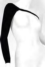
Circular-knit ¾ -finger glove
Circular-knit gauntlet

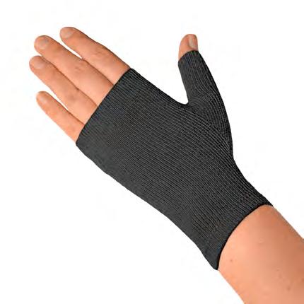
the limb during prolonged wear.1,2,7,14‑17 Therefore, circular‑knit garments are generally most suited to mild oedema where comfort and cosmesis are priorities.1,18 Circular‑knit garments are designed for daytime use only.
Circular‑knit technology uses a fixed number of needles, limiting shape adaptation, with stiffness variation relying on inlay yarn tension and stitch height.7 Therefore, circular‑knit garments are typically available in standard ready‑to‑wear sizes, which are best suited to limbs with minimal distortion. However, there are a few made‑to‑measure options that are available for limbs with minor anatomical irregularities.
Some circular‑knit garments incorporate specialist features, such as softer elbow inserts, improved grip tops and integrated arm‑and‑hand compression. These specialist garments are intended to improve functional outcomes, comfort, mobility and adherence by addressing common challenges, such as gaps in pressure application and ensuring continuity from hand to upper arm.19,20 For example, a circular‑knit sleeve with integrated gauntlet has been found to provide consistent and non‑overlapping compression, for better management of volume and symptoms in the hand as well as the arm.20 Circular‑knit garments may be designed to be layered with other textiles.



Stiffer circular‑knit garments are a distinct category that falls between traditional circular‑knit and flat‑knit garments (Figure 3).1 These hybrid garments are made with a similar knitting technique to traditional circular‑knit garments. However, they use high‑modulus inlay yarns to resist garment fatigue and provide increased stiffness, as well as a denser pattern of interlocking loops to resist expansion.7 The resulting garments tend to be 1.5–2 times stiffer than traditional circular‑knit garments, with better oedema containment.14 Stiffer circular‑knit garments can deliver moderate or high levels of pressure, and they can be layered for added compression.
Flat‑knit garments are manufactured with flatbed machines (Figure 4).7 These machines have rows of large‑gauge needles that can knit relatively thick yarns and textiles with a comparatively coarse texture, with fewer needles per inch (Figure 5). Inlay yarns provide these garments with their controlled compression dosage and elastic properties.7 Compared to circular‑knit textiles, flat‑knit textiles typically offer higher stiffness, better containment and more even distribution.7 They can also be more effective at bridging skin folds without cutting into the skin or causing a tourniquet effect.1,2,21 Flat‑knit garments vary in stiffness and can deliver low, moderate or high levels of pressure. They are intended for daytime use only. Flat‑knit textiles are typically produced in flat sheets that are stitched together to produce a garment post‑knitting. These stitched garments are easy to customise, but they have seams that may be cosmetically undesirable or influence pressure distribution. Variation in knitting pattern affords variation in stiffness and containment (Figure 6).
Flat knitting also allows adjustments in needle count to alter fabric width and shape, allowing flat‑knit garments to be customised to adapt to anatomical variations in the natural contours of the body.7 A better fit enhances patient comfort and wearability, and custom sizing can support distorted anatomical shapes to help resist rolling, especially for areas with anatomical irregularities or increased sensitivity. This adaptability makes flat‑knit garments valuable for addressing moderate‑to‑severe swelling or complex limb shapes that fall outside standard sizing ranges. However, there are also some seamless flat‑knit garments, produced with circular V‑bed and glove flatbed machines, which avoid seams at the expense of containment and


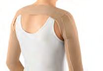

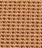

customisability.16,22,23 Flat‑knit garments are available with textured textiles that produce lymphatic alternating pressure profiles (LAPPs).1
The term ‘flat‑knit’ can describe any compression garment knitted on a flatbed machine, and the term alone does not guarantee that the garment has been engineered to the RAL standards. To ensure the necessary stiffness and containment for effective lymphoedema management, compression garments should be sourced from reputable companies with rigorous unbiased external quality checks.
Adjustable wraps are versatile compression garments with circumferential dimensions that can be modified to accommodate changes in volume or deliberately increase or decrease pressure (Figure 7). The wrap is secured with elastic drawstrings, hook‑and‑loop fasteners or hook‑and‑hole closures. A tight fit will increase the resting pressure, and pressure should be applied according to prescribed dosage, potentially guided by inbuilt or augmentative marking systems. The dosage can be re‑adjusted as necessary, such as following oedema reduction.
Adjustable wraps tend to be stiffer than circular‑knit or flat‑knit garments, exerting relatively high working pressures during movement and muscle contractions. However, adjustable wraps can vary considerably in stiffness between moderately elastic short‑stretch wraps to completely inelastic rigid wraps, depending on the construction method and textiles used.13 The level of stiffness can impact the garment’s dynamic capacity, so adjustable wraps should not be assumed to function identically, and research is warranted to compare the stiffness of different adjustable wraps for the upper limb and trunk.
Adjustable wraps can be made from several materials, such as neoprene, breathable neoprene‑like fabric or spacer fabrics, with significant functional differences. Whether the wrap is designed to interlace or overlap to form a rigid sleeve impacts its functional applications. Both ready‑to‑wear and made‑to‑measure adjustable wraps are available.
Adjustable wraps are an effective option during the acute phase of lymphoedema management, where flexibility, comfort and repeatable dosing are key. They are also user‑friendly, with controlled, customisable compression that enables patients to participate actively in care, while improving adherence. Adjustable wraps are suitable for use in the daytime and at night. Studies have found adjustable wraps to be particularly effective for initial limb‑volume reduction.1,24
Decongestive wraps are a distinct category of adjustable wrap designed for reduction during the acute phase of oedema management (Figure 8). Decongestive wraps offer an alternative or supplement to the traditional gold standard for complete decongestive therapy with custom application of multi‑layered, short‑stretch bandages with incongruent foam. Decongestive wraps are made from similar textiles as adjustable wrap and tend to be stiff, exerting high working pressures and low resting pressures.

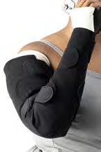


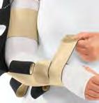
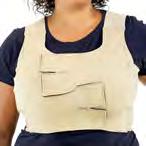
Decongestive wraps for upper extremities and trunk are usually made to measure in custom sizes to fit different limb shapes. These can also be resized and trimmed to accommodate oedema reduction over time. This degree of customisation makes these wraps difficult to label with an accurate dosage. In addition to custom options, there are also some ready‑to‑wear decongestive wraps that can be selected based on the volume of oedema intended to be reduced.
Night‑time garments are compression garments primarily designed for use at night, although they are also safe for daytime use (Figure 9).25,26 They are constructed differently to circular‑knit, stiffer circular‑knit or flat‑knit garments, which manufacturers explicitly label for daytime use. In comparison, night‑time garments tend to exert lower pressures, which supports long‑term self‑management by improving adherence, sleep quality and patient satisfaction.25,26 Night‑time garments are clinically effective26 at to reducing oedema volume, maintaining arm shape and preventing rebound swelling,25,27,28 as well as softening fibrotic tissue.27‑29 The need for night‑time garments may be influenced by refill speed and pattern.30
Night‑time devices for the upper limb, breast and trunk are available in a variety of patient‑centred options, based on their textile and construction. This includes different pressure and stiffness profiles suitable for mild, moderate or severe oedematous presentations, as well as breathable materials, differing thickness profiles and outer elastic or inelastic sleeves to enhance dynamic compression. There are night‑time textured textiles with LAPPs, including channelled designs, padded foam blocks and spacers, used to enhance lymphatic stimulation and pressure distribution. Night time garments, available in ready to wear or custom sizing, are often easy to apply, which can be of particular benefit for older patients.25 This may involve slip on application with an elastic oversleeve or self adjusting using hook and loop fasteners similar to adjustable wraps.27,29,31
Trunk garments refer to compression bras, t‑shirts or vests (Figure 10). Trunk garments are typically made from circular‑knit textiles, but they may also incorporate flat‑knitted, warp‑knit or stretch‑woven textiles for greater stiffness. Unlike seamless circular‑knit garments for the limbs, trunk garments are cut and sewn to achieve the desired shape and fit and thus have seams. This produces garments that are flexible, comfortable and aesthetically pleasing.
Trunk garments are constructed from a variety of textiles. Although some available items may exhibit characteristics of
elastic, stiffer‑circular knit or flat‑knit garments, trunk garments have been designated as a distinct category due to limited manufacturer disclosure regarding textile composition and construction. This can also make dosage, stiffness and containment difficult to determine, presenting a particular challenge, considering that attention to these properties is crucial to selecting the right device for effective management of particular oedematous presentations.
Some trunk garments incorporate internal pockets designed to hold specialised focal pads, enabling simple application of


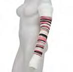

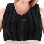

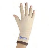


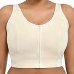

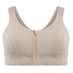
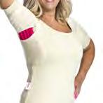
targeted pressure over areas of asymmetry, fibrosis or localised skin change. These pads may feature LAPP‑generating textured textiles to support lymphatic stimulation and tissue softening.
Focal pads are placed over specific areas, such as on the breast and chest wall, to help address local issues and more evenly distribute compression pressures (Figure 11). Localised issues might be areas of tissue congestion or fibrosis following radiation treatment for breast cancer, for example. Focal pads may comprise a single piece of dense foam or a series of foam chips. Focal pads can enhance the overall effectiveness of the garment, as well as assist with softening fibrosis and support the improvement of tissue quality. For example, use of focal pads under elastic garments can provide critical support for fatty tissue in breast oedema by bridging skin folds and preventing garments from rolling or cording, helping mitigate the risk of impaired lymphatic drainage in the affected area, thereby optimising therapeutic outcomes.
Donning/doffing aides facilitate proper application and removal of compression garments to enhance compliance with the compression regimen. A variety of styles are available and can be matched to the user’s individual need or physical abilities (Figure 12).
The STRIDE framework offers a structured and practical approach to classifying and understanding compression options, particularly for the upper extremity, breast and trunk. The classification of compression garments into distinct categories and subcategories enhances awareness of the wide range of designs and textile properties available to address diverse patient needs. Clinicians can leverage this organisational model to make more informed decisions, tailoring garment selection to therapeutic goals while considering patient preferences, lifestyles and functional requirements.
For industry partners, adopting this standardised framework streamlines product development and marketing strategies, providing clearer differentiation and communication of distinct

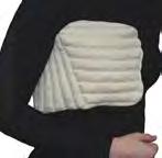


© Sigvaris, L&R, Medi
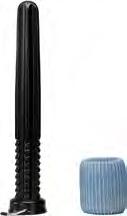
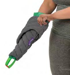

product benefits within defined categories. By increasing awareness of these distinctions, the framework fosters trust and confidence among consumers, empowering them to better understand their options. Additionally, the integration of this model into industry standards holds great promise for advancing innovation, improving accessibility and expanding the scope of compression therapies to manage lymphoedema presentations more effectively. Ultimately, this approach benefits all stakeholders—clinicians, consumers and industry—by optimising outcomes and driving progress in the field of compression therapy.
Declaration of interest: Sponsored by Haddenham Healthcare and the US Medical Compression Alliance (USMCA); Brandy McKeown has been a paid consultant or speaker for HMP global/Wound Source, L&R, LymphaPress, Juzo and Sigvaris; Suzie Ehmann has been a paid consultant or speaker for L&R, Urgo, OVIK, Compression Dynamics and Medline; Sandi Davis has been a paid consultant for Koya Medical; Karen J Bock has been a paid consultant for Pure Medical
References
1. Bjork R, Ehmann S. S.T.R.I.D.E. professional guide to compression garment selection for the lower extremity. J Wound Care. 2019; 28(S6a):1–44. https://doi.org/10.12968/jowc.2019.28.Sup6a.S1
2. Reich-Schupke S, Stücker M. Round-knit or flat-knit compression garments for maintenance therapy of lymphedema of the leg? Review of the literature and technical data. J Dtsch Dermatol Ges. 2019; 17(8):775–784. https://doi.org/10.1111/ddg.13895
3. Liu R, Lao TT, Little TJ, Wu X, Ke X. Can heterogeneous compression textile design reshape skin pressures? A fundamental study. Textile Res J. 2018; 88(17):1915–1930. https://doi.org/10.1177/0040517518779254
4. Chassagne F. [Biomechanical study of the action of compression bandages on the lower leg]. 2017. https://theses.hal.science/tel 01848712 (accessed 1 October 2025)
5. Kumar B, Das A, Alagirusamy R. Effect of material and structure of compression bandage on interface pressure variation over time. Phlebology. 2014; 29(6):376–385. https://doi. org/10.1177/0268355513481772
6. Al Khaburi J, Nelson EA, Hutchinson J, Dehghani Sanij AA. Impact of variation in limb shape on sub bandage interface pressure. Phlebology. 2011; 26(1):20–28. https://doi.org/10.1258/phleb.2010.009082
7. International Lymphoedema Framework. Template for practice: compression hosiery in upper body lymphoedema. 2009. https://www. lympho.org/uploads/files/files/Upper_body.pdf (accessed 20 August 2025)
8. Hettrick H, Ehmann S, McKeown B, Bender D, Blebea J. Selecting appropriate compression for lymphedema patients: American Vein and Lymphatic Society position statement. Phlebology. 2023; 38(2):115–118. https://doi.org/10.1177/02683555221149619
9. Karafa M, Karafova A, Szuba A. The effect of different compression pressure in therapy of secondary upper extremity lymphedema in
women after breast cancer surgery. Lymphology. 2018; 51(1):28–37
10. Phillips N, Lawrance S. Haddenham Comfiwave: a unique compression device for lymphoedema treatment. Br J Community Nurs. 2020; 25(S4):S23–S30. https://doi.org/10.12968/bjcn.2020.25.Sup4.S23
11. Williams AF, Williams AE. ‘Putting the pressure on’: a study of compression sleeves used in breast cancer related lymphoedema. Journal of Tissue Viability. 1999; 9(3):89–94. https://doi.org/10.1016/ S0965 206X(99)80024 2
12. Hirai M, Niimi K, Iwata H, Sugimoto I, Ishibashi H, Ota T, Nakamura H. Comparison of stiffness and interface pressure during rest and exercise among various arm sleeves. Phlebology: The Journal of Venous Disease. 2010; 25(4):196–200. https://doi.org/10.1258/phleb.2009.009064
13. Benigni J P, Uhl J F, Filori P, Balet F, Penoel L. Adjustable compression wraps: stretch, interface pressures and static stiffness indices. International Angiology. 2023; 42(3). https://doi.org/10.23736/ S0392 9590.23.04957 X
14. Bjork R, Leonard M, Parker N, Pour EB. Novel, high containment circular knit garment provides improved edema management for patients with chronic edema to include lymphedea. Nashville (TN): International Lymphoedema Framework; 2018
15. van der Wegen Franken CP, Mulder P, Tank B, Neumann HA. Variation in the dynamic stiffness index of different types of medical elastic compression stockings. Phlebology. 2008; 23(2):77–84. https:// doi.org/10.1258/phleb.2007.006018
16. Hampton S. Elvarex compression garments in the management of lymphoedema. Br J Nurs. 2003; 12(15):925–929. https://doi. org/10.12968/bjon.2003.12.15.11425
17. van Geest AJ, Veraart JC, Nelemans P, Neumann HA. The effect of medical elastic compression stockings with different slope values on edema. Measurements underneath three different types of stockings. Dermatol Surg. 2000; 26(3):244–247. https://doi. org/10.1046/j.1524 4725.2000.09200.x
18. Hirai M, Niimi K, Iwata H, Sugimoto I, Ishibashi H, Ota T, Nakamura H. Comparison of stiffness and interface pressure during rest and exercise among various arm sleeves. Phlebology. 2010; 25(4):196–200. https:// doi.org/10.1258/phleb.2009.009064
19. Everett J, Lawrance S. Use of Haddenham Venex armsleeve for lymphoedema management in clinical practice. Br J Community Nurs. 2019; 24(S10):S12–S18. https://doi.org/10.12968/bjcn.2019.24.Sup10.S12
20. Pugh S, Stubbs C, Batchelor A. Managing upper limb lymphoedema with use of a combined armsleeve compression garment. Br J Community Nurs. 2017; 22(S10):S38–S43. https://doi.org/10.12968/ bjcn.2017.22.Sup10.S38
21. Hirsch T, Arns H, Schleinitz J, Fiedler HW. An innovative flat-knit compression garment for lymphoedema patients led to better outcomes: a multicentre study. J Wound Care. 2024; 33(4):220–228. https://doi.org/10.12968/jowc.2024.33.4.220
22. Miller A. Impact of seamless compression garments on limb functionality, comfort and quality of life. Br J Community Nurs. 2017; 22(S10):S26–S37. https://doi.org/10.12968/bjcn.2017.22.Sup10.S26
23. Elwell R, Heal D, Lister L. Impact of JOBST(®) Elvarex(®) knee and elbow functional zones on quality of life. Br J Community Nurs. 2017; 22(S10):S58–S67. https://doi.org/10.12968/bjcn.2017.22.Sup10.S58
24. Silva J, Araújo RDD, Aguiar SS, Fabro EAN, Pinto MVM, Thuler LCS, Bergmann A. Efficacy, safety of and adherence to adjustable compression wraps in the control phase of breast cancer related lymphedema: A randomized controlled trial. Clin Rehabil. 2024; 38(11):1481–1494. https://doi.org/10.1177/02692155241270921
25. McNeely ML, Dolgoy ND, Rafn BS et al. Nighttime compression supports improved self management of breast cancer–related lymphedema: a multicenter randomized controlled trial. Cancer. 2022; 128(3):587–596. https://doi.org/https://doi.org/10.1002/cncr.33943
26. Brunelle CL, Ag AG. The important role of nighttime compression in breast cancer related lymphedema treatment. Cancer. 2022; 128(3):458–460. https://doi.org/10.1002/cncr.33942
27. Mazur S, Szczęśniak D, Tchórzewska-Korba H. Effectiveness of Mobiderm Autofit in the intensive phase of breast cancer-related lymphedema treatment: a case series. Lymphat Res Biol. 2023; 21(6):608–613. https://doi.org/10.1089/lrb.2022.0079
28. Bertsch T. Evaluation of a novel night‑time compression garment: a prospective observational study. Br J Community Nurs. 2018; 23(11):535–541. https://doi.org/10.12968/bjcn.2018.23.11.535
29. Todd M, Stubbs C, Pugh S. Mobiderm Autofit: an adjustable sleeve that enables patients to self manage lymphoedema. Br J Community Nurs. 2018; 23(S4):S14. https://doi.org/10.12968/bjcn.2018.23.Sup4.S22
30. Ehmann S, McKeown B, Davis S et al. Using the STRIDE algorithm for compression selection in upper body lymphoedema. J Wound Care. 2025; 34(S11C):S36–S48
31. Dhar A, Srivastava A, Pandey RM, Shrestha P, Villet S, Gogia AR. Safety and efficacy of a Mobiderm compression bandage during intensive phase of decongestive therapy in patients with breast cancer related lymphedema: a randomized controlled trial. Lymphat Res Biol. 2023; 21(1):52–59. https://doi.org/10.1089/lrb.2021.0104
Suzie Ehmann, Brandy McKeown, Sandi Davis, Karen J Bock, Justine Whitaker and Naomi Dolgoy
Abstract
This second iteration of STRIDE is an evidence-based algorithm for compression selection, extended to cover the upper limb, trunk and breast. The STRIDE algorithm is patient-centred and complexity - informed, encompassing the site, shape and size of oedematous swelling; impact of tissue texture on textile types; 24-hour refill patterns; patient-specific issues; pressure dosage among other textile characteristics; and oedema etiology and staging. This article details the elements of the STRIDE algorithm and presents practical tools for its application.
Keywords: STRIDE algorithm | Compression garment selection | Upper b ody lymphoedema | Breast and truncal oedema | Textile stiffness and containment | Patient centred therapy | Tissue texture assessment | Daytime and night‑time compression | Evidence b ased clinical framework | Individualised management
Lymphedoema presents unique challenges for each individual presentation. Effective management of this condition relies heavily on the selection of an appropriate compression regimen. Selection requires a detailed understanding of the condition in general and how it can affect different regions of the body, as well the individual’s specific presentation and needs. It also requires an understanding of the capabilities of different compression garments and accessories.
Overview
STRIDE is an algorithm for compression selection that emerged as a critical response to a historically narrow focus on dosage alone. 1,2 While the initial iteration of STRIDE was focussed on compression for the lower limb, 1 this second iteration extends STRIDE to also cover lymphoedema of the upper limb, breast and trunk. The second iteration of STRIDE aims to embody the same key characteristics as the original:
▪ Complexity-informed: embracing the dynamic and multifaceted nature of compression, bridging gaps in traditional metrics and incorporating the intricate interplay of textile characteristics, including pressure (dosage), stiffness and distribution
▪ Patient-centred: highlighting the importance of individualised assessment and treatment tailored to address the diverse needs of people living with lymphoedema
▪ Evidence-based: developed with a methodologically pluralistic approach founded in clinical expertise, textile science and medical literature
▪ Adaptable: offering standards that can evolve with the advancing landscape of compression options, clinical practice and research
▪ Practical: providing comprehensive guidance in a structured framework for clinical providers. The STRIDE recommendations are founded on an understanding of both lymphoedema pathophysiology and the science of compression therapy. The acceptability and applicability of the revised STRIDE algorithm has been validated with the input and consensus of lymphoedema clinicians through a Delphi study.3
STRIDE is an acronym for six key factors in compression selection:
1 Shape
2 Texture
3 Refill
4 Issues
5 Dosage
6 Etiology.
Shape involves identifying the distribution of oedematous swelling throughout the body through a thorough history and clinical examination. The identified locations, contours and measurements help determine the most appropriate choice of compression products.
The first step is to locate all specific sites of the body affected by lymphoedematous swelling. Areas of the upper body likely to be affected include the hand, arm, shoulder, breast, chest and trunk, independently of one another or in combination (Figure 1). The site determines the type of compression garment used, such as sleeves for the upper limb; gauntlets or gloves for the hand; t‑shirts and tanks/vests for the whole trunk; bras or breast bands for the breast; or abdominal corsets for the abdomen (Figure 2).
Suzie Ehmann, DPT, PhD, CWS, CLT L ANA, CLWT, Lymphedema
Therapist, McLeod Health Seacoast, Little River, SC, USA
Brandy McKeown, OTR/L, CLT L ANA, CLWT, Lymphedema
Therapist, International Lymphedema and Wound Training Institute and the Lymphedema Center, USA
Sandi Davis, PT, DPT, CLT L ANA, CLWT, President, Davis Care Physical Therapy, New York City, NY, USA
Karen J Bock , PT, PhD, CWS, CLT L ANA, Assistant Professor, Rockhurst University, Kansas City, MO, USA
Justine C Whitaker, MSc, RN, PhD(c), Director & Nurse Consultant, Northern Lymphology, and Senior Lecturer, University of Central Lancashire, Preston, UK
Naomi Dolgoy, MOT, PhD, CLT, Assistant Professor, University of Alberta, Canada
All affected sites should be covered with compression products. Oedema affecting adjoining sites can be covered with a one‑piece combination garment to avoiding overlapping coverage at the juncture between sites, which could otherwise increase focal pressure and contribute to congestion. 4 Examples include long gloves for the hand and forearm; full‑length sleeves for the forearm and upper arm; and full‑length sleeves with attached gauntlets for the hand, forearm and upper arm.
The second step is to describe the shape of the affected sites. Arms can be described by how much their circumferential profile tapers from the shoulder to wrist (Figure 3):
▪ Cylindrical profiles show normal gradual tapering towards the wrist, often with smooth contours, as is typical in healthy individuals, early stage lymphoedema or balanced body habitus
▪ Columnar profiles show minimal tapering towards the wrist, with a straight appearance, common in cases with generalised swelling or reduced muscle tone
▪ Tapering profiles show exaggerated tapering towards the wrist, potentially reflecting underlying tissue loss, disuse or anatomical variation.
Sites can be described by their contours, meaning whether transitions in circumference between adjacent anatomical landmarks are subtle or pronounced:
▪ Subtle contours feature gradual circumferential changes, such as a smooth curve from elbow to wrist, typical of early stage presentations
▪ Pronounced contours feature sharp circumferential changes over short distances, such as a narrow wrist next to a swollen forearm or a sharply defined elbow crease, often seen in advanced, fibrotic or postoperative presentations. The shape of the affected site helps determine the suitability of ready‑to‑wear or custom garments, as well as if a garment needs to be supported with compression accessories. For example, an arm that has atrophied due to wasting, disuse or disease progression may have reduced volume, a columnar or tapering profile and pronounced sunken contours, especially in the deltoid or forearm regions. There features can complicate garment fit and containment and thus need to be accounted for. The predefined shapes of ready‑to‑wear garments are usually designed to accommodate generic body shapes, such as arms with subtle natural curves, columnar forms and tapering profiles. A ready‑to‑wear sleeve can be laid flat to compare its shape against the contours of a patient’s limb. Unusual body
shapes are more likely to require custom garments, which are more likely to be made with flat‑knit technology, although custom circular‑knit options are also available. An arm with pronounced or complex anatomic contours may not be a good fit for a standard columnar sleeve, which would risk areas of greater or lesser pressure. An atrophied arm may not be a good fit for an sleeve with a sewn‑in elbow contour, which could risk pocketing at the elbow, reducing compression effectiveness. If a wider oedematous area has distinct points of contouring, swelling or tissue changes, these challenging areas can be more effectively targeted with focal pads. Focal pads can be used to match tissue textures that differ from the surrounding skin. They can also be used to bridge skin folds, such as a longer trunk garment that extends over the inframammary fold or middle of the abdominal protrusion and thus risks folding back onto itself and disordering the pressure applied.
The third step is to measure the length and circumference of each affected site, with circumference measurements at key points such as the wrist, forearm and upper arm (Figure 4). The patient’s measurements should then be compared with the stated sizing information for potentially suitable available products. It is worth consulting detailed and precise sizing for specific points on the body, rather than general size categories, as the latter can vary between products, even those from the same manufacturer. A ready‑to‑wear garment may not match all the patient’s measurements, warranting a custom garment to ensure delivery of the required dosage.
Texture covers the types of tissue on the surface of the skin and how these inform the types of textile used for compression.
Tissue texture refers to the quality of the skin and/or subcutaneous tissues affected by oedema and thus to be
covered by compression products. Textures may vary across areas of the body, so different oedematous sites should be assessed separately and regularly. Tissue texture can be assessed with Stemmer’s sign, the Bjork bowtie test and the pitting scale.1,5,6 Tissue can also be gently palpated to assess skin feel and pliability, alongside visual assessment of its appearance. The skin’s texture can then be categorised according to STRIDE as either normal, watery, doughy, woody, fatty or fragile (Table 1). Using consistent terminology for tissue texture can minimise the subjective variability of this assessment.1 These tissue types exist on a continuum, and classification is down to clinical judgement. Different tissue types may be present in different areas of the same patient’s body.
Watery tissue is soft and pliable and may not exhibit fibrotic tissue changes. It easily pits, can be deeply pitting and rebounds quickly. Stemmer’s sign and the Bjork bowtie test should be negative. Watery tissue tends to worsen with dependency and improve with elevation. Watery oedemas are straightforward to reduce and manage with compression, usually requiring compression products only during the day.
Textile recommendation: Watery tissue responds quickly to adjustable wraps or multi layered bandages. Once the volume has been reduced, long term management can be achieved with circular knit garments at lower doses. However, deeply pitting watery oedemas may require stiffer circular knit garments or flat knit garments to prevent creasing or folding into joint lines.
Doughy tissue is relatively soft, feels like putty and indicates early fibrotic changes. It is slow to rebound, taking over 30 seconds, and may be deeply pitting. The Bjork bowtie test and Stemmer’s sign may be positive.1
Table 1. STRIDE assessment of tissue texture1
Texture Feel
Fibrosis Pitting Stemmer’s sign Bjork bow tie test
Watery Soft and pliable Non-fibrotic Easily pitting and quickly rebounding Negative Negative
Doughy Fairly soft (putty-like)
Somewhat fibrotic
Deeply pitting, rebounding after 30 seconds
Woody Firm Severely fibrotic Non-pitting Positive Positive
FattySpongy, squishy, nonpliable, inelastic
Fragile Thin, delicate, inelastic
Non-fibrotic Non-pitting
Textile recommendation: Doughy tissue typically benefits from day and night compression. Stiffer garments provide the necessary containment, and textured textiles support the softening of fibrotic tissue to promote lymphatic drainage.
Woody tissue is firm in consistency and indicates advanced fibrotic changes. It shows very minimal to no pitting, even with deep pressure. The Bjork bowtie test and Stemmer’s sign will be positive.1
Textile recommendation: Woody tissue tends to require both day and night compression. The use of stiffer textiles, such as those seen in flat knit garments, are most effective for containment. Textured textiles are essential to warm and soften the fibrotic tissue, aiding in soft tissue remodelling and lymphatic capillary regeneration.1
Fatty tissue (specifically the abnormally fatty tissue associated with lipoedema) tends to be spongy or squishy feeling, lacking the normal pliability and elasticity of healthy skin. Abnormal fat tends to have poor connective architecture and support, resulting in loss of tissue elastic recoil, which negatively affects tissue tolerance for compression forces.1
Textile recommendation: Abnormal fatty tissue tends to require stiffer and thicker textiles that can bridge across creases and effectively shape and support the oedematous area. Thinner, elastic garments would tend to roll into creases and sink between folds, increasing risk of pressure and friction. Fatty tissue in breast and trunk oedema may also benefit from use of focal pads to evenly distribute pressure across areas with diverse tissue textures and complex anatomical contours.
Fragile tissue tends to feel thin, delicate and inelastic, and it may present with active loss of integument, such as stasis dermatitis or metastatic lesions. Fragile tissue is often impaired, and it can
be prone to tears, bruises, breaks, fissures, lipomas, cysts, blebs and blisters. Fibrosis may be present, patchy or absent, depending on underlying pathology. Fragile tissue may or may not pit, depending on fluid status and tissue integrity. Stemmer’s sign may be either positive or negative, with interpretation limited by skin fragility. The Bjork bowtie test is contraindicated due to risk of further skin damage. Fragile tissue is particularly common in older people.
Textile recommendation: Fragile tissue should be factored into garment selection. Fragile tissue can be protected via: circular knit garments with double covered inlay yarns to reduce friction when donning; layering an underliner with a secondary garment; adjustable wraps; donning and doffing aids; and silicone lotion for garment applications.1
The textile used for compression therapy can have significant physiological effects on oedema reduction, tissue softening, tissue oxygenation and scar reduction.7‑11 The key is to match the tissue texture with the appropriate compression textiles.1 Therefore, understanding the properties and suitability of different textiles is crucial for optimising outcomes.1
STRIDE proposes two distinct but overlapping categorisation systems for compression garments and textile types. Each compression garment will belong to one of seven garment categories: circular‑knit garments, stiffer circular‑knit garments, flat‑knit garments, adjustable wraps, decongestive wraps, night‑time garments and trunk garments. All compression garments are also made from a textile conforming to one or more of five types: elastic textiles, stiff textiles, textured textiles, adjustable textiles and layered textiles. These textiles will fit on a continuum between elastic and stiff, and they may also be textured, adjustable and/or layered.
Within the same category of compression product or textile type, there is often considerable variation in interface pressures, stiffness and distribution, influenced by the materials and manufacturing processes used.12 Therefore, products and textiles in the same category, even from the same manufacturer, should not be presumed to be interchangeable.
Elastic textiles, compared with stiff textiles, have greater stretch and recoil. They produce working pressures that are relatively similar to their resting pressure (dosage), expressed as a lower static stiffness index (SSI). Clinically, elastic textiles tend to exert higher resting pressures than stiffer equivalents. Moreover, they have a lower containment effect, making them less robust and effective in containing severe oedema, bridging skin folds and shaping fatty tissue.1
However, elastic textiles are used in many circular‑knit garments, including sleeves, gloves and vests, that are relatively inexpensive and aesthetically appealing, as well as available in a wide range of sizes. Elastic garments are most appropriate for mild‑to‑moderate oedema with soft, watery tissue and preserved skin elasticity.
Stiff textiles, compared with elastic textiles, have minimal stretch or recoil and more resistance to expansion when the body swells or muscles contract.1,13 In stiff textiles, the difference between working pressure and resting pressure (dosage) is relatively large, expressed as a higher SSI. This allows for dynamic performance, with lower resting pressures and higher intermittent working pressures during movement and posture changes.1 Thus stiff textiles, including stiffer circular‑knit garments and flat‑knit garments, generally provide superior therapeutic performance in oedema reduction and prevention, as well as venous haemodynamics.14‑17 Stiff textiles provide high containment and are useful for managing fibrosis, severe oedema and complex contours. Stiff textiles can be used to bridge fat folds and lobules, shape soft tissue and contain more robust oedemas, with faster refill times. However, these textiles are often made with thick yarn, relatively coarse and expensive to produce, and stiffer garments often require custom sizing for the best fit.
Stiffness is a continuum, and some textiles fall between the extremes of elastic and stiff. These medium‑stiffness textiles balance flexibility with structural support. They have some elastic recoil to conform to movement and shape, thus promoting comfort and mobility, while also having the containment to resist deformation and maintain consistent pressures. This adaptable compression is often suitable for dynamic anatomical areas or mild asymmetry.
Textured textiles
Textured textiles refer to compression fabrics engineered with surface variation—such as ridges, channels or patterned density—that create a non‑uniform pressure profile across the skin. Unlike smooth or uniformly elastic garments, these designs produce alternating zones of higher and lower interface pressure, which may stimulate lymphatic flow more dynamically than consistent compression alone. This has a multidimensional therapeutic effect, delivering repeatable, morphology‑adapted compression.18‑20 These alternating zones of high and low pressure are known as lymphatic alternating pressure profiles (LAPPs).1 LAPPs have been shown to create a micro‑massage that impacts microcirculation and stimulates dermal lymphatics. LAPPs are thought to achieve this by mechanically deforming the skin and subcutaneous tissue below, thus stretching the anchoring filaments of lymphatic capillaries and
promoting movement of fluid from interstitial tissue into lymphatic capillaries. This fluid movement, enhanced by compression, stimulates the formation of new lymphatic capillaries. LAPPs have been shown to reduce oedema, resolve trophic changes and enhance tissue quality by actively warming, softening and breaking up fibrotic tissue.1,10,20‑25
Textured textiles are commonly integrated into some flat‑knit garments, night‑time garments and focal pads. These devices can be of variable thickness and designed for daytime or night‑time use, depending on individual presentations and needs. The texture can be achieved through thicker yarns, specialised knit patterns, spacer fabrics and waved, chipped and/or channelled foam, such as padded foam blocks or dense foam chips in vertical or diagonal channels. Likewise, multilayered constructions can produce raised or patterned surfaces, such as spacer fabrics that feature a three‑dimensional architecture, often with two outer layers connected by vertical filaments, creating an alternating terrain profile. This design promotes airflow, cushioning and a gentle micromassage effect during movement, which supports lymphatic stimulation and contributes to tissue mobilisation. Evidence shows that these features can help soften fibrotic tissue and reduce oedema.7,26 Garments available in elastic, medium‑stiffness and stiff variations may be marketed as light, regular and firm, respectively.
Adjustable textiles are compression devices with circumferential dimensions that can be modified to accommodate changes in volume, increasing or decreasing the pressures applied to optimise oedema management and improve comfort. Adjustable garments include all adjustable wraps, all decongestive wraps and some night garments. These garments empower independent adjustment, with the ease of donning and doffing varying based on the type of closure system, whether elastic drawstrings, hook‑and‑loop fasteners or hook‑and‑hole closures. There are options available for one‑handed closures and side‑bending closures to address decreased strength and flexibility. Adjustable textiles are typically made from stiffer materials.
Layered textiles are two or more compression textiles that are layered over one another to achieve a therapeutic effect that would not be possible with a single layer. The main advantage of layered textiles is that they tend to be stiffer than the sum of their individual components, enhancing their overall dynamic performance. The overall performance of layered compression should be evaluated on an individual basis, as each textile component impacts the system’s dynamic performance. Layering can be particularly useful for addressing the anatomical features of the upper limb, breast and trunk.
There are multi‑part compression kits designed to be layered, such as night‑time garment kits that include both an inner compression garment and an outer elastic sleeve. The outer sleeve may be designed to both increase pressure and create a smoother surface that reduces friction against sheets and snagging.
Layering can also be achieved with separately purchased products. For example, focal pads can be placed under a
compression garment to accommodate the complex surface anatomy typically found in the trunk and upper limb to ensure even distribution of pressure, helping maintain comfort, mobility and cosmesis. Focal pads can also be used to target local differences in tissue texture, such as firmer tissue over the ribcage, areas of radiation‑induced fibrosis or isolated oedema in the dorsum of the hand.
Refill refers to the rate at which a compressed lymphoedematous site changes in volume over time and according to activity levels. After a compression system has been removed, lymphoedema gradually increases over time, especially during activity, but it may also temporarily subside with elevation or rest. Refill can be controlled with strict adherence to an appropriate compression regimen, along with independent strategies for reducing lymphoedema.
Refill can be categorised by the time it takes for a limb to swell beyond the garment size after it has been removed, with less than 8 hours being rapid, 8–24 hours being moderate and over 24 hours being slow. Refill speed influences the most suitable compression system, with faster refill benefitting from compression systems offering stronger containment. Containment is the compression system’s ability to limit oedema expansion and preserve the optimal shape of the compressed site. Refill patterns determine compression wear schedules and whether patients require compression at night as well as during the day. Lymphoedema that naturally reduces on night‑time rest or has a slow refill can be treated with daytime compression alone. However, persistent presentations with rapid refill typically necessitate continuous compression through the day and the night.1 Refill patterns can vary by anatomical site according to differences in gravitational dynamics. For example, lymphoedema of the upper limb is likely to refill faster in the daytime, where it is typically in a dependent position, which contributes to fluid accumulation–whereas at night it is usually elevated relative to the heart, allowing for natural fluid resorption.
Some people with lymphoedema may be resistant to daytime compression, perhaps because they do not like how it looks, how hot it makes them feel or how it limits their daytime activities. These patients can be trialled on evening and night‑time compression if their refill is slow enough to maintain reductions through the day. Night‑time compression can be achieved with specialist night‑time garments, intended for soft‑tissue remodelling and oedema reduction during sleep (but can also be worn during the day as needed). Regarding other categories of compression system, circular‑knit, stiffer circular‑knit and flat‑knit garments are designed for daytime use only, but adjustable wraps, decongestive wraps, bras, vests and focal pads can generally be used during both day and night.
Issues describes the factors that affect compression therapy and selection of compression garments. Compression selection is dependent on the patient’s functional, social, psychological, medical, cognitive and financial status, as well as flaccid limb and caregiver support. The patient–clinician partnership is predicated on the clinician’s ability to recommend compression that allows patients to complete their daily activity goals while optimally
managing their oedema, and therefore this relationship should include active listening and understanding to tailor interventions to match the individual’s lifestyle goals.27 Successful compression regimens are often highly variable due to a wide array of challenges. 28,29 Compression regimens should be informed by consideration of the following specific factors.1,5–10,30–36
Device issues include garments that fit poorly or are difficult to don or doff, as well as those with poor clinical performance, such as in containment. Ongoing follow‑up with therapists will ensure proper fit and facilitate active compression regimens to optimise oedema management, with review of fit and application of compression in session. Prescribers should meet regularly with industry partners to stay abreast of garment options. Clinicians should use resources offered by compression manufacturers to optimise fit of complex patient presentations. Ongoing follow‑up with the prescribing practitioner or therapists will facilitate continuous successful compression regimens. Patients should be provided with appropriate donning/ doffing aids, such as donning frames, gloves, slide sheets and powered devices, tailored to the patient’s physical abilities and garment type. They should also receive ongoing education and support to ensure safe, independent and consistent use of compression garments. Patients should also have access to appropriate devices and ongoing support for donning and doffing.
Financial issues include the effect of compression use on employment and vocation, as well the cost of compression devices for patients who have to purchase their own. Patients may need to choose between a more expensive optimal device and more affordable but still suitable alternatives. Users can be provided with resources and tools, including information for national oedema support groups and organisations that offer details on finance and resource allocation. As part of the patient–clinician relationship, interventions should match the user’s financial capabilities, including consideration of types of garments and duration of wear. Clinicians should gain knowledge on verbiage appropriate to meet rules and regulations of insurance companies necessary to acquire the permitted annual quantity of garments.
Functional issues include difficulty donning or doffing garments; physical dexterity challenges when wearing a compression device; and how compression may affect sleep, exercise, leisure, social life and activities of daily living, including cooking, cleaning, dressing and eating. Compression should help promote a return to independence in functional activities, which is often a key aim of decongestive therapy in upper‑body oedema.31
This requires an assessment of the patient’s functional capabilities, including hand dexterity and activity tolerance, especially regarding donning and doffing compression garments, as well as their functional needs, such as work requirements and activity preferences. The assessment should then guide the provision of appropriate garments at the right dosage, as well as helpful accessories such as donning/doffing aids. Any remaining
physical challenges associated with prescribed compression garments should be reviewed and addressed with therapeutic advice and support, such as the following:
▪ Offering suggestions for activities of daily living, with examples including extra large gloves for cooking and cleaning to fit over compression garments, simplified clasps and closures in clothing; ongoing low impact movement and/or water based physical activity; and suggestions for pillow and positioning for sleep management
▪ Designing therapy sessions to review specific struggles and needs around donning and doffing
▪ Modifying garment types or 24 hour wear schedules to match job demands and work environments.
▪ Providing information for patients to share with their colleagues should their compression regimen cause issues relating to their workplace and job performance.
Hygiene issues include how compression systems can be an impediment to toileting and washing and may need to be donned before and doffed after. Interventions should include details about self‑care and care of the device, including skincare basics and compression care, such as a washing schedule. A minimum of two garments will ensure that there is one to wear while one is being washed. Users may need support with instructions and/or aids for donning and doffing.
Knowledge issues include a patient’s difficulty understanding the role of compression in oedema management, potentially making them unwilling to participate in a compression regimen. This can be overcome with transparent communication, such as providing resources to enable better understanding of compression in context, as well as use of community resources and peer support groups.
Medical issues include comorbidities, fatigue and complications, such as infections or cellulitis. Medical information must have been reviewed in session to best determine how other health issues may impact compression wear. For example, arthritis can make compression donning/doffing a challenge and may require different garment choice, such as use of adjustable wraps rather than sleeves. Physical fatigue is a critical consideration in compression, particularly for individuals in active cancer treatment. This may require modification of garment wear schedules or use of positioning, such as raising the arm on a pillow, rather than application of garments during times of high fatigue.
Skin hygiene is paramount to reduce risk of infection. Hygiene can be promoted with educational resources, use of gloves in high‑risk activities and maintaining skin health through use of appropriate soaps and products. Cuts and scrapes should be avoided and treated immediately when they occur. Continued care for compression garments, including regular washing, should reduce risk of infection.37
Cellulitis may be indicated by redness, heat, additional swelling and/or mottled skin. If these occur, medical care should be sought immediately, as antibiotic treatment typically needs
to be prescribed. Clinicians must understand when it is appropriate to resume compression following a bout of cellulitis. The general recommendation is 48 hours on antibiotics and improvement in clinical signs prior to resuming the compression regimen.38,39 If clinical symptoms do not improve, immediate follow‑up with a medical provider is warranted.
Psychological issues include anxiety, depression, stress and body‑image distortion. Psychological considerations can be embedded in compression management with referrals to psychological services as required and coordination of service delivery, including support groups and exercise programming. Garment choices should be optimised for cosmesis and to allow activities.
Prioritisation tools can be used for management and balance of productive, leisure and social activities. Clinicians and providers can familiarise themselves with positive social‑media influencers within an appropriate age range who can fashion forms of compression as well as bandaging ideas for the patient to find commonality.
Temperature issues include external and internal temperature when compression is donned. Patients can be encouraged to wear breathable and loose fabrics, such as linen and linen blends, as well as cool themselves with small fans and cooling towels. They can be given compression devices made from textiles with cooling properties, as well as recommended to place garments or wraps in a fridge or freezer before donning.
Dosage refers to the expected range of compression strength delivered by a particular device or needed by a particular patient to effectively manage lymphoedema. Dosage is expressed in millimetres of mercury (mmHg) as a range of interface pressures measured at rest.15,40 For upper‑limb compression, dosage refers to expected compression at the wrist,39 but this is less well defined for the breast and trunk. In vivo interface pressures are determined not only by the textile’s elastic recoil (tension), but also other factors such as limb size and tissue composition.12,41,42 There are two different systems for dosage classification in common use (RAL class and US class), with different range thresholds and generic descriptors. Both of these systems have three classes that can broadly be recommended for mild, moderate and severe oedema, respectively (Table 2). 43 However, it is important to be flexible with these broad recommendations,
Table 2. Lymphoedema compression dosage recommendations43 Pressure RAL
Low Class I (15–21 mmHg)
Moderate Class II (23–32 mmHg)
High Class Ill (34–46 mmHg)
15–20 mmHg Mild
21–30 mmHg Moderate
31–40 mmHg Severe
as the optimal dosage may differ according to individual or local variations in limb size, tissue texture, body composition, oedema distribution and functional ability.
There are usually several ways to achieve the prescribed dosage with different compression products. For example, daytime management of mild‑to‑moderate arm lymphoedema could be achieved with any of the following options:
▪ A low pressure adjustable arm wrap
▪ A moderate pressure stiffer circular knit sleeve
▪ A low pressure flat knit sleeve
▪ A moderate pressure flat knit sleeve
▪ Two layered low pressure circular knit sleeves. Likewise, the overall dosage of trunk or breast compression can be increased by placing focal pads under garments and over challenging areas of swelling.
When selecting a compression textile, dosage should not be the only measure of performance. It is also important to consider dynamic properties, such as stiffness, containment and fatigue. Stiffness (or elasticity) may directly impact haemodynamic effectiveness and comfort, and it is also important to note that the required dosage may be lower with stiffer compression.
Etiology refers to the underlying cause of lymphoedema and includes both the origin and extent of lymphatic dysfunction. These factors influence the site, shape and size of swelling, as well as tissue characteristics and progression.
Current understanding of microcirculation highlights the vascular endothelial glycocalyx layer (EGL) as a critical gatekeeper of fluid filtration from blood capillaries.44 The EGL’s role underscores the importance of lymphatic capillaries, which are responsible for nearly 100% of interstitial fluid resorption. This paradigm positions all forms of oedema on a lymphoedema continuum, ranging from optimal lymphatic function to complete system failure.45 Contributing factors may include congenital abnormalities, mechanical insufficiency, trauma, disease or comorbidities that increase lymphatic load and compromise resorption. These influences often shape—but do not rigidly define—the clinical presentation.
Identifying the etiology of lymphatic dysfunction is essential for guiding compression therapy. While individual presentations vary, certain etiological patterns tend to correlate with typical anatomical involvement and tissue response, as in the following cases:
▪ Patients with post surgical lymphoedema (e.g., following oncologic procedures) may present with swelling in the breast, trunk or upper limb, but the extent and tissue texture can differ widely depending on surgical technique, radiation exposure and healing trajectory.
▪ Patients who are neurologically impaired (e.g., post cerebrovascular accident, amyotropic lateral sclerosis, spinal cord injury, Parkinson’s, multiple sclerosis) may exhibit dependent oedema with altered muscle tone and mobility, requiring compression systems that accommodate asymmetry and fluctuating limb volume.
▪ Patients with melanoma or other malignancies may present with localised or regional lymphatic disruption, often requiring targeted compression strategies that consider surgical margins and scar tissue.
Rather than assuming uniform presentations, clinicians can use the STRIDE algorithm to recognise typical patterns associated with specific etiologies, while accounting for comorbidities, tissue characteristics and functional status. This approach supports differential diagnosis and personalised garment selection across the full spectrum of upper‑body lymphoedema presentations.
In applying STRIDE, each factor should first be considered individually. The algorithm is not necessarily linear, and the factors can be considered in any order. After this, the interrelations of all the factors should be considered collectively. The insights obtained from the combination of individual and collective consideration should then support an informed recommendation for compression.
The algorithm can be applied in practice by first identifying the patient’s needs with the STRIDE checklist for compression assessment (Figure 5), then selecting appropriate garments using the STRIDE chart of garment characteristics (Figure 6). The full STRIDE algorithm (Figure 7) can be used to identify appropriate compression options according to oedema site, diagnosis and stage. The algorithm can be read as follows:
1 Select the right row based on site, etiology and tissue texture
2 On that row, identify the recommended size, textile types, day/night refill pattern
3 Select an appropriate garment type for the recommended textile types, day/night refill pattern and patient issues
4 Match the chosen garment type to the right row to determine appropriate options for pressure dosage and stiffness. Trunks garments as a specific category were intentionally excluded from the dosage column of the algorithm (Figure 7) due to insufficient transparency regarding textile composition and mechanical properties. While some trunks garments may resemble circular‑knit, stiffer circular‑knit or flat‑knit garments, manufacturers do not consistently disclose construction details necessary to determine dosage reliability. Without standardised data on stiffness/elasticity and interface pressure, inclusion would compromise the algorithm’s clinical integrity and reproducibility. For circular knit and stiffer circular knit garments for the trunk, designation of dosage (low/moderate/ high) or stiffness (elastic/regular/stiff) was determined by clinical consensus of the authors.
A 68‑year‑old woman presented with painful breast‑cancer related oedema following lumpectomy and sentinel node biopsy (Figure 8). The swelling in the left breast progressively worsened throughout adjuvant radiation treatment, despite initial compression with a tight‑fitting sports bra.
▪ Shape: there was significant swelling in the breast and lateral chest wall, indicating need for a compression bra. There were pronounced contours, indicating need for a focal pad. There were ready to wear bras available in her size.
▪ Texture: the oedematous breast tissue had a woody texture (demonstrated by palpation, +2 pitting, >30 seconds rebound and a positive Bjork bowtie test), indicating need for a stiffer garment with a textured textile, which could be provided a focal pad.
These structured recommendations should be based on holistic assessment of patient-specific factors and modified according to clinician judgment and patient feedback to optimise comfort, compliance and therapeutic efficacy.
MATCH the site to the garment type:
F A rm → sleeve
F Hand → gauntlet or glove
F W hole upper limb → sleeve with gauntlet
F Breast → bra or breast band
F A bdomen → abdominal corset
F W hole trunk → t-shirt or vest
F IF affected sites are adjacent, THEN consider appropriate one-piece combination garments to optimise coverage
F IF the site has pronounced contours, non-standard profiles or sizes that do not match ready-to-wear options, THEN consider a custom garment
F IF the site has pronounced contours, anatomical variation or asymmetry, THEN consider addressing irregularities, such as with focal pads
MATCH the tissue texture to the textile type:
F Normal → Elastic
F Watery → Elastic/medium stiffness
F Doughy → Medium/stiff and textured
F Woody → Stiff and textured
F Fatty → Medium/stiff and textured
F IF there are areas of more fibrotic tissue, THEN consider a textured garment or textured focal pads
F IF the tissue is fragile, THEN select garments to minimise donning difficulty and preserve skin integrity—potentially using underliners, adjustable wraps, donning/doffing aids and/or silicone lotion
MATCH the refill pattern to the compression schedule:
F Slow in the day and at night → Daytime and/or night-time compression, based on patient preference
F Rapid in the day and slow at night → Daytime compression only
F Slow in the day and rapid at night → Night-time compression only
F Rapid in the day and at night → Daytime and night-time compression
F IF the refill pattern only requires daytime compression, THEN consider additional night-time compression to address tissue-texture changes
F IF a patient is recommended but unwilling or unable to wear compression in the daytime, THEN consider night-time compression instead
F IF cosmesis is a priority, THEN consider clinically appropriate options that align with patient aesthetic preferences (e.g., less bulky or more discreet garments)
F IF temperature control is a priority, THEN consider breathable or thermally adaptive textiles that help minimise heat retention and improve wearer comfort
F IF independent adjustment is a priority, THEN consider an adjustable garment
F IF limited mobility or functional impairment cause challenges with donning/doffing, THEN consider a donning/doffing aid and garments that are easy to apply (e.g., layered textiles)
F IF discomfort or sensory intolerance challenge adherence, THEN tailor garments to patient tolerance for textile thickness, breathability and flexibility
F IF the patient has any other needs or preferences, THEN adjust compression recommendations accordingly
MATCH dosage to presentation:
F Mild oedema → Lower pressures
F Moderate oedema → Moderate pressures
F S evere oedema → Higher pressures
F IF applying compression at night, THEN then use lower pressures for comfort
F IF additional local pressure is needed, THEN consider adding a focal pad
F IF patient-specific issues limit practicality or tolerance of higher pressures, THEN consider lower pressures that support adherence while maintaining therapeutic effect
F IF oedema is secondary to neurological impairment, THEN consider lower-pressure and adjustable garments to accommodate fluctuating volume, impaired donning ability and altered sensory processing—including hypersensitivity, diminished sensation or reactivity to heat and touch
F IF oedema is secondary to cancer-related surgery or radiation, THEN assess the entire lymphatic quadrant for asymmetry, distortion or compensatory patterns, and consider focal pads or structured garments to support contouring, containment and lymphatic redistribution
F IF lymph nodes have been removed or irradiated, THEN consider use of compression designs that direct fluid away from affected zones toward functional lymphatic pathways (e.g., LAPP-aligned garments or directional channelling)
F IF the patient presents with multiple contributing factors, THEN prioritise adaptable strategies that support targeted compression, comfort and adherence—(e.g., layering, adjustable wraps or site-specific combinations)
F IF the patient has etiological factors or comorbidities not covered above, THEN consider how they might impact compression recommendations
Regular/stiff S evere chronic sec. oedema or stage 3 lymphoedema Key: Ready-to-wear
Regular/stiff S tage 2B lymphoedema
M od. chronic sec. oedema or stage 2A lymphoedema
oedema or stage 0 lymphoedema
nction
F inances
S leeve
G love
G auntlet
‑ limb • A rm wrap
Consider the following issues:
▪ Refill: refill was fast and continuous through the day and night, indicating need for continuous day and night compression wear as far as possible, facilitated by alternating a higher pressure garment in the day and a more comfortable garment at night.
▪ Issues: the patient had some initial functional and device issues with securing the focal pad, requiring assistance and patient education. She also had psychological and knowledge issues concerning cosmesis that meant she chose not to wear the focal pad during the day until its therapeutic impact was explained and demonstrated.
▪ Dosage: initial use of a high pressure elastic sports bra failed to deliver the necessary containment, indicating need for a stiffer garment with better containment.
▪ Etiology: the oedema stage determined the dosage. Radiation induced fibrosis necessitated stiffer textiles capable of providing higher working pressures.
The patient started using a moderate‑pressure stiff compression vest with a focal pad in the day and an elastic low‑pressure night‑time compression bra at night. After 1 week, her swelling had visibly reduced, and she reported significantly less pain. She was eventually able to transition to a lighter, more elastic compression shirt, without the focal pad, for a functional yet more comfortable, convenient and cosmetically appealing alternative.
Each aspect of the STRIDE algorithm informed the choice of a suitable compression regimen tailored to the patient’s needs. The case study’s positive results demonstrate the value of this systematic and personalised approach to compression selection.
This second iteration of the STRIDE algorithm represents a significant evolution in compression selection. Extending STRIDE to oedema in the upper limb and trunk greatly increases the number of patients who can benefit from this systematic and personalised algorithm. Moreover, STRIDE's comprehensive and evidence‑based approach to assessment and care can be refined by incorporating advancements in diagnostic imaging and microcirculation science.46 These insights should help empower healthcare providers to navigate the complexities of lymphoedema management and improve patient outcomes across a diverse range of presentations.
There is more that can be done to maximise the impact of the STRIDE algorithm. This includes research into the application of STRIDE and its underlying principles. Moreover, industry partners can incorporate the algorithm in internal and external training, to improve communication with the stakeholders responsible for measuring, fitting and using their compression devices. Likewise, clinicians can adopt the standardised structure and language of the STRIDE algorithm in their documentation to strengthen their case for insurance coverage. Aligning conversations about compression selection with the algorithm could potentially facilitate access to medically necessary compression products. It is hoped that the second iteration of the STRIDE algorithm will be a foundation for new collaborations to improve quality of life for people with lymphoedema.
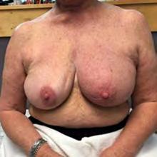
Swollen oedematous left breast at presentation
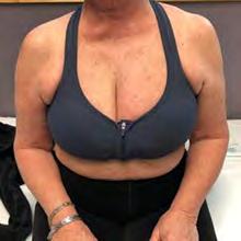
Elastic low-pressure night- t ime compression bra for use at night

Reduced swelling after 1 week of using the compression bra
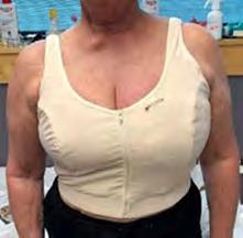
Stiff compression vest with textured focal pad for daytime use
Declaration of interest: Sponsored by Haddenham Healthcare and the US Medical Compression Alliance (USMCA); Suzie Ehmann has been a paid consultant or speaker for L&R, Urgo, OVIK, Compression Dynamics and Medline; Brandy McKeown has been a paid consultant or speaker for HMP global/Wound Source, L&R, LymphaPress, Juzo and Sigvaris; Sandi Davis has been a paid consultant for Koya Medical; Karen J Bock has been a paid consultant for Pure Medical; Justine C Whitaker has been a paid consultant or speaker for Medi and Jobst
References
1. Bjork R, Ehmann S. S.T.R.I.D.E. professional guide to compression garment selection for the lower extremity. J Wound Care. 2019; 28(S6a):1–44. https://doi.org/10.12968/jowc.2019.28.Sup6a.S1
2. Greenhalgh T, Papoutsi C. Studying complexity in health services research: desperately seeking an overdue paradigm shift. BMC Medicine. 2018; 16(1):95. https://doi.org/10.1186/s12916 018 1089
4
3 . Bock KJ, Ehmann S, Dolgoy N et al. Delphi study on the STRIDE algorithm for compression selection in upper b ody lymphoedema. J Wound Care. 2025; 34(S11C):S55–S59
4. Everett J, Lawrance S. Use of Haddenham Venex armsleeve for lymphoedema management in clinical practice. Br J Community Nurs. 2019; 24(S10):S12–S18. https://doi.org/10.12968/bjcn.2019.24. Sup10.S12
5. Goss JA, Greene AK. Sensitivity and specificity of the Stemmer sign for lymphedema: a clinical lymphoscintigraphic study. Plast Reconstr Surg Glob Open. 2019; 7(6):e2295. https://doi.org/10.1097/ gox.0000000000002295
6. Whiting E, McCready ME. Pitting and non p itting oedema. Med J Austr. 2016; 205(4):157–158. https://doi.org/10.5694/mja16.00416
7. Duygu-Yildiz E, Bakar Y, Hizal M. The effect of complex decongestive physiotherapy applied with different compression pressures on skin and subcutaneous tissue thickness in individuals with breast cancer related lymphedema: a double b linded randomized comparison trial. Support Care Cancer. 2023; 31(7):383. https://doi.org/10.1007/s00520 023 07843 y
8. Fernandes MG, da Silva LP, Cerqueira MT, Ibañez R, Murphy CM, Reis RL, Marques AP. Mechanomodulatory biomaterials prospects in scar prevention and treatment. Acta Biomaterialia. 2022; 150:22–33. https://doi.org/10.1016/j.actbio.2022.07.042
9. Mansur AT, Ozker E, Tukenmez Demirci G. A case of elephantiasis nostras verrucosa treated successfully by a new type of compressive garment. Dermatol Ther. 2020; 33(6):e14348
10. Chohan A, Haworth L, Sumner S, Olivier M, Birdsall D, Whitaker J. Examination of the effects of a new compression garment on skin tissue oxygenation in healthy volunteers. J Wound Care. 2019; 28(7):429–435. https://doi.org/10.12968/jowc.2019.28.7.429
11. Ashforth K, Morgner S, VanHoose L. A new treatment for soft tissue fibrosis in the breast - Wounds International. 2011. https:// woundsinternational.com/ journal-articles/a-new-treatment-for-soft-tissue-fibrosis-in-thebreast/ (accessed 1 October 2025)
12. Clark M, Krimmel G. Template for practice: compression hosiery in lymphoedema. 2009. https://woundsinternational.com/ best pr actice s tatements/template for pr actice compression h osiery in l ymphoedema/ (accessed 1 October 2025)
13. Lymphology ISo. The diagnosis and treatment of peripheral lymphedema: 2020 Consensus Document of the International Society of Lymphology. Lymphology. 2020; 53(1):3–19
14. Partsch H, Schuren J, Mosti G, Benigni J-P. The static stiffness index: an important parameter to characterise compression therapy in vivo. J Wound Care. 2016; 25(S9):S4–S10. https://doi. org/10.12968/jowc.2016.25.Sup9.S4
15. Partsch H, Damstra R, Mosti G. Dose finding for an optimal compression pressure to reduce chronic edema of the extremities. Int Angiol. 2011; 30(6):527–533
16. Mosti G, Mattaliano V, Partsch H. Inelastic compression increases venous ejection fraction more than elastic bandages in patients with superficial venous reflux. Phlebology. 2008; 23(6):287–294. https://doi.org/10.1258/phleb.2008.008009
17. Partsch H, Partsch B, Braun W. Interface pressure and stiffness of ready made compression stockings: comparison of in vivo and in vitro measurements. Journal of Vascular Surgery. 2006; 44(4):809–814. https://doi.org/10.1016/j.jvs.2006.06.024
18. Mazur S, Szczęśniak D, Tchórzewska-Korba H. Effectiveness of Mobiderm Autofit in the intensive phase of breast cancer-related lymphedema treatment: a case series. Lymphat Res Biol. 2023; 21(6):608–613. https://doi.org/10.1089/lrb.2022.0079
19. Dhar A, Srivastava A, Pandey RM, Shrestha P, Villet S, Gogia AR. Safety and efficacy of a mobiderm compression bandage during intensive phase of decongestive therapy in patients with breast cancer related lymphedema: a randomized controlled trial. Lymphat Res Biol. 2023; 21(1):52–59. https://doi.org/10.1089/ lrb.2021.0104
20. Todd M, Stubbs C, Pugh S. Mobiderm Autofit: an adjustable sleeve that enables patients to self manage lymphoedema. Br J Community Nurs. 2018; 23(S4):S14. https://doi.org/10.12968/ bjcn.2018.23.Sup4.S22
21. Ehmann S. The biophysical impact of an alternating compression profile (thesis submitred to Nova Southeastern University Florida, US). 2024. https://ezproxylocal.library.nova.edu/ login?url=https://www.proquest.com/dissertations t heses/ biophysical impact alternating compression/docview/3083575791/ se 2?accountid=6579 (accessed 1 October 2025)
22. Brunelle CL, Ag AG. The important role of nighttime compression in breast cancer related lymphedema treatment. Cancer. 2022; 128(3):458–460. https://doi.org/10.1002/cncr.33942
23. Phillips N, Lawrance S. Haddenham Comfiwave: a unique compression device for lymphoedema treatment. Br J Community Nurs. 2020; 25(S4):S23–S30. https://doi.org/10.12968/bjcn.2020.25. Sup4.S23
24. Rutkowski JM, Swartz MA. A driving force for change: interstitial flow as a morphoregulator. Trend Cell Biol. 2007; 17(1):44–50. https://doi.org/10.1016/j.tcb.2006.11.007
25. Boardman KC, Swartz MA. Interstitial flow as a guide for lymphangiogenesis. Circ Res. 2003; 92(7):801–808. https://doi. org/10.1161/01.Res.0000065621.69843.49
26. Bertsch T. Evaluation of a novel night‑time compression garment: a prospective observational study. Br J Community Nurs. 2018; 23(11):535–541. https://doi.org/10.12968/bjcn.2018.23.11.535
27. Bock KJ, Muldoon J. A 24 h our interval compression plan for managing chronic oedema: part 1 t he science and theory behind the concept. J Wound Care. 2022; 31(S2):S4–S9. https://doi.
org/10.12968/jowc.2022.31.Sup2.S4
28. Williams AF. Working in partnership with patients to promote concordance with compression bandaging. Br J Community Nurs. 2012; 17(S10a):S1–S16. https://doi.org/10.12968/bjcn.2012.17. Sup10a.S1
29. Whitaker JC. Lymphoedema management at night: views from patients across five countries. Br J Community Nurs. 2016; 21(S10):S22–S30. https://doi.org/10.12968/bjcn.2016.21.Sup10.S22
30. Dolgoy ND, Al Onazi MM, Parkinson JF et al. The appraisal of clinical practice guidelines for breast cancer related lymphedema. Lymphat Res Biol. 2023; 21(5):469–478
31. Davies C, Levenhagen K, Ryans K, Perdomo M, Gilchrist L. Interventions for breast cancer–related lymphedema: clinical practice guideline from the Academy of Oncologic Physical Therapy of APTA. Phys Ther. 2020; 100(7):1163–1179. https://doi. org/10.1093/ptj/pzaa087
32. Armer J. ONS Guidelines™ for cancer treatment–related lymphedema. Oncol Nurs Forum. 2020; 47(5):518–538. https://doi. org/10.1188/20.ONF.518 538
33. McLaughlin SA, Staley AC, Vicini F et al. Considerations for clinicians in the diagnosis, prevention, and treatment of breast cancer related lymphedema: recommendations from a multidisciplinary expert ASBrS panel: part 1: definitions, assessments, education, and future directions. Ann Surgi Oncol. 2017; 24:2818–2826. https://doi.org/10.1245/s10434–017–5982–4
34. Levenhagen K, Davies C, Perdomo M, Ryans K, Gilchrist L. Diagnosis of upper quadrant lymphedema secondary to cancer: clinical practice guideline from the oncology section of the American Physical Therapy Association. Phys Ther. 2017; 97(7):729–745. https://doi.org/10.1093/ptj/pzx050
35. Koller M, Döller W, Földi E et al. S2k guideline: diagnostics and therapy of lymphoedema. 2017. https://register.awmf.org/assets/ guidelines/058_Ges_D_Lymphologen/058-001le_S2k_Diagnostics_ and_therapy_of_lymphoedema_2019-07-abgelaufen.pdf (accessed 1 October 2025)
36. Queensland Health. The use of compression in the management of adults with lymphoedema. Brisbane: Queensland Health; 2014
37. Abney SE, Ijaz MK, McKinney J, Gerba CP. Laundry hygiene and odor control: state of the science. Elkins CA, ed. Appl Environ Microbiol. 2021;87(14):e03002 20. https://doi.org/10.1128/ AEM.03002 20
38. Bojesen S, Midttun M, Wiese L. Compression bandaging does not compromise peripheral microcirculation in patients with cellulitis of the lower leg. Eur J Dermatol. 2019;29(4):396–400. https://doi.org/10.1684/ejd.2019.3606
39. Dräger S, Kiehne C, Zinser G, Kahle B. Treating cellulitis promptly with compression therapy reduces C‐reactive protein‐levels and symptoms – a randomized‐controlled trial. J Deutsche Derma Gesell. August 112025:ddg.15829. https://doi.org/10.1111/ddg.15829
40. International Society of Lymphology. The diagnosis and treatment of peripheral lymphedema: 2020 consensus document of the International Society of Lymphology. Lymphology. 2020; 53(1):3–19
41. Clark M. EWMA Position document: understanding compression therapy Wounds International. 2003. https://woundsinternational. com/best pr actice s tatements/understanding compression t herapy e wma p osition d ocument w int/ (accessed 1 October 2025)
42. Ehmann S, McKeown B, Davis S, Bock KJ. The science of compression textiles and garments for upper b ody lymphoedema. J Wound Care. 2025; 34(S11C):S19–S29
43. International Lymphoedema Framework. Template for practice: compression hosiery in upper body lymphoedema. 2009. https:// www.lympho.org/uploads/files/files/Upper_body.pdf (accessed 20 August 2025)
44. Bjork R, Hettrick H. Endothelial glycocalyx layer and interdependence of lymphatic and integumentary systems Wounds International. 2018. https://woundsinternational.com/ journal ar ticles/endothelial g lycocalyx layer an d interdependence of l ymphatic an d integumentary s ystems/ (accessed 20 August 2025)
45. Bjork R, Hettrick H. Lymphedema: new concepts in diagnosis and treatment. Cur Dermatol Rep. 2019; 8:190–198
46. Davis S, Ehmann S, McKeown B et al. Anatomy, pathophysiology and assessment of upper- b ody lymphoedema. J Wound Care. 2025; 34(S11C):S5–S18
Suzie Ehmann, Brandy McKeown, Sue Lawrence, Stephanie Moore, Mariam Aldashti and Sandi Davis
Abstract
This article presents six case studies demonstrating use of the STRIDE algorithm for selecting compression garments for upper-body lymphoedema. STRIDE offers a structured, evidence-informed and individualised approach to assessment and selection. It incorporates patients’ multifactorial and evolving needs, as well as textile properties beyond pressure dosage alone to enhance long-term management and overall quality of life.
Keywords: STRIDE algorithm | Upper b ody lymphoedema | Compression garment selection | Flat k nit and circular k nit garments | Textile stiffness and containment | Patient centred therapy | Daytime and night‑time compression | Breast and truncal oedema | Case b ased evidence | Adherence
The following six cases demonstrate how the STRIDE algorithm can be used to select an optimal, appropriate and individualised compression regimen for people with upper b ody lymphoedema. Compression therapy for lymphoedema is well established in the lower limb, but it remains a relatively new frontier for the arm, breast and trunk, where robust evidence, clear guidelines and standardised practices are still lacking. The STRIDE algorithm offers a structured, evidence informed approach that helps clinicians move beyond trial and error methods. STRIDE uses a detailed assessment of a patient’s presentation, functional needs and therapeutic goals to identify and overcome challenges in garment selection, reassessment and adaptation to achieve optimal patient outcomes.
A 71‑year‑old woman diagnosed with metastatic breast cancer presented with left upper‑quadrant lymphoedema and a fungating breast tumour (Figure 1).
Suzie Ehmann, DPT, PhD, CWS, CLT L ANA, CLWT, Lymphedema Therapist, McLeod Health Seacoast, Little River, SC, USA
Brandy McKeown, OTR/L, CLT L ANA, CLWT, Lymphedema
Therapist, International Lymphedema and Wound Training Institute and the Lymphedema Center, USA
Sue Lawrence, RGN, Clinical Nurse Specialist, Buckinghamshire Healthcare Trust, UK
Stephanie Moore, MA, PT, DPT, CLT, CCET, Oncology Physical Therapist, North Kansas City Hospital, Kansas City, MO, USA
Mariam Aldashti, DPT, CLWT, Doctor of Physical Therapy, Kuwait Hospital, Kuwait
Sandi Davis, PT, DPT, CLT L ANA, CLWT, President, Davis Care Physical Therapy, New York City, NY, USA
▪ Site: there was extensive oedema in the entire left arm, hand, breast and chest wall, necessitating comprehensive compression coverage.
▪ Shape: the arm, hand and breast maintained a normal shape, aligning with ready to wear (RTW) sizing.
▪ Size: the left arm circumference was 81% larger than the right.

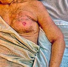

The tissue of the breast and chest wall was woody, suggestive of chronic inflammation and fibrosis, indicating use of textured textiles to soften fibrotic tissue. The left upper‑limb tissue was doughy, with areas of fibrosis and grade 3 pitting, indicating a stiffer garment to afford increased containment, as well as textured textiles. Pitting reduced to grade 1 following complete decongestive therapy (CDT).
The upper limb demonstrated progressive daytime swelling and minimal overnight reduction prior to CDT and remained moderate after CDT. This dynamic change in volume throughout the day in the upper limb demonstrated the need for a night‑time sleeve and glove/gauntlet. The breast showed no overnight refill, meaning it could go without compression at night for increased patient comfort.
▪ Financial issues: cost considerations contributed to the choice of RTW garments, which were more affordable than custom alternatives while maintaining therapeutic efficacy.
▪ Functional issues: post chemotherapy fatigue impacted ability to don/doff garments, initially requiring family assistance, and encouraging lower dosages that are easier to tolerate.
▪ Psychological issues: the patient preferred the cosmetic appearance of sleeves to adjustable wraps.
▪ Day: medium pressure (20–30 mmHg) flat knit combined sleeve and glove; medium pressure (20–30 mmHg) compression bra with focal pad.
▪ Night: low pressure (10–15 mmHg) textured chip sleeve with oversleeve (+10 mmHg).
The patient’s late stage 2 breast cancer‑related lymphoedema required firm compression.
Rationale and outcome
After reduction of limb volume, a medium‑pressure, higher‑stiffness flat‑knit sleeve and glove for daytime use provided strong containment and easier donning compared with an elastic sleeve of the same dosage, as well as being cosmetically acceptable. A medium‑pressure compression bra with a textured focal pad was chosen to address breast involvement with targeted pressure. At night, a low‑pressure textured chip sleeve with oversleeve was chosen to address residual tissue texture, prioritising lymphatic stimulation and skin integrity. No breast compression was included for patient comfort.
The patient’s compression strategy successfully managed swelling, improved tissue pliability and enhanced quality of life by enabling independent use of garments following strength recovery.
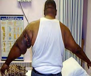
Presentation prior to treatment
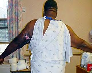
After complete decongestive therapy
A 65‑year‑old man presented with progressive lymphoedema of the upper limb and a persistent wound on the posterior upper arm caused by complications from a dysfunctional dialysis graft (Figure 2). The oedema had persisted for more than 6 months, and no targeted lymphoedema care had been initiated before referral. Treatment began with CDT three times per week, incorporating skin and wound care, manual lymphatic drainage (MLD) and multi‑component bandaging. Between sessions, the patient remained in multi‑layer compression bandages.
▪ Site: significant congestion was observed throughout the upper arm and hand.
▪ Shape: after CDT, there were redundant skin folds in areas of previous swelling, requiring stiff structural support to avoid areas of cording.
▪ Size: the patient’s height (186 cm), weight (422 lb) and arm length could not be adequately covered by RTW garments and thus required a custom garment.
The tissue texture was generally doughy, with grade 2 pitting, a positive Stemmer's sign on the hand and a positive Bjork bowtie test throughout the arm and shoulder. There were prominent distal areas of fibrosis that persisted following CDT and required textured textiles to manage.
Initially, the patient reported no visible changes in limb size throughout the day or night. After CDT, refill was rapid once compression was removed, indicating both daytime and night‑time compression were necessary. The limb volume was highly variable, with dynamic swelling.
Issues
▪ Medical issues: the patient’s dysfunctional arteriovenous fistula eliminated contraindications to compression therapy (alternative vascular access was used for haemodialysis).
▪ Functional issues: the patient had good strength and range of motion, so he was able to independently don/doff compression garments, enabling strong adherence to therapy.
Dosage
▪ Day: custom medium pressure (23–32 mmHg) flat knit sleeve and glove.
▪ Night: low pressure (15–20 mmHg) textured circular knit sleeve.
Etiology
The presentation of late stage 2 lymphoedema required stiff compression to afford adequate containment and address fibrotic tissue changes.
Rationale and outcome
For daytime use, a custom medium‑pressure flat‑knit sleeve and glove were selected to contain fibrotic tissue and bridge skin folds that resulted for dramatic volume loss—circular‑knit options lacked the structural integrity needed to prevent cording and control oedema. At night, a low‑pressure textured circular‑knit sleeve was chosen to address residual tissue texture while prioritising sleep comfort and hand coverage. Following structured compression therapy, the patient achieved stable volume management, improved softening of fibrotic tissue and enhanced limb mobility. Consistent garment adherence ensured long‑term oedema control despite the dynamic nature of the condition.
A 38‑year‑old woman with stage 2B hormone receptor‑positive right breast cancer underwent neoadjuvant chemotherapy, followed by a right mastectomy and axillary lymph‑node dissection. The tissue expander placed during initial surgery was later removed due to erosion, and she subsequently completed

21 radiation treatments to the right chest wall. Approximately 6 months post‑radiation, she developed early‑stage truncal lymphoedema, initially managed with CDT, self‑administered MLD, compression garments, a pneumatic compression pump and upper‑limb exercises. Over the next 3 years, frequent airline travel without compression exacerbated her condition, resulting in clinically progressive lymphoedema—advancing from early to late stage 2—with worsening truncal oedema and tissue texture changes that prompted referral for further lymphatic therapy (Figure 3).
▪ Site: there was swelling in the entire right arm and trunk (but not the hand), especially concentrated in the right upper back, necessitating comprehensive compression coverage.
▪ Shape: the right arm and most of the trunk maintained a normal shape, suitable to RTW sizing. However, post‑mastectomy, the right upper back had asymmetrical contours, requiring a focal pad in the bra for more uniform distribution of compression.
▪ Size: the right arm had swollen by 12% in volume since the prior baseline, and there was increased intra axillary fluid collection compared with previous assessments.
The tissue largely had a stable watery texture. The right upper back showed mild progressive oedema, with grade 2 pitting, requiring stronger compression.
Upright activities led to refill in the day, requiring daytime compression. The mild pitting oedema in the upper quadrant did not refill overnight, allowing for daytime compression only. Night‑time refill in the arm and trunk improved after CDT, allowing the patient to use her pneumatic compression pump nightly instead of wearing a garment while sleeping.
▪ Psychological issues: the patient was highly motivated, ensuring strong adherence to compression routines. She also valued social aspects of dressing, balancing compression needs with personal style, opting for short periods out of compression to wear dresses when socialising. The patient’s prioritisation of cosmesis meant a reluctance to use adjustable wraps or thick flat knit garments.
▪ P re-reconstruction (day only): medium p ressure (20–30 mmHg) bra with focal pad; medium pressure (20–30 mmHg) circular knit sleeve; medium pressure (20–30 mmHg) gauntlet.
▪ Post-reconstruction (day only): low pressure t shirt (15–20 mmHg); medium pressure (20–30 mmHg) stiffer circular knit sleeve.
At the time of referral to lymphoedema management, late stage 2 lymphoedema required structured compression to manage swelling across the upper limb and trunk.
The truncal oedema was initially managed with a medium‑pressure compression bra with a focal pad, chosen to target post‑radiation fibrosis in the upper back and chest wall and to address asymmetrical truncal swelling post‑mastectomy. After breast reconstruction, the patient transitioned to a low‑pressure t‑shirt for greater comfort and coverage.
Upper‑limb oedema was initially managed with a medium‑pressure stiffer circular‑knit sleeve and a medium‑pressure gauntlet. The gauntlet was soon discontinued due to lack of hand swelling. After breast reconstruction, a stiffer circular‑knit sleeve was used to balance cosmesis with functional containment.
A pneumatic compression pump was used to maintain lymphatic control without compromising sleep or garment adherence. The patient’s refill patterns and successful intermittent pneumatic compression (IPC) use meant she did not require night‑time compression garments for maintenance reduction.
The patient’s consistent use of daytime compression and IPC maintained stable reduction of truncal and upper‑limb lymphoedema. These garment choices also allowed her flexibility in personal wardrobe preferences, supporting both her clinical needs and her motivation and desire for social confidence.
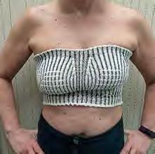
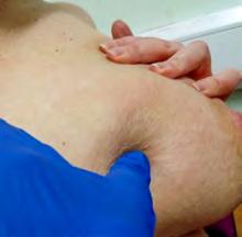

A 50‑year‑old woman developed post‑surgical and radiation‑induced lymphoedema after cellulitis following a wide local excision, sentinel node biopsy and radiotherapy for left breast cancer (Figure 4).
▪ Site: oedema was confined primarily to the distal, medial and lateral breast, with the surrounding chest wall and arms remaining unaffected, requiring a compression bra targeted to breast tissue without affecting adjacent areas.
▪ Shape: scar tissue caused tightness, with associated breast heaviness and cording restrictions to range of motion.
▪ Size: the swelling was moderate, and the breast and trunk size met the criteria for RTW garments.
The breast tissue had a firm peau d’orange texture, with pitting oedema, necessitating a gradual introduction to compression therapy, incorporating focal pads or textured textiles to enhance drainage and soften tissue.
The oedema refilled overnight, causing discomfort and requiring compression during the day and night.
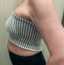
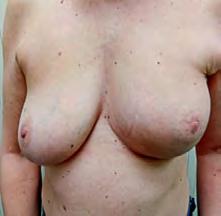

▪ Functional and medical issues: scar tension caused mobility restrictions that were improved with supplementary MLD, exercise and Kinesio.
▪ Day: medium pressure (20–30 mmHg) textured compression bra with focal pads (initial).
▪ Night: low pressure (15–20 mmHg) textured compression (later adopted full time).
The patient’s presentation of late stage 2 breast cancer‑related lymphoedema required stiffer compression and a focal pad support to address tissue texture changes and promote tissue remodelling across the affected area.
For daytime compression, initial trial of supportive sports bras was transitioned to a medium‑pressure textured compression bra with focal pads to target localised breast oedema and post‑radiation fibrosis, while preserving comfort and mobility. A low‑pressure textured (ribbed) bra was initially introduced for night‑time, due to refill, and it was later adopted full‑time for enhanced comfort, control and wearability.
Following ongoing compression therapy, the patient achieved a reduction in swelling, softened tissue texture, natural appearance and greater comfort in daily activities within 2 weeks. The gradual transition from sports bras to medical compression facilitated adaptation, ensuring long‑term adherence.
Figure 5. Case 5
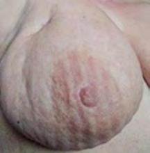
Oedematous and fibrinous breast with linear impressions from a textured bra band
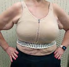
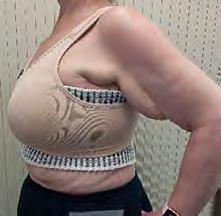
Daytime compression system
A 60‑year‑old woman, diagnosed with grade 2 invasive ductal carcinoma of the left breast, underwent neoadjuvant chemotherapy, followed by lumpectomy and axillary dissection (15 lymph nodes removed). Post‑surgery, she required multiple seroma drainages and incision and drainage of an axillary abscess, which led to cellulitis and the onset of breast and left arm lymphoedema. Due to restricted range of motion, her radiation treatment was delayed (Figure 5).
▪ Site: swelling extended through the entire left upper limb from the fingers through to the left axilla and breast, requiring multi zone compression coverage.
▪ Shape: all areas generally had a normal shape, but there were redundant skin folds in the upper arm that could not be bridged by stiffer circular knit sleeves.1
▪ Size: circumferential measurements aligned with RTW sizing.
The left breast tissue was firm and doughy, with a peau d’orange appearance and deep scarring in the upper lateral quadrant, requiring moderate compression with a textured textile to soften the tissue. The upper limb showed fibrosis and moderate pitting of doughy texture, necessitating a textured textile with moderate containment.
Refill was rapid and persistent, necessitating moderate compression in the day and mild at night. Fluctuations in breast swelling needed to be accommodated with flexible garment selection.
▪ Device issues: initially, textured textiles were challenging to tolerate, leading to a phased transition to higher compression levels.
▪ Functional issues: scar tissue and prior infections limited early compression garment tolerance, requiring gradual adaptation of compression options.
▪ Psychological issues: the patient valued cosmesis and preferred to avoid adjustable wraps for daytime use.
▪ Day: custom medium pressure (23–32 mmHg) flat knit sleeve and glove; low pressure (15–20 mmHg) circular knit compression bra; low pressure (15–20 mmHg) textured breast band.
▪ Night: low pressure (15–20 mmHg) textured night‑time sleeve with finger coverage; low pressure (15–20 mmHg) textured breast band.
The patient’s presentation of late stage 2 breast cancer‑related lymphoedema required stiffer compression and a focal pad support to address areas of firm peau d’orange tissue texture and localised oedema, as well as promote tissue remodelling across the affected area, without compressing unaffected areas in the trunk.
In the daytime, compression for the upper limb was provided with a custom medium‑pressure flat‑knit sleeve and glove to ensure containment. For the trunk, a low‑pressure circular‑knit compression bra was layered over a low‑pressure textured (ribbed) breast band, with the layering providing a combined moderate pressure. The textured textile extended beyond the bra’s circumferential band to address fibrotic tissue, prevent cording along the lateral trunk and ensure uniform pressure distribution.
Night‑time compression was provided with a low‑pressure textured night‑time sleeve with finger coverage, as well as the low‑pressure same ribbed breast band, used alone, to ensure adequate compression, minimise refill, enhance comfort and soften tissue at night. Based on patient feedback, this was adopted full‑time for improved comfort and control.
Following structured compression therapy, the patient achieved reduced swelling, improved tissue pliability and enhanced mobility. The gradual approach to compression garment transition supported sustained adherence and tissue remodelling, leading to better long‑term management of lymphoedema. A multi‑garment approach balanced oedema containment while maintaining patient comfort.
A 70‑year‑old woman presented with stage 2A lymphoedema in the left upper limb, following a left mastectomy performed 7 years previous. While she had previously used an RTW compression sleeve, she had not worn any compression for the past 2 years. The patient reported no prior signs of oedema until a few months before her consultation (Figure 6).
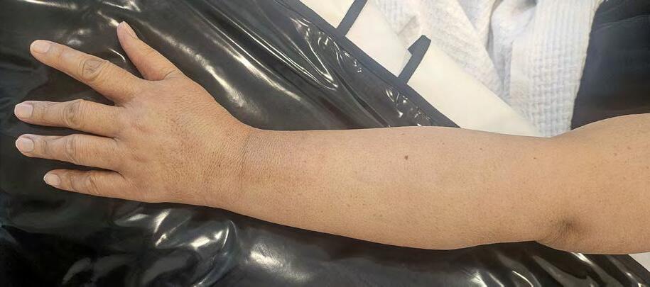
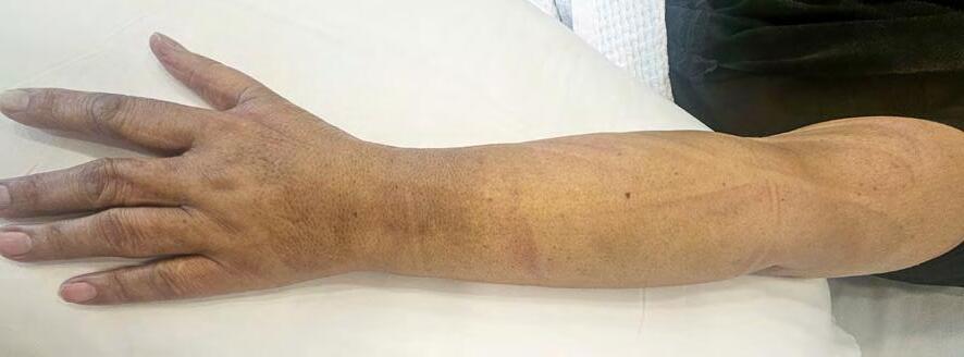
▪ Site: the oedema was localised to the left hand and arm.
▪ Shape: the patient’s hand and arm maintained a normal anatomical shape, making her eligible for standard size RTW garments.
▪ Size: left arm circumferential measurements were 42% greater than the right.
The upper‑limb tissue was doughy, with grade 1 pitting above the elbow and grade 2 pitting from the dorsum of the hand through to the forearm (10‑second rebound). The Bjork bowtie test was positive on the arm and Stemmer’s sign on the dorsal hand, indicating early fibrosis and skin thickening. This warranted stiffer compression textiles to provide better containment, as well as a textured textile to address fibrosis.
Refill was slow, with oedema reoccurring over 24 hours after compression removal. However, refill was persistent through the day and night, with minimal improvement from limb elevation, necessitating night‑time compression.
▪ Functional issues: reduced upper limb strength made donning/doffing difficult, requiring adjustable wraps for improved adherence.
▪ Knowledge issues: the patient had previously found stiff RTW garments uncomfortable, leading to long term avoidance of compression therapy.
▪ Day: medium pressure (20–30 mmHg) flat knit combined sleeve and glove
▪ Night: low pressure (15–20 mmHg) night‑time sleeve
The patient’s late stage 2 lymphoedema following breast cancer treatment required stiff compression to ensure adequate containment of the left upper limb and address early fibrotic changes in the forearm and dorsal hand.
For daytime use, a medium‑pressure flat‑knit combined sleeve and glove were selected to provide firm containment, address early fibrotic changes and prevent further fibrosis, particularly in the forearm and dorsal hand. The flat‑knit textile offered the necessary stiffness and structure to manage pitting and prevent progression, while maintaining anatomical fit given the patient’s preserved limb shape.
Night‑time compression was introduced due to persistent refill and limited response to elevation. A low‑pressure night‑time sleeve was chosen to maintain continued focal treatment and oedema reduction and support tissue softness, with a lower dosage for comfort. The patient’s prior discomfort with stiff garments was addressed through education and gradual reintroduction of compression, balancing therapeutic goals with physical capability and garment tolerance.
Following structured compression therapy, the patient achieved stable oedema containment, improved tissue softness and enhanced adherence.
The case studies presented illustrate how the STRIDE algorithm can be used to guide precise selection of compression garments for the arm, breast and trunk, tailored to a patient’s individual anatomy and clinical challenges.
These cases underline how patient needs are multifactorial, and compression recommendations should include more than dosage (interface pressure). Instead, recommendations should also encompass other textile properties, such as stiffness and containment. Likewise, prioritising comfort can promote patient compliance, which is pivotal to long‑term management. Understanding functional, psychological and financial issues can ensure compression solutions are not just clinically effective but also practical for real‑world adherence. Several of the cases also show how compression needs are dynamic, evolving with the patient’s condition and necessitating continuous reassessment and adaptation.
Case studies can provide insights into the evolving landscape of compression therapy. Moreover, they are a way to build the evidence necessary to guide clinicians in optimal compression selection for upper‑body lymphoedema.
Declaration of interest: Sponsored by Haddenham Healthcare and the US Medical Compression Alliance (USMCA); Suzie Ehmann has been a paid consultant or speaker for L&R, Urgo, OVIK, Compression Dynamics and Medline; Brandy McKeown has been a paid consultant or speaker for HMP global/Wound Source, L&R, LymphaPress, Juzo and Sigvaris; Sue Lawrence has been a clinical advisor to Haddenham Healthcare; Sandi Davis has been a paid consultant for Koya Medical
Karen J Bock, Suzie Ehmann, Naomi Dolgoy, Sandi Davis, Brandy McKeown, Justine Whitaker and Elizabeth Anderson
Abstract
Background: The original STRIDE algorithm covered lower-limb lymphoedema but not the upper body.
Aims: To update the STRIDE algorithm for compression selection to treat lymphoedema of the upper limb, breast and trunk by achieving consensus on the definitions and importance of its six aspects.
Method: Using a modified Delphi framework, clinical experts in the field ranked agreement and gave open-ended feedback over two rounds of surveys, with a >70% threshold for agreement.
Results: In the first round, participants represented five continents (n=36). Characteristics that met the threshold consensus of >70% agreement were then applied to the STRIDE algorithm, and the second survey was developed. In the second round (n=22), the definitions of all elements of the STRIDE algorithm had at least 70% agreement or strong agreement. Shape and issues were the elements most often considered first in compression selection, while refill was least often considered first in selection.
Conclusions: This Delphi study achieved consensus on the descriptions of the elements of the revised STRIDE algorithm for compression in upper-limb, breast and trunk lymphoedema. The STRIDE algorithm can now be used to make clinical decisions on selecting compression garments for the upper body.
Keywords: Upper b ody lymphoedema | Complete decongestive therapy | Delphi consensus method | Compression garment selection | Clinical expert panel | International guideline development | Patient–clinician partnership | Quality of life outcomes | Evidence b ased practice | Complexity informed research
Lymphoedema can significantly impact a person’s overall quality of life.1 Oedema can result from lymphatic damage from an underlying dysfunction or inflammatory processes, or it may be secondary to trauma, cancer treatments or other comorbidities, such as neurological dysfunction.2,3 Lymphoedema is commonly treated with compression therapy as a component of complete decongestive therapy (CDT), with garments used to apply pressure to the affected sites to optimise lymphatic drainage and reduce oedema.2,4 12 However, selecting the optimal compression garment and/or device for lymphoedema in the upper limb, breast and trunk can be challenging due to variations
Karen J Bock, PT, PhD, CWS, CLT L ANA, Assistant Professor, Rockhurst University, Kansas City, MO, USA
Suzie Ehmann, DPT, PhD, CWS, CLT L ANA, CLWT, Lymphedema Therapist, McLeod Health Seacoast, Little River, SC, USA
Naomi Dolgoy, MOT, PhD, CLT, Assistant Professor, University of Alberta, Canada
Sandi Davis, PT, DPT, CLT L ANA, CLWT, President, Davis Care Physical Therapy, New York City, NY, USA
Brandy McKeown, OTR/L, CLT L ANA, CLWT, Lymphedema Therapist, International Lymphedema and Wound Training Institute and the Lymphedema Center, USA
Justine C Whitaker, MSc, RN, PhD(c), Director & Nurse Consultant, Northern Lymphology, and Senior Lecturer, University of Central Lancashire, Preston, UK
Elizabeth Anderson, PhD, RN, CLT , Faculty and Sinclair School of Nursing, University of Missouri, Columbia, MO, USA
in clinical presentations (both the location and severity of the swelling) and variations in the availability of products in different geographical regions.13,14
The literature regarding compression selection focuses more on the lower limb than the upper body. While evidence for upper‑body compression has grown over the past 20 years, there remains a gap in consensus‑driven clinical guidelines for garment selection.4,15‑17 Furthermore, there are notable differences in clinical practice and device availability in the US and other countries.
This study aimed to address the need for a comprehensive evidence‑based guidance on compression selection for upper‑body lymphoedema by producing a new international consensus document built on the original STRIDE algorithm for compression selection, which was limited to lower‑limb lymphoedema.14 The updated STRIDE algorithm required face and content validation to achieve two objectives: to define elements involved in selecting upper‑body compression devices and to establish the importance of these elements to compression selection.
The content of the updated STRIDE algorithm was determined using a modified Delphi method.19 This structured communication technique is widely used to solicit and synthesise expert opinion to achieve consensus on complex issues.18‑20 The Delphi process
is iterative, involving multiple rounds of surveys.21,22 This process was followed for this study. Survey questions and domains were developed from the original STRIDE algorithm. At each round, a series of statements was shared with an expert panel, and participants anonymously indicated their agreement or disagreement on a 5‑point Likert scale and have the option to give open‑ended feedback. These responses were then summarised and shared with the investigators so that the statements could be collectively revised. The iterative process continued until a consensus was reached.22
The study protocol was reviewed by the Rockhurst University institutional review board and deemed no more than minimal risk and exempt from ongoing review (exempt review #2425‑37).
Participants completed informed consent for each round of the survey, conforming to the Declaration of Helsinki guidelines. The survey was developed by the primary investigator, with face validity provided by remaining authors. Content validity was confirmed through previewed surveys and feedback from an international group of lymphoedema therapists and clinicians for cultural and language adaptations to facilitate universal understanding.20
Recruitment of content experts prioritised qualified clinicians active in compression selection for upper‑body lymphoedema. Content experts had to have at least 2 years of relevant clinical experience,18 as well as meet at least one of the following criteria:
▪ In the past month, have worked in a setting with a caseload of at least five patients with upper body lymphoedema
▪ At any point in their career, have had a caseload where at least 30% of patients had upper body lymphoedema
▪ Be currently researching upper body lymphoedema.
Purposive and snowball sampling was instituted, with primary survey dissemination through experts recommended by the research team. Respondents were excluded for the following reasons:
▪ Worked part or full time in the compression garment industry
▪ Failed to provide consent
▪ Completed only part of the survey.
The Delphi survey was created with Survey Monkey (San Mateo, California, US), with statements based on the original STRIDE algorithm and organised into domains.14
The first round covered participant demographic details and the importance of different factors for compression selection. All identified experts were emailed the first survey, with 6 weeks to complete and three reminder emails. A minimum of 30 responses in the first round was set to account for likely attrition in the second round.18
The second round covered agreement with definitions of STRIDE elements and the ranked importance of STRIDE elements to compression selection. All respondents in the first round were contacted again, with a 4‑week deadline and two reminder emails.
Consensus on a statement was set at 70% agreement.18 Statements that met this benchmark were included in the second round, with any open‑ended feedback analysed from a grounded theory perspective to further develop those statements to better describe
1. Participant demographics, n=36
Note: 1Not all demographic questions were compulsory, so some sections may not add up to 36; 2‘Local durable medical equipment providers’, ‘All referrals go to orthotist or practicing lymphoedema therapists in South Africa’
the STRIDE algorithm.23 Through iterative analysis by the research team, characteristics and domains were condensed and clustered into the STRIDE algorithm, and the second survey aimed for agreement on criteria in each section of the algorithm. In the second survey, participants ranked the use of the algorithm categories in order of importance when selecting compression garments and devices.
The participant demographic details are outlined in Table 1. Countries represented included: Australia (n=2), Canada (n=8), Ireland (n=1), Kuwait (n
South
Regarding the importance of factors for compression selection, all but one factor met the threshold of being considered moderately or highly important by 70% of respondents (Figure 1). The exception was hand dominance, at 52.1%, which did not meet the threshold
Limb shape
Tissue composition
Donning/doffing ability
Function during wear
Patient adherence
Oedema distribution
Limb size
Compression dosage
Tissue texture
Wounds or weeping skin
Stiffness
Patient financial burden
Comorbidities
Payor coverage
Day-and-night dynamics
Other functional issues
Climatic issues
Hand dominance
Size Site, size and shape of lymphoedematous swelling
Texture Tissue texture and textile type used in compression garments
Refill How lymphoedema volume changes with activity or throughout the day and the night
Issues Ability to don/doff garments, willingness to participate in compression regimen, function while wearing compression device, cost of device acquisition, physical/dexterity issues, medical issues
Dosage Optimal resting pressure and stiffness to manage lymphoedema
Etiology Comorbid diagnoses, stages of lymphoedema and how these affect compression selection
for consensus. The following were considered highly important by at least 70% of respondents: limb shape, tissue composition, donning/doffing ability, function during wear, patient adherence, lymphoedema distribution, limb size and compression dosage
In the second round, all six definitions of STRIDE elements (Table 2) met the 70% consensus threshold for agreement. Over 60% of participants strongly agreed with definitions of the first five elements, while only 55% strongly agreed on the definition of etiology (Figure 2). Responses to open‑ended questions were analysed and determined to be either agreement, non‑agreement or proposing a different approach for each element definition. When the importance of STRIDE elements to compression selection was ranked, shape and issues were considered the most important elements, while refill was the least important (Figure 3). The trends in the open‑ended feedback informed and mirrored the ranking of the level of importance of each element. There were no comments specific enough to determine trends between any malalignment with the importance of the elements.
In two rounds of Delphi surveys, an international panel of clinical experts reached a consensus on definitions of the six elements of the STRIDE algorithm, as well as on the importance of these elements in compression selection for upper‑body lymphoedema.
The Delphi study findings informed the development of the new STRIDE algorithm for the upper body, creating an innovative approach to compression selection that spans traditional boundaries of dosage and single‑method compression selection therapies. This allows best practice to be standardised and shared with clinicians and the wider medical community to improve care for people with lymphoedema .
The goal of developing the STRIDE algorithm for upper‑body lymphoedema, to optimise outcomes for individuals living with lymphoedema, mirrors the paradigm of a complexity‑informed approach to researching health services and systems.19 It was vital to explore international expert consensus on characteristics of compression selection and note how complexity‑informed
Figure 2. Agreement with definitions of STRIDE elements, second round, n =20 (%)
Figure 3. Ranked importance of STRIDE elements in compression selection, where rank 1 is the most and rank 6 the least important, n =20 (%)
Site, size and shape
Tissue
approaches seek to generate insights with multiple perspectives on an issue.23 The ranking of importance in Figure 3 demonstrates that clinicians focus on different key elements to arrive at a treatment regimen for the patients. A complexity‑informed approach to research embraces the imperfection of datasets and the need for future research to collect more data to keep up with evolving developments in the field.24,25 The generation of the STRIDE algorithm to organise clinical approaches to compression garment selection is the beginning of future research embracing a scientific approach to compression garment selection while emphasising the important role of the patient–clinician partnership in pragmatic, tailored application.24
Clinical implementation of STRIDE would benefit from formal implementation frameworks and standardised assessment of how the algorithm affects outcomes, as well as local and regional adaptations.26,27 Implementation frameworks are designed for assessment of action or practice cycles of knowledge acquisition, use and adjustment to tailor the intervention to the individuals affected, often going back and forth between these steps multiple times.28,29 These frameworks are frequently used in physical medicine and rehabilitation.30 In the US, the Knowledge to Action framework is being applied to the physical therapy clinical practice guideline for diagnosing breast cancer‑related lymphoedema to monitor and evaluate the use of the guidelines.26 Case series and mixed‑method analysis from different countries, geographies/
climates, financial models and healthcare models are needed to explore the complexities of STRIDE in depth. This may generate different versions of STRIDE that are more applicable to specific groups of individuals living with lymphoedema. These versions of STRIDE may identify systemic issues and allow health systems to find political, strategic and financial alignments that improve health quality for their communities.24
Although the STRIDE algorithm is the first framework to detail compression selection for the upper limb, breast and trunk, other organisations are bringing lymphoedema into their frameworks for lower‑limb compression. The Wound, Ostomy, Continence Nurses (WOCN) Society has recently incorporated lymphoedema into its algorithm for compression therapy in the comprehensive management of lower‑limb venous disease and lymphoedema.31 International lymphology organisations have highlighted the impact of chronic oedema on quality of life,32 and numerous national clinician groups have developed clinical practice guidelines for interventions addressing upper‑body lymphoedema.33‑35
The STRIDE Delphi study had several limitations. While the panel was international, there were no surveys from South America, so the transferability of outcomes to this continent cannot be assumed. This survey focussed on experts in upper‑body lymphoedema, so generalisability to the lower limb cannot be assumed.14 Although differences in terminology were controlled through content validity construction, non‑native English speakers
may have interpreted questions differently. Inherent bias should also be considered part of the Delphi process in the non‑random sampling technique involved in recruiting the expert respondents.
This Delphi study reached a consensus on the definitions and importance of all elements of the STRIDE algorithm for selecting compression garments for lymphoedema of the upper limb, breast or trunk. The STRIDE algorithm should help clinicians efficiently and effectively select appropriate compression garments to increase their patients’ quality of life and functional abilities. Application to real‑life cases and further research should help refine and develop this structured approach to compression selection.
Declaration of interest: Sponsored by Haddenham Healthcare and the US Medical Compression Alliance (USMCA); Karen J Bock has been a paid consultant for Pure Medical; Suzie Ehmann has been a paid consultant or speaker for L&R, Urgo, OVIK, Compression Dynamics and Medline; Sandi Davis has been a paid consultant for Koya Medical; Brandy McKeown has been a paid consultant or speaker for HMP global/Wound Source, L&R, LymphaPress, Juzo and Sigvaris; Justine C Whitaker has been a paid consultant or speaker for Medi and Jobst
References
1. Donahue PMC, MacKenzie A, Filipovic A, Koelmeyer L. Advances in the prevention and treatment of breast cancer related lymphedema. Breast Cancer Res Treat. 2023; 200(1):1–14. https://doi.org/10.1007/ s10549 023 06947 7
2 International Society of Lymphology. The diagnosis and treatment of peripheral lymphedema: 2020 Consensus Document of the International Society of Lymphology. Lymphology. 2020; 53(1):3–19
3. Davies C, Levenhagen K, Ryans K, Perdomo M, Gilchrist L. Interventions for breast cancer–related lymphedema: clinical practice guideline from the Academy of Oncologic Physical Therapy of APTA. Phys Ther. 2020; 100(7):1163–1179. https://doi.org/10.1093/ptj/ pzaa087
4. McNeely ML, Dolgoy ND, Rafn BS et al. Nighttime compression supports improved self management of breast cancer–related lymphedema: a multicenter randomized controlled trial. Cancer. 2022; 128(3):587–596. https://doi.org/https://doi.org/10.1002/cncr.33943
5. Brunelle CL, Ag AG. The important role of nighttime compression in breast cancer related lymphedema treatment. Cancer. 2022; 128(3):458–460. https://doi.org/10.1002/cncr.33942
6. Bock KJ, Muldoon J. A 24 hour interval compression plan for managing chronic oedema: part 1 the science and theory behind the concept. J Wound Care. 2022; 31(S2):S4–S9. https://doi.org/10.12968/ jowc.2022.31.Sup2.S4
7. Gregorowitsch ML, Van den Bongard D, Batenburg MCT et al. Compression vest treatment for symptomatic breast edema in women treated for breast cancer: a pilot study. Lymphat Res Biol. 2020; 18(1):56–63. https://doi.org/10.1089/lrb.2018.0067
8. Mosti G, Cavezzi A. Compression therapy in lymphedema: Between past and recent scientific data. Phlebology. 2019; 34(8):515–522. https://doi.org/10.1177/0268355518824524
9. Johansson K, Jonsson C, Bjork Eriksson T. Compression treatment of breast edema: a randomized controlled pilot study. Lymphat Res Biol. 2019. https://doi.org/10.1089/lrb.2018.0064
10. Karafa M, Karafova A, Szuba A. The effect of different compression pressure in therapy of secondary upper extremity lymphedema in women after breast cancer surgery. Lymphology. 2018; 51(1):28–37
11. International Society of Lymphology. The diagnosis and treatment of peripheral lymphedema: 2016 consensus document of the International Society of Lymphology. Lymphology. 2016; 49(4):170–
184
12. Hansdorfer Korzon R, Teodorczyk J, Gruszecka A, Lass P. Are compression corsets beneficial for the treatment of breast cancerrelated lymphedema? New opportunities in physiotherapy treatment a preliminary report. Onco Targets Ther. 2016; 9:2089–2098. https:// doi.org/10.2147/ott.S100120
13. Hettrick H, Ehmann S, McKeown B, Bender D, Blebea J. Selecting appropriate compression for lymphedema patients: American Vein and Lymphatic Society position statement. Phlebology. 2023;
38(2):115–118. https://doi.org/10.1177/02683555221149619
14. Bjork R, Ehmann S. S.T.R.I.D.E. professional guide to compression garment selection for the lower extremity. J Wound Care. 2019; 28(S6a):1–44. https://doi.org/10.12968/jowc.2019.28.Sup6a.S1
15. Blom KY, Johansson KI, Nilsson Wikmar LB, Brogårdh CB. Early intervention with compression garments prevents progression in mild breast cancer related arm lymphedema: a randomized controlled trial. Acta Oncologica. 2022; 61(7):897–905. https://doi.org/ 10.1080/0284186X.2022.2081932
16. Verbelen H, Tjalma W, Dombrecht D, Gebruers N. Breast edema, from diagnosis to treatment: state of the art. Arch Physiother. 2021; 11(1):1–10. https://doi.org/10.1186/s40945 021 0 0103 4
17. Abouelazayem M, Elkorety M, Monib S. Breast lymphedema after conservative breast surgery: an up to date systematic review. Clin Breast Cancer. 2021; 21(3):156–161. https://doi.org/10.1016/j. clbc.2020.11.017
18. Shang Z. Use of Delphi in health sciences research: a narrative review. Medicine. 2023; 102(7):e32829. https://doi.org/10.1097/ md.0000000000032829
19. Pohl J, Held JPO, Verheyden G et al. Consensus based core set of outcome measures for clinical motor rehabilitation after stroke a Delphi study. Front Neurol. 2020; 11:875. https://doi.org/10.3389/ fneur.2020.00875
20. Fernández-Gómez E, Martín-Salvador A, Luque-Vara T, SánchezOjeda MA, Navarro-Prado S, Enrique-Mirón C. Content validation through expert judgement of an instrument on the nutritional knowledge, beliefs, and habits of pregnant women. Nutrients. 2020; 12(4):1136. https://doi.org/10.3390/nu12041136
21. Drumm S, Bradley C, Moriarty F. ‘More of an art than a science’? The development, design and mechanics of the Delphi Technique. Res Social Adm Pharm. 2022; 18(1):2230–2236. https://doi.org/10.1016/j. sapharm.2021.06.027
22. Murphy JP, Rådestad M, Kurland L, Jirwe M, Djalali A, Rüter A. Emergency department registered nurses’ disaster medicine competencies. An exploratory study utilizing a modified Delphi technique. Int Emerg Nurs. 2019; 43:84–91. https://doi.org/10.1016/j. ienj.2018.11.003
23. Conlon C, Timonen V, Elliott-O’Dare C, O’Keeffe S, Foley G. Confused about theoretical sampling? Engaging theoretical sampling in diverse grounded theory studies. Qual Health Rese. 2020; 30(6):947–959. https://doi.org/10.1177/1049732319899139
24. Reed JE, Howe C, Doyle C, Bell D. Simple rules for evidence translation in complex systems: a qualitative study. BMC Medicine. 2018; 16(1):92. https://doi.org/10.1186/s12916 018 1076 9
25. Greenhalgh T, Papoutsi C. Studying complexity in health services research: desperately seeking an overdue paradigm shift. BMC Medicine. 2018; 16(1):95. https://doi.org/10.1186/s12916 018 1089 4
26. Campione E, Wampler M, Newell A. Methodological description of knowledge translation: implementation of clinical practice guidelines into clinical practice. PM&R. 2025. https://doi.org/10.1002/pmrj.13304
27. Darzi A, Abou Jaoude EA, Agarwal A et al. A methodological survey identified eight proposed frameworks for the adaptation of health related guidelines. J Clin Epidemiol. 2017; 86:3–10. https://doi. org/10.1016/j.jclinepi.2017.01.016
28. Nilsen P. Making sense of implementation theories, models and frameworks. Implement Sci. 2015; 10:53. https://doi.org/10.1186/ s13012–015–0242–0
29. Graham ID, Logan J, Harrison MB, Straus SE, Tetroe J, Caswell W, Robinson N. Lost in knowledge translation: time for a map? J Contin Edu Health Prof. 2006; 26(1):13–24. https://doi.org/10.1002/chp.47
30. Moore JL, Mbalilaki JA, Graham ID. Knowledge translation in physical medicine and rehabilitation: a citation analysis of the knowledge to ac tion literature. Arch Phys Med Rehabil. 2022; 103(7s):S256–S275. https://doi.org/10.1016/j.apmr.2020.12.031
31. Ratliff CR, Yates S, McNichol L, Gray M. Compression for lower extremity venous disease and lymphedema (CLEVDAL): update of the VLU algorithm. J Wound Ostomy Continence Nurs. 2022; 49(4):331–346. https://doi.org/10.1097/WON.0000000000000889
32. Moffatt C, Keeley V, Quéré I. The concept of chronic edema—a neglected public health issue and an international response: the LIMPRINT study. Lymphat Res Biol. 2019; 17(2):121–126. https://doi. org/10.1089/lrb.2018.0085
33. Tan M, Salim S, Beshr M, Guni A, Onida S, Lane T, Davies AH. A methodologic assessment of lymphedema clinical practice guidelines. J Vasc Surg Venous Lymph Disord. 2020; 8(6):1111–1118. https://doi. org/10.1016/j.jvsv.2020.05.004
34. Davies C, Levenhagen K, Ryans K, Perdomo M, Gilchrist L. Interventions for breast cancer related lymphedema: clinical practice guideline from the Academy of Oncologic Physical Therapy of APTA. Phys Ther. 2020; 100(7):1163–1179. https://doi.org/10.1093/ptj/ pzaa087
35. Thomas MJ, Morgan K. The development of Lymphoedema Network Wales to improve care. Br J Nurs. 2017; 26(13):740–750. https://doi.org/10.12968/bjon.2017.26.13.740
