OF THE LOWER LIMB
L E G OEDEMA M ANAGE M E N
PRACTICAL GUIDANCE ON DIAGNOSIS, EFFECTIVE TREATMENT AND ONGOING MANAGEMENT

Chronic oedema of the lower limb
Practical guidance on diagnosis, effective treatment and ongoing management
Mark Collier RN ONC BA(Hons) RNT V300
Nurse Consultant and Associate Lecturer in Tissue Viability & Chair of the Leg Ulcer Forum (England and Wales), UK
Adrian Barbul MD, FACS, Professor of Surgery, Vanderbilt University School of Medicine, Nashville, Tennessee, USA
Rona Frances Campbell Clinical Lead Podiatry & Biomechanics, Accelerate CIC, London UK, ORCID 0000-0002-5204-8570
Pascal Filori PhD, MSc Coaching Psychology, Physiotherapist MLD/MT, Mediterranean Institute of Lymphologie, Marseille, France
Lauren Haughey PT, DPT, WCC, Clinical Expert Consult Management, Physical Medicine and Rehabilitation Services, Tennessee Valley Healthcare System, Nashville, Tennessee, USA
Matt Hazledine lives with lymphoedema, advocate, fundraiser, author, public speaker and founder of www.lymphoedemaunited. com, lives in Southeast England, UK
Minna Hellgren Master of Health Care, Tissue Viability Nurse, Helsinki Wound Healing Centre, Helsinki University Hospital, Finland
Alison Hopkins MBE, RN, FQNI, MSc, Strategic Impact Adviser, Accelerate CIC, London, UK
Kirsi Isoherranen Adjunct Professor, Specialist in Dermatology and Allergology, Head Physician of Helsinki Wound Healing Centre, Helsinki University Hospital and Helsinki University, Finland, ORCID 0000-0002-0253-2567
Heli Lagus MD, PhD, Plastic Surgeon, Chief Physician for Plastic Surgery, Helsinki University Hospital, Helsinki, Finland, ORCID: 0000-0002-4216-1668
Hayley Ryan RN, MBA, PGWM, AICGG, Cert IV TAA, PhD candidate, School of Nursing and Midwifery, College of Health, Medicine and Wellbeing, University of Newcastle, New South Wales, Australia, ORCID: 0000-0002-5134-1927
Christine Moffatt CBE, RN MA PhD FRCN Emeritus Professor University of Nottingham, Chair of International Lymphoedema Framework, Visiting Professor Wound and Lymphoedema Centre, Bispebjerg Hospital, Copenhagen, London, UK, ORCID 0000-0002-2436-0129
Critical Reviewers
Arthur Bagonza MPH, PhD Executive Director, Ehud Disability Foundation, ILF Chair Elect, Uganda, ORCID: 0000-0002-2167-7294
Franco Bassetto MD, Professor, Italy, ORCID: 0000-0003-4105-8252
Paul Bobbink PhD, Lecturer, Switzerland, ORCID: 0000-0001-6407-455X
Alfio Luca Costa MD, PhD, Italy, ORCID: 0000-0001-6118-4278
Stacey Bradshaw B.AppSc.(Occupational Therapy), Diploma in Wound Care, Australia, Director and lead educator for the Australian Institute of Lymphoedema, ORCID: 0009-0000-0774-2937
Guido Ciprandi MD, PhD, Professor, Italy, ORCID: 0000-0003-0125-5534
Nele Devoogdt PT, PhD, Professor, Leuven, Belgium, ORCID: 0000-0002-8117-7080
Samantha Holloway RN, Reader/Programme Director, Wales, UK, ORCID: 0000-0002-3307-8248
Andrea Pokorná RN, PhD, Professor of Nursing, Czech Republic, ORCID 0000-0002-1305-6455
Paulo Ramos RN, CNS Community Care and Wound Care, MSc, Portugal, ORCID: 0000-0002-3293-9325
Corinne Scicluna Ward PhD, Senior Lecturer in Tissue Viability Nurse, Malta, ORCID: 0000-0003-3818-8136
Joan Enric Torra-Bou, PhD, MScN, RN, Spain, ORCID: 0000-0001-9410-4668
Evelien Touriany RN, PG, Wound Management Coordinator, Belgium
Corresponding author: Mark Collier, e-mail: collier.mark@btinternet.com
Editorial support: European Wound Management Association (EWMA) secretariat, Jan Nikolai Kristensen, e-mail: jnk@ewma.org
This article should be referenced as: Collier M, Barbul A, Campbell RF, Filori P, Haughey L, Hazledine M, Hellgren M, Hopkins A, Isoherranen K, Lagus H, Ryan H, Moffatt C. Chronic Oedema of the Lower Limb. Practical guidance on diagnosis, effective treatment and ongoing management. J Wound Management. 2025;26(3 Sup1):S1–S88 doi: 10.35279/jowm2025.26.03.sup01
Authorisation for use of pictures showing body parts has been obtained from all patients involved.
Editor and author responsibilities:
Introduction: Mark Collier, Christine Moffatt; Section 1: Heli Lagus; Section 2: Lauren Haughey and Adrian Barbul; Section 3: Pascal Filori; Section 4: Mark Collier; Section 5: Rona Frances Campbell, Minna Hellgren and Alison Hopkins; Section 6: Hayley Ryan; Section 7: Matt Hazledine; Section 8: Kirsi Isoherranen; Summary and way forward: Mark Collier, Christine Moffatt.
© EWMA 2025 Copyright of published material and illustrations is the property of the European Wound Management Association. However, provided prior written consent for their reproduction, including parallel publishing (for example via a repository), obtained from EWMA via the Editorial Board of the Journal, and proper acknowledgement, such permission will normally be readily granted. Requests to produce material should state where material is to be published, and, if it is abstracted, summarised, or abbreviated, then the proposed new text should be sent to Journal of Wound Management Editor for final approval. Although EWMA has taken great care to ensure accuracy, EWMA will not be liable for any errors of omission on inaccuracies in this publication.
Published by the European Wound Management Association, Nordre Fasanvej 113, 2, 2000 Frederiksberg, Denmark, www.ewma.org Email: ewma@ewma.org
The EWMA “Chronic Oedema of the Lower Limb: Practical Guidance on Diagnosis, Effective Treatment and Ongoing Management” document is supported by Essity, Lohmann + Rauscher, Solventum and Urgo.
Glossary and terminology
Term Definition
Bandage interface pressure
Cohesive bandage
Compression bandage
Elastic system
Four-layer bandage (4LB)
Graduated compression
Inelastic system
Long-Stretch bandage
Medical compression systems
Multi-component system
Padding layer
Short-stretch bandage
Static Stiffness Index
Sub-bandage layer
Sub-bandage pressure
The pressure exerted by a compression material or device on the skin.
Bandage that sticks to itself but not to skin or hair; often used as a final securing layer.
An elastic or inelastic bandage applied to exert controlled pressure for managing venous disease or chronic oedema.
Compression systems made of elastic materials maintaining pressure during rest.
Compression system involving padding, light conforming bandage, elastic compression and cohesive retention. Now understood to be a multicomponent system.
Compression highest at the ankle, decreasing proximally to promote venous return.
Systems using rigid materials, offering minimal stretch and higher-pressure during movement.
Elastic bandage (>100% extensibility), maintaining compression even at rest. Common in USA and Australia.
Broader term encompassing bandage kits, hosiery kits, and adjustable compression devices such as wraps.
A compression system made of two or more different types of compression materials, can be a mix of elastic and inelastic bandages. Also known as multi-layer system.
Also see the section below on International Differences in Terminology
Non-compressive material (such as cotton, foam) used to protect vulnerable areas under compression and to increase pressure at a certain sport on the limb. If padding contains structure, it creates also a massage effect.
Inelastic bandage without elastic fibres in the textile, providing high working and low resting pressure. Non-stretch <10% elongation, short stretch 10–100% elongation. Common in Europe.
Also see the section below on International Differences in Terminology.
The difference between the standing and the resting pressure. An SSI of >10mmHg is in the inelastic range creating higher working pressure and lower resting pressures.
The layer closest to the skin underneath the compression bandages, often including dressings or padding.
The pressure exerted by the compression material on the skin. Critical for therapeutic outcomes; measured in mmHg.
Also see the section below on International Differences in Terminology.
International differences in terminology
Term/Concept
Multi-component (also known as multi-layer)
Short-stretch bandage use
Four-layer system
Multi-component Both terms used ‘Multi-layer’ preferred ‘Multi-component’ common
Standard practice
Traditional standard but phased out with simpler systems
Sub-bandage pressure Key clinical measure
Emerging use
Known but less used
Less common
Used with adaptations
Increasing focus
Less clinical focus
Standard practice
Phasing out for simpler systems
Essential clinical measure
Introduction
Aims and objectives
The overall aim of this document is to introduce the non-specialist healthcare professional to the importance of the recognition of chronic oedema and to promote an appreciation of the different clinical variations. It is anticipated that this will enable earlier intervention in patient care by providing effective management strategies including compression therapy and, if “locally” available, earlier referral of the patient to a specialist practitioner or clinic/centre that deals with patients with more complex forms of chronic oedema. It is also anticipated that specialist practitioners will use the document as an additional resource for themselves and their colleagues. The document also includes recommendations on manging wounds associated with chronic oedema. Historically these clinical services have often run independently despite the growing evidence that wounds will not heal unless chronic oedema is controlled and conversely wounds may precipitate chronic oedema. This also marks an important partnership between the two international organisations: The European Wound Management Association (EWMA) and the International Lymphoedema Framework (ILF).
The document will also:
• Expand on existing treatment, reflecting the clinical reality in different countries. A review of evidence-based wound care is included in this document, as in 2019 the ILF undertook an international epidemiology study which highlighted the link between chronic oedema and wounds,1 and Burian2 further highlighted the benefits of compression therapy being used for the management of wounds associated with chronic lower limb oedema.
• Introduce flowcharts to guide current clinical practice and ongoing management
• Build on the previous evidence-based Lower Leg Ulcer Diagnosis and Principles of Treatment
Chronic oedema is the epidemiological term now commonly used in place of lymphoedema, irrespective of the multiple aetiologies and risk factors that cause lower limb swelling
that persists for three months or more.1 It has also been argued that secondary lymphoedema and chronic oedema are in essence the same condition, as it results either as a consequence of the overload or failure of the lymphatic system. 4 Other clinicians have suggested that chronic oedema 5 should be the umbrella term used to refer to all oedema. Nonetheless, it is important to recognise that acute oedema can also be attributed to a failure or overload of the lymphatic system, such as following an acute injury. Acute oedema requires early investigation to determine the cause and to minimise the risk of complications such as wounds and cellulitis. Prompt treatment is recommended.5
For the purposes of this document, the term ‘chronic oedema’ will be used to refer to conditions that involve the presence of chronic oedema for three months or more.1
The use of this terminology allows for the different aetiologies and risk factors that lead to this heterogeneous problem.
It is important that all healthcare professionals (HCP) are able to distinguish between lower limb swelling (acute oedema) associated with the ‘normal’ wound healing process and chronic oedema. Inflammation (first phase of wound healing) is characterised by the classic signs of heat and local discolouration of the skin (such as redness in Caucasian patients), pain, swelling and possibly raised temperature (associated with cellulitis). The overall function of inflammation is to neutralise and destroy any toxic agents at the site of an injury and to restore tissue homeostasis.6 Therefore, any limb swelling should resolve as the wound healing process progresses (see also chapter 4). If this does not occur naturally and prompt action is not taken by HCPs, the patient may develop chronic oedema as a consequence.
Primary lymphoedema is not a single pathology but includes a very heterogeneous group of clinical conditions that are often diagnosed in adulthood but may also present in children and adolescents. Overall, primary lymphoedema includes all lymphatic anomalies that cause lymphatic swelling and present a clinical course in successive stages. Involvement of multiple body segments may occur
dependant on the lymphatic anomaly and requires a very high level of specialist assessment and management due to the frequent co-morbidities and other systemic involvement.
Primary lymphoedema can be associated with various genetic syndromes (chromosomal abnormalities) or it can be idiopathic and therefore isolated from other comorbidities.7
As a dynamic clinical challenge, chronic oedema has been associated with a number of comorbidities — ranging from venous insufficiency and chronic heart failure to infection and trauma and it can also be noted when managing patients with many long-term conditions such as cancer and diabetes, all of which require careful assessment and management to promote effective healing.8
Although it is acknowledged that chronic oedema has potentially life-threatening consequences, the prevalence and impact of the condition remain poorly understood, particularly in community care settings.9,10
Consequently, in 2015 the ILF initiated a number of L ymphoedema IM pact and PR evalence INT ernational (LIMPRINT) epidemiology studies in 9 countries with 40 sites to report on this major growing health issue (see Table 1).
Although data in Table 1 provides an indication of the size of the problem, it is recognised that the true extent of the problem is not known.
This document, a companion to the EWMA document published in 2023 titled Lower Leg Ulcer Diagnosis and
Treatment, is a celebration of international collaboration bringing together authors representing the multi-disciplinary team and organisations such as the ILF and EWMA, among others.
Within this publication, you will find practical information on: differing types of chronic lower limb oedema; wound and skin care considerations; compression therapy; a patient’s perspective of living with lower limb lymphoedema; health economy perspectives; and a number of helpful flowcharts to guide your future practice.
The editors and authors are mainly responsible for their identified sections of the document; they have also all contributed through peer-review and editing to other sections. Further, international peer-review has been conducted by experts drawn from EWMA and ILF.
This document does not discuss primary lymphoedema. Three papers relating to the current classification of primary lymphoedema are listed in the references.11,12,13 Management of complex chronic oedema requires Complex Decongestive Therapy (CDT) which can only be delivered by suitably trained specialist practitioners, generally working in specialist centre. This document also does not deal with surgical techniques or exercise, an integral component of CDT. We acknowledge, that lymphatic filariasis is the largest cause of secondary chronic oedema worldwide and while CDT is used in this patient group the challenges of providing effective treatment in low resource settings are beyond the scope of this document.
Observations in primary care: patients with diabetes 32.1% women, 27.9% men; heart failure 27.3% women,14% men; peripheral arterial occlusion 5.5% women,1.9% men; associated with a wound 73.6% women, 37.9% men; cellulitis affected 628 patients (24.7%); concurrent leg ulcer affected 688 patients (47.8%)
Observations: 90% plus were older than 45 years; 72.06% had a history of cellulitis, although only 10.2% had been hospitalised within the last year; 39.71% had an associated open wound
Observations: This study involved both hospital and Lymphoedema and Primary Care Services (LPCS). The above results are from the LSPC arm of the study.
Table 1. A summary of selected LIMPRINT prevalence studies.9,10,11
1. Pathophysiology of chronic oedema of the lower limb
Learning points
• An overview of formation of different types of chronic lower limb oedema both on capillary and cellular level, including cellular events
• Familiarisation with the factors affecting fluid movement across the capillary wall and the transport of accumulated fluid by lymphatics
• Recognising risk factors for different types of chronic lower limb oedema and to understand the mechanisms behind them
• Awareness of how aging contributes to chronic oedema
The pathophysiology of oedema is multifaceted, involving complex mechanisms of fluid imbalance. Normally, fluid balance between the capillaries, interstitial spaces, and the lymphatic system is tightly regulated. However, in conditions like venous hypertension, the pressure within the veins increases, leading to leakage of fluid into surrounding tissues. Similarly, lymphatic obstruction impairs the drainage of this fluid, resulting in oedema. Excessive swelling can compress blood vessels and capillaries, impairing the delivery of oxygen and nutrients to the surrounding tissue.14
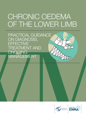
Circulation is a fine-tuned system composed of an arterial and venous network with a complementary network of lymphatics. The arterial system delivers oxygen and nutrition to organs and tissues. The venous system returns the deoxygenated blood and metabolic waste. The lymphatic system absorbs the excess fluid, transports dietary fats and is responsible for immune surveillance.15 In the lower legs, critical events such as exchange of gases, nutrition and metabolic products take place at capillary beds (see Figure 1). Under physiological conditions, the fluid and electrolytes cross the capillary wall by capillary filtration. The filtrated fluid serves as a medium for diffusion of oxygen and nutrition delivered from capillaries to surrounding tissue (=interstitium) and return of carbon dioxide (CO 2) and metabolic waste from the interstitium to capillaries.16 The CO2 is then transported to the lungs and the waste products to the kidneys, to the intestine and to the skin for excretion via the venous system.17
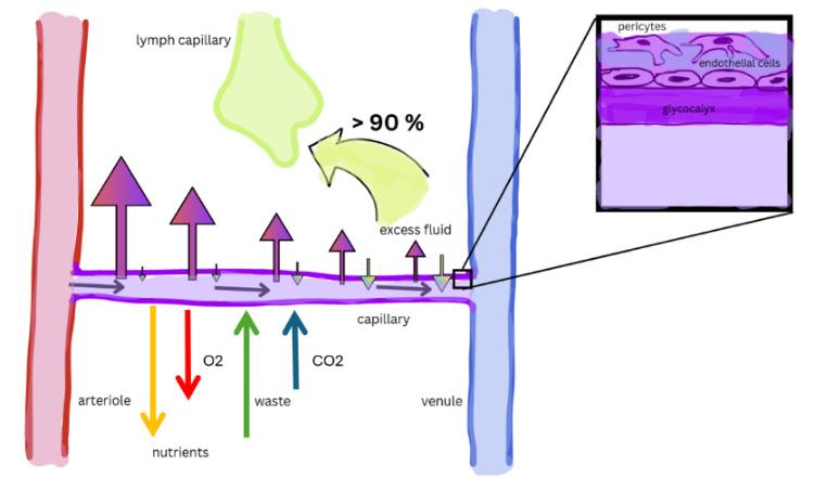
Figure 1: Capillary filtration, diffusion and lymphatic return/ collection of fluid. Credits: Heli Lagus, Helsinki University Hospital and Helsinki University, Finland. Figure inspired by Figure 55 from Ahmed Aziz’s Anatomy and Physiology to the Cardiovascular System for Nursing Students (2019, p. 90).
Under physiological conditions, there is a balance between the fluid filtration from the capillaries and the drainage of the fluid. Previously, it was assumed that the venous system would absorb over 90% of extracellular fluid, but the current view suggests that most of the excess fluid is transported via lymphatics and returned to circulation,16 and the venous reabsorption of fluid would only be transient under special circumstances.18,19,20 Imbalance may occur when there is an increased amount of capillary filtration and/or the removal of the fluid by the lymphatic system is disturbed or exceeds its capacity. All factors increasing the capillary filtration and/or hindering the clearance of excess fluid may result in oedema.
Starling’s revised hypothesis of capillary filtration
Capillary filtration depends on three main factors: hydrostatic pressure, oncotic (colloid osmotic) pressure and capillary permeability. The net amount of fluid filtered from capillaries to surrounding tissue can be estimated by Starling’s Equation. This classic hypothesis of fluid movement across the capillary wall was originally proposed by Ernest Starling in 1896 and it was formulated as an early form of equation by Eugene Landis in 1927.21
Capillary hydrostatic pressure pushes fluid out of capillaries into the interstitial space (extracellular space in tissue surrounding the vessels), whereas interstitial hydrostatic pressure pushes fluid back into capillaries.
Capillary oncotic pressure pulls fluid into capillaries due to plasma proteins attracting fluid. Interstitial fluid oncotic pressure pulls fluid out of capillaries due to proteins in the interstitial space. Proteins like albumin and haemoglobin are effective osmotic compounds that attract water. Lower capillary oncotic pressure due to hypoalbuminemia or diseases like diabetes increases fluid movement from capillaries to interstitial spaces.
Since the publication of Starling’s equation, some significant discoveries affecting the capillary filtration have been made: 1) the presence of a semipermeable endothelial glycocalyx functioning as a sieve; 2) precapillary sphincter regulating blood flow to the capillary; and 3) pore-like breaks of endothelial intercellular tight junctions of the capillary wall permeable to macromolecules and plasma proteins.22 As a consequence, an amendment to the Starling’s Equation has been proposed (See Figure 2).
Endothelial glycocalyx
Capillary permeability is an important regulator of fluid movement through the capillary wall. Endothelial cells of capillaries and other blood vessels have a lining of special extracellular matrix termed the endothelial glycocalyx layer. Glycocalyx maintains selective vascular
permeability functioning like a sieve. A gel-like glycocalyx consists of proteoglycans containing glycosaminoglycan molecules and glycoproteins, and it is negatively charged.24 It was earlier looked upon a passive barrier, but due to its mechanical and electrostatic properties, glycocalyx has been found to limit passage of larger and/or more negatively charged particles out of blood vessels.25
Glycocalyx exerts multiple actions. It has been suggested that glycocalyx acts together with endothelial cells to control movement of fluid across the capillary wall. In addition to the sieve function, glycocalyx also contributes to mechanical signal transduction and molecular bioavailability. It regulates cellular signalling, sensing the extracellular environment and cell adhesion. It inhibits leukocyte diapedesis (extravasation) and coagulation by binding antithrombin III. 26 It shows anti-inflammatory properties,27 and it protects endothelial cells from oxidative stress. Recent findings suggest that collecting lymphatic vessels also have a lining of continuous glycocalyx and its breach may compromise the lymphatic pumping function, thereby contributing to the formation of chronic oedema.28
Formation of oedema
The formation of oedema at the capillary level depends on four critical factors: capillary and interstitial hydrostatic pressure; capillary and interstitial oncotic pressure ; capillary permeability; and lymphatic flow. The first three affecting capillary filtration and the last affecting the clearance of the excess fluid. When capillary hydrostatic pressure increases or capillary oncotic pressure decreases,
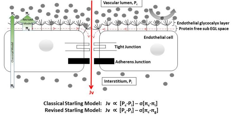
Figure 2: Classical Starling Equation and the differences between the classical and revised Starling principle. 23 Diagram by Trung, D. T. et al, 2020.
Legend: Jv= Fluid flow; Pc = capillary hydrostatic pressure; Pi = interstitial hydrostatic pressure; πc = capillary plasma colloid osmotic pressure (COP); πg = sub-glycocalyx COP; πi = interstitial COP; α =is proportional to.
or both, more fluid is pushed out than pulled in leading to fluid accumulation in the interstitial space.
The factors which influence the formation of oedema include:
Increase in capillary hydrostatic pressure
Factors increasing the hydrostatic pressure in capillaries include the same factors that increase venous hypertension, such as heart failure, venous obstruction, effect of gravitation due to prolonged sitting or standing, sodium and fluid retention, acute renal failure and some medications. See section Risk factors for CVI for more information.
Decrease in capillary oncotic pressure
Diseases like liver cirrhosis or nephrotic syndrome that either lead to reduced production of blood proteins, especially albumin or increase their loss or degradation or both are causes that reduce the capillary oncotic pressure. Malnutrition is a possible cause behind decreased capillary oncotic pressure. See the section on Risk factors for CVI for more information.
Increase of capillary permeability
Degradation of glycocalyx (termed glycocalyx shedding) increases capillary permeability. Protease-enzymes are responsible for the degradation. Various factors can cause degradation of glycocalyx: inflammation via tumour necrosis factor (TNF)-α and reactive oxygen species (ROS), heparanase, hypoperfusion/ischemia, hyperglycaemia, bacterial toxins and growth factors,29 high sodium and even female sex hormones may cause degradation of glycocalyx.30
Moreover, other factors may increase capillary permeability, such as inflammatory mediators, histamine and bradykinin, and leukotriene B4 (LTB4).31 In diabetic patients, binding advanced glycation end (AGE) products to a specific receptor on protein or red blood cells, results in increased vascular permeability. Nitric oxide (NO) mediates vascular tone and vascular permeability. Prostacyclin modulates both vascular pressure and permeability. Activated platelets release thromboxane A2, which takes part in the regulation of permeability.32
Lymphatic flow
The lymphatic system plays a crucial role in collecting and returning excess fluid to the bloodstream. Lymphatic
flow removes fluid, cellular debris and nutrients to exit the interstitium. In normal conditions there is a balance between filtration and lymphatic outflow.
Lymphatic flow increases in response to tissue oedema, lowering interstitial oncotic pressure by dilution and removing interstitial proteins. 29 However, when the lymphatic system is overwhelmed, fluid accumulates in the tissues, resulting in oedema.
Formation of oedema in lower extremities
Oedema is a common clinical condition seen in various diseases. Aetiology may be multifactorial. The accumulation of fluids in lower limbs may be caused by superficial and/ or deep venous reflux, deep venous obstructions, or by disturbances of the lymphatic system or diseases, such as cardiac, renal, and hepatic diseases, as well as medications. Also, obesity and immobility may cause oedema. Oedema can result from any factors that lead to increased capillary hydrostatic pressure, decreased capillary oncotic pressure, increased capillary permeability, or lymphatic dysfunction or obstruction, or their combination.
Acute oedema may also form from increased capillary permeability due to inflammation caused by trauma such as burn injuries, leading to cytokine release, insect bites, cellulitis or allergic reactions.33
In lower limbs, oedema is often caused by chronic venous insufficiency or secondary chronic oedema. When not diagnosed and treated early both conditions may lead to complications like chronic oedema, chronic skin changes and even non-healing wounds.
Chronic venous insufficiency (CVI)based oedema
Venous system
In lower extremities the venous system comprises of a superficial system, a deep system below the muscular fasciae, and connecting perforating veins. Valves in the veins prevent the backflow of blood against gravity. Venous valves are bicuspid structures ensuring that blood flows in one direction. The average number of venous valves varies by vein segment. Posterior tibial veins contain an average of 8 to 19 valves, whereas anterior tibial and peroneal veins contain an average of 8 to 11 valves.34 There are approximately 140 to 150 perforating veins in the leg, of which the ones on medial calf region are
considered clinically the most significant. Perforators normally drain venous blood from superficial to deep veins, but incompetent perforator veins have been linked to CVI.35 See Figure 3.
Venous one-way circulation is assisted by muscle pumps, mainly the calf muscles,36 in addition to negative pressure intra-abdominally and intra-thoracically.37 Even though the calf is the most significant muscle pump with a normal ejection fraction of approximately 65%, the thigh muscle pump ejection fraction is around 15 %38, and the foot serves as the initial pump, initiating the movement of blood.39
The pathophysiology of CVI and venous leg ulcers (VLUs) results from disturbances of the delicate interplay of mechanical, cellular and molecular factors. The stages of chronic venous disorders are internationally characterised by standard classification: Clinical Etiology Anatomy and P athophysiology (CEAP) into clinical classes C0–C6. 41 Chronic oedema encompasses CEAP clinical classes C3–C6. CVI includes also VLU as the most severe manifestation of CEAP classification,41 which are complex and multifaceted diseases, involving inflammation, vascular remodelling, changes in microcirculation and a cascade of cellular and molecular responses leading to persistent oedema, hypoxia and eventually ulcer formation.
Figure 3: Anatomy of the lower extremity venous system. Meissner MH. Pathophysiology of varicose veins and chronic venous insufficiency. In: Hallett JW, Mills JL, Earnshaw JJ, Reekers JA, eds. Comprehensive Vascular and Endovascular Surgery. 1st ed. Mosby; 2004:571–589. Reproduced with permission from Meissner MH, 2004.40
In CVI the veins in the lower extremities fail to return blood efficiently to the heart, leading to persistent elevation of venous pressure (venous hypertension) and oedema. Impaired calf muscle pump function leads to blood pooling, increased venous pressure, and ultimately chronic venous insufficiency. Venous dysfunction in lower extremities occurs when veins in the legs become damaged. CVI may result from valvular insufficiency of either the deep or superficial veins, or perforator valves, or due to venous obstruction such as thrombosis, or muscle pump dysfunction or their combination.42
Venous hypertension promotes inflammation
Inflammation plays a crucial role leading to vascular changes, valve dysfunction, and chronic venous hypertension. In addition to increased venous pressure these processes result in reflux, blood pooling, hypoxia, oxidative stress and inflammation. Persistent venous pressure causes capillaries to dilate, leading to fluid overload and peripheral oedema. There is a secondary enlargement, elongation, dilation, and tortuosity of capillaries which can be viewed as hallmarks of venous microangiopathy. 42 Deoxygenated venous blood accumulates in the interstitium, leading to hypoxia and venous ischemia.43 Increase of blood pressure and mechanical stretching of the venous wall activate both leukocytes and endothelial cells.
Reduced shear stress on endothelial cells triggers the release of vasoactive agents and inflammatory molecules such as TGF-β1, TNF-α, IL-1, and other cytokines, as well as matrix metalloproteinases (MMPs), which degrade the extracellular matrix and impair wound healing. Glycocalyx damage and release of inflammatory mediators contribute to endothelial dysfunction. The increased production of ROS causes oxidative stress. Leukocytes accumulate and increase of membrane adhesion molecules (such as ICAM-1) then facilitate adhesion of leukocytes cells (such as neutrophils, macrophages, T lymphocytes, and mast cells) to the endothelium and their transmigration through the vessel wall into the inflamed tissue.44, 45 See Figure 4.
This inflammatory process leads to gap formation between endothelial cells due to endothelial cells’ actin/myosin filament contraction. These gaps increase permeability and leak plasma proteins into the interstitial space. Also, histamine, bradykinin or leukotriene B4 can increase the vascular permeability. 46 The release of histamine from mast cells is likely to cause itching. Persistent venous
hypertension and bacterial contamination affect wound healing via mechano-transduction and inflammatory pathways.
Iron increases oxidative stress and promotes macrophage pro-inflammatory M1 phenotype
The widened gaps between the endothelial cells also enable also the leakage of erythrocytes into the interstitium. Release of ferritin and ferric oxide from erythrocytes further increases oxidative stress, additional metalloproteinase activation and tissue damage. Iron overload further exacerbates the problem by maintaining macrophages in a pro-inflammatory/inflammatory M1 state,47 instead of wound healing promoting M2 state.
Role of MMPs
Increased expression of MMPs and other proteinases in capillaries leads to breakdown of the vascular extracellular matrix including glycocalyx. This degradation disrupts the structural integrity of the capillary wall, causing abnormal vascular permeability contributing to fluid leakage and oedema.49,50,51
Endothelial cells, infiltrating leukocytes, vascular smooth muscle cells, resident fibroblasts and keratinocytes release MMPs. The dynamic balance between MMPs and tissue inhibitors of matrix metalloproteinases is essential for proper wound healing, while increased levels of MMPs lead to tissue degradation. MMPs regulate pathological remodelling of the extracellular matrix. Among their multiple other tasks, MMPs regulate signaling molecule availability, activate pro-inflammatory cytokines, degrade or activate growth factors and their receptors, and contribute to the formation of a proinflammatory, degradative, and prothrombotic microenvironment. Proinflammatory cytokines further induce MMP expression and downregulate tissue inhibitors of matrix metalloproteinases, creating a vicious circle.52,53
Venous hypertension promotes fibrosis
The inflammation and fibrotic process damage the valves within the veins; also, the non-valvular segments may be affected. Inflammation in the vein walls leads to the loss of elastin while collagen levels increase, leading to thickening, calcification and scar tissue formation of the vessel wall. These changes in vasomotor tone cause reflux (reversed blood flow), impaired venous emptying, and chronic venous hypertension. Venous hypertension causes fibroblasts to
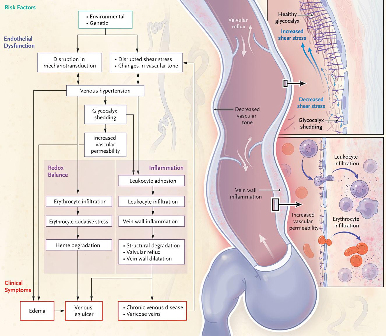
Figure 4: Description of the development of CVI. Reproduced with permission from Fukaya et al, 2024.48
develop a myofibroblast phenotype, increasing skin tension and possibly leading to skin separation in response to injury.
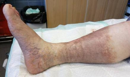
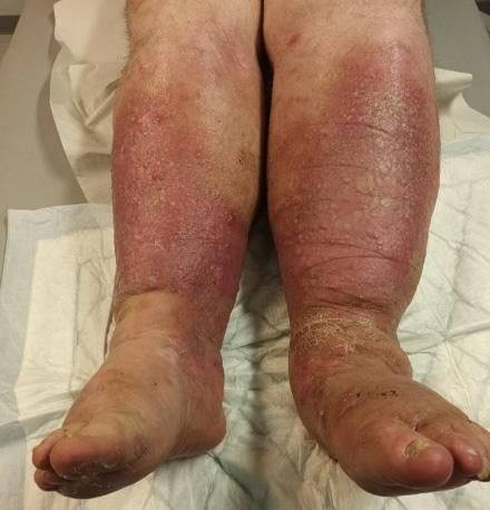
See Figures 5 and 6 for typical clinical signs of venous insufficiency.
Risk factors for CVI
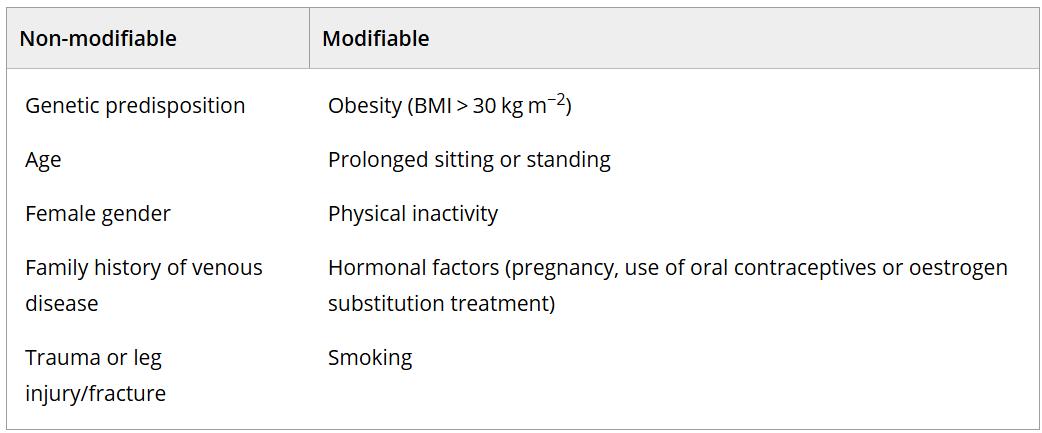
Multiple predisposing/risk factors for developing CVI have been identified. Typical risk factors include increasing age, family history, smoking, obesity, immobility, occupation, posture, female sex, pregnancy, oral contraceptive use, previous deep venous thrombosis (DVT) and history of leg injury.47, 54 Prolonged standing and sitting are recognised as risk factors for the development of CVI.55 Non-thrombotic
iliac vein obstruction such as May-Thurner syndrome also poses a risk for CVI.54
Factors that are associated with increased capillary hydrostatic pressure include DVT, pericarditis, pulmonary hypertension, liver failure or cirrhosis, and right-sided heart failure. Risk factors involving increased plasma volume are renal failure, heart failure and some medications.57
Factors that lead to a decrease in oncotic pressure , typically due to hypoalbuminemia, occur in several diseases such as renal disease where the loss of albumin occurs, whereas in hepatic disease, such as cirrhosis and chronic liver disease, the cause is inadequate/insufficient albumin synthesis, or malabsorption or malnutrition of proteins.17
Less common are genetic factors, such as mutations in the hemochromatosis gene and Factor XIII genes, that can increase the risk of CVI and affect healing outcomes. Polymorphisms in fork head box protein C2 (FOXC2), CADASIL, desmulin dysregulation and MMPs are also linked to CVI. Also, syndromes such as Klippel–Trenaunay, Park–Weber, and Ehlers–Danlos predispose to the development of CVI.52 Low serum magnesium levels pose a potential risk for CVI development.
Chronic complications of diabetes mellitus include micro- and macro-vascular changes which may increase vascular permeability and lymphatic collecting vessel hyperpermeability and lead to chronic oedema.58, 59 See Table 2.
The most common medications that increase the risk of CVI
Vasodilators (some antiepileptics, antidepressants, antipsychotics, antiparkinsonians, antihypertensives), and opioids can increase capillary pressure or permeability, leading to oedema. Hormones, glucocorticoids and NonSteroidal Anti-Inflammatory Drugs (NSAIDs) may cause oedema by sodium retention.48
Chronic oedema
Lymphatic system
Lymphatic capillaries, collector lymphatic vessels (pre- and post-nodal collectors), lymph nodes, lymphatic trunks and lymphoid organs form the lymphatic system, together with lymph fluid and lymphatic cells. The lymphatic system is a low-pressure system, typically between 1 and 20mmHg.60 See an overview of the lymphatic system in Figure 7.
Figure 5: Hyperpigmentation, varicose veins, spider veins and corona phlebectatica on ankle. Credits: Minna Hellgren, Helsinki University Hospital, Finland
Figure 6: Chronic oedema and stasis dermatitis. Credits: Minna Hellgren, Helsinki University Hospital, Finland
Table 2: Risk factors for chronic venous disease.56 Table by Krizanova, O. et al, 2024
Initial lymphatics (also called lymphatic capillaries) merge into larger vessels that contain smooth muscle, enhancing peristaltic movement. Unidirectional valves prevent the retrograde flow. The structure between two valves is called a lymphangion. The lymph is propelled one-way, assisted by a pump of lymphatic smooth muscle cells, valves and compression of skeletal muscles.15 The lymphatic system maintains the fluid balance. It drains, collects and returns the infiltrated excess interstitial fluid back to circulation. It
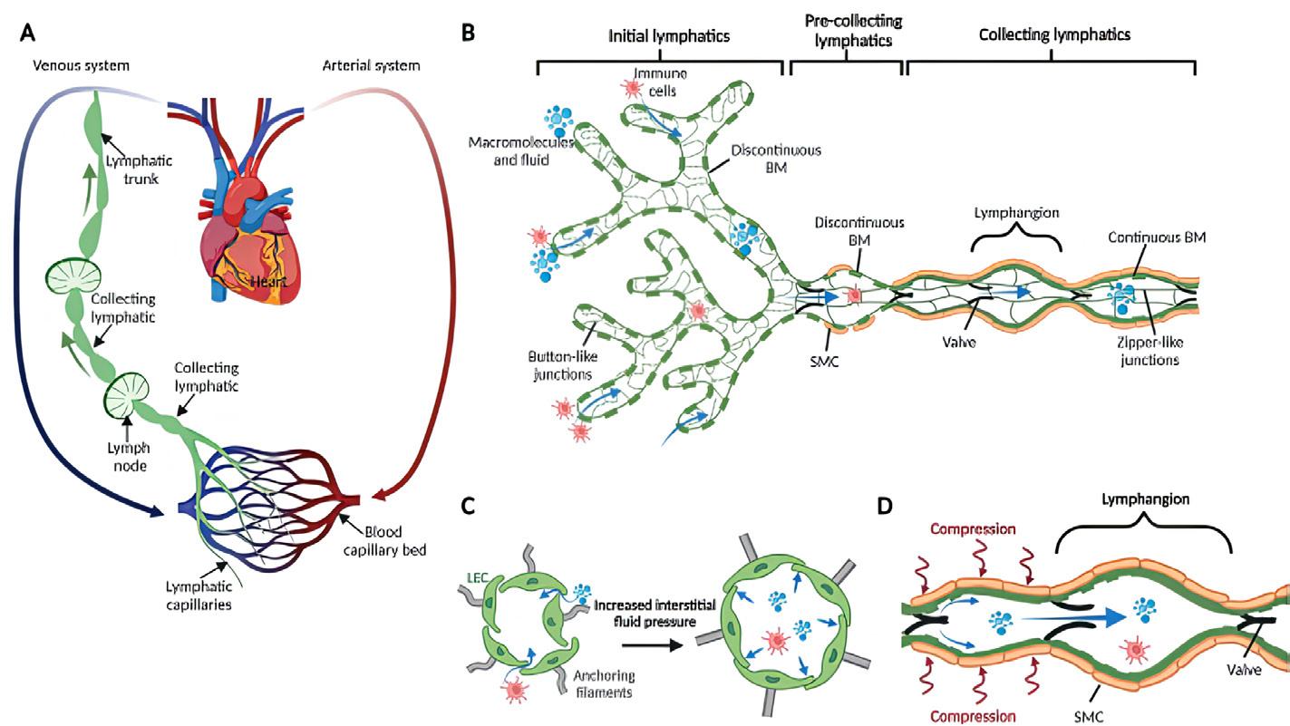
Figure 7: Structure and function of the lymphatic system.61 Diagram by Angeli, V. & Lim, H. Y., 2023
A: The lymphatic vasculature (green) forms part of the circulatory system. Fluid that extravasates from the blood capillary bed into the tissue interstitium is absorbed into initial lymphatics vessels and flows through larger collecting lymphatic vessels that actively transport lymph fluid into draining lymph nodes before returning into the venous system via the thoracic duct.
B Interstitial fluid, macromolecules and immune cells leave the tissue interstitium to enter discontinuous button-like initial lymphatic vessels that lack a continuous basement membrane. Collecting lymphatic vessels have a continuous basement membrane, smooth muscle cell coverage to provide contractile activity to assist blood flow and intraluminal valves to prevent lymph backflow. Collecting lymphatic endothelial cells (LECs) are organised into tight continuous zipper-like junctions and do not absorb fluid from surrounding tissues.
C Initial lymphatic vessels are composed of overlapping LECs that allow interstitial components to enter the vessels when interstitial pressure is high. The overlapping cells also act as valves, preventing fluid from leaking out. Anchoring filaments connect LECs to the surrounding extracellular matrix and facilitate fluid, macromolecule and cell entry into initial lymphatic vessels.
D The collecting lymphatic vessels are composed of several lymphangion’s that propagate lymph flow. Coordinated contraction/ expansion of each lymphangion and opening/closing of intraluminal valves ensure efficient lymph transport. BM: basement membrane, SMC: smooth muscle cell.61
has a critical role in immune surveillance and defending the body against infection. The lymphatic system also facilitates the absorption and transportation of dietary fats.
In physiological conditions, blood vessels leak plasma and proteins at the capillary bed into the interstitial space, driven by an imbalance in hydrostatic and osmotic pressure. The infiltrated lymph fluid contains immune cells, proteins, lipids, lipoproteins, electrolytes and bacteria.62 Fluid accumulation increases interstitial pressure and drives protein-rich interstitial fluid into lymph capillaries, which drain lymph via primary valves. Lymph capillaries form from overlapping lymphatic endothelial cells that are connected to the extracellular matrix by anchoring filaments. As the amount of fluid increases, interstitial pressure increases, and the overlapping of lymphatic endothelial cells opens letting the fluid and cells to enter. The anchoring filaments prevent the initial lymphatics from collapsing as the fluid volume increases. As the tension or pressure due to the volume increase rises it leads to fluid flow down the pressure gradient. Unlike collecting lymphatics, the initial lymphatics do not possess valves. See Figures 7 and 8. The lymphatic system propels lymph one-way using an intrinsic pump from lymphatic smooth muscle cells, extrinsic pressure from skeletal muscle contractions and arterial pulsation.
As shown in Figure 8, during expansion, the interstitial fluid can enter the lymphatics through the endothelial microvalves, because the intralymphatic pressure is lower than the interstitial fluid pressure. Compression of surrounding tissues forces the lymph towards the collecting lymphatics, whose smooth muscle can spontaneously contract. The valve-containing part of a lymph vessel and the adjacent portion of the vessel before the next valve form a functional unit called the lymphangion, which is able to contract or expand.60
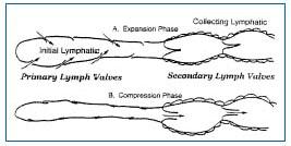
Figure 8: Two-valve system in lymphatics: primary valve in the initial lymphatic and secondary valve in the collecting lymphatic. Reproduced with permission from Stücker et al, 2008.60
Two forces push lymphatic fluid forward: intrinsic forces (lymphangion contractions) and extrinsic forces (muscle movement, and arterial pulsation). Lymphatic valves prevent backflow and are crucial for driving lymph flux against gravity. The transport system is unidirectional. Collecting lymphatic vessels contract to push lymphatic fluid to lymph nodes, lymphatic trunks and ducts, draining via the subclavian veins. The superficial systems drain lymph from skin and subcutaneous tissue, whereas the deep system drains muscles, joints, and nerves.63 The superficial and deep systems are connected via perforating vessels. Lymph nodes filter lymph fluid, concentrating post-nodal lymphatic fluid by reabsorbing water. Normal lymphatic flow is 2–3 litres per day.64
Chronic oedema forms as a result of the impaired return of interstitial fluid into the intravascular space secondary to dysfunction of the lymphatic system.
Primary lymphoedema
Primary lymphoedema is caused by rare developmental lymphatic vascular anomalies/malformations. Dysplasia, hyperplasia, hypoplasia, or aplasia of the lymphatic system in the lymphatic vessels, nodes, or both, causes primary lymphoedema. These malformations are congenital slowflow vascular malformations. They have typically dilated lymphatic vessels and cystic-like areas filled with lymphatic fluid. Primary lymphoedema results from genetic mutations, which can be isolated or part of a complex syndrome. Most cases are inherited as an autosomal dominant trait with incomplete penetrance and variable expression. Currently, less than a third of primary lymphoedema patients have identifiable genetic mutations, often in the vascular endothelial growth factor C signalling pathway.65 Sporadic cases are the most common, accounting for ~60% of primary lymphoedema.66
More than 40 different genetic defects (such as VEGFR-3, CCBE1, FOXC2, GATA2, GJC2, PTPN14, SOX18, CCBE1, FAT4, ADAMTS3, FBXL7, GJC2, KIF11, ITGA9, REEKIN, PIEZO1, EPHB4, CALCRL, and CELSR1) have been identified and associated with anomalies in the lymphatic system, leading either to underdeveloped lymphatic structures or poor lymphatic outflow abilities.65 However, defining the genetic defect in primary lymphoedema has little impact on clinical management. The most current and widely accepted classification of primary lymphoedema is St George’s 202011 refined classification algorithm. It is
based on the age of onset, areas affected by swelling and associated clinical features. The lymphatic anomalies are divided into five main categories:
1. Vascular malformations associated anomalies and lymphatic malformations
2. Syndromic lymphoedema
3. Lymphoedema with prenatal or postnatal systemic involvement
4. Congenital onset lymphoedema (<1 year)
5. Late-onset lymphoedema (>1 year)
A more detailed description of primary lymphoedema is beyond the scope of this document.
Secondary chronic oedema
Secondary chronic oedema is much more common than the primary form arising from damage or obstruction to the lymphatic system, often due to external factors or tissue injury. This can include trauma, surgical procedures like lymph node dissection, vascular surgeries, radiation therapy, or chemotherapy (especially with taxanes). 67 Additionally, factors like infections, malignancies, and postthrombotic syndrome can impede lymphatic flow and lead to the condition.65
Chronic venous disease can cause secondary lymphatic damage by increased capillary filtration, which in the end overloads lymphatic fluid transportation. In the western world, CVI it is the most common form of secondary chronic oedema, 19, 68 whereas the next most common cause of secondary chronic oedema has been suggested to be malignancy or its treatments.69, 62 Secondary chronic oedema of the upper limbs is most often associated with breast cancer and its treatment. The most common cause of secondary chronic oedema worldwide is lymphatic filariasis, transmitted in endemic areas by the mosquito. (See the comment about lymphatic filariasis in the Introduction). All in all, the prevalence of secondary chronic oedema is underestimated, and the condition is poorly recognised.3, 10
For more details on secondary chronic oedema see Chapter 3: Conservative treatment in the management of (secondary) chronic oedema.
Both primary lymphoedema and secondary chronic oedema share chronic swelling, inflammation, adipose deposition and fibrosis but they differ in patient responses and disease courses.
Risk factors of secondary chronic oedema
Factors increasing the risk of secondary chronic oedema include but are not limited to genetic abnormalities, obesity, physical inactivity, radiation and infections. Cellulitis and erysipelas lead to damage of cutaneous lymphatics.
Pathophysiology of chronic oedema formation
Chronic oedema is caused by abnormal accumulation of lymphatic fluid and macromolecular proteins into the interstitium when lymph transport is reduced or impaired. When oedema occurs, oxygen tension decreases resulting in inflammation (see Figure 9).
Role of inflammation
Some authors see secondary chronic oedema as a chronic inflammatory disorder 71 or as a chronic inflammatory condition. Inflammation and inflammation-induced lymphangiogenesis and adipose expansion are key pathological features in the pathophysiology of secondary chronic oedema (Figure 9). Fluid accumulation activates inflammatory cascades and adipose cell differentiation and leads to both protein and lipid accumulation in the interstitial spaces. In surrounding tissue, the release of pro-inflammatory molecules promotes the migration and activation of inflammatory cells such as dendritic cells, macrophages, neutrophils and T-helper cells.72 The inflammation is mediated by increased expression of cytokines, such as interleukin (IL)-13, TNF-α, IL-6, IL-8, and monocyte chemoattractant protein-1. Persistent lymph fluid
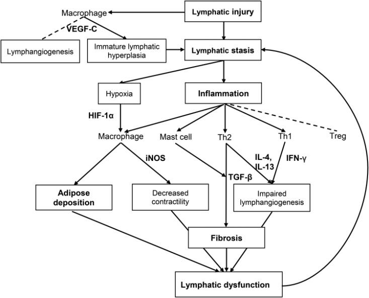
Figure 9: Cellular and molecular mechanisms involved in the pathology of lymphoedema. Reproduced with permission from Sung et al. 2021.78
accumulation maintains chronic inflammation, promoting both adipocyte proliferation and collagen deposition. The deposition of fibroadipose tissue, fibrosis and further damage to lymphatics leads to notable increase in limb girth. This kind of adipose deposition is not dependent on caloric intake or weight gain, but patients with obesity tend to have a more severe presentation.72
Role of T-cells
Inflammatory cells, particularly CD4+ T cells —Th1, Th2, and regulatory T cells (Treg) — infiltrate oedematous tissues. Activation of CD4+ T cells leads to impaired lymphangiogenesis and fibrosis. Activation and accumulation of CD4+ cells is required for adipose deposition after lymphatic injury, whereas inhibition of Th2 differentiation decreases adipose deposition. The number of infiltrating CD4+ cells correlates with the severity of chronic oedema.73
Th2 cells
The accumulated lymphatic fluid triggers Th2 immune response leading to chronic inflammation. Th2 inflammatory responses cause progressive lymphatic dysfunction, soft tissue fibrosis and inhibition of collateral lymphatic vessel formation. Th2 cells enhance fibrosis by producing profibrotic cytokines (IL-4, IL-13, TGF-β1) and by activating M2 macrophages. IL-4 and Il-13 both impair lymphangiogenesis, promote Th2 cell differentiation, macrophage activation, and fibrosis, whereas TGF- β 1 promotes fibrosis and negatively regulates lymphatic vessel regeneration.73
Blockade of Th2 differentiation prevents and treats secondary chronic oedema. Treg cells limit pathological changes — their depletion exacerbates oedema and fibrosis. Depletion of CD4+ cells or inhibition of IL-4, IL13, TGF- β 1 decreases fibrosis and improves lymphatic function.74
Th1 and Th17 cells
Th1 cells produce IFN-gamma, which impairs lymphangiogenesis and activates macrophages. Th17 cells produce IL-17A, which inhibits lymphatic vessel formation.73
LTB4 is a potent inflammatory lipid LTB4 recruits T cells and promotes Th17 differentiation. It is associated with chronic diseases like obesity and type
II diabetes. LTB4 has a pro-lymphangiogenic effect at low concentrations and inhibitory effect at high concentrations. Blockade of LTB4 reduces infiltration of macrophages, neutrophils, and CD4+ T cells.75
Role of macrophages in chronic oedema
Macrophages exhibit multiple and complex roles in the development of chronic oedema. Macrophages regulate inflammation, immunity, and tissue repair and they contribute to adipose metabolism and lymphangiogenesis. Aggregated macrophages participate in the degradation of extracellular proteins.
Macrophages can exacerbate lymph stasis by modulating inducible NO synthase (iNOS) expression. Under the influence of prostaglandin PGE2 macrophages and smooth muscle cells increase NO production within perilymphatic tissues. NO causes lymphatic vessel dilatation and reduces pumping capacity, contributing to chronic oedema. Increased iNOS and NO levels inhibit collecting lymphatic contraction, contributing to disease progression.76
Type M1 inflammatory macrophages and type M2 reparatory macrophages show different functions. Most macrophages present in chronic oedema are M2 differentiated.77
Proteins of lymphatic fluid attract macrophages, stimulating collagen production by fibroblasts, and enhancing stimulation of fibroblasts, keratinocytes, and adipocytes. Diversity of other factors attract macrophages to the site as well and promote their proliferation. Chronic oedema and obesity may directly enhance macrophage migration and proliferation — free fatty acids from necrotic adipose cells serve as chemoattractant for macrophages.77 Cytokines produced by CD4+ inflammatory cells such as IFNgamma, IL-4 and IL-6, regulate macrophage migration and proliferation.
Activated macrophages produce and release cytokines and growth factors that stimulate:
• Lymphatic endothelial cells promotion to lymphangiogenesis (VEGF-C, VEGF-A)
• Fibroblasts to increase collagen production (TGF-β1)
• Adipocytes (in late-stage) leading to adipose deposition
• Keratinocytes leading to hyperkeratosis73
In early stages of chronic oedema hypoxia-inducible factor-1 α (HIF-1 α ) modulates VEGF-C/VEGFR-3
signalling. Macrophages produce VEGF-C and VEGF-A which promote lymphangiogenesis. On the other hand, macrophage released TGF-β1, like Th2 cytokines, inhibits lymphangiogenesis.78
Macrophages can enhance either pro- or antifibrotic effects depending on their phenotype and the circumstances. M2 phenotype macrophages mediate anti-fibrotic functions through regulation of CD4+ T cells and promote lymphangiogenesis through VEGF-C production. In chronic oedema depletion of macrophages promotes an increase in collagen deposition, fibrosis, and impaired lymphatic function.77 The exact mechanisms of how macrophages regulate fibrosis in chronic oedema remain to be elucidated.
M2 repair type macrophages decrease in chronic oedema adipose tissues, leading to imbalance with M1 proinflammatory macrophages.78
In chronic oedema, macrophages produce IL-6, which regulates chronic inflammation and decreases adipose deposition. Inhibition of IL-6 increases adipose deposition, suggesting its homeostatic role in chronic oedema.78,79
Macrophages induce adipogenic transcription factor proliferator-activated receptor gamma (PPAR-γ) expression, contributing to inflammatory cytokines and adipose tissue inflammation.
In late stages, macrophage depletion results in reduced VEGF-C, increased Th2 cells, and collagen deposition, exacerbating fibrosis.
It has been suggested that activity of macrophages in chronic oedema aims to decrease inflammation and to reduce or inhibit fibrosis.77
Tissue fibrosis formation
The development of hypoxia and subsequent activation of HIF-1α initiates immune cell migration and fibrosis.78 Tissue fibrosis is promoted by overactivity of Th2 cells, secretion of IL-4, IL-13, and TGF-β1. TGF-β1 promotes differentiation of fibroblasts into myofibroblasts, increasing collagen production and extracellular matrix deposition. Lymphatic vessels become progressively fibrosed and occlude due to smooth muscle cell proliferation. Myofibroblasts play a role in tissue repair but lead to fibrosis in pathological conditions.75
Adipocyte differentiation and adipose tissue deposition
Lymphatic injury and fluid stasis activate differentiation of local adipocytes; lymphatic fluid is a potent activator of adipocyte differentiation and lipid storage. Fat accumulates near leaky lymphatic vessels, and the leaking fluid induces adipocyte differentiation. Free fatty acids from lymphatic fluid directly promote adipocyte proliferation and differentiation, increasing expression of adipogenic markers.74
Adipose tissue deposition is promoted by lymphatic fluid stasis. In animal studies it has been shown that lymphatic fluid stasis leads to lipid accumulation, subcutaneous fat deposition and increased number of adipocytes.78 Adipose tissue deposition further decreases lymphatic function and contributes to disease progression. Hypertrophic fat lobules compress and collapse their feeding lymphatic capillaries, leading to fluid and lipid transport disruption, resulting in further fat accumulation in the periphery.80 Accumulation of lymphatic fluid has been associated with several adipogenic transcription factors regulating adipogenesis, leading to adipocyte differentiation and lipid accumulation. Lymphatic fluid stasis increases insulin and IGF-2 levels, promoting adipogenesis with adipogenic transcription factors such as peroxisome proliferator-activated receptor γ (PPAR-γ), CCAAT/enhancer-binding protein-alpha (CEBP- α ) and fatty acid binding protein 4 (FABP4), and adiponectin, an adipogenic transcription factor and an endocrine hormone that also serves as a late marker of activated adipocytes. Accumulation of adipose tissue results in increased secretion of adipokines (adiponectin, resistin).81
Cholesterol balance alterations in adipocytes modulate metabolic and inflammatory functions. Cholesterol, carried by LDL and HDL lipoproteins, plays a critical role in lipid transport in lymph. Reverse cholesterol transport depends on efficient lymphatic transport. 80 Cholesterol accumulation and adipose tissue hypertrophy contribute to tissue changes. Disruption of lymphatic channels impairs cholesterol return to systemic circulation, leading to cholesterol accumulation in affected limbs and adipose remodelling.74
The sequence and the co-dependence of events in evolving chronic oedema are not fully understood.
Chronic oedema leads to inflammation and fibrosis impeding lymphatic draining
Both CVI-based oedema and secondary chronic oedema share similar pathological features (see Figure 10). A key characteristic of both conditions is ongoing inflammation with the release of proinflammatory cytokines. In CVI, venous hypertension causes fluid to leak from blood vessels into the surrounding tissue, triggering a chronic inflammatory process, whereas in secondary chronic oedema there is a damage to the lymphatic system, which impairs its ability to drain interstitial fluid. This damage activates chronic immune responses that promote inflammation and fibrosis, creating a feedback loop that further restricts lymphatic function. Prolonged inflammation leads to increased fibrosis, or scarring, of the tissue in both CVI-based oedema and secondary chronic oedema. In both CVI oedema and secondary chronic oedema, the combination of fluid overload, inflammation, and fibrosis create a self-perpetuating cycle where the tissue changes exacerbate the underlying problem. This process can significantly impact on lymphatic function and overall tissue health.
Oedema impairs wound healing
The exact mechanism of how oedema disturbs wound healing is not fully understood.82 On the other hand, there is an increasing body of high-level evidence showing how
treating oedema and preventing oedema improve wound healing, especially concerning VLU.83,84
Oedema occurs as a physiological response to acute injury.85 Excessive swelling can compress blood vessels and capillaries restricting blood flow, also lymphatics may be compressed. Oedema increases the diffusion distance. All these reduce the delivery of oxygen and nutrients to the wound, which ultimately hampers the healing process.14 Also the clearance of metabolic waste is impaired. 86 Oedema changes the environment to make it less supportive of wound healing. Oedema fluid can suppress the essential cellular activity required for new tissue growth and collagen fibre formation. Excess fluid may create a favourable environment for the proliferation of bacteria, increasing the risk of wound infection, and reducing the integrity of the skin and subcutaneous tissue, making it prone to injury. 87 Conditions such as impaired venous return and lymphatic drainage may create or maintain an environment conducive to infection, which can complicate wound healing and potentially lead to chronicity.14
Excess exudate increases the risk for moisture-associated skin damage (MASD). Excess moisture, particularly from chronic wounds, can overhydrate the surrounding skin, impair the skin’s protective barrier, and cause maceration and excoriation. The corrosive effects of enzymes in the exudate can further damage the skin and delay or impair wound healing.88
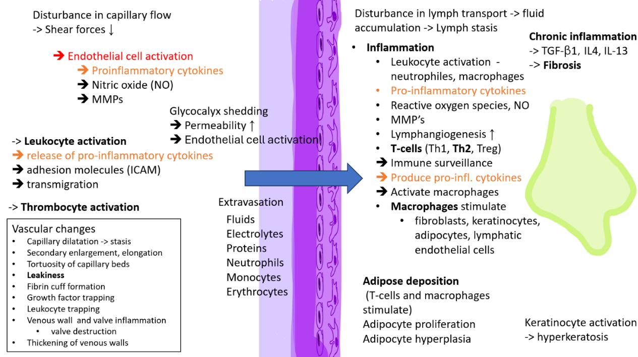
Figure 10: Cellular key events in formation of oedema. Credits: Heli Lagus, Helsinki University Hospital and Helsinki University, Finland.
It has been suggested that in chronic oedema fibrin cuffs (accumulation of fibrin in the capillary bed) may hinder the delivery of oxygen. The capillaries may be plugged by leukocytes leading to faulty healing. 89 Leukocyte sequestration, fibrin cuffs and oxygen-free radicals have been thought to increase the protein permeability, further damaging the lymphatic system. 90 In CVI even local ischemia may have a role in wound development83 and in delays in wound healing.
In the context of wound care, it is crucial to recognise the impact of oedema on the various phases of wound healing, haemostasis, inflammation, proliferation and remodelling.91 Oedema exacerbates the inflammatory phase by prolonging the presence of pro-inflammatory mediators, which can delay the transition to the proliferative phase, where tissue formation and healing occur.14,85
When persisting in the proliferation phase, oedema leads to relatively increased concentration of proteins resulting in attraction of even more fluid into interstitium and leading to the overwhelming of the lymphatic system and possibly sustained insufficiency. In the remodelling phase persisting oedema may result in connective tissue infiltration and fibrosis with elevated protein content.85
Ageing increases the risk of oedema
Ageing causes pathological structural and functional alterations to both blood vessels and lymphatic vessels increasing the risk of developing both CVI-based oedema and chronic oedema due to these age-induced changes.
Ageing and CVI-based oedema
With older age the prevalence of CVI and VLU increase, peaking among adults above 65 years.92 Higher age is also associated with many factors that are known to increase the risk of CVI, such as reduced mobility, limited ankle mobility, reduced calf muscle strength (leading to increased venous pressure) and speed of gait, poor nutrition and multimorbidity. 92, 93 Inactivity and reduced mobility in conjunction with older age may reflect poor general health and/or worsened oedema. 94
CVI age-related changes in the venous system result in weakening and structural changes in both venous walls and the valves. The intima of the veins becomes thicker and connective tissue accumulates in the subintima. There is a decrease in elastin which is accompanied by increased
rigidity and reduced contractility, as well as mechanical weakness of the vessel wall. Endothelial cells become senescent and more susceptible to apoptosis expressing increased levels of inflammatory cytokines, growth factors and MMPs. Endothelial cell degeneration leads to a decreased number of cells, which in turn results in exposure of the basement membrane to inflammatory proteins and leukocytes, leading to an inflammatory microenvironment in the venous walls. The increased permeability enables diffusion of cytokines and other inflammatory substances. With increased age there is an increase in expression of adhesion molecules such as ICAM-1 and VCAM-1 both in endothelial cells and in smooth muscle cells (SMCs) promoting platelet adhesion, and thereby a thrombus formation, as well as adhesion of leukocytes facilitating their transmigration.95 Similar alterations are seen also in lymphatics.
Ageing and chronic oedema
Ageing has a significant impact on the structure and the function of lymphatics driving changes that contribute to the formation of chronic oedema. Ageing augments the risk of increased permeability, decreased pump activity, and delayed immune response in lymphatic system.96
With advancing age, the lymphatic draining function decreases due to capillary rarefaction and lower transport capacity of collecting lymphatics. Decreased capillary density may result from reduction in production of lymphangiogenic factors, declined capability of lymphatic endothelial cells to regenerate, as well as from changes in composition and stiffness of extracellular matrix.
Due to ageing the vascular walls, also lymphatic vessel walls — especially in lymphatic collectors — typically show decreased numbers of SMCs and increased lymphatic diameter. Aged collecting lymphatic vessels are dilated and less contractile. The decreased number of SMCs leads to reduced pumping capacity, which is worsened by the decrease in nitric oxide.97
On cellular level aging causes also senescence of cells which in turn induces lymphatic dysfunction. The permeability of lymphatic vessels is increased due to aging-induced loss of endothelial glycocalyx, and to the production of inflammatory cytokines.98 The increased loss of cellular junctions and basement membrane proteins leads to shedding of glycocalyx and increase of permeability.
Increased permeability may also lead to an increase in penetration of antigens and bacterial products in the adipose tissue activating immune cells of adipose tissue.97 Increased inflammation results in tissue fibrosis. In addition, ageing associated basal activation of peri-lymphatic mast cells inhibits the recruitment of immune cells and delays immune response.96
Summary
The fluid balance in the interstitium depends both on the amount of fluid filtrated from capillaries and on the capability of the lymphatic system to return the accumulated fluid to circulation. Oedema forms when the amount of accumulating fluid exceeds the draining capacity of the lymphatics.
• Chronic leg oedema forms in the capillary bed due to accumulation of fluid caused by increased capillary infiltration and/or overwhelmed or disturbed drainage of fluid by lymphatics.
• Increased capillary filtration may be caused by increased capillary hydrostatic pressure (due to actions such as venous hypertension) decreased capillary oncotic pressure (due to actions such as hypoalbuminemia) and/or increased capillary permeability (due to actions such as glycocalyx shedding).
• Lymphatic flow may be disturbed by rare developmental lymphatic vascular anomalies or malformations (primary lymphoedema) or due to tissue damage, lymphatic vessel obstruction, or lymphatic vessel derangement (secondary chronic oedema).
• Prolonged inflammation causes permanent, irreversible damage to lymphatics.
• Chronic oedema impairs wound healing.
• Ageing causes pathological structural and functional alterations to both blood vessels and lymphatic vessels increasing the risk of chronic oedema.
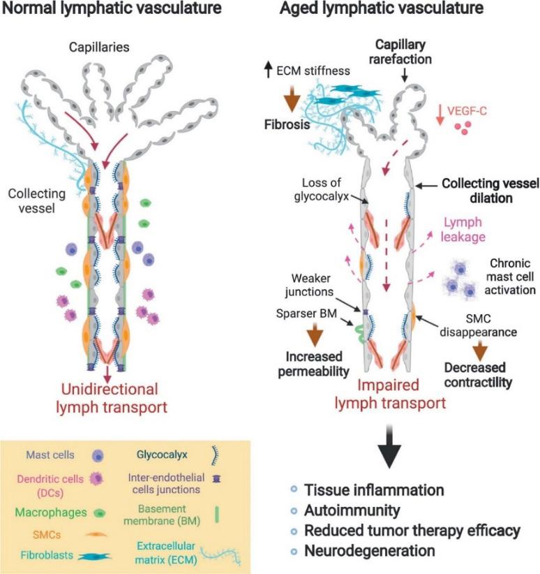
Figure 11: Development and ageing of the lymphatic vascular system.99 Diagram by González-Loyola, A. & Petrova, T. V., 2021
2. Lower leg swelling: diagnosis and management
Learning points
• The crucial need for proper differential diagnosis
• The need for increased awareness of technologies to aid in diagnosis
• The importance of measurements in treatment and the monitoring of lkpost-care success
• The future direction of care
Introduction
Chronic oedema is a poorly understood condition, often misdiagnosed and treated incorrectly and with great delay. Many primary care providers are challenged by patients that present with limb swelling in order to provide prompt diagnosis and treatment.100
In summary from Chapter 1, chronic oedema is swelling caused by impaired return of tissue fluid into the intravascular space secondary to dysfunction of the lymphatic system. It is divided into primary lymphoedema and secondary types.
Secondary chronic oedema is much more common than primary lymphoedema and is caused by mechanical, external or invasive factors which impede lymphatic flow. Worldwide, the most common aetiology of secondary chronic oedema is bacterial filariasis. In the United States and Europe, the most common causes of secondary chronic oedema are chronic venous insufficiency and surgical resection of lymph nodes after cancer treatment.101 Secondary chronic oedema of the upper limbs is most often associated with breast cancer and its treatment.72 Filariasis and podoconiosis will not be addressed in the scope of this chapter.
When oedema occurs, the leakage of fluid and macromolecular proteins into the interstitium results in inflammation (thereby) causing adipose tissue deposition and fibrosis. The fibrosis and deposition of adipose tissue results in significant increase in limb girth. A degree of pitting is present in almost all patients, but pitting may be mild relative to the severity of increased limb girth depending
on the amount of adipose tissue deposition. This adipose deposition is not dependent on caloric intake or weight gain, but patients with obesity tend to have a more severe presentation.101,72
Differential diagnosis
Initial assessment
Signs and symptoms of chronic oedema, such as pitting oedema that in in the lower extremities does not spare the feet or toes, hyperkeratosis (thickening of the skin’s outer layer), an orange rind appearance of the skin, a wart like skin presentation called papillomatosis, lymphangioma (benign, fluid-filled cysts in the lymphatic system), and lymphorrhoea (lymph leakage onto the skin) are often present. The Stemmer sign is commonly used as a quick indication of chronic oedema by attempting to pinch and lift the skin fold between the second and third toe. If unable to lift the skin at the base of the second and third toes, that is a positive Stemmer sign. Diagnosing chronic oedema demands a thorough history and physical examination, including the age of onset, medication, travel history, and family history.100,101,72 Prompt diagnosis and treatment is necessary to prevent secondary complications, such as secondary vasculitis, erysipelas and cellulitis and to halt the progression into the chronic phase of the disease.
Although beyond the scope of this chapter, a word of caution regarding correctly identifying cellulitis versus erysipelas, as both are bacterial infections of the skin. Erysipelas primarily impacts the epidermis, superficial lymphatics and occurs abruptly with a bright red raised rash. Cellulitis impacts the dermis and subcutaneous structures, including the lymphatics, with a generalised pink diffuse rash, slower to spread than erysipelas. However, cellulitis may spread rapidly due to lymphatic impairment leading to severe systemic impacts.
Lower extremity oedema can be caused by numerous other pathologies, such as deep vein thrombosis, venous insufficiency, infection, trauma, and many chronic medical diseases, such as heart failure, pulmonary hypertension, pregnancy, cirrhosis, kidney diseases, and medications,
including calcium channel blockers, gabapentin or pregabalin, non-steroidal anti-inflammatory drugs, oral contraceptives, steroids and thiazolidinediones. 101 The practitioner should determine if the oedema is unilateral or bilateral. Unilateral presentation is less frequent, however, it may be seen in certain cases of primary lymphoedema. It may also occur due to long periods of immobility or an episode of erysipelas or celulitis. Bilateral swelling is the normal presentation associated with chronic medical conditions mentioned elsewhere in this document, e.g., neurological conditions and cancer treatment.102
Staging of secondary chronic oedema
One of the common approaches to staging the disease is to address the impact to the skin. This can be a challenging task with varying aetiologies and no uniform standards. A commonly used method in staging secondary chronic oedema is a Stage 0 to Stage III scale promulgated by the International Society of Lymphology.102
Zero indicates a subtle change in skin and limb volume that are difficult to detect and can take months to years to progress. This stage is also referred to as the subclinical stage by many practitioners. Stage I is characterised by pitting oedema caused by leaking lymphatic fluid; the pitting of the area of the skin has not yet developed fibrotic texture and can respond to elevation. Stage II is notable for excess fat deposits, fibrosing of the skin, and permanent subcutaneous and cutaneous accumulation of fluid. Stage III is a progression from Stage II with permanent accumulation of fluid and continued changes in skin texture, as well as enlarging subcutaneous fat deposits.103,102
Within each stage there are sub-stages based on limb circumferential measures: 5–20% increase is considered mild; 20–40% moderate; and over 40% severe. This percentage scoring has lost favour with many and often Stage II is defined as early or late Stage II. Other staging options include the World Health Organisation and Mayo Clinic’s four-stage system.104 Challenges in diagnosis and staging include comorbidities, such as diabetes, congestive heart failure, obesity, repetitive infections, genetics (such as Turner Syndrome) and peripheral vascular disease. Staging does not address pathology, genetic contributions or immunohistological changes.102
Diagnostic tools
Taking a thorough history with a physical examination is the
critical first step to a diagnosis of chronic oedema which is primarily based in the clinical examination. Once chronic oedema is suspected, the use of diagnostic tools is based on clinical expertise, aetiology, developing technologies and resources available. 102,105 There is an ongoing need for protocols in diagnostics, validation of both diagnostic and treatment techniques as well as longitudinal studies in various settings.105,106 Few facilities have all the tools to define, measure and monitor chronic oedema. The following addresses the modalities of diagnostics with a brief description of their mechanisms and the relative portability which becomes essential in rural or remote areas around the world.
Circumferential limb volumes
Flexible non-stretch tape measure is used to determine circumferential limb volume, useful particularly when an unaffected comparative limb exists. The measurements are taken at regular intervals to capture the length of limb both in the affected and contralateral limb (if there is one). This option is inexpensive, can be used repeatedly by clinicians and can establish baseline values as well as monitor progress. It is useful in identifying early stage chronic oedema with only a 2cm differential between limbs and can be used in any clinical environment. 105 Studies shows strong interrater validity and reliability,107 however, it cannot identify the underlying aetiology. Water displacement can also be used to determine limb volume and is portable; however, measuring entire limbs is very challenging versus a foot or hand by submersion of the body part in water and measuring the water displaced by the body part. Values are only gross volume values, and this method fails to identify the aetiology of the oedema. Lastly, Perometry can be used to determine limb volume via an infrared scanner. This provides fast, reliable measurements; it does require investment in equipment that is not portable that resembles a large box frame that the limb or head is passed through.103
Diagnostics that can address staging of oedema, as well as skin quality versus limb volume are as follows:
Ultrasound (US)
Ultra-high frequency (UHF) US, conventional high frequency (CHF) US and elastography belong in this diagnostic category. These tools focus on the tissue thickness and fibrotic quality of the tissue via emitting sound waves reflected off the tissues to provide a representation of
tissue quality.108 These tools are non-invasive, portable, and have a low cost associated with the investment of the technology. 101,103,105,108 It can cover small or large areas but of limited depths (less than 10mm). It requires trained professionals, and the equipment requires some financial investment. US is the standard tool when ruling out deep venous thrombosis and venous disease.102 A novel approach to the use of US in diagnostics is the use of contrast dye subcutaneously.106 US can be little more than a laptop and a transducer head with a frequency generator that is the size of a brief case or smaller.
Elastography
Uses US to look at the stretch and lack of suppleness of an area. This can be particularly helpful in diagnosis and in measuring the impact of treatment. Elastography, much like an US machine, is portable and uses ultrasonic waves but at low frequency of vibrations to create a picture of the tissue’s stiffness.105 An associated device to measure the mechanical properties of skin via suction, in replacement of US, commonly used in the cosmetic industry of western cultures, has been applied to lymphatic limbs.109
Tonometry
This technique can measure tissue induration in localised skin areas, and the data is reported in Newton 103. It is a fast and portable option that can be used as a measure for treatment success as skin pliability changes or softens. Disadvantages include the requirement of trained personnel and a considerable financial investment.103 Most individuals will be familiar with the look of tonometry in optic care.
Another device measures the resistance of indentation to indicate induration of the skin (this is called indentometry).
Tissue Dielectric Constant (TDC)
TDC is used to measure the skin to fat ratio in localised areas and is an option in high risk individuals, such as oncological patients with head, neck or breast cancers. Comparisons to a contralateral area can also be carried out. The use of an electromagnetic signal applied to the skin with a partial refraction of that signal is calculated via a complex algorithm to determine the TDC.108 The measurement displayed is expressed as percentage of water content (PWC). This indicates the water content of the skin assessed. It can only assess to depths of 0.5-5mm and is limited to smaller areas. It is easy to use, non-invasive and good for localised evaluation with multiple
forms of oedema.103 The equipment itself looks much like the transducer head of an ultrasound machine corded, and some uncorded, to a display about the size of a lunchbox. As with all technology, the units have become smaller and more portable.
Bioimpedance
Bioimpedance is a noninvasive technique that utilises the differential conductance of electrical currents in fat versus fluid. It uses different frequencies to give a picture of the body composition and help identify pockets of interstitial fluid between the comparative limbs. 103 This is helpful in the early stages of chronic oedema in identifying fluid accumulation and therefore is often used in high risk groups, including oncology patients. It has sensitivity but poor ability to address tissue fibrosis in the later stages of chronic oedema. 103,105,108 These relatively expensive machines can be the size of an upright medical weight scale or as small as a hand-held remote.
It is important to note that none of the technologies, regardless of sophistication are interchangeable with each other in measurements. Selection of a technology should be based on the goals of the provider, based on the presentation of the individual, what technology is available, and ability to continue to use that technology to measure success.105
Advanced diagnostic technologies are not portable, require large financial investments in both machinery and professional training, and require facilities capable of housing them. There is no single imaging device that is inclusive of all parameters for diagnostics in chronic oedema.101 The following is a list of these options:
3D Imaging
This less common tool includes the use of a scanner that is bulky and a laptop with software to calculate total limb volumes. It is quick to administer and helpful with body parts that are challenging to measure, such as hands and feet. This is an advanced form of the tape measure with improved accuracy, however, the distortion of limbs in later stages of chronic oedema is compromised. This technology can help give dimensional views of twodimensional imaging as in CT, MRI and US. 103,102,105 The equipment is similar to a scanner that with the purchase of software can provide three dimensional images with regular intervals of 4mm measures of limbs. This can be
very helpful with customisation of compression garments for long term wear.103 Again, technology continues to evolve particularly in the realm of portability.
Advanced imaging
Magnetic Resonance Imaging (MRI) uses high powered magnets and radio waves to generate three-dimensional pictures of body structures. MRI is often paired with injected gadolinium. Without any contrast material, this image technique is non-invasive. The MRI is very accurate, allowing for the identification of the aetiology of obstructions to assist in staging. 103,108 Differential diagnosis from other oedemas is a key feature of this imaging.100,105 Also, this imaging allows for investigation at greater depths, however, it cannot differentiate pathological adipose tissue from normal adipose.101, 102 When using the International Society of Lymphology (ISL) staging of chronic oedema, the images produced have been described as scattered beads and branch-like in Stage I versus a capillary pattern in Stage II when imaging was done on the foot.110 MRI does not use radiation.
Computerised Tomography (CT) is an advanced form of radiographic imaging. A computer program creates an image in three dimensions by compiling cross-sectional images that appear to show ‘slices’ of the body part. This does expose the patient to radiation. CT can be used alone or paired with lymphoscintigraphy. CT is helpful in gynaecological cancers that obstruct the lymphatic system in the lower extremities. CT can be used diagnostically, can monitor treatment effectiveness and can be used to help rule out obstructions, such as deep vein thrombi. These technologies have shielded walls in the rooms where the scanning machines are housed, and the machines themselves are enormous in size. Both CT and MRI can clearly define the classic honeycomb appearance of the subcutaneous tissue seen in chronic oedema.101,103,105,108 Others have described bead-like or branching imaging as a signature of chronic oedema.110
Lymphoscintigraphy
Lymphoscintigraphy is considered the primary reference standard of diagnostic imaging for chronic oedema 111,112 . It involves the injection of a radioactive tracer known as Tc99m subcutaneously or intradermally into the web spaces of the affected extremities.111,112 The particles in the tracer are too large to permeate vascular capillaries, and can migrate only via the lymphatics.111 The movement
of the tracer through lymphatic vessels is captured via sequential, static images at time periods of 20 minutes, 1–2 hours, and 3–4 hours, although there is regional variation in protocols.111,112 The images provide visualisation of the functional status of the deep lymphatic system, including how lymph fluid is draining, and the functionality of the lymph nodes in the area under study. Further static images can also be captured 24 hours after the initial injection of the tracer to determine the effect of rest and elevation. Movement of the tracer from the site of injection to the inguinal lymph nodes in a functioning lymphatic system is expected to occur within 1 hour, and uptake of the tracer in the liver is expected within 2–4 hours.111 Diagnosis of lymphatic dysfunction is determined by absent, delayed, or asymmetric drainage from the site of injection within the expected timeframes.111,112 Lymphoscintigraphy has been shown to have high sensitivity and specificity, however the images produced are of poor resolution and lack anatomic detail.111,112
Indocyanine green lymphography (ICG-L) and nearinfrared fluorescence lymphatic imaging (NIRF-LI)
The newest technologies in diagnostic lymphatic imaging are ICG-L and NIRF-LI. ICG contrast dye is injected intradermally and binds to large proteins like albumin, which permeate lymphatic capillaries.112 As light in the NIR portion of the light spectrum is not absorbed readily by human tissue components, shining an NIR laser light on the skin excites the ICG dye, and provides visibility of the superficial lymphatic anatomy to a depth of 2cm.112,113 ICG dye is not radioactive and therefore can be helpful in eliminating radiation exposure while showing, in real time, dermal backflow and the lymph movement through the lymphatic structures. This dynamic imaging technique can be used to identify stagnation of lymph in the interstitial spaces, as well as superficial lymphatic vessels including capillaries, pre-collectors, and collectors.112 ICG-L is potentially more effective in detecting both symptomatic and asymptomatic disease than lymphoscintigraphy, as the initial stages of chronic oedema present in the superficial structures.113
The combined images produced by the advanced technologies of MRI, CT, lymphoscintigraphy, ICG-L and NIRF-LI are very clear and help define structures that dramatically aid in the care of those with chronic oedema be it planning for surgery, finding obstructions or viewing the impact of non-invasive therapies.114 Standardisation
of techniques is still lacking with regards to injection sites and there has been debate about some advanced imaging lacking sensitivity to the early stages of chronic oedema.102,105,108
Diagnostic selection can be based on ease of use, sensitivity to changes post treatment or the need to identify area of an obstruction. Other considerations in technology selection are the expense of technology, access to technology, geographic and financial limitations of those with chronic
oedema among many other individual considerations the clinician must weigh.
It is important that healthcare professionals are aware of what each assessment specifically measures, in order to accurately interpret the readings in the context of their patient. No one assessment or diagnostic test in isolation can provide a complete picture of aetiology, stage, or potential for treatment responses. Table 3 is a summary of each assessment and diagnostic test, their
Table 3: Overview of assessment and diagnostic tests. Credits: Stacey Bradshaw, Director and lead educator for the Australian Institute of Lymphoedema, Australia
Circumferential measures Assessment
Water displacement Assessment
• Fluid, adipose tissue, muscle, bone
• Combined as a singular measure
• Flexible nonstretch tape measure
• Wrapped around the limb at skin tension
• As above
Perometry Assessment
• As above
• Limb is submerged in water; volume calculated by displacement
• Limb volume is determined by how much water volume is displaced
• Infrared scanner passes over limb and calculates volume
• Inexpensive
• Fast
• Portable
• Can be completed in any position, if required
• Does not require the patient to move if they are unable to
• Easily determines volume without formulas
• Good inter-rater reliability
• Poor inter-user reliability
• Lacks sensitivity
• Unable to definitively differentiate if tissue or fluid is affecting measurement change
• Provides no information on aetiology
• Difficult to submerge whole limb, particularly for less mobile patients
• Infection control considerations of cleaning the vessel after use
• Cannot be used with patients who have open wounds unless waterproof dressing applied
• Provides no information on aetiology
• Fast
• Reliable
Ultrasound Assessment and diagnostic
• Subcutaneous tissue quality and thickness (Assessment)
• Deep vein thrombosis or venous disease (diagnostic)
Tonometry Assessment
• High-frequency sound waves are emitted and reflect off tissues
• Non-invasive
• Portable
• Low cost
• Can differentiate oedema from fibrosis
• Can identify and quantify, or rule out, presence of venous disease or thrombosis
• Expensive
• Not portable
• Requires calibration
• Requires specific patient positioning – not suitable for patients with movement limitations
• Provides no information on aetiology
• Limited depth of image
• Specific training required
• Image quality and results are dependent on skill of the operator
• Unable to definitively determine aetiology in all cases
• Hardness and resistance of tissue
• Pressure is applied to skin and is measured in Newtons
• Fast
• Portable
• Can quantify fibrotic changes
• Can measure treatment effectiveness
• Does not provide information on limb volume, fluid composition, or aetiology
Tissue dielectric constant
Assessment
• Localised percentage water content of tissue
Strengths
• Radiofrequency wave is emitted, and the reflected signal is analysed and calculated
Bioimpedance spectroscopy
Assessment
CT Diagnostic
• Fluid, adipose tissue, muscle
• Calculated independently
• Low level, alternating electrical current is sent through tissues, measuring resistance/ conductivity of different tissues and structures
• Fast
• Portable
• Simple to use
• Non-invasive
• Able to detect where oedema is localised to
• Can provide an objective measure to subjective symptoms
• Fast
• Highly sensitive to early fluid changes
• Graphical representation of results
• Adaptable reports
• Evidence-based protocols and standard deviations available for secondary upper limb lymphoedema
• Can detect sub-clinical lymphoedema in upper limb
• Devices have inclusive safe working loads
• Can be taken in standing or sitting
Limitations
• Expensive
• Not sensitive enough to measure treatment effect
• Shallow depth
• Inability to differentiate fluidrelated tissue fibrosis from other tissue types, ie reading may be within normal limits, but patient’s lymphoedema has advanced to fibrosis
• Unable to determine aetiology
• Expensive
• Optimal sensitivity is reliant on a baseline measurement prior to lymphatic dysfunction
• Lower limb protocols and standard deviations limited
• Inability to differentiate fluid-related tissue fibrosis from other tissue types, for example, fluid reading may be within normal limits, but patient’s lymphoedema may have advanced to fibrosis
• Reading can be impacted by implantable devices, extremely dry skin, and neuropathy
• Unable to determine aetiology
• Obstructions in the lymphatics, such as masses or thrombi
• Takes multiple cross-sectional x-rays
• Can differentiate masses from normal tissue
• Can determine if masses are causing lymphatic obstruction
• Non-invasive if contrast is not used
• Can be used for people with movement limitations
• Very large
• Requires a specifically designed room
• Can only be interpreted by trained specialists
• Radiation exposure
MRI Diagnostic
• As per CT
• Uses a magnetic field and radio waves to disrupt and realign protons within the body, which releases radio signals that forms images of the tissues
• Clearer images
• Greater detail
• Can differentiate pathological adipose tissue from normal adipose tissue
• Can determine if masses are causing lymphatic obstruction
• Non-invasive if contrast is not used
• Can be used for people with movement limitations
• Very large
• Requires a specifically designed room
• Can only be interpreted by trained specialists
• Cannot be used if the patient has any metal implantable devices
Lymphoscintigraphy Diagnostic
• Deep collectors and lymph nodes
• Obstructions
• Radioactive tracer is injected into the tissues, absorbed into the lymphatics, and its movement through the lymphatics is captured by static x-ray images at certain time points
• Visibility of patent deep lymphatic structures
• Can identify factors, such a mass, which may be contributing to lymphatic obstruction
• Can identify absence of lymphatic structures
• Images lack clarity
• Requires injection of radioactive tracer
• Can only be administered by trained specialists
• Cannot definitively determine aetiology
• Does not provide information on limb volume or tissue composition
ICG-L Diagnostic
• Superficial lymphatic structures
• ICG contrast dye is injected intradermally, binds to proteins and is absorbed into the lymphatics. Shining nearinfrared light on the skin excites the dye and makes the dye visible.
strengths and limitations, and what information they add to the holistic and ongoing assessment process.
Treatment
Unfortunately, treatment is not synonymous with cure, as currently there are no cures for secondary chronic oedema.
Initial care for secondary chronic oedema is complex decongestive therapy (CDT). This modality begins with meticulous skin care, compression therapy, and exercise. Manual lymph drainage (MLD) is still used in many centres, but is not supported by robust evidence. The modality seeks to reduce limb volume, optimise skin health and retain gains by compression garments. It is followed by a maintenance stage of continuous application of compression therapy.101,102,115 The exclusive use of MLD is not robustly supported by research.101,102,115 Clinicians attempt to reduce limb volume and protect the skin to avoid cellulitis, skin changes and disease progression.101
Nonsurgical adjunct treatments include exercise, weight reduction and skin care.101,102 MLD can gently push fluid toward functioning lymphatics, while exercise uses gross muscle action to push fluid up against gravity.116 The additional benefit of physical activity is the enhancement of the short stretch bandage compression. Weight reduction reduces stasis of lymphatic fluid in the interstitial tissue and facilitates manual decompression. 100 Pneumatic compression pumps have been credited with reducing the incidence of cellulitis, but their use appears to be best in the short term.101
Surgical options attempt to correct the underlying obstruction to improve lymph flow. Long term studies have not been undertaken and there is great debate about the effects of surgery, as this is compounded by the long-term concurrent use of compression.
• Non-radioactive dye
• Greater clarity of images
• Real-time visibility of lymphatics
• Can distinguish between dermal backflow, dermal re-routing, and patent superficial vessels
• Provides patients with an individualised drainage pathway
• Limited accessibility
• Requires injection
• Expensive
• Only visible to a depth of 2cms
• Does not provide information on aetiology, limb volume or tissue composition
Surgery includes anastomosing of venules to lymph ducts termed VA lymphovenous anastomosis or vascularised lymph node transplantation (VLNT) in which healthy lymph nodes are transferred from a donor site to an area with damaged lymphatic drainage.115 Another surgical option is debulking of subcutaneous tissue, also called liposuction, which is non-selective and risks creating more oedema. The associated imperative of absolute adherence to longterm compression makes this a less valuable option to many.101 Although reports of surgical success have been published, these interventions require microsurgical expertise, and results are often not duplicated. Moreover, any surgical consideration has inherent risk and should be very cautiously considered. Surgical interventions are reserved for the most complex patients and are beyond the scope of this document.101
Use of compression pumps can be part of a plan of care for those with chronic oedema, as it can be used regardless of aetiology and can be applied and used in the home environment. The sleeves are pulled over the limb and pneumatic compression gently ‘milks’ the limb. Many advocate its use, however, the efficacy, as recorded by the ISL consensus102 in 2023, is not fully substantiated and the risk of damaging skin as well as pushing fluid from the lower limbs into the genitalia are real possibilities.101,102,117 Special consideration to the impact of obesity in treatment of chronic oedema should be given. Specific issues to be anticipated are: body folds that increase the risk of cellulitis, decreased mobility to engage in active muscle pump with multilayered dressings, and limitation of technology to accommodate for large body habitus needs.118 Interventions that have not proven fruitful include limb elevation in later stages of oedema, MLD, low level lasers, shock wave or US to reduce tissue stiffness and infrared thermal therapy.102
Pharmacological interventions include the use of anti-
inflammatory and antimicrobial drugs, diuretics, and lymphangiogenic agents, but success is limited.12 The exception is in the treatment of filariasis when in the blood stream. Other treatments that have been investigated to some degree include: pharmacological attempts to intervene in the inflammatory process and fibrotic process; use of growth factors impacting endothelial cells to improve lymphatic function via nonsteroidal, anti-inflammatory drugs; and regulation of cytokine expression.101,116, 102 Another avenue of research has been to investigate connections between cardiovascular disease and chronic oedema for potential treatments.101
Diuretics are commonly prescribed to treat the condition but should not be considered first line treatment and their success is limited.100,102 Those individuals with comorbidities that already use this therapy are an exception and use of diuretics is not directly related to the chronic oedema present. Diuretics, of which there are several classes, work on the kidneys to induce diuresis and are first line treatment for heart failure or hypertension; they have no effect on the lymphatic system and have little effect on lymphatic fluid accumulation that occurs in the tissues of patient with chronic oedema.119
Reassessment is essential to measure the impact of intervention(s). Many of the diagnostic tools are logically used to define an individual’s response to the treatment modalities. Remembering that no diagnostic tool is interchangeable with another and consistency in the tool selection is critical. Sometimes the use of one modality is used for surgical planning (as in pathological obstruction) and another tool is used to measure responses to treatment. Imperative in this process of intervention and reassessment is the impact to the individual with chronic oedema. The social isolation from disfigurement, pain, drainage, odour, and decreased mobility can have a profound impact on the individual and their support systems. Compounding issues that are very real can be as broad as access to care, access to materials and supplies in appropriate care, finding qualified clinicians, funding and available technologies.72,118 This impacts the individual, families, caregivers, communities with lost participation, and financial burdens.116 This, in turn, can lead to misdiagnosis, as well as inappropriate treatments. A mandate for a best practice approach to all medical care includes consideration of chronic oedema, despite no current curative therapies.
Summary
Identification of chronic oedema is the first step in appropriate and accurate treatment of those living with chronic oedema. The end goal is understanding the mechanisms of developing chronic oedema and equipping providers to manage the condition in a practical manner, regardless of where the individual lives.
3. Conservative treatment in the management of chronic oedema
Learning points
Integrative treatment approach:
• Compression therapy: Intensive or reduction phase and maintenance phase
• Adapted physical activity: Limited filtration increases lymphatic return and functional mobility, and decreases the risk of chronic pain and fibrosis
• Emotional and psychological support: Cognitive Behavioural Therapy, Acceptance and Commitment Therapy, patient associations
Multidisciplinary care imperative:
• Requires a patient-centred, evidence-informed framework
Secondary chronic oedema significantly impacts the quality of life of those affected, with effects that are numerous and varied. These include persistent pain, reduced mobility, morphological changes to the affected limb, and significant psychological effects. Rockson77 highlights the psychosocial changes associated with secondary chronic oedema, including deterioration of body image and changes in social interactions.
Treatment
The management of lower limb secondary chronic oedema through physiotherapy is primarily informed by the principles of complex decongestive therapy (CDT), an integrative approach designed to reduce oedema, prevent complications and enhance patients’ quality of life. CDT is principally comprised of MLD, the utilisation of inelastic multi-layer bandages, specific exercises and skin care.
CDT has historically been categorised into two phases: There is a consensus that the first choice of treatment of chronic oedema is a conservative treatment, also called decongestive lymphatic therapy (DLT). This involves an intensive phase of daily drainage, combined with bandages and physical activity, for one to four weeks, and aims to reduce the volume of the limb. This is followed by a maintenance phase.
In the case of pitting oedema, the aim is to maximally reduce the oedema volume. This phase consists of skin care, multilayer bandaging and exercise therapy and manual lymph drainage, if needed. Once there is no or minimal pitting and the patients have received education to improve their self- management skills, the maintenance phase starts, which aims at stabilising the results obtained in the previous phase.
During the maintenance phase, skin care, exercises and lymph drainage are continued but bandaging is replaced by low-stretch compression garments. The involvement of professionals can be minimised in this phase.
Unfortunately, this ideal intensive treatment model is not sufficiently adhered to in the actual management of chronic oedema, as many outpatient services are unable to see patients more than two to three times a week.
However, the goal of treatment remains the same: to reduce the volume of the affected limb, improve function and prevent complications, such as recurrent skin infections.
MLD
Although MLD is widely used in many countries to improve the quality of life of people with secondary chronic oedema, its effectiveness as a stand-alone treatment is still debated. Indeed, Devoogdt‘s randomised controlled trial documents a lack of evidence for the use of MLD.120,121
Several studies have shown modest clinical results, highlighting that MLD alone does not significantly reduce secondary chronic oedema volume compared to combination therapies. For example, a systematic review of the literature showed that while MLD may temporarily improve oedema and heaviness, its impact on lymph volume remains limited when used alone.122 Furthermore, randomised controlled trials, although conducted in the context of upper limb involvement after breast cancer, have shown that the addition of MLD to CDT does not provide significant additional benefit in terms of lymph volume reduction.123, 124, 125
However, another meta-analysis concluded that MLD may contribute to pain management and overall well-being, although its effect on the prevention of postoperative secondary chronic oedema remains uncertain.126
Recent studies have suggested that the effectiveness of MLD may be optimised by combining it with complementary techniques, such as low frequency vibrotherapy, which has been shown to significantly improve therapeutic outcomes compared with MLD alone.127 This study has only shown potential benefit of MLD on lymphatic transport. However, this does not mean that it has a clinical effect. Moreover, fluoroscopy-guided MLD was not effective for reduction of limb volume and for accumulation of fluid at the level of the trunk.128
Compression therapy
Compression therapy, which is discussed in more detail in Chapter 5, is a fundamental part of secondary chronic oedema management, used both in the intensive phase to rapidly reduce oedema and in the maintenance phase to stabilise the results achieved. In addition to the level of pressure applied, the stiffness of the compression device is a critical factor in its effectiveness.129
The rigidity applied by the compression device provides intermittent compression during muscle activation, which promotes lymphatic return by reproducing the effect of a physiological pump on the lymphatic system. The work of Hugo Partsch et al130 and others has shown that the use of inelastic bandages not only significantly reduces oedema volume but also improves lymphatic function, particularly by reducing venous pressure and promoting lymphatic drainage.
In the intensive phase, rigid multicomponent bandages have been shown to be particularly effective, as they exert a high working pressure during the patient’s movements, thus optimising the absorption of interstitial fluid.131 On the other hand, in the maintenance phase, flat-knit medical compression stockings which are stiffer are preferred rather than round-knit.
Research by Mosti et al132 has also confirmed that highrigidity (stiff) devices allow a faster reduction in lymphatic volume and a greater reduction in oedema when the patient walks, compared with elastic solutions. This intermittent compression induced by the stiffness of the bandage stimulates the lymphatic and venous systems, making it
an essential parameter to consider in order to maximise the effectiveness of the compression treatment.
Stiff compression systems are critical to the success of secondary chronic oedema treatment. Clinical studies suggest that optimising stiffness reduces oedema by stimulation of resorption by lymph capillaries, stimulating contraction of lymph collectors and reducing filtration out of the blood circulation.
Complementary treatment methods
Exercise
In addition to the fundamental role of compression, exercise is an integral part of lower limb CDT. A detailed discussion of the use of exercise in CDT is beyond the scope of this document but is of vital importance.
Walking, stationary cycling and swimming are gentle aerobic exercises that stimulate lymphatic return and help to maintain good cardiovascular fitness without overloading the lymphatic system.133
Moderate resistance exercises using weights or elastic bands can improve muscle strength and stimulate lymphatic circulation. It is important to start with low loads and gradually increase them according to the patient’s tolerance.134,135
Swimming or exercising in water provides an environment where hydrostatic pressure acts as a natural restraint on the affected limb, facilitating lymphatic drainage. In addition, the weightlessness of water reduces stress on the joints and decreases the risk of injury.136
Regular exercise can help maintain a long-term reduction in oedema volume, improve mobility and reduce the pain associated with secondary chronic oedema. Physical activity also improves overall cardiovascular fitness, reducing the risk of co-morbidities, such as cardiovascular disease, which are often associated with prolonged immobilisation.133
Skin care
It is also important to monitor the condition of the skin and feet of patients with secondary chronic oedema, as they are more prone to infection. During oedema therapy sessions, strict hygiene and appropriate skin care should be practised to reduce the risk of complications and prevent skin dryness, which can cause cracks and
promote bacterial infections. It is also important to protect the skin from trauma to prevent infection and minimise complications associated with secondary chronic oedema. The concepts of skin care, complications and infection are discussed in more details in Chapter 4.
Pain management
Pain management is another important issue in the management of chronic oedema. Stagnation of lymphatic fluid in the subcutaneous tissues promotes a vicious inflammatory cycle that exacerbates fibrosis and exacerbates compression of nerve and muscle structures, resulting in chronic pain.
The thickening of the skin and the progressive induration of the tissues, consequences of the proliferation of fibroblasts and the underlying fibrosis, lead to a loss of elasticity and sensory impairment, which can manifest as neuropathic or mechanical pain.137
This pain is exacerbated by muscle contractures and limited mobility in the affected joints, especially the ankles and knees, which reduces motor function and quality of life in patients.138
Local ischaemia caused by chronic tissue compression contributes to the development of slow-healing, painful skin ulcers. These lesions not only increase the risk of serious infection but also impose therapeutic limitations on the patient’s physical rehabilitation by restricting the use of certain drainage and compression techniques.
The management of pain in chronic oedema must therefore be multidisciplinary, integrating physical treatments: adapted compression therapy, specific exercises, analgesic and anti-inflammatory drugs.139
Individualised assessment and ongoing adaptation of the therapeutic protocol are essential to improve patient comfort and maintain functionality.
Psychosocial aspects
In addition, chronic oedema has a profound impact on patients’ quality of life, leading to impaired body image, reduced mobility and social isolation. These difficulties promote anxiety, depression and reduced self-esteem and require appropriate psychosocial management.140
Cognitive behavioural therapies (CBT) are effective in helping patients to better manage their emotions and negative thoughts, improving their psychological wellbeing and ability to cope with the disease. In addition, acceptance and commitment therapy (ACT) encourages patients to accept their condition while pursuing personal goals, thereby reducing psychological distress related to body image.141
Support groups provide an important space for sharing, reducing feelings of isolation and facilitating access to practical advice on secondary chronic oedema. 142 As explained by Bobbink, 143 in patients with venous ulcers, ongoing therapeutic education, combining selfmanagement (exercise, compression) and psychological support, allows patients to better control their condition and maintain a better quality of life in the long term.144
Future prospects
Intelligent compression technologies may represent a significant advance for patients with advanced chronic oedema in the future. By incorporating real-time sensors into bandages or other advanced compression system, pressure can be automatically adjusted according to changes in limb volume, improving the effectiveness of treatment. The use of breathable and lightweight materials also allows for better tolerability and prolonged use.
Interdisciplinary collaborations between engineers and clinicians aim to create more ergonomic devices, reduce skin irritation and improve treatment adherence.145 These innovations could transform the daily management of chronic oedema. These advances must undergo research to determine not only clinical efficacy but also cost effectiveness.
Advances in cellular therapy with mesenchymal stem cells offer new opportunities, as these cells promote lymphatic regeneration and reduce chronic inflammation, although further studies are needed.146
In summary
The treatment of chronic oedema is based on an integrative approach combining compression therapy with adapted physical activity, emotional management and technological innovations. Compression remains essential: rigid multilayer bandages in the intensive phase allow a more effective reduction of oedema, while compression stockings are
preferred in the maintenance phase to stabilise the results obtained. However, the research has not yet been carried out to determine the ideal combination of therapies.
Adapted physical activity plays a key role in stimulating lymphatic return and reducing functional complications. Walking, swimming and moderate resistance exercises improve mobility and prevent chronic pain associated with tissue fibrosis.
Emotional management is also important, as secondary chronic oedema has a profound effect on patients’ body image and psychological well-being. CBT and ACT help to reduce anxiety and emotional distress. Access to patient associations and support groups encourages sharing of experiences, reduces feelings of isolation and improves adherence to treatment.
Finally, the future will bring intelligent bandages or other compression systems that can automatically adjust pressure according to changes in limb volume. Cell therapy, particularly with mesenchymal stem cells, could promote lymphatic regeneration and improve drainage. An innovative, multidisciplinary approach remains essential to optimise care and sustainably improve patients’ quality of life.
4. Skin and wound care considerations
Learning points
• An understanding of relevant anatomy and physiology helps HCPs understand the condition a patient presents with and assists with early recognition and holistic assessment of the patient and their presenting condition
• Patients with wounds and any associated symptomatology associated with chronic oedema, should have their wound(s) and symptoms managed using an evidence based/best practice approach with the aim of improving potential healing outcomes
• Regular and thorough skin care and the recognition of any skin changes associated with chronic oedema of the lower limb is vital in order to maintain the patient’s skin integrity. The careful management of skin problems is crucial in order to minimise the risk of infection
The skin is the largest organ of the body and would cover a surface area of over 2sq meters. It acts as a protective barrier to potentially harmful forces, chemical agents and micro-organisms.147 However, when the protective barrier is damaged, such as by trauma, or compromised as a result of lower limb chronic oedema (associated with or without a wound) the body endeavours to stimulate a healing process. The wound healing process is complex and involves several molecular (growth factors) and cellular mechanisms (Table 4), that if disrupted by a disease process will lead to delayed healing.148,149 The many factors that can result in a delay in the normal wound healing process have been previously identified in detail.150 All of these factors can increase a patient’s risk to bacterial colonisation, trap growth factors and other peptides and matrix proteins and further contribute to the risk of delayed healing and a decline in lymphatic function.151
The normal wound healing process has been described as involving four components within three major phases.
Table 4: External and internal causes of a wound, creating an open or closed wound. Credit: Mark Collier, UK.
A wound may be defined as ‘the interruption of the normal continuity of the tissues’ External
Creating an open wound
• (Abrasions) The skin rubs or brushes against either a rough surface or a smooth surface at high speed
• (Lacerations) Tear-like wounds with irregularly torn edges caused by trauma or contact with an object
• A skin tear is a traumatic wound caused by mechanical forces, including removal of adhesives. Severity may vary by depth (not extending through the subcutaneous layer152
• Medical adhesive related skin injury (MARSI) – skin damage related to the use of medical adhesive products or devices such as tapes, electrodes, wound dressings, stoma products, wound closure strips and medication patches153
Creating a closed wound
• Due to ischemia or stasis. Ischemia is the result of reduced blood supply caused by the narrowing or blockage of blood vessels, which leads to poor circulation. Stasis is caused by immobilisation (or difficulty moving) for long periods or failure of the regulating valves in the veins, which leads to blood pooling and failing to flow normally to the heart (impaired circulation). If prolonged, this may develop into lower limb ulceration, venous or arterial pressure ulcers or injuries
Table 4: External and internal causes of a wound, creating an open or closed wound. Credit: Mark Collier, UK continued
External cause
• (Incisions) The result of a surgical procedure or skin cut with a sharp object such as a scalpel or knife, usually linear with smooth edges
• (Puncture) Small rounded wounds that result from objects with thin pointed tips, such as needles, nails or teeth (human or animal)
• (Penetrating) Any object or force that breaks through the skin to the underlying organs or tissue
• (Gunshot) Penetrating wounds that are exclusively caused by bullets from firearms (when there is both an entry and exit wound, the former is usually smaller).
Creating a closed wound
• (Contusions) Direct blunt trauma that can damage the small blood vessels and capillaries, muscles and underlying tissue, as well the internal organs or bone; a common type of sports injury
• (Haematomas) Damage to the small blood vessels and capillaries resulting in blood collecting and pooling in a limited space
• (Crush injuries) The result of an external high-pressure force that squeezes part of the body between two surfaces
The three phases are: a) Inflammation: In the first 24hrs this incorporates haemostasis (often referred to as a separate phase of wound healing in the literature); the late stage (24 to 72 hours) involves cleansing, removal and ingestion of bacteria within the wound margins; b) Proliferation/ Regeneration, the wound fills with connective tissue which gradually increases in strength; and c) Remodelling/ Maturation , rearrangement of collagen fibres within the wound margins and epithelialisation.148
One of the main physiological symptoms associated with chronic lower limb oedema, if poorly managed, is cellulitis154 which may lead to the production of leaking body fluid or the production of excess exudate. Exudate has been referred to as, ‘fluid leaking from a wound’. 155 It plays a central role in the wound healing process, as although it is composed mainly of water, it also contains electrolytes, nutrients, proteins, inflammatory mediators,
Internal cause
• Mainly noted in cases of prolonged uncontrolled diabetes mellitus, where high blood sugars, derivative proteins and metabolites accumulate and damage the nervous system. The patients are usually unaware of any trauma or wounds, mainly due to loss of sensation in the affected area (neuropathy), such as in diabetic foor ulcers and Charcot or neuropathic joints
• Uncontrolled or prolonged medical illnesses can lead to impairment of immune system functions, diminishing the circulation and damaging other organs and systems, including the skin. Illnesses decrease the ability of the human body to defend itself against infections, inflammations, ulcers or wounds. A weakened immune system can also delay or prevent normal wound healing process
protein digesting enzymes (such as MMPs, growth factors and waste products), as well as various types of cells (such as neutrophils, macrophages and platelets).152 Transudate, such as the body fluid produced in lymphorrhoea, is an ultrafiltered fluid secreted or leaked from the circulatory system due to an underlying medical condition that has altered the venous blood pressure, which when the pressure get too high leaks156,157 This body fluid initially appears on the surface of the skin as beads of fluid.158 It is important to note that this secreted fluid is different in composition from wound exudate as it contains less protein and cells.156 However, excess fluid of any composition on the skin for any ‘prolonged’ period of time (which can be as little as a few minutes dependent upon the nature of the fluid), will impair the protective nature of the stratum corneum – the outer layer of the skin, increasing the risk of skin injury whatever the cause of the fluid accumulation.159 The stratum corneum (see Figure 12) is 25 times weaker
Table 5: Growth factors involved in skin wound healing. Adapted from Bennett et al151
Growth factors involved in skin wound healing
Growth Factor
Epidermal Growth Factor (EGF)
Fibroblastic Growth Factor (FGF-2)
Insulin Growth Factor (IGF-1)
Keratinocyte Growth Factor (KGF)
Platelet Derived Growth Factor (PDGF)
Transforming Growth Factor Beta 1 (TGF-β1)
Vascular Endothelial Growth Factor (VEGF)
Secretory cells
Macrophages/keratinocytes
Fibroblasts/endothelial cells
Fibroblasts/endothelial cells/neutrophils
Fibroblasts
Macrophages/platelets
Platelets/macrophages/fibroblasts/ keratinocytes/ neutrophils/endothelial cells
Neutrophils/endothelial cells/platelets
Biological effect
Proliferation of fibroblasts and keratinocytes
Proliferation of fibroblasts and keratinocytes
Proliferation & differentiation of keratinocytes, fibroblasts and endothelial cells
Proliferation and migration of keratinocytes
Activation of neutrophils and fibroblasts/proliferation of fibroblasts and endothelial cells
Angiogenesis/extracellular matrix remodelling/fibroblast differentiation
Angiogenesis
The final stage of skin wound healing is the remodelling phase, which is dependent on the mechanisms started in the inflammatory phases.
at a relative humidity of 100% than at a relative humidity of 50%.160 In summary, the higher the relative humidity of the skin, the more susceptible it is to damage.
Skin care
Important for both HCPs and patients who should also be taught/encouraged to be involved in their own skin care, if physically and cognitively capable.
The presence of moisture on a patient’s skin — whatever the source, can be damaging to the skin’s protective barrier. Therefore, all HCPs, when managing a patient with chronic lower limb oedema or a chronic venous ulcer, should also implement a structured skin care regimen along with any other management interventions that have been clinically indicated, further to a comprehensive, systematic and holistic assessment process.
Skin conditions, such as cellulitis and erysipelas, are commonly associated with patients who have chronic oedema and/or a venous leg ulcer and, therefore, the
overall aim of any planned interventions should be the maintenance of the patient’s skin integrity and the careful management of skin problems in patients with chronic oedema in order to minimise any risk of infection.161
Other common skin conditions associated with chronic oedema include hyperkeratosis, papilloma and mycosis leading to bacterial cellulitis:
• Hyperkeratosis – thickening of the stratum corneum (outer layer of the skin) often associated with an abnormal amount of keratin
• Papillomatosis cutis – a severe complication of chronic oedema and venous insufficiency characterised by hyperkeratotic, papillomatous and verrucous skin lesions
• Mycosis – also known as fungal infections are caused by primary pathogenic (disease causing) and opportunistic fungal pathogens
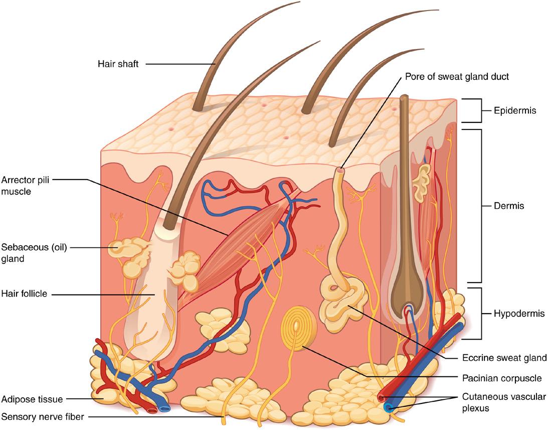
Figure 12: Diagram by: Open Histology – Body Systems, University of Galway, https://openpress.universityofgalway. ie/openhistology3/chapter/layers-of-the-skin/
General principles of good skin care should include:
• Wash daily, whenever possible, using pH neutral soap, natural soap or a soap substitute, and pat dry thoroughly
• Ensure skin folds, if present, are clean and dry
• Monitor affected and unaffected areas of the patient’s skin for cuts, abrasions or insect bites, paying particular attention to any areas affected by sensory neuropathy
• Apply emollients as clinically indicated or prescribed
• Encourage the patient to avoid the use of scented products on their skin
Particularly in hot climates, vegetable-based products are preferable to those containing petrolatum or mineral oil.161 For individuals with intact skin, the condition of their skin (Figure 12) should be optimised though the application of an emollient at night. Patient’s with dry skin, which may appear flaky, scaly or rough to touch, may also complain of associated itching. In this clinical situation, emollients should be applied twice daily (as well as after washing) to aid rehydration. If the heels are deeply cracked, emollients and the use of an occlusive dressing may help, and the patient should be referred to local dermatology guidelines.161
For home or hospital-based management of acute cellulitis/ erysipelas exclude the presence of other infections, such as those with a systemic component like: venous eczema; contact dermatitis; intertrigo; microtrauma and fungal infection; acute deep vein thrombosis; thrombophlebitis; acute lipodermatosclerosis; and lymphangiosarcoma (Stewart-Treves syndrome)
• Swab any exudate or likely source of infection, such as cuts or breaks in the skin
• Before commencing prescribed antibiotics establish: the extent and severity of the rash. Mark and date the edge of the erythema; the presence and location of any swollen and painful regional lymph nodes; the degree of systemic upset (alteration of the individuals ‘normal’ blood values); the erythrocyte sedimentation rate (ESR) or C-reactive protein (CRP), white cell count, and procalcitonin level.
• Prescribe antibiotics as soon as possible, after considering swab results and any bacterial sensitivities if clinically indicated.
• When the patient is resting in bed/on bed rest, elevate the limb, administer appropriate analgesia for example paracetamol or a NSAID, and increase fluid intake
• Avoid MLD
• If tolerated, continue compression at a reduced level or switch from compression garments to reduced pressure multicomponent/multilayer inelastic bandaging
• Avoid long periods without compression.
• Recommence the patient’s usual compression and levels of activity once pain and inflammation are sufficiently reduced for the patient to tolerate
• Educate patient/carer about symptoms to look out for, when to seek medical attention, risk factors, and the use of antibiotic prophylaxis, if clinically indicated.161,162
Finally, a useful strategy that can help guide all HCPs when developing their individual patient’s skin care regimen is that of CLEANSE, PROTECT and RESTORE (CPR). CPR was first suggested in this context following an International Consensus Panel in 2014 that met to consider evidence deficits in current understanding and best practice in relation to patients who suffered from incontinence associated dermatitis (IAD).163
• Cleanse — cleanse the skin to remove any contaminants/excess fluid (from whatever source) prior to the application of a clinically indicated skin protectant as part of an assessment skin care regimen planned in conjunction with the patient
• Protect — protect the skin to avoid or minimise the exposure to and the effects of any contaminants/excess fluid
• Restore — patients may also benefit from ongoing interventions to support and maintain the skin barrier function using a clinically indicated skin care product163
Principles of lymphorrhoea, exudate and wound assessment and management
All wounds on patients’ lower limbs, such as venous leg ulcers, related exudate levels and the surrounding (peri-wound) skin should be assessed using a holistic framework (Figure 13). This assessment incorporates a systematic wound assessment model that is familiar to HCPs undertaking the assessment within their own clinical environments and other members of the multidisciplinary team (MDT). The MDT includes medical, nursing staff and physiotherapists (generalist and specialist), dieticians and others working in both primary and secondary care settings as clinically required and locally available, work in or collaborate with that clinical setting. A holistic approach can be described as dealing with the whole person and not just their clinical condition in isolation.
All wound assessments should be fully documented using physical documentation or an electronic recording
system familiar to all HCPs within the clinical setting, in order for that information to provide a baseline for subsequent reassessment and documentation of the wound/wound fluid. This information can help all members of the MDT identify improvement or deterioration of the wound or medical condition early. 165 Ideally wound and chronic oedema services should be able to refer patients to each other’s HCPs services and have access to each other’s body of knowledge (electronically) or, as is now beginning to occur, offer an integrated service to improve outcomes. The process of wound assessment for all healthcare practitioners involves HCPs having:
• knowledge of relevant anatomy (the skin) and physiology (the ‘normal’ wound healing process)
• the ability to identify factors that may interfere with the ‘normal’ wound healing process
• the ability to collect subjective (patient’s pain experience) and objective (wound dimensions) data
• the ability to analyse and interpret the information that has been gained (as documented)
• the ability to identify the patient’s problems and needs through discussion with the patient; their relatives/ carers and/or other healthcare practitioners164
Chronic oedema can occur in any part of the body but is most commonly seen in the lower limbs due to dependency and gravity. Lymphorrhoea is defined as leaking lymphatic fluid (lymph) though the skin surface, also referred to as ‘wet legs syndrome’. It is a sign of severe uncontrolled chronic
Figure 13: A holistic wound management model.164 Credit: Mark Collier, UK.
oedema, whereas very early signs of chronic oedema are erythema; shiny skin and the appearance of beads of fluid which leaks from the affected oedematous area, increasing the risk of cellulitis/infection and skin damage.166 The most common lower limb wound type that has been associated with chronic oedema is a chronic venous leg ulcer. A leg ulcer (wound) has been defined as, ‘An ulcer that originates on or above the malleolus but below the knee that takes more than two weeks to heal.’167
In clinical settings excess exudate (see Table 6) is often referred to as ‘wound fluid’ or ‘wound drainage’,168 however, it can more appropriately be defined as, ‘material composed of serum, fibrin, and white blood cells that escapes into a superficial lesion (a wound) or area of inflammation’.169
Table 6: Consequences of too much exudate.
Credits: Mark Collier, UK.
Too much exudate
Exudate leaking
Embarrassment
Peri-wound skin changes
Malodour
Maceration
Pain
Increased risk of infection
May indicate the presence of infection158
Effects on a patient’s Health Related Quality of Life (HRQol) –both physical and psychological
Restriction of daily activities of living – work; shopping; cooking; socialising with family, friends and usual social circles (such as clubs and church)170
In 2019 the ILF undertook an international epidemiology study, which highlighted the link between chronic oedema and wounds.1 The effects of chronic oedema, lymphorrhoea and ‘hard to heal’ wounds, such as chronic venous leg ulcers, have significant effects for patients, not least on their quality of life,150,171 such as:
• Increased social isolation and loneliness, embarrassment and reliance on health care contacts
• The inability to safely mobilise or use stairs with a tendency to sleep in a chair rather than a bed
• The inability to wear normal shoes or slippers, increasing the risk of falls and resulting in a more limited choice of clothes which has an adverse effect on the patient’s body image and potentially increases further their social isolation
• Increased pain and limb heaviness which could exacerbate concurrent conditions, such as arthritis
• Anxiety, fear, depression, embarrassment and loss of control
• Increased risk of cellulitis which often results in a hospital admission and/or recurrent infections
• Practical difficulties keeping wound dressings in place if the patient is actively involved in their own care
• Changes in peri-wound skin quality, such as maceration, MASD and a compromised barrier function
• Delayed wound healing
• An increased risk of developing cellulitis, which often results in a hospital admission
• Chronic oedema patients report that the condition affects their ability to work, and many give up work as a result4
• Another consequence is increased costs for the health care system in which they are being managed172
The approach to management for all of the above clinical conditions has been previously identified and should involve various members of the MDT in order to address the following common clinical issues:
• The underlying condition causing the oedema and/or other factors affecting wound healing
• The provision and use of compression therapy and oedema management
• The encouragement of exercise/the maintenance of or increased mobility
• The promotion of evidence-based best practice skin and wound care
• Tailored patient education; taking into consideration individuals’ health literacy levels
• The prevention of oedema and wound recurrence.2
The Chronic Oedema Wet Legs Pathway
The Chronic Oedema Wet Leg Pathway173 was developed in 2017 further to an educational activity of the Lymphoedema Wales Clinical Network, in the United Kingdom (UK). On the conclusion of this activity, it became clear to the participants that many patients in Wales with chronic oedema and lymphorrhoea were being routinely managed ineffectively by the application of a wound dressing and a simple tubular stockinette. This approach provided no compression
or support to the patient’s lower limb. Not surprisingly, therefore, with no active compression therapy involved in their management plan, patients were being reviewed at least once a day as the lymph fluid had soaked through the chosen dressing and stockinette. At that time, the authors (of the Wet Leg Pathway) could find no guidelines or evidence-based pathways in the literature to support the prompt use of compression before an arterial assessment, including an Ankle Brachial Index (ABI) measurement had been undertaken.
The Chronic Oedema Wet Leg Pathway was created to support the efficient, effective and prompt management of patients affected with chronic oedema and lymphorrhoea, in order to reduce the risks of further complications associated with the delayed application of compression, such as cellulitis, hard-to-heal wounds and falls.4
The pathway outlines four levels of interventions for the management of a patient with chronic oedema/wet legs. In summary they are;
Level 1 — Support only protect and comfort
This level is particularly useful in the short term, during the acute phase of cellulitis, where skin integrity is compromised or in end-of-life situations.
• An ABI/TBI (Toe-Brachial Index) is not required. The latest British Lymphology Society paper indicates that an ABI assessment is not reliable in severe chronic oedema patients174
• Wash and dry affected leg(s) using an appropriate method and cleanse wound/remove excess exudate if clinically indicated
• Moisturise the skin with a non-allergenic emollient
• Apply wound absorbent dressings/pads as per local guidance/formulary or national guidelines
• Apply layer of tubular stockinette (size dictated by the size of the patient’s lower limb)
• Apply wool bandages to affected lower limb, ensuring a cylindrical shape is achieved and all bony prominences are covered and protected. Start at the base of the toes and apply in a spiral fashion up to just below knee.
• Cover with a final layer of tubular stockinette (size as clinically dictated).
• Apply toe bandages if toes are oedematous (if suitably trained to do so).
• Reassess at every patient interaction.
Level 2 — provides less than class one compression (14–17mmhg) but does provide a therapeutic level of compression to treat chronic oedema, as it also promotes patient adherence with tolerable compression. Follow the above instructions and then;
• Apply one layer of short stretch inelastic bandage or a cohesive bandage stretched in a spiral application, starting at the base of the toes to below knee, using a 50% overlap if clinically indicated (patient does not have a conic leg shape).
Re-assess at each bandage change and act as clinically indicated consider Level Three/Four or administer compression garments
Level 3 — Follow instructions for Level Two and then; Ensure an arterial assessment including an ABI is completed.
• Apply a second layer of short stretch inelastic bandage or a cohesive bandage at full stretch in an opposite spiral application from the base of the toes to just below the knee with a 50% overlap up to the knee.
Re-assess at each bandage change and act accordinglyconsider Level Four or administer compression garments
Level 4 — If the patient’s chronic oedema or lymphorrhoea is not improving, then:
• Collaborate with the local secondary chronic (lymphoedema) service to increase the layers of short stretch inelastic bandage or a cohesive bandage. The service can also support and advise on compression for oedema in knees and thighs.
The main aim of the interventions is to reduce/manage the patient’s oedema, heal wounds and stop lymphorrhoea.
Once this has been achieved compression therapy (bandages/wraps/garments such as hosiery) ideally should continue to be worn daily by the patient as clinically indicated and in accordance with the patient’s wishes/ local availability.
The full Chronic Oedema Wet Leg Pathway document and supporting appendices can be accessed via https:// sbuhb.nhs.wales/go/discharge-support/pathways-andservices/lymphoedema-service/lymphoedema-servicefolder/use-wet-leg-pathway/
Importance of compression therapy
A number of international guidelines now support the use of mild compression therapy in the absence of red flag symptoms/conditions (National Wound Care Strategy Programme) even before obtaining an ABI assessment. Evidence reviewed has indicated that the benefits of compression therapy outweigh any risks for patients.175 Other international organisations, such as EWMA and the ILF, have recently reviewed the evidence supporting the use of compression therapy. This has been detailed previously in the ILF Best Practice Documentation on the Management of Lymphoedema and in Chapter 8 of a EWMA document entitled Lower Leg Diagnosis and Principles of Treatment. The full documents can be accessed via https://www.lympho.org/uploads/files/files/ Compression-bandaging-final,pdf and https://ewma.org/ wp-content/uploads/2023/12/EWMA_Lower_Leg_Ulcer_ Diagnosis_web.pdf Nonetheless, it is worth noting that the debate in the literature as whether to use mild (up to 20mmHg) or strong (40–60mmHg) compression before an ABI assessment has been undertaken continues. Mosti (2019) 176 and others have indicated that strong compression is both safe and clinically effective until there is evidence of critical ischaemia.
The evidence supporting the use of compression therapy, indicates it should be applied as soon as possible, unless contra-indicated.
Wound care principles
The main components of care for patients with lower leg wounds and chronic oedema were identified by Keast, Burian, Moffatt et al in 20212 as:
• Manage underlying conditions – such as congestive cardiac failure; diabetes; venous insufficiency and peripheral arterial disease (PAD)
• Compression therapy – crucial for the control and reduction of oedema; improvement of the patent’s skin condition and to stimulate wound healing, if the patients vascular status permits. Modified compression can be used up to the point of critical ischaemia by suitably qualified HCPs177
• Other oedema management – prescription of appropriate drug therapy as prescribed and the supplemental use of intermittent pneumatic compression therapy (IPC) along with compression bandaging, if clinically indicated
• Wound care: Debridement178 – removal of non-viable tissue at every dressing change and wound bed cleansing with evidence-based solutions
• Microbial management – control of bioburden as a result of appropriate debridement and wound cleansing
• Exudate control – compression therapy in combination with appropriate wound dressings
• Skin Care: Minimise risk of broken skin and cellulitis/ infection; use of pH balanced soaps; application of emollients as clinically indicated; minimise the effects of any inflammation/dermatitis by the use of prescribed corticosteroids, which should be applied as per manufacturer’s instructions prior to the application of an emollient; application of barrier products, as clinically indicated, and patients with diabetic neuropathy should be encouraged to frequently inspect their skin, in particular at likely pressure points (from footwear)
• Promote exercise – consider referral to a physiotherapist if the discussion and the provision of patient education regarding appropriate exercises (in whatever format) have not made a difference
• Promote lifestyle changes 179 –control of diabetes; smoking cessation; control alcohol intake; concordance with agreed management plan as discussed and developed with the patient
• Pain management – important to distinguish if the pain is nociceptive (perception or sensation) or neuropathic (nerve-related) in origin180
• Weight loss – in particular if the chronic oedema is obesity-induced181
• Offloading – mandatory for all patients with a diabetic foot ulcer/pressure injury,supported with appropriate encouragement, discussion and education
• Assess for psychological impact – set appropriate goals in conjunction with the patient and care givers as applicable2
Compression therapy (see Chapter 5) should be applied in conjunction with appropriate ongoing wound care undertaken in light of current best practice guidelines150,182,161,183 and local protocols, based on the availability of appropriate management products. Burian2 highlighted the benefits of compression therapy being used for the management of wounds associated
with chronic lower limb oedema, as it has been reported to have positive therapeutic effects on the inflammatory process, the patient’s vascularity and it minimises chronic oedema formation.
Chronic oedema has been associated with a number of wound types, such as venous leg ulcers; trauma from compression (tight fitting shoes may lead to an unhealed wound in patients with diabetes and neuropathy for example); wounds as a result of an incidence of acute cellulitis and a wound from a skin biopsy site that has failed to heal.
The wound and skin care elements of the care of a patient with a chronic venous leg ulcer or any chronic wound on the lower limb, as indicated previously, should be undertaken as part of a holistic, structured and personalised strategy, in accordance with best practice guidelines and local protocols based on the availability of appropriate wound management products.150,182,161,183
An example of an acronym that includes all of elements that should be considered when developing a personalised wound care strategy, is that of TIMERS (Figure 14).
Readers are also encouraged to refer to Wound Infection in Clinical Practice, International Wound Infection Institute (IWII)
Chronic venous leg ulcers are just one example of a wound associated with the production of excess amounts of exudate. In this author’s experience and others, staining of dressings (in particular on secondary dressings and bandages) and exudate leakage can be particularly distressing to patients and their carers. Leakage or strikethrough can cause embarrassment and if not appropriately assessed and managed, can lead to patient reports of wound odour, which may also be an indication of increased wound bioburden or infection.158
If exudate is not managed properly, the wound/exudate assessment process should identify one or more of the following:
• delayed wound healing times184
• increases in the incidence of peri-wound skin damage (such as change of colour/skin stripping) and the risk of increased bacterial burden within the wound margins/ infection150
• increases in the number of dressing changes required
• increases in the time and number of interventions required by healthcare practitioners to manage the wound/exudate
• increases in the overall cost-effectiveness of patient management
• changes in patients’ perceived HRQoL
In all cases in which excess wound exudate is assessed, evidence-based interventions should be actioned, with the aim of, and in order to re-establish an ‘optimum’ moisture balance within the patient’s wound margins (not too wet/ not too dry).164 Currently, it is important to note that there is no internationally accepted standard method for measuring the rate of exudate production nor is there an accepted ‘norm’.168
Wound dressings and materials
A number of research studies about a range of ‘modern’ interactive wound dressings and some traditional products – alginates; hydrocolloids (under compression); topical hydrogels; gauge and saline; manuka honey; foams and antimicrobials – were reviewed in detail for their effectiveness by Prost and Atkin.185
In an effort to manage large amounts of leaking body fluid/excess exudate, an increasing number of wound management materials have been developed in recent years such as those with hydration-response technology,186 and those whose composition includes superabsorbent polymers (SAP), which have been developed to help clinicians manage a patient’s leaking body fluid/wound exudate more effectively. Recently, this author reviewed some of the available evidence related to superabsorbent wound dressing materials 187 – laboratory and clinical, however it should be pointed out that to date no dandomised controlled trials involving these wound dressing products and hydration response technology products (comparative or otherwise) have been published.
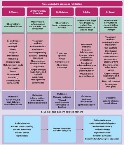
Many absorbent materials used in non-occlusive permeable dressings allow the moisture to be absorbed and then evaporate into the atmosphere. This property has been quantified as the moisture vapour transferability rate (MVTR), 188 However, it should be noted that this characteristic is not strictly relevant to the action of a superabsorbent product, because these products have been designed to retain large amounts of moisture rather than to facilitate evaporation. Many traditional absorbent dressings include a soft polymer core with or without an absorbent pad (National Institute for Health and Care Excellence189) and these are generally best suited for use with lightly to moderately exuding wounds. Dressings that include SAPs as part of their design are commonly referred to as ‘superabsorbers’. These dressings expand on absorption of fluid. However, it has been pointed out that ‘superabsorbent’ is a comparative not an absolute term.190 As with all wound management products, health professionals should read the manufacturer’s instructions prior to using the product.
Summary
The recognition of a patient’s wound associated with chronic oedema and their overall skin condition is vital, as are accurate holistic assessment and evidence based/best practice management. In combination with compression as clinically indicated, this clinical approach should improve the patient’s healing potential; improve/maintain their skin condition; have a positive effect on their health related quality of life and minimise the risk of wound/skin infection(s).
Figure 14: TIMERS. Reproduced with permission from Atkin et al., 2019.150
5. Compression therapy: the gold standard for the management of chronic oedema of the lower limb
Learning points
• An introduction to compression therapy and why this is so critical for the successful management of lower leg oedema. There is a growing acceptance that non or sub-optimal use is a patient safety issue and creates harm when therapeutic treatment is not provided
• Compression therapy must be delivered at a therapeutic dose to provide effective treatment to the leg. Understanding the variety of compression materials and their effects provide opportunities for tailoring treatment to the individual
• Musculoskeletal health and mobility improve the efficacy of compression therapy and well-being of the individual
• The need for compression therapy is lifelong in chronic oedema management and thus expertise and partnership are critical to enable tolerance
Introduction
Compression therapy is the cornerstone of chronic oedema management, whether or not associated with a wound,191 and provision of compression is often described as ‘the gold standard’ part of a treatment plan, delivering clear evidence-based therapy. Compression is critical in both an intensive intervention stage and in maintenance phase, requiring a good knowledge of materials and the different types of compression therapy available. Choosing the most suitable compression system from a range of bandages, medical compression stockings and adjustable compression wraps requires understanding patient need; this is often enhanced with multi-professional co-operation. There is clear evidence about its non-use and the suboptimal use of compression therapy alongside a growing use of reduced levels of compression.192 The EWMA Compression Therapy Campaign193 has produced multiple resources in different languages with the simple aim of increasing the use and understanding of compression therapy and its important role in the management of chronic oedema and lower leg injuries.
Compression is underrated and under used creating a significant health care burden and deleterious impact on lives.194 Unfortunately, effective application and often tolerance of the therapy by the patient is dependent on the skill of the practitioner. In practical terms, it is common for HCPs to lack confidence in using this critical therapy thereby creating poor access for patients. The variation in use across any healthcare system is stark; a survey found that older people and those being managed at home had less access or usage when compared to use of compression in specialist clinics.192 When people are not in receipt of evidence-based treatment, it should be no surprise that quality of life suffers. There is now a large body of evidence that describes people’s experience and the destructive impact of non-healing wounds caused by chronic oedema on their life,195, 196, 197; delayed wound healing is associated with the poorest quality of life. The link between their experience of pain and isolation has not been linked to sub-optimal use of therapeutic compression, and further studies are required to understand this more clearly.
Compression as a therapeutic intervention
The benefits and therapeutic impact of compression therapy as an intervention is little understood by generalist practitioners and is often seen as a task within a treatment plan, despite decades of research that has shown its clinical and cost effectiveness. Thus, to help HCPs in their understanding of this important intervention, there is a growing emphasis in publications on the effect of compression on the limb and the role of its physical properties. 198, 199, 200 There is now a challenge to clarify circumstances that have previously been cited as being contraindications for compression.201, 202, 203 The importance of compression usage in a risk management approach is gaining ground to support clinicians in their decision making and to articulate the rationale for choices being made. Being cautious in implementation is now a preferred term rather than describing contraindications.203
The rationale for the use of compression extends beyond chronic oedema management and venous ulceration; compression therapy is the cornerstone of good management of lower leg conditions. 204 The worth of compression for chronic oedema and all lower leg wounding is accepted practice by specialists but compression therapy position documents have focused primarily on its use in chronic oedema and venous ulceration. There are now a growing number of publications that emphasise the use of compression in wounds of arteriovenous origin, such as pyoderma gangrenosum and those with mild to moderate PAD.205,206,203 A recent consensus document has proposed a new term to promote the use of compression for leg ulcers with mixed aetiology, caused by combined arterial and venous insufficiency or CAVI ulceration.207 For compression to be effectively applied, graduated compression was considered gold standard, that is the compression is applied with higher pressure at the ankle and reducing in dosage to the calf. While recognising the worth of graduated compression in the more immobile group, and this profile within garments, Mosti (2025)236 reports the importance of applying progressive compression in the mobile group, that is a higher pressure at the calf level than the ankle, supporting greater venous pumping function and thus greater efficacy. This recognition demonstrates the need for an HCP to keep up to date with the developing science of this critical therapy.
Understanding the critical benefits of compression therapy is essential if the HCP is going to persuade patients to undertake this therapy. There is a great deal of discussion and reference about patients’ non-compliance with compression, even though we are fully aware that there is a culture of under use of effective evidence-based therapy and a negative approach by nurses and physicians (See Chapter 7). Specialists in this field acknowledge that tolerance is much improved when people are persuaded of its benefits, understand how it works, and that the technique used is actually comfortable. Compression bandaging and even choosing the right garment could be described as art; enabling tolerance is a clinician’s problem to solve.199 Unfortunately, the view that ‘a little compression is better than none’ may create further problems due to this being sub-optimal and thus ineffective. Often the intention of commencing mild compression is to demonstrate its wearability and impact. However, if the compression acts sub-optimally then there is the danger that use of mild
compression actually reduces the patient’s and clinician’s belief in compression as a successful intervention. HCPs and especially specialists need to be alert to this problem; ineffective compression will not control the oedema, inflammation, exudate or pain. Thus, our focus needs to be on enabling people to have optimal compression of a dose that meets their clinical needs.
Effects of compression therapy
For HCPs to use strong compression with confidence, there is the need to understand four critical benefits of compression therapy (see Table 7):
1. Counteracts the impact of gravity. It is important to understand the impact of gravity on any limb. When humans are upright especially in roles with long periods of standing, gravity will be a significant contributor to any delayed healing. Compression counteracts the effect of gravity and subsequent venous hypertension and is useful for all types of ulcers, being considered a ‘most reasonable basic remedy’ for the lower limb.200
2. Improvement of venous function . Compression therapy reverses venous ambulatory hypertension and valves in superficial veins can become competent again.208 The compression system creates a supportive casing to the limb and calf-muscle and in walking, the work of the calf muscle is augmented by this therapy; the pressure the limb receives is dependent on the materials used and subsequent stiffness. Providing a therapeutic level of compression therapy restores muscle-pump efficiency by lowering venous pressure once the superficial and perforator reflux are controlled. The role of an activated calf-muscle pump is critical to reducing venous hypertension and requires good ankle range of movement (see Table 7); reduced ankle mobility impacts on calf function and is known to delay lower leg wound healing.209
3. Reduction in oedema. Excessive oedema causes leg pain, reduces mobility and creates recurrent infection (cellulitis) when unmanaged. Excessive exudate from leg ulceration can be attributed to unmanaged oedema, the source of the exudate or sub-optimal compression therapy. The impact of venous hypertension and oedema in the limb is described in Chapter 1 and compression is the critical therapy to reverse these effects. A recent cellulitic episode is present in 29-32%
of patients at assessment 3,210 with a proportion of these requiring admission for IV antibiotics for cellulitis or sepsis. A retrospective review 210 found that with managed services and the provision of compression garments episodes of cellulitis reduced to 2.12% and admissions to 0.5%. This data speaks of the significant impact good oedema management with garments has on any health economy as well as quality of life.
4. Anti-inflammatory properties . Chronic inflammation is associated with the development of lipodermatosclerosis and subcutaneous fibrosis from ambulatory venous hypertension. (See Chapter 1 on the role of oedema in exacerbating the inflammation). There is a growing body of evidence detailing the direct and immediate impact of compression in reducing pro-inflammatory cytokines. 211,212 The limb and associated skin changes are impacted positively by compression therapy, softening the fibrosis and the livid nature of the lipodermatosclerosis. This immediate anti-inflammatory effect will also reduce pain; this can seem counterintuitive, but it is postulated that compression therapy releases analgesic mediators.208 This underscores the need for HCPs to have deep knowledge of the benefits of compression therapy in order to ‘sell’ this difficult therapy to their patients199 and thereby create swift positive outcomes for patients. However, the dose or strength of the compression therapy or sub-bandage pressure the limb receives is critical for efficacy; a sub-optimal dose of compression will not provide the benefits described.
Table 7: Benefits of compression to the oedematous limb
• Counteracts the impact of gravity; tall people and those in standing occupations may need extra strong compression with a high stiffness index
• Improves venous function, improves function of the venous valves and calf-muscle function when walking; ankle mobility is essential
• Direct impact on oedema, reducing pain, infection and improving function
• Anti-inflammatory impact, reducing pain, aiding healing of wounds and skin changes
Augmenting compression therapy through movement
The importance of musculoskeletal health and movement in
lower extremity chronic oedema
Bipedalism (walking upright) plays an important function in human life, but this important function comes at a cost when fluid moves from the lower extremity (LE) into the trunk. Lymphatic fluid in the body is subject to the force of gravity. It is critical for HCPs to understand the challenge that the presence of gravity poses to the lymphatic system to move fluid from the foot into the trunk against this powerful force.213
To counteract the force of gravity, the lymphatic system relies heavily on extrinsic forces via musculoskeletal (MSK) movement and on intrinsic forces through lymphangion contraction for fluid movement. The body has developed specialised veno-muscular pumps in the MSK system to move fluid from a distal to proximal direction in the LE.214 When these pumps are activated, they exert pressure on the lymphatic vessels, and this facilitates fluid movement within the lymphatic system. There are three pumps in the LE in the foot, calf and posterior thigh. The key pump in the LE is the calf muscle pump (CMP).215 The main joint that operates the CMP is the ankle joint. The development of a ‘stiff ankle’ is associated with ambulatory hypertension and chronic oedema (CO) in the LE and is a common MSK impairment in people that have LE chronic oedema.216,217
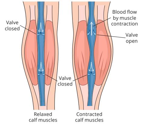
Figure 15: Calf muscle pump. Reproduced with permission from Nair et al, 2024.203
Issues relating to mobility impairment and reduced physical activity are widely reported in people with LE chronic oedema.218,219,220 Mobility commonly relates to the movement of oneself by changing body position or location or by transferring from one place to another221 and physical activity is defined as any voluntary movement produced by the MSK system resulting in energy expenditure.221 Sub optimal or impaired movement and mobility can result in reduced physical activity and rebound oedema and this can then lead to skin changes, cellulitis wounds and/or lymphorrhoea.222 People with LE chronic oedema should be encouraged to move regularly, be physically active and maintain optimal MSK health as this is crucial for lymphatic function and preventing other chronic conditions that can lead to impaired mobility.223,224
Movement is medicine! There is emerging evidence that foot orthoses and certain types of footwear can improve functional movement enhancing muscle pump function and this improves lymphatic function in the LE in conjunction with compression therapy.225,226
Therefore, it is important to be vigilant to any MSK impairments or conditions that will affect optimal mobility. Optimising mobility, movement and physical activity is an important cornerstone part of care – not an adjunct223 and will optimise compression therapy. The management of MSK issues (within scope of practice), early referral to
allied health professions or other services is key in providing essential treatments like orthotics, exercises and footwear which complement and enhance compression. This will help to mitigate the complications that result in LE chronic oedema through impaired movement and mobility and ensure people remain physical activity, and mobile though their life course, which enhances quality of life.224, 220
Sub-bandage pressures
Sub-bandage pressure is the average pressure measured in mmHg that the limb receives from the garment or compression bandage. Unfortunately, classes of compression have international variation thus it is more helpful to simply reference the expected sub-bandage pressure exerted on the limb in mmHg only. It is also more helpful to name the system being used, rather than the more colloquial terms of light or strong compression unless if referencing the standard proposed in this international consensus document.168
• Venous and lymphatic systems in the LE are subject to the force of gravity
• Fluid movement in the lymphatic system is facilitated extrinsically through the veno-muscular pumps in the lower limb
• These pumps work optimally when we walk because the cyclical, repetitive movement of walking mimics a pumping action
• Changes in the way we walk, move and a reduction in PA will negatively influence the movement of lymphatic fluid from the LE to the trunk and this will exacerbate CO
• Promoting and managing movement, PA and mobility is an important part of care — not an adjunct to it
Describing the compression therapy being applied as a dose has been a positive addition to our language; this creates an emphasis on compression as a therapeutic intervention similar to medication. It is common for guidelines to state that compression dosage for the therapeutic intervention needs to be at least 40mmHg and anything less might be considered sub-optimal for most lower limbs227 especially the wider limb; in addition, the height of the person or their standing duration and the presence of extensive lymphatic disorders may require extra strong compression to be therapeutic. However, the amount of compression required will vary depending on the type of oedema or cause of the ulceration; compression dosage of 20-30 mmHg can be effective in the presence of mild chronic oedema but increased to at least 40mmHg in the presence of deep venous disease or in the presence of venous ulceration.
Contraindications and caution
Compression therapy is known to have many beneficial features for treating lower leg oedema, but it has been associated also with side effects and risks. General contraindications for compression therapy, according to Nair et al,203 are with individuals with severe neuropathy involving sensory loss, extra-anatomical bypass or severe PAD. According to Rabe et al,228 severe cardiac insufficiency and true allergies to compression materials are also contraindications for compression therapy.
Table 8: Benefits of addressing MSK health and movement
A recent consensus document 203 provides information on compression usage in combined arterial and venous insufficiency where arterial insufficiency is mild to moderate, and the venous element of the presenting issues are predominant. This document helpfully directs HCPs in this challenging area to use compression therapy when the oedema or ulceration is predominantly impacted by venous hypertension; the dosage may require modification to a moderate level to mitigate risks, so safe and optimal use of compression therapy can be promoted for proactive management.
In the presence of mild to moderate arterial insufficiency, there is mounting evidence of the role of compression therapy in enhancing peripheral perfusion. 202,203 Compression therapy above 20mmHg is now only contraindicated in severe peripheral arterial disease and decompensated heart failure.203 Even in these circumstances, when an oedematous limb presents a risk to quality of life from excoriated skin or harmful and painful leg ulceration, mild compression may be advised under surveillance.
These are the exceptions, but compression hesitancy stems from a fear of inadvertently creating compression damage. While HCPs need to be clinically competent, rather than creating renewed focus on good assessment and evaluation, this fear has often meant the avoidance of compression therapy altogether. Non-use of compression needs to be seen as an avoidable patient safety issue: unmanaged oedema or ulceration creates patient harm and wasted workload, yet it is not recognised as a hazard. Patients need and have a right to receive evidence-based care, and for the oedematous limb or lower leg wounding, this is primarily compression therapy.200 Caution needs to be used when the diagnosis of the wound aetiology is unknown, in the presence of moderate peripheral vascular disease and in the presence of significant pain. In these circumstances, starting with mild compression of around 20mmHg is judicious, alongside evaluating the impact and ensuring compression levels are increased, if required and tolerated.
Challenges in getting compression right
It is important to use the correct compression device to tackle oedema and the site of the oedema. This can create tricky clinical situations especially in inexperienced hands. When excessive oedema is reducing due to the compression device used, it is important to evaluate the
technique and the products used. Advice often needs to be sought if the compression system is not addressing the oedema enough or when there is slippage. Slippage creates poor tolerance due to its impact on pain, pressure injuries or quality of life. Usually most of the reduction happens in the first week. Moffatt et al229 found that inelastic compression bandages dropped 50% pressure in 2 hours and two-thirds pressure after 24 hours. It showed that the danger of slippage can be reduced with technique or using multicomponent systems that have an elastic element that will adjust or adapt to this reduction. Remeasurement of a compression system is critical when there is a reduction in volume and/or shape. Here is an example of fast reduction of severe oedema with compression bandaging where the forefoot remained a clinical challenge, if the ideal outcome was to be achieved, an improved bandaging technique to the forefoot was required:
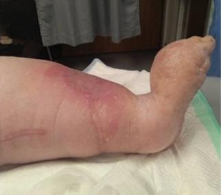
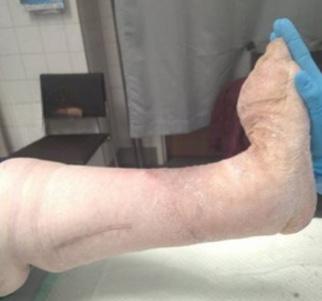
Figures 16, 17: Examples of the difficulty of perfecting oedema management. Credits: Minna Hellgren, Helsinki University Hospital, Finland.
Figure 16 demonstrates the difficulty of perfecting oedema management in a real world homecare setting, managed
by generalist staff with specialist advice only. This example demonstrates how an ideal outcome was difficult to achieve when oedema was not managed early enough or when treatment itself creates additional complexities to manage. This patient had severe oedema and suboptimal inelastic compression was used, resulting in significant oedema to the forefoot (Figure 16). Using the same compression regime, but with an improved technique, oedema to the leg was significantly reduced 5 weeks later (Figure 17) and demonstrates the visible reduction in inflammation. However, the forefoot, while reduced still required a focus from the specialist clinician for forefoot and toe bandaging followed by toe caps once overt oedema has been reduced. This initial reduction gave confidence to both the patient and generalist clinician enabling treatment to continue with the expectation that a better shape to the limb and foot could be achieved.
The role of the ABI
Before starting compression therapy especially with lower leg ulcers, it is important to make an arterial assessment by pedal pulse palpation in the anterior tibialis and/or dorsalis pedis. If absent or the individual is diagnosed with PAD or severe peripheral arterial occlusive disease (PAOD), ankle pressure and ABI or toe pressure is measured, and any subsequent risks for compression therapy require exploration. It is common across Europe for nursing clinicians in the assessment of leg ulceration to be required to obtain an ABI before instigating compression therapy. Low ABI, ankle pressure or toe pressure (Table 9) can indicate risk of soft tissue damage and necrosis caused by insufficient arterial nutrition with the usage of compression therapy. Compression is contraindicated with PAOD if ABI is below 0.5–0.6,230,231,203,228 if ankle pressure is below 60mmHg and toe pressure is below 30mmHg. Although ABI is a reliable measurement for early diagnosis of peripheral vascular disease, caution should be taken with individuals with diabetes. ABI is unreliable with diabetesassociated atherosclerosis and vessel wall calcification revealing normal or high values of ABI measurements.232
It is important to recognise the specialism and international variation in the approach to the role of the ankle brachial pressure index. The British Lymphology Society states that in the absence of cardiovascular disease and signs of PAD, routine ABI is not required233 due to the limitations inherent in this diagnostic tool, which may be elevated or low in the
presence of excessive oedema because the pulse signal cannot easily be heard. If reduced arterial flow is suspected, then referral for vascular opinion or a cautious approach to mild/moderate compression is recommended.
Table 9: Metrics of arterial flow to the foot indicating arterial insufficiency. Table adapted from Nair et al 203
Ankle pressure (absolute ankle systolic pressure)
- Systolic pressure of 50–100mmHg indicates mild to moderate arterial insufficiency
- Systolic pressure below 50mmHg indicates severe arterial insufficiency
ABI = ankle pressure is divided by the brachial systolic pressure
- ABI between 0.4–0.8 suggests moderate arterial insufficiency
- ABI below 0.4 indicates severe arterial insufficiency
Toe pressure (absolute toe systolic pressure)
- Systolic pressure of 30–60mmHg indicates mild to moderate arterial insufficiency
- Systolic pressure below 30mmHg indicates severe arterial insufficiency
TBI = Toe pressure is divided by the brachial systolic pressure
- TBI below 0.7 indicates arterial insufficiency
Choosing a compression system
The choice of type of compression depends on different factors: access of the health care provider to the compression system, the tolerance and self-management skills of the patient and the goal of the compression. Compression textiles and systems have different properties and characteristics and are selected by health care professionals in different stages in the process of compression therapy.198 Often healthcare units have limited access to compression systems and have to use what is available at the time and this often varies across countries.
Inelastic systems (short-stretch) are able to exert higher pressures than elastic systems (and with greater comfort). Moreover, inelastic compression systems are more effective than elastic systems in improving the haemodynamic impairment underlying venous ulceration.
The tolerance of the individual is central to the provision of compression therapy. It’s important to evaluate who is managing the compression therapy or whether the individual is able to apply and remove the system by themselves and whether it supports their lifestyle.203 When the goal is to reduce existing lower limb oedema, different compression bandages are chosen, such as inelastic or elastic compression, often based on country specific recommendations. Multicomponent bandage kits may use a combination of compressive materials234 plus noncompressive padding or treatment layers which can be impregnated with zinc or calamine.235, 203 The application of compression bandages is usually done by health care professionals, but access to compression therapy can be challenging and use in practice is inconsistent.192 Another option is to reduce oedema with adjustable wraps, which gives the individual the possibility of self-application and is not dependent on healthcare professionals. Many compression devices can be used or need to be used in combination for the therapeutic impact required (See Chapter 7).
Compression therapy with existing wounds
When using a compression system with wounds, the compression system should be re-assessed by health care professionals to ascertain that the wound is on a healing trajectory and the oedema and, therefore, the exudate is being actively managed. If the wounds are highly exuding and there is a need to use excessive amounts of dressings frequently to absorb the exudate, the healthcare practitioner needs to evaluate the efficacy of the chosen compression system and whether the dosage of compression being provided is at a therapeutic level for this patient and their
clinical needs. Uncontrolled exudate often requires the use of compression bandages or compression wraps for ease of dressing management until exudate is controlled when compression garments could then be used. If the wounds are epithelialising well and there is no longer a need for thick superabsorbent wound dressings, it is likely that the oedema is now well controlled, enabling the use of hosiery kits with supported self-management. Hosiery kits can be used until the wounds are completely healed and oedema is reduced and then the team can reevaluate the need for long term compression hosiery in order to prevent recurrence.
When oedema has been successfully reduced depending on whether the individual has leg ulcers or manageable oedema, the next critical step is future prevention of wound or oedema recurrence. Prevention is usually managed with adjustable wraps, hosiery kits or compression garments. On the other hand, if the chronic oedema is complex and difficult to control, the management continues with combinations of compression materials, such as bandages, wraps and hosiery.229 Measured and fitted compression garments are renewed every 4–6 months and used as long-term compression therapy with manageable oedema.
Compression bandages and adjustable wraps in combination bring increased sub-bandage pressure, stiffness and comfort to its user. Stiffness is the ability to resist expansion of the limb in inelastic compression devices, and the dosage is the inward pressure from the compression textile due to its properties. 198 The static stiffness index (SSI) describes the ‘difference between the standing and resting pressure’; 234 an inelastic or multicomponent system has a higher SSI. Thus, choosing
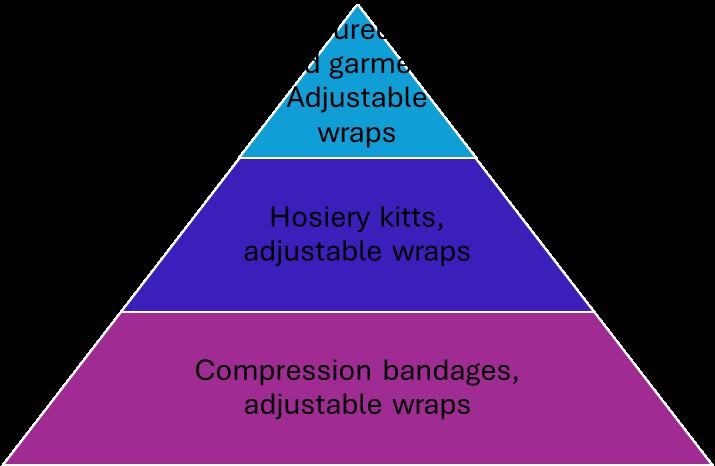

Management of oedema
Reduction
Figure 18: An example of types of compression systems to select with manageable oedema: from reduction to management of oedema. Credits: Minna Hellgren, Helsinki University Hospital, Finland.
a system that has a higher SSI has a greater impact on reducing venous hypertension when walking. 235 The stiffness resists the muscle contraction and creates high pressure peaks imitating the normal function of valve. Nair et al203 describe high peak pressures with sitting and standing during passive and active exercise and thus stiff compression can be used in patients with restricted mobility. Issues like static and dynamic stiffness have become very useful in the clinical setting but the term stiffness has never been fully evaluated in randomised control trials.
Compression bandages
Compression bandages can be described as inelastic or elastic and this indicates the level of stiffness provided to the limb. Elastic materials elongate to >100% of their original length and have the ability to return to their initial length after being stretched and thus follows the limb as the oedema reduces. Inelastic bandages are either non-stretchable or short stretch, with elongation of 10–100%. These definitions provided by Mosti et al236 are important in our understanding of materials and the impact of our decision making in the clinical environment. The problem we have is that the pressure exerted in the application of compression bandages is determined by the clinical operator and this has been shown to vary widely and more often at suboptimal level.237,238 Compression systems with performance indicators can address this issue239 and provide a guide to the clinician where the leg is of a standard width and shape increasing the confidence of the HCP in applying the desired level and preventing over and under stretching of the fabric. Hands on education of HCPs alongside the use of sub-bandage pressure monitors has been shown to create improved levels of compression therapy that are therefore more likely to provide the therapeutic intervention required.238 240
Compression bandages in combination bring increased sub-bandage pressure, stiffness and comfort to its user.
Compression bandages can be applied in different techniques, which Nair et al 203 have presented very descriptively in their consensus document with a guiding figure. Compression therapy must continue constantly, otherwise oedema will rapidly recure. An example of a technique used is that of the Unna Boot; this is a high stiffness bandage traditionally composed of cotton gauze impregnated with zinc oxide paste and sometimes with calamine. Beyond its compressive effect, the Unna Boot
is is thought to exert an anti-inflammatory effect that contributes to wound healing. The soothing effect of the zinc-oxide promotes comfort and reduces irritation. 241 The semi-rigid structure is thought to stabilise the leg and enhances comfort during walking, all contributing to wound healing.242
Maintenance of compression bandages
With the exception of multicomponent single-use bandages, compression bandages are reusable as long as they preserve elasticity and stiffness and are clean. With oedema reduction and management, it is important to maintain and evaluate the bandages every time they are taken off. The bandages may be stained with wound exudate, skin creams and ointments, other tissue fluids or other stains and will need washing. Washing and drying instructions should be followed as per product instructions. If they have lost elasticity or stiffness, they should be replaced with new ones.
Padding/wadding
Padding or wadding with synthetic padding material under the bandages on the bony prominences is recommended to avoid pressure injuries and for protection of the skin. The synthetic material can be, for example, polyester, gauze, polyurethane foam or foam cubes encased between two non-woven bandages. The padding material may need to be purchased separately from the compression bandage or may be included in a kit with compression bandages. Even though padding is needed under compression layers, excessive padding should be avoided. Excessive padding can cause bulkiness, additional heat and bunching and can decrease the amount of pressure given by the bandages resulting a non-therapeutic compression, however bony areas vulnerable to pressure may need extra padding.229,203 (See Figure 19.)
Some multicomponent compression kits may have zinc or calamine impregnated foam or gauze to use under the compression layers. This provides topical treatment for skin changes caused by oedema and might help to reduce inflammation, eczema and superficial wounds.
Inelastic bandages with reduced stretch
These bandages have low resting pressures and high working pressures as they work with muscle contraction. The stiffer material is resistant to increasing oedema. Stretching or elongation is much reduced and between
10–90% thereby short stretch bandages reduce oedema quickly and must be re-adjusted often in the beginning of therapy as required.235 Shorts stretch bandages create a casing to the limb and because of this are more tolerated during rest and don’t need to be removed while resting.
Elastic bandages with high extension or stretch
Elastic bandages are often referred to as long stretch bandages. They have high elasticity and lower working pressure because they do not provide resistance to muscle contraction. This means that when standing, walking or keeping legs down, they don’t resist to increasing oedema. These bandages stretch twice their original length and have high pressure while resting. Long stretch bandages can be easy to apply but should be used with caution with patients who have arterial disease or dermatoporotic skin and should be removed during resting because of the high resting pressure.229,235
Multicomponent and multilayer bandage systems
Multicomponent systems can be confused with multilayer systems. All compression bandages provide multiple layers due to overlap. Multicomponent is the term for
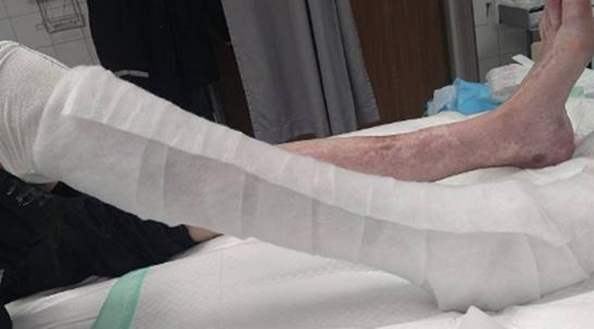
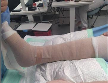
a system where there are a number of compression components within the same kit or chosen system.234 Multicomponent systems can therefore be composed of two or more compression bandages and the kit may include non-compressive products, such as padding and fixing; the multiple components create greater stiffness. Some of the systems have guiding pressure indicators helping the application to reach therapeutic compression. Multicomponent systems can be worn up to a week continuously and can reduce oedema rapidly. They are efficient for the treatment of chronic oedema and like all compression bandages, need to be applied by HCP with proper training and education. 229,235
Figure 21 demonstrates the effect of an intensive bandage regime over a five week period within a specialist service.
This 63-year-old gentleman had chronic oedema in his right leg, significantly impacting his ability to work as a mental health nurse. He was wearing temporary footwear at the initial presentation. He agreed to an intensive bandaging, daily for five weeks with focussed ankle joint mobilisation with a resistance band. This resulted in significant reduction in oedema, reshaping and reduction in skin folds, softer tissues and improved ankle range of movement. Note at week four, the impact on the presentation from the open-cell cubic foam padding used beneath the inelastic compression bandaging to improve skin condition. At week six he was issued a made-to-measure garment and at this stage was now able to wear retail footwear. He was very pleased with the outcome.
Long-term management of oedema
After the pitting oedema is reduced and controlled, compression therapy is usually continued for the longterm management of oedema. Compression stockings or adjustable wraps are measured and fitted to help manage oedema and need to be replaced every six months or more often, if the patient has prolonged standing or needs to wash their compression garments often. The individual needs to be educated to put the stockings on by themselves and take them off, using an aid if required. Compression stockings or garments often enable people to wear their normal shoes and manage their activities of daily living. According to Bjork & Ehmann198 there are various categories of compression garments: circular knit, stiffer circular knit, flat knit, adjustable wraps and night garments. Every garment has its own advantages and disadvantages
Figure 19: Extra-padding on the tibia. Credits: Minna Hellgren, Helsinki University Hospital, Finland
Figure 20: Inelastic bandages. Credits: Minna Hellgren, Helsinki University Hospital, Finland
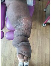
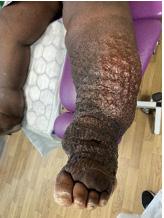
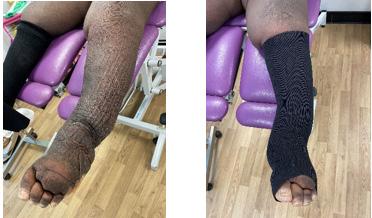
Figure 21: Effect of an intensive bandage regime over a five week period in a specialist service.
Top left: Week 1. Top right: Week 4, note that open-cell cubic foam padding was used for a period of time under bandaging to improve fluid movement (see specific creasing/shaping on the limb). Bottom left: Week 5. Bottom right: Week 6. Credit: Accelerate CIC, London, UK
and should be selected based on the oedema presented by the individual, their healthcare status and need.
Round knit and stiffer circular knit stockings
Round knit garments, previously known as circular knit, are knitted in a tubular fashion either a standard size ready - to - wear or custom made-to-measure. These garments are finer in their structure and cosmetically more appealing than other garments. The stiffer round knit garments are 1,5 or 2 times stiffer than round knit garments and are more resistant to garment fatigue and expansion. It is a hybrid option to circular knit and flat knit garments.198
Flat knit stockings
Flat knit garments are made in a linear fashion in flat sheets with seams or circular fashion that are seamless. These garments have thicker yarns than circular knit garments and due to their inherent stiffness, are better at bridging skin folds and less likely to cause a tourniquet effect or cut in to the skin.198
Adjustable wraps
Adjustable compression wraps (ACW) are a form of gradient compression devices that are cut and sewn from textiles. In comparison to circular knit or flat knit textile variants, ACW tend to exhibit greater stiffness. ACW utilise a range of closures including hook and loop (commonly known as Velcro), elastic drawstrings, and bra-type hook and hole closures. Depending on the design, the textile material may interlace, overlap, or form a rigid sleeve.234
They come with different pieces covering the foot, leg, knee and thigh and there are a variety of designs and physical features available. The textile can be light or firm, ready-to-wear or custom-made. The more excessive the oedema, the firmer garment is needed. The elasticity varies from inelastic to short stretch and long stretch.
These systems can be used in place of compression bandages for oedema reduction and the individual can adjust the system themselves. Montero 235 describes that adjustable wraps positively effects quality of life and provides autonomy to those who do not have access to healthcare professionals to change compression bandages. It is important to advise the individual how to apply the adjustable wraps and to re-adjust daily and as the oedema reduces.198 They can be used in managing oedema after it has been reduced especially when self-application of a garment is not possible. Once again, like compression bandages, the pressure exerted in the application and its efficacy is determined by the user, thus education is critical. Many clinicians find that these do not always work with severe shape distortion; particular areas of difficulty are the foot, knee and thigh.
Night garments
The difference between night garments and other compression garments is the material, which is often waved or chipped foam. They also have a special flat knit spacer. It warms and softens the fibrotic tissue and has circumferential compression. It may be more comfortable to sleep with. The stiffness and the fatigue of the garments vary in different textiles, and the dosage is related to resting pressures. Night garments are designed to be comfortable and safe to sleep with, however, they can also be used during the day.198
The ongoing process of compression therapy
Compression therapy should not only be considered as a form of therapy but also as a process that has a goal.203 Depending on the situation, compression therapy is usually a life-long process to a person with chronic leg oedema.243,203,244 The compression therapy process starts with the assessment of the individual and leg oedema, and this can be enhanced by a multidisciplinary team. After making the diagnosis and care plan, compression therapy is planned and organised considering the different characteristics of the individual and how the variety of available compression systems can help reach the therapeutic goal. The assessment for the correct compression therapy will include site of the oedema or ulcer, the type of oedema (see below) and previous response to any compression therapy, the aetiology of wounds present and any impact of reduced mobility or dexterity. The care plan should also include appropriate interventions, motivation of the individual and establishment of the goal of compression therapy. It is also important to look ahead with the patient so that they are aware of what is required after reduction of oedema, the need for ongoing compression to prevent recurrence and management of expectations. After the successful reduction of oedema the re-evaluation of long-term compression therapy begins with considering the characteristics of the individual and compression textile.243, 203, 198
Assessment for compression
The assessment of the individual and the appearance of the oedema is made at the beginning of the compression therapy process. The aim is to diagnose the underlying
Ongoing compression therapy process:
aetiology and its severity with a multidisciplinary team to make a plan of care, interventions and follow-ups. It´s important from the beginning to ensure that the individual understands the life-long treatment of compression therapy. Assessment also involves the individual’s tissue texture, limb size, location of swelling, quality of oedema and patient functional level. Limb shape and distribution of oedema is to be evaluated by circumferential measurements from the ankle and calf.198 The measurement of the foot is critical as are the toes for toe caps, if required. After compression is started, new measurements can be made and compared to the previous measurements to evaluate oedema reduction; this can bring confidence in the therapeutic value of the compression.
Tissue texture should be evaluated, so that the right kind of compression textile is chosen. For example, a watery tissue texture that is soft, pits easily and is deeply pitting is reduced quickly with compression bandages or adjustable wraps. When the oedema is reduced, compression garments are typically used only during the day with round knit for low dosage, or for deeply pitting tissue, stiffer circular knit or flat knit. Putty consistency in tissue texture pits deep with firm prolonged pressure and requires stiffer compression garments both daytime and nighttime. When the tissue texture is woody and does not pit on application of deep pressure, stiff garments with higher compression pressures are required to warm and soften the fibrotic tissue. When the skin is thin and fragile, it can easily bruise and tear and needs to be considered when choosing compression textiles. To prevent fragile skin being damaged, choose circular knit garments with double-covered inlay yarns. To help when donning the garment, use an under-liner and
Assessment, diagnosis, care plan of leg oedema
Start and organization of compression therapy + attainable goal (long-term maintenance) Interventions and follow up's
Reduction of oedema (+ healing of ulcers) >Reevaluation of long-term compression therapy
Long-term management with compression garments and continued follow up's
Figure 22: Ongoing compression therapy process. Credit: Minna Hellgren, Helsinki University Hospital, Finland
secondary garment and apply silicone lotion to the skin. Other options are to use using donning and doffing devices or adjustable wraps.198 It´s also important to evaluate the extent of the oedema. Compression textiles are mostly applied below the knee, but if oedema extents above the knee, compression textiles should be applied above the knee to the thigh level as needed.
Commencing compression therapy
It is important to explain the purpose and properties of compression therapy to the individual as it starts and continues. When they understand the rationale and benefits of compression therapy, the more motivated they will be to use it. However, we need to recognise the impact of the wider determinants of health on an individual to engage with healthcare; the oedema or wound may be one issue among many that they have to manage.245 Hopkins and Smith245 also recognise the problematic culture within leg ulcer management that impacts negatively on compression usage and also creates a culture of patient blaming. This has been described eloquently by Morgan and Moffatt246 noting how patients can be labelled as ‘bad, difficult or disobedient’. This labelling or marginalisation impacts the person’s care and outcomes.247 The term non-compliance is detrimental to patients because they will not get the care
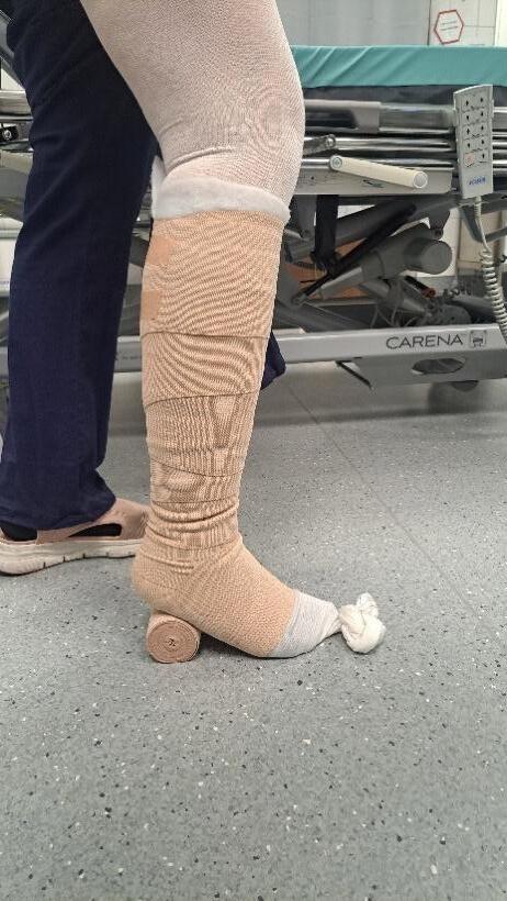
they require and to staff because they stop exploring and bringing creativity to the partnership that should be present in the assessment and planning.
There will, as with any treatment, be hesitancy in accepting a treatment that impacts on body image and lifestyle. Clinicians need to work with this hesitancy so that deep understanding is achieved; it is essential that clinicians pause and truly listen to the individual and their needs, to ensure they are in the centre of the treatment planning and options are explored. This can only be done when the clinician is knowledgeable about the options and choices of materials and is open and able to adjust treatment in response to the patient’s experience.248 Supporting selfmanagement is essential, as is identifying the individual’s capacity to self-care. Patient education promotes shared responsibility for their treatment. Education can be with videos, pictures, guides, links to online resources, anything that’s appropriate in the situation.203 Shared planning will be based on knowledge and understanding and this requires both parties to listen attentively to cues so that all opportunities are explored, enabling use and tolerance of this critical therapy. Knowing the pathway from oedema reduction to management and the rationale for choice or bandages, wraps or garments will bring successful outcomes.
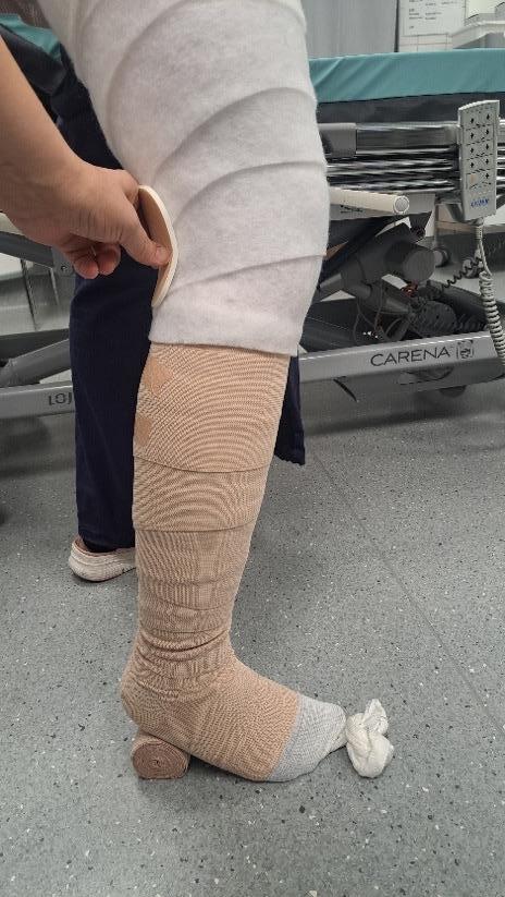
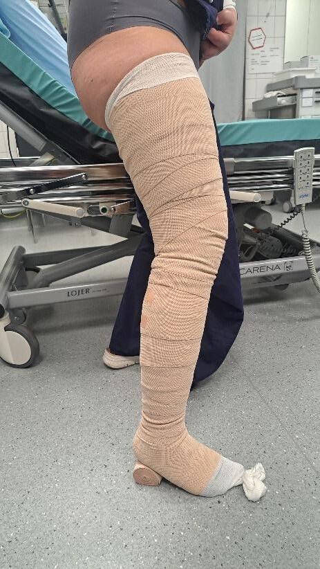
Figure 23: Compression with inelastic bandages. Credits: Minna Hellgren, Helsinki University Hospital, Finland.
From left to right: compression with inelastic bandages from lower leg to thigh level. Notice the padding placed posteriorly on the knee.
Re-evaluation after reduction of oedema
When assessing the individual after the oedema is reduced, there are tools that can be used when choosing the right long-term compression textile. STRIDE acronym that guides the compression selection process198 (see Table 10):
Table 10: The STRIDE acronym. Figure adapted from Bjork et al 198
S=Shape of the limb
T=Texture of the oedema and soft tissues
R=Refill dynamics of the oedema
I=Issues as in contraindications or precautions
D=Dosage, working and resting pressure
E=Etiology underlying aetiologies that contribute to oedema presentation compression therapy needs
The Venous Clinical Severity Score (VCSS) can be used at follow-up to provide an objective measurement of impact.249 The VCSS was developed as an evaluative tool that responds to changes in disease severity over time and for treatment. It’s based on the worldwide standard classification CEAP that describes the clinical features of chronic venous disease. The VCSS includes evaluation of pain, varicose veins, venous oedema, skin pigmentation, inflammation and induration with the scale options being none, mild, moderate and severe. It also includes the active ulcer number, duration and size and the use of compression therapy.
Long-term management with follow-ups
After selecting the garments, it is important to make sure that the individual is trained to put on and take off the garments with the right technique. This needs to be closely monitored. In order to support life-long management and reduce reoccurrence of any wound, it is important for the patient to have access to an experienced healthcare professional who will regularly assess the garments’ textile fatigue and strength, evaluate the presence or changes of oedema and the skin integrity of the limb (see Chapter 6).
Conclusion
This chapter has demonstrated the critical importance of compression therapy and how optimising mobility enhances the successful management of oedematous
limbs and the majority of lower limb wounds. It is incumbent on the clinician to understand the benefits and properties of this therapy in order to optimise its use, provide it with skill and gain acceptance by patients. To use this therapy safely, the clinician must be knowledgeable about products and material, how to adjust the products used and application techniques and how to manage risks in the presence of complexity or moderate arterial insufficiency. Compression therapy is both a skill and an art and HCPs need to challenge its sub-optimal use.
6. Clinical pathways for lower limb oedema management
Learning points
• Five essential clinical pathways to support the multidisciplinary care of individuals with lower limb oedema, with a focus on assessing, managing, and intervening in lymphatic-related conditions
• The pathways serve to simplify complex clinical decision-making while ensuring consistency across various healthcare settings
• They are designed to be practical and accessible for clinicians at all levels while maintaining necessary technical detail
Before selecting an appropriate clinical pathway, it is essential to assign a basic CEAP classification. This ensures that management decisions, including compression therapy, perforator intervention, or specialist referral, are mapped to the disease class and underlying aetiology. Incorporating CEAP at the outset supports consistency in care planning and alignment of interventions with disease severity and pathophysiology.
Pathway 1: Clinical assessment of the lower limb to identify chronic oedema
Goal: Distinguish whether lower limb swelling is due to chronic oedema or other acute causes.
Clinical history
• Onset and duration of swelling (acute versus >3 months =chronic oedema)250
• Family history (Primary lymphoedema, genetic syndromes)251
• Surgical, cancer, or trauma history (risk for secondary chronic oedema)252
• Living in an area endemic to lymphatic filariasis253
• Comorbidities: CHF, CKD, liver disease, obesity, diabetes, conditions causing immobility such as MS, or stroke254
• Drugs associated with swelling, such as calcium channel blockers or cortico-steroids
• Previous history of cellulitis or repeated use of antibiotics
• Previous or current leg ulceration, deep vein thrombosis, varicose vein surgery, PAD
Physical examination
• Skin changes: dry skin, callus, hyperkeratosis, papillomatosis, lymphangioma, mycosis in skin folds255
• Pitting versus non-pitting oedema (indicative of ISL late stage II)
• Presence of positive Stemmer’s sign154
• Symmetry (unilateral/bilateral) and changes in limb distortion
• Observation for signs of cellulitis or lymphorrhoea256
• Assessment for function and neurological changes, such as neuropathy
Diagnostic tools
• Bilateral limb circumreference:257
• May be useful in early screening where clinical oedema is suspected but subtle
• Volume estimation (using the truncated cone formula):
• For more accurate assessment of swelling:
• Measure circumferences every 4cm from the ankle upwards.
• Use the formula: V=(1/12)*h*(C²+Cc+c²)
• V=volume of the segment
• h=height (distance between the two circumference measurements)
• C=larger circumference of the segment
• c=smaller circumference of the segment
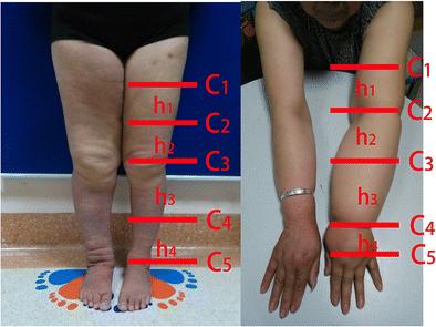
Figure 24: Calculation of upper/lower extremity volume using a summed truncated cone model. Reproduced with permission from Li et al., 2017.257
• Perometry (infrared optoelectronic volumetry):
• Rapid and accurate; does not assess feet or digits
• Limited availability in most community or long-term care settings
• Bioimpedance Spectroscopy (BIS)258
• Detects extracellular fluid accumulation in tissues using electrical impedance
• Particularly useful in identifying subclinical chronic oedema and early-stage lymphatic dysfunction
• Provides L-Dex score; values above 10 suggest lymphatic involvement
• Can be repeated over time to assess progression or response to therapy
• Requires training and consistent electrode placement for reliability
• Ultrasound (Doppler and Tissue Assessment)254
• Doppler ultrasound is essential to exclude DVT as a cause of swelling
• Also used to:
• Assess venous insufficiency (such as reflux or obstruction)
• Evaluates dermal and subcutaneous thickness (indicators of fibrosis or lipodermatosclerosis)
• Detects fluid pockets or changes in dermal thickness in chronic oedema
• Tonometry259
• Measures tissue resistance or stiffness by assessing the indentation depth made by a probe
• High stiffness may indicate fibrotic tissue, often found in Stage II/III chronic oedema
• Quick, non-invasive, and may correlate with chronicity
• Tissue Dielectric Constant (TDC)259
• Assesses localised skin water content by measuring the dielectric constant of skin tissue
• Useful to differentiate lymphatic versus venous or other causes of oedema
• Can monitor changes in response to therapy (such as compression).
• Reference value: TDC >30 suggests increased local fluid.
Decision Point
If findings indicate severe uncontrolled oedema, skin fibrosis, and poor pitting, suspect chronic oedema and refer for specialist assessment.260
Pathway 2: Wound care in lower limb chronic oedema
Goal: Provide a structured approach to managing wounds in oedematous limbs.
Wound Assessment
• Aetiology: venous, arterial, mixed, pressure, trauma, post-cellulitis261
• Wound bed: slough, granulation, necrosis
• Exudate: volume, consistency, odour
• Note: It is important to distinguish wound exudate and lymphorrhoea (see Table 11)
• Surrounding skin: maceration, eczema, infection
Management plan
1. Cleanse wound using non-cytotoxic solutions
2. Debride as appropriate, such as mechanically264
3. Manage exudate with absorbent or foam dressings265
4. Apply compression if indicated (see Pathway 4)
5. Monitor for infection259
6. Pain management systemic or topical
7. Document progress265
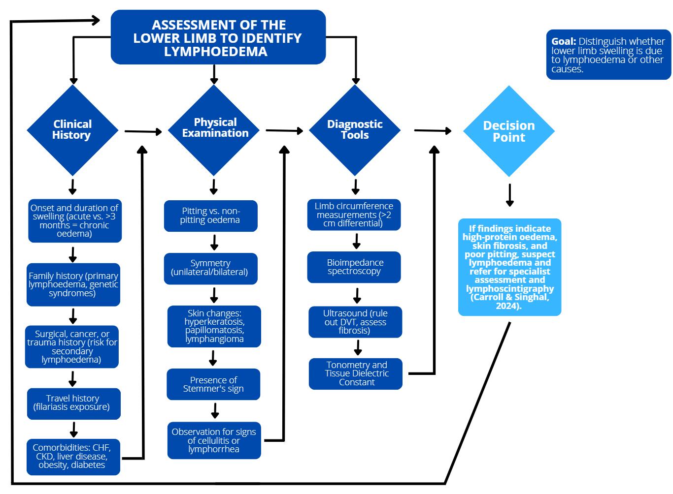
25: Pathway 1: Assessment of the lower limb to identify chronic oedema. Credit: Hayley Ryan, Australia.
Table 11: Exudate assessment: Differentiating wound exudate from lymphorrhoea262,263
Feature Wound exudate
Source Local tissue response to wound healing, inflammation or infection
Appearance Varies: serous (clear), serosanguinous, purulent (thick/yellow/green)
Lymphorrhoea
Leakage of lymph fluid through intact or broken skin due to lymphatic overload
Clear to pale yellow, watery, non-viscous
Viscosity Can range from thin to thick (esp. in infection) Always low viscosity – runny and thin, like plasma
Volume Related to wound size, depth and infection Often high-volume, persistent, and difficult to manage
Onset Usually correlates with wound development or deterioration May occur even in absence of an open wound; worsens with dependency
Odour May have odour if infected Typically, odourless
Effect on skin
Maceration, inflammation, peri-wound dermatitis
Association Often seen with infected or non-healing wounds
Response to dressings May respond to absorbent dressings and antimicrobial control
Severe maceration, widespread wetness, risk of secondary breakdown
Associated with chronic oedema, ISL Stage II/III
Often saturates dressings quickly; requires CDT or compression
Diagnostic tip Swab may reveal pathogens if infected Swab will typically be sterile (unless secondarily infected)
Figure
Table 12: Suspected osteomyelitis: Clinical signs and symptoms267
Feature Description
Persistent or deep ulcer
Exposed or palpable bone
Sudden worsening of ulcer
Ulcer ≥6 weeks duration, particularly over bony prominence (such as heel, metatarsal heads)
Probe-to-bone test is positive: use a sterile metal instrument to gently probe the ulcer base — if bone is felt, high likelihood of osteomyelitis. Refer urgently for further guidance.
Enlargement, increased exudate, or undermining without another clear cause
Signs of systemic infection Fever, rigors, elevated CRP/ESR/WBC count, but may be absent in older adults
Local signs Redness, warmth, swelling, or increased pain, especially if disproportionate to appearance
Discharge Foul-smelling or thick exudate with possible sinus tract formation
Referral criteria
• Wound deterioration over 2 weeks
• Signs of osteomyelitis (see table 12) or spreading infection
• Atypical ulcers (vasculitis, malignant)266
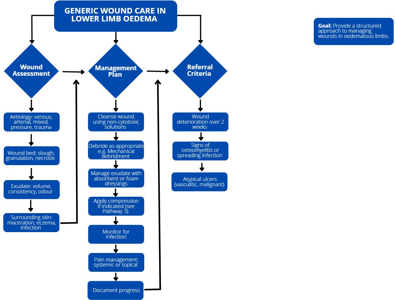
Figure 26: Pathway 2: Wound care in lower limb chronic oedema. Credit: Hayley Ryan, Australia
Pathway 3: Skin assessment in lower limb chronic oedema
Goal: Prevent skin breakdown and identify early signs of complications.
Baseline Assessment
• Integrity: Cracks, fissures, skin tears, open wounds, lymphorrhoea
• Texture: Hyperkeratosis, papillomatosis, callus, warts, dryness or scaling
• Colour: Erythema (inflammation/cellulitis), pallor (poor perfusion), hemosiderin staining
• Moisture level: Maceration, dryness, excessive moisture, presence of lymphorrhoea256
• Signs of infection: Heat, tenderness, erythema, odour, bullae, satellite lesions, skin sloughing
• Other lesions: Papillomatosis cutis, mycosis (fungal infection), eczema, dermatitis
Ongoing monitoring
• Reassess weekly or at every dressing change in highrisk individuals (such as those with diabetes, immobility, or recurrent cellulitis)
• Document:
o Skin changes (such as dryness → maceration → breakdown)
o Fluid levels (dry skin versus exudate versus lymphorrhoea)
o Peri-wound condition
• Educate patient/carer to report changes early259
Skin care protocols
Apply a Cleanse – Protext – Restore approach.
Table 13: The Cleanse – Protext – Restore approach. Credit: Hayley Ryan, Australia.
Step Interventions
Cleanse pH-balanced soap or substitute; pat skin dry, paying attention to skin folds
Protect Apply emollients (urea-based for hyperkeratosis); barrier creams for MASD
Restore Daily emollients (twice daily for dry skin); occlusive dressings for cracked heels
Additional Considerations:268
• Antifungal treatments for tinea pedis or mycosis
• Topical corticosteroids for dermatitis (short-term use only, under guidance)
• Avoid petroleum-based products in hot climates (risk of occlusion, friction, or burn risk if in contact with open fires)
• Encourage patient involvement in self-care and hygiene routines
Decision point
If skin shows recurrent breakdown or infection despite care, refer to dermatologist or specialist wound nurse.
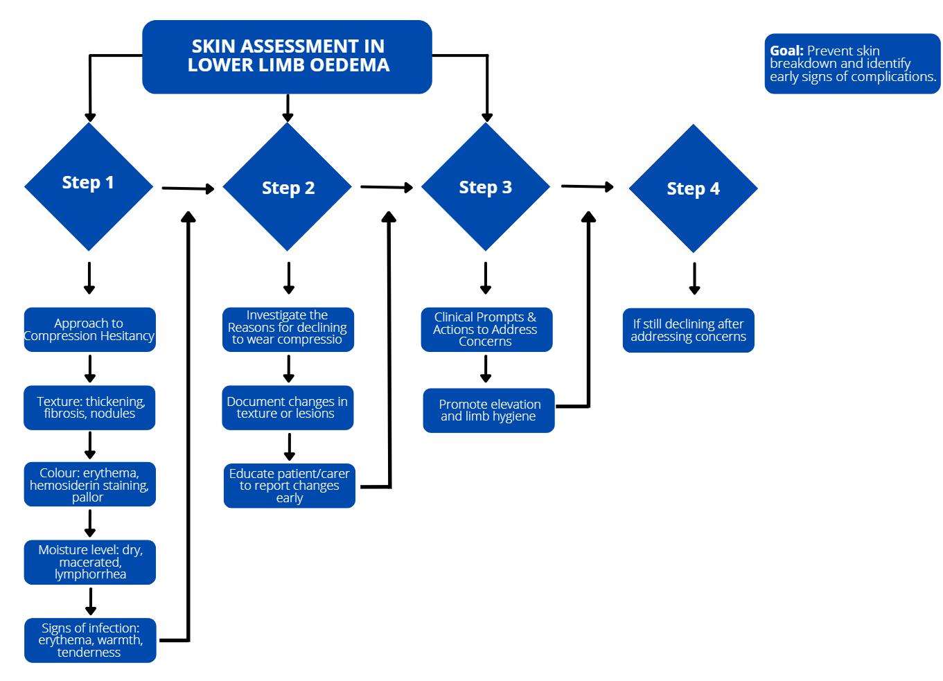
Figure 27: Pathway 3: Skin Assessment in Lower Limb Chronic Oedema. Credit: Hayley Ryan, Australia.
Pathway 4: compression therapy –decision-making guide
Goal: Support evidence-based initiation of compression.
Assessment steps
1. Rule out absolute contraindications (such as severe PAD, critical ischaemia265,269
2. Evaluate ABI or toe pressures254 if unable to record refer for vascular advice
3. Assess patient mobility, skin integrity, tolerance, and cognitive status269
Mobility
• Fully mobile → suitable for most compression types
• Reduced mobility (such as shuffling gait, uses mobility aids)
o Monitor for pressure points/friction
o Ensure that multilayer bandaging is applied effectively to promote mobility
• Bed- or chair-bound
o Apply effective multi-layer bandaging or compression wraps to assist with the additional effects of gravity when legs are dependent. Sequential intermittent pneumatic compression may be helpful as an adjunctive to other forms of compression
o Elevation is critical if the patient can tolerate it
Skin integrity
• Fragile or thin skin (such as aged skin, steroid use)
o Use soft padding and protective interfaces
o Monitor closely for skin tears or shearing
• Broken skin/ulcers
o Choose dressings compatible with compression
Tolerance (Physical and sensory): Assess for:
• Pain or hypersensitivity
• History of hesitancy with compression (past intolerance?)
o Begin with low-pressure systems or wraps if tolerance is poor
o Consider trials of short-stretch bandages or
adjustable wraps
Cognitive status
• Fully independent → able to apply/remove self-care garments
• Mild cognitive impairment
o May need carer assistance or simplified systems (such as wraps versus stockings)
• Moderate-severe impairment (such as dementia)
o Assess risk of garment removal, non-compliance, or distress
o Consider if compression is still appropriate or whether conservative care goals take precedence
o SIPC under supervision may be better tolerated in some cases
Initiating compression
Start with a therapeutic level of pressure
• Use an appropriate compression system
• Monitor and adjust compression type weekly according to oedema reduction and changes in the tissues
Red flags: When to pause or refer
• Sudden increase in pain (determine cause)
• Non-healing wounds despite compression
• Suspected arterial compromise270
• Static improvement in swelling
• Repeat episodes of infection
Decision point
Initiate compression once vascular status is confirmed and adapt to individual patient profile.
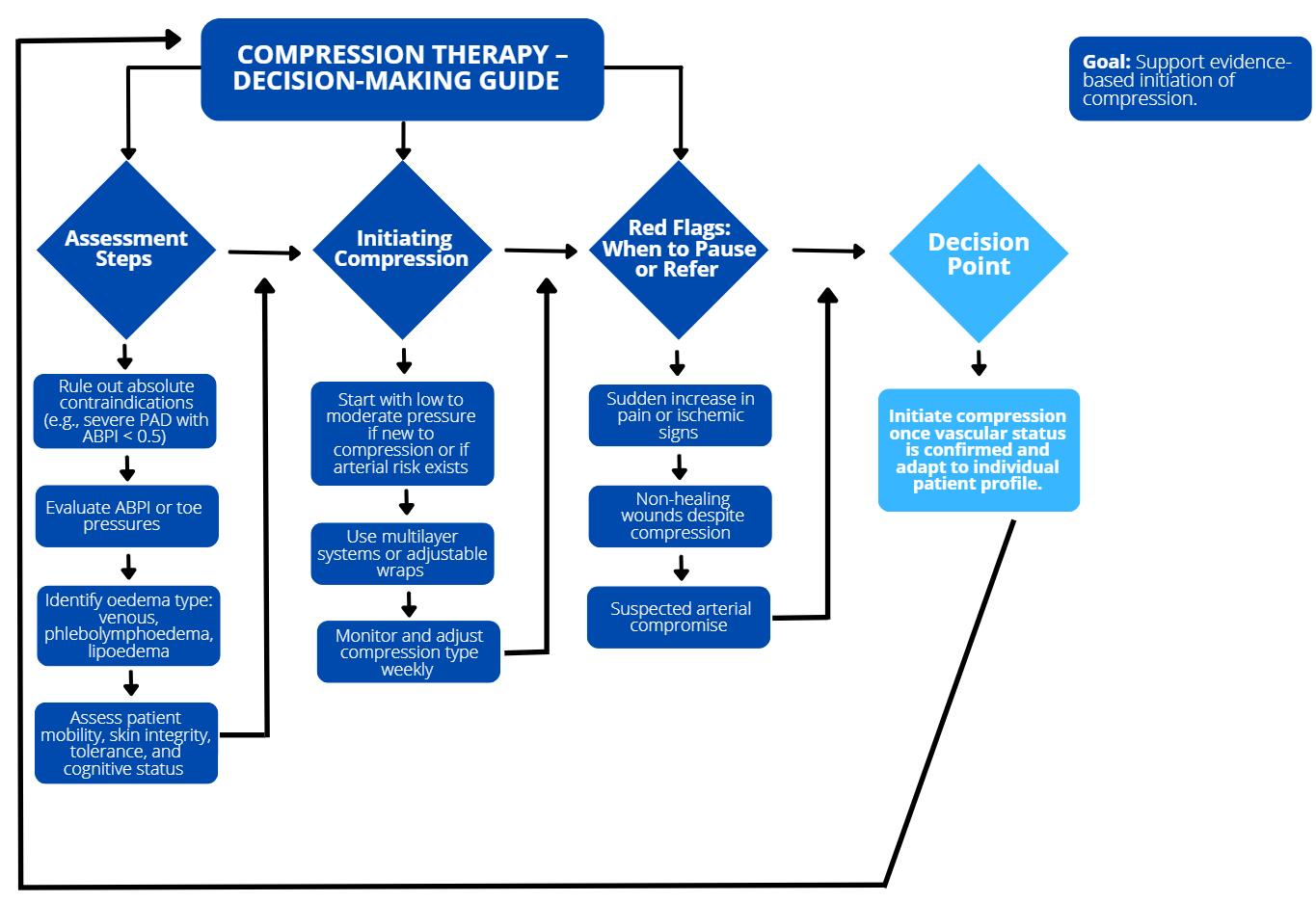
Common barriers to compression adherence
Goal: To increase the long-term adherence to compression therapy in order to maintain stability and minimise the risk of complications.
Approach to compression hesitancy
Always use the appropriate therapeutic level of compression as clinically indicated
Investigate the reasons for declining to wear compression
Ask: “Can you tell me why you’re finding the compression difficult?”
Common reasons to explore:
• Pain or discomfort
• Too hot/uncomfortable
• Bandages/stockings cutting into skin
• Difficulty applying/removing
• Claustrophobic feeling
• Aesthetic concerns
• Perception that compression is “feminine” or not masculine
• Concerns about how compression affects body image or sexual confidence
• Cultural reasons
• Lack of understanding of benefits
• Previous negative experiences
See Table 14.
Clinical prompts and actions to address concerns
Figure 28: Pathway 4: Compression therapy decision-making guide. Credit: Hayley Ryan, Australia.
Possible responses Clinician prompts and actions
“Can you tell me where exactly it hurts when you wear your compression?
Is the discomfort constant, or does it worsen at certain times, for example when standing, walking, sitting?
“It hurts”
“It’s too hot”
Does it feel tight in one area more than others, such as toes, ankle, calf, knee?
Have you noticed any redness, marks, or changes to your skin after wearing it?
Do you have any known conditions like neuropathy or restless legs?”
“Does the heat make you sweat, feel itchy, or cause irritation?
Have you noticed any skin reactions, like rashes or redness, after wearing compression in hot weather?
Have you tried removing them temporarily — if so, how long does it take before you feel relief?
Consider placing the compression in the freezer for a short period before application
Trial Alternatives (if it is the last option)
• Trial intermittent pneumatic compression (IPC) if bandaging is intolerable
• For those with significant heat sensitivity, a period of wraps with breaks and reapplication may improve adherence
“It cuts into my skin” “We can add padding or adjust the fit to prevent that.”
“I can’t put it on”
“I don’t understand why I need it”
“It just feels like something women wear” or “It looks too feminine”
“It makes me feel unattractive” or “It affects how I feel during intimacy”
“Would a wrap/hosiery system be easier for you? I can also show you some donning and doffing aids.”
“Let me show you how it helps prevent ulcers/reduce chronic oedema and improve circulation.”
“There is a range of discreet, neutral-toned options available that were specifically designed with men in mind. Some styles resemble athletic wear, and others come in darker colours or more subtle designs that may feel more comfortable and familiar to wear.”
“That’s a really valid concern. Many people feel the same. There are discreet options that can be removed temporarily if needed, and we can explore alternatives like wraps or garments that are less visible and easier to manage in intimate settings.”
Table 15: Clinical prompts and actions to address concern. Credit: Hayley Ryan, Australia.
Clinical prompts and actions to address concerns
Identified patient reported barrier Suggested clinical actions
• Rule out any changes in arterial disease (ABI/doppler; reassess ABI/TBI that may help confirm whether the pain is due to reduced arterial circulation)
Pain/discomfort
Pain/discomfort
• Assess for correct compression level, adjust padding; assess for other causes of pain, such as arthritis or neuropathic pain;
• Re-measure the limb; trial alternative size/length garmets; different types of fabric or customfitted garments if there is substantial limb distortion or difficulty controlling swelling
• Trial wrap systems, low-pressure compression, or sequential intermittent pneumatic compression with additional compression
• Apply compression first thing in the morning before getting out of bed, when the legs have been elevated overnight, and the swelling is at its most reduced
• If their legs ache during the day, encourage them to lie flat with their legs elevated above their heart for a short period
Table 14: Common reasons for declining to wear compression and possible clinician responses. Credit: Hayley Ryan, Australia.
Too hot
Cutting in
• Garment timing adjustments
• Recommend removal during rest periods when the leg is elevated (if safe and guided by a specialist)
• Seek guidance from your healthcare provider about alternative compression materials
• If heat or discomfort is an issue, your provider may recommend different compression options that can help manage oedema effectively while improving comfort; always consult your clinician before making any changes to your compression regimen
• Layering considerations
• Avoid layering unnecessary liners or socks underneath
• Educate patients on minimal layering techniques where oedema control is still possible with less heat build-up
• Skin comfort measures
• Regularly inspect skin for maceration, fungal infections, or heat rash
• Re-measure limb for correct sizing or choice of garment
• Add extra padding to bony prominences
• Consider use of graduated compression wraps
• Choose a flat knit rather than circular knit garment to reduce the tourniquet effect
• Inclusion of the knee/elbow functional zone, which is a much softer material at the joints may also assist
• Other options may include custom-made compression garments, which can be tailored to fit individual limb shapes and provide more precise and comfortable support, particularly in cases where standard sizing is ineffective or not tolerated
Difficulty applying Educate on donning devices available, caregiver support, consider wraps versus stockings
Claustrophobic/ sensory issues
Stepwise compression introduction, education, partial day wear progressing to full day
Aesthetic concerns Discuss below-knee options, colours, discretion under clothing
Cultural/personal Respect preferences, involve family, and seek alternative approaches
Lack of understanding
Provide education about risks of no compression, visual aids, videos, lived experience stories For example say: “Your limb will increase in volume/size if compression is not worn daily.”
If still declining after addressing concerns
Clinical actions:
• Provide written education on the risks of untreated oedema/ulceration
• Engage in motivational interviewing techniques Involve a multidisciplinary team (GP, vascular, wound specialist)
• Document discussion and the patient’s informed decision
• Regular review: revisit compression conversations at each visit
• If completely declined: implement supportive care measures (such as elevation, skin care, exercise)
Pathway 5: CDT for specialist chronic oedema therapists — indications and cessation
Goal: Identify appropriate timing for initiating, continuing or ceasing CDT. This treatment can only be carried out in centres where staff have been trained in CDT.
When to start CDT
• Confirmed diagnosis of primary lymphoedema or chronic oedema (Stage II or III)
• Evidence of limb volume increase and tissue hardening255
• Functional impact (mobility, pain, ADL restrictions)
Components of CDT
• Skincare, including control of mycosis271
• Compression Therapy265
• Exercise259
• Patient and care giver education
• Lifestyle and risk reduction
o Maintain healthy weight and mobility
o Avoid limb trauma
o Elevate limb when resting, ideally above the heart level
o Psychological support if chronic oedema impacts body image or mood
When to cease or modify CDT
• Positive effect: When chronic oedema is stable, such as with no significant changes in limb volume, no lymphorrhoea, no mycosis
• Negative effects: No improvement after 8–12 weeks despite adherence259
• Negative effects: Development of new complications (such as infection)
• Positive effect: Patient is ready to move from the transition to maintenance phase (compression garments, self-care)
Decision point
If patient stabilises or reaches a plateau with no reversible changes, switch to long-term management and education which must start at the very first visit. If not stable, consider referral for more specialist advice.
Conclusion: integrating pathways for optimised care
These five clinical pathways form the backbone of a comprehensive, evidence-informed approach to managing lower limb oedema and its associated complications.
Figure 29: Pathway 5 CDT for specialist chronic oedema therapists – indications and cessation. Credit: Hayley Ryan, Australia.
Together, they provide a structured framework for early assessment, timely initiation of complex decongestive and compression therapies, targeted wound and skin care, and proactive patient support. Sustained clinical improvement also depends on recognising and addressing the realworld challenges patients face in adhering to compression therapy. By proactively exploring the reasons behind hesitancy, such as pain, discomfort, heat intolerance, difficulty with application, or lack of understanding, clinicians can tailor interventions that are both effective and acceptable to the individual. This person-centred approach ensures that care is not only evidence-based, but also realistic, respectful, and responsive to each patient’s needs and preferences. These pathways support interdisciplinary collaboration, shared decision-making, and ongoing patient education. When implemented consistently, they help reduce morbidity, prevent avoidable complications such as cellulitis and ulceration, and ultimately enhance the quality of life for those living with chronic oedema and lymphatic disorders.
7. How to live better with lower limb lymphoedema – a patient’s perspective
Learning points
• An insight into what it is like to live with lower limb lymphoedema, including the mental and physical challenges experienced, along with the daily rituals and responsibilities of the patient to self-manage their condition incorporating the four cornerstones: Compression, skincare, healthy lifestyle and massage
• Unless you have lymphoedema, it’s almost impossible to understand the significant lifechanging impact this indiscriminate disease has on ordinary people
Introduction
I am honoured to write about my experiences living with lower limb lymphoedema, as this document is aimed at HCPs. Although I live in the UK, I have spoken with many people around the world with lymphoedema, who share very similar challenges regardless of the country they live in.
As a UK resident, my first-hand experiences of healthcare are clearly based in the UK. However, much of the content in my chapter will be relatable regardless of geography.
At the point of writing, I approach the 14th anniversary of being diagnosed with lower limb lymphoedema, a chronic incurable condition/disease. In June 2011 at the age of 40, I fell poorly extremely quickly with flu-like systems and a temperature of over 40oC. At the point of uncontrollable convulsions and feeling delirious, my wife took me to Accident and Emergency at our nearest hospital, where I was soon admitted. A few days later, after many tests, I was told I had severe cellulitis which had spread quickly up my left leg. Once the strong intravenous medication had cleared the agonising infection, it became apparent that my leg had swollen considerably.
While being bed-ridden in acute pain for two weeks in hospital, a vascular surgeon visited me and delivered my diagnosis in a matter-a-fact, cold, unsympathetic manner, along the lines of: “You’ve got lymphoedema. It’s incurable. You need to wear a stocking, which you can collect from the supplies department before you leave.”
And here is my first appeal to the medical professionals reading this. Please be sensitive and compassionate to the person when you are delivering a diagnosis of lymphoedema, and please signpost them to trusted information and advise them of their nearest lymphoedema clinic where they can seek ongoing treatment.
Figure 30: Left leg with severe cellulitis in 2011. Credit: Matt Hazledine, UK272
Lymphoedema is life-changing in many ways, creating daily physical challenges as well as the often-underestimated impact on one’s mental wellbeing and self-esteem, which is documented later in this chapter.
Since diagnosis, my experiences include practically all compression aids, treatments, therapies, and surgeries available, all of which have assisted me in my journey to live better with lymphoedema. Although only my personal experiences are shared, the content in this chapter is relevant to anyone who has lymphoedema, in addition to people who care for, personally or professionally, people living with this incurable condition.
It’s a privilege to write in this publication, enabling me to provide an insight into how lymphoedema has affected my life, from both a negative and positive perspective, which is comparable with many people I have spoken with around the world. I am NOT alone, and I am NOT a lone voice!272
It is hoped that this chapter helps healthcare professionals to become more aware of the obstacles we face in receiving the right information, the appropriate treatment and a degree of understanding and sensitivity towards our mental, as well as physical, health during the 40–60 minutes typically available at our six-monthly clinic appointments. You can help educate and empower people to self-manage and live better with lymphoedema.
Mental challenges with lymphoedema
At the time of diagnosis, my left leg was almost 60% bigger than my right leg. The hospital provided two Class 2 circular knit stockings (off the shelf) but, frustratingly, no information about what lymphoedema is and no guidance on where to find expert help, or how to treat or manage it. The physical challenges are covered shortly but the first and probably most ignored element during the period post-diagnosis, is the effect on one’s mental health and self-esteem. Due to the numerous challenges faced by the all too few NHS lymphoedema clinics (especially in the UK), time with the patient at half-yearly clinic appointments is precious. There is much for the therapist to do including checking skincare, measuring and calculating limb volume, measuring for, and ordering, compression garments, all within 40–60 minutes. It is, therefore, understandable that sometimes there isn’t the time to ask the patient: “How are you?” and “How are you really?” The problem with asking this question could result in the patient really opening up and possibly causing the appointment to overrun, delaying the next patient’s appointment slot, potentially resulting in following appointments becoming shorter to catch up, or your working day becoming longer.
All too often people with lymphoedema share with me that they don’t know anyone else with the same condition. They feel alone, isolated, abandoned and need help as there isn’t a clinic or therapist local to them. Lymphoedema care from the NHS is a postcode lottery in the UK, with only two interdisciplinary centres of excellence in England now in London and Derby.
Where can people who are struggling to come to terms with their diagnosis, or who are looking for support and solutions go for help? Many patient organisations throughout Europe can be easily accessed and provide trusted information and support, in a patient-friendly language. Google search is a reliable method to find patient organisations in your home country. Although based in the UK, the Lymphoedema
Figure 31: Lymphoedema in left leg, diagnosed 2011. Credit: Matt Hazledine, UK272
Support Network 273 and Lymphoedema United 274 have members and followers not just in the UK and Europe, but across the world, uniting the lymphoedema community. The ILF also has a wide network of patient organisations and runs concurrent conferences for patients at its international conferences.
The first few months after diagnosis are usually very tough for the patient (and consequently their partner). The reality of having a chronic incurable life-changing condition is distressing, causing a range of emotions including anger (why me?), confusion (what is it?), worry (will it get worse?), embarrassment (I look different) and all this precedes acceptance, which can take months, often years. They are NOT alone! Many people have shared their stories on the Meet the Members section of the website274 (and in books275,276), to help others who may feel isolated. Their accounts are relatable and connect with people going through the same journey, while also providing reassurance and hope that life will get better and there is a strong lymphoedema community that provides support and understanding.
Patient support groups often meet regularly for tea, coffee and an opportunity to chat to others who understand the mental and physical challenges of living with lymphoedema. Sadly, the Covid-19 pandemic caused many groups to disband, leaving people alone and sometimes vulnerable. The popularity of online support groups has increased, as we become more reliant on social media platforms like Facebook, to connect with others. There are many online support groups to choose from. The quickest way to find them is to go to Facebook Groups and type into the search bar “lymphoedema support group” or just “lymphoedema”. The list of groups will appear with a bio. They are free to join, and some ask you a series of questions before accepting you as a member.
One of the most popular online support groups in Europe is called Lymphoedema Sufferers UK277 which has over 5000 members. It is run by Tom Neilson.278
The British Lymphology Society 279 states that there are approximately 450,000 people in the UK alone, living with lymphoedema. Judging by the number of members above, it’s clear that many don’t know how to, or where to unite with others, or they just don’t want to. Men, in particular, often bury their heads in the sand and don’t talk about
their mental wellbeing or health issues. That was one of the main reasons the annual lymphoedema charity golf day was created280 to unite more people with lymphoedema to meet, talk and raise money for charity. Since 2022 many friendships have been formed and a significant amount raised for lymphoedema charities.
My second appeal to healthcare professionals is to direct their patients to these organisations and support groups, which can provide ideas, solutions or just some empathetic understanding when required.
It is important, though, to recommend trusted and recognised organisations, as there is much misinformation on the internet. Searches on Google could include: “Lymphoedema Patient Groups”, “Lymphoedema Registered Charities”, “Lymphoedema Patient Support Groups” for example.
Physical challenges with lymphoedema
Some of the physical challenges shouldn’t be a surprise to you, as it can be obvious on visual inspection that the limb or limbs are swollen, large, heavy and cumbersome.
The most important fact that people with lymphoedema need to be strongly informed about is that they are vulnerable to infection, in particular cellulitis, which as we know can lead to sepsis if not treated quickly and adequately. Professor Vaughan Keeley281 was instrumental in writing important medical guidance to educate both healthcare professionals, GPs and people living with lymphoedema about the symptoms of cellulitis and what action to take should you suspect cellulitis. The PDF document Guidelines on the management of cellulitis in lymphoedema282 can be viewed and downloaded from the British Lymphology Society website.279
In my experience, the toughest challenges faced were soon after diagnosis when my leg was at its biggest. This included the need to wear, and the embarrassment of wearing, a full-length compression stocking. The other factor most people struggle with is finding clothing and shoes that fit and look good. Huge disappointment and frustration were experienced, when none of my trousers, jeans or shoes (well, the left foot anyway) would fit anymore. I couldn’t get any trousers beyond my huge knee and calf. It was personally devastating as I went from wearing smart suits to wearing big baggy jeans and trainers, while working in a professional office environment
and conducting staff appraisals and attending meetings. That significantly affected my self-confidence, self-esteem. Due to embarrassment and my male ego, I couldn’t wear shorts in public for 10 years, hiding my legs from others to prevent them from staring and the inevitable questions that followed, such as “What happened to your leg?” This isn’t uncommon; people often hide the problem away rather than face inquisitive questions from strangers.
Now, thanks to my wife finding solutions online, I have many clothes and shoes that help me to look more like me again. This has helped me to regain my self-confidence, enabling me to present at many lymphoedema conferences and seminars wearing shorts, showing off my made-tomeasure compression stockings in all their glory, prompting questions that I am now willing to answer. Some of the clothing and shoe solutions, along with compression garments are shared on the website.274
Due to the increased size of my leg, I faced other physical challenges, such as misjudging the distance of objects and
often banging my leg into a desk or table. Whenever I sat at a table and squeezed my leg underneath, it regularly resulted in my leg knocking the table and toppling the glasses over, drawing attention to me. The extra weight of my left leg caused problems on the right side of my body, leading to overcompensation. This included issues with my gait and lower back pain that was later diagnosed as a disc bulge and had to be controlled by having several steroid injections. In 2023, after two years of excruciating hip pain again on the right side, I was fortunate to have a total hip replacement. This was life-changing and meant I could walk again without searing pain, meaning I could get back on the golf course which was also very beneficial for my physical and mental health.
A daily challenge which can exacerbate back pain, and shouldn’t be underestimated by the medical profession, is the physical process of donning and doffing a compression garment. I wear a Class 4 Super, made to measure stocking with 60–90mmHg and believe me, as one gets older it’s harder to put this on every morning. How does someone in their 60s or 70s with less strength cope with this task. It’s a workout. Unfortunately, none of the donning devices I’ve experienced yet, of which there are many, can cope with
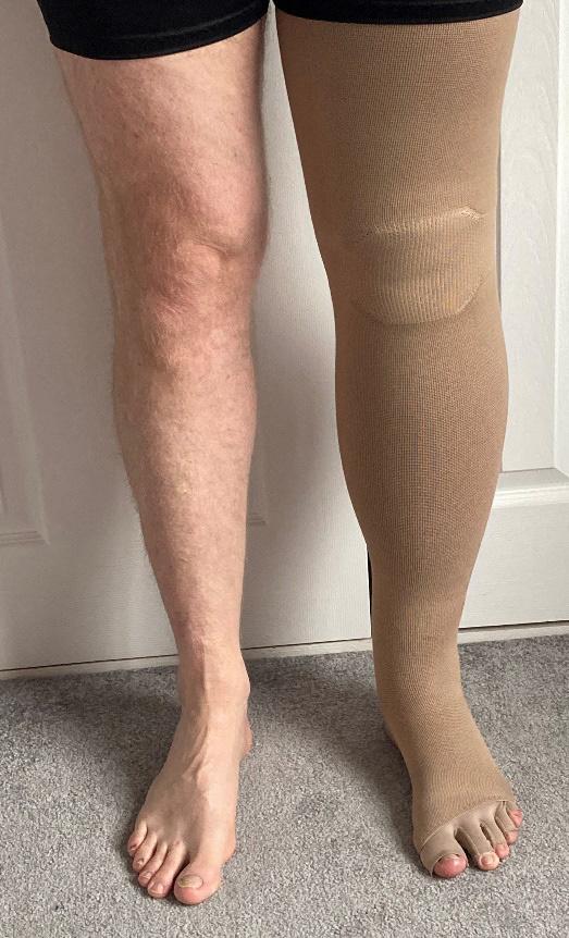
Figure 32: Clothing solutions. Credit: Matt Hazledine, UK.
Figure 33: Wearing Class 4S Compression Garment 60-90mmHG. Credit: Matt Hazledine, UK.
the donning process of a Class 4S garment. With lower limb lymphoedema, we are mostly fortunate to have two hands and the strength and dexterity to put the stocking on. Now imagine how difficult that could be for someone with lymphoedema in their dominant arm, meaning they have to don with their weaker arm. During presentations to healthcare professionals, I ask the audience how many of them have worn a Class 2 or 3 garment for two weeks. Not many have and they are urged to try it to experience just one part of the daily self-management treatment plan.
Receiving the appropriate treatment from a lymphoedema specialist
As mentioned earlier, too many people that require a formal diagnosis and ongoing treatment from a lymphoedema specialist do not have a local lymphoedema clinic and, therefore, do not receive the appropriate care. This is a genuine example of a post code lottery in the healthcare system. Lymphoedema is often referred to as the Cinderella disease, as there isn’t the funding available and, therefore, not the quantity of specialists interested in working in this sector. I’m aware of people who travel for several hours for their six-monthly clinic appointment, and some have even moved house to ensure they are in the catchment area of a lymphoedema clinic.
The challenge of finding a qualified lymphoedema therapist or clinic in the UK has been made slightly easier thanks to the British Lymphology Society’s283 register on its website. By selecting your region on the map or menu, a list of your nearest clinics and therapists is shown on the map, with their contact details and area of expertise listed below. The patient can then ask their GP for a referral, if the service is available on the health service, or they can pay privately if necessary.
It is worth checking whether the professional lymphoedema organisation in your home country has a directory to point people to, as required. Even though I am based in the UK, this seems to be a global issue as I have been contacted by people in Spain, India, and the US asking me to help them find a local lymphoedema clinic or specialist consultant. Fortunately, I have been able to point them to a recognised clinic in their home country.
Unbelievably, some lymphoedema clinics are turning people away because their BMI is too high, and they are told to lose weight before the treatment can begin. When
my leg was at its largest, my BMI reading meant that, on the chart, I was obese. Actually at 6’3” (190cm) tall and considering that I was carrying approximtely 5–6litres/kilos of extra weight in my leg, I wasn’t obese. Therefore, how can BMI be the most used method of assessing someone with lymphoedema? Surely, waist size seems to be the most obvious measurement of being overweight, doesn’t it?
In my personal opinion, the main problem of turning people away at the earliest point of diagnosis, is not providing them with the most appropriate treatment and advice on self-management. If the correct compression garments are not prescribed as soon as possible, it could result in the limb getting bigger, meaning that it will become more difficult to reduce the swelling the longer the patient is left to their own devices. The person could then become less mobile due to the extra weight of their limb(s), which could create additional health problems. The person could become more isolated and less sociable, affecting their mental health and self-esteem. In some cases, the person becomes wheelchair bound and reclusive, just living day to day with only their spouse to support them. What quality of life is this for a human being? All it would take is for the person to receive the appropriate information, treatment, compassion, and encouragement at the earliest stage after diagnosis. This would give them a fighting chance of reducing and controlling the swelling by prescribing the appropriate compression garments, skin emollients and thereafter, having regular six-monthly clinic appointments.
An area that many others with lymphoedema think would be helpful and constructive, is for the therapist or specialist to provide trusted information about lymphoedema and explain why the four cornerstones of self-management are so important. If the person understands what could happen to their limb and their health if they choose not to comply with the guidelines, then hopefully through educating, empowering, and encouraging, they become concordant and perhaps develop into a ‘perfect patient’, which is a win for all concerned.
As we know, the four cornerstones of self-management are:
Compression
Most people living with lymphoedema will endure the daily ritual of donning and doffing of compression garments.
Some garments, typically circular knit, can be supplied ‘off the shelf’, whereas more bespoke made-to-measure garments, typically flat knit, can be produced to order specific to their limb shape and size.
Skincare
Skin care is an essential part of daily self-management for people who have lymphoedema or who are at risk of developing lymphoedema. Keeping your skin intact is the first line of defence against developing infection in the affected area and looking after your skin helps it to perform this important function.
Healthy lifestyle
A healthy lifestyle incorporates a balanced diet and exercise, or movement, as it is sometimes referred to now. Unlike the heart, the lymphatic system doesn’t have a pump, so we need to help it along. When we move our bodies, our muscles pump and we breathe deeper. These actions increase the flow of lymph around the body. This prevents or reduces any swelling, as well as helps our body get rid of bacteria and other unwanted substances.
Lymphatic massage
There are various forms of lymphatic massage techniques, which can be performed manually by a trained expert, the patient or by using a machine called a pneumatic compression pump.
The gentle stretching techniques used in manual lymphatic drainage stimulate the contractions of the lymphatic vessels, helping to move the lymph forward, while pressure changes and circular movements of the skin cause more lymph to be formed from the fluid, protein and waste products present in the tissues. These techniques can help to reduce swelling, bruising and inflammation, and may also reduce pain.
Trusted information and guidance
There is an abundance of information and images available online and the most popular route to find out about a subject is to google it. This can result in misinformation and some quite scary photos of people with huge limbs, darkened cracked skin, toenail infections and severe cellulitis. There are organisations run by people with lymphoedema that provide trusted content, written in a user-friendly language, by some of the most experienced, qualified and renowned lymphoedema experts in the world.
In an ever-changing world, the younger generation would much rather watch a two-minute video online than read an information leaflet or a book. To accommodate this, there are a series of self-management videos to easily demonstrate how to look after their lymphoedema by teaching them how to perform self-lymphatic drainage (SLD) effectively.284 There are a varied range of videos available on YouTube 285 including interviews with some of the key individuals from the lymphoedema sector including medical professionals, product suppliers, patient organisations, and people with the condition sharing their personal experiences and solutions.
The British Lymphology Society is a registered charity, whose members are typically HCPs practising in the lymphoedema sector. There is a wealth of information on their website 279 and they encourage HCPs to join to benefit from Continuing Professional Development accredited learning, webinars and conferences, as well as trusted information, including an informative video about lymphoedema.286 It is highly recommended that you refer patients to recognised professional and/or patient-based organisations or charities in your home country, rather than to google a generalist, which may provide misinformation.
Another organisation founded by healthcare professionals for both healthcare professionals and patients, is the International Lymphoedema Framework (ILF).287 The ILF has created frameworks that are working across the world including North America, Europe, Asia, Africa and India. The national frameworks are non-profit organisations that work nationally to uphold the philosophy of the ILF. They are committed to putting patients at the heart of their activities, to collaborating in multidisciplinary partnerships between all stakeholders in their countries, and to support the mission of the ILF which is to improve the lives of people living with lymphoedema and related conditions worldwide.
One of their most successful projects is LIMPRINT288 which stands for Lymphoedema IMpact and PRevalence – INTernational Lymphoedema Framework. LIMPRINT is an international study aimed at capturing the size and impact of chronic oedema in different countries and health services around the world.
Summary
The important message here is that it should be, ideally, the responsibility of healthcare professionals to direct their
patients to reputable sources of trusted information, written by lymphoedema experts. There are posters and postcards available from these organisations to display in the clinic or waiting room.
With professional help and advice, the mental and physical wellbeing of any person living with lymphoedema will significantly improve. By educating, empowering and encouraging them to self-manage their chronic condition with confidence, this will improve their overall quality of life.
People with lymphoedema can manage their condition and not allow it to manage them.
By uniting our caring lymphoedema community, no one living with this chronic disease should ever feel alone.
8. Epidemiology and health economy perspectives on chronic oedema
Learning points
• The importance of recognising, preventing and treating lower leg chronic oedema to reduce both direct and indirect costs
• The cost savings include not only healthcare systems but also social care systems and persons with chronic oedema of the lower limb
• According to several estimations, outpatient chronic oedema of the lower limb and chronic oedema management programs have the potential to result in substantial savings to the health care system
Although the incidence and prevalence of chronic oedema is increasing, it remains an under-recognised and undertreated problem. It has been estimated that 300,000 people in Canada are affected by chronic oedema, and in USA, 3–5 million people are affected by secondary lymphoedema.289,90 Up to 80% of morbidly obese people attending lymphoedema services are thought to suffer with an element of lymphoedema . 290 Despite the high burden of chronic oedema to health care systems, socio-economic studies are scarce. In addition, we have very limited data on the true costs of secondary chronic oedema to patients, families, health care systems and society in general. Some estimations have been published; Moffatt et al 291 estimated that for every £1 spent on lymphoedema treatment, £100 was saved in terms of hospital admission costs.
When reporting total cost-of-illness, the cost categories can be divided into direct and indirect costs. The direct costs include drugs (topical and systemic), therapeutic measures, physician/nursing fees, inpatient costs, compression therapy costs and travel costs. The indirect ones include costs for the society resulting from inability to work. 292 As chronic oedema patients are becoming younger, the indirect costs become more significant.
From a socio-economic point, the prevention and treatment of chronic oedema is of particular interest. On the other
hand, if treatment of chronic oedema fails and an ulcer appears, the costs are even higher. For instance, the mean one-year cost for leg ulcers has been reported to be in US $US11,000 per patient293 and in the UK £4788.294
A German, observational cross-sectional study including 348 patients with lymphoedema and lipo-lymphoedema revealed that the total costs per patient and year were €5784. In this study, the main cost drivers were costs for manual decongestive therapy and disability costs. They concluded that an early-treatment approach may help lowering costs.292 An Australian randomised controlled trial study with 84 participants reported a $A21,483 cost-saving per participant receiving compression therapy in patients with chronic oedema, as compression therapy was able to reduce hospitalisation due to cellulitis.295
A study investigating the economic impact of compression in the acute phase of deep vein thrombosis in 865 patients reported that mean healthcare costs per patient were €417.08 for multilayer compression bandaging, €114.25 for hosiery, and €105.86 for no compression. Differences in HRQoL were not found. The investigators concluded, that in the indication of acute deep vein thrombosis, compression hosiery is the preferred method.296
A multicentre study was performed in four countries and compared in 165 patients the standard of care (inelastic bandages) and two-layer compression system within decongestive lymphatic therapy. The conclusion was that there was no difference in the QoL improvement in the standard care (inelastic bandages) and two-layer bandages. The number of treatment episodes was higher in those treated with standard care (8.15 versus 6.37), but the overall treatment cost was higher with two-layer bandages (£890.7) compared with standard care (£723).297
In conclusion, further studies are needed to investigate the cost-of-illness of chronic oedema and the costeffectiveness of different treatment methods in the different aetiologies of chronic oedema of the lower limb. Recent
studies indicate that if effective treatment is prescribed and can reduce the risk of developing chronic oedema, ulceration and hospitalisations, the treatment costs are far lower than costs resulting from no treatment.
Summary:
• Chronic oedema can lead to significant both direct and indirect costs if not recognised, prevented and treated properly.
• Investing in proper compression therapy in patients with chronic oedema significantly reduces costs by preventing cellulitis and leg ulceration.
• Investing in proper education programmes for patients with chronic oedema significantly reduces costs by improving early detection, timely intervention and longterm effect.
9. Summary and the way forward for practitioners
This document is an international collaboration between two different organisations, the European Wound Managment Associatian (EWMA) and the Internation Lymphoedema Framework (ILF), who have different foci and, therefore, the content reflects their combined knowledge of managing chronic oedema and wounds, topics rarely discussed together.
This document discusses chronic oedema and does not seek to address primary lymphoedema in any depth, nor incorporate surgical techniques used in the management of patients with chronic lower limb oedema. The editors acknowledge that lymphatic filariasis is the largest cause of secondary chronic oedema worldwide, but it is also beyond the scope of this document.
This document aims to highlight the complex and multifactorial nature of recognising and treating chronic lower limb oedema. In clinical practice, it is vital that all HCPs recognise and can distinguish between lower limb swelling (acute oedema) associated with the ‘normal’ wound healing process and chronic oedema, in order to improve patients’ potential clinical outcomes. If unsure, the receiving HCP should refer the patient to an appropriate member of a multi-disciplinary team who has the knowledge/experience to assist with diagnosis and advise on management.
A multidisciplinary holistic assessment and management plan has been promoted throughout the document to enable all HCPs take account of the many clinical manifestations associated with chronic lower limb oedema and to promote appropriate management. Furthermore, a multi-disciplinary approach should assist all HCPs to optimise the time they spend with their patients.
Throughout this document the authors have identified learning outcomes associated with the content of their chapters. These outcomes have been designed to help non-specialist practitioners feel more confident when managing patients with chronic lower limb oedema. For the specialist practitioner, it is anticipated that these learning objectives will support current practice or stimulate the development of new learning.
Although this document has covered many clinical aspects of assessing and treating chronic lower limb oedema, nonetheless it is acknowledged that the research evidence base related to the subject matter is often poor and is still developing. The current literature referred to in the document reflects many of the clinical controversies that still need to be addressed.
Undoubtably one of the key international priorities is to develop a set of outcome measures that can be used across this diverse population. This is very challenging given the heterogenous population and requires cooperation between organisations to achieve this. This document reflects the importance of including patient voices in developing approaches to care, as well as partnership with the medical device industry which is critical in product development. Future perspectives will doubtless include a greater understanding of the potential for surgery, particularly micro-surgical techniques. Nevertheless, the core components of care, such as skin care, exercise and compression therapy will remain pivotal. The expectation of patient populations is also changing with a greater focus on supported self-management and increased patient choice.
The patient profile is becoming more complex due to associated co-morbidities, such as morbid obesity, cardiovascular disease, reduced mobility and longevity of end of life. The strong association seen within the epidemiology studies highlights the strong increased prevalence associated with an aging population and it is therefore vital that clinicians working in this sector recognise and manage chronic oedema before complications such as cellulitis occur.
There is a huge diversity in the way in which health services are organised and reimbursed internationally with many low-income countries having little or no healthcare. Despite the growing evidence of the number of people affected with chronic oedema there are still many countries that lack any specialist services able to manage these complex patients. These national differences produce significant challenges when discussing optimising care. Despite these challenges
we hope this document can provide relevant information that can support improvement in the care of the many people who live and suffer with these conditions.
We would like to thank all the authors and reviewers who have given their time and expertise in bringing this document together.
Finally, to all readers of this document, we wish you success in your future professional practice.
References
1. Keeley, V. et al. LIMPRINT in Specialist Lymphedema Services in United Kingdom, France, Italy, and Turkey. Lymphat. Res. Biol. 17, 121–126 (2019).
2. Keast, D. H., Burian, E. A., Moffatt, C. J., Nørregaard, S. & Karlsmark, T. International Lymphoedema Framework: The dilemma of wounds in chronic oedema/lymphoedema. J. Wound Care 30, 7–20 (2021).
3. Moffatt, C. J. et al. Lymphoedema: an underestimated health problem. QJM Mon. J. Assoc. Physicians 96, 731–738 (2003).
4. Thomas M, Morgan K, & Larence P. The Chronic Oedema Wet Leg Pathway. (2022).
5. Mortimer, P. S. & Rockson, S. G. New developments in clinical aspects of lymphatic disease. J. Clin. Invest. 124, 915–921 (2014).
6. Collier, M. Understanding wound inflammation. Nurs. Times 99, 63–64 (2003).
7. Mrhac, L., Bukovansky, K., Kafkova, J. & Kraft, O. Primary Congenital Lymphedema. Clin. Nucl. Med. 43, 515–517 (2018).
8. Gasparis, A. P., Kim, P. S., Dean, S. M., Khilnani, N. M. & Labropoulos, N. Diagnostic approach to lower limb edema. Phlebology 35, 650–655 (2020).
9. Moffatt, C. J. et al. Prevalence and Risk Factors for Chronic Edema in U.K. Community Nursing Services. Lymphat. Res. Biol. 17, 147–154 (2019).
10. Keast, D. H., Moffatt, C. & Janmohammad, A. Lymphedema Impact and Prevalence International Study: The Canadian Data. Lymphat. Res. Biol. 17, 178–186 (2019).
11. Gordon, K. et al. Update and audit of the St George’s classification algorithm of primary lymphatic anomalies: a clinical and molecular approach to diagnosis. J. Med. Genet. 57, 653–659 (2020).
12. Brouillard, P. et al. Primary lymphoedema. Nat. Rev. Dis. Primer 7, 77 (2021).
13. Devoogdt, N. et al. The VASCERN PPL working group patient pathway for primary and paediatric lymphoedema. Eur. J. Med. Genet. 67, 104905 (2024).
14. Nickles, M. A., Ennis, W. J., O’Donnell, T. F. & Altman, I. A. Compression therapy in peripheral artery disease: a literature review. J. Wound Care 32, S25–S30 (2023).
15. Ozdowski, L. & Gupta, V. Physiology, Lymphatic System. in StatPearls (StatPearls Publishing, Treasure Island (FL), 2025).
16. Scallan, J., Huxley, V. H. & Korthuis, R. J. Pathophysiology of Edema Formation. in Capillary Fluid Exchange: Regulation, Functions, and Pathology (Morgan & Claypool Life Sciences, 2010).
17. Lent-Schochet, D. & Jialal, I. Physiology, Edema. in StatPearls (StatPearls Publishing, Treasure Island (FL), 2023).
18. Martin-Almedina, S., Mortimer, P. S. & Ostergaard, P. Development and physiological functions of the lymphatic system: insights from human genetic studies of primary lymphedema. Physiol. Rev. 101, 1809–1871 (2021).
19. Poojari, A., Dev, K. & Rabiee, A. Lipedema: Insights into Morphology, Pathophysiology, and Challenges. Biomedicines 10, 3081 (2022).
20. Huxley, V. H. & Scallan, J. Lymphatic fluid: exchange mechanisms and regulation. J. Physiol. 589, 2935–2943 (2011).
21. Michel, C. C., Woodcock, T. E. & Curry, F.-R. E. Understanding and extending the Starling principle. Acta Anaesthesiol. Scand. 64, 1032–1037 (2020).
22. Woodcock, T. Fluid Physiology Part 1: Volume and Distribution of Water and Its Major Solutes Between Plasma, the Interstitium and Intracellular Fluid. in Rational Use of Intravenous Fluids in Critically Ill Patients (eds Malbrain, M. L. N. G., Wong, A., Nasa, P. & Ghosh, S.) 47–74 (Springer International Publishing, Cham, 2024). doi:10.1007/978-3-031-42205-8_2.
23. Trung, D. T., Trieu, H. T. & Wills, B. A. Microvascular Fluid Exchange: Implications of the Revised Starling Model for Resuscitation of Dengue Shock Syndrome. Front. Med. 7, 601520 (2020).
24. Diaz, J. A., Gianesini, S. & Khalil, R. A. Glycocalyx disruption, endothelial dysfunction and vascular remodeling as underlying mechanisms and treatment targets of chronic venous disease. Int. Angiol. 43, (2025).
25. Moore, K. H., Murphy, H. A. & George, E. M. The glycocalyx: a central regulator of vascular function. Am. J. Physiol.-Regul. Integr. Comp. Physiol. 320, R508–R518 (2021).
26. Ushiyama, A., Kataoka, H. & Iijima, T. Glycocalyx and its involvement in clinical pathophysiologies. J. Intensive Care 4, 59 (2016).
27. Aldecoa, C., Llau, J. V., Nuvials, X. & Artigas, A. Role of albumin in the preservation of endothelial glycocalyx integrity and the microcirculation: a review. Ann. Intensive Care 10, 85 (2020).
28. Gianesini, S. et al. Human collecting lymphatic glycocalyx identification by electron microscopy and immunohistochemistry. Sci. Rep. 13, 3022 (2023).
29. Milford, E. M. & Reade, M. C. Resuscitation Fluid Choices to Preserve the Endothelial Glycocalyx. Crit. Care 23, 77 (2019).
30. Wang, J. et al. Factors influencing glycocalyx degradation: a narrative review. Front. Immunol. 15, 1490395 (2025).
31. Bihari, I., Guex, J.-J., Jawien, A. & Szolnoky, G. Clinical Perspectives and Management of Edema in Chronic Venous Disease-What about Ruscus? Med. Basel Switz. 9, 41 (2022).
32. Wautier, J.-L. & Wautier, M.-P. Vascular Permeability in Diseases. Int. J. Mol. Sci. 23, 3645 (2022).
33. Goyal, A., Singh, B. & Afzal, M. Peripheral Edema. in StatPearls (StatPearls Publishing, Treasure Island (FL), 2025).
34. Alberto Caggiati. The venous valves of the lower limbs. Phlebolymphology https://www.phlebolymphology.org/the-venous-valves-of-the-lower-limbs/ (2013).
35. Kuyumcu, G., Salazar, G. M., Prabhakar, A. M. & Ganguli, S. Minimally invasive treatments for perforator vein insufficiency. Cardiovasc. Diagn. Ther. 6, 593–598 (2016).
36. Black, C. M. Anatomy and Physiology of the Lower-Extremity Deep and Superficial Veins. Tech. Vasc. Interv. Radiol. 17, 68–73 (2014).
37. Mohamed K. KAMEL & John BLEBEA. Pathophysiology of edema in patients with chronic venous insufficiency. Phlebolymphology https://www. phlebolymphology.org/pathophysiology-of-edema-in-patients-with-chronic-venous-insufficiency/ (2020).
38. Williams, K. J., Ayekoloye, O., Moore, H. M. & Davies, A. H. The calf muscle pump revisited. J. Vasc. Surg. Venous Lymphat. Disord. 2, 329–334 (2014).
39. Gore, M. Chronic Venous Insufficiency of Lower Extremity. Indian J. Surg. 85, 112–120 (2023).
40. Meissner, M.H, Hallett, J.W, Mills, J.L, Earnshaw, J.J., & Reekers, J.A. Pathophysiology of varicose veins and chronic venous insufficiency. in Comprehensive Vascular and Endovascular Surgery 571–589 (Edinburgh, 2004).
41. Lurie, F. et al. The 2020 update of the CEAP classification system and reporting standards. J. Vasc. Surg. Venous Lymphat. Disord. 8, 342–352 (2020).
42. Eberhardt, R. T. & Raffetto, J. D. Chronic Venous Insufficiency. Circulation 130, 333–346 (2014).
43. Krizanova, O. et al. Chronic venous insufficiency and venous leg ulcers: Aetiology, on the pathophysiology-based treatment. Int. Wound J. 21, e14405 (2023).
44. Chi, Y.-W. & Raffetto, J. D. Venous leg ulceration pathophysiology and evidence based treatment. Vasc. Med. 20, 168–181 (2015).
45. Santler, B. & Goerge, T. Chronic venous insufficiency – a review of pathophysiology, diagnosis, and treatment. JDDG J. Dtsch. Dermatol. Ges. 15, 538–556 (2017).
46. Bihari, I., Guex, J.-J., Jawien, A. & Szolnoky, G. Clinical Perspectives and Management of Edema in Chronic Venous Disease-What about Ruscus? Med. Basel Switz. 9, 41 (2022).
47. Crawford, J. M., Lal, B. K., Durán, W. N. & Pappas, P. J. Pathophysiology of venous ulceration. J. Vasc. Surg. Venous Lymphat. Disord. 5, 596–605 (2017).
48. Fukaya, E. & Kolluri, R. Nonsurgical Management of Chronic Venous Insufficiency. N. Engl. J. Med. 391, 2350–2359 (2024).
49. Alexander, J. S. & Elrod, J. W. Extracellular matrix, junctional integrity and matrix metalloproteinase interactions in endothelial permeability regulation. J. Anat. 200, 561–574 (2002).
50. Kruppa, P. et al. Lipedema-Pathogenesis, Diagnosis, and Treatment Options. Dtsch. Arzteblatt Int. 117, 396–403 (2020).
51. Cabral-Pacheco, G. A. et al. The Roles of Matrix Metalloproteinases and Their Inhibitors in Human Diseases. Int. J. Mol. Sci. 21, 9739 (2020).
52. Raffetto, J. D., Ligi, D., Maniscalco, R., Khalil, R. A. & Mannello, F. Why Venous Leg Ulcers Have Difficulty Healing: Overview on Pathophysiology, Clinical Consequences, and Treatment. J. Clin. Med. 10, 29 (2021).
53. Kandhwal, M. et al. Role of matrix metalloproteinase in wound healing. Am. J. Transl. Res. 14, 4391–4405 (2022).
54. Patel, S. K. & Surowiec, S. M. Venous Insufficiency. in StatPearls (StatPearls Publishing, Treasure Island (FL), 2025).
55. Ortega, M. A. et al. Understanding Chronic Venous Disease: A Critical Overview of Its Pathophysiology and Medical Management. J. Clin. Med. 10, 3239 (2021).
56. Krizanova, O. et al. Chronic venous insufficiency and venous leg ulcers: Aetiology, on the pathophysiology-based treatment. Int. Wound J. 21, e14405 (2024).
57. Kattula, S. R. S. T., Avula, A. & Baradhi, K. M. Anasarca. in StatPearls [Internet] (StatPearls Publishing, 2023).
58. Davis, M. J., Zawieja, S. D. & King, P. D. Transport and Immune Functions of the Lymphatic System. Annu. Rev. Physiol. 87, 151–172 (2025).
59. Li, Y. et al. Diabetic vascular diseases: molecular mechanisms and therapeutic strategies. Signal Transduct. Target. Ther. 8, 152 (2023).
60. Olivier STÜCKER, Catherine PONS-HIMBERT, & Elisabeth LAEMMEL. Towards a better understanding of lymph circulation. Phlebolymphology 15, (2008).
61. Angeli, V. & Lim, H. Y. Biomechanical control of lymphatic vessel physiology and functions. Cell. Mol. Immunol. 20, 1051–1062 (2023).
62. Sleigh, B. C. & Manna, B. Lymphedema. in StatPearls (StatPearls Publishing, Treasure Island (FL), 2025).
63. Pathophysiology, classification, and causes of lymphedema - UpToDate. https://www.uptodate.com/contents/pathophysiology-classification-and-causes-of-lymphedema.
64. Grada, A. A. & Phillips, T. J. Lymphedema. J. Am. Acad. Dermatol. 77, 1009–1020 (2017).
65. Brix, B. et al. Biology of Lymphedema. Biology 10, 261 (2021).
66. Hespe, G. E., Nitti, M. D. & Mehrara, B. J. Pathophysiology of Lymphedema. in Lymphedema (eds Greene, A. K., Slavin, S. A. & Brorson, H.) 9–18 (Springer International Publishing, Cham, 2015). doi:10.1007/978-3-31914493-1_2.
67. Jeong, S. H. et al. Association between chemotherapy and the risk of developing breast cancer-related lymphedema: a nationwide retrospective cohort study. Support. Care Cancer 33, 143 (2025).
68. Gasparis, A. P., Kim, P. S., Dean, S. M., Khilnani, N. M. & Labropoulos, N. Diagnostic approach to lower limb edema. Phlebol. J. Venous Dis. 35, 650–655 (2020).
69. Grada, A. A. & Phillips, T. J. Lymphedema: Pathophysiology and clinical manifestations. J. Am. Acad. Dermatol. 77, 1009–1020 (2017).
70. Ly, C., Kataru, R. & Mehrara, B. Inflammatory Manifestations of Lymphedema. Int. J. Mol. Sci. 18, 171 (2017).
71. Jiang, X. et al. Hypoxia and Hypoxia-Inducible Factors in Lymphedema. Front. Pharmacol. 13, 851057 (2022).
72. Bowman, C. & Rockson, S. G. The Role of Inflammation in Lymphedema: A Narrative Review of Pathogenesis and Opportunities for Therapeutic Intervention. Int. J. Mol. Sci. 25, 3907 (2024).
73. Brown, S., Dayan, J. H., Kataru, R. P. & Mehrara, B. J. The Vicious Circle of Stasis, Inflammation, and Fibrosis in Lymphedema. Plast. Reconstr. Surg. 151, 330e–341e (2023).
74. Azhar, S. H., Lim, H. Y., Tan, B.-K. & Angeli, V. The Unresolved Pathophysiology of Lymphedema. Front. Physiol. 11, 137 (2020).
75. Lee, S.-O. & Kim, I.-K. Molecular pathophysiology of secondary lymphedema. Front. Cell Dev. Biol. 12, 1363811 (2024).
76. Dayan, J. H., Ly, C. L., Kataru, R. P. & Mehrara, B. J. Lymphedema: Pathogenesis and Novel Therapies. Annu. Rev. Med. 69, 263–276 (2018).
77. Ghanta, S. et al. Regulation of inflammation and fibrosis by macrophages in lymphedema. Am. J. Physiol. Heart Circ. Physiol. 308, H1065-1077 (2015).
78. Sung, C., Wang, S., Hsu, J., Yu, R. & Wong, A. K. Current Understanding of Pathological Mechanisms of Lymphedema. Adv. Wound Care 11 , 361–373 (2022).
79. Cuzzone, D. A. et al. IL-6 regulates adipose deposition and homeostasis in lymphedema. Am. J. Physiol. Heart Circ. Physiol. 306, H1426-1434 (2014).
80. Azhar, S. H., Lim, H. Y., Tan, B.-K. & Angeli, V. The Unresolved Pathophysiology of Lymphedema. Front. Physiol. 11, 137 (2020).
81. Lee, S.O & I.K. Molecular pathophysiology of secondary lymphedema. Front. Cell Dev. Biol. 12, 1363811 (2024).
82. Armstrong, D. G. Improvement in Healing With Aggressive Edema Reduction After Debridement of Foot Infection in Persons With Diabetes. Arch. Surg. 135, 1405 (2000).
83. Keast, D. H., Burian, E. A., Moffatt, C. J., Nørregaard, S. & Karlsmark, T. The dilemma of wounds in chronic oedema/lymphoedema. J. Wound Care 30, 7–17 (2021).
84. Burian, E. A. et al. Wounds in chronic leg oedema. Int. Wound J. 19, 411–425 (2022).
85. Villeco, J. P. Edema: A Silent but Important Factor. J. Hand Ther. 25, 153–162 (2012).
86. Villeco, J. P. Edema: A Silent but Important Factor. J. Hand Ther. 25, 153–162 (2012).
87. Villeco, J. P. Edema: A Silent but Important Factor. J. Hand Ther. 25, 153–162 (2012).
88. Dissemond, J. et al. Moisture-associated skin damage (MASD): A best practice recommendation from Wund-D.A.CH. JDDG J. Dtsch. Dermatol. Ges. 19, 815–825 (2021).
89. Armstrong, D. G. Improvement in Healing With Aggressive Edema Reduction After Debridement of Foot Infection in Persons With Diabetes. Arch. Surg. 135, 1405 (2000).
90. Keast, D. H., Despatis, M., Allen, J. O. & Brassard, A. Chronic oedema/ lymphoedema: under-recognised and under-treated. Int. Wound J. 12, 328–333 (2015).
91. Almadani, Y. H., Vorstenbosch, J., Davison, P. G. & Murphy, A. M. Wound Healing: A Comprehensive Review. Semin. Plast. Surg. 35, 141–144 (2021).
92. Li, J., Kumi, M. & Ochoa Chaar, C. I. Management of chronic venous insufficiency in older adults. Semin. Vasc. Surg. S0895796725000468 (2025) doi:10.1053/j.semvascsurg.2025.06.005.
93. Mayrovitz, H. N., Aoki, K. C. & Colon, J. Chronic Venous Insufficiency With Emphasis on the Geriatric Population. Cureus (2023) doi:10.7759/cureus.40687.
94. Burian, E. A. et al. Wounds in chronic leg oedema. Int. Wound J. 19, 411–425 (2022).
95. Molnár, A. Á. et al. The aging venous system: from varicosities to vascular cognitive impairment. GeroScience 43, 2761–2784 (2021).
96. Shang, T., Liang, J., Kapron, C. M. & Liu, J. Pathophysiology of aged lymphatic vessels. Aging 11, 6602–6613 (2019).
97. González-Loyola, A. & Petrova, T. V. Development and aging of the lymphatic vascular system. Adv. Drug Deliv. Rev. 169, 63–78 (2021).
98. Shang, T., Liang, J., Kapron, C. M. & Liu, J. Pathophysiology of aged lymphatic vessels. Aging 11, 6602–6613 (2019).
99. González-Loyola, A. & Petrova, T. V. Development and aging of the lymphatic vascular system. Adv. Drug Deliv. Rev. 169, 63–78 (2021).
100. Lomeli, L. D., Makin, V., Bartholomew, J. R. & Burguera, B. Lymphedema vs lipedema: Similar but different. Cleve. Clin. J. Med. 91, 425–432 (2024).
101. Carroll, B. J. & Singhal, D. Advances in lymphedema: An under-recognized disease with a hopeful future for patients. Vasc. Med. Lond. Engl. 29, 70–84 (2024).
102. Executive Committee of the International Society of Lymphology. The Diagnosis and Treatment of Peripheral Lymphedema: 2023 Consensus Document of The International Society of Lymphology. Lymphology 56, 133–151 (2023).
103. Vargo, M. et al. Current diagnostic and quantitative techniques in the field of lymphedema management: a critical review. Med. Oncol. 41, 241 (2024).
104. Lymphedema: Diagnosis and treatment - Mayo Clinic. Mayo Clinic Medical Professionals Physical Medicine and Rehabilitation https://www.mayoclinic.org/medical-professionals/physical-medicine-rehabilitation/news/ lymphedema-diagnosis-and-treatment/mac-20436554 (2018).
105. Torres-Guzman, R. A., Bailey, V., James, A. J., Drolet, B. C. & Karagoz, H. Current Diagnostic Measures for Lymphedema: A Comprehensive Review. Ann. Plast. Surg. 93, S132–S135 (2024).
106. Fujimoto, Y., Yuri, Y. & Tamiya, H. Skin mechanical properties measured with skin elasticity measurement device in patients with lymphedema: Scoping review. Skin Res. Technol. 30, e13861 (2024).
107. Heroes, A.-K. et al. Reliability, concurrent validity, and clinical feasibility of measurement methods determining volume in patients with lower limb lymphedema and healthy controls. Disabil. Rehabil. 47, 2384–2398 (2025).
108. Zhang, M. et al. Magnetic resonance lymphography findings across different clinical stages of lower limb lymphedema. J. Plast. Reconstr. Aesthetic Surg. JPRAS 96, 129–135 (2024).
109. Tartaglione, G. Advantages of the intradermal lymphoscintigraphy. World J. Radiol. 16, 241–246 (2024).
110. Li, J. et al. Using four-point subcutaneous injection of lymphatic contrast-enhanced ultrasound to diagnose lymphedema of lower extremity. Quant. Imaging Med. Surg. 14, 4965–4971 (2024).
111. Basavalingu, D. et al. Nuclear Medicine Lymphoscintigraphy: Applications and Technical Overview. RadioGraphics 45, e240207 (2025).
112. Vargo, M. et al. Current diagnostic and quantitative techniques in the field of lymphedema management: a critical review. Med. Oncol. 41, 241 (2024).
113. Figueroa, B. A., Lammers, J. D., Al-Malak, M., Pandey, S. & Chen, W. F. Lymphoscintigraphy versus Indocyanine Green Lymphography—Which Should Be the Gold Standard for Lymphedema Imaging? Lymphatics 1, 25–33 (2023).
114. Markarian, B. et al. Assessment Modalities for Lower Extremity Edema, Lymphedema, and Lipedema: A Scoping Review. Cureus 16, e55906 (2024).
115. Murray-Ramcharan, M., Escurra, M. F., Donaldson, B. & Raza Rizvi, S. A. Patient-Centered Outcomes of a Lymphedema Pump in Patients with Symptomatic Lower Extremity Edema. Ann. Vasc. Surg. 108, 333–337 (2024).
116. Bowman, C. & Rockson, S. G. The Role of Inflammation in Lymphedema: A Narrative Review of Pathogenesis and Opportunities for Therapeutic Intervention. Int. J. Mol. Sci. 25, 3907 (2024).
117. Kitayama, S. Diagnosis and Treatments of Limb Lymphedema: Review. Ann. Vasc. Dis. 17, 114–119 (2024).
118. Gallagher S. The Challenges of Treating Patients with Obesity and Lymphedema. Today’s Wound Clin. (2020).
119. Pugh, S., Ives, A. & Stubbs, C. Challenges of lymphoedema management in an ageing population. Br. J. Community Nurs. 23, S6-12 (2018).
120. Tambour, M., Holt, M., Speyer, A., Christensen, R. & Gram, B. Manual lymphatic drainage adds no further volume reduction to Complete Decongestive Therapy on breast cancer-related lymphoedema: a multicentre, randomised, single-blind trial. Br. J. Cancer 119, 1215–1222 (2018).
121. Devoogdt, N. et al. Manual lymph drainage may not have a preventive effect on the development of breast cancer-related lymphoedema in the long term: a randomised trial. J. Cancer Rehabil. 64, 245–254 (2018).
122. Müller, M., Klingberg, K., Wertli, M. M. & Carreira, H. Manual lymphatic drainage and quality of life in patients with lymphoedema and mixed oedema: a systematic review of randomised controlled trials. Qual. Life Res. Int. J. Qual. Life Asp. Treat. Care Rehabil. 27, 1403–1414 (2018).
123. Huang, T.-W. et al. Effects of manual lymphatic drainage on breast cancer-related lymphedema: a systematic review and meta-analysis of randomized controlled trials. World J. Surg. Oncol. 11, 15 (2013).
124. Ezzo, J. et al. Manual lymphatic drainage for lymphedema following breast cancer treatment. Cochrane Database Syst. Rev. 2015, (2015).
125. Liang, M. et al. Manual lymphatic drainage for lymphedema in patients after breast cancer surgery: A systematic review and meta-analysis of randomized controlled trials. Medicine (Baltimore) 99, e23192 (2020).
126. Wanchai, A. & Armer, J. M. Manual Lymphedema Drainage for Reducing Risk for and Managing Breast Cancer-Related Lymphedema After Breast Surgery: A Systematic Review. Nurs. Womens Health 25, 377–383 (2021).
127. Schneider, R. Low-frequency vibrotherapy considerably improves the effectiveness of manual lymphatic drainage (MLD) in patients with lipedema: A two-armed, randomized, controlled pragmatic trial. Physiother. Theory Pract. 36, 63–70 (2020).
128. Devoogdt, N. et al. The ICC Compression Questionnaire: A Comprehensive Tool to Evaluate Compression Materials or Devices Applied in Subjects with Lymphedema or Chronic Venous Disease. Lymphat. Res. Biol. 20, 191–202 (2022).
129. Mosti, G. Relevance of stiffness of compression material on venous hemodynamics and edema. Veins Lymphat. 2, 9 (2013).
130. Partsch, H., Schuren, J., Mosti, G. & Benigni, J. P. The Static Stiffness Index: an important parameter to characterise compression therapy in vivo. J. Wound Care 25 Suppl 9, S4–S10 (2016).
131. Damstra, R.J., et al. Multilayer bandaging versus elastic compression in lymphoedema treatment: a randomized controlled study. Phlebology (2013).
132. Mosti, G., et al. The impact of compression bandage stiffness on venous edema and hemodynamics. J. Vasc. Med. (2015).
133. Brown, J. C. et al. Physical activity, daily walking, and lower limb lymphedema associate with physical function among uterine cancer survivors. Sup-
port. Care Cancer Off. J. Multinatl. Assoc. Support. Care Cancer 22, 3017–3025 (2014).
134. Hsu, Y.-Y., Nguyen, T. T., Chou, Y.-J. & Ho, C.-L. Effects of exercise on lower limb lymphedema in gynecologic cancer: A systematic review and meta-analysis. Eur. J. Oncol. Nurs. Off. J. Eur. Oncol. Nurs. Soc. 70, 102550 (2024).
135. Fukushima, T. et al. Immediate effects of active exercise with compression therapy on lower-limb lymphedema. Support. Care Cancer 25, 2603–2610 (2017).
136. Maccarone, M. C., Venturini, E., Menegatti, E., Gianesini, S. & Masiero, S. Water-based exercise for upper and lower limb lymphedema treatment. J. Vasc. Surg. Venous Lymphat. Disord. 11, 201–209 (2022).
137. Meyer, M., Müller, A.-K., Yang, J., Ŝulcová, J. & Werner, S. The role of chronic inflammation in cutaneous fibrosis: fibroblast growth factor receptor deficiency in keratinocytes as an example. J. Investig. Dermatol. Symp. Proc. 15, 48–52 (2011).
138. Chima, C., Murray, B., Moore, Z., Costello, M. & George, S. Health-related quality of life and assessment in patients with lower limb lymphoedema: a systematic review. J. Wound Care 31, 690–699 (2022).
139. Damstra, R. J., Hendrickx, A. A., van Duinen, K., Klinkert, P. & Dickinson-Blok, J. L. Diagnosis and treatment of primary and secondary lymphedema. Ned. Tijdschr. Geneeskd. 166, D6139 (2022).
140. Ridner, S. H. The psycho-social impact of lymphedema. Lymphat. Res. Biol. 7, 109–112 (2009).
141. Park, S. et al. Mindfulness-Based Cognitive Therapy for Psychological Distress, Fear of Cancer Recurrence, Fatigue, Spiritual Well-Being, and Quality of Life in Patients With Breast Cancer-A Randomized Controlled Trial. J. Pain Symptom Manage. 60, 381–389 (2020).
142. Hunter, E. G., Gibson, R. W., Arbesman, M. & D’Amico, M. Systematic Review of Occupational Therapy and Adult Cancer Rehabilitation: Part 2. Impact of Multidisciplinary Rehabilitation and Psychosocial, Sexuality, and Return-to-Work Interventions. Am. J. Occup. Ther. Off. Publ. Am. Occup. Ther. Assoc. 71, 7102100040p1-7102100040p8 (2017).
143. Bobbink, P., Gschwind, G., Larkin, P. & Probst, S. Making Conscientious Decisions: Engaging in Venous Leg Ulcer Self-Management Following Nurse-Led Patient Education. Qual. Health Res. 35, 901–915 (2025).
144. Ahmed, R. L., Prizment, A., Lazovich, D., Schmitz, K. H. & Folsom, A. R. Lymphedema and Quality of Life in Breast Cancer Survivors: The Iowa Women’s Health Study. J. Clin. Oncol. 26, 5689–5696 (2008).
145. Mosti, G. & Cavezzi, A. Compression therapy in lymphedema: Between past and recent scientific data. Phlebology 34, 515–522 (2019).
146. Tashiro, K., Yoshioka, Y. & Ochiya, T. Extracellular Vesicles from Adipose-Derived Stem Cells Relieve Extremity Lymphedema in Mouse Models. Plast. Reconstr. Surg. 152, 1011–1021 (2023).
147. Abdo, J. M., Sopko, N. A. & Milner, S. M. The applied anatomy of human skin: A model for regeneration. Wound Med. 28, 100179 (2020).
148. Calvin, M. Cutaneous Wound Repair. Wounds 10, 12–32 (1998).
149. Gushiken, L. F. S., Beserra, F. P., Bastos, J. K., Jackson, C. J. & Pellizzon, C. H. Cutaneous Wound Healing: An Update from Physiopathology to Current Therapies. Life 11, 665 (2021).
150. Atkin, L. et al. Implementing TIMERS: the race against hard-to-heal wounds. J. Wound Care 28, S1–S50 (2019).
151. Bennett, S. P., Griffiths, G. D., Schor, A. M., Leese, G. P. & Schor, S. L. Growth factors in the treatment of diabetic foot ulcers. Br. J. Surg. 90, 133–146 (2003).
152. LeBlanc K et al. Best Practice Recommendations for the prevention and management of skin tears in aged skin. Wounds International https:// woundsinternational.com/best-practice-statements/istap-best-practicerecommendations-prevention-and-management-skin-tears-aged-skin/ (2018).
153. Fumarola, S. et al. Overlooked and underestimated: medical adhesive-related skin injuries. J. Wound Care 29, S1–S24 (2020).
154. Burian, E. A. et al. Factors associated with cellulitis in lymphoedema of the arm – an international cross-sectional study (LIMPRINT). BMC Infect. Dis. 24, (2024).
155. Wound exudate and the role of dressings: a WUWHS consensus document – Wounds International. https://woundsinternational.com/consensus-documents/read-more-wound-exudate-and-role-dressings-wuwhs-consensus-document-2/.
156. Sturkie, P. D. & Griminger, P. Body Fluids: Blood. in Avian Physiology (ed. Sturkie, P. D.) 102–129 (Springer New York, New York, NY, 1986). doi:10.1007/978-1-4612-4862-0_5.
157. Melanie McCann. Lymphorrhoea. Lymphoedema Support Network - LSN https://www.lymphoedema.org/information/useful-articles/lymphorrhoea/ (2024).
158. M Romanelli, K Vowden, & D Weir. Exudate management made easy. (2010).
159. Collier, M., Potts, C. & Shaw, E. The use of a coverlet system for the management of skin microclimate. Br. J. Nurs. 23, S28–S35 (2014).
160. Clark, M et al. Pressure ulcer prevention: Pressure, shear, friction and microclimate in context – a consensus document. in Microclimate in context (Wounds International, 2010).
161. Best Practice for the Management of Lymphoedema: an international consensus – Wounds International. https://woundsinternational.com/consensus-documents/best-practice-for-the-management-of-lymphoedema-an-international-consensus/ (2006).
162. f26e37e0dbea7439d330d77035350fc4.pdf.
163. Dimitri Beeckman. Incontinence-associated dermatitis: moving prevention forward – Wounds International. www.woundsinternational.com (2015).
164. Collier, M. What is, how to manage, and what are the effects of wound exudate? Introducing the 3M Exudate Pathway. Br. J. Community Nurs. 28, S27–S38 (2023).
165. Mark Collier. The elements of wound assessment. Nursing Times https:// www.nursingtimes.net/tissue-viability-and-wound-care/the-elements-of-wound-assessment-01-04-2003/ (2003).
166. Compression Hosiery, Second edition – Wounds UK. https://wounds-uk. com/best-practice-statements/compression-hosiery-second-edition/ (2015).
167. National Wound Care Strategy Programme. Recommendations for Leg Ulcers . https://www.nationalwoundcarestrategy.net/wp-content/uploads/2024/07/NWCSP-Leg-Ulcer-Recommendations-final-version-15.07.2024.pdf (2024).
168. World Union of Wound Healing Societies. WUWHS Consensus Document – Wound Exudate, effective assessment and management – Wounds International. https://woundsinternational.com/world-union-resources/wuwhs-consensus-document-wound-exudate-effective-assessment-and-management/ (2019).
169. Merriam-Webster. Exudate. Merriam-Webster.com Dictionary (2018).
170. Franks, P. J. & Moffatt, C. J. Health related quality of life in patients with venous ulceration: use of the Nottingham health profile. Qual. Life Res. Int. J. Qual. Life Asp. Treat. Care Rehabil. 10, 693–700 (2001).
171. Thomas, M., Coveney, E., Pike, C., Morgan, K. & Noble-Jones, R. Exploring the impact of lymphoedema on individuals and if lymphatic venous anastomosis surgery effects perceptions on quality of life: A qualitative study. Eur. J. Oncol. Nurs. 44, 101720 (2020).
172. Guest, J. F., Fuller, G. W. & Vowden, P. Cohort study evaluating the burden of wounds to the UK’s National Health Service in 2017/2018: update from 2012/2013. BMJ Open 10, e045253 (2020).
173. Thomas M, Morgan K, & Larence P. The Chronic Oedema Wet Leg Pathway. Lympohoedema Wales (2022).
174. Elwell, R. & Sneddon, M. Introducing the British Lymphology Society position paper on ankle brachial pressure index. Br. J. Community Nurs. 24, 206–211 (2019).
175. Understanding Compression Therapy Programme: How should you use compression therapy? EWMA https://ewma.org/resources/understanding-compression-therapy/.
176. Mosti, G. & Cavezzi, A. Compression therapy in lymphedema: Between past and recent scientific data. Phlebol. J. Venous Dis. 34, 515–522 (2019).
177. Andriessen, A. et al. Compression therapy for venous leg ulcers: risk factors for adverse events and complications, contraindications – a review of present guidelines. J. Eur. Acad. Dermatol. Venereol. 31 , 1562–1568 (2017).
178. Strohal, R. et al. EWMA Document: Debridement: An updated overview and clarification of the principle role of debridement. J. Wound Care 22, S1–S52 (2013).
179. Resource Library. EWMA https://ewma.org/resource-library/.
180. Holloway S et al. Holistic Management of Wound-Related Pain An overview of the evidence and recommendations for clinical practice. J. Wound Manag. Off. J. Eur. Wound Manag. Assoc. (2024) doi:10.35279/ jowm2024.25.01.sup01.
181. Sudduth, C. L. & Greene, A. K. Current Overview of Obesity-Induced Lymphedema. Adv. Wound Care 11, 392–398 (2020).
182. Kirsi, I. et al. Lower Leg Ulcer Diagnosis & Principles of Treatment: Including Recommendations for Comprehensive Assessment and Referral Pathways. J. Wound Manag. 24, 1–76 (2023).
183. Wound Care People. Best Practice in the Community: Chronic Oedema (2019).
184. Nuutila, K. & Eriksson, E. Moist Wound Healing with Commonly Available Dressings. Adv. Wound Care 10, 685–698 (2021).
185. Prost S & Atkin L. Lower leg ulcer diagnosis and principles of treatment. J. Wound Manag. Off. J. Eur. Wound Manag. Assoc. 1–76 (2023) doi:10.35279/jowm2023.24.02.sup01.
186. Lev-Tov, H. A. & Hermak, S. Hydration response technology dressings for low to excessively exuding wounds: a systematic review. J. Wound Care 33, 383–392 (2024).
187. Collier, M. A clinical observational case series evaluation of a superabsorbent dressing on exudating wounds. Br. J. Nurs. Mark Allen Publ. 33, S2-10 (2024).
188. Bainbridge, P., Browning, P., Bernatchez, S. F., Blaser, C. & Hitschmann, G. Comparing test methods for moisture-vapor transmission rate (MVTR) for vascular access transparent semipermeable dressings. J. Vasc. Access 24, 1000–1007 (2023).
189. Wound Care Handbook. JWC (2025) https://www.woundcarehandbook. com/about.
190. Journal of Wound Care. Wound Care Handbook: The Comprehensive Guide to Product Selection 2023–2024. in (MA Healthcare London, 2023).
191. World Union of Wound Healing Societies (WUWHS). Principles of Best Practice: Compression in Venous Leg Ulcers. A Consensus Document https://tagungsmanagement.org/icc/images/stories/PDF/compression_ vlu_consensus.pdf (2008).
192. Hopkins, A. & Samuriwo, R. Comparison of compression therapy use, lower limb wound prevalence and nursing activity in England: a multisite audit. J. Wound Care 31, 1016–1028 (2022).
193. Compression Therapy Knowledge Centre. European Wound Management Association (EWMA) https://ewma.org/compression-therapy/.
194. Atkin, L. et al. Making Legs Matter: A Case for System Change and Transformation in Lower-Limb Management. J. Wound Care 30, S1–S25 (2021).
195. Cunha, N., Campos, S. & Cabete, J. Chronic leg ulcers disrupt patients’ lives: A study of leg ulcer-related life changes and quality of life. Br. J. Community Nurs. 22, S30–S37 (2017).
196. Green, J., Jester, R., McKinley, R. & Pooler, A. The impact of chronic venous leg ulcers: a systematic review. J. Wound Care 23, 601–612 (2014).
197. Leren, L. et al. Pain in persons with chronic venous leg ulcers: A systematic review and meta-analysis. Int. Wound J. 17, 466–484 (2020).
198. Bjork, R. & Ehmann, S. S.T.R.I.D.E. Professional Guide to Compression Garment Selection for the Lower Extremity. J. Wound Care 28, 1–44 (2019).
199. Alison Hopkins. Increasing the use and impact of compression therapy: understanding the barriers to optimal provision. J. Wound Manag. Off. J. Eur. Wound Manag. Assoc. (2023) doi:10.35279/jowm2023.24.03.03.
200. Partsch, H. & Mortimer, P. Compression for leg wounds. Br. J. Dermatol. 173, 359–369 (2015).
201. Alagha, M., Alagha, A., Lowery, A. & Walsh, S. R. “Veins first” versus “artery first” approach for management of mixed arterial venous leg ulcers (MAVLU): Systematic review and meta-analysis. Phlebol. J. Venous Dis. 40, 144–152 (2024).
202. Mosti, G., Iabichella, M. L. & Partsch, H. Compression therapy in mixed ulcers increases venous output and arterial perfusion. J. Vasc. Surg. 55, 122–128 (2012).
203. Nair, H. K. et al. Leg ulceration in venous and arteriovenous insufficiency: assessment and management with compression therapy as part of a holistic wound-healing strategy. J. Wound Care 33, S1–S31 (2024).
204. Jan Schuren. (PDF) Compression unravelled. https://www.researchgate. net/publication/235338213_Compression_unravelled.
205. Kirsi Isoherranen et al. Lower leg ulcer diagnosis and principles of treatment. J. Wound Manag. Off. J. Eur. Wound Manag. Assoc. 1–76 (2023) doi:10.35279/jowm2023.24.02.sup01.
206. Elena C Montero & Kirsi Isoherranen. Compression therapy beyond venous leg ulcers. J. Wound Manag. Off. J. Eur. Wound Manag. Assoc. (2023) doi:10.35279/jowm2023.24.03.01.
207. Mosti, G. Graduated compression is not mandatory. J. Wound Care 34, 457–458 (2025).
208. Partsch, H. Compression Therapy of the Legs: A Review. J. Dermatol. Surg. Oncol. 17, 799–805 (1991).
209. Araki, C. T. et al. The significance of calf muscle pump function in venous ulceration. J. Vasc. Surg. 20, 872–879 (1994).
210. O’Neill, C. Cellulitis Reduction Monitoring: A Proposed Quality Indicator for Lymphoedema Services. (2024).
211. Beidler, S. K. et al. Inflammatory cytokine levels in chronic venous insufficiency ulcer tissue before and after compression therapy. J. Vasc. Surg. 49, 1013–1020 (2009).
212. Murphy, M. A., Joyce, W. P., Condron, C. & Bouchier-Hayes, D. A Reduction in Serum Cytokine Levels Parallels Healing of Venous Ulcers in Patients Undergoing Compression Therapy. Eur. J. Vasc. Endovasc. Surg. 23, 349–352 (2002).
213. Partsch, H. Intermittent pneumatic compression in immobile patients. Int. Wound J. 5, 389–397 (2008).
214. Uhl, J.-F. & Gillot, C. Anatomy of the veno-muscular pumps of the lower limb. Phlebology 30, 180–193 (2015).
215. Lattimer, C. R., Franceschi, C. & Kalodiki, E. Optimizing calf muscle pump function. Phlebol. J. Venous Dis. 33, 353–360 (2018).
216. Tessari, M., Tisato, V., Rimondi, E., Zamboni, P. & Malagoni, A. M. Effects of intermittent pneumatic compression treatment on clinical outcomes and biochemical markers in patients at low mobility with lower limb edema. J. Vasc. Surg. Venous Lymphat. Disord. 6, 500–510 (2018).
217. Apkhanova, T. et al. The Relationship Between Decreased Ankle Range of Motion and Dysfunction of the Muscle-Venous Pump of The Lower Leg In Patients With Lymphedema of the Lower Extremities. Arch. Phys. Med. Rehabil. 102, e93–e94 (2021).
218. Tedeschi, R. Biomechanical alterations in lower limb lymphedema: Implications for walking ability and rehabilitation. Phlebol. J. Venous Dis. 38, 496–502 (2023).
219. Noble-Jones, Rhian, Lynne Rowley, & Chris Rowley. Clinical innovation: wider collaboration on lymphoedema research is needed — footwear and gait analysis – Wounds International. 8.1, 21–24 (2017).
220. Forner-Cordero, I., Furtado, F., Cervera-Deval, J. & Forner-Cordero, A. Ground reaction force patterns during gait in patients with lower limb lymphedema. Acta Fisiátrica 23, (2016).
221. Organization, W. H. International Classification of Functioning, Disability and Health : ICF. (World Health Organization, 2001).
222. Robyn Bjork & Heather Hettrick. Endothelial glycocalyx layer and interdependence of lymphatic and integumentary systems – Wounds International. Wounds Int. 9, 50–55 (2018).
223. O’Neill, C. & Harris, R. Lymphoedema and Chronic Swelling. Lower Limb and Leg Ulcer Assessment and Management. Wiley Press 43–105 (2024).
224. Campbell, R. F. Musculoskeletal Factors in Leg Ulcers: Assessment and Management. in Lower Limb and Leg Ulcer Assessment and Management (eds Mitchell, A., Ritchie, G. & Hopkins, A.) 188–225 (Wiley, 2024). doi:10.1002/9781119908241.ch4.
225. Manuela De Michele, Monica Mastrullo, Gianluca Melotto, & Roberto Tedeschi. Phlebological insole: Can it help in the lymphoedema treatment? A scoping review. Phebology https://journals.sagepub.com/ doi/10.1177/02683555231162291 (2023).
226. Robledo-Donascimento, Y. et al. Assessing the Influence of Unstable Footwear on Lower Limb Lymphedema: A Comparative Clinical Trial. Top. Geriatr. Rehabil. 40, 158–164 (2024).
227. Lower Limb and Leg Ulcer Assessment and Management (Wiley, 2024). doi:10.1002/9781119908241.
228. Rabe, E. et al. Medical compression therapy of the extremities with medical compression stockings (MCS), phlebological compression bandages (PCB), and medical adaptive compression systems (MAC). Hautarzt Z. Dermatol. Venerol. Verwandte Geb. 72, 37–50 (2021).
229. Moffatt, Christine et al. Compression Therapy: A Position Document on Compression Bandaging. 10–18 https://www.lympho.org/uploads/files/ files/Compression-bandaging-final.pdf (2020).
230. Aboyans, V. et al. 2017 ESC Guidelines on the Diagnosis and Treatment of Peripheral Arterial Diseases, in collaboration with the European Society for Vascular Surgery (ESVS): Document covering atherosclerotic disease of extracranial carotid and vertebral, mesenteric, renal, upper and lower extremity arteriesEndorsed by: the European Stroke Organization (ESO) The Task Force for the Diagnosis and Treatment of Peripheral Arterial Diseases of the European Society of Cardiology (ESC) and of the European Society for Vascular Surgery (ESVS). Eur. Heart J. 39, 763–816 (2018).
231. Weller, C. D., Team, V., Ivory, J. D., Crawford, K. & Gethin, G. ABPI reporting and compression recommendations in global clinical practice guidelines on venous leg ulcer management: A scoping review. Int. Wound J. 16, 406–419 (2019).
232. Abouhamda, A., Alturkstani, M. & Jan, Y. Lower sensitivity of ankle-brachial index measurements among people suffering with diabetes-associated vascular disorders: A systematic review. SAGE Open Med. 7 , 2050312119835038 (2019).
233. Best Practice Statement: Ankle Brachial Pressure Index (ABPI). https:// lymphoedemaeducation.com.au/wp-content/uploads/2018/10/BLS-ABPI-A4.pdf (2018).
234. Mosti, G. et al. Compression terms: Defining terminology of compression therapy - An international compression club consensus document. Phlebol. J. Venous Dis. 02683555241313422 (2025) doi:10.1177/02683555241313422.
235. Conde Montero, E., Serra Perrucho, N. & de la Cueva Dobao, P. Principios teórico-prácticos de la terapia compresiva para el tratamiento y prevención de la úlcera venosa. Actas Dermo-Sifiliográficas 111, 829–834 (2020).
236. Mosti, G. et al. Compression terms: Defining terminology of compression therapy - An international compression club consensus document. Phlebol. J. Venous Dis. (2025) doi:10.1177/02683555241313422.
237. Protz, K. et al. Compression therapy: scientific background and practical applications. JDDG J. Dtsch. Dermatol. Ges. 12, 794–801 (2014).
238. Keller, A., Müller, M. L., Calow, T., Kern, I. K. & Schumann, H. Bandage pressure measurement and training: simple interventions to improve efficacy in compression bandaging. Int. Wound J. 6, 324–330 (2009).
239. Hanna, R., Bohbot, S. & Connolly, N. A comparison of inferface pressures of three compression bandage systems. Br. J. Nurs. 17, S16–S24 (2008).
240. Tidhar, D., Keren, E., Brandin, G., Yogev, M. & Armer, J. M. Effectiveness of compression bandaging education for wound care nurses. J. Wound Care 26, 625–631 (2017).
241. Santa Lucia, G., Snyder, A., Plante, J., Ritter, A. & Elston, D. Unna boot efficacy in dermatologic diseases. J. Am. Acad. Dermatol. 85, 267–269 (2021).
242. Polignano, R., Boneado, P., Gasbarro, S. & Allegra, C. A randomised controlled study of four-layer compression versus Unna’s Boot for venous ulcers. J. Wound Care 13, 21–24 (2004).
243. Chronic lower leg ulcer. Current Care Guidelines. www.kaypahoito.fi https:// www.kaypahoito.fi/ (2021).
244. Isoherranen K et. al. Lower leg ulcer diagnosis and principles of treatment. J. Wound Manag. Off. J. Eur. Wound Manag. Assoc. (2023) doi:10.35279/ jowm2023.24.02.sup01.
245. Hopkins, A. & Smith, C. Personalised Care in Leg Ulceration. in Lower Limb and Leg Ulcer Assessment and Management 318–352 (John Wiley & Sons, Ltd, 2024). doi:10.1002/9781119908241.ch7.
246. Morgan, P. A. & Moffatt, C. J. Non healing leg ulcers and the nurse–patient relationship. Part 1: the patient’s perspective. Int. Wound J. 5, 340–348 (2008).
247. Green, J. & Jester, R. Challenges to concordance: theories that explain variations in patient responses. Br. J. Community Nurs. 24, 466–473 (2019).
248. Bobbink, P., Gschwind, G., Larkin, P. & Probst, S. Making Conscientious Decisions: Engaging in Venous Leg Ulcer Self-Management Following Nurse-Led Patient Education. Qual. Health Res. 35, 901–915 (2025).
249. Vasquez, M. A. et al. Revision of the venous clinical severity score: Venous outcomes consensus statement: Special communication of the American Venous Forum Ad Hoc Outcomes Working Group. J. Vasc. Surg. 52, 1387–1396 (2010).
250. Burian, E. A. et al. Wounds in chronic leg oedema. Int. Wound J. 19, 411–425 (2022).
251. Brix, B. et al. Biology of Lymphedema. Biology 10, 261 (2021).
252. Rockson, S. G., Keeley, V., Kilbreath, S., Szuba, A. & Towers, A. Cancer-associated secondary lymphoedema. Nat. Rev. Dis. Primer 5, (2019).
253. Sleigh, B. C. & Manna, B. Lymphedema. in StatPearls (StatPearls Publishing, Treasure Island (FL), 2025).
254. Gasparis, A. P., Kim, P. S., Dean, S. M., Khilnani, N. M. & Labropoulos, N. Diagnostic approach to lower limb edema. Phlebology 35, 650–655 (2020).
255. Ly, C. L., Kataru, R. P. & Mehrara, B. J. Inflammatory Manifestations of Lymphedema. Int. J. Mol. Sci. 18, 171 (2017).
256. Bowman, C. & Rockson, S. G. The Role of Inflammation in Lymphedema: A Narrative Review of Pathogenesis and Opportunities for Therapeutic Intervention. Int. J. Mol. Sci. 25, 3907 (2024).
257. Li, K. et al. Efficacy and safety of far infrared radiation in lymphedema treatment: clinical evaluation and laboratory analysis. Lasers Med. Sci. 32, 485–494 (2017).
258. Brown, S., Dayan, J. H., Kataru, R. P. & Mehrara, B. J. The Vicious Circle of Stasis, Inflammation, and Fibrosis in Lymphedema. Plast. Reconstr. Surg. 151, 330e–341e (2023).
259. Scherer, K. & Khilnani, N. Evaluation and Management of Patients with Leg Swelling: Therapeutic Options for Venous Disease and Lymphedema. Semin. Interv. Radiol. 38, 189–193 (2021).
260. Carroll, B. J. & Singhal, D. Advances in lymphedema: An under-recognized disease with a hopeful future for patients. Vasc. Med. Lond. Engl. 29, 70–84 (2024).
261. Krizanova, O. et al. Chronic venous insufficiency and venous leg ulcers: Aetiology, on the pathophysiology-based treatment. Int. Wound J. 21, e14405 (2023).
262. Elia, R & Maruccia, M. Lymphedema and Wound Care. in Pearls and Pitfalls in Skin Ulcer Management 649–660 (Springer International Publishing, Cham, 2024).
263. Gethin G et al. Palliative wound care - Recommendations for the management of wound-related symptoms. J. Wound Manag. Off. J. Eur. Wound Manag. Assoc. (2025) doi:10.35279/jowm2025.26.01.sup01.
264. Thomas, D. C. et al. The role of debridement in wound bed preparation in chronic wound: A narrative review. Ann. Med. Surg. 71, 102876 (2021).
265. Nickles, M. A., Ennis, W. J., O’Donnell, T. F. & Altman, I. A. Compression therapy in peripheral artery disease: a literature review. J. Wound Care 32, S25–S30 (2023).
266. Raffetto, J. D. & Mannello, F. Pathophysiology of chronic venous disease. Int. Angiol. J. Int. Union Angiol. 33, 212–221 (2014).
267. Aiken, K. T., Elliott, L. & Da Costa, M. Acute Osteomyelitis: How to Recognize, Diagnose, and Treat—A Narrative Review. J. Nurse Pract. 20, 104899 (2024).
268. Ushiyama, A., Kataoka, H. & Iijima, T. Glycocalyx and its involvement in clinical pathophysiologies. J. Intensive Care 4, 59 (2016).
269. De Maeseneer, M. G. et al. Editor’s Choice – European Society for Vascular Surgery (ESVS) 2022 Clinical Practice Guidelines on the Management of Chronic Venous Disease of the Lower Limbs. Eur. J. Vasc. Endovasc. Surg. 63, 184–267 (2022).
270. Fukaya, E. & Kolluri, R. Nonsurgical Management of Chronic Venous Insufficiency. N. Engl. J. Med. 391, 2350–2359 (2024).
271. Moffatt, C., Keeley, V. & Quere, I. The Concept of Chronic Edema-A Neglected Public Health Issue and an International Response: The LIMPRINT Study. Lymphat. Res. Biol. 17, 121–126 (2019).
272. Matt Hazledine. Lymphoedema United https://lymphoedemaunited.com/ team/matt-hazledine/.
273. Cuthbert, M. Lymphoedema Support Network – LSN – Because Lymphoedema Matters. Lymphoedema Support Network - LSN https://www. lymphoedema.org/.
274. Lymphoedema United Ltd. Lymphoedema United https://lymphoedemaunited.com/.
275. How to LIVE BETTER with Lymphoedema – Meet the Experts. Lymphoedema United https://lymphoedemaunited.com/books/.
276. Books. Lymphoedema United You are NOT alone! https://lymphoedemaunited.com/books/.
277. Lymphoedema Sufferers UK - Support Group | Facebook
278. Neilson, Tom. Case Study of Member 14. Lymphoedema United 279. BLS Home. https://www.thebls.com/.
280. CHARITY GOLF DAY. Lymphoedema United https://lymphoedemaunited. com/information/charity-golf-day/.
281. Professor Vaughan Keeley. Lymphoedema United https://lymphoedemaunited.com/meet-the-experts/professor-vaughan-keeley/.
282. Guidelines on the management of cellulitis in lymphoedema. https://www. thebls.com/documents-library/guidelines-on-the-management-of-cellulitis-in-lymphoedema.
283. thebls. The BLS Directory of Lymphoedema Treatment Services. https:// www.thebls.com/directory.
284. Cuthbert, M. Self-Management Videos. Lymphoedema Support Network - LSN https://www.lymphoedema.org/information/self-management-videos/ (2019).
285. Matt Hazledine LymphUnited YouTube Channel. YouTube https://www. youtube.com/channel/UCWdQOCk9ycMKoRdPbeQcrkw.
286. Video - What is Lymphoedema? – The BLS. https://www.thebls.com/ pages/what-is-lymphoedema.
287. International Lymphoedema Framework Conference. https://www.lympho. org/index.php.
288. ILF LIMPRINT. https://www.lympho.org/limprint.
289. Wang, W. & Keast, D. H. Prevalence and characteristics of lymphoedema at a wound-care clinic. J. Wound Care 25, S11-12, S14-15 (2016).
290. International Lymphoedema Framework. Best Practice for the Management of Lymphoedema – 2nd Edition: Compression Therapy: A Position Document on Compression Bandaging. (2012).
291. Moffatt, C. J. et al. Lymphoedema: an underestimated health problem. QJM Mon. J. Assoc. Physicians 96, 731–738 (2003).
292. Gutknecht, M. et al. Cost-of-illness of patients with lymphoedema. J. Eur. Acad. Dermatol. Venereol. JEADV 31, 1930–1935 (2017).
293. Chan, B. et al. Cost-of-illness studies in chronic ulcers: a systematic review. J. Wound Care 26, S4–S14 (2017).
294. Urwin, S., Dumville, J. C., Sutton, M. & Cullum, N. Health service costs of treating venous leg ulcers in the UK: evidence from a cross-sectional survey based in the north west of England. BMJ Open 12, e056790 (2022).
295. Webb, E. et al. Compression Therapy Is Cost-Saving in the Prevention of Lower Limb Recurrent Cellulitis in Patients with Chronic Edema. Lymphat. Res. Biol. 21, 160–168 (2023).
296. Amin, E. E. et al. Clinical and economic impact of compression in the acute phase of deep vein thrombosis. J. Thromb. Haemost. JTH 16, 1555–63 (2018).
297. Franks, P. J. et al. Quality of Life and Costs Within Decongestive Lymphatic Therapy in Patients with Leg Lymphedema: A Multicountry, Open-Label, Prospective Study. Lymphat. Res. Biol. 19, 423–430 (2021).