OFF THE TAP
By Rigel Smith

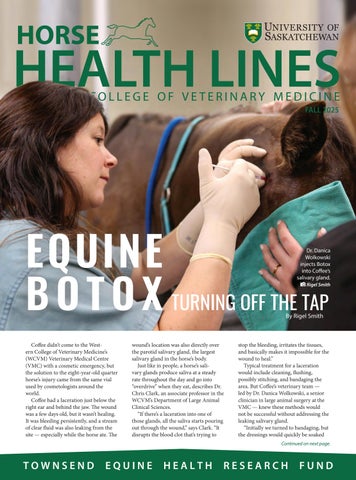
Coffee didn’t come to the Western College of Veterinary Medicine’s (WCVM) Veterinary Medical Centre (VMC) with a cosmetic emergency, but the solution to the eight-year-old quarter horse’s injury came from the same vial used by cosmetologists around the world.
Coffee had a laceration just below the right ear and behind the jaw. The wound was a few days old, but it wasn’t healing. It was bleeding persistently, and a stream of clear fluid was also leaking from the site — especially while the horse ate. The
wound’s location was also directly over the parotid salivary gland, the largest salivary gland in the horse’s body.
Just like in people, a horse’s salivary glands produce saliva at a steady rate throughout the day and go into “overdrive” when they eat, describes Dr. Chris Clark, an associate professor in the WCVM’s Department of Large Animal Clinical Sciences.
“If there’s a laceration into one of those glands, all the saliva starts pouring out through the wound,” says Clark. “It disrupts the blood clot that’s trying to
stop the bleeding, irritates the tissues, and basically makes it impossible for the wound to heal.”
Typical treatment for a laceration would include cleaning, flushing, possibly stitching, and bandaging the area. But Coffee’s veterinary team — led by Dr. Danica Wolkowski, a senior clinician in large animal surgery at the VMC — knew these methods would not be successful without addressing the leaking salivary gland.
“Initially we turned to bandaging, but the dressings would quickly be soaked
Continued on next page.
Dr. Danica Wolkowski injects Botox into Coffee’s salivary gland. Rigel Smith
EQUINE BOTOX
as if you had run them under a tap,” says Wolkowski. “That is a strong indicator of saliva contamination.”
The team needed a way to temporarily stop the flow of saliva to give the wound a chance to heal. After searching online through veterinary journals, the veterinary team found a paper that outlined a similar case in the United Kingdom.
The U.K. veterinarians turned to a solution more often found in a cosmetology office or human hospital than a barn — botulinum toxin, known by the brand name Botox. Clark says the paper provided a detailed “road map” that the WCVM team could adapt to their case.
While many people associate Botox with cosmetic procedures, its medical uses far predate its role in smoothing wrinkles.
“Botulinum toxin is an incredibly potent substance produced by the bacteria that causes botulism — a very dangerous type of food poisoning,” Clark explains. “But in very small, controlled doses, it’s been used in human medicine for decades.”
Botox consists of a heavily diluted concentration of botulinum toxin that can be used to target overactive muscles or glands and provide therapeutic benefit.
To picture how botulinum toxin works, imagine nerves as water pipes running throughout the house (the
brain) to individual “faucets” — the muscles or glands. Water fills the lines, but these faucets remain off until a signal tells them to activate and turn on the tap. Botulinum toxin interrupts that signal pathway. While it doesn’t damage the faucet or the pipe itself, it blocks the tiny chemical messengers that activate the tap.
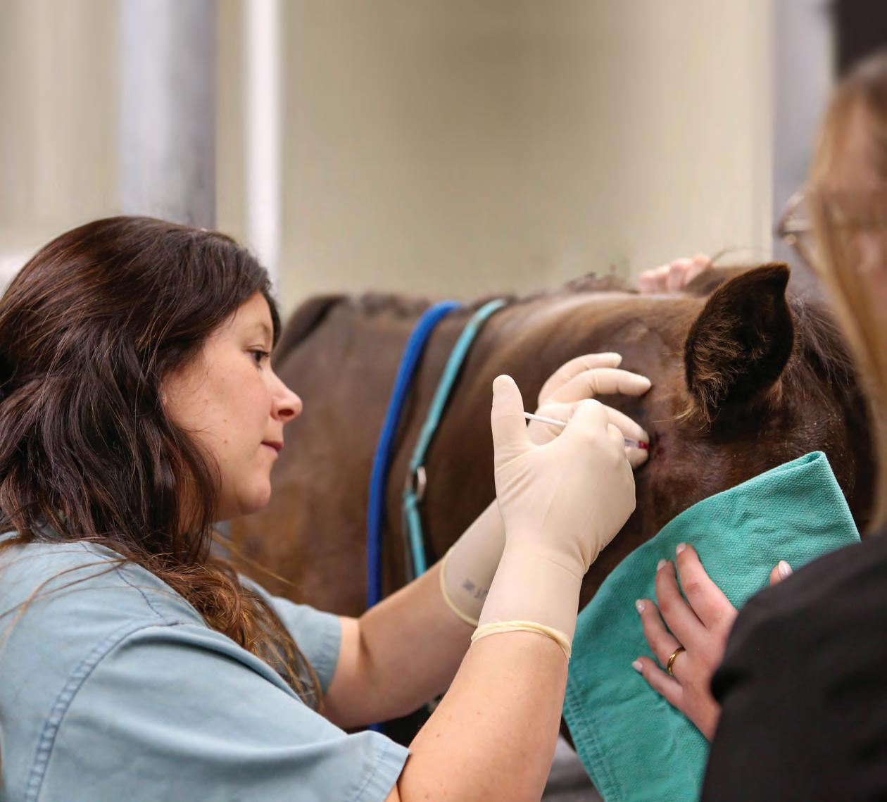
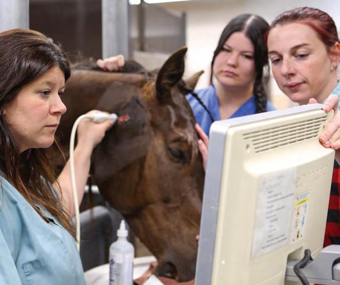
In Coffee’s case, the wounded salivary gland was like a broken tap that wouldn’t stop leaking. Guided by ultrasound imaging, Wolkowski used 20 carefully placed needles to perform Botox injections that temporarily paralyzed the horse’s salivary gland.
“It was like turning off a tap,” says Clark. “It dried up almost immediately
and you could literally see the healing process start.”
The effects last for approximately one month until the body rebuilds the connection and normal function returns. But while the benefits are significant, using Botox isn’t without risks.
“You need to be very careful how much you’re injecting in each site and make sure it’s going into the right place,” says Clark.
While Botox will never be a go-to treatment for every wound, Wolkowski and Clark see it as a valuable option for challenging cases that involve an injury to the salivary gland.
Read the full story at tehrf.ca.
Horse Health Lines is the news publication for the Western College of Veterinary Medicine’s Townsend Equine Health Research Fund (TEHRF). Visit tehrf.ca for more information. Send comments and article reprint requests to:
Myrna MacDonald, Editor Horse Health Lines WCVM, University of Saskatchewan 52 Campus Drive, Saskatoon, SK S7N 5B4 horse.health@usask.ca
Horse Health Lines design and layout: Priddy Design
WCVM clinicians use ultrasound imaging to map Coffee’s salivary gland for Botox injections.
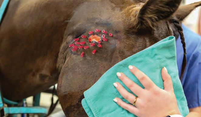
Needles for the Botox injections are positioned around Coffee’s wound.
Rigel Smith
Rigel Smith
HARD TO STOMACH
Veterinarian alerts horse owners about dangers of bale net wrap
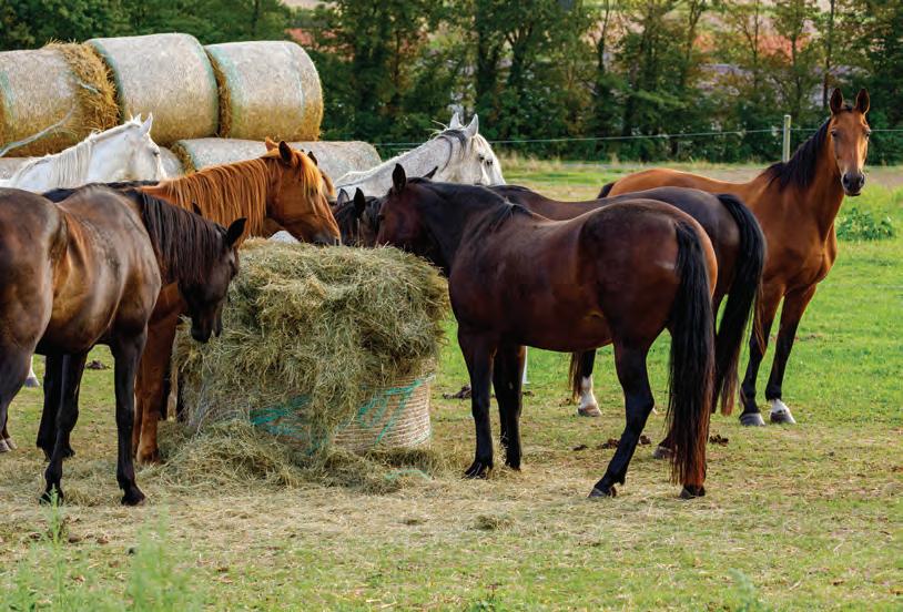
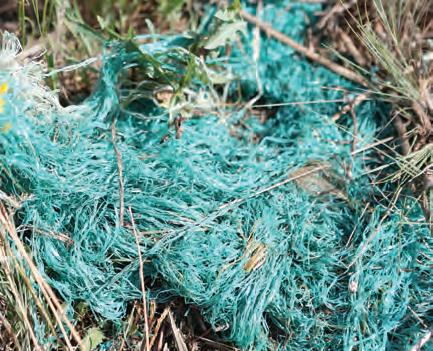
From a distance, there’s nothing unusual about the round hay bale sitting in a round bale feeder — it’s just more feed for the acreage’s horses leaning in to grab mouthfuls of hay. But mixed in with the hay lies an insidious danger: overlooked remnants of plastic net wrap so fine that it slips past the eyes of both humans and horses.
Bale net wrap is a thin, plastic mesh used to bind large round hay bales. Known for its ability to shed water and preserve feed quality, it has largely replaced baling twine on farms across the Prairies. But the finer material is also harder to remove completely and increases the risk of accidental ingestion in horses.
Dr. Alannah Friedlund, a veterinarian and large animal surgical resident at the Western College of Veterinary Medicine (WCVM), is no stranger to this growing issue.
“The wrap is so fine that the horses — even though they’re pretty discriminate eaters — can get a mouthful of it without realizing that they’re actually ingesting it,” says Friedlund.
Once swallowed, net wrap doesn’t break down inside the horse’s digestive tract.
“This net wrap makes it all the way to the small colon, which is the very last stop before the rectum,” says Friedlund. “This is where it seems to get caught in every single case.”
The small colon helps absorb moisture and form fecal balls. As it compresses material inside, the net wrap gets twisted around these balls, forming what Fried-
By Rigel Smith
lund describes as a “rosary-style chain” that the horse can’t pass.
By the time the horse is brought to the college’s Veterinary Medical Centre (VMC), they usually present with typical signs of colic such as bloating and difficulty passing solid feces. Initial treatment with fluids and pain medication rarely works in these cases.
“Normally these horses will just get even more gas distended, more painful,” says Friedlund. “We perform an enterotomy (an incision) of the small colon and that’s when we find, instead of fecal balls, this mass of material.”
The veterinary team can remove the material during surgery, but Friedlund says the recovery rate is about fifty-fifty. Some horses recover well, while others suffer from complications such as colon damage or abdominal contamination.
The cost of colic surgery, which can range from $6,000 to $8,000, can also be challenging for clients.
“It’s a lot easier to just take that wrap off the bale,” points out Friedlund.
Unfortunately, there’s no safer alternative to net wrap on the market. While baling twine is easier to remove, it’s considered less effective for preserving feed quality. That’s why Friedlund says awareness is critical.
“Even a small amount of net wrap can cause a pretty devastating outcome for the horse and owners,” says Friedlund. “But if you’ve never been in that situation before, you probably just don’t realize how significant the problem can be.”
SAFE FEEDING PRACTICES
Remove all net wrap before feeding.
Always remove 100 per cent of the plastic net wrap from hay bales before feeding. Even small remnants left between layers or stuck to the bottom can be dangerous.
Consider alternative feeding methods.
Use slow feeders or hay nets, or you can manually fork hay to make it easier to identify and remove potential hazards.
Avoid processing bales with net wrap.
Feeding chopped hay that’s been processed with net wrap can distribute long strands of plastic throughout feed. Always remove wrap before processing.
Regularly check fields and paddocks.
Look for discarded net wrap or other debris in feeding areas. Even windblown plastic can be a risk.
Know when to call a veterinarian.
If your horse shows signs of colic (bloating, rolling or reduced fecal output), call your veterinarian right away.
iStock
Céline Grimard
Tiny cargo-filled messengers may be the answer to how horse embryos communicate with the mother in early pregnancy and could potentially be used to heal other parts of the equine body.
Extracellular vesicles (EVs) are produced by all the body’s cells, and like miniature Pony Express riders, they carry vital information that allows those cells to communicate with each other.
Dr. Claire Card is a professor and equine reproduction specialist at the Western College of Veterinary Medicine (WCVM). She and her graduate student, Dr. Josefina Ghersa, are looking at EVs specifically produced by the uterus of a pregnant mare and the embryo so they can better understand the issue of early pregnancy loss in horses and how it can be prevented.
“We think that this type of research will eventually lead to an understanding of therapies that will improve the uterine environment, decrease pregnancy losses, and save people a lot of money, time and frustration,” says Card.
Previous studies have shown early pregnancy loss rates between 7.9 and 17 per cent in mares. In older animals, there’s a lower chance of becoming pregnant plus a higher pregnancy loss rate (up to 36
MINIATURE MESSENGERS
By Lynka Itogawa
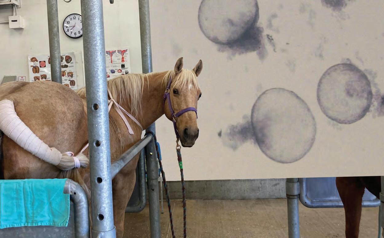
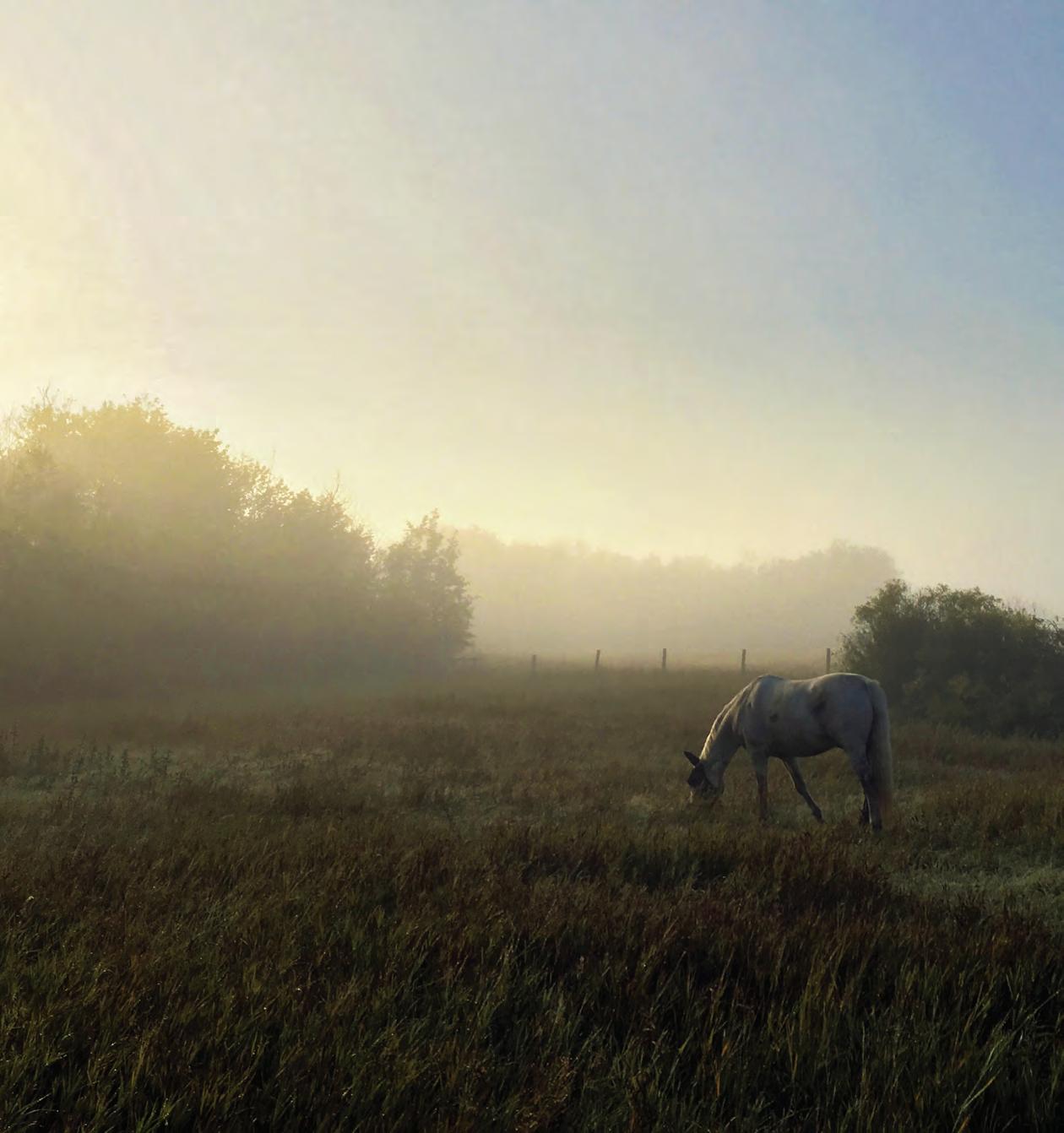
Top left: one of the study’s mares. Top right (background): microscopic view of
per cent in older mares) — two important factors since many valuable performance horses aren’t bred until later in life.
“As a reproductive specialist, pregnancy loss is one of the most devastating and disappointing events that happens in our profession,” says Card.
Unlike all other domesticated animals, the early communication between the embryo and the mother — maternal recognition of pregnancy or MRP — remains a scientific mystery in horses.
Card and her research team are working to solve this biological riddle with the support of a five-year research grant from the Natural Sciences and Engineering Research Council of Canada (NSERC).
To help reduce the number of horses needed for a study and associated costs, WCVM researchers are investigating the use of trophoblastic vesicles (TRVs). When an embryo is divided into fragments in a petri dish, each fragment grows into new embryo-like structures or TRVs. They release hormones and tiny extracellular vesicles — just like those found in an embryo.
Finding a way to freeze and thaw TRVs will allow the WCVM research team to study them year-round without requiring large numbers of mares.
Once grown, the researchers use TRVs for imaging or freeze them in various conditions to see if they are still alive after thawing. As well, the WCVM team analyzes the media to identify and measure how much hormone and EVs are released by the TRVs.
WCVM scientists are also interested in the regenerative medicine properties of EVs. They may have the potential to deliver substances that promote healing in horses’ bodies.
While much work still needs to be done, EVs are showing great potential as powerful tools for equine health — something that Card and her research team are working hard to realize.
Lynka Itogawa is a third-year WCVM student and was the 2025 TEHRF summer research student.
Read the full story at tehrf.ca.
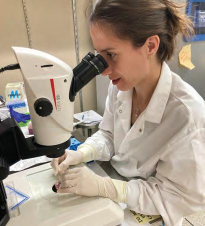
Dr. Josefina Ghersa, theriogenology resident at the WCVM. Lynka Itogawa
trophoblastic vesicles (TRVs). Lynka Itogawa
THE FASTING FACTOR
By Rigel Smith
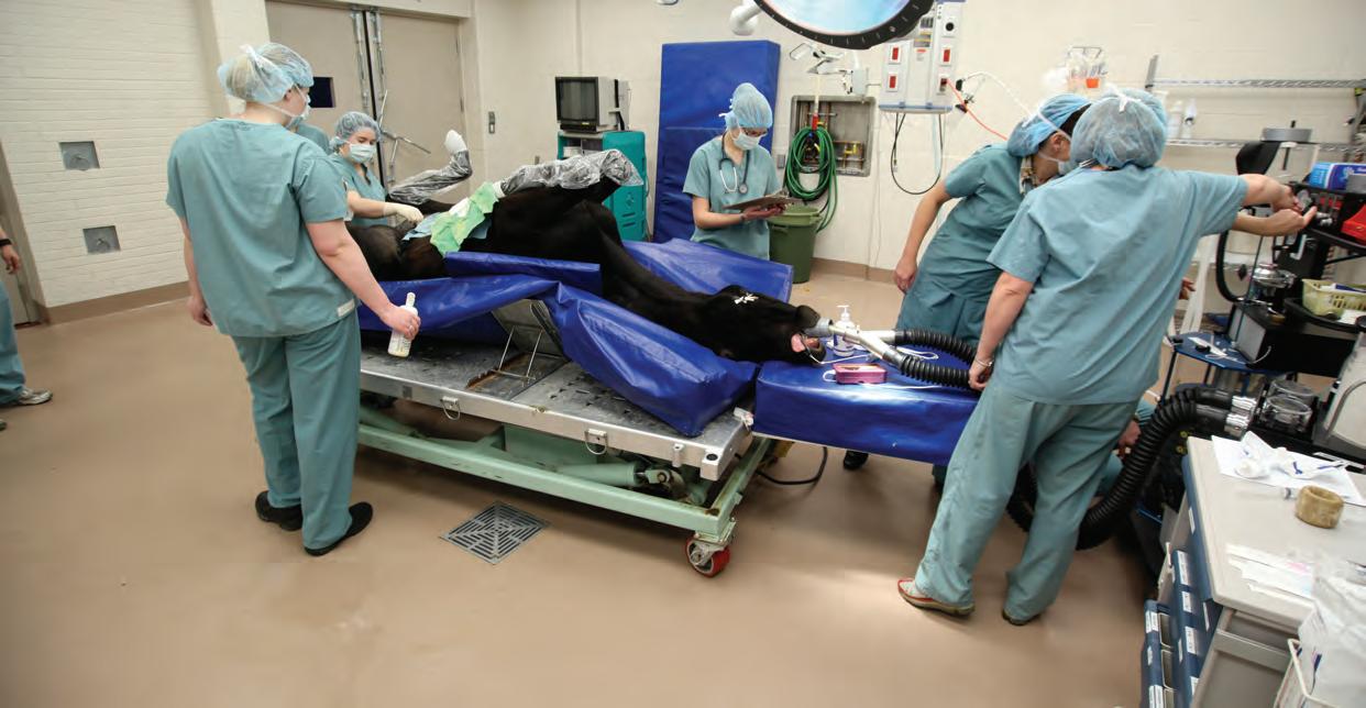
NEW STUDY SEEKS TO IMPROVE ANESTHESIA OUTCOMES FOR HORSES
When a horse goes under anesthesia, even a routine procedure can carry serious risks. Researchers at the Western College of Veterinary Medicine (WCVM) are exploring whether a simple change — such as adjusting when a horse last ate — could help reduce those dangers.
Dr. Shannon Beazley, a veterinary anesthesiologist and associate professor at the WCVM, is leading the two-phase study with financial support from the Townsend Equine Health Research Fund.
“Horses have a higher rate of mortality under anesthesia compared to other species,” says Beazley. “This is due to a variety of factors, including how well they ventilate, their blood pressure, how they recover, and even if they panic when they wake up.”
Unlike dogs or cats, horses aren’t built to lie on their sides or backs for long periods. That position can compromise their ability to ventilate and lead to dangerously low blood oxygen levels — a complication called hypoxemia.
“When I was trained, we fasted horses before anesthesia because of their large intestinal tracts,” says Beazley. “When they’re full of gut contents, they’re heavy,
and when we lay them down, that weight can push on the diaphragm and make it harder for them to breathe.”
The study’s first phase focuses on what happens during anesthesia. Beazley’s team will evaluate eight horses split into two groups — one group fasted for 12 hours and one group not fasted — and measure how well each group ventilates during anesthesia.
During the study, they’ll collect arterial blood samples to assess oxygen and carbon dioxide levels and use a new technology called electrical impedance tomography (EIT). This non-invasive method uses a belt wrapped around the horse’s thorax (chest) to track how air is distributed in the animal’s lungs in real time.
Beazley explains EIT technology is used routinely in human intensive care settings to adjust ventilators but isn’t commonly found in veterinary practices.
Her team will collect EIT and blood gas data at several points — before, during and after anesthesia — to help build a clearer picture of how fasting affects equine lungs.
The study’s second phase shifts focus to the recovery period — specifically, if fasting affects gastrointestinal function
after surgery. Colic and other gastrointestinal issues are known risks following anesthesia in horses, and some veterinarians wonder if fasting might contribute to those problems.
“One of the recommendations now is that for shorter procedures, we don’t fast them,” says Beazley. “The idea is to maintain GI (gastrointestinal) motility so that when they get up, their guts are still moving.”
The research team will observe each horse for 24 hours post-operatively to monitor for signs of colic. Those findings could have real-world impact — not just for veterinarians, but for owners, too.
“Even with all these strategies to improve equine anesthesia in the last 20 to 30 years, we haven’t really improved mortality rates,” says Beazley. “We’re slowly chipping away and trying new things.”
Her upcoming study is part of that effort.
“If it turns out that fasting — or not fasting — can make a difference in outcomes, that’s something we can implement right away,” says Beazley.
“It doesn’t cost anything, and it could reduce complications. That’s a win for us and a win for the horses.”
A WCVM surgical team prepares an anesthetized horse for surgery. Michael Raine
Bubbles and Biscuit
By Rigel Smith
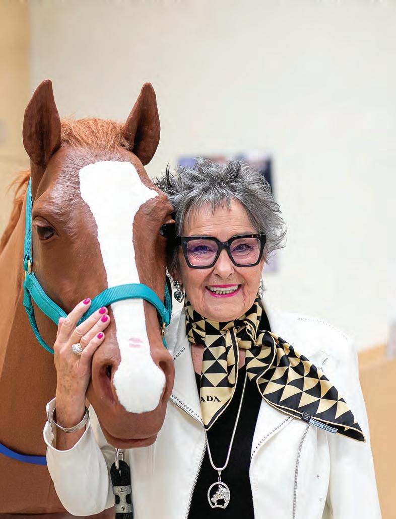
When Dr. June Donaldson (MBA, EdD) saw Bubbles the mare model for the first time, she did what anyone might do when reuniting with a beloved childhood horse: she kissed it on the nose.
“I just walked right up and kissed her,” she says. “It was just so touching.”
The horse, of course, wasn’t real. But to Donaldson, the life-sized equine model constructed of fibreglass and synthetic rubber felt remarkably familiar.
Built by Calgary-based Veterinary Simulator Industries (VSI), the horse is modelled after her grandparents’ draft horse, Bubbles, who lived on their farm in the County of Leduc, Alta. The mare’s broad back, gentle eyes and striking blaze — now forever adorned by a lipstick-kiss imprint — look just like Donaldson’s adored childhood friend.
Donaldson, now a bestselling author, describes Bubbles as “the main attraction” of farm visits. She was a gentle horse you could crawl under, paint toenails on, or pile five kids onto for a ride.
“My brother could stand on her back with a Superman cape while she galloped down the laneway,” Donaldson recalls. “The farm was never the same after she was gone.”
The mare model came paired with a 34-kilogram foal named Biscuit in honour of Seabiscuit, a 1930s American thoroughbred racing superstar. Donaldson has long been involved in preserving Seabiscuit’s legacy — particularly that of his Canadian-born jockey, Red Pollard — through her work with the Seabiscuit Heritage Foundation.
The idea of donating the models to the veterinary college first took root during a tour of the WCVM’s BJ Hughes Centre for Clinical Learning in October 2024. A passing comment made by the centre’s manager, Carolyn Cartwright, stuck with Donaldson.
“She said to us, ‘What we really could use is a mare and foal,’” says Donaldson. Driving home to Calgary, the idea rolled around in her mind. With encouragement from her partner Rob, Donaldson decided to make it happen: “It just felt right.”
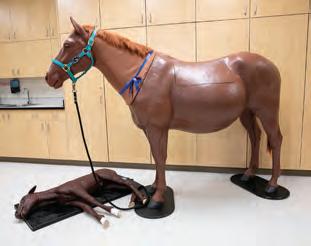
Less than three months later, a large wooden box arrived at the doors of the WCVM. Carefully packed inside stood the 15-hand equine birthing simulator, with her foal tucked neatly underneath.
Thanks to Donaldson, the models now give WCVM students the rare chance to practise foaling scenarios in a safe hands-on environment.
“Foaling is a critical time for the mare and the foal, and things can happen quickly,” says Dr. Gillian Muir, WCVM dean. “It’s not necessarily best for the mare to have a group of students present, and often there isn’t time to teach when it’s happening live.”
Students can also use Bubbles to learn how to insert a tube in a horse’s esophagus and stomach — a technique often used in equine colic cases.
“Simulation is a game changer,” says Muir. “When we bring alumni here … they are just so impressed by what we can offer our students now.”
For Donaldson, the donation is both a tribute and an investment in future veterinary professionals.
“This magnificent facility offers both the book smarts and the hands-on skill,” she says. “And when you put those together, you end up with people who are confident, who are self-assured, and who can hit the floor running to get the job done.”
Read the full story at tehrf.ca.
Dr. June Donaldson and Bubbles. Christina Weese
Equine models Bubbles and Biscuit are helping WCVM students gain hands-on experience. Christina Weese
AWARDS FOR EQUINE-FOCUSED STUDENTS
Two WCVM students earned awards recognizing their equine health achievements during the WCVM’s annual spring awards ceremony on June 4.
Dr. Rachel Haner, a 2025 graduate of the WCVM’s Doctor of Veterinary Medicine (DVM) program, earned the Saskatchewan Speed Horses Association Award. Valued at $3,000, the annual award is given to WCVM graduates who have demonstrated exceptional patient advocacy and compassionate care during equine clinical rotations in their fourth year. Haner, who is originally from Oakbank, Man., has accepted a one-year equine internship at Burwash Equine Services near Cochrane, Alta.
The evening’s second equine-focused honour was the Equine Foundation of Canada (EFC) Graduate Student Scholarship. Candas Rolls of the EFC presented the $5,800 award to Dr. Josefina Ghersa, an equine theriogenology resident and master’s student who is originally from Buenos Aires, Argentina. The annual scholarship supports graduate students who show demonstrated interest and scientific merit in equine research. See below (at right) for a summary of Ghersa’s research work.
GRADUATE RESEARCH IN PROGRESS
Each year, WCVM graduate students who conduct equinefocused research can apply for graduate awards from the Townsend Equine Health Research Fund for financial support. Two award recipients — Drs. Bukola Alaba and Josefina Ghersa have provided research updates (summarized below).
• Dr. Bukola Alaba is a Master of Science student whose work is co-supervised by Drs. Suraj Unniappan and James Carmalt. Her graduate research focuses on fibrocartilage, the unique type of cartilage that makes up the articular surface of the horse’s temporomandibular joint (TMJ). While researchers know a lot about hyaline cartilage that’s found in the equine stifle and fetlock, very little is known about fibrocartilage. This study will help scientists better understand the biological differences between these two forms of cartilage.

RESEARCH IN PRINT
A roundup of WCVM-related equine research articles that were recently published in peer-reviewed journals.
Ghersa J, Friedlund A, Thomas K, Wobeser B, Card C. “Recurrent preputial sarcoids in a miniature donkey.” Clinical Theriogenology. May 2025. doi: 10.58292/CT.v17.12221.
Ing S, Pinard C, James-Jenks EM, Leis ML. “A retrospective survey of equine ocular diseases evaluated at a referral hospital in Ontario (2011 to 2021).” Canadian Veterinary Journal. Jan. 2025. 66(1):98-101.
Polo M, Huby FD, Uehlinger FD, Rubin JE. “Survey of the antimicrobial susceptibility of Escherichia coli isolated from horses admitted to the Western College of Veterinary Medicine, Saskatoon, Saskatchewan.” Canadian Veterinary Journal. April 2025. 66(4):435-439
Visit tehrf.ca for more news updates.
• Dr. Josefina Ghersa is a theriogenology resident and Master of Science student whose supervisor is Dr. Claire Card. Ghersa’s research project focuses on the maternal recognition of pregnancy (MRP) in horses, the signal produced by the embryo alerting the mare’s body of the pregnancy. The researchers are investigating several avenues to understand MRP in this large, multiyear project.
Ready to take a closer look at equine research at the WCVM?
Visit tehrf.ca (click “Research updates”) to read full summaries.
Dr. Bukola Alaba
Dr. Josefina Ghersa
Dr. Rachel Haner
“Our practice (Paton & Martin Veterinary Services) began to make contributions to the fund on behalf of clients when their horses passed away. We have found this to be a gratifying contribution and have been humbled by the responses that we have received from many of our clients. I think that it is very helpful for them to know that their horses have been honoured in such a fashion. The fund gives horse owners the additional opportunity to contribute to this very wor thwhile cause: supporting vital research in the areas of equine health.”
Dr. David Paton (DVM’78) WCVM alumnus and TEHRF donor
Pay tribute to the lives of your patients, clients and loved ones by making a donation to the Townsend Equine Health Research Fund (TEHRF) through its memorial program. Each time you give to the fund, we will send a letter to the client or loved one’s family acknowledging your gift to the equine health fund.