CHAPTER 2 EQUIPMENT SUPPLIES
Richard A. Gasalberti MD, Isaac J. Kreizman MD, Marina Kokova MD
When performing the procedure one must consider the proper type and length of needles, and be prepared for difficulties and possible complications in case ideal equipment is not available. Size of the patient, sex, age and type of procedure matter. For instance, a larger patient would need a longer needle, a sharp needle instead of a pencil point needle may lead to nerve damage, a spinal needle used for epidural access would lead to higher rates of dural punctures. However, the basic need for most of the equipment remains the same.
TYPES OF NEEDLES
1. Quincke needle — needle with a sharp tip and cutting edge. Its sharp bevel facilitates penetration of the needle into the skin. Is commonly used for intramuscular, intravenous injections, biopsies etc. Most of the spinal needles have this type of bevel.
2. Pencil point needle — is classified as “blunt” type of needle and has conical shaped tip with the bevel on the side of the needle shaft. May penetrate structures as delicate as an artery if it is immobile.
3. Bullet tip needle — is somewhat similar to previous type of needle but its tip is completely blunt, which makes it safer for types of procedures which are performed close to important structures like nerves and blood vessels. It’s bevel is at the very tip of the needle. As the tip is completely blunt and cannot penetrate the skin, a sharp-tipped introducer must be used.
4. Chiba needle — this type of needle has a very short angled bevel with a noncutting edge which makes it suitable for procedures such as a lumbar sympathetic block or celiac plexus block when a needle goes through delicate issues on its way to the target
5. Day needle — it is Coudé needle with bullet tip ie noncutting tip which decreases the possible damage to delicate tissues.
6. Huber needle — the tip has a noncoring edge, which is safer for the silastic material over the entry port. Specially designed angles shaft keeps the needle flat against the surface of the skin. Most often used for accessing a sub-cutaneous reservoir or a port during a continuous infusion eg long term epidural infusion.
7. Touhy needle — has the slight curve at the very tip of the needle and the opening on the side which makes the tip blunter and helps guide a catheter at an angle to prevent a dural puncture.
5 CHAPTER 2 - EQUIPMENT SUPPLIES
8. Hustead needle — is a type of epidural needle. Distinctive features are a short bevel, and a rounded “heel”. This design helps to decrease possible damage to the catheter if it is withdrawn through the needle.
9. RX Coudé needle — this is an epidural access needle with a bevel that makes a flat surface so that it may be place parallel to the ligamentum flavum/dura. Such a design decreases risk of dural puncture. Selection of needles, medications, and equipment for specific types of procedures:
1. Intra-articular injections
• Sterile gloves
• Sterile fenestrated drape
• 4 × 4 gauze soaked with povidone-iodine solution (Betadine)
• 30-gauge 0.5- or 1-in needle for subcutaneous infiltration
• 25-gauge 2-in needle, 25-gauge 3.5-in Quincke needle, 22-gauge 3.5-inch Quincke needle, 22-gauge 5-in Quincke needle
• Quincke needles are styleted, and have a relatively dull tip that does not cut into articular cartilage as readily as a standard sharp beveled needle
• 3-cc syringes
• Extension tubing to facilitate syringe changes from local anesthetic, to contrast, to injection solution. It also keeps the syringe and hands out of the way of the radiographic picture during fluoroscopic injections.
• Skin marker
• Radiographic marker for fluoroscopic identification of structures
• Paperclip or similar object for ultrasound identification of structures
2. Tendon injections:
• 22- or 25-gauge 1.5- to 3.5-in spinal needle
• 30-gauge subcutaneous needle
• 5 cc syringe for local anesthetic/deposteroid
• 3 cc syringe for contrast
• Extension tubing
3. Myofascial pain
• Isopropyl alcohol or chlorhexidine
• 25-gauge 1.5-in needle
• 3, 5, or 10 mL syringe for medications
4. Trigger Point Injections
6 CLINICIANS GUIDE TO OPIOID SPARING PAIN MANAGEMENT
• Sterile gloves
• Betadine and/or alcohol solution or other antiseptic solution
• Ethyl chloride spray
• 1% lidocaine (usual anesthetic of choice) and/or 0.25% or 0.5% marcaine, +/− steroid
• 10-cc syringe
• 25-gauge 1.5-in needle for superficial muscles and 25-gauge 3.5in needle for deeper muscles
5. Neuromas
• Sterile gloves
• Betadine and/or alcohol solution or other antiseptic solution, eg, chlorhexidine
• Ethyl chloride spray
• 1% lidocaine (usual anesthetic of choice) and/or 0.25% or 0.5% marcaine, +/− steroid (40 mg depo-medrol, 3 to 6 mg betametha- sone, and 2 to 4 mg dexamethasone).
• 10-cc syringe
• 25-gauge 1.5-in needle for superficial neuromas and 22- to 25-gauge 3.5-in needle for deeper neuromas
• Image guidance (fluoroscopy, ultrasound)
6. Steroids and viscosupplements injections
• 25-gauge 1.5-in needle
• 22-gauge 2-in needle (an 18-gauge needle may be required for large-volume aspiration)
• 3-cc syringe for local anesthetic (optional)
• 10-cc syringe for aspiration
• 10-cc syringe for intra-articular steroid/anesthetic
7. Fluoroscopy or US guided glenohumeral joint injections:
• 22-gauge 3.5-in spinal needle or, for ultrasound-guided injections, a 22-gauge echogenic needle, which will be connected to extension tubing
• 22-gauge 1.5-in needle to prepare the injectate medication
• 25-gauge 1.5-in needle for local anesthetic
• 6-cc syringe for local anesthetic
• 6-cc syringe for medications
• 3-cc syringe for fluoroscopically guided procedures with contrast
• 6-cc syringe for GH effusion aspiration (if applicable)
• Ultrasound machine or fluoroscope
• Extension connector tubing
7 CHAPTER 2 - EQUIPMENT SUPPLIES
8. Stellate ganglion block.
• 22-gauge 2.5-in Chiba or stellate block needle
• 25-gauge 1.5-in needle
• 3-mL syringe for skin local anesthetic
• 3-mL syringe for contrast
• 5-mL syringe for injectate
• 12-in extension tubing
• Kelly clamp
9. Lumbar sympathetic block
• 18-gauge needle to use as radiological marker at L2
• Sterile towels
• Multiple 10-mL syringes with local anesthetic
• Sterile gloves
• Marking pen
• 6- to 7-in, 22- or 25-gauge spinal needles
• 3-mL syringe with 30-gauge needle to anesthetize skin
• 18-gauge introducer
• 5-mL syringe for contrast with extension tubing
10. Lumbar facet joint injections
• 2 ml and 10 ml syringes
• 25 G needle
• 22 G spinal needle, end-opening
• Non-ionic radio-opaque contrast medium
• ECG, BP, and SpO2 monitors
• Resuscitation equipment
• C-arm fluoroscopy or ultrasound
8 CLINICIANS GUIDE TO OPIOID SPARING PAIN MANAGEMENT
CHAPTER 3
COMMON SYNDROMES – UPPER EXTREMITY
Richard A. Gasalberti MD, Isaac J. Kreizman MD, Kokova Marina MD
The upper limb consists of the shoulder girdle formed by the clavicle and scapulae, humerus, ulna and radius, the wrist composed of the carpal bones, and the hand formed by the metacarpals and phalanges.
ACROMIOCLAVICULAR JOINT INJURY
Anatomy. The acromioclavicular joint is a diarthrodial articulation between the articular surfaces of the acromial process and the clavicle, covered by the hyaline cartilage. The joint is safely stabilized by 3 ligaments coracoclavicular ligament, acromioclavicular and oracoacromial ligaments, which covers the acromioclavicular joint capsule lined by a synovial membrane. Fibrocartilage lines the articulating surfaces of the joint. (see Fig. 1)
Pathophysiology. AC joint injuries are common. The most common etiology is direct force applied to the superior aspect of the acromion that may cause the acromioclavicular and coracoclavicular ligaments disruption, or lateral trauma which causes axial load on the joint space(motor vehicle accidents, sports or accidental trauma, falling on outstretched hands). Overuse AC joint injury (aka wear and tear injury) is most common in individuals who are involved in sports.
Presentation. Patients will usually present with pain over the acromioclavicular joint, possibly radiating to the shoulder or neck and made worse with movement or use of the arm, they also report a mechanism consistent with that injury. On exam, there may be swelling, bruising, or deformity, tenderness to palpation over the area, limited active or passive range of motion of the shoulder or neck. [17]
Anterior or posterior AC shear test, one-handed shear test (also known as Paxinos test) forced adduction test on hanging arm, Dugas test, AC distraction (bad cop) may be performed for diagnosing. [1, 14, 15]
9 CHAPTER 3 - COMMON SYNDROMES – UPPER EXTREMITY
SHOULDER DISLOCATION
Anatomy. Static stabilizers of the shoulder include the glenohumeral articulation, labrum, glenohumeral ligaments, and the rotator interval. Dynamic stabilizers of the glenohumeral joint include the rotator cuff (see Fig.2), deltoid, long head of the biceps, and the periscapular musculature. [16]
Pathophysiology. Anterior dislocations (95% of shoulder dislocations males>females) are usually due to fall with abduction component, extension of the arm and a force directed posteriorly. Also, anterior dislocations are typically associated with fractures (head of the humerus, greater tuberosity, clavicle or acromion can be involved). Posterior dislocation is generally caused by an extreme muscle contraction (seizures or electric shock), a direct or indirect trauma that occurs with forceful contractions of the internal rotators with the shoulder internally. Inferior dislocations arise from an axial force directed on an abducted shoulder.
Presentation. The patient with anterior dislocation holds the arm slightly abducted, in external rotation. Abduction and internal rotation are limited. The shoulder loses its usual round shape and the humeral head is palpable anteriorly, in the front of the shoulder. Posterior dislocation present with the arm internally rotated and adducted. External rotation and attempted abduction are painful. Inferior dislocation leads to a condition known as luxatio erecta. [2][3].
Provocative tests for anterior shoulder dislocation include anterior apprehension, Jobe Relocation Test, anterior release, anterior load and shift, and the Sulcus sign.18

10 CLINICIANS GUIDE TO OPIOID SPARING PAIN MANAGEMENT
Fig. 1 Anterior aspect of the right shoulder. Subacromial bursa is located below the acromion and superiorly to the tendon of supraspinatus muscle separating it from acromion and deltoid. (by Aziz Abdurakhimov MD.)
ROTATOR CUFF DISEASE
Anatomy. The rotator cuff includes the following muscles: supraspinatus muscle (innervated by subscapular nerve), main function is to abduct the arm; infraspinatus muscle (innervared by the suprascapular nerve) rotates arm externally, teres minor (innervated by the axillary nerve) adducts and externally rotates arm and subscapular (innervated by the subscapular nerve) rotates binternally and adducts arm. (see Fig. 2)

Pathophysiology. Rotator cuff disease etiology is multifactorial. A combination of extrinsic, intrinsic, and biomechanical factors plays a major role in development of rotator cuff injury. The extrinsic factors include repeated impingement of the rotator cuff tendon against different structures of the glenohumeral joint. Impingement and tearing usually begin in the supraspinatus tendon because it passes under the acromion. The conditions owing to these factors include the anterosuperior impingement syndrome, posterosuperior impingement syndrome and anterointernal impingement syndrome. Usually occurs in patients >50 years of age but may also occur in young athletes. [19]
Presentation. Significant pain withabduction above the head abd internal rotation (reaching up the back), weakness of the upper extremity, decreased range of motions, clicking, catching, stiffness, crepitus. Rotator cuff tests include the impingement and topographic tests, combination of which allows determination of whether or not a patient’s symptoms are caused by rotator cuff disease [4][5]. Neer impingement test, Hawkins-Kennedy test, Yocum test and posterior impingement test are useful in confirmation of impingement syndromes. The
11 CHAPTER 3 - COMMON SYNDROMES – UPPER EXTREMITY
Fig. 2 The dorsal scapula muscles of the right side. (by Aziz Abdurakhimov MD.)
topographic tests such as the Jobe test, full can test, Patte test, infraspinatus isolation test, Gerber lift-off test and speed palm up test are considered to be relatively sensitive but not specific. Can occur young athletes, will often hear a “pop” (baseball pitchers). Tears are diagnosed with MRI and often require surgical repair.
ADHESIVE CAPSULITIS (FROZEN SHOULDER)
Adhesive capsulitis (aka frozen shoulder) is an idiopathic, benign, self-limiting condition characterized by pain, loss of both active and passive motion of the shoulder. Affected capsule has no actual adhesions; rather it shows signs of synovitis.
Pathophysiology. The pathologic process involves the anteriosuperior joint capsule, axillary recess, and the coracohumeral ligament. [4][6]. Most common cause is prolonged immobility secondary to shoulder trauma. Adhesive capsulitis is considered to be primary if its etiology is unknown. Secondary capsulitis develops in a set of a known disease such as systemic, extrinsic or intrinsic conditions. There is known significant association between adhesive capsulitis and GM type 1 (high chance of bilateral disease).
Presentation. Somewhat similar to rotator cuff disease. Differentiated by significantly decreases passive range of motion. Most frequently lost motions are shoulder abduction and external rotation. Progressively growing sharp pain at extremities. Also, pain at night with sleep interruption, which may last from 3-9 months. This initial phase is followed by “thawing phase” when symptoms of pain and decrease ROM improve.
SUBACROMIAL BURSITIS
(Subdeltoid Bursitis, Supraspinatus Tendinitis)
Anatomy. The subacromial bursa sac is located in the shoulder joint, separating the superior surface of the supraspinatus tendon (one of four that make up the rotator cuff ) from the overlying coraco-acromial ligament, acromion, and coracoid (the acromial arch) from the deep underlying surface of the deltoid muscle.
Pathophysiology. Most commonly occurs as a result of repetitive or prolonged activities, lots of overhead lifting and forceful pulling, or sports that involve significant amounts of throwing and pitching, placing strain on the subacromial bursa (see Fig. 1).
Direct blow to the shoulder or due to a fall onto the shoulder, elbow or outstretched hand may also be a cause of this condition. Complication includes Frozen shoulder (also known as adhesive capsulitis).
Infections and chronic illnesses or diseases like arthritis, diabetes, and
12 CLINICIANS GUIDE TO OPIOID SPARING PAIN MANAGEMENT
thyroid problems can also increase the likelihood of bursitis. Presentation. Patients complain of gradual onset of aching shoulder pain over a series of weeks or months which may spread down the arm toward the elbow or wrist aggravated by lying on the affected shoulder, using the arm above the horizontal level (painful abduction, internal rotation). [7]
MEDIAL EPICONDYLITIS (Golfer’s Elbow)
Anatomy. The medial epicondyle is the common origin of the forearm flexor and pronator muscles (see Fig. 3). Pathology involves the flexor carpi radialis and pronator teres. Large diffuse tears can also occur in the palmaris longus, flexor digitorum superficialis and flexor carpi ulnaris.
Pathophysiology. Golfer’s elbow is a degenerative problem, not inflammatory. The pathology occurs as result of high-energy valgus stress on the medial elbow created by the overhead throw. An overuse leads to microtearing, collagen breakdown, failure of tendon healing that results in a tendon degeneration.
Presentation. Patients suffer from activity-related pain in the elbow with the most sensitive region located near the origin of the wrist flexors on the medial epicondyle, progressing to pain at rest and loss of functional strength. Also, present with local tenderness over the medial epicondyle and the tendon of the flexor group, without swelling or erythema. [9][10] May also see pain with resisted wrist flexion/ pronation.

13 CHAPTER 3 - COMMON SYNDROMES – UPPER EXTREMITY
Fig. 3 Medial aspect of the right elbow showing flexor muscles and ulnar nerve. (by Aziz Abdurakhimov MD.)
LATERAL EPICONDYLITIS
Is an overuse injury involving the extensor muscles of the forearm originating on the lateral epicondyle of the distal humerus. (see Fig. 4)

Pathophysiology. Shares the same pathophysiology as medial epicondylitis but involves involves the extensor carpi radialis brevis muscle (macroscopic tearing) and less commonly the extensor carpi radialis longus, extensor digitorum, and extensor carpi ulnaris. Any activity involving wrist extension or supination can be associated with overuse of the muscles originating at the lateral epicondyle.
Presentation. Typical presentation is pain just distal to the lateral epicondyle, elbow stiffness often accompanied by localized tenderness over the lateral epicondyle. Patients will commonly have pain with palpation of the lateral epicondyle. Pain can be increased with resisted wrist, second and middle finger extension/supination (Cozen’s sign, Mill’s Test). [9][10]
ULNAR NERVE INJURY
Anatomy. Ulnar nerve is a terminal nerve of a brachial plexus that supplies innervation to muscles in the forearm and hand. Also, carries sensory innervation from skin of the hypothenar eminence and medial 1,5 digits. Muscles innervated by ulnar nerve at the forearm include flexor carpi ulnaris and ulnar half of the flexor digitorum profundus which function is to flex wrist and digits 4 and 5. In hand ulnar innervates muscles of hypothenar compartment, central compartment [11] (palmar and dorsal interossei muscles, lumbricals, and adductor pollicis). (see Fig. 4 and Fig. 5)
14 CLINICIANS GUIDE TO OPIOID SPARING PAIN MANAGEMENT
Fig. 4 Lateral aspect of the right elbow region showing extensor muscles. (by Aziz Abdurakhimov MD.)
Pathophysiology. The ulnar nerve can be damaged at 3 sites, the most common is the cubital tunnel aka “funny bone”, where the ulnar nerve travels through the groove on the posterior surface of the medial epicondyle. Other common sites: at the level at the level of the wrist (wrist lacerations), and at the hand (fractured hook of hamate). Ulnar nerve entrapment may cause denervation and paralysis of the muscles supplied by the nerve.
Presentation. Usually patients present with pain in the medial aspect of the elbow commonly associated by numbness or tingling sensation in 4th and 5th fingers. Later on the course of disease weakness of intrinsic muscles of hand may occur (difficulties with opening the jar or turning the key).
DE QUERVAIN TENOSYNOVITIS
Anatimy. Tendons of extensor pollicis brevis and abductor pollicis longus muscles contained within the first dorsal compartment at the wrist are involved.
Pathophysiology. Swelling and stenosis of the sheath that contains the thumb extensor tendons limits their sliding through the sheath. Histological specimens in De Quervain tenosynovitis shows a thickening and myxoid degeneration consistent with a chronic degenerative process. Most commonly caused by repetitive use of the thumb and forceful grip activities. Most often seen in middle-aged women. Presentation. Presents with severe aching and shooting pain, tenderness and swelling over the radial styloid especially with ulnar deviation or thumb use, may also see skin thickening at the first dorsal compartment over the radial styloid which may forms a visible fusiform. Spasms, occasional burning sensation in the hand, difficulty gripping with the affected side of the hand is also present.
Finkelstein test is pathognomonic: the patient’s thumb is folded into a clenched hand and then the wrist is deviated down to the ulnar side causes pain. Negative grind test [12]
CARPAL TUNNEL SYNDROME
Anatomy. The carpal tunnel is a narrow fibro-osseous tunnel through which 9 tendons passes with the median nerve. Within the carpal tunnel median nerve runs between flexor digitorum superficialis and flexor digitorum profundus. Carpal tunnel syndrome is the most common of the median nerve entrapments with a collection of characteristic symptoms and signs. (see Fig. 5)
Pathophysiology. Anything that causes median nerve compression in the carpal tunnel at the wrist may cause carpal tunnel syndrome,
15 CHAPTER 3 - COMMON SYNDROMES – UPPER EXTREMITY
eg direct trauma, repetitive use, anatomic abnormalities. Eventually, entrapped median nerve may undergoe demyelination followed by axonal degeneration. Middle-aged women and pregnant women are most commonly affected. May cause thenar atrophy. Presentation. Patients with carpal tunnel syndrome have vague pain in the area of thenar eminence and +/- forearm. The pain is frequently accompanied by numbness, tingling sensation over fingers 1-4. The course of disease may later complicated by weakness, persistent numbness, thenar atrophy. There is preserved flexion of the 2/3 digits and normal wrist sensation over the thenar eminence as the branches responsible for these functions arise more proximally. [11][13]

16 CLINICIANS GUIDE TO OPIOID SPARING PAIN MANAGEMENT
Fig. 5 Volar aspect of the right hand showing superficial palmar arch, median and ulnar nerves distribution. (by Aziz Abdurakhimov MD.)
REFERENCES
[1] Rockwood CA Jr, G. D. (1996). Fractures in Adults. Philadelphia: Lippincott-Raven.
[2] Westin CD, G. E. (1995 , May-Jun). Anterior shoulder dislocation. A simple and rapid method for reduction. Am J Sports Med.
[3] Marx JA. Marx JA, H. R. (2002). Medicine: Concepts and Clinical Practice. St. Louis, MO: Mosby.
[4] EA., C. (1934). The shoulder. Boston, MA.
[5] G, W., JP, L., P, B., & E, N. (n.d.). Postero-superior glenoid impingement. Another shoulder impingement. Revue de chirurgie orthopedique et reparatrice de l’appareil moteur .
[6] BJ., L. (1969). The frozen shoulder. Acta Orthop Scand Suppl.
[7] Kristine M Lohr, M. (2015, Oct 13). Bursitis. (M. Harris Gellman, Editor) Retrieved from Medscape: http://emedicine.medscape.com/article/2145588-overview#a5
[8] Safran MR, M. D. (1998). Biceps tendon injuries. Manual of Sports Medicine. Philadelphia, PA: Lippincott Williams & Wilkins.
[9] Hannah GA, W. J. (1994). The elbow in athletics. Sports Medicine Secrets. Philadelphia, PA: Hanley & Belfus.
[10] RP., N. (1993). The Elbow and Its Disorders. Philadelphia, PA: WB Saunders Co.
[11] Thompson, J. C. (2010). Netter’s concise orthopedic anatomy. Philadelphia, PA: Saunders.
[12] Ilyas A, A. M. (2007). De quervain tenosynovitis of the wrist. J Am Acad Orthop Surg.
[13] AAOS. (2009, 12). Carpal tunnel syndrome. Retrieved from American Academy of Orthopaedic Surgeons.: http://orthoinfo.aaos.org/ topic.cfm?topic=a00005
[14] Hyland S, Varacallo M. StatPearls [Internet]. StatPearls Publishing; Treasure Island (FL): Oct 27, 2018. Anatomy, Shoulder and Upper Limb, Clavicle.
[15]Wurm M, Beirer M, Biberthaler P, Kirchhoff C. [Clavicular fractures : Diagnostics, management and treatment]. Unfallchirurg. 2018 Dec;121(12):983-998.
[16]. Turkel S.J., Panio M.W., Marshall J.L., Girgis F.G.Stabilizing mechanisms preventing anterior dislocation of the glenohumeral joint J. Bone Joint Surg., 63 (8) (1981), pp. 1207-1217
[17]. Doyscher R, Kraus K, Finke B, Scheibel M. [Acute and overuse
17 CHAPTER 3 - COMMON SYNDROMES – UPPER EXTREMITY
injuries of the shoulder in sports]. Orthopade. 2014 Mar;43(3):202-8.
[18]. Lo I.K., Nonweiler B., Woolfrey M., LitchfieldR., Kirkley A.An evaluation of the apprehension, relocation, and surprise tests for anterior shoulder instability
Am. J. Sports Med., 32 (2004), pp. 301-307
[19]. State of the Art for Minimally Invasive Spine Surgery A. Dezawa, , P. -Q. Chen, , and J. -Y. Chung
CHAPTER 3 UPPER EXTREMITY CODING & BILLING
Amy Turner RN BSN MMHC CPC CHC
Documentation is the driving factor in choosing the correct CPT code. All components of a procedure must be recorded to allow for proper billing of a patient encounter. Always document: specific location, medication administered and imaging guidance.
Injection into a joint is dependent on the size of the joint the medication is administered into. Each joint injection has two codes to choose from dependent on whether ultrasound guidance is used. Joint injections are separated into major, intermediate and small joints. Joint injections may be reported bilaterally using modifier 50. If performing a unilateral injection, use of the LT or RT modifier is recommended to designate laterality. Use of unspecified ICD-10 codes should not be used as it is apparent which joint is being injected.
• AC joint is considered an intermediate joint- 20605 with no ultrasound, 20606 with ultrasound
• Glenohumeral joint is considered a major joint- 20610 with no ultrasound, 20611 with ultrasound
Injections into the tendon should be clearly documented as to whether the tendon sheath or the tendon origin or insertion is injected- ex lateral epicondylitis. Tendon/ligament injections may be reported bilaterally using modifier 50. If performing a unilateral injection, use of the LT or RT modifier is recommended to designate laterality. Use of unspecified ICD-10 codes should not be used as it is apparent which side is being injected.
• 20550- Single tendon sheath, or ligament, aponeurosis (eg. “plantar fascia”)
• 20551- Single tendon origin/insertion
Carpal tunnel syndrome may be amenable to a carpal tunnel injection.
18 CLINICIANS GUIDE TO OPIOID SPARING PAIN MANAGEMENT
This procedure has a specific code. Carpal tunnel injections may be reported bilaterally using modifier 50. If performing a unilateral injection, use of the LT or RT modifier is recommended to designate laterality.
• 20526- Injection, therapeutic (eg, local anesthetic, corticosteroid), carpal tunnel
Joint InjectionsCodeImagingNotes
Major (shoulder, Hip, Knee) 2061077002- fluoroUse Modifier 50 if Bilateral
Major (shoulder, Hip, Knee) 20611
Intermediate (wrist, ankle, AC joint)
Intermediate (wrist, ankle AC joint)
*includes US Use Modifier 50 if Bilateral. Must retain image
2060577002- fluoroUse Modifier 50 if Bilateral.
20606
*includes US Use Modifier 50 if Bilateral. Must retain image
Small (fingers, toes) 2060077002- fluoroUse Modifier 50 if Bilat
Small (fingers, toes)
Single tendon sheath, or ligament
*includes US Use Modifier 50 if Bilateral. Must retain image
Use Modifier 50 if Bilateral
TPI- 1-2 muscle2055276942- USBill per muscle not per “stick”. Cannot bill bilat
TPI 3+ muscles2055376942- USBill per muscle not per “stick”. Cannot bill bilat
19 CHAPTER 3 - COMMON SYNDROMES – UPPER EXTREMITY
20604
2055077002-fluoro 76942-
US
2055177002-fluoro 76942-
Single tendon origin/insertion
US Use Modifier 50 if Bilateral
20 CLINICIANS GUIDE TO OPIOID SPARING PAIN MANAGEMENT
CHAPTER 4
COMMON SYNDROMES LOWER LIMB
Ali Guy M.D, Antigona Lajqi
The lower extremity consists of the hip joint formed by the pelvis and femur, the knee joint formed by the femur and the tibia, the ankle joint formed by the tibia, fibula and talus, and the foot comprised by the metatarsals and phalanges.
HIP INJURIES
Iliotibial Band Syndrome (ITBS)
Anatomy. The illiotibial band is a thick band of dense fibrous fascia originating from the iliac crest and inserts into the lateral epicondyle of the tibia. The iliotibial band extends throughout the entire length of the lateral thigh, aiding adjacent muscles in extension, abduction, and lateral rotation of the hip. [1]
Pathophysiology. Friction created by iliotibial band movement against the lateral femoral epicondyle lead to inflammation and irritation. This type of injury is most common in runners and athletes, and through activities that require repetitive knee flexion and extension, and pronation of forefoot with excessive internal rotation of the tibia. Muscular imbalance, training errors, and chronic overuse can cause and further exacerbate symptoms. Lidocaine injections of nonsteroidal anti-inflammatory drugs to large muscle bodies of the hip and Gluteus Maximus can reduce inflammation produced by muscle spasms. Physical therapy treatment for this condition is also beneficial with focus on flexibility, massage, and muscle strengthening of effected areas. [2]
Presentation. Common symptoms of illiotibial band injuries include lateral knee and/or hip pain and tenderness over the lateral femoral epicondyle. Referred pain throughout the lateral aspect of the thigh is also common for this injury. Ober’s test, Renne Test, and Compression test can be used to diagnose iliotibial band syndrome. [1]
Iliopsoas Bursitis
Anatomy. The iliopsoas muscle acts a primary hip flexor and share a role with adjacent muscles in truck stabilization. The iliopsoas tendon forms a complex incorporating tendons from originating muscles that make up the iliopsoas muscle. The iliopsoas muscle itself originates from two separate structures; iliacus and psoas major. Insertion into the lesser trochanter of the femur serve to connect movements of the hip with the femur and lower extremities. [3]
Pathophysiology. Development of iliopsoas bursitis can be attributed
21 CHAPTER 4 - COMMON SYNDROMES - LOWER LIMB
to overuse, hypertonicity, rheumatoid arthritis, and prolonged strain. The synovial sac between the iliopsoas tendon and anterior portion of the hip joint articular capsule becomes irritated with repeated flexion of the hip joint. This injury is most commonly found in weight lifters, athletes, runners, and gymnasts. Ultrasound, MRI, and x-ray imaging can be used to effectively diagnose iliopsoas bursitis. Anti-inflammatory non-steroid drugs and physical therapy are common options to treat and manage pain. More invasive treatment options include injection of corticosteroids directly into inflamed bursa. [2]
Presentation. The most common complaint of patients with iliopsoas bursitis is groin pain focused in the anterior portion of the hip. Palpable tenderness during physical examination of the tendon attaching to the small trochanter can also be an indication of iliopsoas bursitis. Back pain can be an indication of this condition as the psoas major muscle originates from the transverse processes of L1-L5.
Osteoarthritis
Pathogenesis. Osteoarthrosis is the most prevalent form of arthritis that can affect any joint in the body. Protective articular cartilage that line the end of bones deteriorate and cause bones to rub against one another establishing joint pain. Multiple risk factors the contribute to the progression of cartilage deterioration. Biological changes that occur with aging affect the body’s ability to maintain cartilage and joint tissue. Females are more susceptible to this condition than male counterparts, as well as individuals who are overweight. Pressure from excessive weight placed on joints can gradually wear down articular cartilage and promote osteoarthritis. Overuse and excessive trauma to joints can also accelerate the rate of osteoarthritis development. [4]
Diagnosis. Understanding risk factors can help diagnose osteoarthritis as well as diagnostic imaging and physical examination. X-rays can assist in the diagnosis of osteoarthritis when observed joint space narrowing is present due to the lack of articular cartilage. Physical examination of palpable tenderness, crepitation, stiffness, and decreased range of motion are indicators used to diagnose osteoarthritis.
Treatment. Treatment for osteoarthritis remains palliative as the damage to joints has already been established. Management of pain can be done through invasive and non-invasive means depending on severity. Physical therapy, occupational therapy, and aquatic therapy are non-invasive treatment measures to improve muscle strength of surrounding muscles of osteoarthritic joints. NSAIDs can be administered to alleviate pain by reducing inflammation that surrounds joints. More invasive options of treatment include intra-articular injections, Platelet-rich plasma injections, and surgical intervention through joint
22 CLINICIANS GUIDE TO OPIOID SPARING PAIN MANAGEMENT
replacement in more severe cases. [4]
Greater Trochanter Bursitis
Pathogenesis. Greater trochanter bursitis is caused by inflammation of the surrounding bursae that superimpose the greater trochanter. Development of this condition can also arise from excessive friction of the greater trochanter over three other major bursae structures; gluteus minimus bursa, subgluteus medius bursa, subgluteus maximus bursa. [5] [6]

Presentation. Localized lateral hip pain and palpable tenderness over the greater trochanter are symptoms commonly associated with this condition. These symptoms can be exacerbated by abduction, flexion, and extension of the hip joint. Gait abnormalities brought on by overuse, obesity, muscular imbalance, spasticity, trauma, and osteoarthritis are common attributes to this disorder. [5]
Treatment. The use of nonsteroidal anti-inflammatory drugs and injections of corticosteroids directly into trochanteric bursae are effective methods for pain management. Physical therapy can be effective treat the underlying causes of abnormal gait, with a focus improve gait by muscle strengthening and flexibility.
KNEE INJURIES
Medial and Lateral Meniscal Tears
Anatomy. The medial meniscus and lateral meniscus are thin pads of fibrous cartilage located between the femur and tibia. These structures provide support and protect the knee by absorbing shock induced by impact, distributing load throughout the joint, and joint stabilization.
23 CHAPTER 4 - COMMON SYNDROMES -
LOWER LIMB
Figure 1. Anterior view of the hip joint.
Pathophysiology. The medial and lateral menisci are susceptible to injuries or tears caused by sudden and forceful twisting or rotating of the knee. There is little capability to heal a torn meniscus due to the nominal blood supply characteristic of fibrocartilage. These injuries can be treated by non-surgical means of physical therapy, or through surgical intervention depending on severity and classification of the tear. Surgical treatments include arthroscopy or meniscectomy in order to repair or remove the damaged meniscus, respectively. MRI studies are the best way to diagnose meniscal tears. [7]
Presentation. Patients with a meniscal tear present symptoms of localized medial or lateral knee pain along the joint line, stiffness, decreased range of motion, and locking of the knee. McMurray, Apley test, ‘Bounce’ test, Thessaly test can also be conducted in order to diagnose a meniscal tear.
Chondromalacia
Pathogenesis. Chondromalacia is a condition that occurs when there is degeneration of cartilage located posteriorly to the patella. This lack of cartilage forces the patella to come into contact with the femur causing inflammation and pain in the knee joint. Chronic overuse, obesity, malalignment of the patella, trauma, and instability of the patella are contributing factors of chondromalacia. [2]
Presentation. Localized anterior knee pain is the primary complaint for individuals with chondromalacia. Physical examination indications include swelling, palpable tenderness, and malalignment of the patella. MRI studies can effectively help diagnose chondromalacia by observing changes in joint facet patellar cartilage and the femoropatellar joint.
Treatment. The treatment of chondromalacia depends on severity of the condition. Non-invasive treatment includes physical therapy and stabilizing the patella with the use of a knee brace. NSAIDs are also effective in minimizing pain due to inflammation caused by contact of the patella against the femur. Surgical options are a likely course of treatment in more severe cases of chondromalacia. Surgical procedures include release of the lateral patellar release, realignment of the patella, and arthroscopy. [2]
Baker’s Cyst
Anatomy. Baker’s cysts are benign cysts associated with coexisting intra-articular knee conditions including osteoarthritis and meniscal tears. Synovial fluid from the knee joint builds up in the posterior compartment of the knee, specifically in the gastrocnemius- semimembranosus bursa, as a result of injury or damage to the structures of the
24 CLINICIANS GUIDE TO OPIOID SPARING PAIN MANAGEMENT
knee. [8]
Presentation. Posterior knee pain is a common symptom of a Baker’s cyst. Physical examination of tenderness with palpation of the posterior knee, stiffness, calf pain and swelling are indicators for diagnosing baker’s cysts. The best method for diagnosis is through MRI studies, in which this condition can be differentiated from all other possible diagnoses.
Treatment. Injection of corticosteroids can decrease the size of Baker’s cysts and provide symptom relief. In more severe cases, surgical intervention to remove walls of the cyst and prevent future fluid buildup can decrease the likelihood of recurring cyst formation and alleviate pain.
ANKLE INJURIES

Tarsal Tunnel Syndrome
Background. Tarsal tunnel syndrome is a disorder resulting from posterior tibial nerve or respective tibial nerve branch compression within the tarsal tunnel. Nerve compression can occur from a multitude of factors including inadequate arch support, diabetes, and injury to the ankle. Individuals that lack the proper arch support for stabilization of the heel can cause surrounding muscles to become strained. Sprains and bone growths caused by injury or structural abnormalities can cause inflammation to structures surrounding the tarsal tunnel. Diabetes can also cause swelling to the lower extremities contributing nerve compression. [9] [10]
Presentation. Radiating pain continuing with burning sensation and

25 CHAPTER 4 - COMMON SYNDROMES - LOWER LIMB
Figure 2. Anterior aspect of the knee with ligaments, muscles, tendons, and bones.
numbness and tingling in the feet and/or toes are indications of tarsal tunnel syndrome. Observation of lower extremity weakness, sensation loss, and positive Tinel’s test during physical examination are also common in individuals with this condition. Electromyography testing is used to diagnose tarsal tunnel syndrome by measuring electrical activity in response to nerve stimulation. [10]
Treatment. NSAIDs can help reduce inflammation surrounding the tarsal tunnel alleviating pressure on the tibial nerve. Injections of lidocaine and cortisone have been shown to effectively manage and eliminate paresthesia. Braces can also be used to stabilize the ankle and eliminate excessive pronation, supination, plantarflexion, and dorsiflexion to relieve nerve compression. Surgery may be indicated in more severe cases.
Inversion Sprains
Anatomy. The most common type of ankle sprains occur from inversion stress on the lateral collateral ligaments. The anterior talofibular ligament is the most frequent site of injury that arises from lateral ligament compartment. Individuals who suffer from lateral ankle sprains present higher risks of recurrent injury as well as ankle instability and post-traumatic ankle osteoarthritis. [10] [11]
Pathophysiology. X-ray imaging should be used to rule out possible ankle fractures, as well as MRI studies to determine the specific ligament effected if no fracture is present. Pain and tenderness with palpation over the lateral side of the ankle, effusion, and ecchymosis are indictors of ankle sprains observed during physical examination. Treatment. Due to potential high risks of recurrent injury, treatment of ankle injuries should be effective and implemented appropriately. Immobilization of the ankle joint allow for damaged structures to heal properly. Cam boots are commonly used for mild to moderate sprains, and plaster casts for more severe cases. NSAIDs can be used to manage and reduce inflammation. Physical therapy should resume once immobilization device is removed, focusing on balance training, increasing range of motion, improving muscle strength, and coordination. More invasive treatments for severe cases include cortisone injections and possible surgical intervention. [11]
Eversion Injuries
Anatomy. Eversion sprains occur when the ankle is forced into excessive eversion and dorsiflexion. The deltoid ligament is a common site of injury for eversion sprains. However, due to the strength of the medial ligament, tibial fractures are more frequent than tears in deltoid ligament for this type of injury. [10]
26 CLINICIANS GUIDE TO OPIOID SPARING PAIN MANAGEMENT
Pathophysiology. X-rays must be done to rule out tibial fractures in order to determine an accurate course of treatment. If a fracture is ruled out, MRI studies can be used to identify the ligament structure is damaged. Pain and tenderness with palpation and ecchymosis along the anteromedial aspect of the ankle are indictors of acute deltoid ligament sprains. Radiating pain and weakness in the affected lower extremity, tenderness, and ecchymosis are common symptoms of tibial fractures as a result of eversion injuries. [12]

Treatment. Immobilization devices should be ordered to progress healing of damaged ligaments for acute injuries to the deltoid ligament. Physical therapy with an emphasis on stability, muscle strengthening, coordination is an effective method of treatment. NSAIDs and cortisone injections can be administered to reduce inflammation in the ankle joint. If a fracture is present, open reduction and internal fixation surgery is needed. [12]
COMPLEX REGIONAL PAIN SYNDROME
Complex Regional Pain Syndrome (CPRS)/RSD
Background. Complex regional pain syndrome is chronic neurological pain resulting from multiple factors including chemical burns, postherpetic neuralgia, trauma, result of surgery, electrical injuries, and diabetes. The sympathetic nervous system controls involuntary functions in the body that influence regulation of blood flow rate and pressure. Nerve damage and abnormal responses from the sympathetic nervous system can lead to uncontrolled vasoconstriction obstructing blood flow to an effected area resulting in chronic severe pain. There are two divisions of CRPS, differentiated by nerve involvement. Individuals with type I CRPS have no confirmed nerve damage, whereas type II CRPS develops as a result of damage to one or more nerves. [14] [15]
Pathophysiology. Hyperactive responses from the sympathetic ner-

27 CHAPTER 4 - COMMON SYNDROMES - LOWER LIMB
Figure 3. Lateral view of the ankle joint with ligaments, muscle, and tendons.
vous system to tissue damage alter inflammatory and circulatory function within the body. Treatment option vary with regards to severity and classification of symptoms. Trigger point injections to large muscle groups surrounding affected extremity can alleviate pain. Other effective treatment options include repeated sympathetic nerve blocks, in which a numbing agent is injected into the space surrounding the nerve associated to the affected extremity. [14]

Presentation. Complex regional pain syndrome can be effectively diagnosed using the International Association for the Study of Pain criteria. This criteria system compares symptom sensitivity and specificity for sensory, vasomotor, sudomotor, and motor/trophic factors. Clinical diagnosis can be made by process of eliminating other possibilities. Palpation and physical examination of the affected extremity show changes in color and temperature due to lack of blood flow, tremor, edema, spasticity. Pain associated with burning sensations, numbness, and tingling are common symptoms. Diagnosing complex regional pain syndrome can be achieved through orthostatic blood pressure tests, Valsalva’s maneuver, and the response of heart rate from deep breathing and tilt test. [13] [15]

28 CLINICIANS GUIDE TO OPIOID SPARING PAIN MANAGEMENT
Figure 4. Common symptoms of Complex Regional Pain Syndrome
REFERENCES
[1] Khaund, Razib, and Sharon H. Flynn. “Iliotibial Band Syndrome: A Common Source of Knee Pain.” AAFP Home, 15 Apr. 2005, www.aafp. org/afp/2005/0415/p1545.html.
[2] Pecina MM, Bojanic I. Overuse Injuries of the Musculoskeletal System. Boca Raton: CRC Press; 1993.
[3] Tyler TF, Fukunaga T, Gellert J. Rehabilitation of soft tissue injuries of the hip and pelvis. Int J Sports Phys Ther. 2014;9(6):785-97.
[4] Murphy NJ, Eyles JP, Hunter DJ. Hip Osteoarthritis: Etiopathogenesis and Implications for Management. Adv Ther. 2016;33(11):1921-1946.
[5] Segal NA, Felson DT, Torner JC, et al. Greater trochanteric pain syndrome: epidemiology and associated factors. Arch Phys Med Rehabil. 2007;88(8):988-92.
[6] Reid D. The management of greater trochanteric pain syndrome: A systematic literature review. J Orthop. 2016;13(1):15-28. Published 2016 Jan 22. doi:10.1016/j.jor.2015.12.006
[7] Mordecai SC, Al-Hadithy N, Ware HE, Gupte CM. Treatment of meniscal tears: An evidence based approach. World J Orthop. 2014;5(3):233-41. Published 2014 Jul 18. doi:10.5312/wjo.v5.i3.233
[8] Frush TJ, Noyes FR. Baker’s Cyst: Diagnostic and Surgical Considerations. Sports Health. 2015;7(4):359-65.
[9] Ormeci T, Mahirogulları M, Aysal F. Tarsal tunnel syndrome masked by painful diabetic polyneuropathy. Int J Surg Case Rep. 2015;15:103-6.
[10] Cailliet, Rene Foot and Ankle Pain. 2nd ed., F.A. Davis, 1997.
[11] Vuurberg G, Hoorntje A, Wink LM, et al. Diagnosis, treatment and prevention of ankle sprains: update of an evidence-based clinical guideline Br J Sports Med 2018;52:956.
[12] Lötscher P, Lang TH, Zwicky L, Hintermann B, Knupp M. Osteoligamentous injuries of the medial ankle joint. Eur J Trauma Emerg Surg. 2015;41(6):615-21.
[13] Harden RN, Bruehl S, Perez RS, et al. Validation of proposed diagnostic criteria (the “Budapest Criteria”) for Complex Regional Pain Syndrome. Pain. 2010;150(2):268-74.
[14] Birklein F, Dimova V. Complex regional pain syndrome-up-todate Pain Rep. 2017;2(6):e624. Published 2017 Oct 5. doi:10.1097/ PR9.0000000000000624
[15] Hooshmand H. Chronic Pain: Reflex Sympathetic Dystrophy: Prevention and Management. Boca Raton: CRC Press.; 1993
29 CHAPTER 4 - COMMON SYNDROMES - LOWER LIMB
CHAPTER 4 LOWER LIMB CODING & BILLING
Amy Turner RN BSN MMHC CPC CHC
The lower extremity consists of the hip joint formed by the pelvis and femur, the knee joint formed by the femur and the tibia, the ankle joint formed by the tibia, fibula and talus, and the foot comprised by the metatarsals and phalanges. The joints, tendons, ligaments and muscles of the lower limbs are commonly sites of interventions to help a patient manage their pain.
Injection into a joint is dependent on the size of the joint the medication is administered into. Each joint injection has two codes to choose from dependent on whether ultrasound guidance is used. Joint injections are separated into major, intermediate and small joints. Joint injections may be reported bilaterally using modifier 50. If performing a unilateral injection, use of the LT or RT modifier is recommended to designate laterality. Use of unspecified ICD-10 codes should not be used as it is apparent which joint is being injected.
• ·20610- Major joint/bursa injection (hip/knee/trochanteric bursa) without ultrasound guidance
• ·20611- Major joint/bursa injection (hip/knee/trochanteric bursa) with ultrasound guidance
• ·20605- Intermediate/bursa joint injection (ankle) without ultrasound guidance
• ·20606- Intermediate/bursa joint injection (ankle) with ultrasound guidance
• ·20600- Small joint/bursa injection (toes) without ultrasound guidance
• ·0604- Small joint injection (toes) with ultrasound guidance
Injections into the tendon should be clearly documented as to whether the tendon sheath or the tendon origin or insertion is injected- ex lateral epicondylitis. Tendon/ligament injections may be reported bilaterally using modifier 50. If performing a unilateral injection, use of the LT or RT modifier is recommended to designate laterality. Use of unspecified ICD-10 codes should not be used as it is apparent which side is being injected.
• ·20550- Single tendon sheath, or ligament, aponeurosis (eg. “plantar fascia”)
• ·20551- Single tendon origin/insertion
Genicular nerve blocks are frequently performed for patients with knee pain. CPT assistant and the Medicare Correct Coding Initiative
30 CLINICIANS GUIDE TO OPIOID SPARING PAIN MANAGEMENT
have stated that even though the superior medial, inferior medial and superior lateral genicular branches are blocked, correct coding is 64450 x 1 (Other peripheral nerve block). The AMA has proposed a specific CPT code for genicular nerve blocks effective January 1, 2020.
Genicular radiofrequency ablation per CPT assistant should be reported as 64640 x 3 (Destruction, other peripheral nerve). Keep in mind that pulsed RF is to be reported with an unlisted code, 64999. Documentation should always include the temperature and length of time of lesioning.
Treatment for complex regional pain syndrome in the upper and lower extremities frequently includes sympathetic blockade. Lumbar sympathetic blocks may be billed bilaterally with the use of modifier 50. If performing unilateral injections, addition of the LT and RT modifier to indicate laterality is recommended. This procedure has a medically unlikely edit of 1 meaning that payers only expect to see this code billed once per day. This code doe not include fluoroscopic guidance and may be reported separately.
• ·64520- Injection, anesthetic agent; lumbar or thoracic (paravertebral sympathetic)
31 CHAPTER 4 - COMMON SYNDROMES - LOWER LIMB
Joint InjectionsCodeImagingNotes Major (shoulder, Hip, Knee) 2061077002- fluoroUse Modifier 50 if Bilateral Major (shoulder, Hip, Knee) 20611 *includes US Use Modifier 50 if Bilateral. Must retain picture Intermediate (wrist, ankle) 2060577002- fluoroUse Modifier 50 if Bilateral. Intermediate (wrist, ankle) 20606 *includes US Use Modifier 50 if Bilateral. Must retain picture Small (fingers, toes) 2060077002- fluoroUse Modifier 50 if Bilat Small (fingers, toes) 20604 *includes US Use Modifier 50 if Bilateral. Must retain picture Piriformis, psoas injection 2055376942/77002No bilateral applies. If ultrasound used must retain image.
32 CLINICIANS GUIDE TO OPIOID SPARING PAIN MANAGEMENT Genicular Nerve Block 64450 report x 1 only per CPT assistant/Medicare CCI Genicular Nerve RF 64640Report 64640 x 3 Lumbar Sympathetic Block 6452077003Use modifier 50 for Bilateral Single tendon sheath, or ligament 2055077002-fluoro 76942- US Use Modifier 50 if Bilateral Single tendon origin/insertion 2055177002-fluoro 76942- US Use Modifier 50 if Bilateral TPI- 1-2 muscle2055276942- USBill per muscle not per “stick”. Cannot bill bilat TPI 3+ muscles2055376942- USBill per muscle not per “stick”. Cannot bill bilat
CHAPTER 5
PHARMACOLOGICAL TREATMENT
Charles E. Argoff, M.D., Andrew Dubin, M.D., Erica Wegrzyn, BS, PharmD, Grace Ford, M.D., Robert Duarte, M.D., Jeffrey Fudin, BS, PharmD, FCCP, FASHP
Introduction:
It has been our collective experience that despite the increased awareness, recognition and utilization of invasive approaches to managing chronic pain as described elsewhere in this handbook, many people being treated with such approaches, in addition, continue to concurrently utilize pharmacologic approaches to pain reduction. The information that follows, was presented in the New York Pain Society’s inaugural pain management handbook and is presented again as it remains as relevant as ever. The information contained within this chapter is meant as a brief guide to many of the non-opioid pharmacologic strategies currently available. Emphasis is placed on those pharmacologic approaches commonly used in the management of musculoskeletal pain, one of the most common type of chronic non-cancer pain that we encounter as pain management providers. In the future, we hope to report upon new pharmacologic approaches to the management of chronic musculoskeletal pain as they become available.
Acetaminophen (acetoacetic acid-p-phenetidide, APAP) has long been considered a first line oral agent for the management of the pain associated with osteoarthritis (OA). Notwithstanding, recent data supports that APAP has no advantages for OA compared to placebo.
(1) The advantages of APAP include the overall safety profile, although it may elevate liver enzymes with usual doses when used regularly beyond 2 weeks. In general APAP is not associated with significant gastrointestinal risk, and renal toxicity is not generally problematic except for higher than recommended doses. Caution should be exercised as there is a clear risk for hepatic-toxicity with long term exposure at high doses. Previously acceptable dosing guidelines for APAP included doses of up to 4 grams per day. With newer data showing an increased risk for liver toxicity as doses increase, more recent suggestions to limit the total dose to no more than 3 grams per day for over-the-counter dosing and 4 grams per day under the direction of a prescribing clinician. Monitoring of liver function enzymes (LFT’s) is appropriate for patients utilizing chronic APAP. When compared to nonsteroidal anti-inflammatories (NSAIDS), APAP offers advantages of avoiding adverse impact to platelet function. This can be of particular benefit in patient’s status post-surgery, trauma, or in a neurosurgical ICU status-post intracranial bleed. More recently the use of intrave-
33 CHAPTER 5 - PHARMACOLOGICAL TREATMENT
nous (IV) APAP has become a popular mode of intervention for pain control for several reasons. The IV form offers advantages of rapid absorption, higher peak serum concentrations when compared to orals and does not undergo the classic hepatic first pass effect. Whereas oral APAP absorption will be slowed in patients on concomitant opioids due to delayed gastric emptying, this is bypassed with IV APAP. While all of the above would seem to make IV APAP an attractive option for pain control, its major utility when compared to oral (PO) APAP is more rapid onset of action and twice the maximum concentration with an equal area under the curve compared to equal doses by the oral route. Data does indicate better pain control within the first 30 minutes of dosing by IV but no difference between 1 to 6-hours post dose when PO and IV dosing are compared. Furthermore, the overall use of opioids was not different between the groups. A particular and somewhat unique issue associated with the use of IV APAP is the potential for symptomatic hypotension status post use. Clearly this limits the utility of the medication in a more mobile pain patient population where the risk for orthostatic hypotension may be compounded by the use of IV APAP . ( 2,3,4)
Nonsteroidal anti-inflammatory drugs (NSAIDs) encompass a broad group of medications. Salicylates have a long history in the management of both Rheumatoid and OA. Propionic acid derivatives including but not limited to ibuprofen, flurbiprofen, naproxen, and ketoprofen have also been used for years. Acetic acid derivatives such as sulindac, indomethacin, and tolmetin can also be used. Failure to respond to one class of NSAID does not mean that they are ineffective. Changing class from an acetic acid derivative to a propionic acid derivative or visa versa, or even within the same class may at times prove effective.
34 CLINICIANS GUIDE TO OPIOID SPARING PAIN MANAGEMENT
Table 1: NSAID Chemical Classes
Carboxylic Acids
Acetic Acids
Salicylic Acids Carboand Heterocylic Acids
Salicylic Acids
Propionic Acids
Enoloic Acids
Non-Acidic Acids
Fenamic Acid Oxicams
Aspirin Diflunisal
Ketorolac
Etodolac
Sulindac Indomethacin
Tolmentin
Aspirin Diflunisal
Ibuprofen
Ketoprofen
Naproxen
Flurbiprofen
Fenoprofen
Oxaprozin
Nabumetone
Traditional NSAIDs mechanistically block prostaglandin synthesis through the inhibition of cyclooxygenase-1 (COX-1) and cyclooxygenase-2 (COX-2). COX-1 is considered constitutional under normal circumstances and is in part responsible for platelet activity by activating thromboxane synthesis and produces protective prostaglandins within the gut. COX-2 activity is generally inducible and following an injury or illness is responsible for pain, inflammation, and fever. COX-2 is also readily present as a constitutional enzyme in the bowel and macula densa of the kidney.
Traditional NSAIDs block COX-1 and COX-2 and therefore, provide analgesia but also can cause GI distress and increased bleeding risk which is attributed to COX-1 activity. Selective COX-2 inhibitors, are associated with fewer GI side effects due to limited COX-1 inhibition. Additionally, through a negative feedback loop, COX-2 specific inhibitors stimulate prostacyclin which in turn results in more clotting therefore mitigating the increased bleeding risk associated with traditional NSAIDs. Notwithstanding, this pharmacological mechanism have the unfortunate outfall of increasing risk for thromboembolism.
The major drawbacks to NSAIDS, as a class of medications, include
35 CHAPTER 5 - PHARMACOLOGICAL TREATMENT
Mefenamic Acid Meloxicam Piroxicam
the well documented risks of both GI and renal toxicity. These risks increase with long term use and patient age. The risk for renal toxicity is further increased in patients with underlying diabetes and hypertension. COX -2 specific NSAIDs are associated with fewer GI risks compared to classic NSAIDs, but this has only been demonstrated in short term studies. The most COX-2 specific NSAIDs in order are: etodolac, then meloxicam, followed by celecoxib. There are no well controlled studies to show this holds for long term use. The risk for renal toxicity does not appear to be any different. COX-2 specific inhibitors carry the same black box warning as NSAIDs. As such caution should be the watch word with the long-term use of NSAIDs including cyclooxygenase inhibitors. (5,6,7)

Figure 1 Reprinted with permission, Dr. Jeffrey Fudin.
http://paindr.com/wp-content/uploads/2015/12/Relative-Selectivity-of-NSAIDs-as-Inhibitors_edit.pdf
Questions have been raised as to whether or not NSAIDS are independently associated with increasing blood pressures. Proposed mechanisms include possible impact on prostaglandin synthesis, cardiac output, or renal function. Other questions regarding NSAIDs and the potential impact on blood pressure in hypertensive patients is whether the impact, if there is one, is across all classes of antihypertensive or unique to specific classes of antihypertensive medications. Data from the literature indicates that NSAIDS as a broad class of medications is implicated in raising supine mean pressures on average 5mm of Hg. In general, NSAID use was associated with a more signifi-
36 CLINICIANS GUIDE TO OPIOID SPARING PAIN MANAGEMENT
cant increase in BP in treated hypertensive patients who were on beta blockers and/or vasodilators. When diuretics were used the increase in BP was significantly blunted. (8,9)
Accuracy/ Flaws Questionable accuracy; based on small number; extrapolated inappropriately
Based on US mortality a decade earlier
Coxibs had elevated risk of cardiovascular disease, and less GI bleed c/t traditionals Coxibs did not raise the risk of all-cause mortality
NSAIDs are also associated with increased risk for adverse cardiovascular events and stroke. A systematic review of community based controlled observational studies by McGettigan and Henry out of the UK sheds light on potential risks of NSAIDs while also highlighting the uniqueness of each NSAID. While the propionic acid derivatives on a whole seem to be associated with lower risk, naproxen was consistently associated with significantly less cardiovascular risk, especially for stroke, than ibuprofen despite both being propionic acid derivatives. The risk for adverse outcome also appears to be dose dependent with ibuprofen, not with naproxen again another point of divergence within the same class of drug. (10,11)
More recently data suggests that chronic use of NSAIDs could be
37 CHAPTER 5 - PHARMACOLOGICAL TREATMENT
Singh, 1999(24)Cryer, 2005(26) Tarone, 2004(25) Number of NSAID Deaths 16,500 3200 48/1,000 person-years
Table2 Studies Comparing NSAID Mortality
Arthritis, Rheumatism,
Medical
System (ARAMIS)1
Data Source
and Aging
Information
Study Type 1999 observational study 2004 observational study 2010 observational study
Based on US mortality data accumulated from 1990s Medicare beneficiaries with a diagnosis of rheumatoid arthritis or osteoarthritis
Annual deaths of
NSAID-induced GI bleeding
associated with an increased risk for second hip fracture after a primary hip fracture in both men and women. Data, however, is mixed and largely dependent on animal studies with randomized control studies in humans lacking. While fracture healing in animals has been examined, some studies may have examined healing early at only 21 days after fracture, and animal data does not necessarily predict a direct human correlation. Several explanations for a possible NSAID impact on bone health can be posited. It has been suggested that NSAIDs as a broad class may directly and negatively impact on bone remodeling. COX-2 is involved in cortical bone remodeling and by inhibiting the production of needed inflammatory prostaglandins to drive bone remodeling. In essence the balance between resorption and remodeling could be skewed. It is also possible that the population of patients who take NSAIDs chronically may be at higher risk to fall at baseline which in turn independently increases their risk for second hip fracture and other non-vertebral as well as vertebral fractures. (12, 13)
Controversy also exists in the use of NSAIDs for post-operative orthopedic procedures with many providers avoiding use due to concerns for healing time. Presently, there are no randomized controlled trials in humans to support such an avoidance of NSAIDs post-operatively, with only limited short term studies completed in animal models. There is, however, evidence that poor pain control post-operatively is associated with an increased risk for the development of complex regional pain syndrome (CRPS). (13, 14)
Another frequently asked questions regarding NSAIDs and surgery is when should an NSAID be discontinued pre-operatively. As previously discussed COX-2 specific NSAIDs are associated with lower bleeding risks compared to traditional NSAIDs and therefore the answer to this question is not generalizable to the NSAID class as a whole. As such, it is important for providers to consider bleed risk of the planned procedure, and COX-2 specificity of the NSAID as well as the half-life, as agents with increased COX-2 specificity and shorter half-lives may allow for discontinuation closer to surgery. (15)
Data from the UK makes note that out of 8 million people in the UK with OA, approximately half of that group takes NSAIDs on a regular basis, and that this contributes to an annual estimated 2000 deaths from NSAID side effects in the UK. An increased risk for adverse cardiac events led to the withdrawal of several COX-2 specific NSAIDs and broad class restrictions and precautions in patients with cardiovascular pathology. Given these concerns the role of NSAIDs in the longterm management of OA needs to be carefully considered and other alternatives need to be fully explored. (5,6,7)
Given the trepidation over using systemic anti-inflammatory medi-
38 CLINICIANS GUIDE TO OPIOID SPARING PAIN MANAGEMENT
cations as a long-term agent, other options have been considered in terms of delivery and ways to limit systemic load. Topical NSAIDs are an option that has gained traction.
Topical NSAIDs:
Topical NSAIDS offer the potential advantage of the focal targeted application of an anti-inflammatory analgesic agent with the advantage of a decreased side effect profile. Other types of topical agents will be discussed later in this chapter. Topical NSAIDs may have utility in managing the pain of OA, both as an analgesic agent and an anti-inflammatory agent. Recent research has revealed that OA while not a classic inflammatory arthropathy does have a clear component of inflammation associated with it typically manifesting as a secondary synovitis in response to such factors as joint trauma, mal alignment, obesity etc. Topical NSAIDs have been shown in a meta-analysis to reduce joint pain and improve function, with loss of efficacy with use beyond 4-weeks duration. Given that the effectiveness of topical NSAIDs appears to wane after 4 weeks, they may have their greatest utility for management of flares of pain. A major benefit of topical NSAIDs when compared to oral agents is their side effect profile. (16,17,18)
While topical and oral preparations both carry the same black box warning, which is applied to the broad class of NSAIDS, the significantly lower plasma concentrations clearly decrease the risk for systemic toxicity. Data from Roth and Fuller indicates that the use of topical diclofenac preparations at a concentration of 1.5% applied QID was associated with a statistically significant lower incidence of gastrointestinal adverse events, as well as significantly lower incidence of adverse cardiovascular events, as well as laboratory abnormalities in terms of LFT elevation or changes in serum creatinine when compared to oral dosing. Work by Holt et al clearly demonstrates the marked differences between systemic loads seen with application of topical diclofenac both at 1.5% and 2 % compared with 75mg BID oral diclofenac. At steady state, peak exposure from oral diclofenac resulted in systemic exposure states that were approximately 60-80 fold greater than topical preparations. Moreover, and potentially very interesting was that topical preparations of diclofenac were noted to be eliminated 4-6 times slower when compared to oral diclofenac. In summary, the data is consistent with a systemic exposure utilizing the 2 % formulation at approximately 7 % of that noted with comparable dose of oral diclofenac at steady state. (18,19,20) Diclofenac patch (Flector), has been found to have similarly systemic peak serum concentrations. In fact, on day 4 of patch application, serum levels were found to be less than 1% of the serum levels compared to a single 50mg oral dose of diclofenac. (21) This provides support for the use of topical NSAIDs
39 CHAPTER 5 - PHARMACOLOGICAL TREATMENT
for localized pain in populations generally thought to be contraindicated for NSAID use, such as geriatric patients or those with decreased renal function.
In general, when one looks at topical NSAIDs as a class, the most common adverse events are associated with local site reaction to the topical preparation. The most common noted reaction was skin dryness and this was noted in approximately 25-40% of individuals.
Potential issues associated with topical NSAIDs and what in part may ultimately limit their utility include issues of tissue penetration. Various vehicles have been used to try and optimize skin penetration while at the same time limiting issues of skin sensitivity. (22)
Micronized NSAIDs:
Recent developments are pairing nanotechnology and drug design. Micronized NSAIDs are allowing for increased efficacy with an improved safety profile, by reducing the drug’s particle size and increasing surface are. This increases the rate of dissolution and absorption allowing for similar efficacy with much lower doses. Studies have found micronized NSAIDS to have similar Cmax levels, decreased time to Tmax and lover AUC. At the time of publication FDA approved micronized NSAID formulations include: indomethacin (Tivorbex), diclofenac (Zorvolex), and meloxicam (Vivlodex). (23)
Future of NSAIDs
Research is ongoing to further improve the safety profile and efficacy of NSAIDs, particularly, hydrogen sulfide releasing therapies in combination with an NSAID. Studies are currently examining Several H2S-releasing NSAIDs including diclofenac, naproxen indomethacin, ketorolac and aspirin.
Skeletal Muscle Relaxants
Skeletal muscle relaxants remain an ill-defined and confusing enigma to many since they produce a range of divers effects that remain poorly defined for clinicians. (Table 3).(24-7) Nevertheless, they are commonly prescribed for musculoskeletal pain. The class includes carisoprodol, chlorzoxazone, cyclobenzaprine, metaxalone, methocarbamol, and orphenadrine.(28) Each of these are FDA approved for the relief of discomfort associated with an acute, painful musculoskeletal conditions. Baclofen and tizanidine are FDA-approved for the treatment of spasticity due to multiple sclerosis, spinal cord disease or injury. Benzodiazepines, most notable diazepam, are also prescribed for adjunctive relief of skeletal muscle spasm.
Many of these medications are indicated for use during an episode of acute low back pain. Acute low back pain may include local pain
40 CLINICIANS GUIDE TO OPIOID SPARING PAIN MANAGEMENT
and tenderness, muscle spasm, and limited range of motion, but what actually constitutes painful muscle spasm remains controversial (29) Muscle spasm may be a variant of the myofascial pain presentation, and as such not really a spasm. (30)
At the level of the peripheral muscle, tissues are comprised of intrafusal fibers that signal changes in muscle length. These lie in parallel with extrafusal muscle fibers that normally serve to contract or stabilize joints. When muscle tissue is stretched the intrafusal fibers stretch resulting is an increase in neural discharges carried by afferent nerve fibers. This signal is transmitted to the dorsal horn and synapses with alpha-motoneurons in the ventral horn, producing excitatory postsynaptic potentials. The result is a type of negative feedback, with muscle contraction of the intrafusal muscle fibers where the original stretch signal originated. These muscle fibers also maintain an efferent component, facilitated by small gamma-motoneurons that originate in the ventral horn of the spinal cord and travel together with the alpha-motoneurons that innervate extrafusal muscle fibers. The gamma-motoneurons adjust the sensitivity of the muscle fibers and regulate muscle tension over a wide range of muscle lengths. This complex system of afferent and efferent signaling through the motorneurons when at homeostasis leads to stabilization of muscle structures.
In the dorsal horn a complex network of excitatory and inhibitory interneurons mediates motor reflexes in response to deep and cutaneous stimulation. Such reflexes mediate ipsilateral flexion and contralateral extension in response to noxious stimuli to coordinate a protective or escape response. Impulses from cutaneous afferents travel through the dorsal horn of the spinal cord and terminate on excitatory interneurons, which in turn terminate on presynaptic terminals of the intrafusal fibers further promoting excitation at the ventral horn alpha-motoneuron. Inhibitory centers in the bulbar reticular formation and facilitatory centers from several brain regions further regulate both corticospinal and reflex muscle activity. (31,32)
Excitatory neurotransmitters in the CNS play a major role in the modulation of movement in the spinal cord and include substances like glutamate, aspartate, and substance P. These neurotransmitters are released from the terminals of primary afferent fibers to mediate reflexes that enhance motor tone at the spinal level. (33) Gamma-aminobutyric acid (GABA) is a major inhibitory CNS neurotransmitter, that emanates from supraspinal and interneuronal inputs. GABA is believed to play a major role in presynaptic inhibition of motor neurons in the dorsal horn. (33)
A reflex increase in muscle tone activates polysynaptic reflexes and produces hyperexcitability of alpha and/or gamma motorneurons.
41 CHAPTER 5 - PHARMACOLOGICAL TREATMENT
(34) In chronic muscle spasticity the processes is more complicated with pathology from supraspinal CNS descending pathways that produce excessive excitation or diminished inhibition of alpha-motoneurons in the dorsal horn.(35)
Mechanism of Action
The exact mechanism of action for these diverse agents is not clear although a variety of mechanisms have been proposed. (Figure 1) The substance mephenesin, an early muscle relaxants, in animal models affected monosynaptic and polysynaptic reflexes. (36,37) Subsequent animal data showed that mephenesin and methocarbamol prolonged the refractory period of skeletal muscle by a direct action on skeletal muscle fibers. (38) Frankly, surprisingly little has been described about the effects of commonly prescribed skeletal muscle relaxants such as cyclobenzaprine, methocarbamol, carisoprodol, and chlorzoxazone on neurotransmission..
The pharmacologic capacity of other commonly used drugs is less well characterized in specific relation to muscle spasm. Diazepam, a benzodiazepine, suppressed polysynaptic reflexes in cats, but required doses higher than would be used clinically. (39) Benzodiazepines in general, act by potentiating the postsynaptic effects of GABA within the CNS. (35) Baclofen (parachlorophenyl GABA) is a lipophilic derivative of GABA that binds to GABAB but not to GABAA receptors and may exert its effect, in part, by inhibiting certain excitatory neurotransmitters. (33) Tizanidine, is an a2-adrenergic receptor agonist that may also act by decreasing spinal excitatory amino acid release. (40)
Centrally-acting Sedative-hypnotic Muscle Relaxants
Carisoprodol
Carisoprodol, although not a controlled substance in the United States, is hepatically metabolized to meprobamate, a schedule IV controlled substance. Meprobamate produces physical and psychological dependence. (41-46) Substance abuse appears to be problematic with carisoprodol, probably due to meprobamate formation. In recent years, several states have begun treating carisoprodol as a controlled substance within their state formularies. Due to the dependence potential, carisoprodol should be cautiously tapered as opposed to immediately discontinued following long-term use. In 2007, the European Medicines Agency recommended the suspension of marketing authorization for carisoprodol-containing products for its 12 member states concluding that the risk of their use is greater than the benefits. (47)
42 CLINICIANS GUIDE TO OPIOID SPARING PAIN MANAGEMENT
Chlorzoxazone
Chlorzoxazone may be less effective than the other skeletal muscle relaxants. (48) Chlorzoxazone does not have any significant drug interactions, but does have a significant adverse effect profile that includes a rare idiosyncratic hepatocellular reaction. (49,50)
Metaxalone
Metaxalone does not have any significant drug interactions and appears to have a fairly benign side effect profile. Hemolytic anemia and impaired liver function nay occur, but are uncommon. Nevertheless, fatalities attributed to the use of metaxalone have been reported. (51,52) Metaxalone is contraindicated in patients with severe renal or hepatic impairment. There are few published placebo-controlled studies of metaxalone for musculoskeletal pain. (53)
Methocarbamol
Methocarbamol is available in an oral form and a parenteral form for IV or IM use. Complications with the parenteral form include pain, skin sloughing, and thrombophlebitis. There are few published studies comparing it to placebo for the treatment of musculoskeletal pain. (54)
Antihistamine Muscle Relaxant Orphenadrine Citrate
Orphenadrine is a derivative of diphenhydramine, (yes, the over the counter medication) and accordingly exhibits antihistaminic and anticholinergic properties. There have been reports of severe adverse reactions with parenteral use (e.g. anaphylactoid reaction). Orphenadrine’s anticholinergic actions have been noted to produce significant adverse effects at high dosages, e.g. tachycardia, palpitations, urinary retention, blurred vision. (55)
TCA-Like Muscle Relaxant
Cyclobenzaprine
Cyclobenzaprine is more structurally and pharmacologically similar to the tricyclic antidepressants, particularly amitriptyline, than it is to the centrally acting sedative-hypnotic skeletal muscle relaxants. As with the other skeletal muscle relaxants, cyclobenzaprine does not act directly on muscle tissue. (56) It is interesting to note that the 5 mg dose resultsin similar clinical efficacy with less sedation than the 10 mg dose. (57).
The value of muscle relaxant monotherapy remains uncertain.
43 CHAPTER 5 - PHARMACOLOGICAL TREATMENT
This appears to apply to cyclobenzaprine as well. In an open-label study of patients with acute neck or low back pain associated with muscle spasm who were randomized to be treated for seven days with either cyclobenzaprine 5 mg orally three times daily alone or with cyclobenzaprine 5 mg orally three times daily in combination with ibuprofen at doses of 400 mg orally three times daily or 800 mg three times daily, no significant treatment differences were found among these groups. (58)
Since cyclobenzaprine has a similar adverse event profile as the tricyclic antidepressants, one might want to avoid using cyclobenzaprine and a tricyclic antidepressant concurrently unless the combination is truly clinically indicated. Anticholinergic side effects including dry mouth, urinary retention, and constipation occur with cyclobenzaprine. Use of cyclobenzaprine is contraindicated in the setting of arrhythmias (with fatal consequences reported), congestive heart failure, hyperthyroidism, or during the acute recovery phase of a myocardial infarction. Concurrent use with pro-serotonergic agents such as selective serotonin reuptake inhibitors (SSRIs) may predispose patients to life-threatening serotonin syndrome. (59)
Concomitant use of cyclobenzaprine with tramadol may place patients at higher risk for developing seizures. (56) Concomitant use of cyclobenzaprine with monoamine oxidase inhibitors or use within 14 days after their discontinuation is contraindicated. Cyclobenzaprine can enhance the effects of all medications with CNS depressant activity. Older patients appear to have a higher risk for CNS related adverse reactions, e.g. hallucinations and confusion, when using cyclobenzaprine. Since withdrawal symptoms have been noted with the sudden discontinuation of chronic cyclobenzaprine, use of a medication taper is advised
GABA Agonist Muscle Relaxants
Diazepam
Diazepam is the most commonly prescribed and references d benzodiazepine in the treatment of muscle spasms. (60) It has hypnotic, anxiolytic, antiepileptic, and antispasmodic properties. Sedation and abuse potential are the main concerns with this agent and class. It is important to slowly taper this agent after long-term use, as opposed to abrupt removal to avoid any withdrawal symptoms.
Baclofen
Studies have shown baclofen to have superior efficacy than diazepam. (25) Baclofen is unique in that it can be administered intrathecally in cases of severe spasticity and for patients who do not tolerate or have
44 CLINICIANS GUIDE TO OPIOID SPARING PAIN MANAGEMENT
failed oral therapy. Baclofen should be tapered slowly after long-term use to avoid a withdrawal reaction and rebound phenomena. It should be used with caution in the elderly and for patients with renal impairment.
Central Alpha – 2 Agonist Muscle Relaxants
Tizanidine
Tizanidine is related chemically to clonidine, but has significantly less antihypertensive effect. (61). The main adverse effect for most patients with this agent is sedation. (62) Currently tizanidine is FDA approved for the management of increased muscle tone associated with spasticity resulting from central nervous system disorders, such as multiple sclerosis or spinal cord injury. Two studies report use of tizanidine in of back pain or muscle spasm, either alone or in combination with ibuprofen, and another reports effectiveness in myofascial pain.(63-65)
A multicenter, placebo-controlled study evaluated the efficacy and safety of tizanidine in the treatment of low back pain; tizanidine was found to provide more pain relief and less restriction of movement than placebo. Drowsiness was the most common side effect but acute low back pain patients, this effect may actually be desired, especially at night. (63) A study of 105 patients with acute low back pain who received tizanidine 4 mg orally three times daily with ibuprofen 400 mg orally three times daily or ibuprofen 400mg orally three times daily compared to placebo. The results suggested that the tizanidine/ibuprofen combination was more effective for moderate or severe acute low back pain than ibuprofen only. (64).
Acute Low Back Pain
Available data indicate that skeletal muscle relaxants are more effective than placebo to relieve acute low back pain. (58) Unfortunately, most of the data are dated, and are derived form studies for which the designs and analyses that would not be acceptable today. No data clearly show that any one agent as more efficacious than another. Some data suggest that chlorzoxazone may be less effective than other drugs, and as such, puts into question the use of this agent. (48,66)
Most clinical guidelines list skeletal muscle relaxants as optional agents for use individually or in combination with a non-steroidal antiinflammatory agent (NSAID). The federal clinical practice guideline published in 1995 specifically noted that skeletal muscle relaxants alone or in combination with an NSAID were no more effective than using an NSAID alone.(67) This conclusion has been supported in systematic reviews. (48,66) Skeletal muscle relaxants have been shown to more effective than placebo for patients with acute LBP with respect to
45 CHAPTER 5 - PHARMACOLOGICAL TREATMENT
outcomes such as short-term pain relief, global efficacy and improvement of physical outcomes.(68-71) A meta-analysis of cyclobenzaprine studies for acute low back pain concluded that, despite limitations in the available evidence, the combination of a NSAID with cyclobenzaprine may be appropriate(72) It is probably best to consider the use of skeletal muscle relaxants as an adjunct or alternative to NSAIDs- this is especially important for patients for whom NSAID toxicity is a concern or when NSAID monotherapy proves suboptimal.
Systematic reviews have been published regarding the randomized controlled trials of muscle relaxants in the treatment of low back pain. (48,66,73) These concur that there is strong evidence that muscle relaxants are more effective than placebo for acute low back pain, but do not indicate superiority of a specific type of muscle relaxant. Muscle relaxants also appear to be useful in acute cervical pain presentations. (74-76)
Baclofen and tizanidine are well established for the treatment of spasticity secondary to upper motor neuron or spinal disorders. (35, 40, 77) Limited clinical evidence exists for the treatment of acute muscle spasm with baclofen. As noted previously, tizanidine has some evidence but can be extremely sedating especially at analgesic doses.
Chronic Low Back Pain
Despite the common use of skeletal muscle relaxants, relatively few data clarify their appropriateness in the treatment of chronic back pain. (48, 73) No skeletal muscle relaxant has an indication for use in chronic back pain yet they are often prescribed on a long term basis. (78) When used in acute back pain, skeletal muscle relaxants are used to treat muscle spasms and associated pain during the normal recovery period of 1 to 3 weeks. Since this also correlates with the time course that most patients recover from their acute injury, it is difficult to discern the exact nature of the utility for these medications.
Skeletal muscle relaxants have CNS depressant effects and must be used with caution, particularly for patients with concomitant use of alcohol, anxiolytics, opioid analgesics, or other sedating medications. There is strong evidence that skeletal muscle relaxants are associated with increased risk for adverse effects related to the central nervous system.(48,66,73) Patients appear to benefit from less pharmacotherapy, especially avoiding substances that may cloud cognitive and functional capacities. (79)
Topical Analgesic Balms
Applying medicines topically is an ancient practice, that, while often perceived as pragmatic can become quite problematic. Many ancient
46 CLINICIANS GUIDE TO OPIOID SPARING PAIN MANAGEMENT
cultures utilized a variety of natural substances (e.g. herbs and plants) for a variety of medicinal uses, including analgesia. Today a variety of topical remedies is available to patients with painful conditions, primarily as over-the-counter (OTC) analgesic balm, many of which have been available for decades. The majority of these preparations contain counter-irritants such as camphor, menthol, and salicylates either alone or in combination with each other or a variety of other medicinal ingredients. Capsaicin, a counter-irritant, and non-salicylate NSAIDs are also available in prescription and OTC topical formulations. Lidocaine and a variety of other substances used topically as well for musculoskeletal pain.
Topical drug administration would appear to maintain many potential benefits, especially in pain presentations that have a defined local and peripheral component. (80) The most obvious benefit is avoiding effects common with systemic administration of analgesics, e.g. adverse effects, drug interactions, need for an effective serum concentration. At the same time topical administration can be prone to a variety of limitations, often inversely related to the benefits of topical application. Benefits and limitations are summarized in Table 4. Direct topical drug application appears to avoid numerous problems that occur with systemic administration of medications. This is especially true for NSAIDs, where toxicity with systemic administration can be very difficult for many patients. As described below, topical NSAIDs appear to be useful for some acute pain presentations, (e.g. soft tissue injuries and postsurgical pain).
NSAIDs have the most evidence base among the topical analgesics. Moore and colleagues conducted a meta-analysis reviewing analgesic efficacy for acute pain related to soft tissue trauma, sprains, and strains. (81) They also analyzed pain relief for chronic pain conditions, such as osteoarthritis and tendonitis. The number needed to treat (NNT) was 3.9 for the acute pain conditions and 3.1 for the chronic pain conditions. The authors noted that local skin reactions were uncommon (3.6% of patients) in the studies. As could be expected, systemic adverse effects were extremely uncommon at less than 0.5% of patients exposed to this drug class. The evidence is not as compelling for the role of topical NSAIDs when compared to oral administration. (82) While topical administration does not appear to afford the same therapeutic profile, this route of administration is also better tolerated and may be of benefit in patients who would not otherwise be able to use an NSAID orally.
Topical Counterirritants
Topical counterirritants comprise a group of substances primarily for use by patients in a variety of OTC analgesic compounds. These
47 CHAPTER 5 - PHARMACOLOGICAL TREATMENT
include capsaicin, camphor, menthol, and salicylates which appear to provide analgesic benefit by desensitizing peripheral nociceptive receptors. Galeotti and colleagues suggested that menthol’s analgesic properties may be mediated through selective activation of kappa-opioid receptors (83).
Capsaicin appears to have the best evidence for use among the topical analgesics, primarily in osteoarthritis. Analgesic activity for capsaicin is attributed to depletion of substance P from peripheral nerve terminals. This requires both time and consistent dosing. A recent systematic review of topical capsaicin for musculoskeletal pain found an NNT of 8.1 for pain relief. (84) A variety of guidelines list capsaicin as a useful adjunct for use in patients with osteoarthritis. The European League Against Rheumatism (EULAR) and the American College of Rheumatology both recommend capsaicin for the treatment of pain in osteoarthritis.(85,86) The main issue related to capsaicin use is the adverse effect profile, which occurs to some extent with all patients, and is an expected consequence of the mechanism of action. In the aforementioned systematic review, side effects were problematic in approximately one-third of the patients. (84) Typical experiences include local adverse reactions such as pain upon application, burning, stinging, and redness at the site of application. This adverse effect profile is probably the biggest disadvantage for this medication, causing either early discontinuation or reduced patient compliance leading to absence of efficacy. (87,88)
The other counter irritants can be classified as rubefacients, including salicylates. There are few good efficacy data for these medications, probably in part because these substances have been used for so long. The benefit of these agents may also be due to the actual administration process, i.e. rubbing, causing increased stimulation in the area.
While salicylates may also have activity similar to other NSAIDs, their topical mechanism remains poorly elucidated. Mason and colleagues reviewed the use of topical salicylates for acute musculoskeletal pain. (89) These authors noted that topical salicylates produced a significant reduction in pain compared to placebo, with a NNT of 2.1. The benefit of this medication class for chronic use is limited by both lack of efficacy data and the potential for adverse effects with continued administration.
The reader may be aware that there are various compounding pharmacies that sell mixtures of various medications to be applied topically- the role of these preparations in the management of musculoskeletal pain is difficult to assess. No such compounded preparation is FDA approved and while there are many anectdotal reports of good
48 CLINICIANS GUIDE TO OPIOID SPARING PAIN MANAGEMENT
outcome, there are no high qualtiiy published studies.
Conclusion
Skeletal muscle relaxants and topical analgesic balms comprise a cadre of substances that are commonly used for a variety of pain conditions. Skeletal muscle relaxants have value for acute back pain, mainly as adjunctive agents with other forms of analgesia and physical therapy. The use of these agents in chronic pain conditions remains controversial. This is in part due to the lack of efficacy data available for the use of these substances in chronic back pain conditions. Moreover, these agents maintain a substantial adverse effect profile that often is counter productive for patients with chronic pain.
Topical analgesic balms are commonly used for self-care in acute painful conditions. Capsaicin is the one substance within this group with potential value in osteoarthritis. The main challenge with this substance is managing expectations and adverse effects.
49 CHAPTER 5 - PHARMACOLOGICAL TREATMENT
REFERENCES
1. Smith J. APAP ineffective against osteoarthritis pain. Rheumatology News. 2016 Mar 17. Accessed 2017 Mar 26. Available: http://www.mdedge.com/rheumatologynews/article/107398/osteoarthritis/APAP-ineffective-against-osteoarthritis-pain
2. Toms L1, McQuay HJ, Derry S, Moore RA. Single dose oral paracetamol (APAP) for postoperative pain in adults. Cochrane Database Syst Rev. 2008 Oct 8;(4)
3. Dan C. Nichols, Pramit a. Nadpara, Perry D Taylor, Gretchen M. Brophy Intravenous Versus Oral APAP for Pain Control in Neurocritical Care Patients Neurocrit Care. 2016 Dec;25(3):400-406
4. McNicol ED, Ferguson MC, Haroutounian S, Carr DB, Schumann R. Single dose intravenous paracetamol or intravenous propacetamol for postoperative pain. Cochrane Database Syst Rev. 2016 May 23;(5)
5. Jüni P, Rutjes AW, Dieppe PA. Are selective COX 2 inhibitors superior to traditional non steroidal anti-inflammatory drugs? BMJ. 2002 Jun 1;324(7349):1287-8.
6. McKellar G, Madhok R, Singh G. The problem with NSAIDs: what data to believe? Curr Pain Headache Rep. 2007 Dec;11(6):423-7. Review.
7. García Rodríguez LA, Barreales Tolosa L. Risk of upper gastrointestinal complications among users of traditional NSAIDs and COXIBs in the general population. Gastroenterology. 2007 Feb;132(2):498-506.
8. Johnson AG1, Nguyen TV, Day RO. Do nonsteroidal anti-inflammatory drugs affect blood pressure? A meta-analysis. Ann Intern Med. 1994 Aug 15;121(4):289-300.
9. Snowden S1, Nelson R. The effects of nonsteroidal anti-inflammatory drugs on blood pressure in hypertensive patients. Cardiol Rev. 2011 JulAug;19(4):184-91.
10. McGettigan P1, Henry D Cardiovascular risk with non-steroidal anti-inflammatory drugs: systematic review of population-based controlled observational studies. 9 PLoS Med. 2011 Sep;8
11. Henry D, McGettigan P. Cardiac and renal failure due to NSAIDs: a review of the epidemiological literature. Inflammopharmacology. 1999;7(3):311-38.
12. Konstantinidis I1, Papageorgiou SN, Kyrgidis A, Tzellos TG, Kouvelas D. Effect of non-steroidal anti-inflammatory drugs on bone turnover: an evidence-based review. Rev Recent Clin Trials. 2013 Mar;8(1):48-60.
13. Shah V, Fudin J. Is it safe to take NSAIDS following orthopedic sur-
50 CLINICIANS GUIDE TO OPIOID SPARING PAIN MANAGEMENT
gery? Paindr.com. 2015 Jan 7. Accessed: 2017 Mar 26. Available: http:// paindr.com/is-it-safe-to-take-nsaids-following-orthopedic-surgery/
14. Moseley GL, Herbert RD, Parsons T, et al. Intense pain soon after wrist fracture strongly predicts who will develop complex regional pain syndrome: prospective cohort study. J Pain. 2014;15(1):16-23
15. Younan M, Atkinson TJ, Fudin J. A Practical Approach to Discontinuing NSAID Therapy Prior to a Procedure. Practical Pain Management. 2013 Nov/Dec; 13(10):45-51.
16. Derry S, Conaghan P, Da Silva JA, Wiffen PJ, Moore RA. Topical NSAIDs for chronic musculoskeletal pain in adults. Cochrane Database Syst Rev. 2016 Apr 22;4:CD007400.
17. Bruyère O, Cooper C, Pelletier JP, Maheu E, Rannou F, Branco J, Luisa Brandi M, Kanis JA, Altman RD, Hochberg MC, Martel-Pelletier J, Reginster JY A consensus statement on the European Society for Clinical and Economic Aspects of Osteoporosis and Osteoarthritis (ESCEO) algorithm for the management of knee osteoarthritis-From evidence-based medicine to the real-life setting. Semin Arthritis Rheum. 2016 Feb;45(4 Suppl):S3-11
18. Roth SH, Fuller P. Diclofenac topical solution compared with oral diclofenac: a pooled safety analysis. J Pain Res. 2011;4:159-67
19. Roth SH, Fuller P. Pooled safety analysis of diclofenac sodium topical solution 1.5% (w/w) in the treatment of osteoarthritis in patients aged 75 years or older. Clin Interv Aging. 2012;7:127-37
20. Holt RJ, Taiwo T, Kent JD. Bioequivalence of diclofenac sodium 2% and 1.5% topical solutions relative to oral diclofenac sodium in healthy volunteers. Postgrad Med. 2015 Aug;127(6):581-90
21. Flector [package insert]. Bristol, TN: King Pharmaceuticals, Inc.; 2012.
22. Gulin SJ, Chiriac A. Diclofenac-Induced Allergic Contact Dermatitis: A Series of Four Patients. Drug Saf Case Rep. 2016 Dec;3(1):15.
23. Fudin J, Gottwald J. Nano-Formulated NSAIDs: A New Dawn for Safe Use. PharmacyTimes. 2015 Sept 16. Accessed: 2017 Mar 25. Available: http://www.pharmacytimes.com/contributor/jeffrey-fudin/2015/09/nano-formulated-nsaids-a-new-dawn-for-safe-use
24. Elenbaas JK. Centrally acting oral skeletal muscle relaxants. Am J Hosp Pharm 37(10):1313-23, 1980.
25. Waldman HJ. Centrally acting skeletal muscle relaxants and associated drugs.
J Pain Symptom Manage 9(7):434-41, 1994.
26. Balano KB. Anti-inflammatory drugs and myorelaxants. Phar-
51 CHAPTER 5 - PHARMACOLOGICAL TREATMENT
macology and clinical use in musculoskeletal disease. Primary Care 23(2):329-34, 1996.
27. Patel AT, Ogle AA. Diagnosis and management of acute low back pain. Am Fam Physician 61:1779–86, 1789–90, 2000.
28. Jackson KC. Evaluation of Skeletal Muscle Relaxant Use for Acute Musculoskeletal Pain and Injury in Ambulatory Care. The Journal of Pain 4(2 Suppl 1):84 (934), 2003.
29. Johnson 1989. The myth of skeletal muscle spasm. Am J Phys Med Rehabil 68:1, 1989.
30. Rivner MH. The neurophysiology of myofascial pain syndrome. Curr Pain Headache Rep 5(5):432-40, 2001.
31. Magoun HW, Rhines R. An inhibitory mechanism in the bulbar reticular formation. J Neurophysiol 9:165–171, 1946.
32. Schreiner LH, Lindsley DB, Magoun HW. Role of bralitory systems in maintenance of spasticity. J Neurophysiol 12:207–216, 1949.
33. Davidoff RA. Antispasticity drugs: mechanisms of action. Ann Neurol 1985;17:107–116.
34. Stanko JR. A review of oral skeletal muscle relaxants for the craniomandibular disorder (CMD) practitioner. J Craniomandib Pract 8:234–243, 1990.
35. Young RR, Delwaide PJ. Drug therapy. Spasticity. N Engl J Med 304:28–33, 1981.
36. Henneman E, Kaplan A, Una K. A neuropharmacological study on the effect of myanesin (Tolserol) on motor systems. J Pharmacol Exp Ther 97:331–341, 1949
37. Latimer CN. Action of mephenesin upon three monosynaptic pathways of cat. J Pharmacol Exp Ther 118:309–317, 1956.
38. Crankshaw DP, Raper C. Some studies on peripheral actions of mephenesin, methocarbamol and diazepam. Br J Pharmacol 34:579–590, 1968.
39. Ngai SH, Tseng DTC, Wang SC. Effect of diazepam and other central nervous system depressants on spinal reflexes in cats: a study of site of action. J Pharmacol Exp Ther 153:344–351, 1966.
40. Wagstaff AJ, Bryson HM. Tizanidine: a review of its pharmacology, clinical efficacy and tolerability in the management of spasticity associated with cerebral and spinal disorders. Drugs 53:435–452, 1997.
41. Littrell RA, et al. Carisoprodol (Soma): A New and Cautious Perspective on an Old Agent. Southern Medical Journal; 86(7): 753-756, 1993
52 CLINICIANS GUIDE TO OPIOID SPARING PAIN MANAGEMENT
42. Bailey DN, Briggs JR. Carisoprodol: an unrecognized drug of abuse. Am J Clin Pathol 117(3):396-400, 2002.
43. Reeves RR, Carter OS, Pinkofsky HB, Struve FA, Bennett DM. Carisoprodol (soma): abuse potential and physician unawareness. J Addict Dis 18(2):51-6, 1999.
44. Reeves RR, Carter OS, Pinkofsky HB. Use of carisoprodol by substance abusers to modify the effects of illicit drugs. South Med J 92(4):441, 1999.
45. Rust GS, Hatch R, Gums JG. Carisoprodol as a drug of abuse. Arch Fam Med 2(4):429-32, 1993.
46. Elder NC. Abuse of skeletal muscle relaxants. Am Fam Physician 44(4):1223-6, 1991. 24.
47. European Medicines Agency Press Release. European Medicines Agency recommends suspension of marketing authorizations for Carisoprodol-containing medicinal products. Doc ref. EMEA/520463/2007. www.emea.europa.eu
48. Tulder MW van, Touray T, Furlan AD, Solway S, Bouter LM. Muscle relaxants for non-specific low back pain (Cochrane Review). In: The Cochrane Library, Issue 3, 2003. Oxford: Update Software.
49. Powers BJ, Cattau EL Jr, Zimmerman HJ. Chlorzoxazone hepatotoxic reactions. An analysis of 21 identified or presumed cases. Arch Intern Med. 146(6):1183-6, 1986.
50. Jackson KC. Low Back Pain Pharmacotherapy. Drugs Today (Barc). 40(9):765-72, 2004.
51. Moore KA, Levine B, Fowler D. A fatality involving metaxalone. Forensic Sci Int. 149(2-3):49-51, 2005
52. Poklis JL, Ropero-Miller JD, Garside D, et al. Metaxalone (Skelaxin)-related death. J Anal Toxicol. 28(6):537-41, 2004.
53. Dent RW, Ervin DK. A study of metaxalone (Skelaxin) vs. placebo in acute musculoskeletal disorders: A cooperative study. Curr Ther Res Clin Exp. 18(3):433-40, 1075
54. Tisdale SA, Ervin DK. A controlled study of methocarbamol (Robaxin) in acute painful musculoskeletal conditions. Curr Ther Res Clin Exp 17(6):525-30, 1975.
55. Gareri P, De Fazio P, Cotroneo A, Lacava R, Gallelli L, De Fazio S, De Sarro G. Anticholinergic drug-induced delirium in an elderly Alzheimer’s dementia patient. Arch Gerontol Geriatr. 44 Suppl 1:199-206, 2007.
56. Flexeril [package insert]. Fort Washington, PA: McNeil Consumer &
53 CHAPTER 5 - PHARMACOLOGICAL TREATMENT
Specialty Pharmaceuticals; April 2003.
57. Borenstein DG, Korn S.. Efficacy of a low-dose regimen of cyclobenzaprine hydrochloride in acute skeletal muscle spasm: results of two placebo-controlled trials. Clin Ther. 25(4):1056-73, 2003.
58. Childers MK, Borenstein D, Brown RL, et al. Low-dose cyclobenzaprine versus combination therapy with ibuprofen for acute neck or back pain with muscle spasm: A randomized trial. Curr Med Res Opin. 21(9):1485-93, 2005.
59. Keegan MT, Brown DR, Rabinstein AA. Serotonin syndrome from the interaction of cyclobenzaprine with other serotoninergic drugs. Anesth Analg. 103(6):1466-8, 2006.
60. Cherkin DC, Wheeler KJ, Barlow W, Deyo RA. Medication use for low back pain in primary care. Spine 23:607–14, 1998.
61. Coward DM. Tizanidine: neuropharmacology and mechanism of action. Neurology. 44(11 Suppl 9):S6-10, 1994.
62. Smith HS, Barton AE. Tizanidine in the management of spasticity and musculoskeletal complaints in the palliative care population. Am J Hospice and Palliative Care. 17(1):50-58, 2000.
63. Berry H, Hutchinson DR. A multicentre placebo-controlled study in general practice to evaluate the efficacy and safety of tizanidine in acute low back pain. J Int Med Res. 16(2):75-82, 1988.
64. Berry H, Hutchinson DR. Tizanidine and ibuprofen in acute low back pain: results of a double-blind multicentre study in general practice. J Int Med Res. 16(2):83-91, 1988.
65. Malanga GA, Gwynn MW, Smith R, Miller D. Tizanidine is Effective in the Treatment of Myofascial Pain Syndrome. Pain Physician. 5(4):422-32, 2002.
66. van Tulder MW, Koes BW, Bouter LM. Conservative treatment of acute and chronic nonspecific low back pain. A systematic review of randomized controlled trials of the most common interventions. Spine 22(18):2128-56, 1997.
67. Bigos SJ, Bowyer OR, Braen GR, et al. Clinical Practice Guideline Number 14: Acute Low Back Problems in Adults. Rockville, MD: U.S. Department of Health and Human Services, Agency for Health Care Policy and Research; December 1994. Publication 95-0642.
68. Barrata R. A double-blind study of cyclobenzaprine and placebo in the treatment of acute musculoskeletal conditions of the low back. Current Therapeutic Research 32(5):646-652, 1982.
54 CLINICIANS GUIDE TO OPIOID SPARING PAIN MANAGEMENT
69. Berry H, Hutchinson D. A multicentre placebo-controlled study in general practice to evaluate the efficacy and safety of tizanidine in acute low-back pain. The Journal of International Medical Research 16:75-82, 1988.
70. Lepisto P. A comparative trial of dS 103-282 and placebo in the treatment of acute skeletal muscle spasms due to disorders of the back. Therapeutic Research 26(4):454-59, 1979.
71. Gold R. Orphenadrine Citrate: Sedative or Muscle Relaxant?. Clinical Therapeutics 1(6):451-453, 1978.
72. Browning R, Jackson JL, O’Malley PG. Cyclobenzaprine and back pain. A meta-analysis. Arch Intern Med 161:1613–20, 2001.
73. Chou R, Peterson K, Helfand M. Comparative efficacy and safety of skeletal muscle relaxants for spasticity and musculoskeletal conditions: a systematic review. J Pain Symptom Manage. 28(2):140-75, 2004.
74. Dillin W, Uppal GS. Analysis of medications used in the treatment of cervical disk degeneration. Orthop Clin North Am 23(3):421–433, 1992.
75. Basmajian JV. Cyclobenzaprine hydrochloride effect on skeletal muscle spasm in the lumbar region and neck: two double-blind controlled clinical and laboratory studies. Arch Phys Med Rehabil 59:58–63, 1978
76. Basmajian JV. Reflex cervical muscle spasm: treatment by diazepam, phenobarbital or placebo. Arch Phys Med Rehabil 64:121–124, 1983.
77. Davidoff RA. Antispasticity drugs: mechanisms of action. Ann Neurol 17:107–116, 1985.
78. Dillon C, Paulose-Ram R, Hirsch R, et al. Skeletal muscle relaxant use in the United States; data from the Third National Health and Nutrition Examination Survey (NHANES III). Spine. 15;29():892-6, 2004.
79. Hare BD, Lipman AG. Uses and Misuses of Medication in the Treatment of Chronic Pain. In Hare BD, Fine P (editors), Chronic Pain, Problems in Anesthesia, Philadelphia, JB Lippincott Co., 1990.
80. Stanos SP. Topical agents for the management of musculoskeletal pain. J Pain Symptom Manage. 33(3):342-55, 2007
81. Moore RA, Tramer D, Carroll PJ, et al. Quantitative systematic review of topically applied non-steroidal anti-inflammatory drugs, Br Med J 316 (7128), pp. 333–338, 1998.
82. Lin J, Zhang LW, Jones A, Doherty M. Efficacy of topical non-steroidal anti-inflammatory drugs in the treatment of osteoarthritis: me-
55 CHAPTER 5 - PHARMACOLOGICAL TREATMENT
ta-analysis of randomised controlled trials, Br Med J 329: 324-6, 2004.
83. Galeotti N, DiCesare Mannelli L, Mazzanti G et al. Menthol: a natural analgesic compound. Neuruosci Lett 2002 Apr 12;322(3):145-8.
84. Mason L, Moore RA, Derry S, et al. Systematic review of topical capsaicin for the treatment of chronic pain, Br Med J 328 : 991-4, 2004.
85. Jordan KM, Arden NK, Doherty M, et al. EULAR recommendations 2003: an evidence based approach to the management of knee osteoarthritis. Report of a Task Force of the Standing Committee for International Clinical Studies Including Therapeutic Trials (ESCISIT), Ann Rheum Dis 6 (12), pp. 1145–1155, 2003.
86. American College of Rheumatology Subcommittee on Osteoarthritis Guidelines, Recommendations for the medical management of osteoarthritis of the hip and knee: 2000 update, Arthritis Rheum 43(9), pp. 1905–1915, 2000.
87. Bley KR. Recent developments in transient receptor potential vanilloid receptor 1 agonist-based therapies, Expert Opin Investig Drugs 13(11), pp. 1445–1456, 2004.
88. Szallasi A. Vanilloid (capsaicin) receptors in health and disease, Am J Clin Pathol 118(1): 110–121, 2002.
89. Mason L, Moore RA, Edwards JE, et al. Systematic review of efficacy of topical rubefacients containing salicylates for the treatment of acute and chronic pain, Br Med J 328 (2004): 995-7, 2004.
56 CLINICIANS GUIDE TO OPIOID SPARING PAIN MANAGEMENT
Pharmacotherapies Commonly Used for Muscle Spasm
Seizures with tramadol and MAOIs;
effects with tricyclic antidepressants
57 CHAPTER 5 - PHARMACOLOGICAL TREATMENT
Table 3
Drug OnsetDurationCommon Dosing Side EffectsImportant Drug Interactions Sedative Carisoprodol (Soma) 30 min 4-6 hours 350 PO QID ataxia, dizziness, drowsiness, N/V, withdrawal potential Additive effects with alcohol and other CNS depressants Chlorzoxazone (Parafon Forte) ~ 1 hour 3-4 hours250–750 mg PO TID-QID. dizziness, drowsiness, headache, N/V Metaxalone (Skelaxin) 1 hour4-6 hours400-800 mg PO TID dizziness, drowsiness, headache, N/V, rash Methocarbamol (Robaxin) 30 min (PO) N/A750-1000 mg PO QID blurred vision, dizziness, drowsiness TCA Like Cyclobenzaprine (Flexeril) ~ 1 hour 12-24 hours 5-10 mg PO TID drowsiness, dizziness, dry mouth Additive effects with alcohol and
sants;
Additive
other CNS depres-
Antihistamine
Orphenadrine (Norflex) 1 hour (PO) 4-6 hours100 mg PO BID
tachycardia, lightheadedness, N/V, dry mouth
Additive effects with alcohol and other CNS depressants; Coadministration with propoxyphene can lead to confusion, anxiety, and/or tremors GABA
fatigue, hypotension, ataxia, respiratory depression
Potentiation of effects when taken with phenothiazines, opioids, barbiturates, MAOIs
5 mg PO TID titrated up to 40–80 mg/day
drowsiness, slurred speech, hypotension, constipation, urinary retention
Antidepressants (short-term memory loss); additive effects with imipramine
dry mouth, dizziness, hypotension, increased spasm/tone
Additive effects with alcohol and other CNS depressants; reduced clearance with oral contraceptives
58 CLINICIANS GUIDE TO OPIOID SPARING PAIN MANAGEMENT
Diazepam (Valium) 30 minutes (PO) Variable, depending on elimination 2-10 mg PO TID sedation,
Type
Baclofen
3-4
4-6
(Lioresal)
days (PO) 30 min (IT) Variable (PO)
hours (IT)
Tizanidine (Tizanidine) 2 weeks Variable2-8 mg PO TIDQID drowsiness,
Central Alpha 2 Agonists
Table 4
Benefits and Limitations of Topical Analgesics (adapted from ref 79)
Benefits Limitations
• Avoid need for oral absorption
• Avoid metabolic complications and systemic adverse effects
• Ease of dose termination in the event of untoward side effects.
• Direct access to the target site Convenient administration
• Improved patient acceptance and adherence
• Alternative route when oral not viable (e.g. patient with emesis)
Figure 2
• Absorption pharmacokinetic issues due to molecular size, lipophilicity, and skin permeability
• Topical enzymatic activity may occur and reduce efficacy.
• Localized skin irritation, such as erythema can occur.
Agents by Proposed Mechanism of Action
CNS Depressants
Antihistamine
orphenadrine
Sedatives
carisoprodol, chlorzoxazone, metaxalone, methocarbamol
TCA-like
cyclobenzaprine
Central Alpha-2 Agonists
tizanidine
GABA Agonists
baclofen
benzodiazepines
59 CHAPTER 5 - PHARMACOLOGICAL TREATMENT
60 CLINICIANS GUIDE TO OPIOID SPARING PAIN MANAGEMENT
CHAPTER 6
MIGRAINE MANAGEMENT
Grace Forde, MD, Robert A. Duarte, MD and Charles Argoff, MD.
MIGRAINE MANAGEMENT
The Landmark Study revealed that of patients presenting to a primary care office with headache and having a normal physical examination, once a secondary head- ache has been ruled out, 94% of them will fulfill IHS criteria for migraine: 76% for defInite migraine and about 18% for probable migraine.
INTERVENTIONAL TREATMENT FOR MIGRAINE
Onabotulinum Toxin A
Onabotulinum toxin type A (Botox) is derived from the anaerobic bacteria Clostridium botulinum. It has been FDA approved for the treatment of Chronic Migraine since October 2010. CM are defined as greater than 15 headache days a month for more than 3 months and at least 8 of these days have migraine features. These headaches should last at least 4 hours.
Onabotulinum toxin A plays a role in regulating pain pathways by impairing release of substance P, glutamate, and calcitonin gene related peptide (CGRP). Although the exact mechanism of action of onabotulinum toxin type A in the prophylactic treatment of CM has not been fully elucidated, the current notion is that one component com- prises inhibition of neuropeptide and neurotransmitter release from peripheral trigeminal sensory nerve terminals, and this consequently mitigates development of peripheral sensitization and, secondarily, central sensitization (Fig. 1).
The injections are performed every 12 weeks using a 30 guage ½ inch needle. The BOTOX is reconstituted with preservative free normal saline injecting 4 cc into a 200 unit vial or 2 cc into each 100 unit vial.
The protocol states 5 units into each specified site totaling 155 units per treatment. At the clinician’s discretion, an additional 40-45 units can be injected resembling the follow my pain protocol. However, the studies to do generally support the additional units. The entire procedure can be done in the sitting position. If the patient is prone to lightheadedness, the procedure can be performed in the lying supine position for the forehead and temple region injections. Patient should be advised to wait approximately 4 weeks following injections to expect results although benefit has been seen at the 2 week mark. To determine success from BOTOX, three separate trials should be performed. If significant benefit has not occurred by the 9 month mark, discontinuation of this intervention should be considered.
61 CHAPTER 6 - MIGRAINE MANAGEMENT
The most common adverse events were mild to moderate in severity. Only neck pain (8.7%) and muscular weakness (5.5%) were reported in greater than or equal to 5% of patients treated with onabotulinum toxin A. Eyelid and eyebrow ptosis are other side effects that are seen if proper technique is not observed.

PERIPHERAL NERVE STIMULATION FOR THE TREATMENT OF PRIMARY HEADACHES
Despite the advances made in the treatment of headaches over the last few decades, subsets of patients either do not achieve adequate pain relief or cannot tolerate the side effects of typical migraine medications. Electrical stimulation of peripheral nerves via an implantable pulse generator seems to be a good alternative for patients with treatment-refractory headaches.27 However, this remains an off-label use in the United States. Before the trial, patients should have tried multiple rescue and preven- tive medications.
Several clinical trials show considerable evidence supporting the use of peripheral nerve stimulator (PNS) for headaches not responding to conservative therapies.
62 CLINICIANS GUIDE TO OPIOID SPARING PAIN MANAGEMENT
Fig. 1. Botox injection sites for migraines. (Courtesy of Allergan USA, Inc, Irvine, CA; with permission. BOTOX® is a registered trademark of Allergan, Inc.)
However, the mechanism by which PNS improves headaches or predicts who will benefit from PNS stimulation remains uncertain. The decision to use PNS should be individualized based on patient suffering and disability.
When a trial or permanent PNS implantation is considered, it is important to evaluate cost-effectiveness and psychological evaluation.
A psychological evaluation is essential and should be performed to rule out second- ary gain, drug abuse, unresolved legal issues, as well as somatization or untreated depression.
Most chronic headache patients receive nerve blocks such as occipital nerve block or supraorbital nerve block before proceeding with a PNS trial. Whether positive response to nerve blocks with short-acting local anesthetics is a predictor of success- ful PNS trial is debatable.
Contraindications include anatomic defect or infection at the implant site, bleeding tendency, terminal disease, need for a cardiac pacemaker, pregnant or nursing, need for future MRI imaging, and metal allergy.
Infection is a serious complication, and lead migration is the most common complication.
There are many good reviews about neurostimulation in headaches. One such article is “Pearls and pitfalls: Neurostimulation in headaches.
Migraine trigger site deactivation surgery should be considered a last ditch effort in patients with severe disability from chronic refractory migraines.
63 CHAPTER 6 - MIGRAINE MANAGEMENT
REFERENCES
1. Levin M. The international classification of headache disorders, 3rd edition (ICHD III)—changes and challenges. Headache 2013;53(8):1383–95.
2. Lipton RB, Bigal ME, Diamond M, et al. Migraine prevalence, disease burden and the need for preventative therapy. Neurology 2007;68:343–9.
3. Whitcup SM, Turkel CC, DeGryse RE, et al. Development of onabotulinumtoxinA for chronic migraine. Ann N Y Acad Sci 2014;1329:67–80.
4. Schulman EA, Lake AE, Goadsby PJ, et al. Defining refractory migraine and re- fractory chronic migraine: proposed criteria from the Refractory Headache Spe- cial Interest Section of the American Headache Society. Headache 2008;48: 778–82.
5. Lee P, Huh BK. Peripheral nerve stimulation for the treatment of primary head- ache. Curr Pain Headache Rep 2013;17:319.
6. Falowski S, Wang D, Sabesan A, et al. Occipital nerve stimulator systems: review of complications and surgical techniques. Neuromodulation 2010;13:121–5.
7. Schwedt TJ, Dodic DW, Trentman TL, et al. Response to occipital nerve block is not useful in predicting efficacy of occipital nerve stimulaton. Cephalalgia 2007; 27:271–4.
8. Jurgens TP, Leone M. Pearls and pitfalls: neurostimulation in headache. Cepha- lalgia 2013;33(8):512–25.
CHAPTER 6- MIGRAINE CODING & BILLING
Amy Turner RN BSN MMHC CPC CHC
The procedure for injection of botulinum toxin is reported with CPT code 64615. Use of modifier 50 is not appropriate for this code as the code specifically states “bilateral”. Documentation should include the name of each muscle injected and the number of units injected.
• 64515- Chemodenervation of muscle(s); muscle(s) innervated by facial, trigeminal, cervical spinal and accessory nerves, bilateral (eg, for chronic migraine).
Correct reporting of administration of onabotulinum toxin A (J0585) for relief of migraine headaches is extremely important to ensure proper reimbursement of this medication. This medication is reported per unit injected.
64 CLINICIANS GUIDE TO OPIOID SPARING PAIN MANAGEMENT
• J0585- Injection, onabotulinumtoxin A, 1 unit
Medicare requires the usage of the JW modifier to report medication wastage. The usual dosage of toxin injected is 155 units therefore 45 units is wasted. In this situation, documentation should clearly indicate “155 units given, 45 units wasted”. Correct reporting of this would be:
• J0585 x 155 units
• J0585- JW modifier x 45 units
Refer to your payor policies regarding requirements for reporting wastage.
65 CHAPTER 6 - MIGRAINE MANAGEMENT
66 CLINICIANS GUIDE TO OPIOID SPARING PAIN MANAGEMENT
CHAPTER 7
PLATELET-RICH PLASMA
Richard A. Gasalberti MD, Isaac J. Kreizman MD, Aziz Abdurakhimov MD, Michael Garbulsky PA
Platelet-rich plasma (PRP), also known as autologous conditioned plasma, is a autologous mixture of highly concentrated platelets rich in growth factors, which are actively secreted by platelets and playing important role in tissue healing. PRP contains a 3- to 5-fold increase in growth factor concentrations which is optimal for would healing.
Preparation:
There is a wide variation in the reported protocols for standardization and preparation of PRP. Varying on PRP cell content and fibrin structure four main families of preparations can be easily defined according to Ehrenfest et al. (2009) [1]
1.Pure Platelet-Rich Plasma (P-PRP), such as cell separator PRP, Vivostat PRF or Anitua’s PRGF—preparations without leucocytes and with a low-density fibrin network, named P-PRP gel after activation.
2.Leukocyteand Platelet-Rich Plasma (L-PRP), such as Curasan, Regen, Plateltex, SmartPReP, PCCS, Magellan, Angel or GPS PRP—preparations with leucocytes and with a low-density fibrin network, named L-PRP gel after activation. L-PRP may be detrimental to the healing of injured tendons because it induces catabolic and inflammatory effects on tendon cells. L-PRP could benefit early-phase healing because of its ability to fight off infections, whereas P-PRP could be used for late-stage healing because of its anabolic effects [8] .
3.Pure Plaletet-Rich Fibrin (P-PRF), such as Fibrinet —preparations are without leucocytes and with a high-density fibrin network. An activated gel form of this product does not allow it to be injected.
4. Leukocyte- and Platelet-Rich Fibrin (L-PRF), such as Choukroun’s PRF—preparations with leucocytes and with a high-density fibrin network.
Nowadays a PRP is widely used in different clinical scenarios, such as orthopedics, surgery and healing therapies, as a growth factor pool for tissue regeneration, pain reduction with functional improvement. Studies into its clinical efficiency are not definite and one of the main rea-
67 CHAPTER 7 - PLATELET RICH PLASMA
sons for this is that different PRP preparations are applied, prompting different responses that cannot be compared. Platelet quantification and the growth factor content definition must be defined in order to understand molecular mechanisms behind PRP regenerative strength. PRP preparation by process known as differential centrifugation has been greatly simplified so that it can be used in the office setting as well as the operating room. It is very important for physician to know which FDA-cleared PRP devices produced the definite platelet concentrations and release of a therapeutic level of bioactive growth factors. Numerous factors contribute to platelet concentration gradient such as the platelets size, the biological difference among individuals and hematocrit variability. Centrifugation process must be sterile and precisely suited to platelet separation and sequestration in definite concentrations without damaging or lysing them. Centrifugal acceleration, time, blood volume, and minimization of the platelet gradient before sampling are significant aspects to ensure reproducible compositions within the autologous nature of PRP. According to Dugrillon et al [2] more attention should be given to the quality of PRP than to the number of platelets concentrated. There is another study by Kececi et al., showing that a definite platelet concentration might be obtained by adjusting centrifugation force individually according to the personal baseline value [4]. There are many protocols for preparation of PRP each having its own standardized parameters and claimed results. The basic principle PRP preparation is a 2-phase centrifugation process. PRP method briefly described below.
• Collect of around 34 cc of venous blood from patient in tubes (8.5ml x 4 tubes) that containing anticoagulants. Most protocols use large bore needles (>22) to draw the blood. Anticoagulants with citrate and dextrose of sodium citrate are recommended.
• First spin. Centrifuge tube at a low speed
• Transfer the supernatant plasma into another empty sterile tube
• Second spin. Centrifuge tube at a higher speed
• Remove the upper 2/3rd of platelet-poor plasma
• Gently shake the tube to homogenize the lower 1/3rd which is PRP with platelet pellets at the bottom of the tube
• 5ml of homogenized PRP ready to use Also, there is another 2-phase centrifugation process called Buffy coat method, where whole blood stored at 20°C to 24°C before “high spin” centrifugation. There is three layers are formed after first spin, the top layer consisting of platelet poor plasma, the middle layer consisting of
68 CLINICIANS GUIDE TO OPIOID SPARING PAIN MANAGEMENT
platelets and WBCs and the bottom RBCs layer. The top platelet poor plasma layer is removed and the middle buffy-coat layer is transferred to another sterile tube after that it is centrifuged at low speed to separate WBCs from Platelets.
Currently, the majority of orthopaedic applications for PRP can be grouped into 1 of 4 categories: chronic tendinopathies, acute ligamentous injuries, muscle injuries, and intraoperative augmentation. PRP has been used to induce a local healing response in conditions such as rotator cuff tendinopathy and rupture, lateral epicondylitis, patellar tendinopathy, achilles tendinopathy, and plantar fasciitis. Many researchers claim effectiveness of PRP use in patients with knee osteoarthritis by alleviating joint inflammation, cartilage destruction and bone damage, and repairing joint tissue [6],[7]. The recent systematic review and meta-analysis on efficacy of platelet-rich plasma as conservative treatment in orthopaedics indicated very borderline effectiveness of PRP and does not support the use of PRP as conservative treatment in orthopaedics [5]. According to Lim et al., PRP may yield better outcomes in the long term, over 24 weeks, for patients with rotator cuff tendinopathy compare to corticosteroids which are effective in the short-term pain reduction and functional improvement [3] . Use of different PRP preparations with varying dosage and qualities, patient-related factors such as age, gender, disease history, different injection methods and different protocols makes the real effectiveness of PRP use unclear.
69 CHAPTER 7 - PLATELET RICH PLASMA
REFERENCES
[1] Dohan Ehrenfest DM, Bielecki T, Mishra A, Borzini P, Inchingolo F, Sammartino G, et al. In search of a consensus terminology in the field of platelet concentrates for surgical use: Platelet-rich plasma (PRP), platelet-rich fibrin (PRF), fibrin gel polymerization and leukocytes. Curr Pharm Biotechnol. 2012;13:1131–7. [PubMed]
[2] Dugrillon A, Eichler H, Kern S, Klüter H. Autologous concentrated platelet-rich plasma (cPRP) for local application in bone regeneration. Int J Oral Maxillofac Surg. 2002;31:615–9. doi: 10.1186/s13287-0150172-4 [PubMed]
[3] Lin MT1, Chiang CF1, Wu CH2, Huang YT3, Tu YK3, Wang TG1. Comparative Effectiveness of Injection Therapies in Rotator Cuff Tendinopathy: A Systematic Review, Pairwise and Network Meta-analysis of Randomized Controlled Trials. Arch Phys Med Rehabil. 2019 Feb;100(2):336-349.e15. doi: 10.1016/j.apmr.2018.06.028. [PubMed]
[4] Kececi Y, Ozsu S, Bilgir O. A cost-effective method for obtaining standard platelet-rich plasma. WOUNDS. 2014;26:232–8. [PubMed]
[5] Massimo Franchini,corresponding author1,2 Mario Cruciani,1,3 Carlo Mengoli,1 Giuseppe Marano,1 Simonetta Pupella,1 Eva Veropalumbo,1 Francesca Masiello,1 Ilaria Pati,1 Stefania Vaglio,1 and Giancarlo M. Liumbruno1. Efficacy of platelet-rich plasma as conservative treatment in orthopaedics: a systematic review and meta-analysis. Blood Transfus. 2018 Nov; 16(6): 502–513. [PubMed]
[6] Huang G1, Hua S2, Yang T1, Ma J1, Yu W1, Chen X1. Platelet-rich plasma shows beneficial effects for patients with knee osteoarthritis by suppressing inflammatory factors. Exp Ther Med. 2018 Mar;15(3):3096-3102
[7] Francesca Salamanna, Francesca Veronesi, Melania Maglio, Elena Della Bella, Maria Sartori, and Milena Fini. New and Emerging Strategies in Platelet-Rich Plasma Application in Musculoskeletal Regenerative Procedures: General Overview on Still Open Questions and Outlook. Biomed Res Int. 2015; 2015: 846045.
[8] Yiqin Zhou, Jianying Zhang, Haishan Wu, MaCalus V. Hogan, and James H-C. Wangcorresponding author. The differential effects of leukocyte-containing and pure platelet-rich plasma (PRP) on tendon stem/progenitor cells - implications of PRP application for the clinical treatment of tendon injuries. Stem Cell Res Ther. 2015; 6(1): 173. Stem Cell Res Ther. 2015; 6(1): 173. Published online 2015 Sep 15. doi: 10.1186/ s13287-015-0172-4
70 CLINICIANS GUIDE TO OPIOID SPARING PAIN MANAGEMENT
CHAPTER 7-PLATELET-RICH PLASM CODING & BILLING
Amy Turner RN BSN MMHC CPC CHC
Platelet rich plasma injections have been assigned a Category-III code by the AMA. Code 0232T should reported when platelet rich plasma is injected into any site.
• 0232T- Injection(s), platelet rich plasma, any site, including image guidance, harvesting and preparation when performed
• This code should not be reported in conjunction with in CPT codes used for tendon, ligament or joint injections: 20550, 20551, 20600, 20604, 20605, 20606, 20610, 20611, 20926, 36415, 36592, 76942, 77002, 77012, 77021, 86965, 0481T)
Review of payer policies/precertification is recommended a many payer consider this therapy investigation/experimental.
71 CHAPTER 7 - PLATELET RICH PLASMA
72 CLINICIANS GUIDE TO OPIOID SPARING PAIN MANAGEMENT
CHAPTER 8 CERVICAL SPINE
Sekhar Upadhyayula, MD., Pablo Cuate, Pre-Med
The purpose of this guide is to inform and help physicians that are interested in practicing pain management procedures. This guide should not be used in replacement of actual training but as a supplement for what is learned in training sessions.
CERVICAL ESI
Indications:
The cervical interlaminar epidural approach is useful in treating a wide variety of pain symptoms such as radicular pain associated with discs, spinal stenosis but also herpes zoster flareups

Cervical epidural tips: The space in the cervical epidural area is narrowest. Therefore considerable experience and expertise is required before attempting this. Because of the close proximity and presence of the spinal cord, the patient should be alert and able to communicate during needle placement and access of the epidural space. A pillow under the chest allows forward flexion of the neck and can double the size of the epidural space giving a greater margin of safety.


A saline filled loss of resistance (LOR) syringe provides greater safety when combined with a continuous loss resistance technique. The rationale is that noncompressible nature of the saline in the loss resistance syringe allows for better tactile feel while immediately entering the epidural space.
Anatomy:
A cervical vertebra has a spinous process that melds into bilateral laminae. The epidural space is anterior to the laminae, lateral to the pedicles and posterior to the vertebral body; therefore access into space is between the interlaminar space slight paramedian.
In the lumbar region, however, the spinous process attaches to the
73 CHAPTER 8 - CERVICAL SPINE
lamina almost horizontally allowing for a midline perpendicular approach. The epidural space is a potential space that surrounds the layers of the spinal canal. The contents include valveless epidural veins and lymphatics and an abundance of connective tissue and fat. The importance of the vasculature cannot be overemphasized as the valveless veins can increase with increases in intra-abdominal pressure reducing the size of the space. This is important as abdominal pressure can increase the chances of inadvertent ‘wet tap’.
Hanging drop Technique: Previous technique using sitting position, with a drop of saline at the end of the epidural needle which is advanced until the saline drop is ‘sucked’ in by the negative epidural pressure s less commonly used. This is not as sensitive as the continuous saline LOR technique.
Technique:
With the patient in the prone position, a pillow is positioned under the chest to allow forward flexion of the neck. Intravenous access is maintained and identification of the midline spinous processes, the C7/T1 space is identified.
After anesthetizing the region with lidocaine, a 20gauge Tuohy epidural needle is advanced until contact with the bony lamina is made. The needle is then gently “walked” off of the superior part of the lamina until ligament flavum is engaged.
At this point using a saline-filled loss of resistance syringe continuous advancement is made until loss of resistance is encountered. Confirmation with contrast is made noting the typical pattern of spread within the epidural space, notably a soap bubble appearance. Verbal communication with the patient is essential at this point, followed by incremental injection of a non-particulate steroid. The needle is removed from the epidural space and removed with gentle aspiration.
Post-Procedure:
The patient is assisted off the table and transferred to the recovery room for 15-20 minutes.
CERVICAL MEDIAL BRANCH BLOCK
Indications:
Cervical Medial Branch Blocks are performed typically when a patient presents pain in the zygapophysial joints. Medial Branch Blocks are chosen over intra or peri articular joint blocks because of the superior diagnostic specificity.
74 CLINICIANS GUIDE TO OPIOID SPARING PAIN MANAGEMENT
Anatomy:
The zygapophysial joints lie posterolaterally at the junction of the lamina and pedicle. The zygapophysial joints are true joints with cartilaginous surfaces with a synovial lining. Each vertebra has a superior and inferior articular process which form the joint. The cervical facet joints are angled parallel to the axial plane and become steeper in a cephalad-caudad direction towards the lower neck. This allows for a wide range of flexion, extension, and rotation.
Technique :
The fluoroscopic tube is placed direct posterior to view the anatomy of the of the pillar. The overlying skin over C3-C7 pillar is infiltrated with 1% lidocaine 1ml, and a 25 gauge blunt bevel spinal needle with a curved tip is advanced to the ‘waist’ of the articular pillar.

With the tip of the needle in its final position at the waist of the articular pillar, appropriate anterior-posterior and lateral images should be documented. On the lateral view, the tip of the needle is at the midpoint of the trapezoid shape of the articular pillar.
Each level is injected with a non-particulate steroid and 1% lidocaine, total volume not to exceed 0.5 ml per level. (the low volume improves the diagnostic specificity).
Note: It is easier to document needle tip position at higher cervical levels because of the absence of the lateral image impeding shoulders. At lower levels, it is often helpful to have an assistant gently pull down the relaxed shoulders to help extend the neck and get a better view. However, in those with more musculature, shorter next, and a beefier build, it may be challenging to get a true lateral. In this case, a ‘swimmers’ view may be useful.
Cervical medial branch blocks at levels C1, C2, C3, and C4, are often

75 CHAPTER 8 - CERVICAL SPINE
easier to perform with the patient lying on their side allowing for more direct gun barrel view and axis to the articular pillar.
Post Procedure:
The patient is assisted off the table and transferred to the recovery room for 15-20 minutes. Prior to discharge, facet provocation is tested on the side with pain and the percentage improvement documented.
CERVICAL MEDIAL BRANCH BLOCK RF
Indications:
Cervical Medial Branch Blocks RF is performed typically when a patient presents pain in the zygapophysial joints and after a minimum of two trials of CMBB have been performed with significant relief. Medial Branch Blocks are chosen rather than lumbar epidurals because of the superior diagnostic specificity that is located upon physical examination from patients.
Anatomy:
The zygapophysial joints lie posterolaterally at the junction of the lamina and pedicle. The zygapophysial joints are true joints with cartilaginous surfaces and a true synovial lining. Each vertebra has a superior articular process and the inferior articular process. The cervical facet joints are angled parallel to the axial plane and become steeper in a cephalad-caudad direction towards the lower neck. This allows for a wide range of flexion, extension, and rotation.
Technique:
The fluoroscopic tube is angled directly posterior AP, to view the anatomy of the of the pillar. The overlying skin is infiltrated with lidocaine, and then a 22 gauge curved tip RF needle is inserted and advanced to the mid position (waist) of the pillar and periosteum contacted. The needle is then advanced under true lateral fluoroscopy to the middle third of the articular pillar while constantly maintaining osseous contact. Sensory and motor stimulation is performed with the patient


76 CLINICIANS GUIDE TO OPIOID SPARING PAIN MANAGEMENT
verbalizing accurate location of pain in the cervical area. Then 0.25 ml of lidocaine 1% is injected to anesthetize the medial branch nerve and also to improve the size of the lesion. An RF lesion with T: 80 degrees and duration of 90 seconds is created. The cannula tip is repositioned 1mm up and 1 mm down from the original point. Dexamethasone is injected at the end of the procedure. RF needle are removed.
Post-Procedure:
The patient is assisted off the table and transferred to the recovery room for 15-20 minutes, ice can be used to decrease pain. Typical results are seen in one to two weeks.
77 CHAPTER 8 - CERVICAL SPINE
REFERENCES
Bogduk, N. (2012). Clinical and Radiological Anatomy of the Lumbar Spine. Elsevier - Health Sciences Division.
Narouze, S. N. (2010). Atlas of Ultrasound-Guided Procedures in Interventional Pain Management. Springer New York.
Raj, P. P., Lou, L., & Erdine, S. (2008). Interventional Pain Management: Image-guided Procedures. Saunders/Elsevier.
Rathmell, J. P. (2011). Atlas of Image-Guided Intervention in Regional Anesthesia and Pain Medicine. Wolters Kluwer Health.
Waldman, S. D. (2009). Pain review. Philadelphia, PA: Saunders/Elsevier.
CHAPTER 8 CERVICAL SPINE CODING & BILLING
Amy Turner RN BSN MMHC CPC CHC
Cervical Spine procedures often seen in pain management include cervical epidural injections, facet joint injections, medial branch blocks and radiofrequency ablation. With all injections please be aware of payer guidelines. Many payers have specific guidelines surrounding medical necessity and frequency.
Cervical transforaminal injections may be reported bilaterally by adding modifier 50.
62320Interlaminar cervical epidural injection without imaging guidance
62321Interlaminar cervical epidural injection with imaging guidance (fluoroscopy or CT)
(Do not report 62321 in conjunction with 77003, 77012, 76942)
64479Cervical transforaminal injection, with imaging guidance (fluoroscopy or CT); single level
(for transforaminal cervical injection with ultrasound guidance, use 0228T)
64480Cervical transforaminal injection with imaging guidance; each additional level
This is an add on code and no modifier is required. (for transforaminal epidural injection under ultrasound guidance, use 0299T)
Cervical MBB and intraarticular facet joint injections share the same codes. Please remember that MBB are billed per level not per medial
78 CLINICIANS GUIDE TO OPIOID SPARING PAIN MANAGEMENT
branch blocked. Example: Cervical medial branch block performed at right C 4 and right C5 = 1 level (C4-5). Medial Branch blocks and facet joint injections may be reported bilaterally by adding modifier 50. If performing unilateral injections addition of the LT and RT modifier to indicate laterality is recommended. If using ultrasound guidance refer to 0213T-0218T.
64490Paravertebral facet joint (or nerves innervating that joint) with imaging guidance (fluoroscopy or CT), cervical single level
64491Paravertebral facet joint (or nerves innervating that joint) with imaging guidance (fluoroscopy or CT), second level (Do not report 64491 more than once per day) This is an add on code and no modifier is required.
64492Paravertebral facet joint (or nerves innervating that joint) with imaging guidance (fluoroscopy or CT), third and any additional level (Do not report 64492 more than once per day) This is an add on code and no modifier is required.
Cervical Radiofrequency levels are counted per level not per medial branch destroyed. Example: Cervical medial branch radiofrequency performed at right C4 and right C5 = 1 level (C4-5). Medial branch radiofrequency may be reported using modifier 50. If performing unilateral injections addition of the LT and RT modifier to indicate laterality is recommended. These codes have a 10-day global period.
64633Destruction by neurolytic agent, paravertebral facet joint nerve(s), with imaging guidance (fluoroscopy or CT): cervical or thoracic, single facet joint
64634Destruction by neurolytic agent, paravertebral facet joint nerve(s), with imaging guidance (fluoroscopy or CT): cervical or thoracic, each additional facet joint (list separately in addition to code for primary procedure) This is an add on code and no modifier is required.
79 CHAPTER 8 - CERVICAL SPINE
80 CLINICIANS GUIDE TO OPIOID SPARING PAIN MANAGEMENT
CHAPTER 9
THORACIC SPINE
Sekhar Upadhyayula, MD., Pablo Cuate, Pre-Med
THORACIC ESI
Indication:
Similar to the lumbar interlaminar epidural, a thoracic interlaminar epidural approach is useful in treating a wide variety of pain symptoms including herniated discs and zoster radiculitis.
Anatomy:
Because of the sharp angulation of the spinous processes in the thoracic region accessing the epidural space is often best achieved from a para median approach. Because of the close proximity and presence of the spinal cord in this region, patient cooperation and appropriate communication is vital for safety and success. A thoracic vertebra has a spinous process that melds into bilateral laminae.
Technique: Using fluoroscopic guidance, the T5 through T12 Thoracic spine is visualized in AP view and lateral views. The skin over the desired inter-space is anesthetized with 1% lidocaine via a 27 gauge needle. A 20 G Tuohy epidural needle is inserted through the anesthetized skin and advanced into the anesthetized interspace under fluoroscopic guidance. Bony contact is made over the lamina, and the needle is walked forward and upwards until a LOR (loss of resistance) using saline encountered. A thoracic epidurogram is performed with Isovue 200, 2ml via the epidural needle under live fluoroscopic views. A mixture solution of 6 ml total NS 0.9% (4 ml) and Dexamethasone 14 mg is slowly injected into the epidural space in divided doses.


Post-Procedure:
The patient is assisted off the table and transferred to the recovery room for 15-20 minutes.
81 CHAPTER 9 - THORACIC SPINE
THORACIC MEDIAL BRANCH BLOCK
Indications:
Thoracic Medial Branch Blocks are performed typically when a patient presents pain in the zygapophysial joints. Medial Branch Blocks are chosen rather than thoracic epidurals because of the superior diagnostic specificity that is located upon physical examination of patients. Two medial branches of the dorsal rami innervate each joint from L1-L2 through L4-L5. In order to accurately localize the joints, oblique and anteroposterior views must be done to confirm the placement of needles.
Anatomy:
The zygapophysial joints lie posterolaterally at the junction of the lamina and pedicle. The zygapophysial joints are true joints with cartilaginous surfaces and a true synovial lining. Each vertebra has a superior articular process and the inferior articular process. Unlike the lumbar facet joints, the thoracic facet joints are steeply angled allowing for limited flexion, extension, and rotation. This angulation also makes it very difficult to do an intra-articular injection successfully.
Technique:
The pedicle is located in the AP fluoroscopy view. The overlying skin and deeper layers were infiltrated with 1% free lidocaine with a 27 gauge needle. A 25-gauge, 3.5 inches spinal needle is advanced to the dorsum and pointed to the junction of the transverse process and pedicle. The mixture solution with 0.25% Bupivacaine and Kenalog is injected through the needle. An additional level of medial branch nerve block is performed at desired levels.


Post-Procedure:
The patient is assisted off the table and transferred to the recovery room for 15-20 minutes. Prior to discharge, test the pain score of the patient using facet provocation on the side with pain and document
82 CLINICIANS GUIDE TO OPIOID SPARING PAIN MANAGEMENT
the percentage amount on the operative note.
THORACIC MEDIAL BRANCH BLOCK RF
Indications:
Thoracic Medial Branch Blocks RF is performed typically when a patient presents pain in the zygapophysial joints and after a minimum of two trials of TMBB have been performed with significant relief.

Technique:
The fluoroscopic tube is placed directly posterior to view the anatomy of the thoracic facet SAP. The overlying skin is infiltrated with 1% lidocaine 1 ml, and then a 50-mm-long RF cannula with 5-mm active tip is inserted and advanced to the junction of the SAP with the transverse process. The same is repeated for the other levels. Sensory at 50 Hz and motor at 2 Hz. No radiating thoracic pain, and sensory is similar to where the pain is located. The sensory (50Hz and 1 volt) and motor (2Hz and 3 volts) simulations is performed with localizing respond in the cervical area. Then 0.5 ml of lidocaine 1% is injected to anesthetize the nerve and also improve the size of the lesion.

An RF lesion with T: 80 degrees and duration of 60 seconds is created. Post procedure 0.25% Bupivacaine and Dexamethasone is injected through the needle. An additional level of medial branch nerve block is performed at desired levels.
Post-Procedure:
The patient is assisted off the table and transferred to the recovery room for 15-20 minutes, ice can be used to decrease pain. Typical results are seen in one to two weeks.
83 CHAPTER 9 - THORACIC SPINE
REFERENCES
Bogduk, N. (2012). Clinical and Radiological Anatomy of the Lumbar Spine. Elsevier - Health Sciences Division.
Narouze, S. N. (2010). Atlas of Ultrasound-Guided Procedures in Interventional Pain Management. Springer New York.
Raj, P. P., Lou, L., & Erdine, S. (2008). Interventional Pain Management: Image-guided Procedures. Saunders/Elsevier.
Rathmell, J. P. (2011). Atlas of Image-Guided Intervention in Regional Anesthesia and Pain Medicine. Wolters Kluwer Health.
Waldman, S. D. (2009). Pain review. Philadelphia, PA: Saunders/Elsevier.
CHAPTER 9 THORACIC SPINE CODING & BILLING
Amy Turner RN BSN MMHC CPC CHC
Thoracic epidural injections, facet joint injections, medial branch blocks and thoracic radiofrequency utilize the same codes as for the cervical spine. With all injections please be aware of payer guidelines. Many payers have specific guidelines surrounding medical necessity and frequency.
62320 Injection(s), of diagnostic or therapeutic substance(s) (eg, anesthetic, antispasmodic, opioid, steroid, other solution), not including neurolytic substances, including needle or catheter placement, interlaminar epidural or subarachnoid, cervical or thoracic; without imaging guidance
62321Injection(s), of diagnostic or therapeutic substance(s) (eg, anesthetic, antispasmodic, opioid, steroid, other solution), not including neurolytic substances, including needle or catheter placement, interlaminar epidural or subarachnoid, cervical or thoracic; with imaging guidance (ie, fluoroscopy or CT)
Notes:
(Do not report 62321 in conjunction with 77003, 77012, 76942)
Thoracic MBB and intraarticular facet joint injections share the same codes. Please remember that MBB are billed per level not per medial branch blocked. Example: Thoracic medial branch block performed at right T 7 and right T8 = 1 level (T8-9). Medial Branch blocks and facet joint injections may be reported bilaterally by adding modifier 50. If
84 CLINICIANS GUIDE TO OPIOID SPARING PAIN MANAGEMENT
performing unilateral injections addition of the LT and RT modifier to indicate laterality is recommended. These codes have a 10-day global period.
If using ultrasound guidance refer to 0213T-0218T.
64490Injection(s), diagnostic or therapeutic agent, paravertebral facet (zygapophyseal) joint (or nerves innervating that joint) with image guidance (fluoroscopy or CT), cervical or thoracic; single level
64491Injection(s), diagnostic or therapeutic agent, paravertebral facet (zygapophyseal) joint (or nerves innervating that joint) with image guidance (fluoroscopy or CT), cervical or thoracic; second level (List separately in addition to code for primary procedure)
Notes:
(Use 64491 in conjunction with 64490) This is an add on code and no modifier is required.
64492Injection(s), diagnostic or therapeutic agent, paravertebral facet (zygapophyseal) joint (or nerves innervating that joint) with image guidance (fluoroscopy or CT), cervical or thoracic; third and any additional level(s) (List separately in addition to code for primary procedure)
Notes:
(Do not report 64492 more than once per day) (Use 64492 in conjunction with 64490, 64491) This is an add on code and no modifier is required.
Thoracic radiofrequency levels are counted per level not per medial branch destroyed. Example: Cervical medial branch radiofrequency performed at right T 7 and right T8 = 1 level (T8-9). Medial branch radiofrequency may be reported using modifier 50. If performing unilateral injections addition of the LT and RT modifier to indicate laterality is recommended. These codes have a 10-day global period.
64633Destruction by neurolytic agent, paravertebral facet joint nerve(s), with imaging guidance (fluoroscopy or CT): cervical or thoracic, single facet joint
64634Destruction by neurolytic agent, paravertebral facet joint nerve(s), with imaging guidance (fluoroscopy or CT): cervical or thoracic, each additional facet joint (list separately in addition to code for primary procedure) This is an add on code and no modifier is required.
85 CHAPTER 9 - THORACIC SPINE
86 CLINICIANS GUIDE TO OPIOID SPARING PAIN MANAGEMENT
CHAPTER 10 LUMBAR SPINE
Sekhar Upadhyayula, MD., Pablo Cuate, Pre-Med
LUMBAR ESI
Indications:
The lumbar interlaminar epidural approach is useful in treating a wide variety of pain symptoms. Given that that lumbar epidural space can hold large quantity of medication, bilateral and multilevel spinal problems can be treated.
Anatomy:
A lumbar vertebra has a spinous process and melds into bilateral laminae. The epidural space is anterior to the laminae, lateral to the pedicles and posterior to the vertebral body; therefore access into space is between the interlaminar space. In the lumbar regions, the spinous process attaches to the lamina almost horizontally allowing for a midline perpendicular approach.
Technique: Using fluoroscopic guidance, the L2-S1 lumbar spine is visualized in AP view and lateral views. The skin over affected interspace is anesthetized with 1% lidocaine via a 27-gauge needle. A 20 G Tuohy needle is inserted through the skin and advanced into the anesthetized interspace under fluoroscopic guidance. The needle position in the epidural space is confirmed by loss of resistance (LOR) technique and confirmation with multiple AP, and lateral views of fluoroscopy and contrast enhancement of the space. Negative needle is confirmed. An epidurogram is performed with injecting Isovue 200, 2ml via the epidural needle under live fluoroscopic views. The contrast material can be seen flowing up and down often outlining the epidural space and the nerve roots at that segment laterally. A mixture solution of 6 ml total NS 0.9% (4 ml) and Kenalog (Triamcinolone) 80 mg (2 ml) is slowly injected into the epidural space in divided doses without resistance. Following the


87 CHAPTER 10 - LUMBAR SPINE
injection, the needle is slowly removed while continuously aspirating to remove any steroid from the needle tip and any blood in the subcutaneous tissues.
Post-Procedure:
The patient is assisted off the table and transferred to the recovery room for 15-20 minutes.
LUMBAR SYMPATHETIC BLOCK


Indications:
Typically a procedure to help patients with complex regional pain syndrome (CRPS) and ischemic pain. In doing a sympathetic block, one is blocking the regional sympathetic ganglia.
Anatomy:
While synaptic innervations are extending from the second to fourth vertebrae, the majority of the innervations pass through the second and third lumbar vertebrae. Preganglionic fibers leave their corresponding nerve roots, join the sympathetic chain and synapse within the ganglion.
Technique:
Under the 45 degrees oblique view of fluoroscopy, the vertebral bodies of L2L5 is visualized. The fluoroscopic tube is directed posterior to view the anatomy of the L3 vertebral body. The C arm is rotated to visualize an oblique view, and endplates were squared. Then the C arm is rotated obliquely to visualize the gradual lateral disappearance of the transverse process, ‘sunset view.’ The overlying skin is infiltrated with 1% lidocaine. A 22-gauge, 5-inch spinal needle is advanced to the lateral aspect of the superior 1/3 of the vertebral body and walked medially and anteriorly. PA and lateral views are taken as needle continues to advance. In lateral view, the needle tip is located just one mm in front of the anterior border of the vertebral body. Contrast is injected via the 22 g needle. The contrast is seen to flow up and down anterior to the vertebral bodies in the pre-

88 CLINICIANS GUIDE TO OPIOID SPARING PAIN MANAGEMENT
vertebral space.
0.25 % Bupivacaine 15 ml in divided doses is then injected, and the contrast is seen to spread upwards and medially. The needle is withdrawn and punctures sites covered with sterile band-aids.
Post-Procedure:
The patient is assisted off the table and transferred to the recovery room for 15-20 minutes. Patient should begin to exhibit pain relief after procedure and temperature should be monitored. A successful block will demonstrate increase in temperature in the limb and a decease in pain.
LUMBAR MEDIAL BRANCH BLOCK

Indications:
Lumbar Medial Branch Blocks are performed typically when a patient presents pain in the zygapophysial joints. Medial Branch Blocks have superior diagnostic specificity Two medial branches of the dorsal rami innervate each joint from L1-L2 through L4-L5. In order to correctly localize the joints, oblique and anteroposterior views must be done to confirm the placement of needles.
Anatomy:
The zygapophysial joints lie posterolaterally at the junction of the lamina and pedicle. The zygapophysial joints are true joints with cartilaginous surfaces and a true synovial lining. Each vertebra has a superior articular process and the inferior articular process. The lumbar facet joints are angled with an oblique orientation allowing for flexion, extension, and rotation.
Technique:
Under the 45 degrees oblique view of fluoroscopy, the vertebral bodies of L4-L5 is visualized. The fluoroscopic tube is placed directly posterior to view the anatomy of the sacral ala. A 25-gauge, 3.5 inches spinal needle is advanced to the su-


89 CHAPTER 10 - LUMBAR SPINE
perior and medial aspect of the sacral ala and touched to the periosteum. The ‘eye’ of the Scotty Dog view. At this level, a solution of 0.25 % bupivacaine 0.5 ml and 5 mg Triamcinolone is injected.
L1-L4 location: The fluoroscopic tube is angled between 5 to 15 degrees, and the oblique view is obtained to maximal show the most medial aspect of the transverse process of level. This is the ‘Scotty Dog” view. The ‘eye’ of the dog is targeted. This is the location of the medial branch nerve. A curved 25-gauge, 3.5 inches spinal needle is advanced to the junction of the transverse process with the superior articulating process. At this level, a solution of 0.25 % bupivacaine 0.5 ml and 5 mg Triamcinolone is injected.
Post-Procedure:
The patient is assisted off the table and transferred to the recovery room for 15-20 minutes. To ensure diagnostic accuracy, facet provocation maneuvers and reproduction of pain should be documented before the procedure, and then 30 minutes after the procedure. At least 75% reduction in symptoms should be expected to conclude a predominant facet pain origin. Prior to discharge, facet provocation is tested on the side with pain and the percentage improvement documented.
LUMBAR MEDIAL BRANCH BLOCK RF


Indications:
Lumbar Medial Branch Blocks RF is performed typically when a patient presents pain in the zygapophysial joints and after a minimum of two differential trials of LMBB have been performed with significant relief. Two medial branches of the dorsal rami innervate each joint from L1-L2 through L4-L5. In order to accurately localize the joints, oblique and anteroposterior views must be done to confirm the placement of needles.
Note: Because of the requirement for placement of a larger needle, which is painful, my preference is to sedate the patient, anesthetize and then place all needles, and then allow the patient to awaken, perform motor testing and
90 CLINICIANS GUIDE TO OPIOID SPARING PAIN MANAGEMENT
then sensory testing before the radiofrequency ablation.
Anatomy:
The zygapophysial joints lie posterolaterally at the junction of the lamina and pedicle. The zygapophysial joints are true joints with cartilaginous surfaces and a true synovial lining. Each vertebra has a superior articular process and the inferior articular process. The lumbar facet joints are angled with an oblique orientation allowing for flexion, extension, and rotation.
Technique:
The fluoroscopic tube is angled between 5 to 15 degrees, and the oblique view is obtained to maximal show the most medial aspect of the transverse process of L3-L5. The overlying skin is infiltrated with 1% lidocaine via a 27-gauge needle. A 100-mm-long cannula with 10mm curved active tip is inserted and advanced to the junction of the transverse process with the superior articulating process.
The sensory (50Hz and 1 volt) and motor (2Hz and 3 volts) is performed with most and strong localizing response within the paravertebral and hip area. There should not be any stimulation below the knees. Then 0.5 ml of lidocaine 1% is injected to anesthetize the nerve and also improve the size of the lesion. An RF Rhizotomy lesion with 80 degrees for 90 seconds is created.

Then a solution of 0.25 % bupivacaine 0.5 ml and 5 mg Triamcinolone is injected to decrease post op pain and reduce risk of neuritis.
Post-Procedure:
The patient is assisted off the table and transferred to the recovery room for 15-20 minutes, ice can be used to decrease pain. Typical results are seen in one to two weeks.
SACROILIAC JOINT BLOCK
Indications:
It is often difficult to distinguish the pain from the SIJ and the lumbosacral area, but some common symptoms are localized pain in the lower back and upper buttock. The SIJ is a diarthrodial joint with hyaline cartilage on the sacral side and fibrocartilage on the iliac side. This is a relatively safe procedure, however, one should be careful not to advance the needle past the ventral joint capsule and into the pelvis.
91 CHAPTER 10 - LUMBAR SPINE
Needle placement can be confirmed with lateral views, although usually done from an anteroposterior view. Pre-procedure documentation of sacroiliac joint pain, positive Patrick’s maneuver, and local palpation should be documented. Thirty minutes post procedure, documentation is again made of the degree of pain relief.


Anatomy:
The SIJ are structures formed by the sacrum medially and the ilium of the pelvis laterally; with the majority of the connection between them as a stable fibrocartilaginous connection. The synovial joint space spreads from the posterior to the inferior extent of the SI apposition.
Technique:
Using fluoroscopic guidance, the sacroiliac joint is visualized in a cephalo-caudal, obliquely in a direction until the maximum view of the joint is seen. The skin is anesthetized with lidocaine via a 27-gauge needle. A 22 g Blunt bevel spinal needle is inserted through the skin and advanced into the anesthetized skin into the inferior part of the SI joint under fluoroscopic guidance. An arthrogram is performed with injecting Isovue 200. A mixture solution of preservative free NS 0.9% and Kenalog 40 mg is slowly injected. Following the injection, the needle is slowly removed while continuously aspirating to remove any steroid from the needle tip and any blood in the subcutaneous tissues.
Post-Procedure:
The patient is assisted off the table and transferred to the recovery room for 15-20 minutes.
SACROILIAC JOINT BLOCK RF

Indications:
This procedure is performed after an SIJ has been proven to provide relief for the patients. Similar to an SIJ the Radiofrequency Ablation
92 CLINICIANS GUIDE TO OPIOID SPARING PAIN MANAGEMENT
(RF) on the SIJ is a diarthrodial joint with hyaline cartilage on the sacral side and fibrocartilage on the iliac side. Needle placement can be confirmed with lateral views, although it should be noted that this procedure is done from an anteroposterior view. It is also important to note that before the procedure the grounding pad is correctly connected to the patient.
Anatomy:
The SIJ are structures formed by the sacrum medially and the ilium of the pelvis laterally; with the majority of the connection between them as a stable fibrocartilaginous connection. The synovial joint space spreads from the posterior to the inferior extent of the SI apposition.
Technique:
The fluoroscopic tube is placed direct posterior to view the anatomy of the sacral ala. The overlying skin is infiltrated with lidocaine, and 100-mm-long cannula with 10-mm active tip is inserted and advanced to the superior and medial aspect of the sacral ala and touched to the periosteum.

Sensory (50Hz and 1 volt) and motor (2Hz and 3 volts) simulations is performed with localizing respond in the paravertebral and the hip area. The RF lesion with T: 80 degrees and duration of 90 seconds is created. The cannula is changed position 1mm up and 1 mm down from the original point, and the stimulation and RF lesion is repeated. .
Post-Procedure:
The patient is assisted off the table and transferred to the recovery room for 15-20 minutes.
GANGLION IMPAR BLOCK

Indications:
Ganglion impar blocks, also known as Ganglion of Walther is used to treat chronic, recalcitrant perineal or pericoccygeal pain when it is considered that there is a sympathetic pain. Specific conditions
93 CHAPTER 10 - LUMBAR SPINE
for which ganglion impar blocks are used include SMP or CRPS I/II of the perineum, coccygodynia, and perineal hyperhidrosis. Although this is one of the easiest procedures to perform, care must be taken to avoid inadvertent puncture into the posterior rectal vault.
Anatomy:
The ganglion is typically about 0.5 cm in length and is located in the midline, anterior to the first and second coccygeal vertebra and dorsal to the rectum. Given the fact that the ganglion gets fibers from the lumbar and sacral portions of the sympathetic and parasympathetic nervous system, the ganglion creates sympathetic innervation to the genitalia and pelvic viscera.
Technique:
Using fluoroscopic guidance, the L2-S1 lumbar spine is identified in AP view and lateral views. In a lateral view, the coccyx is seen. The skin over the coccyx is anesthetized with lidocaine. A 22 G 2-inch needle is inserted through the skin and advanced under fluoroscopic guidance in lateral view in order to ensure the needle does not enter the rectum. This needle is advanced to the anterior aspect of the sacrococcygeal junction. Injection of contrast in live fluoroscopy to confirm correct placement of the needle. Once confirmed the steroid mixture solution is slowly injected in divided doses without resistance.


Post-Procedure:
The patient is assisted off the table and transferred to the recovery room for 15-20 minutes. Before discharging follow-up with the patient as they should begin to experience immediate relief.
94 CLINICIANS GUIDE TO OPIOID SPARING PAIN MANAGEMENT
Racz
Indications:
Patients with localized radicular neurologic deficits who have not had pain relief through various methods and therapies ultimately undergo decompressive and or spinal fusion. Post laminectomy pain syndrome involves irritation of nerve roots possibly from tethering scar tissue. The Racz catheter procedure is performed to break down scar tissue that has formed from the surgery, and provide a targeted ganglion delivery of medication. This approach is used by placing a catheter into the epidural space directed through the caudal canal to the medial foramen of the nerve roots.


Anatomy:
The five fused sacral vertebrae form the triangular sacrum, which attaches in a wedge-like manner between the two iliac bones. The dorsally convex sacrum lies superior to the fifth lumbar vertebra and caudad to the coccyx. On the anterior concave surface, there are four pairs of unsealed anterior rami of the upper four sacral nerves. The posterior sacral foramina are smaller than their anterior counterparts. The sacral cornua are the bony projections which consist of remnants of the inferior articular process that project down the sides of the sacral hiatus. The sacral hiatus is covered with the sacrococcygeal ligament.
Technique:
The L2-S1 lumbar spine is visualized in the AP view and lateral views. The sacrum, coccyx, and cornu are visualized. In a lateral view, after the cornue is palpated, the skin over the caudal hiatus is anesthetized with 1% lidocaine. A 16 G Tuohy Racz needle is inserted through the skin and advanced through the anesthetized caudal hiatus. The needle position in the caudal epidural space is confirmed by loss of resistance to air (LOR) technique and confirmation with multiple PA and lateral views of fluoroscopy and isovue contrast enhancement of the caudal space. The bevel of the needle is turned towards the ventrolateral aspect of the caudal canal of the affected side. A stainless steel spiral-tipped Racz epidural catheter placement is passed through
95 CHAPTER 10 - LUMBAR SPINE
the needle and guided into the target region. Multiple passes can be made, and the tip of the catheter placed close to scar tissue. Periodic contrast enhancement identifies the scar tissue and areas of flow of medication impediments. 0.9% normal saline (PF) 10 ml is injected through the catheter directly into the filling defect area to stretch the scar tissue.
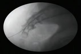
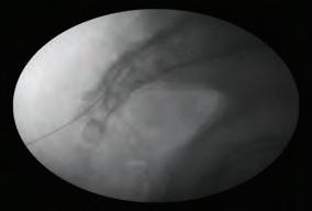
After carefully aspiration with negative presenting blood and CSF, the mixture solution 5 ml with 1% Lidocaine and Dexamethasone, 10-14 mg is injected through the catheter direct to scar tissue. The R-K needle and the Racz catheter were removed as one unit to avoid catheter shear.
Post-Procedure:
The patient is assisted off the table and transferred to the recovery room for 15-20 minutes.
CAUDAL ESI
Indications:
The caudal epidural approach is useful in treating pain syndromes such as lumbar radiculopathy, spinal stenosis, vertebral compression fractures, postherpetic neuralgia, and pelvic syndromes. Given that that caudal approach is safe, simple, requires no LOR and provides minimal pain while also allowing steroids to be administered via this route, it is beneficial to patients with low back surgery that have contraindications. Also because the caudal epidural approach is non-selective, volumes as high as 10-12cc can be used to reach more superior lumbar structures.
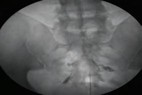
Anatomy:
The five fused sacral vertebrae form the triangular sacrum, which attaches in a wedge-like manner between the two iliac bones. The dorsally convex sacrum lies superior to the fifth lumbar vertebra and caudad to the coccyx. On the anterior concave surface, there are four pairs of unsealed anterior rami of the upper
96 CLINICIANS GUIDE TO OPIOID SPARING PAIN MANAGEMENT
four sacral nerves. The posterior sacral foramina are smaller than their anterior counterparts. The sacrospinal fluids and multifidus muscles effectively prevent leakage of drugs injected into the sacral canal. The sacral cornua are the bony projections which consist of remnants of the inferior articular process that project down the sides of the sacral hiatus. The sacral hiatus is covered with the sacrococcygeal ligament.
Technique:
The caudal approach can be completed by placing the patient in a prone position followed by preparation of the area that will be palpated with an antiseptic solution. A fenestrated sterile drape is placed over the prepped skin followed by the placement of the middle finger of the non-dominant hand at the tip of the coccyx. Once the sacral hiatus is located, the overlying skin and subcutaneous tissues is anesthetized with 1% lidocaine. Using a 20-gauge Touhy needle drive the needle through the skin at a 45-degree angle and pass the sacrococcygeal ligament. As one is driving the needle through the ligament, you will feel a “pop.” Once the needle passes the ligament into the caudal spinal canal, the needle will reduce its angle to the plane of the sacrum and an additional advancement of 1 to 2 cm can be made. Once the needle is in the projected space, AP and lateral views are taken followed by injection of contrast in live fluoroscopy to confirm correct placement of the needle. Once confirmed the steroid with a saline solution in a 20cc is injected through the needle and finally, the needle is removed.

Post-Procedure:
The patient is assisted off the table and transferred to the recovery room for 15-20 minutes.
TRANSFORAMINAL ESI
Indications:
These injections often allow more targeted localization of individual nerve roots either for therapeutic or pure diagnostic purposes by accessing the dorsal root and ganglia. Access via the lateral intervertebral foramen can also lead to the spread of medication into the epidural space.
Transforaminal epidural stared injections can be performed two different ways. The first technique is also called the ‘safe’ triangle technique where the spinal nerve root forms the hypotenuse of the triangle and
97 CHAPTER 10 - LUMBAR SPINE
access is directed in a gun barrel view directly in a Scottie dog view to the “dog tag” of the Scottie dog. The nerve emerges from this location. Despite its name, however, the safe triangle approach is not necessarily “safe” as inadvertent vascular injection is possible.
Physicians often use the Kambin triangle approach. This involves an initial Scottie dog view, walking the needle gently to contact the bone of the superior articular process about midway up. The curved needle is then gently walked laterally until it is walked off of the bone at which point it is turned immediately until the only contact is made again. By repeating this back and forth several times, the needle tip is driven around anterior and in close approximation to the superior articular process avoiding any neurovascular structures.
Anatomy:
The ventral and dorsal roots of the spinal nerves descend in the vertebral canal in their foramina, which faces laterally. The superior articular process of the lower vertebra and inferior articular process of the upper vertebra form the posterior wall. The spinal nerve lies superior and anterior to the foramen, just inferior to the pedicle.
Technique, S1: Lateral and AP views are taken, and the sacral superior end-plate spine is squared and the opening of the S1 foramen identified. A 25 G curved tip spinal needle is advanced through anesthetized in line with the fluoroscope (gun barrel) until sacrum bone is contacted. The needle is then walked off into the foramen through the tactile resistance of the ligament. The fluoroscope is turned laterally, and upon injecting contrast, one can view the nerve root anteriorly.


L1 through L5 location:
Lateral and AP views are taken, and the spine is squared off. The appropriate vertebral body of is located using fluoroscopy. The fluoroscope is maneuvered in a cephalocaudal direction until the caudal border is perpendicular to the camera view. The fluoroscope is then rotated laterally until the pedicle and superior articulating facet is in line. The target is the ‘dog tag” in this Scotty dog view. A skin wheal is raised using lidocaine, and the tissue planes is infiltrated. Acurved spi-
98 CLINICIANS GUIDE TO OPIOID SPARING PAIN MANAGEMENT
nal needle is introduced in line with the fluoroscope until the pedicle is encountered at the six o’clock location. The needle is then walked off into the foramen.
Isovue is injected via the needle and dye flow along the nerve root and towards the epidural space at the same vertebrae level is noted. Real time injection of contrast avoids vascular injection. 0.25% preservative free bupivacaine and Dexamethasone (non-particulate) is injected via the needles. At the end of the injection, the needles are removed.
Post-Procedure:
The patient is assisted off the table and transferred to the recovery room for 15-20 minutes. There may be temporary weakness if the root is anesthetized
99 CHAPTER 10 - LUMBAR SPINE
REFERENCES
Bogduk, N. (2012). Clinical and Radiological Anatomy of the Lumbar Spine. Elsevier - Health Sciences Division.
Narouze, S. N. (2010). Atlas of Ultrasound-Guided Procedures in Interventional Pain Management. Springer New York.
Raj, P. P., Lou, L., & Erdine, S. (2008). Interventional Pain Management: Image-guided Procedures. Saunders/Elsevier.
Rathmell, J. P. (2011). Atlas of Image-Guided Intervention in Regional Anesthesia and Pain Medicine. Wolters Kluwer Health.
Waldman, S. D. (2009). Pain review. Philadelphia, PA: Saunders/Elsevier.
CHAPTER 10 LUMBAR SPINE CODING & BILLING
Amy Turner RN BSN MMHC CPC CHC
There are multiple interventional options that can be performed in the lumbar spine. With all injections please be aware of payer guidelines. Many payers have specific guidelines surrounding medical necessity and frequency.
Lumbar interlaminar epidural injection and caudal epidural injections share the same codes. These codes include imaging guidance. These codes should not be reported in conjunction with 77003, 77012, 76942).
62322 Injection(s), of diagnostic or therapeutic substance(s) (eg, anesthetic, antispasmodic, opioid, steroid, other solution), not including neurolytic substances, including needle or catheter placement, interlaminar epidural or subarachnoid, lumbar or sacral (caudal); without imaging guidance
62321 Injection(s), of diagnostic or therapeutic substance(s) (eg, anesthetic, antispasmodic, opioid, steroid, other solution), not including neurolytic substances, including needle or catheter placement, interlaminar epidural or subarachnoid, lumbar or sacral (caudal); with imaging guidance (ie, fluoroscopy or CT)
Notes: (Do not report 62323 in conjunction with 77003, 77012, 76942)
Lumbar transforaminal epidural injections provide an alternative ap-
100 CLINICIANS GUIDE TO OPIOID SPARING PAIN MANAGEMENT
proach to delivering medication to the epidural space and are billed using a separate set of codes. Lumbar transforaminal injections reported bilaterally may be billed with modifier 50. When performing unilateral injections, adding a RT or LT modifier is recommended to designate laterality. Imaging guidance by fluoroscopy or CT is included with these codes. If using ultrasound guidance, please refer to codes 0230T and 0231T.
64483 Injection(s), anesthetic agent and/or steroid, transforaminal epidural, with imaging guidance (fluoroscopy or CT); lumbar or sacral, single level
Notes: (For transforaminal epidural injection under ultrasound guidance, use 0230T)
64484 Injection(s), anesthetic agent and/or steroid, transforaminal epidural, with imaging guidance (fluoroscopy or CT); lumbar or sacral, each additional level (List separately in addition to code for primary procedure)
Notes: (Use 64484 in conjunction with 64483) This is an add on code and does not need a modifier.
Lumbar medial branch blocks and intraarticular facet joint injections share the same codes. Please remember that MBB are billed per level not per medial branch blocked. Example: Lumbar medial branch block reported at right L4 and right L5 = 1 level (L5-S1). Medial Branch blocks and facet joint injections may be performed bilaterally by adding modifier 50. If performing unilateral injections addition of the LT and RT modifier to indicate laterality is recommended.
If using ultrasound guidance refer to 0213T-0218T.
64493Injection(s), diagnostic or therapeutic agent, paravertebral facet (zygapophyseal) joint (or nerves innervating that joint) with image guidance (fluoroscopy or CT), lumbar or sacral; single level
64494Injection(s), diagnostic or therapeutic agent, paravertebral facet (zygapophyseal) joint (or nerves innervating that joint) with image guidance (fluoroscopy or CT), lumbar or sacral; second level (List separately in addition to code for primary procedure)
Notes:
(Use 64494 in conjunction with 64493) This is an add on code and no modifier is required.
101 CHAPTER 10 - LUMBAR SPINE
64495Injection(s), diagnostic or therapeutic agent, paravertebral facet (zygapophyseal) joint (or nerves innervating that joint) with image guidance (fluoroscopy or CT), lumbar or sacral; third and any additional level(s) (List separately in addition to code for primary procedure)
Notes:
(Do not report 64495 more than once per day) (Use 64495 in conjunction with 64493, 64494) This is an add on code and no modifier is required.
Lumbar radiofrequency levels are counted per level not per medial branch destroyed. Example: Lumbar medial branch radiofrequency right L4 and right L5 = 1 level (L5-S1). Medial branch radiofrequency may be reported using modifier 50. If performing unilateral injections addition of the LT and RT modifier to indicate laterality is recommended. These codes have a 10-day global period. CPT states- “Do not report 64635 or 64636 for non-thermal facet joint denervation including chemical low-grade energy, (<80 degrees Celsius), or any form of pulsed radiofrequency. To appropriately report any of these modalities, use 64999.”
64635Destruction by neurolytic agent, paravertebral facet joint nerve(s), with imaging guidance (fluoroscopy or CT): lumbar or sacral, single facet joint
64636Destruction by neurolytic agent, paravertebral facet joint nerve(s), with imaging guidance (fluoroscopy or CT): lumbar or sacral, each additional facet joint (list separately in addition to code for primary procedure) This is an add on code and no modifier is required.
Sacroiliac joint injections are also frequently performed in pain management offices. If performing bilateral SI joint injections, modifier 50 may be reported. If performing unilateral injections, addition of the LT and RT modifier to indicate laterality is recommended. CPT code 27096 required the usage of fluoroscopy or CT guidance. If performing SI Joint injection without fluoroscopy or CT, CPT code 20552 should be reported.
102 CLINICIANS GUIDE TO OPIOID SPARING PAIN MANAGEMENT
27096Injection procedure for sacroiliac joint, anesthetic/steroid, with image guidance (fluoroscopy or CT) including arthrography when performed
Notes:
(27096 is to be used only with CT or fluoroscopic imaging confirmation of intra-articular needle positioning) (If CT or fluoroscopy imaging is not performed, use 20552)
Sacroiliac joint radiofrequency can be accomplished with multiple techniques. Unfortunately, many payers consider radiofrequency of the SI joint investigational/experimental and is not a covered benefit. Be aware of individual payer policies surrounding this therapy.
If performing destruction of the lateral branches of S1, S2, S3, S4 CPT Assistant, June 2012; volume 22: Issue 6 states “When performing individually separate nerve destruction, each peripheral nerve root neurolytic block is reported as destruction of a peripheral nerve, using code 64640, Destruction by neurolytic agent; other peripheral nerve or branch. In this instance, for peripheral nerve root neurolytic blocks (destruction) of L5, S1, S2, and S3, code 64640 should be reported four times. The coder should append modifier 59, Distinct Procedural Service, to the second and subsequent listings of code 64640 to separately identify these procedures.”
For use of the Simplicity probe- CPT Assistant, December 2009; Volume 19: Issue 12 states-“ insertion of a single electrode (having three contacts) at the sacroiliac (SI) joint “to lesion the lateral branches of S1, S2, S3, and S4,” code 64999, Unlisted procedure, nervous system, is reported once. This “SI joint rhizotomy” would be reported once using the unlisted nervous system code 64999.
Lumbar sympathetic blocks may be billed bilaterally with the use of modifier 50. If performing unilateral injections, addition of the LT and RT modifier to indicate laterality is recommended. This procedure has a medically unlikely edit of 1 meaning that payers only expect to see this code billed once per day.
64520Injection, anesthetic agent; lumbar or thoracic (paravertebral sympathetic)
Fluoroscopic guidance may be billed separately- 77003.
Ganglion impar block should be reported as an unlisted procedure, 64999. This question was asked in CPT assistant, September 2007; volume 17: issue 9-
103 CHAPTER 10 - LUMBAR SPINE
“Question: What is the appropriate CPT code to report for a ganglion impar sympathetic block?
AMA Comment: Code 64999, Unlisted procedure, nervous system, should be reported. When reporting an unlisted code to describe a procedure or service, it will be necessary to submit supporting documentation (eg, a procedure report) along with the claim to provide an adequate description of the nature, extent, and need for the procedure and the time, effort, and equipment necessary to provide the service.”
Epidural lysis of adhesions may be reported with one of two CPT codes, 62263 or 62264. These codes are inclusive of imaging guidance (72275, epidurography and 77003, fluoroscopy) and administration of contrast. Both codes require multiple adhesiolysis sessions be performed.
62263 Percutaneous lysis of epidural adhesions using solution injection (eg, hypertonic saline, enzyme) or mechanical means (eg, catheter) including radiologic localization (includes contrast when administered), multiple adhesiolysis sessions; 2 or more days
64636Percutaneous lysis of epidural adhesions using solution injection (eg, hypertonic saline, enzyme) or mechanical means (eg, catheter) including radiologic localization (includes contrast when administered), multiple adhesiolysis sessions; 1 day
104 CLINICIANS GUIDE TO OPIOID SPARING PAIN MANAGEMENT
CHAPTER 11
RADIOFREQUENCY ABLATION. ALEXANDER E. WEINGARTEN MD, BENJAMIN PORTAL MD
General Description:
Radiofrequency ablation (RFA) is a minimally invasive procedure that uses heat to interrupt pain signals in spinal nerves carrying pain signals to the brain. It can provide lasting relief for people with chronic pain, especially in the lower back, neck and arthritic joints.
During the procedure, a high-frequency electrical current runs through an insulated needle with the precise placement of an RF electrode probe on a specific target nerve at the tip of the needle, the electric field causes molecule movement which, in turn, produces thermal energy. With only 90 to 150 seconds of treatment, the RF current alters the adjacent neural environment via thermal and electromagnetic-induced cellular changes. The heat from the tip of the RFA device is targeted to create a small lesion within a nerve, which disrupts the pain signal.
The mechanism of the destruction is charged particles in the tissue such as free ions and proteins succumb to ionic agitation and friction from the high-frequency alternating current. The oscillation of charged molecules generates heat and ultimately results in protein denaturation, cellular membrane disruption, and increased membrane permeability, a neuro destructive process resulting in thermocoagulation. [3] Most practitioners today have adopted target temperatures at about 80°C, well above the neurodestructive threshold but below 95° to 100°C, which produces unwanted sequelae such as charring, tissue adherence to the probe, hematoma, gas formation, and extended damage to adjacent structures.
Randomized control trials have shown Conventional radiofrequency denervation resulted in significant reductions in low back pain originating from the facet joints in patients showing the best response to diagnostic block over the first 12 months when compared with sham procedures or epidural nerve blocks. [2]
Indications and Contraindications:
Over the subsequent decades, RFA has become a widespread and effective treatment to create significant and sustained pain relief. Current and expanding clinical applications of RFA include facial; cervical, thoracic, and lumbar facet; spinal radicular; sacroiliac joint (SIJ); lumbar discogenic; peripheral nerve; intraarticular joint; and sympathetically mediated pain. In addition, other RFA sites include Dorsal Root Ganglion (herniated discs or regional pain syndromes), Splanchnic nerve and the Gasserian ganglion for trigeminal neuralgia.
105 CHAPTER 11 - RADIOFREQUENCY ABLATION
The bulk of clinical data involves conventional RFA; however, over the past decade, modified forms of RF treatments have emerged with four forms of RFA predominate in clinical use: conventional (i.e. continuous) RFA (CRF), pulsed RFA (PRF), water-cooled RFA (WCRF), and bipolar RFA (BRF).
A range of contraindications for RF procedures exist, some of which require clinical judgment. Clear contraindications include coagulopathies, therapeutic anticoagulation, ongoing sepsis, or the presence of nearby invasive lesion such as tumor or infection. Relative contraindications include inadequately treated psychiatric conditions, psychopathology, previous failed RFA to the same target nerve, unrealistic expectations, and associated neuropathic or deafferentation pain syndromes. Anatomic abnormalities may also present a contraindication, especially with the presence of prior surgical instrumentation. Additionally, patients with a pacemaker or spinal cord stimulator must be carefully monitored because of the risk of interaction with the RF equipment. As a consequence of RF lesioning, sensing pacemakers may mistakenly interpret RF signal as intrinsic atrial activity and fail to pace, which would result in asystole for pacemaker-dependent patients.
Equipment and Technique:
To perform RF treatment safely and effectively, an RF generator should also be capable of nerve stimulation and monitoring of electrode temperature, impedance, voltage, and lesion time. Temperature monitoring is accomplished by a thermocouple sensor at the tip of the electrode. For CRF, most practitioners raise the temperature 1°C/sec. Raising the temperature too quickly risks cavitation and unpredictably shaped lesions. Nerve stimulation helps determine electrode to nerve distance. Sensory testing confirms that one is close enough to the target nerve, and motor testing ensures one is far enough away from any important motor innervation. To test sensory stimulation, 50 Hz of electrical current should produce pain or tingling with less than 0.5 to 0.6 V. For motor stimulation, electrical current is set at 2 Hz, and muscle contractions are observed with voltages greater than two to three times the sensory voltage threshold. An impedance monitor is useful for detecting entry into various mediums. During any RF treatment procedure, impedance should remain relatively constant. A changing or abnormal impedance can provide a differential for the culprit clinical situation. For example, a large increase for example might suggest movement from fluid to tissue.
RF electrodes produce little lesion distal to their tip and coagulate transversely. The most reliable coagulation is done if the electrodes are placed parallel to the nerve. Radiological confirmation of electrode placement is essential. When the electrode is placed on the patient’s
106 CLINICIANS GUIDE TO OPIOID SPARING PAIN MANAGEMENT
body, a circuit is complete. An electric field is established around the electrode tip. This field oscillates with alternating RF current causing movement of ions in the tissue. This causes friction in tissue surrounding the catheter tip which produces heat. RF current is a low energy, high frequency (100,000-500,000 hz). [4]
107 CHAPTER 11 - RADIOFREQUENCY ABLATION
REFERENCES
1.Leggett LE, Soril LJ, Lorenzetti DL, et al. Radiofrequency ablation for chronic low back pain: A systematic review of randomized controlled trials. Pain Research & Management: The Journal of the Canadian Pain Society. 2014;19(5):e146-e153.
2. The efficacy of conventional radiofrequency denervation in patients with chronic low back pain originating from the facet joints: a meta-analysis of randomized controlled trials Lee, Chang-Hyun et al.The Spine Journal , Volume 17 , Issue 11 , 1770 - 1780
3. Sluijtel ME, Teixeira A, van Duijn B. Comment on: Erdine S et al.; Ultrastructural changes in axons following exposure to pulsed radiofrequency fields. Pain Pract. 2010;10(3):262; author reply 262–263.
4. Principles and Practice of Pain Medicine, 3e Bajwa ZH, Wootton R, Warfield CA. Bajwa Z.H., Wootton R, Warfield C.A. Eds. Zahid H. Bajwa, et al. New York, NY: McGraw-Hill, ,
RADIOFREQUENCY ABLATION LUMBAR MEDIAN BRANCH NERVES
Facet joint pain accounts for an estimated 15-45% of the causes of lower back pain. Conservative treatments include oral medications, physical therapy, trigger point injections, and medial branch blocks. Medial branch nerve blocks serve both a diagnostic and therapeutic modality, which help confirm the diagnosis of a facet joint etiology of the lower back pain. Radiofrequency ablation is used for treatment of facet joint arthropathy as an etiology for the cause of low back pain following confirmatory diagnosis with median branch nerve blocks. This procedure is performed using fluoroscopic guidance. The patient usually presents with axial low back pain with occasional radiation to the buttocks and lower extremities. Radiofrequency involves the use of thermal energy to ablate the median branch nerves and create facet denervation thereby relieving the pain.
Radiofrequency Denervation in the Lumbar Region
Technique:
The radiofrequency ablation is commonly used for treatment of lumbar medial branch neuropathy/facet arthropathy. The medial branch of the posterior ramus of the spinal nerves supplies the facet joints at its own level of the lumbar spine as well as the next lower level of the lumbar spine. In the reviewing the anatomy of the medial lumbar branch one notes that it runs in a dorsal and caudal direction and lies in the groove on the base of the superior articular process in direct contact with the base of the superior surface of the transverse process. In addition, each medial branch provides nerve supply to several lumbar spinal muscles including the multifidus as well as ligaments
108 CLINICIANS GUIDE TO OPIOID SPARING PAIN MANAGEMENT
and periosteum of the vertebral column. In proceeding with the radiofrequency ablation of the lumbar spine the practitioner needs verbal feedback from the patient during this stimulation aspect of the procedure and heating portion. Heavy sedation is therefore not employed as the patient needs to be awake during the procedure.
The equipment acquired for radiofrequency ablation includes a 10 cm 22-gauge radiofrequency needle with a 5-mm active tip. In addition, a radiofrequency probe, which is compatible to this needle is required, which connects to the radiofrequency generator. In performing a radiofrequency ablation of the lumbar medial branch nerves, the patient is placed in a prone position on a fluoroscopy table. A pillow is placed under the abdomen to reduce the normal lumbar lordosis. The targeted levels are noted and are identified via use of a C arm fluoroscopy unit using an AP view. The C-arm is subsequently rotated to approximately a 15-degree oblique view and until the facet joint is identified. The entry point is identified as the junction of the superior articular process and transverse process. Local anesthetic using 1% lidocaine is injected subcutaneously and the radiofrequency needle in inserted until it makes contact with the bone in the area noted above. Position of the needle is confirmed by fluoroscopy. The needle tip should be in the region of the superior medial aspect of the transverse process at the level to be ablated. A sensory stimulation test is performed at 50 Hz and generally should be positive between 0.3 and 0.6 volts. The patient should feel either a buzzing or pressure sensation in the area of the needle. During the sensory stimulation test if sensations are felt down the ipsilateral extremity, one needs to reposition the needle as it is too close to the segmental nerve at that level. Following repositioning, the sensory stimulation test is then repeated at 50 Hz. This is followed by a motor stimulation test, which is performed at 2 Hz and at 3 volts following a successful sensory stimulation test. There should be no motor stimulation noted in the ipsilateral lower extremity at the settings noted above. During the motor stimulation test if there are contractions occurring in the lower extremity, the needle again must be repositioned since it is too close to a motor nerve. Following successful completion of the sensory and motor stimulation test and following a negative aspiration, 1 cc of lidocaine 1% is injected through the radiofrequency needle prior to the start of radiofrequency ablation. The recommended settings for the ablation are 80 degrees centigrade for 90 seconds. Of note, the L5-S1 facet joint i.e. the medial branch of the L5 nerve is somewhat different than the other lumbar levels due to a change in the anatomy. The L5 medial branch nerve is located at the junction between the superior sacral articular process and the upper border of the sacrum. There is no pedicle located at this level. The C-arm is used and is rotated obliquely for about 15 degrees
109 CHAPTER 11 - RADIOFREQUENCY ABLATION
until the junction between the superior sacral articular process and the upper border of the sacrum is identified. Local anesthetic is then administered at this point and the needle is then inserted until its tip reaches the junction noted above. Following positioning of this needle, a sensory and motor stimulation test is performed prior to the start of radiofrequency ablation.
Equipment:
The needles generally used for this procedure include a 20- or 22-gauge needle with either a 5 mm or 10 mm active tip. A radiofrequency probe, which is sterilized, is inserted through the radiofrequency cannula once the cannula is positioned in its proper location as described above. The probe is connected to a radiofrequency generator using the settings noted above. Following the procedure, Band-Aids are applied to the affected areas and the patient can be discharged immediately assuming that no sedation has been used. With some patients there will be some temporary numbness in the ipsilateral extremity because of the effect of the local anesthetics used during the procedure, which can diffuse and affect the sensory nerves of the lower extremities. Should that be the case, the patient should be kept in the surgical facility until the numbness wears off and the patient should be told that he or she may experience soreness in the anatomical region that the procedure was performed as well as some burning pain in the lower extremity lasting up to two weeks. These side effects generally will resolve in less than two weeks. Additional side effects include infection, bleeding (localized), and muscle spasms.
The same procedure described above can be performed in the region of the cervical medial branch nerves, the stellate ganglion, thoracic sympathetic nerves, as well as lumbar sympathetic nerves. There are shorter needles to be used in the cervical region including a 4-6 cm length radiofrequency needle. The settings on the generator are generally the same and the procedure is performed in a similar fashion in these regions.
Procedure:
The patient is kept awake for this procedure since continuous oral contact with the patient is necessary. Therefore, minimal sedation if any is used. The prone position is used on the fluoroscopic table. A pillow is placed under the abdomen and all pressure points are checked and padded if necessary. The pertinent lumbar vertebral levels are marked, and an anterior posterior fluoroscopic film is obtained. The location for the entry zone at each vertebral level is marked over the junction between the transverse process and the superior articular process. Local anesthetic is infiltrated under the skin and a radiofrequency needle is subsequently inserted and advanced until the needle tip makes con-
110 CLINICIANS GUIDE TO OPIOID SPARING PAIN MANAGEMENT
tact with the bony landmarks noted above. The radiofrequency needle is then connected to the radiofrequency probe after final positioning of the needle. A sensory stimulation test is then performed at 50 Hz and the patient should feel either tingling or pressure in the back at a voltage level of less than 0.5 volts. If the patient complains of radicular pain in the ipsilateral extremity the needle tip is moved slightly, and the sensory stimulation test is performed again to obtain the parameter noted above. Prior to the start of radiofrequency ablation, a motor stimulation test is performed at 2 Hz and 3 volts. The patient should not experience any muscle contractions in the ipsilateral buttock or leg. If contractures are noted in the ipsilateral extremity the needle must be repositioned. Following final position of needle 1 cc of 1% lidocaine is injected through the radiofrequency needle prior to the start of the radiofrequency lesioning. The radiofrequency lesioning is generally performed at 80 degrees centigrade for 60 to 90 seconds. Depending upon which machine is used up to three levels may be lesioned at the same time. Following the procedure, the patient may complain of transient numbness in the ipsilateral extremity due to local anesthesia entering the intervertebral foramen on the ipsilateral side. This side effect is usually short lived and resolves fairly quickly.
In addition, the patient may complain of burning pain in the lower extremities or localized pain at the site of the injection, which usually resolves in about two weeks. Other side effects include infection, backache, and muscle spasm
111 CHAPTER 11 - RADIOFREQUENCY ABLATION
REFERENCES
1. Uematsu S. Percutaneous electrothermocoagulation of spinal nerve trunk, ganglion and rootlets. In: Schmidel HH, ed. Current technique in operative neurosurgery. New York: Grune and Stratton; 1977:469-490.
2. Sluijter M, Van Kleef M. Characteristics and mode of action of radiofrequency lesion. Cur Rev Pain 1998; 2:143-150.
3. Van Kleef M, Sluijter M. Radiofrequency lesions in the treatment of pain of spinal origin. In: Gildenberg P, Tasker R, eds. Textbook of stereotactic and functional neurosurgery. New York: The McGraw-Hill; 1998:1585-1599
4. Bogduk N, Long DM. The anatomy of the so-called ‘articular nerves’ and their relationship to facet denervation in the treatment of lowback pain. J Nuerosurg 1979;51(2): 172-177.
5. Gray D, Bajwa Z, Warfield C. Facet block and neurolysis. In: Waldman SLR, Ross A, et al, eds. Interventional pain management. Philadelphia: WB Saunders; 2001:446-483.
6.Kornick G, Kramarich S, Lamer T, et al. Complications of lumbar facet radiofrequency denervation. Spine 2004; 29(12):1352-1354.
CHAPTER 11 RADIOFREQUENCY ABLATION CODING & BILLING
Amy Turner RN BSN MMHC CPC CHC
Radiofrequency of the medial branches are reported per level not per medial branch destroyed.
• Cervical medial branch radiofrequency performed at right C4 and right C5 = 1 level (C4-5)
• Thoracic medial branch block performed at right T 7 and right T8 = 1 level (T8-9)
• Lumbar medial branch block reported at right L4 and right L5 = 1 level (L5-S1)
Medial branch radiofrequency may be reported using modifier 50. If performing unilateral injections, addition of the LT and RT modifier to indicate laterality is recommended. These codes have a 10-day global period. Fluoroscopic or CT guidance is included in these codes.
64633Destruction by neurolytic agent, paravertebral facet joint nerve(s), with imaging guidance (fluoroscopy or CT): cervical or thoracic, single facet joint
112 CLINICIANS GUIDE TO OPIOID SPARING PAIN MANAGEMENT
64634Destruction by neurolytic agent, paravertebral facet joint nerve(s), with imaging guidance (fluoroscopy or CT): cervical or thoracic, each additional facet joint (list separately in addition to code for primary procedure) This is an add on code and no modifier is required.
64635Destruction by neurolytic agent, paravertebral facet joint nerve(s), with imaging guidance (fluoroscopy or CT): lumbar or sacral, single facet joint
64636Destruction by neurolytic agent, paravertebral facet joint nerve(s), with imaging guidance (fluoroscopy or CT): lumbar or sacral, each additional facet joint (list separately in addition to code for primary procedure) This is an add on code and no modifier is required.
Example: Radiofrequency ablation is performed at the right L2, L3, and L4 medial branches. Correct reporting of this procedure would be:
64635 x 1 RT
64636 x 1 RT
Even though three needles are placed, correct coding is per level/joint denervated.
CPT states- “Do not report 64635 or 64636 for non-thermal facet joint denervation including chemical low-grade energy, (<80 degrees Celsius), or any form of pulsed radiofrequency. To appropriately report any of these modalities, use 64999.”
Sacroiliac joint radiofrequency can be accomplished with multiple techniques. Unfortunately, many payers consider radiofrequency of the SI joint investigational/experimental and is not a covered benefit. Be aware of individual payer policies surrounding this therapy.
If performing destruction of the lateral branches of S1, S2, S3, S4 CPT Assistant, June 2012; volume 22: Issue 6 states “When performing individually separate nerve destruction, each peripheral nerve root neurolytic block is reported as destruction of a peripheral nerve, using code 64640, Destruction by neurolytic agent; other peripheral nerve or branch. In this instance, for peripheral nerve root neurolytic blocks (destruction) of L5, S1, S2, and S3, code 64640 should be reported four times. The coder should append modifier 59, Distinct Procedural Service, to the second and subsequent listings of code 64640 to separately identify these procedures.”
113 CHAPTER 11 - RADIOFREQUENCY ABLATION
For use of the Simplicity probe- CPT Assistant, December 2009; Volume 19: Issue 12 states-“ insertion of a single electrode (having three contacts) at the sacroiliac (SI) joint “to lesion the lateral branches of S1, S2, S3, and S4,” code 64999, Unlisted procedure, nervous system, is reported once. This “SI joint rhizotomy” would be reported once using the unlisted nervous system code 64999.
114 CLINICIANS GUIDE TO OPIOID SPARING PAIN MANAGEMENT
CHAPTER 12 SYMPATHETIC BLOCK
Richard A. Gasalberti MD, Isaac J. Kreizman MD, Aziz Abdurakhimov MD, Marina Kokova MD,
1. Stellate Ganglion Block
Cervical sympathetic block results in interruption of the sympathetic efferent fibers to the upper extremity, head, and neck. It can provide both therapeutic and diagnostic values in patients with a significant sympathetically maintained component to the pain syndrome.
INDICATIONS
Indicated in a variety of disorders related to sympathetic innervation of the head, neck, and upper extremities. Most common are:
• Complex regional pain syndrome 1 and 2
• Acute Herpes zoster (shingles)
• Postherpetic neuralgia (early)
• atypical facial pain
• Bell palsy
• postamputation stump pain
• Neuropathic pain associated with CNS pathology
• Phantom limb pain
• acute peripheral ischemia
• vasospasm
• arteritis
• frostbite
• erythromelalgia
• Raynaud disease
Other less common indications include:
• Hyperhidrosis
• Ménière disease,
• accidental intraarterial injection of intravenous medications
• angina pectoris
More recently, stellate ganglion block has been used for the treatment of patients with hot flashes and posttraumatic stress disorders. 14, 15
CONTRAINDICATIONS
Absolute contraindications: coagulopathy, contralateral pneumothorax, and recent myocardial infarction. Relative contraindications include glaucoma and atrioventricular block are
115 CHAPTER 12 - SYMPATHETIC BLOCK
COMPLICATIONS
When performed properly by an experienced practitioner, the stellate ganglion block has a low incidence of complications. The complications of stellate ganglion block result either from insertion and manipulation of the needle or as a direct result of the injected solution. They may include:
• Horner syndrome (ptosis, miosis, anhidrosis, enophthalm, and nasal congestion)
• RLN block
• Vagus nerve block
• Phrenic nerve block
• Partial brachial plexus block
• Esophageal penetration, mediastinal infection, or emphysema
• Discitis
Most serious are the following:
• Intravascular injection
• Subarachnoid injection
• Pneumothorax
• Retropharyngeal hematoma
2. Thoracic (T2-T3) Ganglion Block
The anterior approaches to the sympathetic trunk in the neck (stellate ganglion) are easily performed and are most commonly used; however, a posterior approach to the sympathetic trunk at the T2, T3 level provides the most reliable and consistent block of sympathetic activity to the upper extremity.
INDICATIONS
• Complex regional pain syndrome 1 and 2
• Herpes zoster (shingles)
• Postherpetic neuralgia (early)
• Postradiation arteritis
• Neuropathic pain associated with CNS pathology
• Phantom limb pain
CONTRAINDICATIONS
Many techniques have been described, but because of the risk of pneumothorax, the posterior approach is generally restricted to patients in whom fascial barriers or anatomic anomalies may prevent diffusion of local anesthetic from its anterior deposition at C6/C7 to the upper thoracic sympathetic trunk or when precise neurolytic procedures are planned.
116 CLINICIANS GUIDE TO OPIOID SPARING PAIN MANAGEMENT
COMPLICATIONS
• Procedure related general complications like infection, bleeding
• Pneumothorax—patient should be monitored for late onset of pneumothorax
• Epidural and/or intrathecal injection, and neurological deficit
• Intercostal neuritis
3.Splanchnic Nerve Blocks
The greater, lesser, and least splanchnic nerves are the major preganglionic of the celiac plexus.
INDICATIONS
• Intra-abdominal cancer
• Chronic abdominal pain (including chronic pancreatitis pain)
• Differential diagnosis of somatic versus visceral pain
• Treatment of patients who have failed to obtain relief from celiac plexus blocks
• Palliation of the acute pain of arterial embolization of the liver for cancer therapy
• Treatment of pain of abdominal “angina” associated with visceral arterial insufficiency
CONTRAINDICATIONS
• Local infection
• Sepsis
• Coagulopathy
• Respiratory insufficiency or pleural adhesions (due to risk of pneumothorax)
• Tumors that distort the relevant anatomy
• An abdominal or thoracic aneurism
COMPLICATIONS
Because this is a sympathetic block, there is a potential for hypotension, and therefore IV access is necessary. Without fluid prophylaxis (500-1000 cc of balanced salt solution), clinically significant hypotension can be expected in 30% to 60% of patients. Patients also need to be warned regarding
• orthostatic hypotension
• and diarrhea as potential side effects.
• Pneumothorax (watch for shortness of breath or chest pain)
• If on the left side, chylothorax is a possibility, due to trauma of the thoracic duct
117 CHAPTER 12 - SYMPATHETIC BLOCK
4. Lumbar sympathetic block
The lumbar sympathetic block results in interruption of the sympathetic efferent fibers to the lower extremities with sparing of the somatic nerves. This provides diagnostic value as to the relative sympathetic contribution to the patient’s pain syndrome. It may also provide therapeutic value in those patients with a significant sympathetically maintained component to their pain.
INDICATIONS
The most common indications:
• CRPS types I (reflex sympathetic dystrophy) and II (causalgia)
• acute herpes zoster
• early postherpetic neuralgia
• postamputation stump pain
• phantom limb pain
• radiation neuritis
• peripheral neuropathy
Blockade of the lumbar sympathetics is also used in conditions with limited blood flow within the small vessels of the lower extremities:
• acute ischemia
• atherosclerosis
• Frostbite
• Erythromelalgia
• Raynaud disease
• Buerger disease
Other less commonly encountered indications:
• Hyperhidrosis
• phlegmasia alba dolens
• Acrocyanosis
• discogenic pain
• accidental intraarterial injection of intravenous medications. 14-17
CONTRAINDICATIONS
• Patient cannot discontinue their anticoagulant medication
• History of bleeding diathesis
• Active infection near the injection site
COMPLICATIONS
The known complications are:
• Intravascular injection
• Subarachnoid injection
118 CLINICIANS GUIDE TO OPIOID SPARING PAIN MANAGEMENT
• Renal trauma
• Ureteral stricture
• Lumbar plexus blockade
• Segmental nerve injury
• Infection
• Ejaculatory failure (bilateral lumbar sympathetic block)
• Discitis
• Psoas necrosis
• Genitofemoral neuralgia
119 CHAPTER 12 - SYMPATHETIC BLOCK
REFERENCES
[1] Garcea G, Thomasset S, Berry DP, Tordoff S. Percutaneous splanchnic nerve radiofrequency ablation for chronic abdominal pain. ANZ J Surg. 2005 Aug;75(8):640–644.
[2] Erdine S, Talu GK. Sympathetic blocks of the thorax. In: Raj PP, Lou L, Erdine S, Staats P, eds. Interventional Pain Management: Image-Guided Procedures. Philadelphia, PA: WB Saunders; 2008:255–265.
[3] Gest TR, Hildebrandt S. The pattern of the thoracic splanchnic nerves as they pass through the diaphragm. Clin Anat. 2009 Oct;22(7):809–814.
[4] Boswell MV, Trescot AM, Datta S et al. Interventional techniques: evidenced-based practice guidelines in the management of chronic spinal pain. Pain Physician. 2007;10:7–111.
[5] Cepeda MS, Carr DB, Lau J. Local anaesthetic sympathetic blockade for complex regional pain syndrome. Cochrane Systematic Rev. 2005:4: art. no. CD004598.
[6] Furlan AD, Lui PW, Mailis A. Chemical sympathectomy for neuropathic pain: does it work? Case report and systematic literature review. Clin J Pain. 2001;17(4):327–336.
[8] Prager J, Aprill C. Complications related to sedation and anesthesia for interventional pain therapies. Pain Med. 2008;9(S1):S121–S127.
[9] Stanton-Hicks MD, Burton AW, Bruehl SP et al. An updated interdisciplinary clinical pathway for CRPS: report of an expert panel. Pain Pract. 2002;2:1–16.
[10] Mercadante S, La Rosa S, Villari P. CT-guided neurolytic splanchnic nerve block by an anterior approach. J Pain Symptom Manage. 2002 Apr;23(4):268–270.
[11] Mercadante S, Nicosia F. Celiac plexus block: a reappraisal. Reg Anesth Pain Med. 1998 Jan-Feb;23(1):37–48.
[12] Moore D. Celiac (splanchnic) plexus block with alcohol for cancer pain of the upper intra-abdominal viscera. In: Bonica J, Ventafridda V, eds. Advances in Pain Research and Therapy. New York: Raven; 1979:357.
[13] Lieberman R, Lieberman S, Cuka D. Celiac plexus block and splanchnic nerve block: A review. Semin Intervent Radiol. 1988;5:213.
14. Mekhail N, and Malak O: Lumbar sympathetic blockade. Tech Reg Anesth Pain Manage 2001; 5: pp. 99-101 View In Article | Cross Ref
120 CLINICIANS GUIDE TO OPIOID SPARING PAIN MANAGEMENT
15. Boas RA: Sympathetic nerve blocks: in search of a role. Reg Anesth Pain Med 1998; 23: pp. 292-305
View In Article
16. Kosharskyy B, and Rozen D: Lumbar discogenic pain. Disk degeneration and minimally invasive interventional therapies [German]. Anasthesiol Intensivmed Notfallmed Schmerzther 2007; 42: pp. 262-267
View In Article | Cross Ref
CHAPTER 12-SYMPATHETIC BLOCK CODING & BILLING
Amy Turner RN BSN MMHC CPC CHC
Treatment for complex regional pain syndrome in the upper and lower extremities frequently includes sympathetic blockade. Lumbar sympathetic blocks and stellate ganglion blocks may be billed bilaterally with the use of modifier 50. If performing unilateral injections, addition of the LT and RT modifier to indicate laterality is recommended. This procedure has a medically unlikely edit of 1 meaning that payers only expect to see this code billed once per day. These code does not include fluoroscopic guidance and may be reported separately.
Blockade of the celiac plexus and neurolysis of the celiac plexus have imaging guidance included in the code description.
Sphenopalatine block performed using a device to deliver medication through the nose should be reported using code 64999, Unlisted procedure, nervous system. This is documented in CPT Assistant, July 2014; Volume 24: Issue 7.
Sympathetic BlocksCodeImagingNotes
Celiac Plexus/Splanchnic64530bundled
Celiac Plexus Neurolysis64680bundledTherapeutic medications included
121
12 - SYMPATHETIC BLOCK
CHAPTER
Lumbar Sympathetic Block 6452077003Use modifier 50 for Bilateral Stellate Ganglion 6451077003
122 CLINICIANS GUIDE TO OPIOID SPARING PAIN MANAGEMENT
CHAPTER 13 SPINAL CORD STIMULATION
Richard A. Gasalberti MD, Isaac J. Kreizman MD, Aziz Abdurakhimov MD, Michael Garbulsky PA
Spinal cord stimulation (SCS) is usually used as an electro-modulator for neuropathic and sympathetically mediated chronic pain. SCS involves percutaneous or surgical implantation of electrodes in the epidural space, with power supplied by an implanted battery. The mechanism of action of SCS has not been fully understood and is probably influences multiple components and levels within the CNS with both interneuron and neurochemical mechanisms. The gate control theory of pain was published by Ron Melzack and Patrick Wall in 1965, which stated that pain is transmitted by transmission cells in the dorsal horns. The theory holds that transmission cells are stimulated by small fibers and inhibited by large fibers acting through the substantia gelatinosa.
Stimurator
Spinal cord stimulators consist of a cylindrical or paddle leads/electrodes, an implantable pulse generator which delivers electrical stimulation that can be modified to achieve an effect, and an extension cable that connects the lead with the generator.

Trial screening kit consists of a Tuohy needle, loss of resistance (LOR) syringe, guidewire, trial screening lead and anchors.
Clinical use, indications and contraindications
The US Food and Drug Administration (FDA) has approved SCS for several indications, the most common of which are complex regional pain syndrome, failed back surgery syndrome (FBSS), chronic peripheral neuropathy or plexopathy, painful peripheral vascular disease.
123 CHAPTER 13 - SPINAL CORD STIMULATION
Also, SCS may be considered for patients with chronic, neuropathic pain for whom standard pain treatments have failed and when there is no indication for surgical intervention to treat the underlying condition.
Other indications include postherpetic neuralgia, multiple sclerosis pain, phantom limb pain, intercostal neuralgia, and spinal cord injury with variable motor and sensory deficits. For patients with cardiac pacemakers and defibrillators, compatibility with SCS should be established prior to trial and ensure stimulation does not result in defibrillator discharge. Prior to surgery all patients undergo psychiatric and pain psychologic evaluation.
General considerations in the selection of SCS for a particular patient are as follows [1]:
• The patient has a diagnosis amenable to this therapy,
• The patient has failed conservative therapy for at least 6 months,
• All significant psychological issues have been ruled out,
• There is no serious untreated drug dependence problem,
• A trial has demonstrated pain relief.
Absolute contraindications to SCS are sepsis, coagulopathy, local inflammation at the site of implantation, obliterated spinal canal due to previous surgery or trauma. Also, contraindications may include prior surgery with epidural scarring, severe stenosis or scoliosis, or spine instability. An immunocompromised condition is a relative contraindication to SCS.
SCS placement
Spinal level for lead placement depends on the location of pain. An electrical current is applied to the dorsal columns, creating paresthesia in the dermatomes whose afferent fibers traverse the regions being stimulated. Adequate extensive training of physician and also an understanding of neuroanatomy, surgical technique, and perioperative patient care are mandated. Traditional SCS devices are efficient of delivering pulse frequencies in the range 2 to 1,200 Hz, with typical application of approximately 40 to 60 Hz. These relatively low-frequency produce a tingling sensation that overlap the pain distribution, with the intent of masking pain perception. 10 kHz high-frequency SCS (HF10 therapy) has been shown to be an effective paresthesia-free alternative, compare to traditional SCS, and may improve the efficacy of SCS for the treatment of isolated low back pain.
Spinal cord trial
124 CLINICIANS GUIDE TO OPIOID SPARING PAIN MANAGEMENT
SCS placement is divided into two stages. The first stage is SCS trial which gives the patient and physician the opportunity to determine whether a permanent implant would adequately alleviate pain and improve quality of life. Aslo, the trial helps to determine an optimal number and location of leads for pain coverage and patient tolerability. Duration of trial stage is usually 3 to 10 days.
Trials are generally performed on awake patients in an aseptic setting outside of the operating room. Using the standard loss of resistance technique, 14-gauge 9-inch Tuohy introducer needle, is percutaneously placed in the dorsal epidural space. The SCS lead is introduced into the epidural space through the epidural needle. Once in the epidural space, the SCS lead is directed under fluoroscopic guidance into the posterior paramedian epidural space and poisoned to the desired anatomic location. Usually for shoulder and/or neck pain leads positioned above C5 and C3 level respectively, for lower back pain at T9 – T10 levels. For optimal position see table below. After the successful epidural lead placement, the epidural needle is carefully withdrawn and lead is firmly anchored to the skin followed by attachement to an external programmable pulse generator.
A trial is considered successful if ≥50 percent pain is relieved during the trial period. Pain relief from SCS can often be realized for days or even weeks after the stimulation has been turned off.
Permanent implantation
The second stage is SCS final implantation. Permanent leads may be percutaneous or paddle leads. Spinal cord stimulator cylindrical leads are placed percutaneously via needle insertion into the posterior epidural space under fluoroscopic guidance. Paddle leads placed into the epidural space via laminotomy or laminectomy by the spine surgeon. After the final spinal cord stimulator implantation, the patient will follow up with the physician to monitor for signs of infection and to review stimulator settings because the initial settings often require slight adjustments in the first few weeks.
Implantation of a permanent SCS system is painful and intolerable in some patients under local anesthesia. The Korean journal of anesthesiology, Lee et al. case report shows that epidural anesthesia can be used to perform the SCS implantation, with a cylindrical type lead, without discomfort if the patient can localize the area of paresthesia [4]
125 CHAPTER 13 - SPINAL CORD STIMULATION
.
Pain Location Lead Tip LevelEntry level Foot only T11-L1 (L1) L2-L3
Table 1. Percutaneous lead implantation guide [2] .
Complications
SCS is a relatively safe and reversible method of pain management. However, there may be both technical and clinical complications. A most common complication of SCS is lead migration which can be visualized radiographically. If the reprogramming of electrodes does not resolve the complication, the leads are removed. Poor anchoring technique and vigorous physical activity can cause the lead fracture.
The risk of infection is high (about 12%) [3] and is a serious complication that may require transplant removal. Prophylactic antibiotics are recommended prior to SCS trial and/or implantation for infection prevention. Active infection is a contraindication to spinal cord stimulator trial and implantation.
A risk for spinal epidural hematomas are rare and increased in patients with coagulopathies. Spinal epidural hematomas may occur weeks after SCS placement and should be suspected if patients with severe pain or new onset neurological deficit.
Other complications include spinal cord trauma in patients under deep or general anesthesia, intraoperative trauma to the neuraxis with dural puncture and cerebrospinal fluid leakage, seromas which spontaneously absorbed within 1-2 months, and tolerance in patients of long term stimulation.
126 CLINICIANS GUIDE TO OPIOID SPARING PAIN MANAGEMENT Pain Location Lead Tip LevelEntry level Anterior thigh T11-T12 L2-L3 Posterior thigh T11-L1 L2-L3 Perineum T11-L1 (midline)L2-L3 Buttock and lower extremityT9-T10 (T11-L1)T12-L1 Lower back T9-T10 (midline)T12-L1 Upper chest wall T1-T2 T4-T6 Upper extremity C3-C5 T1-T3 Hand C5-C6 T1-T3 Shoulder C2-C4 T1-T3 Neck C1-C2 T1-T3 Jaw C2 T1-T3
REFERENCES
[1] J.A. Turner, J.D. Loeser, R.A. Deyo, et al. Spinal cord stimulation for patients with failed back surgery syndrome or complex regional pain syndrome: a systematic review of effectiveness and complications. Pain, 108 (1-2) (2004), pp. 137-147
[2] Spinal Cord Stimulation—Percutaneous Lead Implantation Guide. (NI-2772EN, UC9604345aEN) Minneapolis, MN, Medtronic Inc, 1999, p 11; and Barolat G, Massaro F, He J, et al. Mapping of the sensory responses to epidural stimulation of the intraspinal neural structures in man. J Neurosurg 1993;78:233-239
[3] Rudiger J, Thomson S Neuromodulation. 2011 Mar-Apr;14(2):136-41; discussion 141.Epub 2010 Nov4
[4] Lee SE, Choi RM, Kee R, Lee KH, Jeon S, Jung JW, et al. Epidural anesthesia for permanent spinal cord stimulation with a cylindrical type lead: a case series. Korean J Anesthesiol. 2015;68:179–183. [PMC free article] [PubMed]
CHAPTER 13- SPINAL CORD STIMULATION CODING & BILLING
Amy Turner RN BSN MMHC CPC CHC
Awareness of payer policies is extremely important regarding reimbursement of neuromodulation therapies. With the advancement in technologies, payers are specifying in their policies what platforms are and are not considered medically necessary and covered benefits for patients.
CPT code 63650 has a medically unlikely edit of 2, meaning that Medicare does not expect to see this code reported more than twice on a single day for a single beneficiary. If more than 2 leads are placed, this claim may be denied. However, this is a coding denial and may be appealed with documentation. This is not an appropriate situation in which an advanced beneficiary notice would be obtained as this is a coding denial and not a medical necessity denial.
Spinal cord stimulation trial (Dorsal column or Dorsal root ganglion):
• 63650- Percutaneous implantation of neurostimulator electrode array, epidural
• Fluoroscopic guidance is included in the insertion of the lead.
• This is reported per lead/array.
• Appending modifier 50 is not appropriate.
• This code has a 10-day global.
• Removal of the trial lead in an inherent part of the insertion and is not separately reportable.
127 CHAPTER 13 - SPINAL CORD STIMULATION
• Code L8680 may still be reportable to payers who have not revalued the lead expense into code 63650.
Permanent spinal cord stimulation placement (Dorsal column or Dorsal root ganglion):
• 63650- Percutaneous implantation of neurostimulator electrode array, epidural
• 63685- Insertion or replacement of spinal neurostimulator pulse generator or receiver, direct or inductive coupling.
• Fluoroscopic guidance is included in the insertion of the lead.
• This is reported per lead/array.
• Appending modifier 50 is not appropriate.
• This code has a 10-day global.
Removal of SCS leads and/or generator:
• 63661- Removal of spinal neurostimulator electrode array(s), including fluoroscopy when performed.
• This code would only be reported once even if multiple leads are removed.
• 10 day global
• 63668- Revision or removal of implanted neuromuscular pulse generator or receiver
Replacement of SCS leads and/or generator:
• 63663- Revision, including replacement when performed of spinal neurostimulator electrode percutaneous array(s), including fluoroscopy when performed.
• This code would only be reported once even if multiple leads revised or replaced.
• 10 day global
• 63685- Insertion or replacement of spinal neurostimulator pulse generator or receiver, direct or inductive coupling
CPT has revised the codes for SCS programming to specify that the programming must be performed by a physician or other qualified health care professional. It is not appropriate to report programming codes when the device representative is performing the service.
CPT has also clarified that “Test stimulation to confirm correct target site placement of the electrode array(s) and/or to confirm functional status of the system is inherent to placement and is not separately reported as electronic analysis or programming of the neurostimulator system”. “Test stimulation to confirm correct target site placement of the electrode array(s) and/or to confirm functional status of the system is inherent to placement, and is not separately reported as electronic analysis or programming of the neurostimulator system”
128 CLINICIANS GUIDE TO OPIOID SPARING PAIN MANAGEMENT


 Isaac J. Kreizman MD
Charles E. Argoff MD
Isaac J. Kreizman MD
Charles E. Argoff MD