4 minute read
CHAPTER 1 DEDICATED ANATOMY


Richard A. Gasalberti MD, Isaac J. Kreizman MD, Aziz Abdurakhimov MD
The functions of human spine are support of the body, protection of the spinal cord and spinal nerve roots, and movement of the trunk. The spine develops four anterior to posterior curves, two kyphoses, and two lordoses. The vertebral bone density significantly increases in majority during puberty and reaches a peak during the mid-twenties. In osteoporosis, trabecular and cortical bone lose mass and interconnections despite normal bone mineralization which lead to a loss of elasticity in the bone and an increase in bone fragility which can lead to vertebral compression fractures.
Advertisement
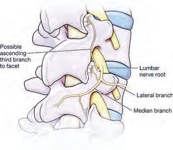
A typical vertebra consists of a vertebral body that acts to support the weight of the human body and a vertebral arch with several unique structures (the pedicles and laminae, superior and inferior articular processes which form a facet joins, transverse and spinous processes which assist movement). Facet joint is a synovial joint which is surrounded by a capsule of connective tissue and produces a synovial fluid to nourish and lubricate the joint. Each facet joint receives innervation from the meningeal branches of the spinal nerves (figure 1). Damage to a facet joint with recruitment and activation of inflammatory cell result in release of inflammatory cytokines that cause irritation and stimulation of the nociceptive nerve endings supplying the joint. Local anesthetic and corticosteroids injections are used to treat pain arising from facet joint [1] .
Intervertebral disc is a clinically important structure in human spine. It composed of three regions known as the annulus fibrosus, nucleus pulposus, and cartilaginous end plate. Nucleus pulposus may cause bulging of the outer annular fibers or herniates though annulus fibrosus. Herniation or bulging of the intervertebral disc may compress exiting spinal roots which can lead to radicular pain. Usually intervertebral disc herniates into the central vertebral canal, affecting the inferior nerves. Posterolateral herniation at L4-L5 or L5-S1 is common due to the thin posterior longitudinal ligament and thicker anterior longitudinal ligament [3]
There are 31 pairs of spinal nerves which exit though intervertebral foramen. Spinal nerves from C1 to C7 exit though intervertebral foramen above the corresponding vertebra. C8 spinal nerve exit below C7 vertebra. All other spinal nerves located below the C8 cervical nerve exit intervertebral foramen below the corresponding vertebra. For example, herniation of intervertebral disc at level L3-L4 affects L4 spinal nerve.
The cervical spine is one of the most complicated articular systems in the body, comprising 76 separate joints. It allows more movement than any other spinal region and is surrounded by a myriad of nerves, vessels, and many other vital structures. The main functions of the cervical spine are to protect the spinal cord, support the skull, and enable diverse head movement. The upper cervical spine includes the atlanto-occipital and atlanto-axial joints which are clinically important structures affected in degenerative diseases. These joints allow the flexion, extension, and axial rotation of the cervical spine. Long standing rheumatoid arthritis frequently involves the cervical spine and causes joint destruction with vertebral subluxation that may lead to sever pain and disability. Due to a high degree of mobility of the atlas relative to the axis the atlantoaxial joint is most often destructed [2] .
The vertebral artery enters the foramen of the transvers process of C6 cervical vertebra and ascends through the remaining foramina. Severe trauma to the cervical region with anterior dislocation of vertebra can lead to occlusion of flow through the vertebral artery.
Knowledge of the innervation of the cervical region provides an understanding of patients presenting with neck pain. C1 innervates the neck muscles. C2 carries sensation from the back of the head and scalp, along with motor innervation to several muscles in the neck. C3C5 contribute to the formation of the phrenic nerve and innervate the diaphragm. The cervical enlargement C5-T1 gives the rise to the rootlets that form the brachial plexus, which innervates the upper limbs.
The thoracic region contains the most vertebrae. The ribs, are attach to the thoracic region and anteriorly to the sternum, provide stability to the thoracic spine and limit movement of the spine. Thoracic spine joints include fibrocartilaginous joint, zygapophysial (Facet) joint, costo-vertebral joint and costo-transverse joint. The facet joints are significant weight bearing joints that tend to bear more weight as time progresses and intervertebral discs reduce in size. Facet joint overload as well as pathologies can lead to osteoarthritis of the joint and ultimately induce pain. These joints are thought to be the source of pain in 48% of the cases of chronic thoracic pain [5]. Facet blocks, which are performed with fluoroscopic guidance, including medial branch periarticular and intra-articular injections are useful in the treatment of joint inflammation.
Prolonged changes in the forces received by the thoracic region can result in loss of thoracic kyphosis with straightening and narrowing of anteroposterior dimensions of thoracic cage which may cause cardiovascular problems and pain [6]. Ischemic injury of the thoracic spine is common due to fine blood supply at the watershed area (T4-T9).
The thoracic sympathetic trunk course inferiorly along vertebral column in the thoracic region connecting sympathetic ganglia. The thorax contains three splanchnic nerves: the greater, the lesser, and the least which are formed from branches of the sympathetic chain.
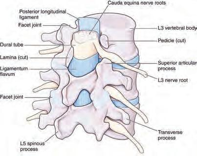
The vertebral bodies of the lumbar region are large with extensive blood supply, compare to thoracic and cervical regions, which is essential in resisting extensive loads placed on the spine (figure 2).
Figure 2. Lumbar spine structures
Lumbar facet joints are extremely significant clinically and associated with lower back pain. Intervertebral disc degeneration may lead to increased stress on the Z joints, causing them to resist greater loads. Intervertebral disc bulging or herniation, with compression of the spinal nerves, that commonly seen at level L4-L5 is also common cause of lower back pain that radiates to lower extremity. The lumbosacral joints are common source of low back pain because these joints receive tremendous amount of biomechanical stress.
References
[1] Standring S et al. (2008). Gray’s anatomy: the anatomical basis of clinical practice (40th ed.). Edinburgh: Churchill Livingstone
[2] Clinical anatomy of the spine, spinal cord, and ANS. Gregory D. Cramer, Susan A. Darby; 3rd ed. Elsevier.
[3] Scapinelli R. (1989). Morphological and functional changes of the lumbar spinous processes in the elderly. Surg Radiol Anat, 11, 129-133.
[4] White AW & Panjabi MM. (1990). Clinical biomechanics of the spine. Philadelphia: JB Lippincott.
[5] Schulte TL et al. (2010). Intra-articular meniscoid folds in thoracic zygapophysial joints. Spine (Phila Pa 1976). [Epub]
[6] Pascoe DD et al. (1997). Influence of carrying book bags on gait cycle and posture of youths. Ergonomics, 40, 631-641.


