A publication from


I founded Arbona Health Hub with the goal of breaking down complex scientific papers into simple-to-read articles. Quickly, that grew into a team of professionals who shared my vision.

A publication from
I founded Arbona Health Hub with the goal of breaking down complex scientific papers into simple-to-read articles. Quickly, that grew into a team of professionals who shared my vision.
Arbona Health Hub started as a passion for sharing science discovery, a platform where I could publish articles summarizing scientific findings in a digestible format. It then grew into a place where all scientists could submit their ideas to be published. This issue of Arbona Health Hub is a collection of notable articles published online at arbonahealthhub.com.
This would not have been possible without all the members that comprise the Board of Directors, as well as every single writer for contributing to our project and seeing the vision that we are trying to achieve. This also could not have been possible without you, the reader, for taking time out of your day to read our work.
Founder, Editor-in-Chief


Alejandro Arbona Lampaya Chairman of the Board


Héctor Cancel Asencio Board Treasurer Sofía

Writtenby:MarcelA.DeJesusVega
PublishedonlineonMarch21,2024
During the past week I came across a publication titled “Aging and the Left Behind: Puerto Rico and Its Unconventional Rapid Aging” (Matos-Moreno, 2022) It was a very insightful read for me and I felt it necessary to share this publication and summarize what I learned from it. I have been hearing about the rapid aging of Puerto Rico (PR) since I can remember I think it is an issue that should be further discussed by the healthcare community and especially the younger generation that aspires to be a part of the improvement of our healthcare system and country
The article presents irrefusable data that supports the premise about Puerto Rico’s rapid aging, provides reasons and confirmation about why this trend is happening, and lists research references that help in knowing about this. The article also predicts the impact that this situation will have on the population concerning existing problems about the sustainability of government services and longterm economic projection Interestingly, the article highlights the impact this will have on caregiving dynamics for the aging population. Finally, the article mentions a set of issues that should be addressed to overcome and prevent most of the challenges
In this article, the authors made it a priority to present and communicate important data that supports the premise that Puerto Rico’s population is aging rapidly The following are some of the data I found to be the most interesting:
1
2020 estimates places Puerto Rico’s population share of adults (65+ years) as the 10th highest in the world.
2
13% of Puerto Rico’s population was 65 years or older, in 2010 This number increased to 21% in 2019.
3
More than 700,000 working-age adults (2064) have out-migrated in the last 15 years
4
5
In 2010 population decreased 22% for the first time compared to last decennial count.
In 2020, population declined an 118% compared to the 2010 count
Old-age support ratio in 2019 is equal to 33.1 older adults per 100 working-age population
6. It is very important to note that the authors use all this information to support the idea that Puerto Rico is aging mainly due to the outmigration of working-age adults The article notes that this ongoing migration wave is a result of the economic instability that Puerto Ricans have been experiencing since the mid2000’s Authors also iterate that reduced births and increased life expectancy further contribute to the rapid aging.

The article mentions the following studies as important references for data regarding gerontological research Note that the following are not the only studies that evaluate aging in Puerto Rico, but the ones that this article has cited.
Puerto Rico Elderly: Health Conditions survey (PREHCO)
It is said that this is the only representative study of older adults in Puerto Rico
PREHCO is an island-wide study of the social, economic, and health conditions that affect older adults. This study took into consideration prevalence and correlates of prominent diseases as health indicators “10/66 Dementia Research Group” survey Puerto Rico Alzheimer ’s Disease Initiative (PRADI)
The article mentions and describes these studies in order to emphasize the “need for additional population-based data sets of a longitudinal nature focused on aging “ Adding that including larger samples will be important as well as considering Puerto Ricans living in other countries aside from the United States
The authors of this article included a section titled as above where they made sure to talk about how and why the older adults in Puerto Rico are a disadvantaged group compared to older adults in the United State in terms of social and physical well-being In my opinion this section was the most insightful The following sentence, extracted from this section of the article, I believe summarizes very well this situation and the concern this will create for government administration:
“Given the poor health and economic profile of the older adult population in Puerto Rico, an expected increase in health care and social services usage associated with rapid population aging could obstruct healthy aging if older adults’ needs are not met ”
Another interesting point that is raised in this section is that “approximately 39 7% of older adults in Puerto Rico live under the poverty level Furthermore, the primary source of income for older adults is Social Security (80 8%) followed by Supplemental Nutrition Assistance Plan (40.7%)”.

The article talks about an “effort” made from Puerto Rico’s government to address the affect of outmigration on population aging and references the approval by the Puerto Rico Legislative Assembly of the “Demographic Challenge Act of 2010” The Demographic Challenge Committee is created with the two main goal of (1) studying demographic phenomena occurring in PR and the factors influencing these processes and (2) to issue recommendations to address the needs of Puerto Rico’s aging population Although very broad goals, they seem to be directed to provide answers to most of the issues with the rapid aging
The committee established 7 policy elements to reduce societal, cultural, and economic crises
The article mentions the following:
Increase the number of births to 60,000 per year
Reduce the number of deaths to 20,000 per year
Reduce net migration to ZERO
Increase the availability of public housing and long-term care for the older adult population
As I was read this, I could not decide whether to laugh or cry as three of these policies just seem absurd and unrealistic to me Additionally, the article notes that “while the committee was tasked with analyzing demographic trends and issuing policy recommendations, there was no commitmentthatthesewouldbeimplemented through legislation or executive action” I very much hope that for the next generation of healthcare professionals in Puerto Rico and future leaders of the country, reading this make you feel a bit angry, or even offended I am sure that when our time comes, we will do a better jobandwon’tlethistorykeeprepeatingitself
As the last point made by the authors of this article, five emerging key issues are noted I believe this is the conclusion and most important part of the argument The authors made sure to present possible solutions to theseproblems
1.
Reductionincaregivingandsocialsupport
Article emphasizes how outmigration and reduced fertility in prior decades will impact life in older adults as they will be experiencing increased isolation and lowersocialparticipation
Older adults would confront more challenges in finding adequate caregivingandsocialsupport
The article notes that more studies should be supported to understand the increased demand for social and health services and adapt to the population’s need
2
Financialinsecurityinlatelife
Recent measures the government as implemented to reduce government spending is creating barriers and challenges for older adults’ financial security
The article notes that new data sources designed to capture monetary transfers andfinancialsupportareneeded
3.
Disparitiesinaccesstohealthcare
Puerto Ricans older adults have unequal health care benefits compared to those residing in the United States, even though monetary contributions are the same
Structural inequalities coupled with increased outmigration of health care professionals worsens health care access for older adults staying in PuertoRico
The article notes that more research is needed to understand and overcome public policy challenges that accentuate disparitiesinhealthcare
Returnmigration
The probable return of older adults that have raised their children in the United States, would increase the pace of populationagingontheisland
Thearticlenotestheimportanceofbeing awareofthis
Measures and government reforms, including pension reductions that affect the already eroded economy, do not provide fertile sole for self-realization, growing, thriving, and establishing roots in Puerto Rico
The article notes that the Government needs to develop programs that incentivize working-age adults to stay or return and contribute to Puerto Rico’s family structure, health care system, and

I believe that if you have come this far and read this article You most likely already know what the conclusion is I would like to end this blog entry with the following quote form the article, thatIthinkdoesagreatjobatsummarizingthe conclusion: “In Puerto Rico, population aging will be substantially felt in all social and economic institutions over the coming years Family composition and dynamics will change, work and retirement will be transformed, built environments will shift purpose, a higher percentage of the population will depend on social security and pensions, and Medicare appropriations will need to support a higher percentage of the population Many institutions will struggle as the workforce declines and must prepare for Puerto Rico’s demographic transformation How Puerto Rico will address the challenge of rapid aging remains unclear andrequiresnewpolicyeffort”
Reference:
Matos-Moreno A, Verdery AM, Mendes de Leon CF, De Jesús-Monge VM, Santos-Lozada AR Aging and the Left Behind: Puerto Rico and Its Unconventional Rapid Aging Gerontologist 2022 Aug 12;62(7):964-973 doi: 101093/geront/gnac082 PMID: 35696667; PMCID:PMC9372893

Writtenby:ChristianG.CorreaGarcía
PublishedonlineonMarch5,2024
Have you ever felt emotional exhaustion, depersonalization, or a diminished sense of personal accomplishment no matter how long you have been studying for your exams? You may be experiencing burnout caused by pressures that accompany medical education, including academic demands, prolonged work hours, and emotional challenges that patient care may provoke This topic has been gaining more attention due to the increasing number of medical students and doctors openly reporting such feelings. Evidence supports the negative effects that burnout may cause, and as the future forefront of healthcare, addressing this issue early is paramount This article aims to shed light on the causes, consequences, and potential strategies for mitigating burnout It urges a collective effort among medical institutions, educators, and students themselves to foster a healthier, more supportive educational environment
What does Burnt Toast Look Like?
Many burnt-out medical students may not realize they have already reached their boiling point; they may be so consumed by their endeavors that they don’t even notice the signs related to burnout
Feelings of stress, frustration, and exhaustion are completely normal when pursuing an MD
However, keeping an eye on how you feel throughout the process may save your career and well-being
If you have experienced any of the following, you may be in the process of burning yourself out:
Low energy levels
Easily irritated
Poor sleep
Emotionally detached from the world
Unable to concentrate
Unable to cope
General feeling of uselessness and having no direction
There are also different types of burnout that you may want to be aware of:
Overload Burnout: When you work yourself too hard to the point of exhaustion This is very common among medical students who incorrectly believe that studying to exhaustion is effective
Under-Challenge
Burnout: Feelings of unfulfillment and unappreciation caused by lack of stimulation/motivation from work or school
This happens to those in very repetitive job positions, where there is a lack of room for growth and opportunities to learn new things and make connections
Neglect Burnout: Feeling unable to keep up with the demands that work/school provide. A lack of organization, structure, and direction can facilitate burnout in your environment This particular subtype of burnout may lead to the infamous “impostor syndrome” many students experience, where they feel inadequate to perform the tasks thrown at them by medical school
Medical students undergo pre-clinical years, focusing on basic sciences in the 1st and 2nd years, and clinical rotations in the 3rd and 4th years in the U.S., where burnout may manifest itself at any time For pre-clinical students, the primary source of stress often stems from the academic rigors required to assimilate vast amounts of medical knowledge in a short amount of time, coupled with the pressure to perform well on exams This intense focus on academic achievement can lead to emotional exhaustion, as students spend long hours studying with little time for rest or personal activities The relentless pursuit of academic excellence can also foster feelings of inadequacy, significantly diminishing their sense of personal accomplishment when they perceive their efforts as never quite enough
Transitioning to clinical rotations introduces a new set of challenges that can exacerbate burnout symptoms Clinical students must apply their theoretical knowledge in real-world settings, dealing directly with patients This novel phase introduces the emotional toll of witnessing suffering and death, the stress of making critical decisions, and often, the physical exhaustion from lengthy shifts These experiences can lead to depersonalization, where students may begin to feel disconnected from their patients and cynical about their role in healthcare The shift from a primarily academic focus to the hands-on, high-stakes environment of clinical practice highlights the need for targeted interventions to address burnout at each stage of medical education, acknowledging the unique pressures faced by pre-clinical and clinical students alike
Research on medical student burnout presents a complex picture, where over half of students reveal encountering burnout symptoms during their education The burden of burnout seems to disproportionately affect female medical students, who report higher levels of psychological distress and exhaustion The prevalence of burnout in medical students also seems to increase as they advance through their medical training, particularly during the transition from pre-clinical to clinical, where patient care responsibilities are summed on top of the school workload This issue is not uniform globally; variations in burnout rates are observed across different countries and regions, influenced by the local educational systems, cultural norms, and available support structures
Not treating burnout symptoms may carry consequences that extend beyond the personal toll on one’s mental and physical health Academic performance and future professional practice may suffer as a consequence of one’s exhaustion, cynicism, and sense of ineffectiveness This, accompanied by a lack of motivation and engagement with their studies, can result in poor quality of patient care as well as poor academic outcomes such as low exam scores and grades (due to impaired critical thinking and decision-making skills). This snowball effect can jeopardize a student’s progression through medical school and their future careers
Beyond the academic and professional ramifications, burnout may pose significant mental health risks, including increased susceptibility to depression, anxiety, and even suicidal ideation among medical students
These mental health issues can have lasting effects, affecting students’ well-being long into their residency and beyond, as they transition into practicing physicians The cycle of burnout thus not only impacts the individuals experiencing it but also feeds into their personal lives and the healthcare system as a whole, potentially compromising patient care and safety Therefore, addressing burnout in medical education is not just a matter of improving student well-being but also a critical step towards ensuring the quality and effectiveness of future healthcare delivery in the United States and Puerto Rico
As mentioned, many students are already experiencing burnout symptoms but have not noticed yet This article is a wake-up call to those students who feel they are stuck in an endless loop of nothingness with no way out The first step in prevention and management is to be aware of the situation and look after yourself
Commit to at least 5 minutes every day to sitting down in a quiet space and asking yourself how you are genuinely feeling Have an internal conversation with yourself and uncover the roots of your stresses Although it seems like a simple task, many find it hard to be alone with their thoughts Talk to yourself and think of possible solutions for what is causing this burden Learning to deal with burnout is a personal journey because everyone works differently
Some good starting points may be:
Retake a hobby you might have abandoned
Commit to exercising at least 3 times a week
Prioritize self-care
Meditate
Surround yourself with the right people
Implement a rest day from studying in your schedule
Seek professional help
Create a connection with yourself to the point you can be aware whenever you are starting to get burnt out Living a healthy lifestyle may also significantly improve your chances of avoiding burnout, this includes having a clean diet and avoiding the consumption of substances that may exacerbate stress su and recreational drugs It you might not have enou or hobbies, but making t for your personal en combating the toaster
Addressing the topic o students is imperative n being of the students th the future of healthcare a produce a ripple effect t individual to the patient care and interaction Th action for medical scho broader medical commun health in a higher spot o and implement strategi and management of culture that acknowl addresses burnout can improvements in both and healthcare deli collaboratively, we can generation of healthcare supported, and equipped of their profession
References:
1
Dyrbye, L N , Thomas, M R , Shanafelt, T D (2006) Systematic Review of Depression, Anxiety, and Other Indicators of Psychological Distress Among U S and Canadian Medical Students Academic Medicine
2
Rotenstein, L S , Ramos, M A , Torre, M , Segal, J B , Peluso, M J , Guille, C , Sen, S , Mata, D A (2016) Prevalence of Depression, Depressive Symptoms, and Suicidal Ideation Among Medical Students: A Systematic Review and Meta-Analysis JAMA
Casarella, J (2022, December 18) Burnout: Symptoms and Signs WebMD Retrieved March 4, 2024, from // / 3


Writtenby:RusselS.Steans
PublishedonlineonMarch7,2024
Health Warning: Fasting is a serious medical practice which should be approached with strong consideration to individual health conditions/nutritional requirements Always consult a healthcare professional if you intend to begin fasting, especially for prolonged periods of time.
Fasting is the practice of abstaining from or limiting food and/or drink consumption over a certain period of time It has been a part of various cultural/religious traditions for centuries and can be utilized towards spiritual, social, and health purposes. For example, many people incorporate fasting into their regular routines or as a part of specific religious observances Fasting periods may range from a few hours to several days, depending on an individual’s needs A fast may involve complete abstinence from food and water (often referred to as “dry fasting”) or it may allow for consumption of certain types of food/drink (e.g. water fasting)[1]
The roots of fasting trace back to ancient cultures where it held spiritual significance, serving as a means of purification, penance, and connection with the divine In ancient Greece, fasting was believed to promote healing and detoxification Hippocrates, often lauded as the father of modern medicine, frequently prescribed fasting as a remedy for various ailments[2]
In the early 20th century, Dr Otto Buchinger and Dr Arnold Ehret advocated for the use of fasting in therapeutic settings Their efforts drew the attention of the scientific community towards fasting as a potential treatment for chronic conditions such as obesity, diabetes, and hypertension.[2,3]
Consequently, modern researchers have focused efforts towards unraveling the physiological impacts of fasting Several studies have shown that fasting can result in health benefits, such as weight loss and improved insulin sensitivity [1,4] Other studies point to the connection between fasting and immune function, noting that timerestricted feeding clinical protocols may result in reduced inflammation and enhanced cellular repair processes (autophagy)[4]
This article will outline this immune connection, focusing specifically on inflammation, and how it may be managed through fasting clinical protocols
Immunometabolism is a field of study which explores the intricate relationship between metabolism and immune cell function It focuses on how metabolic pathways regulate the function of immune cells, and how immune responses, in turn, influence metabolic processes in the body[5]
Today, researchers remain fascinated with the potential to use fasting to modulate the immune system To do so, the pathways which link the immune system to metabolism are actively under investigation
A substantial focus has been placed on nicotinamide adenine dinucleotide (NAD+), a molecule which is widely used in metabolic reactions and throughout the body.[5]
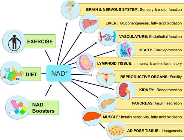
Fasting has been shown to robustly increase levels of NAD+ in the body [5] This “NAD+boosting” is believed to elicit anti-inflammatory effects through the following mechanisms:
Activation of Sirtuin enzymes → Sirtuins are a family of enzymes involved in the regulation of inflammation They rely on NAD+ to function, so NAD+ boosting is believed to improve their inflammation regulation function [5]
Inhibition of PARP enzymes → Poly(ADPribose) polymerases (PARPs) are enzymes that consume NAD+ during DNA repair processes Excessive PARP activation can deplete cellular NAD+ levels, leading to increased inflammation Inhibiting PARP activity helps preserve NAD+ levels and reduces inflammation [4]
Reduction of Oxidative Stress → NAD+ is a key regulator of reduction/oxidation reactions in cells Oxidative stress can promote inflammation by activating proinflammatory signaling pathways. Boosting NAD+ can help maintain adequate NAD+ levels, allowing cells to better counteract oxidative stress and mitigate inflammation [4]
Numerous other biological mechanisms linking fasting and reduced inflammation have been reported. For example, in a recent paper published in Cell Reports, researchers found that higher levels of the important metabolite, arachidonic acid, following fasting can result in
active inhibition of pro-inflammatory mediators such as the NLRP3 inflammasome.[6]
A 2019 study published in Cell notes that reducing dietary intake via fasting can have a regulatory effect on levels of circulating monocyte pool available for inflammatory responses Moreover, it was found that fasting has the potential to improve inflammation without compromising immunity against pathogenic microbes [7] Collectively, these findings highlight the value fasting protocols may provide in anti-inflammatory treatment regimes.
Understanding the links between the immune system and metabolism in the context of fasting is an important task with important implications for patients Identification of new immunometabolic pathways may allow for the development of novel therapeutic agents/strategies for inflammation treatment
For example, if a robust pathway linking NAD+ boosting to reduced inflammation is outlined by researchers, this information could be used to validate the use of NAD+-boosting agents such as nicotinamide riboside, succinic acid, resveratrol, etc for patients burdened with inflammatory disease
With further research, fasting itself may become a more common strategy prescribed by medical professionals to manage the inflammation of their patients
Fasting has deep historical roots in various cultures and traditions, and its application in modern healthcare continues to evolve with ongoing research and clinical practice
The relationship between fasting, NAD+ boosting, and improved immune health is actively being outlined by researchers There is considerable evidence to suggest that NAD+ boosting contributes to an anti-inflammatory environment by regulating various molecular pathways involved in inflammation, enhancing cellular metabolism, and preserving tissue homeostasis
However, while fasting may offer benefits for immune health by promoting cellular renewal, reducing inflammation, and modulating immune responses, prolonged or extreme fasting should be approached with caution, as it may have detrimental effects on immune function
As with any lifestyle or dietary change, it is imperative to consider one’s personal health conditions and consult with a healthcare professional before beginning a fasting regimen
References:
1 Kerndt, P R , Naughton, J L , Driscoll, C E , & Loxterkamp, D A (1982) Fasting: The History, Pathophysiology and Complications. Western Journal of Medicine, 137(5), 379
Patterson R E , Laughlin G A , LaCroix A Z , Hartman S J , Natarajan L , Senger C M , Martínez M.E., Villaseñor A., Sears D.D., Marinac C R , Gallo L C (2015) Intermittent Fasting and Human Metabolic Health Journal of the Academy of Nutrition and Dietetics, 115(8), 1203–1212. https://doi org/10 1016/j jand 2015 02 018
2 Boschmann, M , Michalsen, A (2013) Fasting Therapy – Old and New Perspectives Research in Complementary and Classical Natural Medicine, 20(6), 410–411 https://doi org/10 1159/000357828
3 Rajman, L , Chwalek, K , & Sinclair, D A (2018) Therapeutic Potential of NADBoosting Molecules: The In Vivo Evidence Cell Metabolism, 27(3), 529–547 https://doi org/10 1016/j cmet 2018 02 011
4 Perry, J M , Wu, J , Bley, M , Steans, R S , Meadows, A M , Huffstutler, R D , Tian, R , Griffin, J L , & Sack, M N (2024) Nicotinamide Riboside Augments Human Macrophage Migration via SIRT3-Mediated Prostaglandin E2 Signaling Cells, 13(5), 455 https://doi org/10 3390/CELLS13050455
5 Pereira, M , Liang, J , Edwards-Hicks, J , Meadows, A M , Hinz, C , Liggi, S , Hepprich, M , Mudry, J , Han, K , Griffin, J L , Fraser, I , Sack, M N , Hess, C , & Bryant, C E (2024) Arachidonic acid inhibition of the NLRP3 inflammasome is a mechanism to explain the anti-inflammatory effects of fasting Cell Reports, 43(2), 113700
https://doi org/10 1016/J CELREP 2024 113700
7
6 Jordan, S , et al (2019) Dietary intake regulates the circulating inflammatory monocyte pool Cell, 178(5), 1102 https://doi.org/10.1016/J.CELL.2019.07.050

Writtenby:AveryR.Arsenault
PublishedonlineonMarch12,2024
Private equity (PE) ownership has been a growing trend in the healthcare industry, raising questions about its implications for patient care and the healthcare system as a whole Private equity in healthcare is a form of for-profit ownership of hospitals/medical centers/nursing homes/ etc that invest in healthcare facilities by raising capital from investors and borrowing the rest PE firms are often known as “leverage buyouts” because they primarily use debt to finance acquisitions[1]
PE investors spent more than $200 billion on healthcare acquisitions in 2021 alone, with $1 trillion invested in the past decade Investors are largely taking over physician practices, especially in high-margin specialties like dermatology, urology, gastroenterology, and cardiology A concern is that these firms are monopolizing metropolitan areas. When PE firms acquire multiple providers in the same specialty within a local or regional market, these firms can gain significant market power This has the potential to lead to higher prices or lower pressure, or both, as a result of reduced competitive pressure [1,2]
Why are private equity firms investing in healthcare?
Several factors are driving PE investment, largely the attraction to earning a piece of the $4 trillion healthcare economy With low interest rates, there has been low cost of capital, leading to an increase in influx of investors Additionally, the increasing commercialization of healthcare has opened the door for private investors
While healthcare was traditionally a nonprofit sector, with the increased competition, some nonprofit healthcare organizations have begun to mimic for-profit models Nonprofit, charitable, healthcare systems are stashing capital reserves (money), and sought near-monopoly power among local markets, enabling them to raise prices Lastly, PE firms offer fresh perspectives, money, and hope to solve some of the leading problems for American’s health, with rising concerns over the ongoing failure of the US healthcare system to deliver valuable care. [1]
In a recent Harvard review of PE hospital buyouts, researchers compared how often patients experienced certain outcomes before and after a hospital was acquired by PE This study found that “after a hospital was acquired by private equity, admitted Medicare patients had a 25% increase in hospital-acquired complications, compared with patients admitted before acquisition Patients also had 27% more falls and 38% more bloodstream infections caused by central lines, despite private equity hospitals placing 16% fewer central lines than before the buyout.” [3]
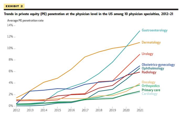
Imagefrom:Abdelhadi,O,Fulton,B D,Alexander L,&Scheffler,R M (2024) PrivateEquity-AcquiredPhysicianPracticesAndMarket PenetrationIncreasedSubstantially 2012-21 Healthaffairs(ProjectHope) 43(3) 354–362 https://doiorg/101377/hlthaff202300152
Additionally, a recent 2023 meta-analysis evaluated the trends in PE ownership on health outcomes, costs to patients or payers, costs to operators and quality, controlling for the risk of bias in study outcomes The study’s biggest takeaway is that PE ownership is associated with increase in healthcare costs to patients or payers, primarily by increased charges and negotiatedhigherrateswithpayers.Also,mixed impacts of PE ownership on healthcare quality, but greater evidence suggesting PE may degrade quality rather than show any improvement Outof55studiesinthereview,21 studies identified at least one harmful impact on PE ownership The most frequent quality measure looked at issues with staffing PE ownership was associated with almost exclusively negative impacts on patient, satisfaction, daily functioning, and general qualityscores 4
Private Equity investment continues to be a dominant force in the commercialization of healthcare across the United States, however, results on the benefit seem limited, and unchecked regulation causes even more concern The ability of PE firms to dominate local healthcare markets, with limited regulation or scrutiny from antitrust authorities exacerbates concerns, and leaves communities vulnerabletoPEmonopoly
WithlimitedsupervisionfromtheFederalTrade Commission, state regulators and policy makers, increased policy regulation is needed, while also evaluating the benefits of patient care,safetyandsatisfaction
While this article touches on the surface of PE investment across the US, more research is needed to fully understand the impact on health outcomes, system costs and how this model compares to other non-US settings Longitudinal studies are needed to understand the full effects of the increasing commercialization of healthcare by PE firms, after this large increase in buyouts from 2021 to now It is known that PE ownership is widely consequential for every patient, provider and payerinvolved
References
Privateequity’sroleinHealthCare CommonwealthFund (2023, November17) https://wwwcommonwealthfundorg/publications/explainer/2023/ nov/private-equity-role-healthcare#: :text=What%20is%20private%20equity%3F,two%20forms%3 A%20private%20or%20public
1 Abdelhadi, O, Fulton, B D, Alexander, L, & Scheffler, R M (2024) Private Equity-Acquired Physician Practices And Market Penetration Increased Substantially, 2012-21 Health affairs (Project Hope),43(3),354–362 https://doiorg/101377/hlthaff202300152 2
Miller, J (2023, December 26) What Happens When Private Equity TakesOveraHospital HarvardMedicalSchoolNewsandResearch https://hmsharvardedu/news/what-happens-when-private-equitytakes-over-hospital 3
Borsa, A, Bejarano, G, Ellen, M, & Bruch, J D (2023) Evaluating trends in private equity ownership and impacts on health outcomes, costs, and quality: systematic review BMJ (Clinical researched),382,e075244 https://doiorg/101136/bmj-2023-075244 4

Writtenby:RicardoA.PérezOjeda
PublishedonlineonMay10,2024
Sublingual drug delivery is a non-invasive method that offers unique benefits compared to traditional oral ingestion or parenteral routes It involves placing a drug under the tongue, allowing it to dissolve and be absorbed directly into the bloodstream through the highly vascular sublingual mucosa This enables rapid drug absorption and immediate systemic effects, bypassing the digestive tract’s harsh environment and avoiding first-pass hepatic metabolism
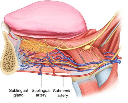
The sublingual region, which lies beneath the tongue, offers an ideal location for swift drug absorption thanks to its distinctive anatomical and physiological characteristics This area is richly vascularized by a network of capillaries that is part of the systemic circulation. As taught, the vessel responsible for sublingual drug absorption is the deep lingual vein The mucosal lining in this
region is notably thin and highly permeable, allowing for rapid drug diffusion This process enables faster drug action, as the compounds do not have to pass through the gastrointestinal tract and thus begin working almost immediately after administration In contrast to other oral sites, the sublingual area experiences decreased saliva flow, which can help prevent premature swallowing and minimize drug dilution
When a drug is ingested orally, it undergoes firstpass metabolism. This process involves the drug traveling from the stomach to the small intestine, where it is absorbed into the bloodstream From there, it is carried to the liver, where a significant percentage of the drug can be broken down before being dispersed throughout the rest of the body This can decrease the effectiveness of the drug and require higher dosages to achieve the intended therapeutic effects
Numerous factors can impact first-pass metabolism One of the most significant is the drug’s properties Some drugs are more susceptible to liver breakdown than others, which can affect their bioavailability. Hepatic enzyme activity can also play a role People with higher levels of liver enzymes may break down drugs more quickly, reducing their efficacy Additionally, research has shown that intestinal metabolism can also impact first-pass metabolism Some medications may be metabolized in the intestines before reaching the liver, decreasing their bioavailability
Scientists have explored new ways to tackle the obstacles presented by first-pass metabolism, including the utilization of prodrugs Prodrugs are initially inactive compounds but become active drugs upon absorption into the body This can increase their bioavailability and alleviate the effects of first-pass metabolism Nitroglycerin, which is employed to prevent and treat angina, Zofran ODT, which is used to prevent nausea and vomiting; and Levsin (hyoscyamine), which is used to regulate gastric secretions and hypermotility in spastic colitis, are some common examples of sublingual medications
Administering medications through the sublingual method is crucial for delivering certain drugs, particularly those that require quick action or may undergo significant firstpass metabolism This area of the mouth is ideally designed to facilitate the efficient absorption of medications directly into the bloodstream The sublingual method not only maximizes the therapeutic advantages of the medication but also improves patient adherence due to its convenience and effectiveness Continuous innovations in drug preparation and delivery technologies are making the sublingual route increasingly appealing for expanding its use in clinical practice, guaranteeing patients receive the most prompt and effective treatment available.
2
3
5
References:
RRIERSYST V25 I5 20 1
Goswami, T , Jasti, B , & Li, X (2008)
Sublingual drug delivery Critical reviews in therapeutic drug carrier systems, 25 5, 44984
https://doi org/10 1615/CRITREVTHERDRUGCA
Katzung, B G , Masters, S B , & Trevor, A J (2020) Basic & Clinical Pharmacology (15th ed) McGraw-Hill Medical; McGraw-Hill 49
Kester, Mark (2012) Elsevier’s Integrated Review Pharmacology || Pharmacokinetics , (), 1–15 doi:10 1016/B978-0-323-07445-2 00001X
Pawar, P , Ghorpade, H , & Kokane, B (2018) Sublingual route for systemic drug delivery Journal of Drug Delivery and Therapeutics
https://doi org/10 22270/JDDT V8I6-S 2097
4 Robertson, D (2017) First Pass Metabolism Nurse Prescribing, 15, 303-305 https://doi org/10 12968/npre 2017 15 6 303


Writtenby:SofíaI.MalavéOrtiz
PublishedonlineonMarch16,2024
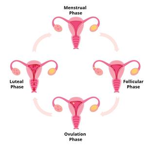
The menstrual cycle usually occurs over a 28-day period but can range anywhere from 21 to 35 days, although this might be different for everyone, and it consists of four phases: menstrual, follicular, ovulation, and luteal. During these phases, the body undergoes hormonal fluctuations specifically for reproductive hormones such as estrogen/estradiol (E2), luteinizing hormone (LH), follicle-stimulating hormone (FSH), and progesterone (P4)
Estrogen stimulates growth of cells that line the uterus
Progesterone maintains the cells that line the uterus
Follicle-stimulating hormone stimulates growth, and maturation of follicles in the ovaries
Luteinizing hormone stimulates eggs to be released from the ovary
The menstrual phase is the first stage of your menstrual cycle, it is also when you get your period Therefore, your hormone levels of estrogen and progesterone decrease causing the thickened lining of your uterus to shed in a combination of blood, mucus, and tissue
The follicular phase is the longest phase of the cycle, and it overlaps with your menstrual phase since it starts on the first day of your period and ends when you start to ovulate In this phase, the pituitary gland in the brain releases folliclestimulating hormone that stimulates your ovaries to produce follicles (eggs)
Eventually, one of these follicles will mature and become dominant while others will be destroyed or reabsorbed by your body Once that’s done your follicle-stimulating hormone levels decrease while your estrogen levels start to increase making the lining of your uterus grow and thicken
The ovulation phase starts with the release of luteinizing hormone, it’s the phase where ovulation takes place Therefore, the ovary releases a mature egg that will travel through the fallopian tube toward the uterus. If fertilization occurs the egg travels to the uterus
The luteal phase is the phase after ovulation and before the menstrual phase In this phase, the follicle that contained the egg in ovulation turns into the corpus luteum The corpus luteum produces progesterone along with some estrogen which helps thicken the lining of the uterus for possible egg fertilization If the egg is not fertilized (no pregnancy occurs) then you’ll pass on to the menstrual phase
Reproductive hormones during the cycle can influence our moods, energy levels, body temperature, and sleep, among many other factors.
Getting enough sleep is essential for our physical and mental health Our body requires sleep for every process, including maintenance, repair, and development, which are crucial for learning, memory, and overall health Any disruptions in our sleep can interfere with our daily functioning, overall well-being, and cognitive function, and can even increase the risk of developing diseases such as Alzheimer’s and dementia Therefore, it is crucial to maintain a consistent sleep schedule and prioritizequalitysleep
Numerous studies have found that women and menhavedifferentsleeppatternsfromanearly age, where women tend to report poorer sleep quality and are at a higher risk for various sleep disorders such as insomnia and sleep apnea During the menstrual cycle, women experience subtlechangesinsleepqualitythroughouttheir reproductive years. The menstrual cycle has been associated with changes in circadian rhythms and sleep architecture, while sleep efficiencyappearstoremainconstantandtends todeclineinlaterreproductiveage
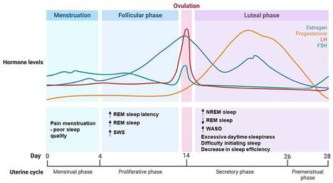
In the menstrual cycle, women have reported poorer sleep quality and more disturbances during the premenstrual week, even without significant menstrual-related complaints. Sleep studies have shown that this may be due to an increase in progesterone, and estrogen levels during the luteal phase, which might also be relatedtoariseinbodytemperatureduringthis phase as well Sleep disturbances can also depend on the regularity of your cycle since women with irregular cycles report more sleep difficultiesthanregularcyclingwomen
Sleep disturbances don’t end in the reproductive years, they also affect peri and post-menopausal women The hormonal changes that women experience during menopause are marked by a gradual decrease in the levels of estrogen and progesterone in their body. These fluctuations are associated with multiple sleep disturbances, including insomnia and increased awakenings Additionally, menopausal women commonly experience hot flashes and night sweats, which disrupt sleep Changes in rapid eye movement (REM) sleep patterns may occur, potentially contributing to sleep disruptions Moreover, hormonal changes may increase the risk of developing sleep-disordered breathing conditions such as sleep apnea Overall, hormonal fluctuations in menopause significantly impact various aspects of sleep, leading to diminished sleep quality and efficiency, and increased vulnerability to sleep disturbances Studies have shown that women who are in menopause are more prone to developing dementia, while combination hormonal therapy, progesterone therapy, and estrogenhormonetherapyhaveshowntolower thisrisk.
Overall, throughout the menstrual cycle, reproductive hormones have a complex role in regulating sleep quality and architecture, with varying effects of estrogen and progesterone at different phases of the menstrual cycle and life stages
References:
1
2
Baker FC, Lee KA Menstrual Cycle Effects on Sleep Sleep Med Clin 2022Jun;17(2):283-294 doi:101016/jjsmc202202004 Epub2022Apr22 PMID:35659080
NowakowskiS MeersJ,HeimbachE SleepandWomen’sHealth Sleep Med Res 2013;4(1):1-22 doi: 1017241/smr2013411 PMID: 25688329; PMCID:PMC4327930
3
Dorsey A, de Lecea L, Jennings KJ Neurobiological and Hormonal Mechanisms Regulating Women’s Sleep Front Neurosci 2021 Jan 14;14625397 doi: 103389/fnins2020625397 PMID: 33519372; PMCID: PMC7840832
4
Gervais NJ, Mong JA, Lacreuse A Ovarian hormones, sleep and cognition across the adult female lifespan: An integrated perspective Front Neuroendocrinol 2017 Oct;47:134-153 doi: 101016/jyfrne201708002 Epub 2017 Aug 9 PMID: 28803147; PMCID: PMC7597864
5
Haufe A, Leeners B Sleep Disturbances Across a Woman’s Lifespan: What Is the Role of Reproductive Hormones? J Endocr Soc 2023 Mar 15;7(5):bvad036 doi: 101210/jendso/bvad036 PMID: 37091307; PMCID: PMC10117379
6
Brzecka A Leszek J Ashraf GM Ejma M Ávila-Rodriguez MF Yarla NS Tarasov VV, Chubarev VN, Samsonova AN, Barreto GE, Aliev G Sleep Disorders Associated With Alzheimers Disease: A Perspective Front Neurosci 2018 May 31;12:330 doi 103389/fnins201800330 PMID: 29904334 PMCID:PMC5990625
7
Massimiliano de Zambotti Ian M Colrain Fiona C Baker Interaction between Reproductive Hormones and Physiological Sleep in Women, The Journal of Clinical Endocrinology & Metabolism, Volume 100, Issue 4,April2015,Pages1426–1433,https://doiorg/101210/jc2014-3892
Whatarethestagesofthemenstrualcycle? BirlaFertility&IVF (2023 July3) https://birlafertilitycom/blogs/stages-of-menstrual-cycle/
8 Women in sleep and breathing Frontiers (nd) https://wwwfrontiersinorg/research-topics/55217/women-in-sleepand-breathing/articles
Writtenby:JavierAVelázquezGonzález
PublishedonlineonJune24,2024
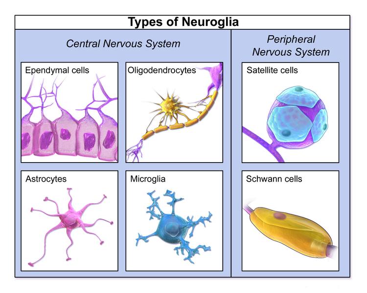
Glioblastoma (GBM) is one of the primary malignant brain tumors derived from glial cells of the central nervous system (Figure 1) Glial cells are responsible for supporting neuronal function, so one can imagine that uncontrolled growth of these cells can lead to serious neurological consequences affecting the functional processes of the brain GBMs originate from astrocytes and masses are found within cerebral hemispheres, as a “butterfly glioma” crossing the corpus callosum (Figure 2) In histopathological slides, GBMs present as poorly differentiated primitive cells with pseudopalisading necrosis (Figure 3)
GBM is the most aggressive of glial tumors, as the average survival rate for patients with GBM is 14-16 months (Mohammed et al 2022) Interestingly, there are groups of patients with GBM termed long-term survivals (LTS) that survive more than 5 years and may have distinguishing molecular, genetic, clinical, and radiographic characteristics that can be very useful for developing novel treatments for all GBM patients. The focus lies on acquiring larger LTS cohorts to make accurate predictions based on the most common characteristics these patients present Through histological, radiological, molecular, genetic, clinical, and demographic data, researchers can develop the framework on how to identify LTS patients and develop the understanding to provide novel treatment for GBM patients
The most common symptoms presented in GBM patients include seizures, confusion, vision problems, facial weakness, ataxia and imbalance, memory problems and personality/behavioral changes Although many of these symptoms can appear in LTS and short-term survivors (STS), Briceno et al found that STS presented predominantly with seizures, headaches, and memory problems/confusion, while LTS primarily presented with difficulty in walking and uncoordinated movements Interestingly, Briceno et al mentioned that a relationship between STS
patients and increased intracranial pressure may be a subject for further analysis Nonetheless, definitive symptomatic clinical data is still needed Most LTS are predominantly younger in age compared to STS
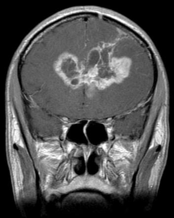
Figure 2. Coronal T1 MRI of a typical “butterfly glioma”
Although this is yet to be a significant determining factor, younger age is indeed associated with longer survival rates, as these patients tend to have concurrent mutations in Isocitrate dehydrogenase (IDH), and O6 methylguanine-DNA methyltransferase (MGMT) promoter methylation, which does provide significant better survival outcomes (Wang et al 2016) In terms of sex, Johnston et al mention that studies that include LTS cohorts have included females predominantly Even though GBM generally affects males more than females, larger cohorts are needed to determine if females have a protective factor that can be explained by sex Looking further into the future, treatments are focusing on sex-specific strategies to treat patients based on their conditions.
Through Next Generation Sequencing, the most common mutations found in LTS are in IDH1, IDH2 and MGMT promoter Although IDH mutation does provide longer survival rates in GBM, it is rarely seen, and patients who had
GBM with IDH mutation were no longer classified as GBM Interestingly, molecular data found that methylation of MGMT is more commonly seen in LTS, and it may provide an underlying mechanism to study further and consider therapeutic routes involving this gene Other molecular findings include a mutated TP53 gene, TERTp mutation, EGFR fusion, and translocation Johnston et al found a 17 Extreme Survival (ES) cohort study that found higher intermediate fibrillary elements and lower small anaplastic elements Still, there is more information to be elucidated, and as genetic techniques become more sophisticated, genomic studies can be applied to treat patients with low survival probability based on distinguishing genetic characteristics found in LTS GBM patients
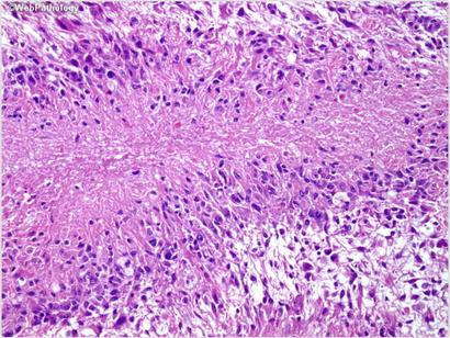
Figure 3 Higher magnification showing nuclear pseudopalisading – aggregation of tumor cells around the periphery of the necrotic areas.
Johnston et al. found that pre-diagnostic MRI analysis of T1 images demonstrated hypointense lesions in LTS patients, with STS presenting with mixed-intensity lesions Gray matter involvement was mostly common in STS patients, but it is not a determinant characteristic and further imaging should be carried out T2/FLAIR hyperintensity was more common in LTS while there were no differences in T2/FLAIR heterogeneous signals between both groups One of the most significant results by show that contrast enhancement is significantly more prominent in wild-type IDH GBM patients Wang et al found that radiological features combined with IDH1 status for predicting the survival outcome of GBM patients showed that tumor contrast enhancement, multi-enhancing foci, and peritumoral edema, were found to be associated with the survival outcomes of GBM patients with IDH mutations.
These findings can be used as guidelines for understanding how these tumors behave and grow
What can Immunotherapy provide to GBM patients?
Parker et al suggested that low clinical success to immunotherapy in GBM is due to inter and intra tumoral heterogeneity, lack of neoantigens, poor antigen presentation, and priming and secretion of immunosuppressive cytokines External immunosuppression, low CD8 T cell activity and frequency, local T cell exhaustion and suppression, and increased frequency of immunosuppressant tumorassociated macrophages are factors that improve tumor survival chances The need lies in developing effective immunotherapies that combat immunosuppression in GBM.
Eventually, the goal is to provide anti-tumor immunity and prolong patient survival time The technique used to deliver the immunotherapy is via convection-enhanced delivery (CED), in which a catheter is inserted into the tumor mass to facilitate drug distribution using the positive pressure generated by the infusion pump The process above allows the delivery of macromolecules that are too large to cross the blood-brain barrier or too toxic to be delivered systematically Preclinical mouse glioma experiments showed that delivery via CED in mouse models leads to the killing of tumor cells and produces secondary immune responses via CD4 and CD8 T cell activation Hopes are that clinical trials in humans can provide similar effects and use these milestones as the starting point for developing novel immunotherapy targeting tumors in all types of GBM patients
Approximately 90% of GBM have poor prognosis due to the invasive and aggressive nature of this tumor Understanding the dynamics in which glial cells develop is a key milestone for initiating strategies to treat patients with low expectations of survival. Developing novel strategies to target malignant brain tumors such as GBM is one step toward improving survival outcomes, alongside generating enough knowledge to treat other types of malignant tumors Other advances include mathematical models to understand and predict tumor growth behavior in a personalized sex-specific manner These unique models focus on patient-specific approaches that could potentially be more applicable to treating GBM patients in a clinical setting
References:
1
Mohammed, S, Dinesan, M, & Ajayakumar, T (2022) Survival and quality of life analysis in glioblastoma multiforme with adjuvant chemoradiotherapy: a retrospective study Reports of practical oncology and radiotherapy: journal of Greatpoland Cancer Center in Poznan and Polish Society of Radiation Oncology, 27(6), 1026–1036 https://doi.org/10.5603/RPOR.a2022.0113
2
Briceno, N, Vera, E, Komlodi-Pasztor, E, Abdullaev, Z, Choi, A, Grajkowska, E, Kunst, T, Levine, J, Lindsley, M, Fernandez, K, Reyes, J, Boris, L, Burton, E, Panzer, M, Polskin, L, Penas-Prado, M, Pillai, T, Theeler, B J, Wu, J, Wall, K, Gilbert, M R (2024) Long-term survivors of glioblastoma: Tumor molecular, clinical, and imaging findings Neurooncology advances, 6(1), vdae019 https://doi org/10 1093/noajnl/vdae019
3 Johnston, S K, Whitmire, P, Massey, S C, Kumthekar, P, Porter, A B, Raghunand, N, Gonzalez-Cuyar, L F, Mrugala, M M, HawkinsDaarud, A, Jackson, P R, Hu, L S, Sarkaria, J N, Wang, L, Gatenby, R A, Egan, K M, Canoll, P, Swanson, K R, & ENDURES consortium (2019) ENvironmental Dynamics Underlying Responsive Extreme Survivors (ENDURES) of Glioblastoma: A Multidisciplinary Team-based, Multifactorial Analytical Approach American journal of clinical oncology, 42(8), 655–661 https://doi.org/10.1097/COC.0000000000000564
5
Wang, K, Wang, Y, Fan, X, Wang, J, Li, G, Ma, J, Ma, J, Jiang, T, & Dai, J (2016) Radiological features combined with IDH1 status for predicting the survival outcome of glioblastoma patients Neurooncology, 18(4), 589–597 https://doi.org/10.1093/neuonc/nov239
4 Parker S, McDowall C, Sanchez-Pérez L, Osorio C, Duncker PC, Briley A, Swartz AM, Herndon JE 2nd, Yu YA, McLendon RE, Tedder TF, Desjardins A, Ashley DM, Gunn MD, Enterline DS, Knorr DA, Pastan IH, Nair SK, Bigner DD, Chandramohan V. Immunotoxin-αCD40 therapy activates innate and adaptive immunity and generates a durable antitumor response in glioblastoma models Sci Transl Med 2023 Feb 8;15(682):eabn5649 doi: 101126/scitranslmedabn5649 Epub 2023 Feb 8 PMID: 36753564; PMCID: PMC10440725
1
Image credits: Cover Image – Adapted from Adeberg S, Bostel T, König L, Welzel T, Debus J, Combs SE A comparison of long-term survivors and short-term survivors with glioblastoma, subventricular zone involvement: a predictive factor for survival? Radiat Oncol 2014 Apr 23;9:95 doi: 101186/1748-717X-9-95
PMID: 24758192; PMCID: PMC4011838
2
Figure 1 – (Blausencom staff (2014) “Medical gallery of Blausen Medical 2014“ WikiJournal ofMedicine 1 (2) DOI:10.15347/wjm/2014.010 ISSN 2002-4436 – Own work)
3
Figure 2 – Adapted from https://radiopaedia.org/articles/butterflyglioma?lang=us
4
Figure 3 – Adapted from https://www.webpathology.com/image.asp? case=738&n=13
Writtenby:GustavoA.GarrigaGarcía
PublishedonlineonMay31,2024
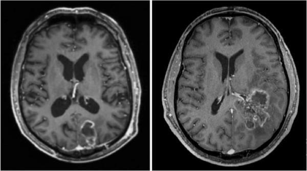
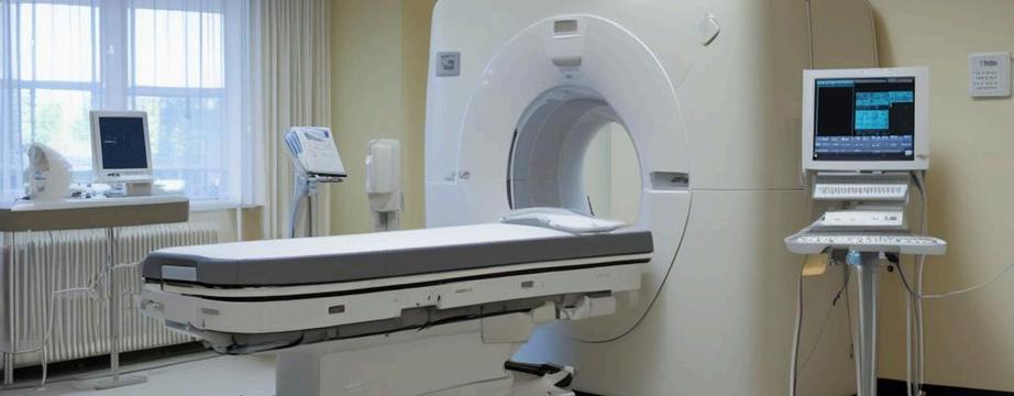
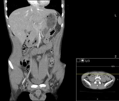
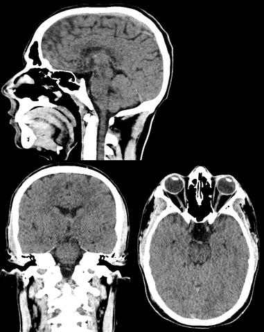
The Computer Tomography (CT) scan represents a widely used and valuable imaging diagnostic tool in modern medicine It enables medical professionals to detect the presence of tumors, traumatic injuries, hemorrhages, and a range of other health abnormalities (Computed Tomography (CT), 2022)
The technique uses a rotating X-ray beam around the patient to generate cross-sectional images, which are then processed and subsequently stacked to create a 3D image However, the procedure requires exposure to ionizing radiation, which poses a potential threat to DNA Ionizing radiation can lead to single-stranded and doublestranded DNA breakages, which may lead to mutations and, ultimately, to cancer. Although the radiation doses used in CT scan procedures are considered generally safe, concerns have been raised regarding children, who are more susceptible to the harmful effects of ionizing radiation, as well as those patients who undergo repeated exposures
A multinational cohort study (EPI-CT) conducted by Gomez et al examined nearly one million individuals who underwent CT scans before age 22 across nine European countries The study revealed a dose-response association between the cumulative radiation in bone marrow from CT scans and the risk of developing hematological
malignancies, including those of lymphoid and myeloid lineage, with doses as low as 10 – 15 milligray (mGy) In addition, when comparing the number of CT procedures rather than the active bone marrow dose, patients who underwent more than one CT scan exhibited a 43% higher risk for hematological and lymphoid malignancies as compared to those who underwent only one CT scan The excess relative risk for all hematological malignancies in children, adolescents, and young adults was 196 per 100 mGy, indicating almost a 2fold increase in relative risk.
In addition, the researchers reported similar risk estimates for lymphoid and myeloid malignancies and acute lymphoma. The investigator’s analyses estimated an excess absolute risk of 17 7 per 100,000 person-years per 100 mGy, suggesting that for every 100,000 pediatric patients examined in a given day, 17 to 18 patients could potentially develop hematological malignancies within 12 years of an eight mGy mean exposure attributed to the ionizing radiation (Gomez et al., 2023). These results suggest that exposure to radiation from imaging procedures in pediatric patients can increase the risk of developing hematological malignancy, highlighting the importance of reducing unnecessary radiation exposure by being cautious and selective when recommending these studies to young patients
Similarly, Miglioretti et al identified an elevated risk of radiation-induced cancer in children undergoing pediatric CT imaging Miglioretti et al conducted a retrospective observational study using data from over seven US healthcare systems from 1996 – 2010, including children younger than 15 years old who were examined via CT scans (152,419 – 371,095 children per year) The study found that cancer risks varied based on age, gender, and the region of the body being imaged Younger patients and girls exhibited an increased risk for solid cancer (projected cases: 13 1 – 33 9 girls vs 6 3 – 14 8 boys per 10,000 CT scans). Furthermore, abdomen/pelvis CT scans, which have a mean effective dose of 10 6 millisieverts (mSv) (same as 10 6 mGy) in children younger than 5 and 14 8 mSv in children 10-14 years old, had the highest risk for radiation-induced solid cancers, especially in the older children
Meanwhile, head CT scans, the most commonly performed pediatric CT, posed a higher risk of leukemia and brain cancer, particularly in children under five years old, which had a projected risk of 1 9 cases of leukemia per 10,000 head CT scans The study projected that four million pediatric CT scans of the head, abdomen/pelvis, chest, or spine in the US per year could cause 4,870 future cancers The majority of projected cancers in exposed girls are breast, thyroid, lung cancer, and leukemia, whereas in exposed boys, about half the projected cancers are in the brain, lung, colon cancer, and leukemia Given that effective doses vary between 0.03 – 69.2 mSv, reducing the highest effective doses (25% upper quartile) might aid in potentially preventing over 43% of the projected cancers (Miglioretti et al , 2013) These projections highlight the potential longterm consequences of radiation exposure from CT imaging pediatric patients Healthcare providers should carefully consider the benefits and necessity of these procedures, especially in the pediatric population, which displays the highest vulnerability to the effects of ionizing radiation due to their sensitivity and longer lifespan to develop cancer.
Additionally, Pearce et al. conducted a retrospective cohort study using data from the National Health Services (NHS) Centers in Great Britain between 1985 and 2002, where patients younger than 22 without a previous cancer diagnosis were included Analysis of leukemia and brain tumors involved 178,604 and 176,587 people, respectively Head CT scans constituted the majority of CTs (64%), followed by abdomen/pelvis (9%) and chest CT (7%) The study demonstrated a significant association between radiation doses from CT scans and subsequent incidents of leukemia and brain tumors Patients who received a dose of 30 mGy had a 3 18 times higher risk of developing leukemia compared to those who received doses of less than five mGy
Compared to patients with a dose of less than five mGy, those receiving a dose of 50-74 mGy had a 2 82 times higher risk of developing brain tumors, and those receiving a dose of 50 mGy or more were 3 32 times as likely to develop brain tumors This implies that exposure to 2-3 head CTs, resulting in a cumulative dose of approximately 60 mGy in the brain, increases the risk of developing brain tumors almost
threefold in children below 15 years old. While exposure to 5-10 head CTs can lead to the accumulation of about 50 mGy in red bone marrow dose, tripling the risk of leukemia in children below 15 years of age. In addition, the study also identified statistically significant associations between CT scans and cancer subgroups, including gliomas, schwannomas, meningiomas, acute lymphoblastic leukemia, and myelodysplastic syndromes (Pearce et al , 2012)
CT scans are valuable medical diagnostic tools that can quickly detect tumors, injuries, and other health abnormalities However, an extensive body of evidence demonstrates a dose-response relationship between cumulative radiation doses from CT scan exposure and the risk of developing radiation-induced malignancies, especially in children Therefore, healthcare providers must exercise caution and prudence when recommending these procedures and participate in informed decision-making with the patient
The multinational cohort study conducted by Bosch de Basea Gomez et al demonstrated a positive association between cumulative radiation from CT scans and the risk of developing hematological malignancies in young individuals, highlighting the potential long-term consequences of repeated exposures to ionizing radiation from CT scans Similarly, Miglioretti et al identified an elevated risk of
radiation-induced cancer in pediatric patients undergoing CT imaging, projecting that thousands of future cancer cases may occur annually in the United States as a result of failing to minimize effective doses. Pearce et al. study reinforced these findings by revealing a positive association between radiation doses from CT scans and subsequent incidents of leukemia and brain tumors in children. Finally, Cao et al multi-continental meta-analysis emphasized that even in adults, cumulative radiation doses from CT scans are associated with a linear increase in cancer risks, with higher doses resulting in a rapid increase in cancer odds These studies collectively provide ample and compelling evidence that even low to moderate doses of ionizing radiation from CT scans can significantly increase the long-term risk of hematological malignancies, solid cancers, leukemia, and brain tumors
Healthcare providers must carefully weigh the benefits and risks of CT imaging, especially in vulnerable populations such as pediatric patients and individuals undergoing repeated scans Alternative imaging techniques such as ultrasound and magnetic resonance imaging should be considered in order to make a diagnosis whenever possible. Patients should also be thoroughly informed about the associated risks before undergoing CT procedures, and discussion with the physician regarding any concerns and questions should be encouraged Additionally, radiation dose optimization strategies are necessary to reduce potential health risks and improve patient safety during CT scans to enhance quality care. Although CT scans are associated with an increased risk of developing cancer, they remain an extremely valuable resource in diagnostic medicine, particularly in emergency cases where prompt diagnosis is needed to take appropriate action Therefore, their usefulness should not be diminished and underappreciated
References:
Bosch de Basea Gomez, M , Thierry-Chef, I , Harbron, R , Hauptmann, M , Byrnes, G , Bernier, M -O , Le Cornet, L , Dabin, J , Ferro, G , Istad, T S , Jahnen, A , Lee, C , Maccia, C , Malchair, F , Olerud, H , Simon, S L , Figuerola, J , Peiro, A , Engels, H , Cardis, E (2023) Risk of hematological malignancies from CT radiation exposure in children, adolescents and young adults Nature Medicine, 29(12), 3111–3119 https://doi org/10 1038/s41591-023-02620-0 1
2. Cao, C.-F., Ma, K.-L., Shan, H., Liu, T.-F., Zhao, S.Q , Wan, Y , Jun-Zhang, null, & Wang, H -Q (2022) CT Scans and Cancer Risks: A Systematic Review and Dose-response Meta-analysis BMC Cancer, 22(1), 1238. https://doi.org/10.1186/s12885022-10310-2
3 Computed Tomography (CT) (2022) National Institute of Biomedical Imaging and Bioengineering. Retrieved February 12, 2024, from https://www nibib nih gov/scienceeducation/science-topics/computedtomography-ct
4. Miglioretti, D. L., Johnson, E., Williams, A., Greenlee, R T , Weinmann, S , Solberg, L I , Feigelson, H S , Roblin, D , Flynn, M J , Vanneman, N , & Smith-Bindman, R (2013) The use of computed tomography in pediatrics and the associated radiation exposure and estimated cancer risk JAMA Pediatrics, 167(8), 700–707 https://doi.org/10.1001/jamapediatrics.2013.311
5 Pearce, M S , Salotti, J A , Little, M P , McHugh, K , Lee, C , Kim, K P , Howe, N L , Ronckers, C M , Rajaraman, P , Sir Craft, A W , Parker, L., & Berrington de González, A. (2012). Radiation exposure from CT scans in childhood and subsequent risk of leukaemia and brain tumours: A retrospective cohort study Lancet (London, England), 380(9840), 499–505. https://doi org/10 1016/S0140-6736(12)60815-
Image Credits:
Figure 1: https://www sanjuanmri net/wpcontent/uploads/2023/08/mri jpg
Figure 2: https://upload wikimedia org/wikipedia/commo ns/f/f5/CT of a normal abdomen and pelvis%2 C coronal plane 44 png
Figure 3:
https://upload wikimedia org/wikipedia/commo ns/9/96/CT of a normal brain %28thumbnail% 29 png
Writtenby:AlejandroArbona-Lampaya
PublishedonlineonJune28,2024
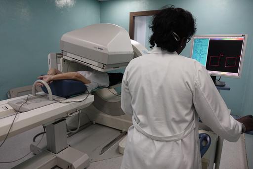
When hearing the word nuclear, what comes to mind? Usually This feels like it belongs in your personal statement and not in this section You can talk about how taking care of your dad reinforced your desire to study medicine, etc., one thinks about nuclear weapons or nuclear power plants, not an everyday diagnostic tool in healthcare Nuclear medicine doesn’t share many similarities with these, but the main thing they all have in common is radiation Nuclear medicine is a specialty that requires completion of a medical degree (MD or DO) and then 4 years of residency training It shares similarities with Diagnostic Radiology However, the main difference is that in nuclear medicine, radioactive material, called radioactive tracers, is used for diagnostic purposes, to monitor the health of organs/tissue, and to target and destroy diseased or damaged organs/tissue.
Radioactive tracers are carrier molecules that are closely bound to a radioactive atom The carrier molecules can differ depending on the objective of the scan. Most nuclear medicine diagnostic procedures include administering a radioactive tracer to a patient via inhalation, oral consumption, or injection into an organ The nuclear medicine physician will choose the most accurate tracer for a given patient, tailoring it so that each patient receives an individualized assessment. The tracer used determines whether the patient will have a Single Photon Emission Computed Tomography (SPECT) or Positron
A SPECT scan provides 3-D images of the distribution of radioactive tracer molecules in the patient’s body The images are computer generated from projection images of the body that are recorded at different angles by the SPECT machine SPECT machines have gamma radiation detectors that can detect the gamma-ray radiation emitted from the tracers injected into the patient
A PET scan can help reveal your tissues and organs’ metabolic or biochemical function Like with SPECT, PET scans use radioactive tracers to show typical and atypical metabolic activity Cancer cells have a higher metabolic activity than normal cells, which makes PET scans useful for cancer detection. PET scans can also be used to detect heart disease and brain disorders like Alzheimer’s and dementia

SPECT scans can detect and monitor the progression of heart disease, such as blocked coronary arteries Coronary arteries supply blood to the heart muscle, and a lack of blood flow can lead to ischemic heart disease, which can cause heart attacks and death Other radiotracers can detect bone diseases, gallbladder disease, and intestinal bleeding SPECT has also been used to help diagnose Parkinson’s disease in the brain and distinguish it from other anatomically comparable movement disorders and dementias As mentioned earlier, PET scans play an important role in cancer detection and heart disease monitoring
What are the risks of using radioactive tracers?
Given its name, nuclear medicine exposes the patient to radiation. This dose is no more than what is experienced during routine chest X-ray or CT exams As a result of radiation exposure, these procedures increase your risk of developing cancer later in life. However, this risk is considered to be minimal compared to the expected benefit from a medically necessary diagnostic imaging exam Physicians agree that the pros of undergoing nuclear medicine examinations outweigh the cons of not undergoing these procedures With imaging, some conditions may be noticed, or confirmation of diagnosis cannot be made, which would lead to better outcomes for the patient
The nuclear medicine team, which consists of nuclear medicine technicians, medical physicists, and nuclear medicine-specialized physicians, is committed to tailoring the dose of radiation needed for each patient so that it’s the lowest effective dose Due to these specialists, nuclear medicine imaging is very safe As a result, only a few people are contraindicated from undergoing nuclear medicine procedures The most common contraindications include pregnancy (to avoid exposure of the fetus to radiation) or allergic reactions to the radioactive tracers
Nuclear medicine is a vital field that uses radioactive tracers for detailed diagnostic imaging, providing insights into the health and function of organs and tissues Despite common associations with nuclear weapons and power plants, in medicine, “nuclear” refers to a safe and powerful tool for diagnosing conditions such as, but not limited to, heart disease, bone disorders, Parkinson’s disease, and various cancers Utilizing SPECT and PET scans, nuclear medicine offers unparalleled accuracy in detecting and monitoring these diseases While radiation exposure poses minimal risk, the benefits of precise diagnosis and effective treatment planning far outweigh
this concern. With careful dose management by specialized professionals, nuclear medicine continues to be an indispensable component of modern healthcare, driving better patient outcomes and advancing our ability to combat complex diseases
References:
1 Nuclear medicine (2024, May 22) Johns Hopkins Medicine
Nuclear medicine (n d ) National Institute of Biomedical Imaging and Bioengineering
https://www nibib nih gov/scienceeducation/science-topics/nuclear-medicine
3
2 Yandrapalli, S , & Puckett, Y (2022, October 3) SPECT Imaging StatPearls - NCBI Bookshelf.
https://www hopkinsmedicine org/health/tre atment-tests-and-therapies/nuclearmedicine
https://www ncbi nlm nih gov/books/NBK564 426/#:~:text=Single%2Dphoton%20emission %20computed%20tomography,and%20funct ionality%20of%20specific%20tissues
4
Positron emission tomography scan - Mayo Clinic (2023, April 18)
https://www mayoclinic org/testsprocedures/pet-scan/about/pac20385078#:~:text=A%20positron%20emission %20tomography%20(PET)%20scan%20is%20 an%20imaging%20test,typical%20and%20aty pical%20metabolic%20activity
5 Image Credits:
Facts about nuclear medicine (2024, February 20) Radiation and Your Health https://www cdc gov/radiation-health/dataresearch/facts-stats/nuclear-medicine.html
1 Figure 2
Figure 1
https://live.staticflickr.com/1826/42436453 085 62fe1c6c00 b.jpg
2 Figure 3
https://media.lex.dk/media/179425/article topimage PET-scanning.jpg
3 Figure 4
https://d2jx2rerrg6sh3.cloudfront.net/imag e-
handler/picture/Brain SPECT with Acetaz olamide Slices thumb.jpg
4
https://images.everydayhealth.com/image s/senior-health/alzheimersdisease/amyloid-pet-imaging-foralzheimers-diagnosis-722×406.jpg? sfvrsn=b234fc7b 1

Writtenby:SahudiNicoleSotoRodríguez
PublishedonlineonJune5,2024
During our studies of the head and neck, a notable topic discussed was ankyloglossia While we defined ankyloglossia through its clinical presentation, our discussion did not encompass its etiology, incidence/prevalence, signs and symptoms, long-term effects, or treatment options
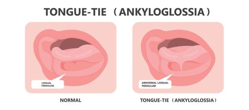
Figure 1 Typical lingual frenulum anatomy compared with ankyloglossia
In class, Ankyloglossia was defined as a condition characterized by the extension of the frenulum of the tongue to the tip, impeding its protrusion Nonetheless, upon extensive searches on the condition, it seems as though achieving consensus on aspects pertinent to this congenital anomaly remains an ongoing endeavor Ankyloglossia, or tongue-tie, is currently defined by the International Affiliation of Tongue-Tie Professionals organization as restricted tongue mobility stemming from a constrictive lingual frenulum Specifically, the frenulum, or “tissue remnant,” is located on the midline between the tongue’s ventral surface and the mouth’s floor
The condition may result in many symptoms, varying in severity
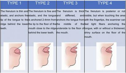
The above-mentioned discord regarding Ankyloglossia’s definition stems from the noticeable need for agreement regarding the classification system and accepted diagnostic parameters for the condition The diagnostic process often relies on various grading tools, albeit vague in their application. Among diagnostic tests, the Coryllos classification is employed to categorize the type of frenulum Nonetheless, the method overlooks the crucial evaluation of tongue function as a diagnostic parameter In contrast, the Hazelbaker Assessment Tool for Lingual Frenulum Function (HATLFF) provides a more comprehensive and complete inspection by examining both the tongue anatomy and the tongue function This tool works through a point system, in which 10 points are assigned for frenulum anatomy and 14 points are allotted for tongue function The scores obtained from this assessment then guide recommendations for surgical intervention. For instance, surgery is not deemed necessary if a function score reaches 14, irrespective of the anatomy score, whereas intervention is usually advised if the anatomy score falls below 8
The plausible causes of Ankyloglossia remain unknown While some studies have implied a potential link between Ankyloglossia and syndromes such as X-linked cleft palate syndrome, Kindler syndrome, Optiz syndrome, and Van der Woude syndrome, these occurrences are uncommon, and Ankyloglossia usually presents in individuals without any congenital pathologies or diseases On the contrary, some studies support the idea that the condition may be inherited genetically, while others hypothesize potential risk factors, such as newborns born to mothers who use cocaine
The lack of agreement on standardized diagnostic criteria and evaluation tools leaves the incidence and epidemiology of this congenital disorder vague While the disease is more common in males, there is no bias based on race Incidence rates vary widely, ranging from as low as 0 1% to as high as 10 7%, with significant increases in recent years, possibly due to the broad definition of the condition Specialists suggest that Ankyloglossia might be overdiagnosed in our society, potentially leading to unnecessary surgical procedures Other factors that may contribute to the increased diagnoses in children include increased awareness of the condition, heightened focus on breastfeeding, its benefits, and the impact of Ankyloglossia on the processes and growth in medical practitioners detecting and treating the condition
Symptoms primarily revolve around the limitations imposed by restricted tongue mobility The condition can present itself during breastfeeding, marked by difficulties such as ineffective latching and persistent latch loss, both of which may ultimately result in inadequate weight gain and failure to thrive For mothers, symptoms include discomfort and pain during breastfeeding, insufficient milk production, and incomplete emptying due to defective suckling This can lead to an array of issues, including nipple infections, ulceration, and bleeding, which may prompt early cessation of breastfeeding, exacerbating the
feeding difficulties, poor weight gain, and failure to thrive in the infant Later in development, the disease hinders the pronunciation of consonants and specific sounds and mechanically impedes various actions such as lip licking, kissing, and performing tongue tricks
The absence of conclusive diagnostic and management criteria for the disease leads to disputes about optimal intervention for its treatment if deemed necessary The most common procedure, frenotomy, is an incision of the lingual frenulum to release the tongue and allow mobility The procedure is typically recommended for patients with outstanding HATLFF scores or who have not responded to alternative non-invasive treatments; prompt intervention is recommended to mitigate potential breastfeeding difficulties
Ethical concerns have been raised regarding the invasive nature of the surgery, particularly as it has not been associated with any beneficial outcomes beyond encouraging continued breastfeeding As such, specialists endorse an initial evaluation by lactation consultants for those presenting with breastfeeding problems to assess for any potential underlying causes, such as airway obstruction, laryngopharyngeal reflux, and craniofacial anomalies, including abnormal palatal, mandibular, or maxillary maturation Maternal facets should also be looked into, including nipple anatomy, position and/or support of the breast, milk supply, and breast infection Also, surgery is contraindicated in infants with neuromuscular disorders, hypotonia, retrognathia, coagulopathies, and micrognathia Still, these contraindications are sometimes dismissed due to the procedure’s perceived low risk Complications are uncommon

Ethical concerns have been raised regarding the invasive nature of the surgery, particularly as it has not been associated with any beneficial outcomes beyond encouraging continued breastfeeding. As such, specialists endorse an initial evaluation by lactation consultants for those presenting with breastfeeding problems to assess for any potential underlying causes, such as airway obstruction, laryngopharyngeal reflux, and craniofacial anomalies, including
abnormal palatal, mandibular, or maxillary maturation Maternal facets should also be looked into, including nipple anatomy, position and/or support of the breast, milk supply, and breast infection. Also, surgery is contraindicated in infants with neuromuscular disorders, hypotonia, retrognathia, coagulopathies, and micrognathia Still, these contraindications are sometimes dismissed due to the procedure’s perceived low risk Complications are uncommon
Ankyloglossia presents challenges due to its unclear definition and diagnostic criteria Causes remain undetermined, with controversies over its association with specific syndromes and genetic inheritance. Diagnosis is challenging due to the lack of standardized tools and possible overdiagnosis Symptoms vary from breastfeeding difficulties in infants to speech impediments and motor constraints later in life Treatment often involves surgical intervention, but ethical concerns persist due to limited evidence of its benefits beyond aiding breastfeeding A comprehensive evaluation by specialists is recommended to identify alternate underlying issues
References:
Becker S, Brizuela M, Mendez MD Ankyloglossia (Tongue-Tie) [Updated 2023 Jun 9] In: StatPearls [Internet] Treasure Island (FL): StatPearls Publishing; 2024 JanAvailable from: https://www ncbi nlm nih gov/books/NBK482 295/
Becker, S , Brizuela, M , & Mendez, M D (2023) Ankyloglossia (Tongue-Tie) In StatPearls StatPearls Publishing
Brzęcka, D , Garbacz, M , Micał, M , Zych, B , & Lewandowski, B (2019) Diagnosis, classification and management of ankyloglossia including its influence on breastfeeding Developmental period medicine, 23(1), 79–87 https://doi org/10 34763/devperiodmed 20192 301.7985
González Garrido, M D P , García-Munoz, C , Rodríguez-Huguet, M , Martin-Vega, F J , Gonzalez-Medina, G , & Vinolo-Gil, M J (2022). Effectiveness of Myofunctional Therapy in Ankyloglossia: A Systematic Review International journal of environmental research and public health, 19(19), 12347.
https://www mdpi com/1660-4601/19/19/12347
Messner, A.H., Walsh, J., Rosenfeld, R.M., Schwartz, S R , Ishman, S L , Baldassari, C , Brietzke, S E , Darrow, D H , Goldstein, N , Levi, J , Meyer, A K , Parikh, S , Simons, J P , Wohl, D.L., Lambie, E. and Satterfield, L. (2020), Clinical Consensus Statement: Ankyloglossia in Children Otolaryngology–Head and Neck Surgery, 162: 597-611
https://doi.org/10.1177/0194599820915457 (https://doi org/10 1177/0194599820915457)
Figure 1 https://kidscorner ca/wpcontent/uploads/2023/09/kids-corner-blog12.jpg
Figure 2
https://oss jocpd com/files/article/202211161629/pdf/46-6%287%29 pdf
Figure 3.
https://dentagama com/img/031020073041Fr enotomy png

Writtenby:AdrielGuevárezGalán
PublishedonlineonMay24,2024
During my studies in medical school regarding the Development of the Male Genital Systems, a topic of discussion were some variants on the penis development Hypospadias (HP) were one of the clinical correlations that were discussed, with an emphasis on the high prevalence on congenital variants in the population of Puerto Rico which affects around 1 to 125 live male birthsThis is second to Ventral Septal Defects, a type of heart defect, on the most prevalent findings HP are defined as failure in closure of the ventral foreskin of the penis that can extend all the way to the scrotum This will result in a variable location of the external urethral orifice, depending on where the failed closure occurred
HP can be further classified on its severity regarding its anatomical location This includes in the glandular penis or anterior (mild), midshaft/distal penis (moderate), or from proximal penis to perineal region (severe) It is believed that the underlying cause for HP are both genetical and environmental factors affect this development Specifically, exposure to either estrogen or androgen like compounds in utero Interestingly, males with HP have been associated to testicular, bladder, and prostate cancer, but also with male infertility (Lo E, et al 2020)
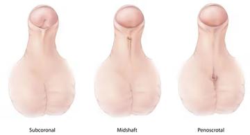
A main endocrinological disruptor that increases the likelihood for developing HP is Diethylhexyl phthalate (DHEP) a common plasticizer used in polyvinylchloride (PVC) plastics. The main mechanism for DEHP is decreasing the levels of Transforming Growth Factor β (TGF-β) It is important to note that this transcription factor is involved in molecular signaling pathways, that in the formation of male genitalia is epithelialmesenchymal transition (EMT) This mechanism is highly repeated in other process of embryology and some cancers, but it is generally a process where epithelial cells change to mesenchymal cells to give rise to other tissues and structures
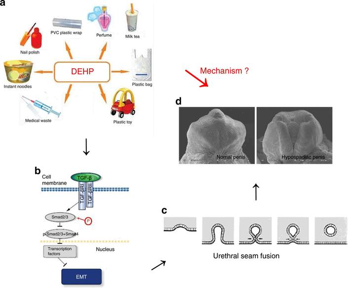
Figure 2. From Zhou et al. 2020: “The mechanism of hypospadias induced by DEHP in rats. The mechanism of hypospadias induced by DEHP was unknown (a → d). DEHP entered the body through various routes (a); gestational DEHP exposure could inhibit urethral EMT mediated by the TGF-β/Smad signal pathway (b); decreased EMT would lead to retardation of urethral seam fusion (c), eventually resulting in the occurrence of hypospadias (d). DEHP diethylhexyl phthalate, EMT epithelial–mesenchymal transition, TGF-β transforming growth factor beta.”
In the male penis, it has been characterized that urethral epithelial cells undergo EMT and fuse with urethral folds to form a urethral fusion plate Eventually both sides are expected to fuse medially, giving the penis’ cylindrical shape If the urethral seam gets halted, that is where hypospadias will develop It has also been characterized that TGF-β/Smad signaling pathway regulates EMT in this region When mice were exposed to DEHP this signaling pathway was inhibited, evidenced by Scanning Electron Microscopy (SEM) with a defect in penis development. Specifically in ventral urethral groove, where failed closure was observed Interestingly in light microscopy preparation, the urethral opening was observed towards the outside, where normally it would close towards the inside
Another aspect to consider is the role of microRNA’s, which are small non-coding RNA molecules that regulate gene expression, that play part in the development of HP microRNA200c (miR-200), has specific action on regulating Zeb1 gene, which further regulates expression E-cadherin and vimentin, which are markers for EMT Also, miR-200 has been found that it can reduce the effects of TGF-β1’s effects on inducing EMT. The specific mechanism is that miR-200 inserts in the 3’ Untranslated
Region (3’UTR) of Zeb1’s messenger RNA, preventing its translation, and thus preventing the EMT Meaning that when miR-200 is reduced and Zeb1 expression is increased, it will most likely result in the development of HP So, a possible therapeutic is suggesting induction of miR-200 to further induce EMT and stimulating closure of ventral urethral groove These findings lead the role on the pathogenesis of hypospadias (Quian, C et al, 2016)
Another morphogen to consider in the development of HP is sonic hedgehog (SHH) Where aberrant expression of SHH can lead to failure of the formation of the genital tubercule
This in part cannot produce the phallus and persistent cloaca, which are structures related to the genitourinary system Interestingly, transcription factors such as FOXA1 and FOXA2 regulate the transcription of SHH. However, only retaining a single copy of FOXA1 but mutation/deletion of FOXA2 retains division of the cloaca but will not form the urethral tube formation leading to the formation of HP. Leading to state that both genes are required to induce SHH to further induce the formation of the urethral tube and preventing the variant of HP. (Gredler, M. et al, 2020).
The development of hypospadias can be explained by different molecular signaling pathways However, the common denominators in the pathways include EMT and proper formation of the urethral tube. Interestingly, distinct environmental hazards play a role in the development of hypospadias
References:
1
Gredler, M L , Patterson, S E , Seifert, A W , & Cohn, M J (2020) Foxa1 and Foxa2 orchestrate development of the urethral tube and division of the embryonic cloaca through an autoregulatory loop with Shh Developmental Biology, 465(1), 23–30 https://doi.org/10.1016/j.ydbio.2020.06.009
3
Lo, E. M., Hotaling, J. M., & Pastuszak, A. W. (2020) Urologic conditions associated with malignancy Urologic Oncology, 38(1), 23–30 https://doi.org/10.1016/j.urolonc.2018.12.018
2. Qian, C , Dang, X , Wang, X , Xu, W , Pang, G , Chen, Y , & Liu, C (2016) Molecular mechanism of MicroRNA-200c regulating transforming growth factor-β (TGF-β)/SMAD family member 3 (SMAD3) pathway by targeting zinc finger E-box binding homeobox 1 (ZEB1) in hypospadias in rats
Medical Science Monitor: International Medical Journal of Experimental and Clinical Research, 22, 4073–4081 https://doi.org/10.12659/msm.896958
4
Zhou Y, Huang F, Liu Y, Li D, Zhou Y, Shen L, Long C, Liu X, Wei G TGF-β1 relieves epithelial-mesenchymal transition reduction in hypospadias induced by DEHP in rats
Pediatr Res 2020 Mar;87(4):639-646 doi: 10 1038/s41390-019-0622-2 Epub 2019 Nov 14 PMID: 31726466

Writtenby:HéctorCancel-Asencio
Have you ever wondered how drugs work to combat neurodegenerative diseases like Alzheimer’s? Welcome back to the second part of this exploration, where we delve deeper into the intricate mechanisms and pharmaceutical options related to Alzheimer’s Disease
You can read the first part by scanning the following QR code:

Alzheimer’s Disease (AD) is the most common form of neurodegenerative disease and poses a significant public health challenge worldwide Despite decades of research, there is still no cure for AD, making prevention the primary focus
In today’s article, we’ll peel back the layers to reveal the science behind Alzheimer’s treatments. Whether you’re a medical professional, researcher, or someone affected by this condition, this article aims to provide valuable insights into the latest advancements in Alzheimer’s treatment strategies
We’ll explore the drugs available in pharmacies, and the treatments they offer Don’t worry, we’ll keep things simple for easy understanding
Before we dive in, I want to express my gratitude to Jessica Tang, a medical student at the University of California, Davis School of Medicine, for her valuable contributions to today’s article Thanks, Jessica, for sharing your expertise with us
In the realm of AD treatment, the pharmaceutical industry offers a range of therapeutic options targeting different aspects of the condition While medications for AD do not offer a cure, it’s significant to recognize that they can enhance the quality of life and extend periods of independence for individuals affected by the condition
Currently, the Food and Drugs Administration (FDA) has given the green light to various drugs designed to tackle different symptoms of Alzheimer’s disease as it progresses through its various stages These stages – mild, moderate, and severe – are determined by how well someone does on tests that check memory, understanding of time and location, and ability to think and reason, mostly falling under the physical and neurological exam We cannot leave behind that these tests are accompanied by brain scans such as magnetic resonance imaging (MRI), computed tomography (CT), and positron emission
tomography (PET), as well as other biomarkers such as blood tests, cerebrospinal fluid tests, and genetic tests
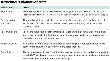
Monoclonal antibodies, such as Aducanumab and Lecanemab, represent a significant advancement as the first disease-modifying therapies for AD Amyloid-beta (A-β) plaques are not cleared by the body as a consequence of the development of AD Aducanumab primarily targets the A-β plaques for clearance, while Lecanemab exhibits a higher affinity for protofibril A-β, which eventually aggregate to form the A-β plaques
Additionally, drugs like Donepezil, Galantamine, and Rivastigmine function by inhibiting acetylcholinesterase, an enzyme responsible for breaking down acetylcholine By doing so, these drugs increase the concentration of acetylcholine at synapses, aiming to improve the symptoms associated with AD These diverse treatment approaches offer hope for alleviating the burden of AD and improving the quality of life for affected individuals
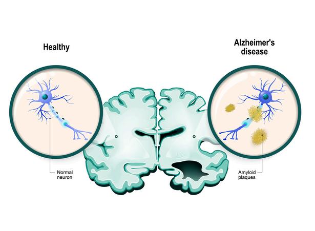
In addition to the pharmaceutical industry’s efforts in Alzheimer’s treatment, ongoing research is shedding light on novel approaches and insights We can see hundreds or even thousands of ongoing research dedicated to finding better understanding and possible drugs that can help for this and many neurological diseases
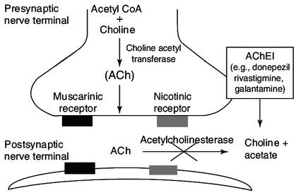
I know this image might look complicated, even the pronunciation is hard but, this is the simplest explanation of how these acetylcholinesterase inhibitors works and their mechanism of action
During my conversation with Jessica Tang, she highlighted key aspects of her research Her studies were centered on AD within the context of tauopathies, which are characterized by abnormal accumulation of tau protein aggregates Specifically, she investigated the effects of Fyn Kinase inhibition on a mouse model of tauopathy Her findings suggested that inhibiting Fyn Kinase could potentially reduce phospho-Tau accumulation in the brain, offering a promising avenue for further exploration
However, recent developments, such as the discontinuation of Adacanumab (published on January 31st of 2024 by Biogen) , have prompted shifts in Alzheimer’s research and treatment strategies The decision to limit coverage of future anti-amyloid therapies by the Center for Medicare and Medicaid Services has redirected researchers’ focus toward alternative drug targets and mechanisms This includes exploring treatments that target inflammation, alongside continued investigation into amyloid and tau-related therapies
Despite advancements, challenges persist in developing effective treatments for Alzheimer’s disease These challenges include the need to diversify drug targets and mechanisms beyond amyloid and tau Looking ahead, nonpharmacological interventions and multidisciplinary approaches hold promise in slowing disease progression
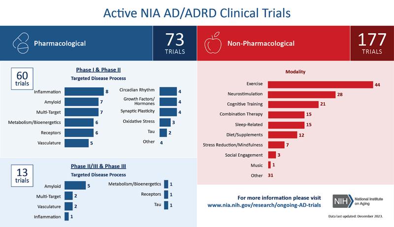
I hope you enjoyed this “simple” breakdown of this complex disease The amount of research behind this and many neurological diseases in order to explain how they develop and attack our body is still far from our understanding and with big limitations since diagnosing them poses a challenge, and medications face obstacles in reaching the brain due to the brain’s distinct blood supply, which is largely isolated from the rest of the body But with this information, you can now understand a little bit more that we, the scientific community, are working toward finding answers without rest Together, let’s continue unraveling the mysteries of the brain, one discovery at a time!

2
3
References:
Tang, S J , Fesharaki-Zadeh, A , Takahashi, H , Nies, S H , Smith, L M , Luo, A , Chyung, A , Chiasseu, M , & Strittmatter, S M (2020) Fyn kinase inhibition reduces protein aggregation, increases synapse density and improves memory in transgenic and traumatic Tauopathy Acta neuropathologica communications, 8(1), 96
1 Miller, G (2020) Seeking a better test for Alzheimer’s Knowable Magazine https://doi org/10 1146/knowable-080720-1
Chowdhury, S , & Chowdhury, N S (2023) Novel anti-amyloid-beta (Aβ) monoclonal antibody lecanemab for Alzheimer’s disease: A systematic review International journal of immunopathology and pharmacology, 37, 3946320231209839
https://doi org/10 1177/03946320231209839
4 Zhang, Y , Chen, H , Li, R , Sterling, K , & Song, W. (2023). Amyloid β-based therapy for Alzheimer’s disease: challenges, successes and future Signal transduction and targeted therapy, 8(1), 248 https://doi.org/10.1038/s41392-023-01484-7
NIA-Funded Active Alzheimer’s and Related Dementias Clinical Trials and Studies (n d ) National Institute on Aging. https://www nia nih gov/research/ongoingAD-trials
5 Moghul, S , & Wilkinson, D (2001) Use of acetylcholinesterase inhibitors in Alzheimer’s disease Expert Review of Neurotherapeutics, 1(1), 61–69. https://doi org/10 1586/14737175 1 1 61
7
6 https://investors biogen com/newsreleases/news-release-details/biogenrealign-resources-alzheimers-diseasefranchise


Arbona Health Hub Invites Premeds, Medical Students, Residents, and Healthcare Professionals to Contribute!
We are extending an open invitation for these groups to submit their articles for consideration.
Why Contribute?
Showcase Expertise: Share your knowledge with our engaged audience.
Impactful Insights: Contribute to vital health discussions, making a positive impact.
Networking Opportunities:
Connect with a community of healthcare professionals and enthusiasts.
Profile Elevation: Boost your online presence and credibility in healthcare.
Volume 1. Issue 1. March-Jun 2024
The Arbona Health Hub publication is published quarterly by the Arbona Health Hub team
Email: arbonahealthhub@gmail.com
Publisher: Arbona Health Hub INC. Caguas, Puerto Rico
Publication Editors: Alejandro Arbona-Lampaya, Avery R. Arsenault
Article Editors: Alejandro Arbona-Lampaya, Avery R. Arsenault, Héctor Cancel Asencio, Marcel De Jesús Vega, Adriel Guevárez Galán, Sofía I. Malavé Ortiz, Ricardo A. Pérez Ojeda
Contributing Writers: Alejandro Arbona-Lampaya, Avery R. Arsenault, Héctor Cancel Asencio, Christian G Correa García, Marcel De Jesús Vega, Gustavo A Garriga García, Adriel Guevárez Galán, Ricardo A Pérez Ojeda, Sahudi N Soto Rodriguez, Russel S Steans, Javier A Velázquez González Read Arbona Health Hub online by