ImageProcessing forAutomatedDiagnosis ofCardiacDiseases
Editedby KalpanaChauhan
DepartmentofElectricalEngineering,CentralUniversityofHaryana, Mahendragarh,India
RajeevKumarChauhan
DepartmentofElectricalEngineering,DayalbaghEducationalInstitute, Agra,India
AcademicPressisanimprintofElsevier 125LondonWall,LondonEC2Y5AS,UnitedKingdom 525BStreet,Suite1650,SanDiego,CA92101,UnitedStates 50HampshireStreet,5thFloor,Cambridge,MA02139,UnitedStates TheBoulevard,LangfordLane,Kidlington,OxfordOX51GB,UnitedKingdom
Copyright©2021ElsevierInc.Allrightsreserved.
Nopartofthispublicationmaybereproducedortransmittedinanyformorbyanymeans,electronicor mechanical,includingphotocopying,recording,oranyinformationstorageandretrievalsystem,without permissioninwritingfromthepublisher.Detailsonhowtoseekpermission,furtherinformationaboutthe Publisher’spermissionspoliciesandourarrangementswithorganizationssuchastheCopyrightClearance CenterandtheCopyrightLicensingAgency,canbefoundatourwebsite: www.elsevier.com/permissions.
ThisbookandtheindividualcontributionscontainedinitareprotectedundercopyrightbythePublisher(other thanasmaybenotedherein).
Notices
Knowledgeandbestpracticeinthisfieldareconstantlychanging.Asnewresearchandexperiencebroadenour understanding,changesinresearchmethods,professionalpractices,ormedicaltreatmentmaybecome necessary.
Practitionersandresearchersmustalwaysrelyontheirownexperienceandknowledgeinevaluatingandusing anyinformation,methods,compounds,orexperimentsdescribedherein.Inusingsuchinformationormethods theyshouldbemindfuloftheirownsafetyandthesafetyofothers,includingpartiesforwhomtheyhavea professionalresponsibility.
Tothefullestextentofthelaw,neitherthePublishernortheauthors,contributors,oreditors,assumeanyliability foranyinjuryand/ordamagetopersonsorpropertyasamatterofproductsliability,negligenceorotherwise,or fromanyuseoroperationofanymethods,products,instructions,orideascontainedinthematerialherein.
LibraryofCongressCataloging-in-PublicationData
AcatalogrecordforthisbookisavailablefromtheLibraryofCongress
BritishLibraryCataloguing-in-PublicationData
AcataloguerecordforthisbookisavailablefromtheBritishLibrary
ISBN978-0-323-85064-3
ForinformationonallAcademicPresspublications visitourwebsiteat https://www.elsevier.com/books-and-journals
Publisher: MaraConner
AcquisitionsEditor: TimPitts
EditorialProjectManager: ChiaraGiglio
ProductionProjectManager: SojanP.Pazhayattil
CoverDesigner: MilesHitchen
TypesetbySPiGlobal,India
Contributors
MeghaAgarwal
DepartmentofElectronicsandCommunicationEngineering,JaypeeInstituteofInformation Technology,Noida,India
RajeevAgrawal
DepartmentofElectronicsandCommunicationEngineering,G.L.BajajInstituteofTechnologyand Management,GreaterNoida,India
V.AjanthaDevi
AP3Solutions,Chennai,TamilNadu,India
M.A.Ansari
DepartmentofElectricalEngineering,GautamBuddhaUniversity,GreaterNoida,India
ArunBalodi
DepartmentofElectronicsandCommunicationEngineering,AtriaInstituteofTechnology, Bangalore,India
KalpanaChauhan
DepartmentofElectricalEngineering,CentralUniversityofHaryana,Mahendragarh,India
RajeevKumarChauhan
DepartmentofElectricalEngineering,DayalbaghEducationalInstitute,Agra,India
I.Lakshmi
DepartmentofComputerScience,StellaMarisCollege,Chennai,India
RajatMehrotra
DepartmentofElectricalEngineering,GautamBuddhaUniversity,GreaterNoida,India
AmolD.Rahulkar
DepartmentofElectricalandElectronicsEngineering,NationalInstituteofTechnology,Goa,India
AnjuSaini
DepartmentofMathematics,GraphicEraUniversity,Dehradun,India
AswiniK.Samantaray
DepartmentofElectricalandElectronicsEngineering,NationalInstituteofTechnology,Goa,India
AmitSinghal
DepartmentofElectronicsandCommunicationEngineering,BennettUniversity,GreaterNoida, India
PragatiTripathi
DepartmentofElectricalEngineering,GautamBuddhaUniversity,GreaterNoida,India
T.Vani
DepartmentofComputerScience,RajeswariVedachalamGovernmentArtsCollege[Affiliatedto UniversityofMadras],Chengalpattu,TamilNadu,India
Preface
Thefieldofmedicalimageprocessingisexpandingdaily,asisitsuseinindustrialandmedicalfields. Therearemanychallengesandopportunitiesinimageprocessingmethodsandongoingresearchis examininghowtousethesemethodstoautomaticallydiagnosediseases.Thisbookexaminesthecurrentandemergingtechnologiesdevelopedfortheautomateddiagnosisofcardiacdiseases.Theconceptsoutlinedinthisbookcanbetestedforresearchpurposesandthenewadvancesinalgorithmscan beappliedinpracticalapplications.Readerswilllearnsomeofthetechniquesusefulforobtainingimagesoftheheart.Thebookpresentsbasicaswellasadvancedconceptsofimageprocessingtechniques.
Chapter1 discussesdifferentheartdiseases,includingirregularitiesthatinfluencethenormalfunctioningoftheheartvalves,theheart’selectricalsystem,andthemusclesandcoronaryarteriesofthe heart.Thefocusofthischapterisheartvalvediseases,especiallythoserelatedtothemitralvalve.In particular,thechapterexaminesthediagnosis,causes,andsymptomsofmitralregurgitation(MR).Itis shownthatechocardiographyisthesuperiorimagingtechniqueinthisdisease.
Chapter2 dealswithmachinelearning(ML)proceduresincardiovascularmultimodalimaging.In particular,thechapterproposesconvolutionneuralnetwork(CNN)modelsforfeaturingthecorrespondencesbetweenmultimodalinformation.Theseportrayalsareadditionallyexpectedtovisualizethe cardiovascularlifestructuresinmoredetailforbetterunderstandingandinvestigation.Inaddition, thechapterexamineshowquantitativeinvestigationcanbenefitwhenthesescholarlyimageportrayals areutilizedindivision,movementfollowing,andmultimodalimageregistration.
Chapter3 depictsthecardiacanatomyindetailforbetterunderstandingandstudy.Inadditiontoanatomicalstudy,thechapterdiscusseshowquantitativeresearchcanbenefitfromtheuseoftrainedimage representationsinsegmentation,motiontracking,andmultimodalimageregistration.Aprobabilistic edge-maprepresentationisimplementedtodefineanatomicalcorrespondenceinmultimodalcardiacimagesandtodemonstrateitsuseinspatialimagealignmentandanatomicallocalization.Inaddition,a novelimagesuper-resolutionsystemisimplementedtoimprovecardiaccinemaMRimages.
Chapter4 offersabriefdescriptionofthetheoreticalstructuresandtheirapplicationsforsegmental cardiacimaging,imageenhancement,andmultimodalimagealignment.Theseanalyticalmethods sharecommongoals:timeefficiency,quantitativeobjectiveevaluation,andenhancementandanalysis ofmultimodalimagedata.Inthissurvey,theauthorsconcentrateonhowthelearningofimagerepresentationwillaccomplishthesegoalsandimprovetheaccuracyandrobustnessofthetechniques applied.
Chapter5 describesfuzzy-baseddespecklingmethodsforechocardiographicimages.Theauthors proposeandanalyzehybridfuzzyfiltersthatintegrateanon-localmeans(NLM)filterwiththreedifferenttypesoffuzzyfilters.Theauthorscomparetheproposedmethodswithfifteendespecklingfilters onstandardtestimagesandechocardiographicimagesofthemitralvalveinthreeviews.Theperformanceofoneproposedhybridfuzzyfilter,HFF3,exhibitedthebestperformancecomparedtothe othersintermsofedgepreservationanddenoisingofspecklenoise.
Chapter6 examinesmachinelearning-basedmedicaldiagnosisasafast,non-invasive,timesaving, andaccuratemethod.Asthismethodisnon-invasive,itispreferredoverexistingmethods.Thechapter explainstheconceptofmachinelearninganditssignificanceinthemedicaldiagnosisofcardiac diseases.
Chapter7 examinestheuseofvariouswavelettransformsincontent-basedimageretrievalfordiagnosisofcardiacdiseases.Itdiscusseswaveletpropertiesandanalyzesretrievalperformanceofvariousorthogonal,bi-orthogonal,andGaborwavelettransforms.Theauthorsevaluatethedifferent wavelettransformsusingseveralcardiacimagedatabases,namely,NEMA,OASIS,andEXACT09, intermsofaverageretrievalprecision(ARP)andaverageretrievalrate(ARR).
Chapter8 illustratesbroadlyconstructedcomputer-aidedapproachesforevaluatingECGsignals. Artificialintelligencetechniquesgivepreciseandmechanicalclassificationsofheartbeatstoidentify arrhythmiasorunexpectedchangesincardiacmorphology.Thesetechniquesarealsousedforautomaticsyndromeanalysis,monitoring,andstratificationbymanagingextendedECGrecordingsfor whichdiagramandphysicalinvestigationscanbemonotonousandtimeconsuming.AIisflexible andcanbepracticallyutilizedinwearableECGdevices,assuringcompetentanddependablemonitoringoftheheartinbothclinicalandresidentialsettings.Thechapteralsoexamines3DcomputersimulationsasinfluentialapparatusesforunderstandingECGresults.
Chapter9 proposesanewregularizationmodelfordetectingECGimageboundaries.Themethod helpsthecurvetoapproachthedesiredboundarieswhilemaintainingsmoothnessforbettervisualization.Theauthorsuseregion-basedsegmentationalongwithspeckledensityasthedatafittingenergyto determineintensityinformationinlocalregions.Theproposedimprovedregularizationandfittingbasedsegmentation(IRFS)techniquewithanewregularizationmodelandfittingfunctionsuccessfully achievedtherightminimaandregionalongwithimprovedcapabilityofthecurvetodrawthedesired boundaries.
Chapter10 considersapubliclyavailabledatasetofcine-MRI(magneticresonanceimaging)imagestodetectheartfailurecaseswith(orwithout)infarction.Localtexture-basedpatternsareusedto extractrelevantinformationfromtheimage.Thechapterexaminesfourdifferenttypesofpatternbasedfeatures:localbinarypattern(LBP),localternarypattern(LTP),differenceofGaussianLTP (DoGLTP),andternaryco-occurrencepattern(LTCoP).Variousmachinelearningclassifiersare employedtodifferentiatebetweennormalheartimagesandheartfailureimages.Performancemetrics arecomputedfortheseclassificationstrategiesandadetailedcomparisonisprovidedtohighlightthe mostaccuratemethodforautomatedidentificationofheartfailure.
Chapter11 isaboutthefusionmethodadoptedinthediagnosisofcardiacdiseases.Theadvancementsinmedicalimagefusionresearchoutlinedinthischapterdemonstratetheimportanceoffusionin improvingcardiacdiagnosis,monitoring,andvisualization.Thealgorithmsusedforcardiacimagefusionmethodscanimproveimagequalityandcanbeusedindifferentapplications.Theprominentapproachestestedoncardiacimagesincludediscretewaveletstransform(DWT),principlecomponent analysis(PCA),andmaximummodel.Theperformanceofthemethodsshowsthatthecombination ofoneormoremethodsofimagefusioniseffectiveincardiacimageanalysis.
Cardiacdiseasesandtheirdiagnosis methods
1.1 Introduction
Theheartisamuscularstructureandacentralcomponentofthevertebratecardiovascularsystem.The heartfunctionsinaclosedloopmanner,thatis,oxygenatedbloodispumpedfromthelungstothe wholebodyanddeoxygenatedbloodispumpedbackfromthelungstothebody.Thetransferofblood fromhearttothebodyiscarriedoutbythearteriesandarterioles,whilethereturningofthebloodis donethroughthevenulesandveins.Bloodtransportisvitaltobringoxygenandnutrientstothebody’s tissuesaswellastoremovecarbondioxideandwasteproducts/chemicals [1].
Thehumanheartislocatedbetweenthelungs.Becauseofslighttiltingofitsapexontheleftsideof thechest,heartrhythmorbeatingoccursinthislocationcausinganillusionthattheheartislocatedon thatside.Thesizeofahumanheartisthatofatightlyclosedfist.Itbeatsabout100,000timesinaday.
Althoughtheheartpumpsblood,deliveringoxygentotheentirebody’smusclesandorgansfor themtofunction,italsoneedsitsownoxygen-enrichedbloodtoworkproperly.Theheartfunctions asalargemuscularpumpwitharteries,veins,andvalves,andanelectricalsystem.Theelectricalsystemtriggerspulse,therebystimulatingthehearttobeat.Theheartmusclesthensqueezethebloodto pushtheoxygenatedbloodthroughouttheentirebodyinonelargearterialcircuitalsystemandthe deoxygenatedbloodthroughthepulmonaryarteriestothelungs.Thetwo,one-wayvalvescreateseparationbetweenthefourdifferentchambers,namely,theleftventricle(LV)andleftatrium(LA),and rightventricle(RV)andrightatrium(RA),forformingthedualpumpsoftheheartadjustingbothrate andflowoftheoxygenatedanddeoxygenatedbloodthroughouteachcardiaccycleorheartbeat.The moreactivityapersonperforms,themoretheheartmusclesmustworktosupplythenecessaryquantity ofbloodtothemusclestobeutilizedduringtheactivity.
Mitralregurgitation(MR)isamitralvalveinsufficiencythatcausesachangeinthesizeand/or shapeoftheLV,affectingitsfunctioningandresultingfromischemicheartdisease [2,3].MRleads tomyocardialinfarction(MI)inabout20%ofcases [4,5].TheseverityofMRincreasesaround30%in patientssufferingfromcoronaryarterydisease(CAD)withischemicLVdysfunction [6].
TherearemanyapproachesavailabletodiagnoseMRthatarehelpfulindeterminingseveritygrade anddysfunction [7–13].Diagnosticmethodsincludeassessmentofregurgitationvolume,orificesize, orifice,andregurgitantorificewiththehelpofechocardiographyorcatheterization.Inaddition,twodimensional(2D)contrastechocardiographyandDopplerechocardiographyareefficientwaysfor assessingMR.Wediscusstheadvantagesofthesetechniqueslaterinthechapter [14–20].
Tobegin,thischapterdiscussesdifferentheartconditionsbycategorizingheartvalvesandtheir relateddiseases,withaspecialfocusonMR.ItalsopresentsvariousdiagnosticmethodsandthequalitativeandquantitativeparametersusefulingradingMRseverity.Finally,thechapterendswithadiscussionofdifferentmodesandtechniquesofechocardiography.
1.2 Heartvalves
Thetwoatrioventricular(AV),one-wayvalvesarethinstructures,havingconnectivetissuesandendocardia.Thesevalves,namely,thebicuspid/mitralandthetricuspidAVvalvesarelocatedbetween theLAandtheLV,andtheRAandRV,respectively.Thetwosemilunar,one-wayvalvesaremadeup ofthreeflaps,eachcomposedofconnectivetissuesandendocardiumaswellasfiberstopreventthe valvesfromflappinginsideout.Theirshapesarelikeahalfmoonandthustheyarecalledthesemilunar

(SL)aorticvalveandSLpulmonaryvalve.Thesevalvesarelocatedbetweentheleftventricleandaorta andbetweentheRVandthestartofpulmonaryartery. Fig.1.1 showsthesevalves.Theheart’sone-way bloodflowismaintainedwiththehelpoffourheartvalves,eachonehavingaspecificpositiononthe exitsofthefourchambers.Thesefourheartvalvesallowonlytheone-wayflowofbloodintheforward directionsandrestrictthebackwardflowofblood.Sequenceofbloodflowisfromtheatria(rightand left)intotheventricles(rightandleft)throughtheopentricuspidandmitralvalves,respectively,as shownin Fig.1.1.Accordingtopressurechangeinthechambers,thereisanopeningorclosingof AVvalves.Theycloseduringtheventricularsystole(contraction)whentheventriclepressureincreasesthepressureinthetwoatria.Thisactionkeepsthevalvessnappedshutandpreventsbackward flowofblood.Thecontractionoftheventriclesleadstoforcedopeningofthepulmonaryandaortic valvestopumpthebloodfromtherightandleftventriclesintothepulmonaryartery(throughopen valves)towardsthelungs,andthroughtheaorticvalvetotheaortaandthebody.Attheendofcontraction,theventriclesbegintorelaxandtheaorticandpulmonicvalvesremainclosedduringthediastole.Backwardflowofbloodintotheventriclesispreventedbythesevalves.Thispatternrepeats againandagain,causingcontinuousbloodflowfromthehearttothelungsandthebody.
1.3 Mitralvalveregurgitation
Tovisualizethemitralvalve(MV),cliniciansmustchooseatechniquethatenhancestheimageaccordingtotheirvisualperceptionandthatworksinaccordancewiththekindofimage [21].Logtransformationdoesnotgivesatisfactoryresults(subjectiveassessment)inthecontrastenhancementof echocardiographicimagesduetohighwhitepixelspreading.Thiswhitespreadingoverlapstheimportantfeatures.Thereasonforthisproblemisthatmorepixelswillshiftinthehigh-intensityvaluewhen thelogtransformationisapplied.Toovercomethisproblem,thefigureof1inEq. (1.1) oflogtransformationisreplacedbyavariable,say, a.Thisoffersaflexiblewaytoanalyzetheimageatdifferent valuesof a.Thisvaluecanbechangedbycliniciansinaccordancewiththeirvisualperceptionsfor bettervisualizationoftheimage.ThenormalMVopenswhentheLVrelaxes(diastole)toallowblood flowfromtheLAandtofilltheLV(decompressed).
DuringsystoleorcontractionoftheLV,thepressureintheLVincreases.Thisincreasedpressure leadstoclosureoftheMVandrestrictsbloodflowfromleakingintotheLA.Atthistime,theblood flowstotheaorta(passingtheaorticvalve)andthebody.Theannulus,leaflets,andsubvalvularapparatusesworkinacomplexmannerfortheproperfunctioningofthevalve.Themitralleaflettissues
FIG.1.1
Classificationofheartvalves.
Table1.1Thelayersofvalvetissues:fibrosa,spongiosa,andatrialis/ventricularis [22].
LayerLocationCompositionFunction
FibrosaFacesthe LV
SpongiosaMiddle layer
Atrialis (Ventricularisfor semilunarvalves)
Facesthe LA
Highconcentrationofcollagen, thickestlayer
Highconcentrationof glycosaminoglycans(GAGs)and proteoglycans(PGs)
Highconcentrationofcollagenand elastinthinnestlayer
Bearsmostoftheloadduring coaptation
Providesshearbetweenouter supportlayersanddiffusesgasses andnutrients
Elastinallowsforstrainwhenthe valveisopen
areorganizedinthreelayers:fibrosa,spongiosa,andventricularis. Table1.1[23,24] describesthe location,composition,andfunctionsoftheselayers.
1.4 Heartdiseases
Heartdiseasesareabnormalitiesthataffectthevalves,functions,electricalsystem,muscles,andarteriesoftheheart.Somecommonheartdiseasesinclude:
•Coronaryarterydisease(CAD)
•Myocardialinfarction(MI)—aseveretypeofheartdisease
•Highbloodpressureorhypertension(HBP)
•Heartvalvedisease
•Cardiomyopathyorheartmuscledisease
•Pericarditis
•Rheumaticheartdisease(RHD)
1.4.1 Coronaryarterydisease(CAD)
CADisadiseaseinwhichadepositcalledplaquegrowsontheinsidewellsofthecoronaryarteriesand restrictsthenormaloxygenatedbloodsupplytoheartmuscles.Itisalmostoftenduetotheprogressive buildupofcholesterolandotherfattymaterials,knownasatheroscleroticplaqueoratheroma,inthe wallsofthecoronaryarteries.Thisprocessisknownasatherosclerosisandcanaffectmanyarteries,not justthoseoftheheart.Asanatheromadevelops,itmaygushintotheartery,narrowingtheartery’s interior(lumen)andpartiallyblockingbloodflow.Calciumaccumulatesinsidetheatheromaovertime. Thesupplyofoxygen-richbloodtotheheartmuscle(myocardium)becomesinadequatewhenanatheromablocksmoreandmoreofacoronaryartery.Toencouragegoodhealth,itisrecommendedto reducedietaryfatintaketonomorethan25–35%ofdailycalories.However,somedoctorsbelieve thattominimizetheriskofcoronaryheartdisease,fatmustbereducedto10%ofdailycalories.AnotherwaytomitigateriskfactorsforCADistoconsumealow-fatdietthatalsotendstolowerelevated totalandLDL(bad)cholesterollevels.
1.4.2 Myocardialinfraction(MI)
InsevereCADdisease,theplaquedepositsuddenlyblocksnormalflowofbloodandheattoheart musclessuchthattheycannotgetenoughoxygenandthusbegintodie.AnMIcausespermanentdamagetotheheartmuscleduetooxygenshortage.AnMImayresultinimpairmentofdiastolicandsystolicfunctionandcanrenderthepatientvulnerabletoarrhythmias.AnMImayalsocauseavarietyof seriouscomplications.
1.4.3 Highbloodpressureorhypertension(HBP)
HBPistheconditionwhentheforceofthebloodflowingthroughbloodvesselsiscontinuouslyvery high.TherearemanycausesofHBP,includingheartdisease,smoking,stress,lackofphysicalactivity, andsoon.
1.4.4 Heartvalvedisease
Improperfunctioningofvalvescreatesvalvediseases.Stenosisistherestrictionofbloodflowdueto narrowingofavalve.MRduetoincompleteclosingoftheMVisthemaintypeofheartvalvedisease.
1.4.5 Cardiomyopathyorheartmuscledisease
Cardiomyopathyisatypeofprogressivediseaseinwhichthereisanabnormalenlargement,thickening,andstiffingoftheheart.Duetotheseabnormalities,theheartmusclesarenotabletopumpblood properly.
1.4.6 Pericarditis
Pericarditisisaconditioninwhichthepericardiumbecomesinflamed.Pericarditisistypicallyacute, meaningitoccursoutofnowhereandcanlastformonths.Thedisordernormallygoesawayafterthree months,butattackscanlastforyears.
1.4.7 Rheumaticheartdisease(RHD)
RHDisachronicheartdiseasethatmayariseduetorheumaticfever,whichisastreptococcal(strepor throat)infection. Fig.1.10 showstheRHDcondition.
1.5 Mitralvalvediseases
MVdiseasesbroadlycanbeofthreetypes:(1)Mitralstenosis:whentheorificeoftheMVnarrowsand restrictsnormaldiastolicbloodflowfromtheLAintotheLV;(2)Mitralregurgitation(MR):backward bloodflowintotheLAduringsystolethatmaybeacuteorchronic;and(3)Mitralvalveprolapse:posteriordisplacementorbendingoftheanterior,posterior,orbothMVleafletstowardstheleftatrium. MRisthefocusofthischapterasitisthesecondmostcommonvalvularlesionafteraorticstenosis (AS).StudyofMRisunderstoodtocoveralltheotherMVdiseases.
1.5.1 Mitralregurgitation(MR)
MRormitralinsufficiencyisthemostcommonvalvedisorder.WhentheheartisaffectedbyMR,there isleakageofbackwardbloodflowthroughtheMVduringthecontractionandareductionintheamount ofbloodsuppliedtothebody.
IfMRdoesnotprogress,thentheamountofMRissmallandthebackwardleakagehasnosignificantconsequence.However,ifthereissignificantMR,thentheLVmustworkhardertofulfillthe oxygenatedblooddemandofthebody.Tomeetthisincreaseddemand,theheartmuscles(i.e.,myocardium)havetodomoreworkandthiscreatesasequenceofchangesinthebloodcirculationsystem. Thesetypesofchangestakealongperiodoftime,sometimesseveralyearsordecades,anddepend upontheseverityoftheregurgitation.ThecausesofMRalsodeterminehowquicklytheheartbegins tofail,thatis,theygivetheinformationoffailureintermsofweakeningofheartapparatus.Weakheart apparatusesarethesourcesofasuddenheartattack.
1.5.2 Causesofmitralregurgitation
MRmayincreasefrommildtomoderatetosevereduetovariouscardiacdiseasesorotherheartvalve abnormalities.Someoftheseinclude:
• Mitralvalveprolapse:Duetodeformationandelongationofvalveleaflets,thenormalcoaptationof theleafletsisrestricted.Thisisknownasmitralvalveprolapse.Duetothisabnormalityinvalve motion,thedirectionofbloodflowispartiallyreversed.ThebloodleaksbackwardfromtheLVto theLA.Mitralprolapsemayrangefrommildtosevere.
• Infectiveendocarditis:Sometimesheartvalvesareinfectedbybacteria,fungus,orsomeother organismsthataffectthebloodstream.Thisisknownasinfectiveendocarditis(IE).Theorganisms sticktothevalvescausinganabnormalstructure,knownasvegetation,togrow.Thisvegetation thickensthevalveandchangesitsdirection,thusrestrictingleafletsfromjoiningduringthevalve’s closingoperation.Endocarditisdevelopsfasteronpreviouslyabnormalheartvalvesthanonnormal heartvalves.
• Rheumaticfever:Rheumaticfeveroccursduetothroatinfection.Ifthisinfectionisnottreated,it cancauseinflammationoftheheartvalvesandothervalvularcomplications.Rheumaticfeveris commonindevelopingcountries.
• Congenitalheartabnormality:MRmayalsooccurinpatientsbornwithabnormalitiesoftheheart.
• Othertypesofheartdisease:Heartattacksandmuscleinjuriesandabnormalitiesmayalsolead toMR.
• Trauma:Whenthevalvechordsarebroken,thereisasuddendisplacementoftheleafletsandthus leafletsarenotabletowithstandtheirnormalposition.Theseflailedleafletsarenotabletojoin, allowingseverevalvularleakage.
1.5.3 Mitralregurgitationsignsandsymptoms
Incasesofmildandmoderateregurgitation,patientsmaynevershowsymptomsorseriouscomplications.EvenpatientswithsevereMRmayshownosignsandsymptoms,althoughsymptomsmayoccur iftheLVbecomesabnormalorifatrialfibrillationorpulmonaryhypertensionoccurs.Pulmonary
hypertensionoccurswhenbloodpressureincreasesinthepulmonaryartery.Thismakesitharderonthe rightsideofthehearttosupplyadequateoxygenatedbloodtothebody.
IftheLVisseverelyenlarged,thereisriskofseriousheartdiseaseandevenheartfailure.These patientstypicallyshowsignsofweakness,shortnessofduringwork,andcollectionoffluidinthelower legsandabdomensleadingtoswellinginthefeet.
1.5.4 Mitralregurgitationdiagnosis
MRmaybediagnosedbyusingastethoscopetolistenforaheartmurmur.Thechangeinheartsoundis duetobackflowofbloodthroughtheMV.Theremaybeotherreasonsforaheartmurmuraswell.Other recommendedtestsforMRdiagnosisinclude:
•ChestX-ray:AchestX-rayshowsapictureofthelargeheartvesselstodeterminesizeandshape.It isalsohelpfulindiagnosinglunginfectionsortheaccumulationoffluidinthelungs.IfX-rayshows enlargementoftheheart,thismayindicatesevereMR.
•Electrocardiogram(ECG):AnECGisaone-dimensionalsignalthatshowstheelectricalactivityof aheartbeat.AnECGmaybehelpfulindetectingdisturbedrhythmsrelatedtocausesofMRlike CAD.Disturbedrhythmscanalsoberelatedtootherabnormalitiesoftheheart.
•Echocardiogram:Anechocardiogramisadirectpictureoftheheart.High-frequencyultrasound wavesareusedtakeapictureoftheheart’schambersanddeterminetheirsizeandshape.Thetest cancapturemovementoftheheartvalvesaswellasthethickness,size,andmotionoftheheartwall. Thetestishelpfulfordeterminingthevolumeofbloodpumpedbytheheartperminute,alsocalled cardiacoutput.Echocardiographycanalsodetectthepressureindifferentchambersandmajor bloodvesselsoftheheart.Mostoften,echocardiographyisdonebyapplyingthetransducerfrom theoutside(i.e.,transthoracicechocardiogram(TTE)),whereassomecasesrequireinsertionofthe transducer(i.e.,transesophagealechocardiogram(TEE)).
EchocardiographyandcolorDopplerechocardiographyhavetheirownutilityintheevaluationofMR severity,however,theyeachexhibittheirownadvantages/disadvantagesandlimitations.Quantitative parametershelpclinicianstogroupMRintocategoriesofmild,moderate,andsevere.DopplerechocardiographyisamethodusedtoconfirmMR.M-modeor2DechocardiographyisnotabletodeterminethespecificsignsofMR.Continuousechocardiographicexaminationallowsthevisualizationof MRprogression.Changescanbeseenbytakingechocardiogramsatdifferenttimeintervals.Volume overloadingintheLAorintheLVistheinitialechocardiographicfeatureofMR.Volumeoverloading occursduetothestrokevolumetransferbetweenthetwochambers.However,thethicknessofthewall isnormal(becausethewallmassincreasesatthetimeofenlargement).Iftheend-diastolicdimension becomesgreaterthan5.5cm,andanoticeablehyperdynamicwallmotionisseenontheinterventricular septum,thenLVvolumeoverloadiseasilyrecognized.
1.6 Cardiacdiseasediagnosismethods
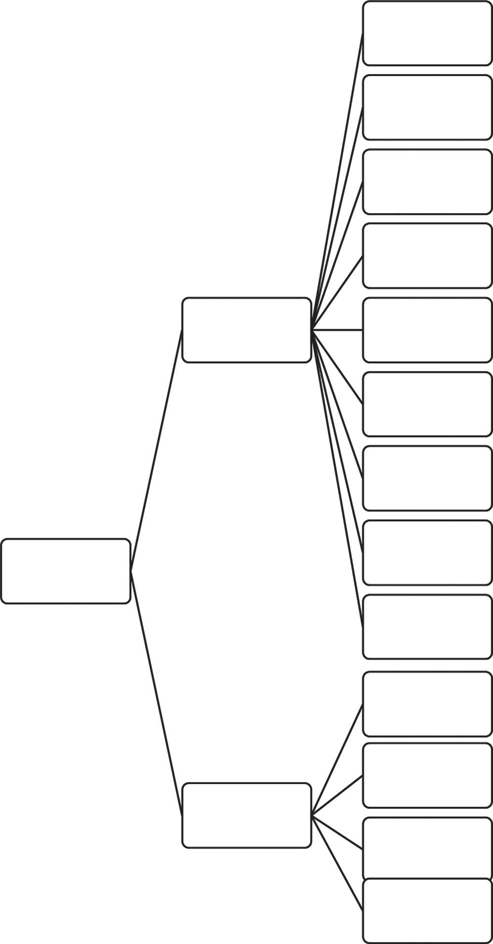
Therearevariousnoninvasiveandinvasivemethodstodiagnosethevariouscardiacdiseases.Each methodhasadvantagesandlimitations. Fig.1.2 showstheclassificationofthedifferentmethods.
Cardiac Diseases Diagnosis Methods
Noninvasive
Electrocardiogram (ECG)
Echocardiography (TT)
Stress ECG and Echocardiography
Carotid ultrasound
Nuclear Stress Test
Holter Monitor
Event Recorder
Cardiac CT scan
Cardiac MRI
Invasive
FIG.1.2
Differentcardiacdiseasesdiagnosismethods.
Cardiac Catheterization
Echocardiography (TTE)
Angiography
Electrophysiology Study
Echocardiographydetectsthesizeofthecardiacchamber,wallmotion,wallthickness,valvemotion,andanatomyofvalves,proximalgreatvessels,andpericardium.Itpresentsalivepictureofcardiacfunctionalityandanatomy.Echocardiographyisasensitivetoolfordeterminingvolumeofpleural andpericardialfluid,toidentifymasslesionsthatmaybeinsideorneartheheart.Thismodalityis effectivefordiagnosingcongenitalheartdiseasesaswellasmyocardialorvalvularpathology.Itis asafeprocedurebecauseTTEisdonewithouttheinsertionofchemicals,whichmaydamagethe myocardium.
1.6.1 Principlesofecho
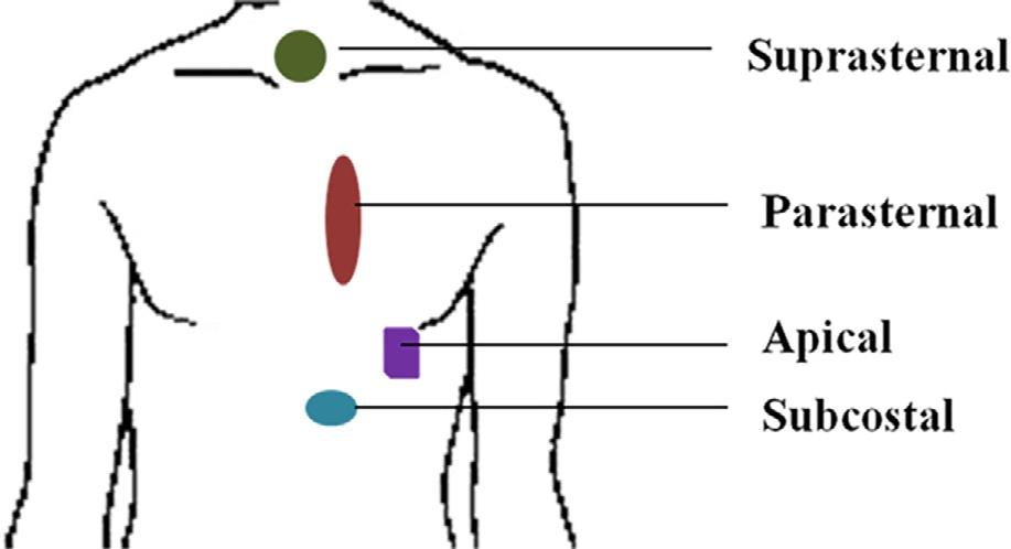
Ultrasoundhashigh-frequency(>20,000Hz),pulsedsoundwaves.Whenultrasoundwavesenterthe tissues,theytransmitthroughthemandarereflectedbackbasedonacousticimpedanceofthetissues. Thedensityofthetissuetimesthevelocityatwhichsoundwavestravelthroughthetissuesistheacousticimpedance.Ultrasoundreflectiondependsuponthemismatchingoftheimpedancesbetweenthe twotissues.Thegreaterthedifference,thegreaterthereflection.Boneshavehighacousticimpedance, whereasairhaslowacousticimpedance.Therefore,bone-tissueandair-tissueinterfacesexhibitgreat mismatchofacousticimpedance,resultinginhighreflectionofultrasoundwaves.Assuch,imagingof deeperstructuresisrestrictedwhentheultrasoundbeamintersectstheair-filledandbonystructurebecausetheyreflectthebeamtoimagetheirouterstructure.Therefore,somesuitableplaceshavebeen selectedtotaketheimageoftheheart.Forechocardiography,intercostalspaceswithinthecardiac windows(wheretheheartisagainstthethorax,withoutinterveninglungs)orfromsubcostalwindows (dependinguponthespecies)areidealforimagingtheheart.
Thespeedofanultrasoundwavevariesdependinguponthetissuetypethroughwhichitispropagating.Thespeedofanultrasoundbeamisapproximately1540m/secthroughsofttissues.Basedon therelationwiththetransducer’sparameters,thesize,thickness,andlocationofthesofttissuescanbe calculatedatanypointandatanyinstant.Thereflection,refraction,andtransmissionrulesoftheultrasoundwavearethelawsofgeometricoptics.Reflection,refraction,andabsorptionoftheultrasound wavesdependuponthedifferenceofacousticimpendencesattheinterfaces.Astheultrasoundbeamis distancedfromthetransducer,itsintensitydecreasesbecauseofthescatter,divergence,absorption,and reflectionofenergyofthewaveattheinterfaceofthetissues.Whentheultrasoundbeamisperpendiculartotheimagestructures,itformsthegreatestreflectiontocreateastrongecho.Thetransducer receivesthesereflectedechoesandcreatesanimageontheultrasoundmachine.Thetransduceractsas areceiverabout99%ofthetime.Theimagesformedbythetransducercanbedisplayedonamonitor, recordedforfutureuse,orprintedonpaper.OpticalCDscanberecordedtoformadatabank.
1.6.2 Modesofechocardiography
Echocardiographycanbeclassifiedintothreemodes:M-mode,two-dimensional(2D,B-mode,orreal time),andDoppler.Onetypeofechocardiographicexaminationcreatesacomplementaryfindingfrom theothermodes.Therefore,thedifferentmodesareperformedsimultaneously.
1.6.2.1
M-modeechocardiography
AhighsamplingrateisusedinM-modeechocardiography.High-clarityimagesareobtainedfroman M-modeechocardiogram,allowingforaccuratemeasurementofcardiacdimensionsandevaluationof cardiacmotion.ItisdifficulttoplacetheM-modebeamattheexactlocationintheheartandthereforeit isdifficulttoobtainclearechoesandcarryoutcriticalmeasurements.Itisalsodifficulttoobtainmeaningfulresultsfromthecalculationsperformedontheobtainedmeasurements.Therightparasternal positionpermitsthestandardviewofM-mode.Toavoidthedisturbancegeneratedbythepapillary musclesintheLV’sfreewall,theM-modecursorshouldbepositionedwithintheheart(therightparasternalshort-axisview).TheMV,LVwall(atthelevelofthechordaetendineae),andaorticroot(aorta/ leftatrialappendage)viewsareincludedthroughthestandardM-modeview.Alinearsweepisaddedto showmotionpatterns,asshownin Fig.1.3
1.6.2.2
Two-dimensionalechocardiography
Areal-timeimageofbothdepthandwidthofaplaneoftissuesisobtainedin2Dechocardiography.Itis possibletotakeunlimitednumberofimagingplanes;however,therearesomestandardviews,which arehelpfulintheevaluationoftheextracardiacandintracardiacstructures.
Moreinformationabouttheshapeandsizeoftheheartisobtainedin2Dechocardiographythanin M-mode.2Dechocardiographyalsogivesthespatialrelationshipsoftheheart’sstructuresduringthe cardiaccycle.BothM-modeand2Drecordingsaredonesimultaneouslyforobtainingmore
FIG.1.3
ExampleofM-modedisplayofMV.
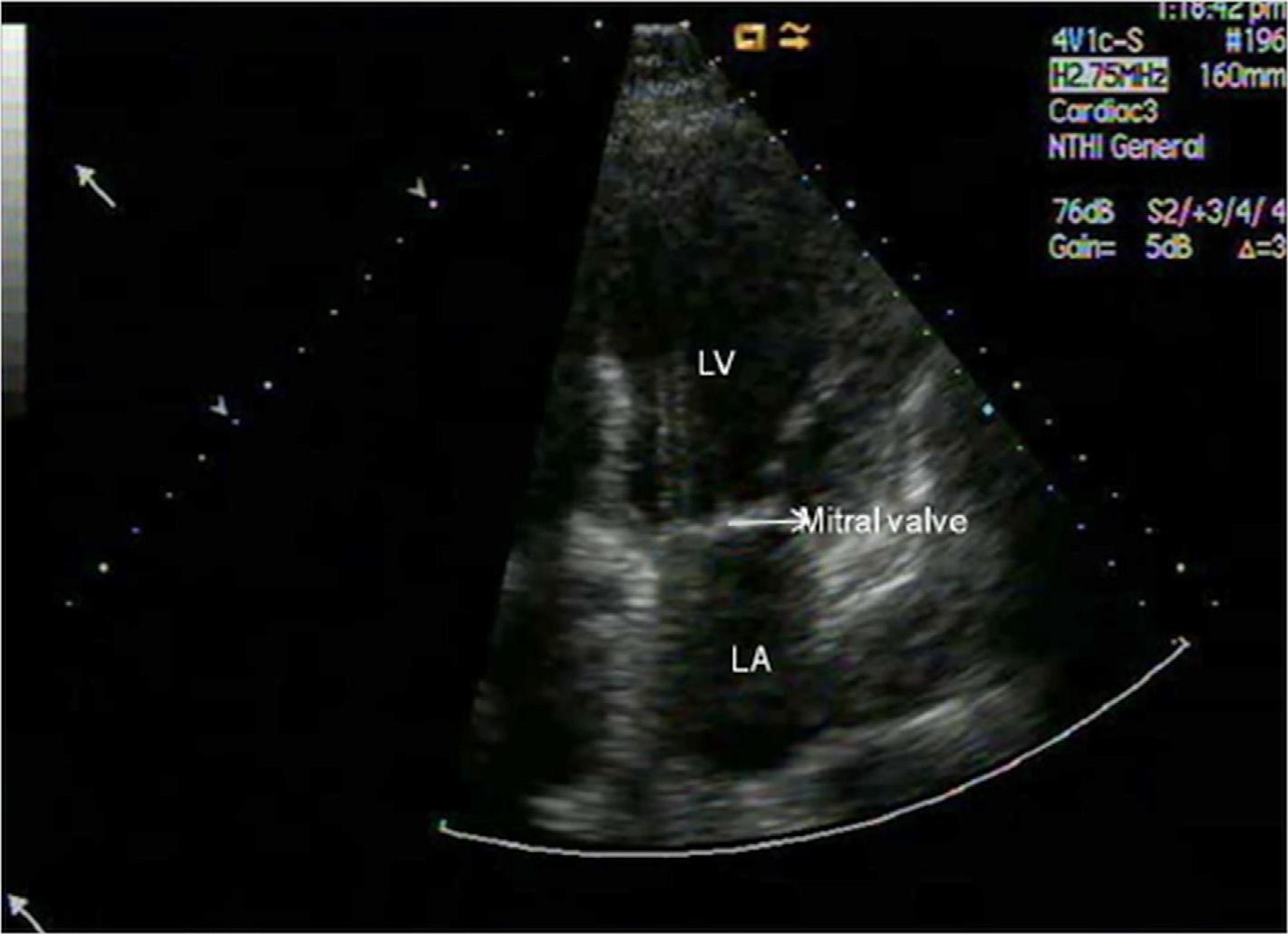
informationaboutcardiacanatomyandclinicalvalues.Thismakesechocardiographyamajordiagnostictool. Fig.1.4 showsanimageobtainedwiththehelpof2Dechocardiography.
1.6.2.3 Dopplerechocardiography
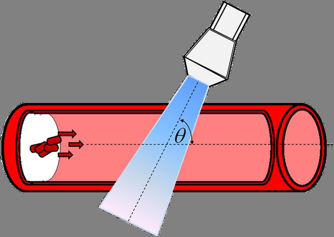
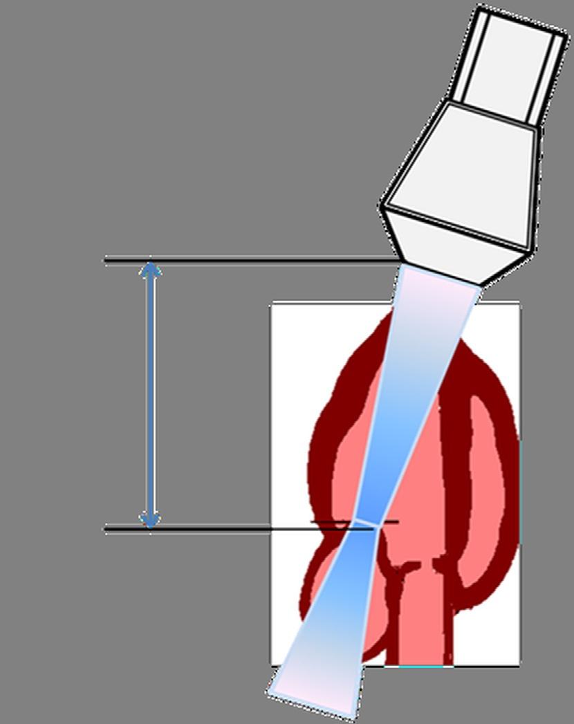
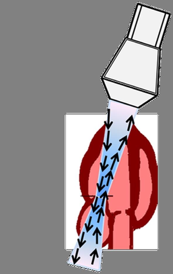
Bloodflowpatterns,velocity,anddirectionareobtainedusingDopplerimaging.ColorDopplerworks basedondetectingchangeoffrequencyofreflectedultrasoundwaves.Thephenomenonisreferredto asDopplershift.Thischangeinfrequencyoccursastheultrasoundwavesreflectoffthemovingblood cells,whichareeithermovingtowardsthetransducerorawayfromthetransducer.Inthisway,color DopplerhelpstodocumentandquantifyinsufficiencyoftheMV,alsoreferredtoasMR.Anaccurate measurementofbloodflowvelocityispossibleifthedirectionofbloodflowispreciselyparalleltothe directionoftheultrasoundbeam.Theresultsbecomeincreasinglyinaccurateastheangle θ ,shownin Fig.1.5,deviatesfromthezeroanglebetweenthebloodflowdirectionandthebeamdirection. Eq. (1.1) describestherelationshipthatdeterminesbloodflowvelocity.
where Fd istheDopplerfrequency, fo istheoriginalfrequency, V isthebloodflowvelocity, c isthelight velocity,and θ istheangleofthetransducer.
Clinically,twotypesofDopplerechocardiographyareemployedingeneral:pulsedwave(PW) Doppler,asshownin Fig.1.6,andcontinuouswave(CW)Doppler,asshownin Fig.1.7.
FIG.1.4 Apical4-chamberviewofMVtakenwith2Dechocardiography.
TheDopplerphenomenon.
ExampleofaPWDoppler.
FIG.1.5
FIG.1.6
InPWDoppler,theultrasoundbeamistransmittedasshortburststoapoint(fixedasthe“sample volume”)onadistancefromthetransducer.TheadvantageofthistypeofDoppleristhatitiseasyto calculatethespectralcharacteristics,bloodflowvelocity,anddirectionfromaspecifiedpointinthe heartorbloodvessel.Themeasuredmaximumvelocityislimitedbecauseofthelimitedpulserepetitionfrequencyandisamajordisadvantage.ThetransducerusedinPWDopplersystemsalternate transmissionandreceptionofultrasound,asintheM-modetransducer(Fig.1.7).Oneoftheadvantages ofPWDoppleristhatitcanprovideDopplershiftdataselectivelyalongtheultrasoundbeamfroma smallsegment,alsoknownas“samplevolume.”
DualcrystalsareusedinCWDopplertosimultaneouslyandcontinuouslysendandreceiveultrasoundwaves.TheflowwithhighvelocitycanalsobemeasuredwithCWbecausethereisnomaximum measurablevelocity(Nyquistlimit).InCWDoppler,thevelocityanddirectionofsampledbloodflow isinthespreadform,notinthespecificarea,whichisadisadvantageofthismodality.Asitsname suggests,continuousultrasoundwavesaregeneratedwithcontinuousultrasoundreception. Fig.1.7 showstwocrystaltransducers,onecrystalforeachfunction,andthedual-functionaccomplishment.
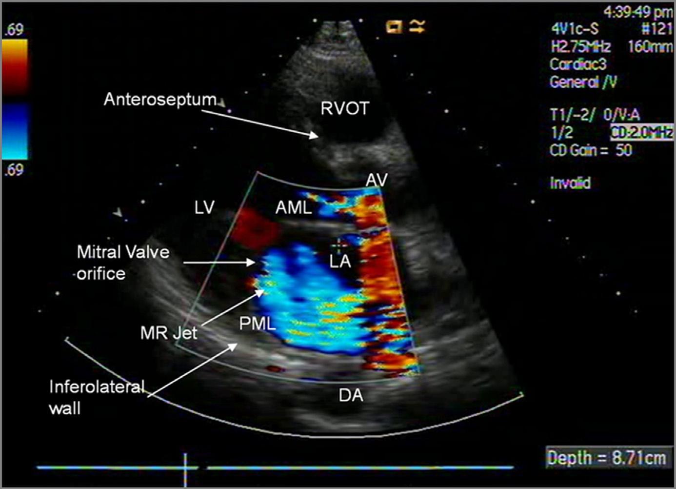
ColorflowDopplerechocardiographyisacombinationofM-modeand2DmodalitieswiththeimagingofbloodflowandisaformofPWDoppler.Multiplescanscanbecarriedoutfortakingmultiple samplesalongthescanlinewiththistechnique.Colorcodingisassignedtothemeanfrequencyshift thatisobtainedfromdifferentvelocityanddirectionsofmanysamplevolumes.Thereareseveraltypes ofmappingavailableforthispurpose.TheBART(blueawayandredtowards)systemismostcommonlyused.Thepresenceofmultiplevelocitiesanddifferencesinrelativeflowvelocitycanbe obtained.Differentmapsthatdependoncolorandbrightnessareusedtoindicatemultiplevelocities.
Continuous Wave
FIG.1.7
ExampleofCWDoppler.
FIG.1.8
ExampleofcolorDopplerimaginginsystolicparasternallong-axisviewMR.
Fig.1.8 showsa2DimageofbloodflowintotheLAatthetimeofsystoleduringMR [25–31].The colors(redandblue)representthedirectionofagivencolorjetandthedifferentvelocities,whichcan berepresentedbyhuesfromdulltobright.Aturbulentjetshowsthemosaicpatternofmanycolors.A 2Ddisplayofflowisshownaccordingtosize,direction,andvelocity.
Meaningofcolor Thereisusefulinformationintheflowmapofanimage.Thecolorredisassigned totheflowtowardthetransducersandthecolorblueisassignedtotheflowawayfromthetransducers.
1.6.3 Two-dimensionalrecordingtechniques
TechniquesforbothM-modeechocardiographyand2Dechocardiographyaresimilar.In2Dechocardiographyastationaryultrasoundbeamisusedasaflashlight.Itilluminatesonlyasmallareaoftheheartat atime.A2Dtransducerislikeacircularsaw,whichisrotatedonthechestatthetimeofrest.Itisrotated aroundthepointusingtheindexmarkprintedonthetransducer.Theindexmarkonthetransducerrepresentstheright-handsideofthemachinedisplay.Amorecomplexmaneuverisnecessaryduring2D examinationsothatanalignedscanplaneisachievedwiththedesiredanatomicaxisoftheheart.
In2Dechocardiographytransducermanipulation,thescanplanepivotsaboutthetransduceraxis when rotating thetransducer.Forexample,therewillbeachangefromtheparasternallongaxistothe parasternalshortaxisifthereisarotationthrough90degrees. Tilting ofthetransducerformaseriesof radialplanes.Theaxisofthetransducerismovedintheplaneofthescanwhen Anglin. Oneexampleis tobringanobjecttothecenterofthefieldofviewatanedge. Fig.1.9 showsthelocationoftheplanesto accesstheheart.
FIG.1.9
Planesforaccessingthehearttoperformechocardiography.
1.6.4 Advantagesandlimitationsofechocardiography
1.6.4.1 Advantages
•portable
•doesnotrestrictclinician,nurse,ortestingequipmentaccesstosickpatients
•canbeperformedintheuprightpositioninseverelyorthopnoeicpatients
•noninvasive,safe,andsuitableforfollow-upinvestigations
•relativelycheap
•widelyavailable
1.6.4.2 Limitations
•Imagequalityisdependentonoperatorskillandpatientanatomyandposition,generallybestinthe leftlateralposition.Itmaybeseverelyimpairedbyairbetweenthechestwallandheart(e.g., hyperinflatedlungsinobstructiveairwaysdisease,patientsonmechanicalventilation,thosewith pneumothoraxorwhoaresupineorintherightlateralposition,etc.).Narrowribspacesandobesity mayalsocausetechnicaldifficulty.
•Informationisoftenqualitativeratherthanquantitative.Significantintra-andinter-observer variationisobservedwhenimagesaresuboptimal.
•Leftatrialappendage,andinadults,superiorvenacavaandmajorityofaortaandpulmonaryarteries abovevalve/rootlevel,cannotbeimaged.
•Imagequalityisgenerallyinferiortotransesophagealecho.
•Offerslimitedcapacityfordifferentiationbetweendifferenttypesoftissuesandfluids.
1.7 Resultsandanalysis
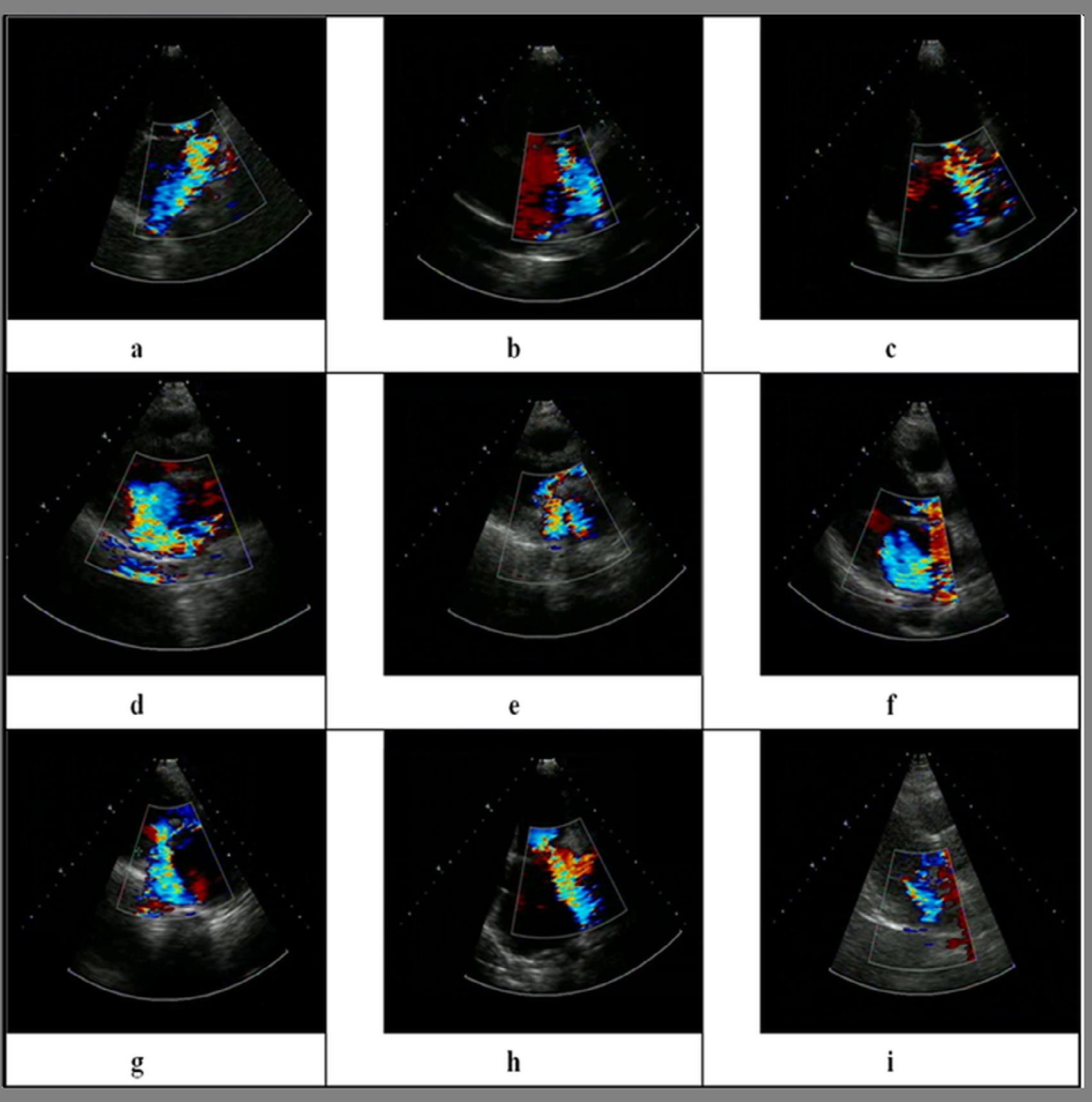
Fig.1.10 showsninecasesofMRimagedwithcolorDopplerechocardiography.Theimagesaretaken atdifferentviewsoftheheart.Themosaicpatternofcolorshowstheregurgitation. Inthenextsection,wepresentananalysisofMRfindingsbasedonregurgitantareaandvena contractawidth.
ImagesfromMR-affectedpatientstakenwithcolorDopplerechocardiography [25]
1.8 Discussion
Table1.2 showsgradesofMRseverityobtainedfromimagingbothregurgitantareaandvenacontracta width.SevereMRisdefinedasjetareagreaterorequalto10cm2.InmoderateMR,venacontracta widthshouldbegreaterthan0.5cmwhenjetareaisintherangeof3tolessthan10.Venacontracta
FIG.1.10
Table1.2SeveritygradeanalysisofMR [25,26].
Images Jetareadrawnby clinician(cm2) Venacontracta width(cm)MRseveritygrade 115.390.58Severe 210.860.61Severe 39.380.46Moderate 413.230.56Severe 59.720.46Moderate 612.940.56Severe 711.290.54Severe 811.860.54Severe 95.590.39Moderate
widthof0.3cmtolessthan0.5cmrepresentsmoderateMR.ValueslessthanmoderateconditionsindicatemildMR,whichisnotrepresentedhere.
1.9 Conclusions
Thischapterdiscussedvarioustypesofheartdisease,withafocusonMR.Itpresentedthesymptoms andcausesofMRaswellasdiagnosticmethodsforthiscondition.Echocardiographywasshowntobe superiortoothermodalities,althoughitdoessufferfromsomelimitations.
References
[1] S.S.Mader,TheCardiovascularSysteminUnderstandingHumanAnatomyandPhysiology,fifthed.,The McGraw-HillCompanies,2004,p.227(Ch.12).
[2] C.L.Reid,D.T.Kawanishi,C.R.McKay,U.Elkayam,S.Rahimtoola,P.A.Chandraratna,Accuracyofevaluationofthepresenceandseverityofaorticandmitralregurgitationbycontrast2-dimensionalechocardiography,Am.J.Cardiol.52(5)(1983)519–524.
[3] M.E.Goldman,V.Fuster,T.Guarino,B.P.Mindich,Intraoperativeechocardiographyfortheevaluationof valvularregurgitation:experiencein263patients,Circulation74(1986)1–143.
[4] M.M.Guerreiro,C.Abreu-Lima,M.R.Gomes,Valueofintraoperative2-dimensionalcontrastechocardiography(2-DCE)forassessingthepresenceandseverityofmitralregurgitation,in:Proceedings1stInternationalSymposiumonEchocardiographyandDopplerinCardiacSurgery,1988,p.69.
[5] M.M.Guerreiro,F.J.Sepulveda,M.R.Gomes,Usefulnessofintraoperativeepicardial2dimensionalechocardiographyinsurgicaldecision,Int.J.CardiacImag.4(1)(1989)71.
[6]S.Zhang,J.P.MarquesdeSa,C.Abreu-Lima,M.Guerreiro,Mitralregurgitationassessmentbyautomated analysisofintracavitarycontrast2-Dechocardiograms,in:ProceedingsComputersinCardiology,1990, pp.671–674, https://doi.org/10.1109/CIC.1990.144309
[7] K.S.Dujardin,M.Enriquez-Sarano,K.R.Bailey,R.A.Nishimura,J.B.Seward,A.J.Tajik,Gradingofmitral regurgitationbyquantitativeDopplerechocardiography:calibrationbyleftventricularangiographyinroutineclinicalpractice,Circulation96(1997)3409–3415.
[8] M.Honey,J.H.Gough,S.Katsaros,G.A.Miller,V.Thuraisingham,Leftventricularcine-angiocardiographyintheassessmentofmitralregurgitation,Br.HeartJ.31(1969)596–602.
[9] S.Blumlein,A.Bouchard,N.B.Schiller,M.Dae,B.F.ByrdIII,T.Ports,E.H.Botvinick,Quantitationof mitralregurgitationbyDopplerechocardiography,Circulation74(2)(1986)306–314.
[10] F.Recusani,G.S.Bargiggia,A.P.Yoganathan,A.Raisaro,L.M.Valdes-Cruz,H.W.Sung,C.Bertucci, M.Gallati,V.A.Moises,I.A.Simpson,Anewmethodforquantificationofregurgitantflowrateusingcolor Dopplerflowimagingoftheflowconvergenceregionproximaltoadiscreteorifice,Circulation83(2)(1991) 594–604.
[11] G.S.Bargiggia,L.Tronconi,D.J.Sahn,F.Recusani,A.Raisaro,S.DeServi,L.M.Valdes-Cruz, C.Montemartini,AnewmethodforquantitationofmitralregurgitationbasedoncolorflowDopplerimaging offlowconvergenceproximaltoregurgitantorifice,Circulation84(4)(1991)1481–1489.
[12] N.Fujita,A.F.Chazouilleres,J.J.Hartiala,M.O.Sullivan,P.Heidenreich,J.D.Kaplan,H.Sakuma,E.Foster, G.R.Caputo,C.B.Higgins,Quantificationofmitralregurgitationbyvelocity-encodedcinenuclearmagnetic resonanceimaging,J.Am.CollegeCardiol.23(4)(1994)951–958.
[13] M.Enriquez-Sarano,J.B.Seward,K.R.Bailey,A.J.Tajik,Effectiveregurgitantorificearea:anoninvasive Dopplerdevelopmentofanoldhemodynamicconcept,J.Am.CollegeCardiol.23(2)(1994)443–451.
[14] F.Grigioni,M.Enriquez-Sarano,K.J.Zehr,K.L.Bailey,A.J.Tajik,Ischemicmitralregurgitation.Long-term outcomeandprognosticimplicationswithquantitativeDopplerassessment,Circulation103(13)(2001) 1759–1764.
[15] S.F.Yiu,M.Enriquez-Sarano,C.Tribouilloy,J.B.Seward,A.J.Tajik,Determinantsofthedegreeoffunctionalmitralregurgitationinpatientswithsystolicleftventriculardysfunction:aquantitativeclinicalstudy, Circulation102(12)(2000)1400–1406.
[16] G.A.Lamas,G.F.Mitchell,G.C.Flaker,S.C.SmithJr.,B.J.Gersh,L.Basta,L.Moye,E.Braunwald,M.A. Pfeffer,Clinicalsignificanceofmitralregurgitationafteracutemyocardialinfarction,Circulation96 (3)(1997)827–833.
[17] T.Kumanohoso,Y.Otsuji,S.Yoshifuku,Mechanismofhigherincidenceofischemicmitralregurgitationin patientwithmyocardialinfarction:quantitativeanalysisofleftventricularandvalvegeometryin103patients withpriormyocardialinfarction,J.ThoracicCardiovasc.Surg.125(1)(2003)135–143.
[18] B.H.Trichon,G.M.Felker,L.K.Shaw,C.H.Cabell,C.M.O’Connor,Relationoffrequencyandseverityof mitralregurgitationtosurvivalamongpatientswithleftventricularsystolicdysfunctionandheartfailure, Am.J.Cardiol.91(5)(2003)538–543.
[19] D.S.Bach,S.F.Bolling,Improvementfollowingcorrectionofsecondarymitralregurgitationinend-stage cardiomyopathywithmitralannuloplasty,Am.J.Cardiol.91(5)(1996)66–69.
[20]F.A.Tibayan,F.Rodriguez,F.Langer,D.Liang,G.T.Daughters,N.B.IngelsJr.,D.CraigMiller,Undersized mitralannuloplastyaltersleftventricularshapeduringacuteischemicmitralregurgitation,Circulation110 (11(Suppl.II))(2004)II98–II102, https://doi.org/10.1161/01.CIR.0000138395.45145.45.
[21] D.H.Adams,R.Rosenhek,V.Falk,Degenerativemitralvalveregurgitation:bestpracticerevolution,Eur. HeartJ.31(2010)1958–1966.
[22] J.D.Bronzino,BiomedicalEngineeringHandbook,vol.2,CRCPress,1999.
[23] J.A.Sethian,LevelSetMethodsandFastMarchingMethods:EvolvingInterfacesinComputationalGeometry,FluidMechanics,ComputerVision,andMaterialScience,seconded.,CambridgeUniversityPress, Cambridge,UK,1999.
[24] R.Gonzalez,R.Woods,DigitalImageProcessing,seconded.,Prentice-HallInc.,2002.
[25] K.Saini,M.L.Dewal,M.K.Rohit,Afastregion-basedactivecontourmodelforboundarydetectionofechocardiographicimages,J.DigitalImag.25(2)(2012)271–278.
[26] K.Chauhan,R.K.Chauhan,A.Saini,Enhancementandde-specklingofechocardiographicimages,in: N.Dey,A.S.Ashour,F.Shi,V.E.Balas(Eds.),SoftComputingBasedMedicalImageAnalysis,Elsevier, 2018,pp.61–79.
[27] K.Chauhan,R.K.Chauhan,Boundarydetectionofechocardiographicimagesduringmitralregurgitation,in: M.Hassaballah,K.Hosny(Eds.),RecentAdvancesinComputerVision:TheoriesandApplications,vol.804, Springer,2018,pp.281–303.
[28] M.L.Dewal,K.Saini,M.K.Rohit,Assessmentofmitralregurgitationseveritywithintensitybasedregion growing,Int.J.HybridInform.Technol.8(6)(2015)45–56.
[29] K.Saini,M.L.Dewal,M.K.Rohit,Levelsetbasedonnewsignedpressureforcefunctionforechocardiographicimagesegmentation,Int.J.Innov.Appl.Stud.3(2)(2013)560–569.
[30] K.Saini,M.L.Dewal,M.K.Rohit,Statisticalanalysisofspecklenoisereductiontechniquesforechocardiographicimages,in:ProceedingsInternationalConferenceonMethodsandModelsinScienceandTechnology,vol.1414,2011,pp.95–99.
[31] K.Saini,M.L.Dewal,M.K.Rohit,Amodifiedhybridfilterforechocardiographicimagenoiseremoval,Int. J.SignalProcess.ImageProcess.PatternRecogn.5(2)(2012)61–72.
Cardiacmultimodalimage registrationusingmachinelearning techniques
Chapteroutline
2.1 Introduction
2.1.1 Imageregistration
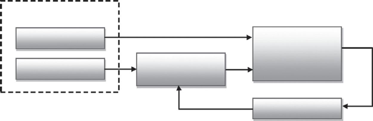
Imageregistration [1,2] isawaytomapinformationfromimageswiththeassistanceofareference image.Theobjectiveofsuchmappingistocoordinatetherelatedimagesbasedoncertainhighlightsto aidintheimagefusionprocess.
Imageregistrationrequirestwo3Dimages:areferenceandafloatingimage.Tobalancetheimages, thereferenceimageisfixedinspacewhilethefloatingimageischangednumericallyuntilitisenlisted
V.AjanthaDevi
tothereferenceimage,asshownin Fig.2.1.Themathematicaltransformfunction,orregistrationtransfer,isassessedfromafewcomparativepropertiesofthetwoimages.
Imageregistrationmethodsdifferinthekindofdatatheyextractfromimagesandinthewaythey decidetheregistrationtransform.Toanalyzethesetechniques,itisimportanttoaggregateimageregistrationinatypicaledge.Imageregistrationcomprisesthreesteps:(1)imagefeatureextraction, (2)registrationtransformcalculation,and(3)qualitymeasurement.
Imagesaredevelopedtoremovespecificfeaturesorboundaries,forexample,edgesormoments. Thesefeaturesarethenutilizedintheregistrationalgorithm.Theregistrationalgorithmdeterminesthe registrationtransformofthereferenceandfloatingimages.Theregistrationtransformiseitherdeterminedscientificallyorassessediterativelyusingtheregistrationalgorithm.Thefundamentalprerequisite [1] ofanyregistrationapproachisthecapacitytodecidepreciselyanddependablythedegreeand natureoffitbetweenimagesandinthismannerthelegitimacyoftheregistrationtransform.
2.1.2 Medicalimageregistration
Medicalimagingisincreasinglybeingutilized [3] fordiseasediagnosisandtreatment.Researchers fromallfields,particularlythoseinneuroscience,useimagingtoexaminemeasuresofillnessanddeterminecourseofdisease.Forresearchpurposes,itisoftenhelpfultolookatvariousimagesacquired frommultiplemodalities,ratherthanasingleimagefromonemodalityobtainedatdifferenttimes [4].
Themeasureofinformationcreatedbyeachprogressivegenerationofimagingframeworksismore prominentthanthatcreatedbythepreviousgeneration.ThispatternissettorepeatwiththedevelopmentofthemultislicehelicalCTscanandmagneticresonanceimaging(MRI) [5,6] frameworkswith higheranglequalities.Thereare,accordingly,potentialadvantagestoimprovingtherouteinwhich theseimagesareanalyzedandconsolidated,asshownin Fig.2.2.Modernizedmethodologiesoffer favorablecircumstances,particularlybyunequivocallymodifyingtheinformationindifferentpictures andarrangingthejoinedimagestoregisterthem.
2.1.3 Cardiacimageregistration
Cardiovascularimagesobtainedthroughvariousmodalitiescangivecorrespondingdata.Inthisway, thecombinationofatleasttwoco-registeredmultimodaldatasetsintoasolitaryportrayalcanhelpin
Optimization Similarity measure
transformation
FIG.2.1 Flowchartofthestudy.
Type of Diagnosing Define
Diagnosing Method Duration
Imaging method
Used to diagnose X-Ray
X-rays are quick, painless tests that produce images of the structures inside Patient body, especially bones.
CT Scan
CT scans use a series of xrays to create cross-sections of the inside of the body, including bones, blood vessels, and soft tissues. MRI
Patient will lie on a table that slides into the scanner, which looks like a large doughnut. The x-ray tube rotates around Patient to take images. Patient will lie, sit, or stand while the x-ray machine takes images. Patient may be asked to move into several positions.
MRIs use magnetic fields and radio waves to create detailed images of organs and tissues in the body.
Patient lie on a table that slides into the MRI machine, which is deeper and narrower than a CT scanner. The MRI magnets create loud tapping or thumping noises.
Ultrasound
Ultrasound uses highfrequency sound waves to produce images of organs and structures within the body.
A technician applies gel to Patient skin, then presses a small probe against it, moving it to capture images of the inside of Patient body.
PET Scan
PEt scans use radioactive drugs (called tracers) and a scanning machine to show how Patient tissues and organs are functioning.
Patient swallow or have a radiotracer injected. Patient then enter a PET scanner (which looks like a CT scanner) which reads the radiation given off by the radiotracer.
Ionizing radiationIonizing radiationMagnetic wavesSound wavesRadiotracers
· bone fractures;
· arthritis;
· osteoporosis;
· infections;
· breast cancer;
· swallowed items;
· digestive tract problems
· injuries from trauma;
· bone fractures;
· tumors and cancers;
· vascular disease;
· heart disease;
· infections;
· used to guide biopsies
Methodofdiagnosingthemedicalimage.
· aneurysms;
· Multiple Sclerosis (MS);
· stoke;
· spinal cord disorders;
· tumors;
· blood vessel issues;
· joint or tendon injuries
· gallbladder disease;
· breast lumps;
· genital/prostate issues;
· joint inflammation;
· blood flow problems;
· monitoring pregnancy
· used to guide biopsies
· cancer;
· heart disease;
· coronary artery disease;
· Alzheimer’s Disease;
· seizures;
· epilepsy;
· Parkinson’s Disease
FIG.2.2
Categorization of the methods
SEGMENTATION
myocardial identification & delineation using techniques such as thresholding, clustering, shape & appearance modeling, etc.
MODEL INDEPENDENT MODEL BASED
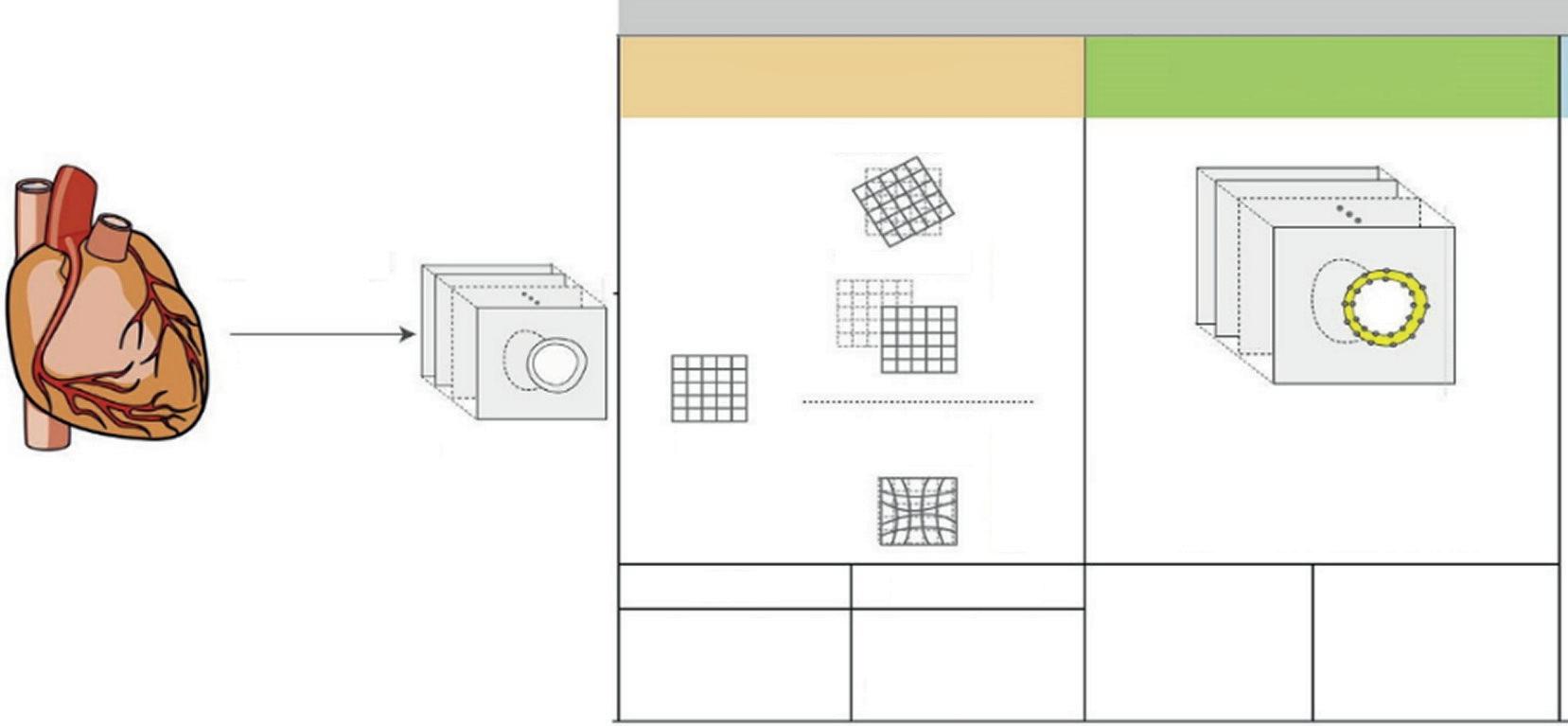
cardiovasculardiagnosis,asshownin Fig.2.3.Theutilizationofamultimodalimagingapproachin medicalpracticeisrestrictedbydisadvantagesinexactimageregistration.Afewmethodologieshave beencreatedtoregister3Dcardiovasculardatasets [7].Thesemethodologiescanbecategorizedby accordingtothefollowingthreekeyaspects:
• theareawherethearrangementchangeischaracterized(searchspace):Forthesearchspace,the inflexiblechangestrategyisthemosteffortlesssinceitutilizesonlysixboundaries(three translationalandthreerotational).Elasticregistrationcanbeusedaswell,however,itsmedical applicationisrestrictedbyhighcomputationalcosts [8].
• thecapacitythatdepictsthenatureofthearrangement(similaritymetricorregistrationmetric): Theresultofasimilaritymetric,whichdemonstratesdecencyofthematch,isamajorquestionin developinganenrollmenttechnique.Obviously,theproximitymetricmustbeefficientinorderto mergetoaglobalaverageforoptimalcoordination,anditmustalsobeprocessedinafairperiodof time.Variousstrategiesutilizetheseparationbetweenchosenmathematicalhighlightsofthe image,forexample,anatomicaltouristspots [9],surfacesonthechest [10,11],orheartsurfaces [12, 13],togatherinformationfromanatomicalfocusesorsurfacedivisionsandcompare.These strategiesarenotalwayseasytoperformconsideringthedisparatedatagivenbyvariousmodalities, particularlywhenadeformityinthemyocardialdividerexists.Consequently,voxel-based closenessmeasurements,forexample,usingcommondata [14–16],don’tmakeassumptionsabout theconnectionbetweenpictures.
• theoptimizationmethodologyusedtodeterminethechangethatexpandsthecharacterized comparabilitywork: Thelaststepofaregistrationcycleisoptimization,whichcanfigurethe changethataugmentsthecharacterizedsimilitudemetricandadjuststheheartdatasets.
CARDIAC IMAGING
- Gated)
Cardiac images in short-axes orientation
Image Warping feature based & intensity based feature based & intensity based
FIG.2.3
Cardiacimageregistrationandsegmentation.