AbouttheEditor
AffiliationsandExpertise
Professor/FounderandChairmanoftheDepartmentofPediatricUrology/FounderandPresident ofPediatricUrologyandRegenerativeMedicine ResearchCenter,SectionofTissueEngineering andStemCellsTherapy,Children’sHospital MedicalCenter,TehranUniversityofMedical Sciences,Tehran,Iran(IRI)/Presidentofthe IranianFetalDiagnosisandTreatment Association.
ProfessorAbdol-MohammadKajbafzadehiscurrentlyHeadofPediatric UrologyandRegenerativeMedicineResearchCenter,SectionofTissue EngineeringandStemCellsTherapy.Hisresearchspecialtyincludes genitourinarycancer,physiology,nephrology,urology,radiology,stemcell andregenerativemedicine.Hehashundredsofscientificpublications includingdefi nitivereferencesinthe fieldsoftissueengineeringand regenerativemedicine.Hisextensiveexperienceinclinical,surgicaland basicscienceresearchmainlytissueengineeringandstemcelltherapyaswell asbio-scaffoldsandinventionofnontoxictechniquesformorethan40 tissuesandwholeorgandecellularizationpavedtheroadtoextrapolatehis knowledgetoclinicalsettingsandusehisexpertisetosolvepatients’ problems[1–53].Over28years(1984–2012),hedesignedhundredsof studieswithmainfocusonthestemcelltherapyandtissueengineeringtissue repairrenaltransplantation.Pulsatinghearttissuewasproducedbyseeding variousheartcellsinhislaboratory,whereliveimagescanbeviewedonsite [54].Thehuman-sizedkidneyhasbeenalsorecellularizedandtransplanted tothelargelaboratoryanimals[55].Since2007,theworksdoneinhisCenter hasbeenfeaturedseventimesonthecoveroftheUSandUKurology journals.The firstcelltherapyforurinaryincontinencewasalsoperformed 12yearsagoathiscenter[56].Renaldamagewasalsorepairedby
autologouscellsforthe firsttimeathiscenter[42].Hisresearchcenterhas focusedprimarilyondesigningcelltherapyapproachesinanimalmodels, newclinicaltrials,andprospectiveclinicaltrials.Hedevelopedhundredsof newideasinvariousaspectsoftissueengineering,stemcellcultureand differentiationtherapy,bonemarrowstemcells,adipose-derivedstemcells, dentalpulpstemcells,endometrialstemcells,musclederivedstemcells,and kidneystemcells.Thisclearlyhelpstounderstandthemechanismofthe diseaseandtoimprovethetherapeuticmethods.Thesealltogether substantiallyaddtohisclinicalandexperimentalexpertiseintranslational medicineandfromthebenchtothebedside.Itclearlyhelpstounderstanding ofthemechanismofdiseaseandimprovementoftreatmentoptions.
References
1.SabetkishS,KajbafzadehAM,SabetkishN,KhorramirouzR,AkbarzadehA,SeyedianSL,PasalarP,OrangianS,BeigiRSH,AryanZ(2015)Whole‐organtissue engineering:decellularizationandrecellularizationofthree‐dimensionalmatrixliver scaffolds.JBiomedMaterResPartA103(4):1498–1508
2.KajbafzadehA-M,Javan-FarazmandN,MonajemzadehM,BaghayeeA(2013) Determiningtheoptimaldecellularizationandsterilizationprotocolforpreparinga tissuescaffoldofahuman-sizedlivertissue.TissueEngPartCMethods19(8):642–651
3.Sharifi E,AzamiM,KajbafzadehA-M,MoztarzadehF,Faridi-MajidiR,ShamousiA, KarimiR,AiJ(2016)Preparationofabiomimeticcompositescaffoldfrom gelatin/collagenandbioactiveglass fibersforbonetissueengineering.MaterSci EngC59:533–541
4.KajbafzadehA-M,PayabvashS,SalmasiAH,SadeghiZ,ElmiA,VejdaniK, TavangarSM,TajikP,MahjoubF(2007)Time-dependentneovasculogenesisand regenerationofdifferentbladderwallcomponentsinthebladderacellularmatrixgraft inrats.JSurgRes139(2):189–202
5.KajbafzadehA-M,ElmiA,TalabSS,EsfahaniSA,TourchiA(2010)Functional externalanalsphincterreconstructionfortreatmentofanalincontinenceusingmuscle progenitorcellautografting.DisColonRectum53(10):1415–1421
6.SemyariH,RajipourM,SabetkishS,SabetkishN,AbbasFM,KajbafzadehA-M (2016)Evaluatingtheboneregenerationincalvarialdefectusingosteoblastsdifferentiatedfromadipose-derivedmesenchymalstemcellsonthreedifferentscaffolds:an animalstudy.CellTissueBank17(1):69–83
7.KajbafzadehA-M,TourchiA,MousavianA-A,RouhiL,TavangarSM,SabetkishN (2014)Bladdermuscularwallregenerationwithautologousadiposemesenchymal stemcellsonthree-dimensionalcollagen-basedtissue-engineeredprepuceandbiocompatiblenanofibrillarscaffold.JPediatrUrol10(6):1051–1058
8.TalabSS,KajbafzadehAM,ElmiA,TourchiA,SabetkishS,SabetkishN,MonajemzadehM(2014)Bladderreconstructionusingscaffold‐lessautologoussmooth musclecellsheetengineering:earlyhistologicaloutcomesforautoaugmentation cystoplasty.BJUInt114(6):937–945
9.TabatabaeiFS,AiJ,KashiTSJ,KhazaeiM,KajbafzadehA-M,GhanbariZ(2013) Effectofdentinematrixproteinsonhumanendometrialadultstem-likecells:invitro regenerationofodontoblastscells.Archivesoforalbiology58(7):871–879
10.KajbafzadehA-M,SabetkishS,HeidariR,EbadiM(2014)Tissue-engineered cholecyst-derivedextracellularmatrix:abiomaterialforinvivoautologousbladder muscularwallregeneration.PediatrSurgInt30(4):371–380
11.AmidiF,HoseiniMA,NiaKN,HabibiM,KajbafzadehAM,MazaheriZ,YaminiN (2015)Malegerm-likecelldifferentiationpotentialofhumanumbilicalcordWharton’sjelly-derivedmesenchymalstemcellsinco-culturewithhumanplacentacellsin presenceofBMP4andretinoicacid.IranJBasicMedSci18(4):325
12.ElmiA,KajbafzadehA-M,TourchiA,TalabSS,EsfahaniSA(2011)Safety,efficacy andhealthrelatedqualityoflifeofautologousmyoblasttransplantationfortreatment ofurinaryincontinenceinchildrenwithbladderexstrophy-epispadiascomplex.JUrol 186(5):2021–2026
13.KajbafzadehA-M,EsfahaniSA,TalabSS,ElmiA,MonajemzadehM(2011)In-vivo autologousbladdermuscularwallregeneration:applicationoftissue-engineered pericardiuminamodelofbladderasabioreactor.JPediatrUrol7(3):317–323
14.Sharifi E,Ebrahimi‐BaroughS,PanahiM,AzamiM,AiA,BarabadiZ, KajbafzadehAM,AiJ(2016)Invitroevaluationofhumanendometrialstemcell‐derivedosteoblast‐likecells’ behaviorongelatin/collagen/bioglassnanofibers’ scaffolds.JBiomedMaterResPartA104(9):2210–2219
15.DehkordiMB,MadjdZ,ChaleshtoriMH,MeshkaniR,NikfarjamL,Kajbafzadeh A-M(2016)Asimple,rapid,andefficientmethodforisolatingmesenchymalstem cellsfromtheentireumbilicalcord.CellTransplant25(7):1287–1297
16.KajbafzadehA-M,EsfahaniSA,SadeghiZ,ElmiA,MonajemzadehM(2012) Applicationofdifferentscaffoldsforbladderwallregeneration:thebladderasa naturalbioreactor.TissueEngPartA18(7–8):882–887
17.KajbafzadehA-M,KhorramirouzR,KameliSM,HashemiJ,BagheriA(2017) Decellularizationofhumaninternalmammaryartery:biomechanicalpropertiesand histopathologicalevaluation.BioResOpenAccess6(1):74–84
18.KajbafzadehA-M,SabetkishS,SabetkishN,MuhammadnejadS,AkbarzadehA, TavangarSM,MohseniMJ,AmanpourS(2015)In-vivotrachearegeneration:fabricationofatissue-engineeredtracheainnudemiceusingthebodyasanatural bioreactor.SurgToday45(8):1040–1048
19.HashemiJ,PasalarP,SoleimaniM,KhorramirouzR,FendereskiK,EnderamiSE, KajbafzadehAM(2018)Applicationofanovelbioreactorforinvivoengineeringof pancreastissue.JCellPhysiol233(5):3805–3816
20.KajbafzadehA-M,TaftiSHA,Mokhber-DezfooliM-R,KhorramirouzR,SabetkishS, SabetkishN,RabbaniS,TavanaH,MohseniMJ(2016)Aorticvalveconduit implantationinthedescendingthoracicaortainasheepmodel:Theoutcomesof pre-seededscaffold.IntJSurg28:97–105
21.KajbafzadehA,SabetkishN,SabetkishS,TavangarS,BeigiRH,TalebiM, AkbarzadehA,NikfarjamL(2015)Lungtissueengineeringandpreservationof alveolarmicrostructureusinganovelcastingmethod.BiotechHistochem90(2):111–123
22.KhorramirouzR,SabetkishS,AkbarzadehA,MuhammadnejadA,HeidariR,KajbafzadehA-M(2014)Effectofthreedecellularisationprotocolsonthemechanical behaviourandstructuralpropertiesofsheepaorticvalveconduits.AdvMedSci59 (2):299–307
23.KajbafzadehA-M,SabetkishS,TourchiA,AmirizadehN,AfsharK,AbolghasemiH, ElmiA,TalabSS,EshghiP,MohseniMJ(2014)Theapplicationoftissue-engineered preputialmatrixand fibrinsealantforurethralreconstructioninrabbitmodel.IntUrol Nephrol46(8):1573–1580
24.KajbafzadehA-M,AbbasiounR,SabetkishS,SabetkishN,RahmaniP, TavakkolitabassiK,ArshadiH(2017)Futureprospectsforhumantissueengineered urethratransplantation:decellularizationandrecellularization-basedurethraregeneration.AnnBiomedEng45(7):1795–1806
25.KajbafzadehA-M,KajbafzadehM,SabetkishS,SabetkishN,TavangarSM(2016) Tissue-engineeredexternalanalsphincterusingautologousmyogenicsatellitecells andextracellularmatrix:functionalandhistologicalstudies.AnnBiomedEng44 (5):1773–1784
26.SabetkishN,KajbafzadehA-M,SabetkishS,TavangarSM(2014)Augmentation cystoplastyusingdecellularizedvermiformappendixinrabbitmodel.JPediatrSurg 49(3):477–483
27.KajbafzadehA-M,KhorramirouzR,KameliSM,FendereskiK,DaryabariSS, TavangarSM,GarajegayehBA(2019)Three-yearefficacyandpatencyfollow-upof decellularizedhumaninternalmammaryarteryasanovelvasculargraftinanimal models.JThoracCardiovascSurg157(4):1494–1502
28.KameliSM,KhorramirouzR,EftekharzadehS,FendereskiK,DaryabariSS, TavangarSM,KajbafzadehAM(2018)Applicationoftissue‐engineeredpericardial patchinratmodelsofmyocardialinfarction.JBiomedMaterResPartA106 (10):2670–2678
29.HashemiJ,PasalarP,SoleimaniM,ArefianE,KhorramirouzR,AkbarzadehA, GhorbaniF,EnderamiSE,KajbafzadehAM(2018)Decellularizedpancreasmatrix scaffoldsfortissueengineeringusingductalorarterialcatheterization.CellTissueOrg 205:72–84
30.NafisiN,AkbariME,MahjoubF,MohseniMJ,SabetkishS,KhorramirouzR, TehraniM,KajbafzadehA-M(2017)Applicationofhumanacellularbreastdermal matrix(ABDM)inimplant-basedbreastreconstruction:anexperimentalstudy.AestheticPlastSurg41(6):1435–1444
31.ElmiA,KajbafzadehA-M,OghabianMA,TalabSS,TourchiA,KhoeiS,RafieB, EsfahaniSA(2014)Analsphincterrepairwithmuscleprogenitorcelltransplantation: serialassessmentwithironoxide–enhancedMRI.AmJRoentgenol202(3):619–625
32.KhorramirouzR,KameliSM,FendereskiK,DaryabariSS,KajbafzadehA-M(2019) Evaluatingtheefficacyoftissue-engineeredhumanamnioticmembraneinthetreatmentofmyocardialinfarction.RegeneratMed14(2):113–126
33.KajbafzadehA-M,TaftiSHA,KhorramirouzR,SabetkishS,KameliSM,OrangianS, RabbaniS,OveisiN,GolmohammadiM,KashaniZ(2017)Evaluatingtheroleof autologousmesenchymalstemcellseededondecellularizedpericardiuminthe treatmentofmyocardialinfarction:ananimalstudy.CellTissueBank18(4):527–538
34.KajbafzadehA-M,AbbasiounR,SabetkishN,SabetkishS,HabibiAA, TavakkolitabassiK(2017)Invivohumancorpuscavernosumregeneration:Fabricationoftissue-engineeredcorpuscavernosuminratusingthebodyasanatural bioreactor.IntUrolNephrol49(7):1193–1199
35.KajbafzadehA-M,KhorramirouzR,SabetkishS,TalebiMA,AkbarzadehA,KeihaniS(2016)Invivoregenerationofbladdermuscularwallusingdecellularizedcolon matrix:anexperimentalstudy.PediatrSurgInt32(6):615–622
36.FarahaniF,YazdiAK,GhasemiM,ShariatpanahiE,KajbafzadehA-M,AmanpourS (2015)Resultsofacellulardermismatrixgraftusedfortympanoplastyinguineapig model.IranJOtorhinolaryngol27(79):95
37.KajbafzadehA-M,MozafarpourS,SeyedianSSL,KhorramirouzR,HojjatiHN (2015)Decellularizeddermalstripasasuburethralslinginaratmodelofstress urinaryincontinence.IntUrolNephrol47(8):1303–1310
38.KajbafzadehAM,KhorramirouzR,AkbarzadehA,SabetkishS,SabetkishN,SaadatP,TehraniM(2015)Anoveltechniqueforsimultaneouswhole‐bodyandmulti‐organdecellularization:umbilicalarterycatheterizationasaperfusion‐basedmethod inasheepfoetusmodel.IntJExpPathol96(2):116–132
39.SabetkishN,SabetkishS,MohseniMJ,KajbafzadehA-M(2019)Preventionofrenal scarringinacutepyelonephritisbyprobiotictherapy:anexperimentalstudy.Probiot AntimicrobProteins11(1):158–164
40.KhorramirouzR,KameliSM,EftekharzadehS,KajbafzadehAM(2017)Application ofomentumasaninvivobioreactorforregenerationofdecellularizedhumaninternal mammaryartery.JBiomedMatResPartA105(10):2685–2693
41.KajbafzadehA-M,KhorramirouzR,MasoumiA,KeihaniS,NabavizadehB(2018) Decellularizedhumanfetalintestineasabioscaffoldforregenerationoftherabbit bladdersubmucosa.JPediatrSurg53(9):1781–1788
42.SabetkishS,SabetkishN,TalebiMA,HalimiS,KajbafzadehA-M(2018)Theroleof nonautologousandautologousadipose-derivedmesenchymalstemcellinacute pyelonephritis.CellTissueBank19(3):301–309
43.SalariSedighH,SaffarpourA,JamshidiS,AshouriM,NassiriSM,Sharifi D, TorkzabanP,KajbafzadehA(2010)Canineperiodontalstemcells:Isolation,differentiationpotentialandelectronicmicroscopiccharacterization.IranJVetSurg5(1–2):19–28
44.SabetkishS,SabetkishN,KajbafzadehA-M(2019)In-vivoregenerationofbladder muscularwallwithwholedecellularizedbladdermatrix:Anovelhourglasstechnique forduplicationofbladdervolumeinrabbitmodel.JPediatrSurg
45.AkbarzadehA,KianmaneshM,FendereskiK,EbadiM,DaryabariSS,MasoomiA, GhazisaeediF,BeigiRSH,SheikhR,KajbafzadehA-M(2019)Decellularisedwhole ovinetestisasapotentialbio-scaffoldfortissueengineering.ReprodFertilDevelop31 (11):1665–1673
46.FatourechiM,FattahianH,KajbafzadehA(2015)Theexperimentalstudyof bio-engineeredfree-cellostrichcorneaasxenograft.
47.KajbafzadehA-M,ElmiA,TalabSS,SadeghiZ,EmamiH,SotoudehM(2010) Autograftingofrenalprogenitorcellsameliorateskidneydamageinexperimental modelofpyelonephritis.CellMed1(3):115–122
48.ElmiA,KajbafzadehA,TalabSS,EsfahaniS,TourchiA,OghabianM,KhooiS (2010)Invivomagneticresonancetrackingoftransplantedmyoblastslabeledwith magneticironoxidenanoparticleinarabbitmodeloffecalincontinence.JPediatrUrol 6:S28
49.SamanST,KajbafzadehA,ElmiA,ShadiAE(2010)AutoaugmentationCystoplasty UsingAutologousSmoothMuscleCellSheetEngineering.JPediatrUrol6:S26–S27
50.KajbafzadehA-M,AzadehE,EmamiH,ShafaattalabS,EsfahaniSA,TourchiA (2009)Restorationoffunctionalanalsphincterbymuscleprogenitorcellstransplantationinarabbitmodeloffecalincontinence.JPediatrUrol5:S85–S86
51.VejdaniK,KajbafzadehA,AmanpourS,GolestaniM(2005)Humanbladderacellular matrixforaugmentationcystoplastyintherabbit.
52.DaryabariSS,KajbafzadehA-M,FendereskiK,GhorbaniF,DehnaviM,RostamiM, GarajegayehBA,TavangarSM(2019)Developmentofanefficientperfusion-based protocolforwhole-organdecellularizationoftheovineuterusasahuman-sizedmodel andinvivoapplicationofthebioscaffolds.JAssistReprodGenet36(6):1211–1223
53.EbrahimSoltaniAR,KajbafzadehA,EzzatiM,EbrahimSoltaniZ,HosseinifarN, MalekiA,NezhadSistaniM(2019)Novelevaluationofsevofluraneanesthetic exposureonthetesticulargermcellsofneonatalmalemice.ToxicolRes8(6):988–993
54.AkbarzadehA,KhorramirouzR,GhorbaniF,BeigiRSH,HashemiJ,Kajbafzadeh A-M(2019)Preparationandcharacterizationofhumansizewholeheartfororgan engineering:scaffoldmicroangiographicimaging.RegeneratMed14(10):939–954
55.KajbafzadehA-M,KhorramirouzR,NabavizadehB,SeyedianS-SL,AkbarzadehA, HeidariR,MasoumiA,AziziB,BeigiRSH(2019)Wholeorgansheepkidneytissue engineeringandinvivotransplantation:Effectsofperfusion-baseddecellularization onvascularintegrity.MatSciEngC98:392–400
56.KajbafzadehA-M,ElmiA,PayabvashS,SalmasiAH,SaeediP,MohamadkhaniA, SadeghiZ,NikfarjamL(2008)Transurethralautologousmyoblastinjectionfor treatmentofurinaryincontinenceinchildrenwithclassicbladderexstrophy.JUrol 180(3):1098–1105
CharacterizationMethods ofAcellularizedTissueandOrgans
LaylaShojaie,YektaRahimi,Masoumeh MajidiZolbin,FaezehDaghigh, andAbdol-MohammadKajbafzadeh
Abstract
Theextracellularmatrix(ECM)ofmammalianorgansandtissueshasbeenapplied asasubstitutescaffoldtosimplifytherestorationandreconstructionofseveraltissues. Suchscaffoldsarepreparedinvarious arrangementsincludingsheets,powders,and hydrogels.Oneofthemoreapplicableprocessesisusingnaturalscaffolds,forthis purposediscardedtissuesororgansarenatu-
L.Shojaie Y.Rahimi M.M.Zolbin
A.-M.Kajbafzadeh(&) PediatricUrologyandRegenerativeMedicine ResearchCenter,SectionofTissueEngineering andStemCellsTherapy,Children’sHospital MedicalCenter,PediatricCenterofExcellence, TehranUniversityofMedicalSciences,Tehran,Iran e-mail: kajbafzd@sina.tums.ac.ir
L.Shojaie
DepartmentofMedicineDivisionofGI/LiverKeck SchoolofMedicine,UniversityofSouthern California,LosAngeles,USA
F.Daghigh DepartmentofPhysiology,TabrizBranch, IslamicAzadUniversity,Tabriz,Iran
F.Daghigh TuberculosisandLungDiseaseResearchCenter, TabrizUniversityofMedicalSciences,Tabriz,Iran
PresentAddress:
A.-M.Kajbafzadeh No.62,Dr.Gharib’sStreet,KeshavarzBoulevard, (PANNEK,#6),1419433151Tehran,Iran
© SpringerNatureSwitzerlandAG2021
rallyderivedbyprocessesthatcomprised decellularizationoffollowingtissuesor organs.Protectionofthecomplexstructure and3D(threedimensional)ultrastructureof theECMisextremelynecessarybutitis predictablethatallprotocolsofdecellularizationendindisruptionofthearchitectureand potentiallossofsurfaceorganizationand configuration.Tissuedecellularizationwith conservationofECMbioactivityandintegrity canbeimprovedbyprovidingwell-designed protocolsregardingtheagentsanddecellularizationtechniquesoperatedduringprocessing. Anoverviewofthecharacterizationofdecellularizedscaffoldsandtheroleofreagnetscan validatetheappliedmethods'efficacy.
Keywords
Acellular Tissueengineering Scaffold Characterize
1.1History
Decellularizedextracellularmatrices(dECM) provideapossiblesupplyofsubstancestogeneratedifferentscaffolds.Todate,thereareno absolutecriteriapreciselytoconfi rmdecellularizedECM.Indeedeffectivedecellularization methodologyisordainedbyvariousfactorssuch astissuedensityandstructure,geometricand
A.Kajbafzadeh(ed.), DecellularizationMethodsofTissueandWholeOrgan inTissueEngineering,AdvancesinExperimentalMedicineandBiology1345, https://doi.org/10.1007/978-3-030-82735-9_1
biologiccharacteristicsneededforthetargeted clinicalpurpose,aswellasthespecificcharacteristicsofthetissueoftheorigin.Eachtissue demandsitsspecificcharacterizationmethods. Indeed,efficientcellandgeneticelementseliminationarecrucialinpreventingimmunerejection oftheconstructtoseededcells.Quantitative metricshavenotbeendescribedfortheterm decellularizationyet.Toevaluatethequalityof decellularizedtissueanditsextracellularmatrix (ECM),multipleaspectsshouldbeexamined. Basedoncurrentliteratureandexperimentsin whichinvivoconstructiveresponsewasestablished,andimmunerejectionofthehostdidnot occur,severalcriteriahavebeensuggested.These minimalcriteriaaresuggestedtobeexercisedto assessthedecellularizationprocessanditsefficacy.Thebasisofthesuggestedcriterialiesinthe amountandqualityofgeneticmaterialthatis remainedintheECM.First,theamountof dsDNAshouldbelessthan50nanogramsper milligramofthedryweightoftheECM.Second, DNAfragmentsdetectedintheECMshouldnot bemorethan200basepairs.Andlastly,histologicalevaluationshouldnotbeabletoidentify geneticmaterialwithhematoxylin-eosinstaining (H&E)or4′,6-diamidino-2-phenylindole(DAPI) (Medberry 2014).
The fi rstandsecondprincipleisconsidereda quanti fiableapproachtoassesstheacellularized ECM,whereasqualitativelyveri fiedbyhistologicalstainingssuchasH&EorDAPI.Quantitiveassessmentisreadilyaccomplishedby availabledsDNAintercalators.Forthesecond criterion,endonucleasessuchasDNaseand RNaseareappliedtobreakdownnucleicacid basepairfragments.Theseenzymes,fortunately, decreasethelengthoffragments,buttheydo littletopartthefragmentsoftheECM.Inpursuit ofdecreasingimmuneresponse,theIntracellular membranecompartment(e.g.,phospholipids) mustbenoticedviaenzyme-basedmeasurement.
ECMisavitalcomponentduringdevelopment,influencingcelldifferentiation,proliferation,andmigration,anditsprominentrolein providingstructuralstabilityandsupportforcells andtissueisindispensable(Medberry 2014). ECM,anon-cellularelementofthetissue
microenvironment,comprisesproteinssuchas collagens,laminins, fi bronectin,andpolysaccharideglycosaminoglycans(GAGs)(Cirulli etal. 2000).Collagensarethemostabundant componentinECM;sofar,theyarethemainaim tomodify.Severalmethodshavebeenappliedto determinetheamountandqualityofECM components.Forexample,tomaintainthecontentofcollagen,histologicalcollagenstainsare used,whereasScanningelectronmicroscopy (SEM)providesmoreinformationaboutthe structureandarchitecture.Additionally,Second harmonicgeneration(SHG)detectsstructural changesincollagen fibersbylossofsignals.
Proteinsthathelpwithstructuralabilities,as wellasmechanicalpropertiesofthetissue, shouldmatchtheoriginaltissuetoareasonable extentsothattheprocesswouldhavemore chancetobesuccessful.ItisassumedSome decellularizationagentsandprotocolsdestroy basicECMelements;forexample,detergents maydisruptcollagenstructure;therefore mechanicalstrengthofECMundergoeschanges ormostdetergentseliminatetheamountofGAG, thusdecreasetheviscoelasticityfeatureofECM (Kezwoń etal. 2016;Conconietal. 2005).
Consequently,mechanicalpropertiessuchas elasticmodulus,viscousmodulus,tensile strength,andyieldstrengthshouldbeassessed. However,allinall,thecharacteristicismainly provisoryonthetypeofthetissueororgan's soughtfunction(WangandGuan 2010).
Theprocessofdecellularizationviaacell removalagentwillaltertheECMcomposition andstructure.Thegoalistotryandminimize thesealterationstohavearobustECM.Thereare severaltypesofagentsandtechniquesavailable fordecellularization.Theagentcanbechemical, biological,physical,ormiscellaneous.Among themostcommonlyusedchemicalagentsare TritonX-100,TritonX-200,andsodiumdodecyl sulfate(SDS).Eachreagenthasitsowncharacteristics.Forexample,SDSandTritonX-100can removemoresignificantthan90%ofnuclear remnants.TritonX-100andTritonX-200have mixedresults;nevertheless,theybothcan removecellsmoreefficaciouslyfromthintissue; however,TrironX-200tendstodisrupttheECM
moreconsiderably.SDSismoreeffectivethan bothandcanremovecellsfromdensetissues,but disruptstheECM,damagesthecollagen,and removesGAGs(Merrittetal. 2010;Guoetal. 2010;Hudsonetal. 2004).Itisworthhighlightingthatalltheseoutcomesdependonthe targettissue.Biologicagentscanbeenzymesor chelatingcompounds.Enzymesliketrypsin, nucleases,anddyspasesarecommonlyused (Yangetal. 2010;Wainwrightetal. 2010). Chelatingagentsarenoteffectivewhenused aloneandshouldbeusedwitheitherenzymesor detergentstobeeffective.Examplesofphysical andmiscellaneousagentsarefreezingand thawing,directforceandpressure,andelectroporation.Thecommonsideeffectofallthe physicalmethodsisECMdisruptionwhichcan happendirectlyandindirectly(Lehretal. 2011; Sellaroetal. 2010;Hashimotoetal. 2010).The structureoftheremainingECMandtheclearanceofcelldebrisandgeneticmaterialcan influencethehostresponse'seffi cacy.Sincethe bestreagentsarestillnot100%effective,wash buffersandextensivewashprocedureshavebeen suggested(Hashimotoetal. 2010;Xingetal. 2015).
1.1.1Hematoxylin–EosinStaining(H &E)
Hematoxylinandeosin(H&E),iswell-known stainingusedtoassesstheoverallhistologic appearanceofsamples,showingcells,cytoplasm, nuclei,andECMconstitution.Hematoxylinand eosinhavedistinctfunctions.Hematoxylinstains nucleicacidsandhasadeeppurplecolor.Eosin stainsproteinsarepink.Inhealthytissue,nuclei arestainedblue,whereasthecytoplasmand extracellularmatrixhavedivergingdegreesof pinkstaining.Nucleolistainwitheosin.Ifabundantpolyribosomesarepresent,thecytoplasm willhaveadistinctbluecast.Thisstainreveals sufficientstructuraldatawithspecificfunctional implications(Fischeretal. 2008).
Forpreparation,thesamplesare fixedinformalinsolutionbeforebeingembeddedinparaffin.Transversalsectionsarecut,dewaxed,and
stained.H&Estainingisperformedaccordingto themanufacturer'sprotocols.Decellularizedtissuecanshowadecreaseorlackofhematoxylinstainednucleiindicatingadequatecellelimination.Suchasseen,inmanystudiesthatbyuseof H&Estain,the firstandsecondcriteriafor decellularizedtissuecanbeestablished.Completecellremovalwasalsoobservedinastudy byPashosetal.withinareasofdensecollagen (Pashosetal. 2017).TheH&Estainofthe decellularizedtissueisalteringthecolorofpink withconsiderablylessblue-purpleportions, indicatingthepresenceofthenucleusinthe nativetissuestain.
1.1.24′,6-Diamidino-2-Phenylindole (DAPI)
Anotherconfirmationtestforcellnucleus removalisstainingthatisbroadlyutilizedfor assessingdecellularizedtissueis4′,6-diamidino2-phenylindole(DAPI).
Forvisualizationofintactandundamaged nuclei,DAPIstainingisused.Thetissuesections areprepared,asmentionedforH&Estaining. Immunofluorescencestainingwillbeperformed, a fluorescentmicroscopewithaSlidebook softwareisutilizedtogetaphotooftheslide.If intactnucleiarepresent,the fluorescencedye willmanifestinthecamera.Inthemajorityof caseswithsuccessfuldecellularization,theDAPI imagewouldshowamuchmorelimited fluorescentdyethaninthenativetissue,andit willapprovethe(Pashosetal. 2017;Crapoetal. 2011).
1.1.3TheMTTCellProliferation Assay
MTT(345dimethylthiazol 2y25 diphenyl tetrazolium)isayellowwater-solubletetrazoliumthatisreducedbymitochondriaoflived cells.Afterreduction,itturnstowater-insoluble purple/blueformazanproduct.Classically10,000 cellssuspendedin100 µlofmediaareincubated with10microLofMTTreagent.After3h,
detergentsshouldbeaddedtothemedia,tolyse thecellsanddissolvethecoloredcrystal.Ethanol orpropanol,acid-isopropanol,acid-isopropanol plus10%TritonX100,mineraloil,dimethyl sulfoxide,allaresuggestedassolubilizedagents. Theamountofpurple/blueformazanproduction thenisdetectedbyspectrophotometryat570nm andisdirectlyproportionaltoanumberofviable cells.MTTisasensitiveandquantitiveassayfor cellproliferationanddeterminingtheabsenceof viablecells(Purpuraetal. 2018)thestrengthof usingMTTassaytoconfi rmdecellularizedECM isevensmallchangesinmetabolicactivitycan provokealterationinMTT.
1.1.4ElectronMicroscopy
Thereare2maincategoriesofelectronmicroscopy:scanningelectronmicroscope(SEM)and transmissionelectronmicroscope(TEM).The criticaldifferencebetweentheseisintheoptics. TEMconveysthebeamofelectronsthrougha thinsampleontoadetector,thencondensedby lensesandhitthesample.TEMisappropriatefor imagingmicroscopicparticlessuchasviruses andorganellesinsidethecells.
Ontheotherhand,SEMissuitableforimaging thesurface,suchastissues,bacteria,cells,and organisms.Inordertoimagethesurfaceofthe sample,SEMutilizesabeamofelectronsina rasterpatternthroughthesampleandprovides informationabouttopography,composition,and directionality(Godwinetal. 2017).
Inordertogetaccurateimages,at first,the samplemustbe fixedinaserialsolutionsuchas glutaraldehydeorparaformaldehydeineither phosphateorcacodylatebuffer.Forbiological samples,distilledwateristherightchoice. Afterward,thesamplemustbeentirelydried, priortoplacinginahighvacuumenvironment. Thesetwotestsconfirmthepreservanceofextra cellularmatrixandlackofdistortioninthe scaffoldandmorespeci ficallytheremovalof organelleremanentinthede-cellularscaffold whichisvitalforimplantationpurpose(Keene andTufa 2018).
1.1.5SecondHarmonicGeneration
In1962,Secondharmonicimaging(SHG)introducedbyKleinmanincrystallinequartz.SHGis nonlinearopticalmicroscopyinwhichtwohigh energyphotonshitamediumanddirectlyconverttoasinglephotonwiththesametotalenergy atdoublefrequency.SHGimagingisusedfor non-centrosymmetricspatialarrangementssuch ascollagen-basedstructureorbirefringentcrystals,membranes,proteins.ThestrengthofSHG isthatstationingproceduresarenotrequiredand alsopreservethemoleculararchitectureand polarizationdependence.SHGintensitydepends onboththesizeandorganizationofthecollagen fibers.Thisimagingtoolrepresentstheintegrity anduniformityofcollagen fibersinthecontext ofscaffoldandanydisorganizationinthestroma ofthetissuecanbedemonstratedwithnoneed foranyspeci ficpretreatmentandprotocols,soit canbeconductedinthefreshtissuestructure (Keikhosravietal. 2014;Leonardetal. 2018).
1.1.6MechanicalProperties
ECMpreservesthethree-dimensionalstructure ofthecellsandmainlyconsistsofcollagen, elastin,proteoglycans.Cellularfunctiondeterminestherelativeamountofeachcomponent. Ithasbeendemonstratedbesidesthesecomponents,othermoleculessuchaslaminin,also playrolestomaintainthestructure,stiffness,and celltocelladhesion.Toassessthemechanical propertiesofECM,thereare2maincategoriesof measurement:
1.Micro-scalemeasurement:atomicforce microscopy,micro-stretching.Forexample, CombiningSHGwithamicro-stretching techniquepresentsapracticalevaluationof thecollagenresidueindecellularizedECM.
2.Macro-scalemeasurement:measurementof theforce-lengthcurveofatissue.Thiscurve isresultedfromchangeindimensionsbya givenmaterialsuchascollagensheetsor elastinbandsduringapplyingaforceor stress.
1.1.7Zymography
Zymographyisasubstrategelelectrophoresisto assesstheamountofmatrixmetalloproteinase Metalloproteinasesareagroupofproteolytic enzymesthatcontributeakeyroleintissue remodeling.Thezymographytechniqueestablishedthesplittingupofproteinsbynonreducing sodiumdodecylsulfate–polyacrylamidegel electrophoresis(SDS–PAGE).Mostcommonly, theentiregeliscomposedofgelatinorcasein (Leonardetal. 2018)Duringelectrophoresis, SDSnon-proteolyticlyactivatesMMPs.After beingseparatedbyelectrophoresisandarenaturationstep;thegelgetsincubatedinabufferof ionizedcalciumandzincat37°CthatisoptimizedformeasuringMMPsactivitytowards distinctsubstrates.Invascularremodelingand vasculardisorders,zymographyhasalsobeen employedtoevaluatethealterationsinMMP activity(Renetal. 2017).
1.2Conclusion
Nowadaysthesescaffoldsareappliedinclinical tissueengineeringandthemostimportantissue withthemisthequalitycharacterizationofscaffoldsinordertomakethemsafeandcompatible withbodycomposition.Sincethesourcetissues forbiologicscaffoldsaretypicallyallogeneicor xenogeneicinderivation,highestdecellularization isnecessary.Thebiologicscaffoldmaterials preparationofmammalianECMentailsdecellularizationofsourcetissues.Suchdecellularization typicallycomprisesexposuretoselectedbiologic chemicalandnon-physiologicagentssuchas detergentsandenzymesandmechanicalforces thatunavoidablycausedisruptionoftheassociatedECM.Theoptimalchoiceofdecellularization protocolscanbereasonablyselectedifthorough informationofthemechanismofdisorderlyaction isconsideredandassumed.
References
CirulliV,BeattieGM,KlierG,EllismanM,RicordiC, QuarantaVetal(2000)Expressionandfunctionof avb3and avb5integrinsinthedevelopingpancreas: rolesintheadhesionandmigrationofputative endocrineprogenitorcells.JCellBiol150(6):1445–1460
ConconiMT,DeCoppiP,DiLiddoR,VigoloS, ZanonGF,ParnigottoPPetal(2005)Tracheal matrices,obtainedbyadetergent-enzymaticmethod, supportinvitrotheadhesionofchondrocytesand trachealepithelialcells.TransplantIntOfficialJEur SocOrganTransplant18(6):727–734
CrapoPM,GilbertTW,BadylakSF(2011)Anoverview oftissueandwholeorgandecellularizationprocesses. Biomaterials32(12):3233–3243
FischerAH,JacobsonKA,RoseJ,ZellerR(2008) Hematoxylinandeosinstainingoftissueandcell sections.CSHprotocols.2008:pdb.prot4986
GodwinARF,StarborgT,SherrattMJ,RosemanAM, BaldockC(2017)DefiningthehierarchicalorganisationofcollagenVImicrofibrilsatnanometreto micrometrelengthscales.ActaBiomater52:21–32
GuoS,RenX,WuB,JiangT(2010)Preparationofthe acellularscaffoldofthespinalcordandthestudyof biocompatibility.SpinalCord48(7):576–581
HashimotoY,FunamotoS,SasakiS,HondaT,HattoriS, NamKetal(2010)Preparationandcharacterizationof decellularizedcorneausinghigh-hydrostaticpressurizationforcornealtissueengineering.Biomaterials31 (14):3941–3948
HudsonTW,LiuSY,SchmidtCE(2004)Engineeringan improvedacellularnervegraftviaoptimizedchemical processing.TissueEng10(9–10):1346–1358
KeeneDR,TufaSF(2018)Ultrastructuralanalysisofthe extracellularmatrix.MethodsCellBiol143:1–39 KeikhosraviA,BredfeldtJS,SagarAK,EliceiriKW (2014)Second-harmonicgenerationimagingofcancer.MethodsCellBiol123:531–546
Kezwoń A,ChromińskaI,FrączykT,WojciechowskiK (2016)Effectofenzymatichydrolysisonsurface activityandsurfacerheologyoftypeIcollagen. ColloidsSurfBBiointerfaces137:60–69
LehrEJ,RayatGR,ChiuB,ChurchillT,McGannLE, CoeJYetal(2011)Decellularizationreduces immunogenicityofsheeppulmonaryarteryvascular patches.JThoracCardiovascSurg141(4):1056–1062 LeonardAK,LoughranEA,KlymenkoY,LiuY,KimO, AsemMetal(2018)Methodsforthevisualizationand analysisofextracellularmatrixproteinstructureand degradation.MethodsCellBiol143:79–95
MedberryCJ(2014)Centralnervoussystemextracellular matrixasatherapeuticbioscaffoldforcentralnervous systeminjury.UniversityofPittsburgh
MerrittEK,HammersDW,TierneyM,SuggsLJ,WaltersTJ,FarrarRP(2010)Functionalassessmentof skeletalmuscleregenerationutilizinghomologous extracellularmatrixasscaffolding.TissueEngPart A16(4):1395–1405
PashosNC,ScarrittME,EagleZR,GimbleJM, ChaffinAE,BunnellBA(2017)Characterizationof anacellularscaffoldforatissueengineeringapproach tothenipple-areolarcomplexreconstruction.Cells TissuesOrgans203(3):183–193
PurpuraV,BondioliE,CunninghamEJ,DeLucaG, CapirossiD,NigrisoliEetal(2018)Thedevelopment ofadecellularizedextracellularmatrix-basedbiomaterialscaffoldderivedfromhumanforeskinforthe purposeofforeskinreconstructionincircumcised males.JTissueEng9:2041731418812613
RenZ,ChenJ,KhalilRA(2017)Zymographyasa Researchtoolinthestudyofmatrixmetalloproteinase inhibitors.MethodsMolBiol(clifton,NJ).1626:79–102
SellaroTL,RanadeA,FaulkDM,McCabeGP,DorkoK, BadylakSFetal(2010)Maintenanceofhuman hepatocytefunctioninvitrobyliver-derivedextracellularmatrixgels.TissueEngPartA16(3):1075–1082
WainwrightJM,CzajkaCA,PatelUB,FreytesDO, TobitaK,GilbertTWetal(2010)Preparationof cardiacextracellularmatrixfromanintactporcine heart.TissueEngPartCMethods16(3):525–532
WangF,GuanJ(2010)Cellularcardiomyoplastyand cardiactissueengineeringformyocardialtherapy. AdvDrugDelivRev62(7–8):784–797
XingQ,YatesK,TahtinenM,ShearierE,QianZ,ZhaoF (2015)Decellularizationof fibroblastcellsheetsfor naturalextracellularmatrixscaffoldpreparation.TissueEngPartCMethods21(1):77–87
YangB,ZhangY,ZhouL,SunZ,ZhengJ,ChenYetal (2010)Developmentofaporcinebladderacellular matrixwithwell-preservedextracellularbioactive factorsfortissueengineering.TissueEngPartC Methods16(5):1201–1211
EsophagusDecellularization
LousinehArakelian,WilliamGodefroy, LionelFaivre,andPierreCattan
Abstract
Inpathologiesoftheesophagussuchas esophagealatresia,cancersandcausticinjuries,methodsforfullthicknessesophageal replacementrequirethesacrificeofhealthy intra-abdominalorganssuchasthestomach andthecolon.Thesemethodsareassociated withhighmorbidity,mortalityandpoor functionalresults.Thereconstructionofan esophagealsegmentbytissueengineering (TE)couldanswerthisproblem.ForesophagealTE,thisapproachhasbeenexplored mainlybyacombinationofmatricesandcells. Inthischapter,wewilldiscussthestudies onfullorganesophagealdecellularization,
L.Arakelian(&) L.Faivre
Unité deThérapieCellulaire,HôpitalSaint‐Louis, AP-HP,Paris,France
e-mail: lousineh.arakelian@aphp.fr
L.Faivre
e-mail: lionel.faivre@aphp.fr
L.Arakelian L.Faivre P.Cattan
InstitutdeRechercheSaintLouis,INSERM, CIC‐BT1427andUMR‐U976,HôpitalSt‐Louis, Paris,France
e-mail: pierre.cattan@aphp.fr
W.Godefroy
Paris,France
P.Cattan
DepartmentofDigestiveSurgery,St‐Louis, Hospital Paris7University,Paris,France
© SpringerNatureSwitzerlandAG2021
includingtheanimalmodels,themethodsof decellularizationandrecellularization.
Keywords
Esophagus Tissueengineering Decellularization Scaffold
2.1History
Theesophagusisatubularholloworgancomposed offourlayers(innermostmucosa,submucosa, muscularispropriaandadventitia)anddifferentcell typesincludingepithelial,glandularandmuscle cells(Poghosyanetal. 2011;KuoandUrma 2006). Ithasbeenshownpreviouslythatextracellular matrix(ECM)caninducetherecruitmentanddifferentiationofcellsintheirrelativecompartments throughitsbiochemicalandbiomechanicalproperties(Reingetal. 2009).Decellularizedorgans havetheadvantageofpreservingthesecomplex properties(Crapoetal. 2011),andveryearlyafter thedevelopmentofdecellularizationmethods, researchgroupsstartedworkingondecellularized tissuesforesophagealTE.
DecellularizedECMofotherorganssuchas skin(Bozuketal. 2006),urinarybladder (Badylaketal. 2005)orsmallintestinalsubmucosa(SIS)(Badylaketal. 2011)havebeentried inanimalmodelsandhumansforseveraltypesof esophagealrepair.Thesematriceswereshownto
A.Kajbafzadeh(ed.), DecellularizationMethodsofTissueandWholeOrgan inTissueEngineering,AdvancesinExperimentalMedicineandBiology1345, https://doi.org/10.1007/978-3-030-82735-9_2
beefficientforsuper ficiallesionsorpartial defects(Badylaketal. 2011).However,nosuccesshasbeenreportedforfullthicknesscircumferentialesophagealreplacementwithnonesophagealmatrices,withorwithoutcells. Therefore,researchersturnedtoorganspeci fic ECMforesophagealTE.
The firstreportondecellularizedesophagus datesbackto2005inaratmodel(Ozekietal. 2006).Porcineesophagihavealsobeendecellularizedinseveralstudieswithsuccess(Koch etal. 2012;Totonellietal. 2013;Lucetal. 2018; Arakelianetal. 2019).
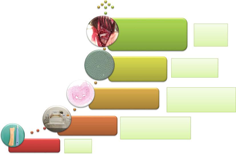
Wewilldiscussthechallengesfacedfora clinicaluseoftheseECMinhumans(Fig. 2.1).
2.2MaterialandMethods
2.2.1AnimalModels
Fullesophagealdecellularizationhasbeen mainlycarriedoutonrat(Ozekietal. 2006;
Urbanietal. 2018)andporcineesophagi(Luc etal. 2018;Arakelianetal. 2019).Eventhough ratesophagihaveservedasanimportantproofof concept,protocolsdevelopedonthissmallanimalmodelcannotbedirectlyappliedtoesophagi correspondingtohumansize.Porcineesophagus hastheadvantageofahighlysimilarstructure andsizecomparedtothehumanone(Ziegler etal. 2016).Forthisreason,porcineesophagus seemstobearelevantmodeltodevelopdecellularizationmethodsthatcanbeusedfora humanesophagealdecellularization.
2.2.2Decellularization
Thedecellularizationoftheesophagushasbeen mainlycarriedoutusingdetergentsincluding SDS,DEOX,tritonX-100orChaps(Mallisetal. 2019).Thedetergentisusedforrupturingthecell membranesandeliminatingcellcontent.CalciumchelatorEDTAhasalsobeenaddedto facilitatecelldetachmentandimprovethe
Clinical applica on
Cell seeding
Characteriza on of the matrix
Method of decellulariza on
•Indica ons
•In vivo efficiency
•Regula ons compliance
•Cell type and source
•Seeding method
•Evalua on of cell viability and prolifera on
•DNA quan fica on
•Biochemical proper es
•Biomechanical proper es
•Immunogenicity/biocompa bility
•Sterility
•Detergent and enzyma c treatment
•Bioreactor
•Rinsing and detoxifica on •Steriliza on
Fig.2.1 Stepstoproduceaclinicalgradeesophagealdecellularization:frombenchtobedside
decellularization(Arakelianetal. 2019).The nature,concentrationandtreatmentperiodcan highlyaffectthequalityofthe finalproduct.For smalleresophagealmodels,lowerconcentrations ormilddetergentscanbeusedfordecellularization.However,forlargermodels,higher concentrationsofstrongerdetergentssuchas SDSandDEOXwereneededandthetreatment periodwasextendedtoseveraldays.
Decellularizationprotocolsshowedthateven thoughinsomeexperimentsintheratesophagi, DNAcanbeeliminatedbycyclesofdetergent treatment(Mallisetal. 2019),inlargeranimal esophagi,thedetergentalonedoesnotremove DNAandthecellnuclei(Arakelianetal. 2019). Therefore,esophagealdecellularizationprotocolsincludeaDNasetreatment.Inthetwo recentdecellularizationstudiesinporcinemodel, onetreatedthedecellularizedmatrix12hwith 2000KunitzunitsofDNase-I(Sigma-Aldrich) (Lucetal. 2018),whereastheotherteamprivilegedashorter3htreatmentwith100u/ml clinicalgradeDNase(Pulmozyme)(Arakelian etal. 2019).
Attheendofdecellularization,aneffi cient rinsingmethodshouldbedevelopedinorderto fullyremovethesedetergentstoavoidcytotoxicity.Insmallanimalmodels,abundantrinsing withwaterorPBSwasreportedtobesufficientto removethesedetergents.Inbiggermodels,the rinsingcyclesweremuchlongeroritcouldbe necessarytouseanabsorbingresinwhichsignificantlyimproveddetergentremovaland reducedcytotoxicity(Arakelianetal. 2019).
Inthe fi rstattemptsofdecellularization, mechanicaltreatment,alongwithenzymaticand detergenttreatment,wasachievedbyplacingthe esophagiunderconstantagitation(Ozekietal. 2006)orbyperfusingtheorganusingaspeed rollerpump(Totonellietal. 2013).Thesemethodsincreaseddetergentandenzymeinfiltration withintheesophagusandimproveddecellularizationcomparedtostaticconditions.However, thesetechnicsworkedbetterforsmallerratesophagicomparedtolargerandthickerporcine ones.Furthermore,theseareopensystemswhich requireahighlevelofmanualmanipulationand
anincreasedriskofcontamination.Therecent decellularizationprotocolsincludedtheuseof bioreactorsforliquidperfusion(Lucetal. 2018) orperfusionandaxialrotation(Arakelianetal. 2019).Theseclosedsystemsincreasedtheefficiencyofdecellularizationandreducedmanual handlingwhichmaybeanadvantageforfuture clinicalapplications.
2.2.3Sterilization
Theesophagusisanorganwhichisinconstant exchangewithextracorporeal,non-sterileenvironment.Itisthereforeimportanttouseasterilizationmethodtopreventbacterialandfungal growththroughoutthedecellularizationoratthe endoftheprocess.Fordecontamination,ateam usedsodiumazide(Lucetal. 2018),amolecule whichcanbehighlytoxic(ChangandLamm 2003)andnotrecommendedforclinicaluse. Othersprivilegedtheuseofantibiotics(ATB)for aninitialdecontamination.Forthispurpose,a mixofATB(gentamycin,clindamycinvancomycinandamphotericinB),previouslyused forvasculargraftapplications,wasvalidatedfor esophagealdecontamination(Arakelianetal. 2019).DuetothehighconcentrationoftheATB, itisimportanttoefficientlyremovethematthe endofthedecellularizationtoavoidtoxicity, whilepreservingthesterilityofthedecellularized matrix.
Anotheroptionisa finalsterilizationwith chemicalorphysicaltreatments.Chemicaltreatmentscanincludeethyleneoxideorperacetic acid(PAA).Thediffi cultywiththesetreatments isthattheseproductsmayremaininthedecellularizedtissueandinducecytotoxicity(Lucas etal. 2017).Furthermore,ithasbeenshownthat PAAcanpreventvascularizationofsofttissues afterimplantationinvivo(Scheffleretal. 2008). Physicalsterilizationincludestreatmentwith gammarays.Eventhoughthistreatment efficientlyremovesbacterial,fungalandviral contaminations,itcancompromisethebiomechanicalpropertiesofthedecellularizedmatrices (Wittetal. 2016).
2.2.4Characterization oftheDecellularized Matrix
Asforotherdecellularizedorgans,therecommendedcriteriatodefineacompletedecellularizationaretovalidatetheabsenceofresidual cells,theeliminationofDNA(lessthan 50 µg/mgofdrymass)andtomakesurethatno residualDNAfragmentsexceeding200bp remainsinthetissue(Crapoetal. 2011).However,theserecommendationscanvaryslightly accordingtothenatureandtheoriginofthe tissue.Furthermore,thegeneralstructure,the bioactivemoleculesandthebiomechanical propertiesshouldalsobemaintainedafter decellularization.
2.2.5DNAQuantification
Indecellularizedesophagi,DNAwasextracted fromthematrixandwasthenquantified.Inall thesestudies,anefficienteliminationofDNA wasdemonstrated(Lucetal. 2018;Arakelian etal. 2019).ForDNAfragmentsize,anelectrophoresisoftheextractedDNAonagarosegel wascarriedoutwhichshowedthatnolargeDNA fragments(morethan200bp)wasvisible.The eliminationofnucleiwasalsoshownbyDAPI staining(Lucetal. 2018;Arakelianetal. 2019; Mallisetal. 2019).
2.2.6GeneralStructure
andComposition
Forthedemonstrationofcellremoval,histology (HESstaining)remainsthestandardmethodof validation(Lucetal. 2018;Arakelianetal. 2019).Furthermore,itisimportanttoshowthat thecomponentsoftheECMsuchascollagens, elastin fibers,glycosaminoglycans(GAGs)and othermoleculesarepreservedafterdecellularization.Intwostudiesofratandporcineesophagealdecellularization,collagenhasbeen quanti fiedusingahydroxyprolineassaykit (Mallisetal. 2019)orstainedwithpicrosiriusred
andanalyzedbyhistochemistry(Arakelianetal. 2019).Thesestudiesshowedthatmostofthe collagenwaspreserved,despitesomelossof structure.Elastin fibershavebeenstainedwith orceinafteresophagealdecellularization,andit wasshownthattheywerehighlypreservedafter decellularization.Finally,GAGquantification withdimethylmethyleneblueassay(DMMB)or stainingwithtoluidineblue(Arakelianetal. 2019)showedthattherewasamajorlossofthese moleculesafterdecellularization.However, immunostainingwithspeci ficantibodiesshowed thatthelossofGAGswasmainlyrelatedto chondroitinsulfates,whereastheheparansulfates anddermatansulfateswerepreserved(Arakelian etal. 2019).TheselasttwocategoriesofGAGs arethemainonesinvolvedinthebiomechanical propertiesofthematrix,aswellasthebinding andthedeliveryofhormonesandgrowthfactors (KjellénandLindahl 2018).Itisimportantto mentionthattheextentoflossofthesemolecules highlydependsonthenatureandconcentration ofthedetergent,aswellasthedurationofthe treatment(Mallisetal. 2019).Itistherefore importanttodevelopaprotocolwhichallowsan efficientdecellularizationwithoutamajorlossof structuralmolecules.
2.2.7BiomechanicalProperties
Thebiomechanicalpropertiesofthedecellularizedesophagihavebeenevaluatedandcompared tothenativeesophagi.Thetwomethodsthat havebeenusedtoevaluatethebiomechanical propertiesareburstpressuretestandtensile strength.Inthedecellularizedesophagi,porosity wasdetectedwhichpreventedthedecellularized esophagifromreachingaburstpoint(Lucetal. 2018).Tensiletestsshowedthatinthetransversalorientation,thedecellularizedandnative esophagihadsimilarproperties.Ontheother hand,inthelongitudinalorientation,thedecellularizedesophagiwerestifferthanthenativeone (Lucetal. 2018;Arakelianetal. 2019).Asfor invivoimplantation,decellularizedesophagi wereeasilyhandledforsurgicalproceduresand wereresistanttosutures.
2.2.8Immunogenicity andBiocompatibility
Oneofthemainpurposesofdecellularizationis toreducetheimmunereactionofthehostto avoidgraftrejectionand fibrosis.Tostudythese propertiesindecellularizedesophagi,an invitro assaywasdevelopedbasedontheproliferation oflymphocytesstainedwith fluorescentmolecule andanalyzedby flowcytometry(Arakelianetal. 2019).Thisassayshowedthatthedecellularized esophagididnotinducelymphocyteproliferation andindicatedtheabsenceofanacute immunogenicity.
However,theimmunereactionisacomplex mechanismandtrueimmunogenicityshouldbe evaluatedinvivo.Inanotherstudy,thisreaction wasevaluatedbyasubcutaneousimplantationof thematrixinnon-immunosuppressedWinstar rats(Lucetal. 2018).After14days,aninduction ofinflammatoryresponsewithinfiltrationof mononuclearcellswasshown.
2.2.9Cytotoxicity
Astheproductsusedfordecellularizationsuchas detergentsandahighdoseofATBaretoxicfor cells,itisimportanttomakesurethattheyare efficientlyremovedafterdecellularization.Itis thereforenecessarytodevelopassaystoanswer thesequestionsefficiently.Indecellularized esophagi,themainassaysusedsofarwerebased ontheevaluationofcellviability,bydirector indirectmethods(Iso10993-5-2009).Inthe directmethod,mesenchymalstromalcells (MSCs)wereseededonthedecellularizedesophagiandtheviabilityandmetabolicactivity wereevaluatedbyneutralredassayandMTT assay,respectively(Lucetal. 2018).Inthe indirectmethod,thedecellularizedesophagiwere incubatedwithcellculturemediumandthe supernatantwasthenusedforBalb/3T3cell culture.Theviabilityofthesecellswasevaluated by flowcytometryafterannexinVand7AAD staining(Arakelianetal. 2019).Thediffi culty withadirectMTTassayisthattheresultingdye isabsorbedbythematrix,anditisdifficultto
haveaccurateandreproducibleresults.The indirectmethodallowstoovercomethisdifficultyandtoevaluatethereleaseoftoxicsubstancesbythematrix.Bothmethodscanbeused forshorttermcytotoxicityevaluation.However, thepresenceofdetergentsandtoxicsubstances shouldbefurtherevaluatedbymassspectrometryandlong-termcytotoxicityshouldalsobe evaluatedinvivo
2.3CellSeeding
2.3.1CellTypesandOrigin
Cellseedingondecellularizedesophagihasbeen exploredinordertofunctionalizethesematrices andtoevaluatethepotentialofcellstoaccelerate tissueregeneration.Forinvivoapplications,itis essentialtoquestionthecelltypesandtheirorigin(autologousorallogeneic),asthischoice conditionsthedesiredmechanismofaction.The firstchoiceistousedifferentiatedcells,organspeci ficornot,suchasepithelialcells(Ozeki etal. 2006;Urbanietal. 2018;Asnaghietal. 2009;Barronetal. 2016;Jensenetal. 2018; Poghosyanetal. 2015;Nakaseetal. 2008), smoothmusclecells(Barronetal. 2016; Poghosyanetal. 2015;Takeokaetal. 2019)and endothelialcells(Takeokaetal. 2019).The functionalizationofthedecellularizedesophagus bythesecellscanbeinducedeitherbyadirect colonizationoftheECMbytheseededcellsor byparacrineeffects.Ithasbeenshownthatsome cellscanindeedsecretefactorsthatcanattract thehostcellsandacceleratetissueregeneration (Marzaroetal. 2006;Xiuunletal. 2009).
Theotheroptionistousenon-differentiated cells.Todate,noclearstemcellniche,ableto giverisetoallthecelltypes,hasbeenidenti fied intheadultesophagus(Seery 2002).Regarding stemcells,anotherpossibilityistoseedthe matrixwithMSCseitheroriginatingfromadiposetissueorbonemarrow(Hassetal. 2011). Thesecellspromotetherecruitmentofpatient cellsinsituthroughparacrineeffects,accelerate re-vascularizationandreduceinflammatoryand scarringprocesses(Lucetal. 2018;Arakelian
etal. 2019;Asnaghietal. 2009;Jensenetal. 2018;Poghosyanetal. 2015;Takeokaetal. 2019;Tanetal. 2013;Francescaetal. 2018; Catryetal. 2017).ItwasshownthatbonemarrowMSCseededonanon-esophagealextracellularmatrixacceleratedmuscleregenerationand re-epithelialisationinapatchesophagoplastyand afullthicknessesophagealreplacementmodels (Tanetal. 2013;Catryetal. 2017).
Beyondthesemechanisticaspects,theorigin ofcellscanleadtosigni ficantconstraints. Indeed,autologouscellswillrequireasampleof thepatient,isolation,amplificationandthenthe constitutionofthesubstitute;whiletheuseof allogeneiccellswillreducetheproductiontime, butraisesthequestionofimmunologicalrejection.Thankstotheirimmunomodulatoryproperties,MSCsareaninterestingsourceforthe recellularizationofdecellularizedesophagi.
2.3.2SeedingMethods
Thedecellularizedesophagusisacylindrical hollowtubewithaninnerandoutersurface.The challengeisthereforetodecidewhichlayer shouldbeseededandhowtodistributethecells evenlyonthematrix.
Celldensity,aswellasthedurationofcell culture in-vitro arefurtherimportantparameters toensurethecolonizationofthematrixbythe cellsandtheirinfi ltration.Fiveteamsshowed veryvariableculturetimes,rangingfrom7to 21days(Ozekietal. 2006;Lucetal. 2018; Arakelianetal. 2019;Urbanietal. 2018).The numberofseededcellsvariesfromonestudyto anotherfrom1.105 toseveralmillionspercm2 (Ozekietal. 2006;Lucetal. 2018;Arakelian etal. 2019;Urbanietal. 2018).Theseparameters couldbedifferentaccordingtocelltypesand theircapacitytoadhereandproliferate.
Sometubularesophagealsubstituteswere seededunderstaticconditions.Cellswere depositedontheoutersurfaceorwereinjected insidethelumenusingapipette(Catryetal. 2017;Poghosyanetal. 2013).However,inmost studies,axialrotationwasappliedtohomogenize celldistributiononthematrix.Thisrotationwas
achievedeithermanuallyatregulartimeintervals (Urbanietal. 2018;Barronetal. 2016;Jensen etal. 2018)orusingacontinuousrotationsystem (Ozekietal. 2006;Arakelianetal. 2019;Urbani etal. 2018;Asnaghietal. 2009;Francescaetal. 2018).Thesesystemsinclude:(1)anaxialrotary bioreactorwithpartialliquidimmersionofthe substitute(Asnaghietal. 2009),(2)anaxial rotatingstirrerwitha filterplugtube(Arakelian etal. 2019),(3)arotatingbioreactorwithafull liquidimmersionofthematrix(Francescaetal. 2018)or(4)aWaverotorbioreactor(Thermonics,Tokyo,Japan)(Ozekietal. 2006).The advantageofusingabioreactorforcellseedingis thatitallowsahomogeneouscelldistribution,as wellasreducingmanualinterventionandabetter controlofoxygenation,pHandcellularmetabolism.Theseparametersareimportantforthe reproducibilityofcellseedingandforafuture clinicalapplicationunderGMPconditions. Urbanietal.clearlydemonstratedthebenefitsof adynamicculture(Urbanietal. 2018).However, theanimalmodelusedbeingtherat,thetranspositiontoahuman-sizedesophagusremainsto bedemonstrated.Cellsheettechnologyis anotheroptionofcellseedingonthedecellularizedesophagi.Thismethodhasbeenexplored usingMSCs.Tosummarize,MSCswereculturedinadishataveryhighconfluenceandthe cellsheetwasrolledaroundadecelluarized esophagus(Lucetal. 2018).Cellsheetseeding canbeimprovedusingthermoresponsivepolymerssuchaspNIPAMwhichallowafullcell sheetdetachmentuponchangingthetemperature. Thismethodhasalreadybeenvalidatedina clinicaltrialforsuper ficiallesionsusingepithelialcells(Yamaguchietal. 2017)andcouldbe usedforseedingofdecellularizedesophagi.
2.4ClinicalApplications
Commercializednon-esophagealdecellularized naturalECMhavepreviouslybeentestedin clinicaltrialsfortreatingesophagealleakswith decellularizedskinorsuper ficialesophageal lesionswithSISpatchestopreventstenosis (Bozuketal. 2006;Badylaketal. 2011).
However,thesemethodshaveneverbeensuccessfullyappliedforfullthicknesscircumferentialreplacementhumans.
Fortheclinicalapplicationofdecellularized esophagi,itisimportanttoconsidertheregulatoryaspectwhichwillbeapplied.InEurope,for example,ifthematrixistobeusedalone, withoutcellseeding,itcouldbeconsideredasan implantablemedicaldevice “IMD” oras “human cells,tissues,andcellularandtissue-basedproduct(HCT/P)”.Oneofthemaindetermining criteriaforchoosingbetweenthesetwocategoriesistheoriginofthedecellularizedmatrix andthenatureoftheprotocol.A finaldecontaminationismandatoryforIMDs.Ahuman matrixcanbetreatedbothasanIMDanda HCT/P,whereasaporcinedecellularizedesophaguscanonlybetreatedasanIMD.Inboth categories,itisnecessarytoshowthesterilityof thematrixandbothcaninvolveaninitial decontaminationwithantibioticsanda final sterilizationusinggammaraysorchemicalssuch asethyleneoxide.ForIMD,thequalitycontrols shouldbecarriedouttoobtainaCEmarkingand thematrixcanbeproducedbypharmaceutical companies.AHCT/P,however,shouldbeproducedinspecialaccreditedfacilitiessuchas humantissuebanks.Inbothcategories,alongtermconservationmethodshouldbevalidated whichcouldincludethepreparationofafrozen matrixbank.
Ifthedecellularizedesophagealmatrixistobe seededwithcellsbeforeimplantation,the final productisconsideredasanadvancedtherapy medicinalproduct(ATMP),correspondingtoa newcategoryofregulations(https://www.ema. europa.eu/en/human-regulatory/overview/ advanced-therapy-medicinal-products-overview). Thismeansthatontopofevaluatingthebiologicalpropertiesandthesterilityofthematrix, thenatureofthecellsandthecultureconditions onthematrixbeforeandafterinvivoimplantationshouldbeevaluated.Thecellsshouldbe isolatedandculturedinaclinicalgradecell culturemedia,andtheoptimalcelldensityas wellasinvitromaturationtimeshouldbeclearly defined.Onceimplantedintheanimal,thepossiblemigrationofthecellswithindifferent
organs,aswellastheirtumorigenicpotential, shouldbecarefullyevaluated.UnlikeMDand tissueproducts,ATMPsneedtobeproducedin authorizedspecialfacilitiessuchasplatformsor pharmaceuticalindustries.
Forallthethreecategories,apre-clinicaltrial inabiganimalmodelisnecessarytoshow theefficiencyofthematrixinesophageal replacement.
Oneofthechallengesforinvivoesophageal replacementisthemethodofvascularization.As theesophagusiscomposedofmicrovessels comingfromtheaortaandthesurrounding organs,itisnecessaryto findavascularization methodtopreventorgannecrosis.Theoption thathasbeentestedinpreviousesophagealtissue engineeringstudieshasbeenamaturationstepin theomentum(Lucetal. 2018;Poghosyanetal. 2015).ThesestudiesshowedthatatubularsubstitutecomposedofSISforesophagealreplacementwassuccessfullyvascularizedbythis option.Thismethodhasalsobeenusedsuccessfullyforthevascularizationofaratdecellularizedesophagusandaporcineone.However, long-termeffi ciencyafterorganreplacement shouldbeevaluatedinvivo.
References
ArakelianL,CailleC,FaivreL,Corté L,BrunevalP, ShamdaniSetal(2019)Aclinical-gradeacellular matrixforesophagealreplacement.JTissueEng RegenMed. https://doi.org/10.1002/term.2983
AsnaghiMA,JungebluthP,RaimondiMT,DickinsonSC, ReesLEN,GoTetal(2009)Adouble-chamber rotatingbioreactorforthedevelopmentoftissueengineeredholloworgans:Fromconcepttoclinical trial.Biomaterials30(29):5260–5269
BadylakSF,VorpDA,SpievackAR,Simmons-ByrdA, HankeJ,FreytesDOetal(2005)EsophagealreconstructionwithECMandmuscletissueinadogmodel. JSurgRes
BadylakSF,HoppoT,NieponiceA,GilbertTW,DavisonJM,JobeBA(2011)Esophagealpreservationin fivemalepatientsafterendoscopicinner-layercircumferentialresectioninthesettingofsuperficial cancer:aregenerativemedicineapproachwitha biologicscaffold.TissueEngPartA[Internet]17 (11–12):1643–50.Availablefrom:http://www.liebertonline.com/doi/abs/https://doi.org/10.1089/ten.tea.2010. 0739
BarronMR,BlancoEW,AhoJM,ChakroffJ,JohnsonJ, CassiviSDetal(2016)Full-thicknessoesophageal regenerationinpigusingapolyurethanemucosalcell seededgraft.JTissueEngRegenMed12(1):175–185
BozukMI,FearingNM,LeggettPL(2006)Useof decellularizedhumanskintorepairesophagealanastomoticleakinhumans.JSLS10:83–85
CatryJ,Luong-NguyenM,ArakelianL,PoghosyanT, BrunevalP,DometTetal(2017)Circumferential esophagealreplacementbyatissue-engineeredsubstituteusingmesenchymalstemcells.CellTransplant [Internet]26(12):1831–1839.Availablefrom:http:// journals.sagepub.com/doi/https://doi.org/10.1177/ 0963689717741498
ChangS,LammSH(2003)Humanhealtheffectsof sodiumazideexposure:aliteraturereviewand analysis.IntJToxicol CrapoPM,GilbertTW,BadylakSF(2011)Anoverview oftissueandwholeorgandecellularizationprocesses. Biomaterials32:3233–3243
HassR,KasperC,BöhmS,JacobsR(2011)Different populationsandsourcesofhumanmesenchymalstem cells(MSC):acomparisonofadultandneonatal tissue-derivedMSC.CellCommunSignal9(1):12
JensenT,WanczykH,SharmaI,MitchellA,SayejWN, FinckC(2018)Polyurethanescaffoldsseededwith autologouscellscanregeneratelongesophagealgaps: anesophagealatresiatreatmentmodel.JPediatrSurg KjellénL,LindahlU(2018)Specificityofglycosaminoglycan–proteininteractions.CurrOpinStructBiol 50:101–108
KochH,GraneistC,EmmrichF,TillH,MetzgerR, AupperleHetal(2012)Xenogenicesophagusscaffolds fixedwithseveralagents:Comparativeinvivo studyofrejectionandinflammation.JBiomed Biotechnol
KuoB,UrmaD(2006)Esophagus anatomyand development.GIMotilonline[Internet],pp1–20. Availablefrom: http://www.nature.com/gimo/ contents/pt1/full/gimo6.html
LaFrancescaS,AhoJM,BarronMR,BlancoEW, SolimanS,KalenjianLetal(2018)Long-term regenerationandremodelingofthepigesophagus aftercircumferentialresectionusingaretrievable syntheticscaffoldcarryingautologouscells.SciRep [Internet]8(1):4123.Availablefrom: http://www. nature.com/articles/s41598-018-22401-x
LucG,CharlesG,GronnierC,CabauM,KaliskyC, MeulleMetal(2018)Decellularizedandmatured esophagealscaffoldforcircumferentialesophagus replacement:proofofconceptinapigmodel. Biomaterials175:1–18
LucasAD,MerrittK,HitchinsVM,WoodsTO, McnameeSG,LyleDBetal(2017)Residualethylene oxideinmedicaldevicesanddevicematerialresidual ethyleneoxideinmedicaldevicesanddevicematerial MallisP,ChachlakiP,KatsimpoulasM,StavropoulosGiokasC,MichalopoulosE(2019)Optimizationof decellularizationprocedureinratesophagusfor
possibledevelopmentofatissueengineeredconstruct. Bioengineering MarzaroM,VigoloS,OselladoreB,ConconiMT, RibattiD,GiulianiSetal(2006)Invitroand invivoproposalofanartificialesophagus.JBiomed MaterResA77(4):795–801 NakaseY,NakamuraT,KinS,NakashimaS, YoshikawaT,KuriuYetal(2008)Intrathoracic esophagealreplacementbyinsitutissue-engineered esophagus.JThoracCardiovascSurg136(4):850–859 OzekiM,NaritaY,KagamiH,OhmiyaN,ItohA, HirookaYetal(2006)Evaluationofdecellularized esophagusasascaffoldforculturedesophageal epithelialcells.JBiomedMaterResPartA PoghosyanT,GaujouxS,ChiricaM,Munoz-Bongrand N,SarfatiE,CattanP(2011)Functionaldisordersand qualityoflifeafteresophagectomyandgastrictube reconstructionforcancer.JViscSurg[Internet]148: e327 e335.Availablefrom: http://ac.els-cdn.com. gate2.inist.fr/S1878788611001093/1-s2.0S1878788611001093-main.pdf?_tid=102588d6-832211e7-8a8b-00000aacb35f&acdnat=1502956978_ 4c9f951fb448fd427801b6ae238e4cf9
PoghosyanT,GaujouxS,VanneauxV,BrunevalP, DometT,LecourtSetal(2013)Invitrodevelopment andcharacterizationofatissue-engineeredConduit \nResemblingesophagealwallusinghumanandpig skeletalmyoblast,Oral\nEpithelialCells,andbiologic scaffolds.TissueEngParta19(19–20):2242–2252
PoghosyanT,SfeirR,MichaudL,BrunevalP,DometT, VanneauxVetal(2015)Circumferentialesophageal replacementusingatube-shapedtissue-engineered substitute:anexperimentalstudyinminipigs.Surg (UnitedStates)[Internet]158(1):266–277. https://doi. org/10.1016/j.surg.2015.01.020
ReingJE,ZhangL,Myers-irvinJ,PhD,CorderoKE, FreytesDOetal(2009)Degradationproductsof extracellularmatrixaffectcellmigrationandproliferation.15(3)
SchefflerSU,CmdJG,CmdJK,PrzybillaD(2008) RemodelingofACLallograftsisinhibitedbyperaceticacidsterilization.ClinOrthopRelatRes 466:1810–1818
SeeryJP(2002)Stemcellsoftheoesophagealepithelium. JCellSci115:783–1789
TakeokaY,MatsumotoK,TaniguchiD,TsuchiyaT, MachinoR,MoriyamaMetal(2019)Regenerationof esophagususingascaffoldfreebiomimeticstructure createdwithbiothree-dimensionalprinting. PLoSONE14(3):1–12
TanB,WeiRQ,TanMY,LuoJC,DengL,ChenXHetal (2013)Tissueengineeredesophagusbymesenchymal stemcellseedingforesophagealrepairinacanine model.JSurgRes182(1):40–48
TotonelliG,MaghsoudlouP,GeorgiadesF,GarriboliM, KoshyK,TurmaineMetal(2013)Detergent enzymatictreatmentforthedevelopmentofanatural acellularmatrixforoesophagealregeneration.Pediatr SurgInt
UrbaniL,CamilliC,PhylactopoulosD,CrowleyC, NatarajanD,ScottoniFetal(2018)Multi-stage bioengineeringofalayeredoesophaguswithinvitro expandedmuscleandepithelialadultprogenitors.Nat Commun
WittTA,MarlerRJ,PislaruSV,RobertD(2016)Lowdosegammairradiationofdecellularizedheartvalves resultsintissueinjuryinvitroandinvivo.AnnThorac Surg101(2):667–674
XiuunlI,XiaozUO,WeizHI,PengyA,HuiixIE, ZhiingyA(2009)Graftsofporcinesmallintestinal submucosawithculturedautologousoralmucosal
epithelialcellsforesophagealrepairinacanine model.ExpBiolMed(Maywood)234(4):453–461 YamaguchiN,IsomotoH,KobayashiS,KanaiN, KanetakaK,SakaiYetal(2017)Oralepithelialcell sheetsengraftmentforesophagealstricturesafter endoscopicsubmucosaldissectionofsquamouscell carcinomaandairplanetransportation.SciRep7(1) ZieglerA,GonzalezL,BlikslagerA(2016)Largeanimal models:thekeytotranslationaldiscoveryindigestive diseaseresearch.Cmgh[Internet]2(6):716–24. https:// doi.org/10.1016/j.jcmgh.2016.09.003