@aestheticsgroup
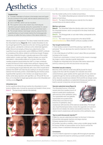
Treatment tips 1. Consider restoration of volume in the temple, brow and upper lid sulcus at three to four points, with the lateral canthal area as an optional extra (Figure 4). 2. Use a soft filler, which can be moulded. 3. The superior sulcus area needs very small aliquots injected supra periosteally along the supraorbital rim, above the orbital septum, staying lateral to the supraorbital notch. 4. The temple hollows need larger volumes, placed into the superficial fascia of each temple behind the frontozygomatic process, to soften the bony contour of the lateral orbital rim. laterally its lateral compartment. The deep medial cheek fat overlies the infraorbital foramen (Figure 5). At the medial infraorbital rim, the tear trough ligament (TTL), attaches to the skin, forming a gentle tear trough groove. Laterally this continues as the orbitomalar ligament (OML). Descent and atrophy of these bony attachments of the superficial musculo aponeurotic system (SMAS) and SOOF contribute to unmasking of the inferior orbital rim and the tear trough. Prolapse of the deep orbital fat, through a naturally weak area of the medial orbital septum, creates eye bags.13 Ageing changes can cause the inferolateral > inferomedial orbital rim to recede, and loss of the maxillary projection (bone) below the orbit14 is a major contributor to laxity and descent of the medial cheek soft tissue. Ageing of the mid-face is a mix of atrophy and descent of soft tissue, which is visible as a worsening tear trough deformity with a loss of the smooth blend between the SOOF and malar fat pad, leading to an abrupt transition between the lid-cheek junction, cheek flattening and mid-face ptosis. Superficial filler injections in the mid-face can weigh tissues down further, whilst deep injections with rigid fillers can “lift and fill” the midface, so it is important to target appropriate areas in the infraorbital and mid-face zones (Figure 5). Infraorbital zone: As the superficial inferior fat pad overlies the infraorbital rim and it tends to deflate early, it should be assessed and treated in three zones Zone 1: The tear trough extends inferolaterally Figure 4
Figure 5
Aesthetics Journal
Aesthetics aestheticsjournal.com
from the medial canthus to the medial corneal limbus. Zone 2: The middle infraorbital groove extends from the medial to lateral corneal limbus. Zone 3: The lateral infraorbital groove extends from the lateral corneal limbus to the lateral canthus. Mid-face sub zones: Zone 4: The infraorbital hollow lies directly under zone 2 and over the infraorbital foramen, which corresponds to the deep medial fat compartment. Zone 5: The infrazygomatic or sub malar hollow corresponds to the medial SOOF. Zone 6: The malar mound corresponds to the lateral SOOF. Lateral Canthal area: Where ROOF continues caudally as SOOF. Tear trough treatment tips: 1. Treat mid-face zones 4, 5 and 6 first, placing a rigid filler pre periosteal.1 This can decrease the need for treatment in the medial tear trough. 2. Use small aliquots of soft filler in zone 1, place filler pre periosteal but deep to the muscle. 3. Avoid over volumisation in zones 1 and 2 as this causes a sausagelike bulge in what is naturally a gentle depression. 4. Palpate the infraorbital bone and place filler below septum (not behind or above it), as this will only worsen any eye bag. Periorbital vascular anatomy Branches of the external carotid artery (ECA) provide the blood supply to the face with the exception of a mask-like area of the central forehead, upper eyelids and the upper part of nose, which are supplied though the internal carotid system (ICA) by the ophthalmic artery. Vascular anastomoses between ECA and ICA are danger zones for the aesthetic practitioner as inadvertent intravascular injection can lead to vascular compromise and permanent blindness.14 Figure 6
Vascular watershed areas (Figure 6) The infraorbital foramen: Infraorbital vessels arise from maxillary branch of external carotid which anastomose with branches of the ophthalmic artery. The supraorbital notch and glabella: Supraorbital and supratrochlear, infratrochlear and external nasal branches of ophthalmic artery anastomose with branches of the external carotid artery. The temple area: Superficial temporal artery crosses the zygomatic arch and 2cm above the arch divides into anterior and posterior branches. The anterior branch anastomoses with branches of the ophthalmic artery. Tips to avoid intravascular injection16,17 1. Mark the vascular watershed and inject ‘on the bone’ in that area. 2. Avoid fast anterograde injections and large volumes. 3. Always aspirate prior to injection. 4. Consider cannulas vs needles, unless using a smaller gauge needle. 5. Choose HA filler as hyaluronidase can be used to remove the product, if there is inadvertent intravascular injection. Conclusions A sound anatomic approach to surgery with thorough pre-operative planning remains the basis for achieving successful cosmetic and
Reproduced from Aesthetics | Volume 2/Issue 4 - March 2015