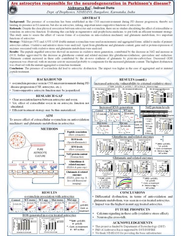
Are astrocytes responsible for the neurodegeneration in Parkinson’s disease? Aishwarya Raj*, Indrani Datta Dept. of Biophysics, NIMHANS, Bangalore, Karnataka, India ABSTRACT Background: The presence of α-synuclein has been established in the CNS microenvironment during PD disease progression, thereby not limiting its presence in DA neurons, but also in astrocytes; risking, important neuro-supportive functions of astrocytes. Rationale: Despite the clear physical association between astrocytes and α-synuclein, there are no studies elucidating the effect of extracellular αsynuclein on astrocytic function. Evaluating this can help in regenerative and prophylactic-medicine, to put forth an efficient treatment strategy. This study aims to assess the effect of various forms of α-synuclein on anti-oxidation machinery and glutamate metabolism, two important functions of astrocytes. Strategy: Wild-type (WT) and A53T/A30P double mutant α-synuclein were used in monomeric and aggregated forms; added to media of primary astrocytes culture. Oxidative and nitrative stress were analyzed. Apart from glutathione and glutamate content, gene and/or protein expression of enzymes associated with oxidative stress and glutamate metabolism were analyzed. Results: The peptide engulfed astrocytes showed an increase in oxidative stress generation, contributed by the decrease in Nrf2 and increase in iNOS, further aggravated by the decrease in glutathione content and related enzymes like glutathione-synthetase, -peroxidase and -reductase. Glutamate content increased in these cells contributed by the de-novo synthesis of glutamate by pyruvate-carboxylase. Decreased GDH expression was observed, with its enzyme activity increased probably to compensate for the increased glutamate content. The highest dysfunction was observed with the mutant aggregated α-synuclein treatment. Conclusion: The presence of α-synuclein did lead to astrocytic dysfunction. The impact was higher in the case of aggregated and/or mutated peptide treatment.
BACKGROUND
RESULTS (contd.)
• α-synuclein presence seen in CNS microenvironment during PD disease progression (CSF, astrocytes, etc.). • Neuro-supportive astrocyte function may be jeopardized.
Increased astrocytic vulnerability to external oxidative stress [**&## - p<0.001, # - p<0.05; * - WT vs Mut, # - Mono vs Agg] **
**
**
**
##
##
##
#
##
##
RESEARCH GAP • Clear association known between astrocytes and α-syn. • Yet, effect of extracellular α-syn in on astrocytic function not elucidated. • Efficient treatment strategy may be thus materialized.
Increased RNS generation WT mono
Control
AIM
**
To assess effect of extracellular α-synuclein on anti-oxidation machinery and glutamate metabolism in astrocytes.
** Mut agg
Mut mono
65.5±0.4%
69.2±0.8%
1. Peptide association (Flow cytometry)
* *
30
**
#
** ** ** **
$$
$$
##
* * ** **
** ** ** **
gg
ag g
on o
M ut a
on o m T W
Treatment
**,##$$ - p<0.001; *,# - p<0.05 * - Cont. vs treatment # - Mono vs Agg; * WT vs Mut
Glutamate and related enzymes $$
$$ @@ #
**
#
**
$ ##
##
##
##
**
**
#
**
**
**
##
**
**,$$,@@ - p<0.001; # - p<0.05 * - C1. vs treatment; # - C2 vs treatement; $ - Mono vs Agg; @ - WT vs Mut
GDH Enzyme kinetics
4.0
3.5
3.0
2.5
2.0
1.5
Control WT mono WT agg Mut mono Mut agg
1.0
0.5 0
100
200
300
400
500
$$
**
##
**
Glutamate & related 6. enzymes (ELISA, gene exp. & live cell fluorescent imaging)
## $$
$$
** ** **
**,##$$ - p<0.001; *,# - p<0.05 * - Cont. vs treatment # - Mono vs Agg; * WT vs Mut
RESULTS
CONCLUSIONS
α-synuclein association with astrocytes
• Differential dysfunction, in terms of anti-oxidation and glutamate metabolism, was seen in α-syn treated astrocytes. • Impact was the highest in mut agg treated astrocytes.
WT mono
0.9±0.2%
Nrf2
4
3
C-Cont.; 1-WT mono; 2WT agg; 3-Mut mono; 4-Mut agg
0
Time (seconds)
Control
2
β-actin
$$ ##
$$ #
10
Nrf2 protein content (Western blot)
Glutathione & 5. related enzymes (ELISA & gene exp.) Cont. WT mono WT agg Mut mono Mut agg
*
20
W T
RNS levels (Flow cytometry
40
NADH f1/f0
4.
** p<0.001 Cont. vs treatme -nt
$$
$$ #
50
Co n tr
PURE ASTROCYTE CULTURE
Glutathione content Glutathione concentration ( M)
3.
1
C
Glutathione and related enzymes
ROS levels (Spectrofluorimetry)
ol
2.
**
**
M ut m
METHODS
62±0.04%
52.3±0.7%
27.2±0.4%
Nrf2 protein levels
WT agg
WT agg
Mut mono
60.8±0.7%
51.8±0.7%
**
**
65.6±0.7%
**
Mut agg
** p<0.001 ** Cont. vs treatment
63.1±0.9%
ROS generated in α-syn treated astrocytes ** ## ## ## ##
** ##
#
#
**&## - p<0.001 # - p<0.05 * - Cont. vs treatment # - WT vs Mut
FUTURE PROSPECTS • •
Calcium signaling in these cells (oxidative stress effect). Neuron-glia cross-talk.
ACKNOWLEDGEMENTS • This project is funded by Department of Biotechnology (DBT) • PhD of Aishwarya Raj is supported by DST-INSPIRE • We thank NIMHANS for providing the basic infrastructure.