

Medical Legal Case Report
361 Medical Malpractice in the Waiting Room: Who Is at Risk?
KP Carpenter, L Walker, RA Lindor
Case Series
365 Use of Point-of-care Ultrasound for Detection of Urethral Foreign Bodies: A Case Series
L Tomasi, M Zampi, M Schroeder, M Cooper, N McIntyre
Case Reports
369 Report of Two Cases: Altered Mental Status and Anisocoria as Presenting Symptoms in Acute Basilar Artery Occlusion
A Ryu, K Chhabra, T George, E Kasparov, M Wali, CC Lee
373 Case Report: Bigeminy with Alternating Injury Pattern Morphologies in a Young Woman After Cardiac Arrest
M Huttner, M McMurray, M Huecker, S Ikram
376 Idiopathic Atraumatic Renal Hemorrhage: Case Report
T Ranson, G Ruddy, Z Ostapowicz, L Joyner
380 Suspected Fat Embolism Syndrome in the Setting of Ballistic Long Bone Fractures: A Case Report
I Husain, D Andrews
385 Paraspinal Compartment Syndrome Associated with Opioid Overdose: A Case Report
T Habiyaremye, K Holz, JR Jackson
389 Orbital Compartment Syndrome as a Complication of Blepharoplasty: A Case Report
L Libet, J Garcia, L Liu
392 An Unusual Presentation of Orbital Compartment Syndrome: A Case Report
J Rosenblum


About Us: Penn State Health is a multi-hospital health system serving patients and communities across central Pennsylvania. We are the only medical facility in Pennsylvania to be accredited as a Level I pediatric trauma center and Level I adult trauma center. The system includes Penn State Health Milton S. Hershey Medical Center, Penn State Health Children’s Hospital and Penn State Cancer Institute based in Hershey, Pa.; Penn State Health Hampden Medical Center in Enola, Pa.; Penn State Health Holy Spirit Medical Center in Camp Hill, Pa.; Penn State Health Lancaster Medical Center in Lancaster, Pa.; Penn State Health St. Joseph Medical Center in Reading, Pa.; Pennsylvania Psychiatric Institute, a specialty provider of inpatient and outpatient behavioral health services, in Harrisburg, Pa.; and 2,450+ physicians and direct care providers at 225 outpatient practices. Additionally, the system jointly operates various healthcare providers, including Penn State Health Rehabilitation Hospital, Hershey Outpatient Surgery Center and Hershey Endoscopy Center.
We foster a collaborative environment rich with diversity, share a passion for patient care, and have a space for those who share our spark of innovative research interests. Our health system is expanding and we have opportunities in both academic hospital as well community hospital settings.
Benefit highlights include:
•Competitive salary with sign-on bonus
•Comprehensive benefits and retirement package
•Relocation assistance & CME allowance
•Attractive neighborhoods in scenic central Pennsylvania

FOR MORE INFORMATION PLEASE CONTACT:
Heather Peffley, PHR CPRP
Penn State Health Lead Physician Recruiter hpeffley@pennstatehealth.psu.edu


Indexed in PubMed and full text in PubMed Central
Rick A. McPheeters, DO, Editor-in-Chief Kern Medical/UCLA- Bakersfield, California
R. Gentry Wilkerson, MD, Deputy Editor University of Maryland School of Medicine
Mark I. Langdorf, MD, MHPE, Senior Associate Editor University of California, Irvine School of Medicine- Irvine, California
Shahram Lotfipour, MD, MPH, Senior Associate Editor University of California, Irvine School of Medicine- Irvine, California
Shadi Lahham, MD, MS, Associate Editor Kaiser Permanente- Orange County, California
John Ashurst, DO, Decision Editor/ ACOEP Guest Editor Kingman Regional Health Network, Arizona
Anna McFarlin, MD, Decision Editor Louisiana State University Health Science Center- New Orleans, Louisiana
Lev Libet, MD, Decision Editor Kern Medical/UCLA- Bakersfield, California
Amin A. Kazzi, MD, MAAEM
The American University of Beirut, Beirut, Lebanon
Anwar Al-Awadhi, MD
Mubarak Al-Kabeer Hospital, Jabriya, Kuwait
Arif A. Cevik, MD United Arab Emirates University College of Medicine and Health Sciences, Al Ain, United Arab Emirates
Abhinandan A.Desai, MD University of Bombay Grant Medical College, Bombay, India
Bandr Mzahim, MD
King Fahad Medical City, Riyadh, Saudi Arabia
Barry E. Brenner, MD, MPH Case Western Reserve University
Brent King, MD, MMM University of Texas, Houston
Daniel J. Dire, MD University of Texas Health Sciences Center San Antonio
David F.M. Brown, MD Massachusetts General Hospital/Harvard Medical School
Edward Michelson, MD Texas Tech University
Edward Panacek, MD, MPH University of South Alabama
Erik D. Barton, MD, MBA Icahn School of Medicine, Mount Sinai, New York
Francesco Dellacorte, MD
Azienda Ospedaliera Universitaria “Maggiore della Carità,” Novara, Italy
Francis Counselman, MD Eastern Virginia Medical School
Gayle Galleta, MD
Sørlandet Sykehus HF, Akershus Universitetssykehus, Lorenskog, Norway
Hjalti Björnsson, MD Icelandic Society of Emergency Medicine
Jacob (Kobi) Peleg, PhD, MPH Tel-Aviv University, Tel-Aviv, Israel
Jonathan Olshaker, MD Boston University
Katsuhiro Kanemaru, MD University of Miyazaki Hospital, Miyazaki, Japan
Amal Khalil, MBA UC Irvine Health School of Medicine
Elena Lopez-Gusman, JD
California ACEP
American College of Emergency Physicians
DeAnna McNett, CAE
American College of Osteopathic Emergency Physicians
John B. Christensen, MD California Chapter Division of AAEM
Randy Young, MD
California ACEP
American College of Emergency Physicians
Mark I. Langdorf, MD, MHPE UC Irvine Health School of Medicine
Jorge Fernandez, MD
California ACEP
American College of Emergency Physicians University of California, San Diego
Peter A. Bell, DO, MBA
American College of Osteopathic Emergency Physicians
Baptist Health Science University
Robert Suter, DO, MHA
American College of Osteopathic Emergency Physicians UT Southwestern Medical Center
Shahram Lotfipour, MD, MPH UC Irvine Health School of Medicine
Brian Potts, MD, MBA
California Chapter Division of AAEM Alta Bates Summit-Berkeley Campus
Christopher Sampson, MD, Decision Editor University of Missouri- Columbia, Missouri
Joel Moll, MD, Decision Editor
Virginia Commonwealth University School of Medicine- Richmond, Virginia
Steven Walsh, MD, Decision Editor Einstein Medical Center Philadelphia-Philadelphia, Pennsylvania
Melanie Heniff, MD, JD, Decision Editor University of Indiana School of Medicine- Indianapolis, Indiana
Austin Smith, MD, Decision Editor Vanderbilt University Medical Center-Nashville, Tennessee
Rachel A. Lindor, MD, JD, Decision Editor Mayo Clinic College of Medicine and Science
Jacqueline K. Le, MD, Decision Editor Desert Regional Medical Center
Christopher San Miguel, MD, Decision Editor Ohio State Univesity Wexner Medical Center
Khrongwong Musikatavorn, MD King Chulalongkorn Memorial Hospital, Chulalongkorn University, Bangkok, Thailand
Leslie Zun, MD, MBA Chicago Medical School
Linda S. Murphy, MLIS University of California, Irvine School of Medicine Librarian
Nadeem Qureshi, MD
St. Louis University, USA Emirates Society of Emergency Medicine, United Arab Emirates
Niels K. Rathlev, MD Tufts University School of Medicine
Pablo Aguilera Fuenzalida, MD Pontificia Universidad Catolica de Chile, Región Metropolitana, Chile
Peter A. Bell, DO, MBA Baptist Health Science University
Peter Sokolove, MD University of California, San Francisco
Robert M. Rodriguez, MD University of California, San Francisco
Robert Suter, DO, MHA UT Southwestern Medical Center
Robert W. Derlet, MD University of California, Davis
Rosidah Ibrahim, MD
Hospital Serdang, Selangor, Malaysia
Samuel J. Stratton, MD, MPH Orange County, CA, EMS Agency
Scott Rudkin, MD, MBA University of California, Irvine
Scott Zeller, MD University of California, Riverside
Steven Gabaeff, MD Clinical Forensic Medicine
Steven H. Lim, MD Changi General Hospital, Simei, Singapore
Terry Mulligan, DO, MPH, FIFEM ACEP Ambassador to the Netherlands Society of Emergency Physicians
Vijay Gautam, MBBS University of London, London, England
Wirachin Hoonpongsimanont, MD, MSBATS Siriraj Hospital, Mahidol University, Bangkok, Thailand
Ian Oliffe, BS Executive Editorial Director
Sheyda Aquino, BS WestJEM Editorial Director
Tran Nguyen, BS CPC-EM Editorial Director
Stephanie Burmeister, MLIS WestJEM Staff Liaison
Cassandra Saucedo, MS Executive Publishing Director
Isabelle Kawaguchi, BS WestJEM Publishing Director
Alyson Tsai, BS CPC-EM Publishing Director
June Casey, BA Copy Editor
Official Journal of the California Chapter of the American College of Emergency Physicians, the America College of Osteopathic Emergency Physicians, and the California Chapter of the American Academy of Emergency Medicine


Available in MEDLINE, PubMed, PubMed Central, Google Scholar, eScholarship, DOAJ, and OASPA.

Editorial and Publishing Office: WestJEM/Depatment of Emergency Medicine, UC Irvine Health, 3800 W. Chapman Ave. Suite 3200, Orange, CA 92868, USA Office: 1-714-456-6389; Email: Editor@westjem.org
Volume 9, no. 4: November 2025 i Clinical Practice and Cases in Emergency
Indexed in PubMed and full text in PubMed Central
This open access publication would not be possible without the generous and continual financial support of our society sponsors, department and chapter subscribers.
Professional Society Sponsors
American College of Osteopathic Emergency Physicians
California ACEP
Academic Department of Emergency Medicine Subscribers
Albany Medical College Albany, NY
American University of Beirut Beirut, Lebanon
Arrowhead Regional Medical Center Colton, CA
Augusta University Augusta GA
Baystate Medical Center Springfield, MA
Beaumont Hospital Royal Oak, MI
Beth Israel Deaconess Medical Center Boston, MA
Boston Medical Center Boston, MA
Brigham and Women’s Hospital Boston, MA
Brown University Providence, RI
Carl R. Darnall Army Medical Center Fort Hood, TX
Conemaugh Memorial Medical Center Johnstown, PA
Desert Regional Medical Center Palm Springs, CA
Doctors Hospital/Ohio Health Columbus, OH
Eastern Virginia Medical School Norfolk, VA
Einstein Healthcare Network Philadelphia, PA
Emory University Atlanta, GA
Genesys Regional Medical Center Grand Blanc, Michigan
Hartford Hospital Hartford, CT
Hennepin County Medical Center Minneapolis, MN
Henry Ford Hospital Detroit, MI
State Chapter Subscribers
Arizona Chapter Division of the American Academy of Emergency Medicine
California Chapter Division of the American Academy of Emergency Medicine
Florida Chapter Division of the American Academy of Emergency Medicine
International Society Partners
INTEGRIS Health
Oklahoma City, OK
Kaweah Delta Health Care District Visalia, CA
Kennedy University Hospitals Turnersville, NJ
Kern Medical Bakersfield, CA
Lakeland HealthCare
St. Joseph, MI
Lehigh Valley Hospital and Health Network Allentown, PA
Loma Linda University Medical Center Loma Linda, CA
Louisiana State University Health Sciences Center New Orleans, LA
Madigan Army Medical Center Tacoma, WA
Maimonides Medical Center Brooklyn, NY
Maricopa Medical Center Phoenix, AZ
Massachusetts General Hospital Boston, MA
Mayo Clinic College of Medicine Rochester, MN
Mt. Sinai Medical Center Miami Beach, FL
North Shore University Hospital Manhasset, NY
Northwestern Medical Group Chicago, IL
Ohio State University Medical Center Columbus, OH
Ohio Valley Medical Center Wheeling, WV
Oregon Health and Science University Portland, OR
Penn State Milton S. Hershey Medical Center Hershey, PA
Presence Resurrection Medical Center Chicago, IL
California Chapter Division of AmericanAcademy of Emergency Medicine
Robert Wood Johnson University Hospital New Brunswick, NJ
Rush University Medical Center Chicago, IL
Southern Illinois University Carbondale, IL
St. Luke’s University Health Network Bethlehem, PA
Stanford/Kaiser Emergency Medicine Residency Program Stanford, CA
Staten Island University Hospital Staten Island, NY
SUNY Upstate Medical University Syracuse, NY
Temple University Philadelphia, PA
Texas Tech University Health Sciences Center El Paso, TX
University of Alabama, Birmingham Birmingham, AL
University of Arkansas for Medical Sciences Little Rock, AR
University of California, Davis Medical Center Sacramento, CA
University of California Irvine Orange, CA
University of California, Los Angeles Los Angeles, CA
University of California, San Diego La Jolla, CA
University of California, San Francisco San Francisco, CA
UCSF Fresno Center Fresno, CA
University of Chicago, Chicago, IL
University of Colorado, Denver Denver, CO
University of Florida Gainesville, FL
University of Florida, Jacksonville Jacksonville, FL
University of Illinois at Chicago Chicago, IL
University of Illinois College of Medicine Peoria, IL
University of Iowa Iowa City, IA
University of Louisville Louisville, KY
University of Maryland Baltimore, MD
University of Michigan Ann Arbor, MI
University of Missouri, Columbia Columbia, MO
University of Nebraska Medical Center Omaha, NE
University of South Alabama Mobile, AL
University of Southern California/Keck School of Medicine Los Angeles, CA
University of Tennessee, Memphis Memphis, TN
University of Texas, Houston Houston, TX
University of Texas Health San Antonio, TX
University of Warwick Library Coventry, United Kingdom
University of Washington Seattle, WA
University of Wisconsin Hospitals and Clinics Madison, WI
Wake Forest University Winston-Salem, NC
Wright State University Dayton, OH
Uniformed Services Chapter Division of the American Academy of Emergency Medicine
Virginia Chapter Division of the American Academy of Emergency Medicine
To become a WestJEM departmental sponsor, waive article processing fee, receive print and copies for all faculty and electronic for faculty/residents, and free CME and faculty/fellow position advertisement space, please go to http://westjem.com/subscribe or contact: Emergency Medicine Association of Turkey Lebanese Academy of Emergency Medicine MediterraneanAcademyofEmergencyMedicine
Stephanie Burmeister
WestJEM Staff Liaison
Phone: 1-800-884-2236
Email: sales@westjem.org
Sociedad Chileno Medicina Urgencia ThaiAssociationforEmergencyMedicine
Indexed in PubMed and full text in PubMed Central
Clinical Practice and Cases in Emergency Medicine (CPC-EM) is a MEDLINE-indexed internationally recognized journal affiliated with the Western Journal of Emergency Medicine (WestJEM). It offers the latest in patient care case reports, images in the field of emergency medicine and state of the art clinicopathological and medicolegal cases. CPC-EM is fully open-access, peer reviewed, well indexed and available anywhere with an internet connection. CPC-EM encourages submissions from junior authors, established faculty, and residents of established and developing emergency medicine programs throughout the world.
395 A Case Report of Thyroid Storm with Cardiovascular Collapse After Propranolol Administration
M Ringer, T Phan, EJ Samones, B Wolk
400 Burkitt Lymphoma Presentation with Oropharyngeal Mass of Tonsillar Fossa: A Case Report
DA Rosario, S Aronson, J Zerzan
404 Female Menstrual Cup Causing Renal Colic, Hydronephrosis, and Ureteral Stricture: A Case Report T
CT Yoshida, A Vu, R Lam, S Donahue
407 Bilateral Carotid Artery Dissection After a Fall: A Case of Horner Syndrome Revealed on Examination
E Spevack, ZM Weisner, E Nokovich, M Joyner, L Exley
411 Managing Foreign Body Airway Obstruction with Magill Forceps: A Case Report
O Sayed, S Garcia, BJ Sandefur
416 A Diagnostic Dilemma—Severe Hyperthermia and Rigidity in a Young Man with Polysubstance Use: A Case Report
JP O’Brien, M Carvey
421 Jaundice in a Returning Traveler—A Rare Manifestation of Mycoplasma pneumoniae Infection: Case Report
A Molin, M Crowe, B Matt, JR Jackson
425 Unilateral Upper Extremity Paralysis Secondary to Hypokalemia and Fasting: A Case Report
A Adler, S Shelbaya, S McCormick
429 Sonographic Visualization of a Tortuous Optic Nerve: Case Report of a Novel Finding on Point-ofCare Ultrasound
L Delicio, A Pearl, VH Tran
432 Chloramine/Chlorine Injury Treated with Noninvasive Positive Pressure Ventilation: A Report of Two Cases
R Fisher, CE Kuschner, MA Goldstein, S Jhaveri, S Mohan, P Sud
436 The Complexity of Weak Rhesus Positivity in Pregnancy: Challenges and Management—A Case Report
M Warner, N Villa, J Winebrenner, S Lewis, L Tjiattas-Saleski
Policies for peer review, author instructions, conflicts of interest and human and animal subjects protections can be found online at www.cpcem.org.
Indexed in PubMed and full text in PubMed Central
Clinical Practice and Cases in Emergency Medicine (CPC-EM) is a MEDLINE-indexed internationally recognized journal affiliated with the Western Journal of Emergency Medicine (WestJEM). It offers the latest in patient care case reports, images in the field of emergency medicine and state of the art clinicopathological and medicolegal cases. CPC-EM is fully open-access, peer reviewed, well indexed and available anywhere with an internet connection. CPC-EM encourages submissions from junior authors, established faculty, and residents of established and developing emergency medicine programs throughout the world.
of Contents continued
439 Dysarthria-Clumsy Hand Syndrome in a Patient with a Caudate Nucleus Stroke: A Case Report
J Niknam, S Al-Zaher, SK Kotikalapudi
443 Transthoracic Echocardiography-guided ECMO Cannulation in the Emergency Department: A Case Report
W Osae, K Gurysh
447
Spontaneous Rupture of a Hepatic Artery Aneurysm: A Case Report, Against the Odds
V Zograbyan, A Hladik, L Espinoza, M Cruz
451 Ogilvie Syndrome in the Setting of Myxedema Ileus: A Case Report
S Mounce, SH Kim, J Waymack
454 Delayed Presentation of Subclavian Artery Pseudoaneurysm Following Blunt Thoracic Trauma: A Case Report
ME Mollman, L Mays
458 A Novel Presentation of Stanford Type A Aortic Dissection with Vaginal Bleeding: A Case Report V Chandramaniya, S Mehta, NK Kapadia, C Harikrishnan, J Custodio
463 Pediatric Abdominal Pain: Boba Tea and Computed Tomography Findings: Case Report J Ewaldt, J Waymack, S Kim
467 Not Just Another Broken Heart: A Case Report of Takotsubo Cardiomyopathy Causing Syncope A Virella, S Jose, J Mirro, A Cohen, N Biewala, M Nelson
Images in Emergency Medicine
471 Primary Choroidal Melanoma in a 30-year-old Woman with Monocular Flashers C Conrad, R Alouidor, AM Matin, JA Klinger, TT Xu, JL Homme
474 Intraprosthetic Dislocation Following Reduction of Dualmobility Total Hip Arthroplasty M Barden, M Benbassat, E Benbassat
Policies for peer review, author instructions, conflicts of interest and human and animal subjects protections can be found online at www.cpcem.org.

Kayla P. Carpenter, BS
Laura Walker, MD, MBA
Rachel A. Lindor, MD, JD
Section Editor: Melanie Heniff, MD,
JD
Mayo Clinic, Department of Emergency Medicine, Rochester, Minnesota
Submission history: Submitted January 27, 2025; Revision received May 4, 2025; Accepted May 28, 2025
Electronically published September 10, 2025
Full text available through open access at http://escholarship.org/uc/uciem_cpcem DOI: 10.5811/cpcem.42039
Introduction: Prolonged emergency department (ED) wait times pose problems for both patients and ED staff. Poor patient outcomes can result in litigation that could have been prevented by faster access to care.
Case Series: We present 10 lawsuits involving patients who experienced poor outcomes allegedly due to inappropriate management in the waiting room. These cases involved allegations of violations of the Emergency Medical Treatment and Labor Act (EMTALA) or general negligence and were levied against both the physicians and hospitals involved.
Conclusion: Both common law and EMTALA’s medical screening exam requirements impose significant obligations on physicians and hospitals to proactively manage patients in the waiting room. Being familiar with these requirements may help minimize legal risks. [Clin Pract Cases Emerg Med. 2025;9(4):361-364.]
Keywords: malpractice; waiting room; EMTALA; negligence.
A 50-year-old male experiencing indigestion and lightheadedness presented to an emergency department (ED) in North Carolina. After a prolonged delay and before receiving any evaluation, the patient left, suffering a fatal cardiac arrest moments later outside the hospital. The patient’s family sued, claiming that his death could have been avoided with faster medical screening. The hospital argued that the patient’s decision to leave was the cause of his death but ultimately settled with his family for $650,000.1
Although this case occurred more than 20 years ago, ED wait times have not improved during that period—a problem exacerbated by patient boarding, inadequate staffing, and increases in non-urgent visits.2 Prolonged wait times pose problems for patient satisfaction, staff satisfaction and perhaps, most importantly, patient safety.3,4 When patients experience poor outcomes that could have been prevented by faster access to care, they may choose to sue. Here, we will examine the legal obligations owed to patients in waiting
rooms and how EDs can best attempt to meet these obligations. The two main avenues that pose legal risk are allegations of Emergency Medical Treatment and Labor Act (EMTALA) violation, or general negligence.
Since its enactment in 1986, EMTALA requires all hospitals that accept Medicare payments to provide a screening exam to patients seeking emergency care and stabilize any emergency medical conditions identified prior to patient discharge or transfer. While these requirements may seem straightforward, the majority of reported EMTALA lawsuits involving waiting room patients revolve around nuances of the screening requirement.
The medical screening exam (MSE) required by EMTALA must be offered to all patients seeking emergency
Medical Malpractice in the Waiting Room: Who Is at Risk?
care, and it must be timely and be designed to identify an emergency medical condition. The Centers for Medicare & Medicaid Services (CMS) does not delineate precise requirements for an MSE, but it requires that it be commensurate with the clinical conditions of the patient and be provided equally to all patients with that condition at that facility. So, patients with a sore throat do not need an MSE that is as extensive as patients with chest pain, but all patients with an equivalent sore throat should receive equivalent MSEs.5 For some conditions, an MSE can be completed within seconds, while for others it cannot be completed within the ED stay, necessitating admission. The CMS notes the MSE “is an ongoing process that begins, but typically does not end, with triage.”6 Allegations of violations under EMTALA’s medical screening requirement are numerous.
One way in which hospitals and physicians are held responsible for violating EMTALA is by not offering an MSE at all. This most frequently arises in situations with patients for whom specialty care is considered more appropriate, in patients exhibiting difficult behavior, and with patients who do not make it to the formal waiting room but still seek ED care. For example, an Ohio ED was reported to the Office of the Inspector General (OIG) for an EMTALA violation after a triage nurse suggested that a pregnant patient in the waiting room seek care at a neighboring hospital with OB services rather than providing an appropriate MSE for her pelvic pain, loss of fluid, and vomiting. Her partner drove her to a facility 30 miles away, where she required an emergency Caesarean section, and her baby was stillborn.7
In South Carolina, an ED was fined for an EMTALA violation after a patient brought in after he was assaulted became combative on arrival; security in the ED waiting room told his mother that they would call police if she did not take him out of the ED, and he never received an MSE.8 Finally, in Nebraska, an ED entered into a settlement agreement with the OIG after its staff ignored the pleas of a patient and his friend seeking emergency care just outside the ED entrance, refusing to assist the patient out of the car and into the ED. Bystanders eventually helped the patient inside, where he subsequently died from a heart attack less than an hour later.9 In each of these cases, a standard MSE is required, and failure to provide one to any patient presenting to the ED may result in penalties. Patients’ difficult behaviors, lack of relevant specialty coverage, or inability to make it to the formal triage desk are not valid justifications for failing to provide an MSE.
A second allegation arising from patients in waiting
rooms under EMTALA is an inappropriate delay in screening. While EMTALA does not provide specific timelines for provision of an MSE, it does require that the exam adequately reflect the acuity of the patient’s symptoms. For example, in a 2021 Florida case, a patient died in the waiting room from complications of COVID-19 after being unassessed for 10 hours.10 In a 2019 Maryland case, a patient who was brought in by paramedics for nausea and vomiting was placed in the hallway to await triage and had three separate seizures over the next 45 minutes before receiving any examination by medical personnel. After his third seizure, he suffered a respiratory arrest and could not be resuscitated.11 Both cases led to allegations of EMTALA violations due to delayed screening and resulted in settlements with the OIG.
A third common allegation under EMTALA is failure to perform an appropriate MSE, often highlighted by a departure from the ED’s own policies and procedures. Medical screening exams are considered processes and not just onetime events; therefore, allegations of delays may occur not just at the initial evaluation but also for re-evaluations. For example, in a case settled with a Florida ED, a man initially presented with dysphagia and underwent a computed tomography of the neck that was reassuring. About nine hours later, while still in the waiting room, he developed chest pain, but when he communicated this to the triage staff, no further tests were ordered other than a blood pressure check. He subsequently died in the waiting room due to a ruptured thoracic aortic aneurysm, and the ED was found to have fallen short in its duty to provide an appropriate MSE in response to his concerns of chest pain.12 In this case, the change in symptoms necessitated a repeat MSE; the initial MSE for the patient’s previous symptoms was not sufficient to meet EMTALA’s requirement for an “appropriate” MSE when he developed additional symptoms.
The EMTALA does not specify the components of an MSE but instead gauges the exam’s appropriateness based on 1) a determination that it was designed to identify an emergency medical condition and 2) a finding that it is uniformly applied to all patients who present to the ED with similar symptoms or conditions.5 Often the MSEs come from the hospital’s internal policies and clinical practice guidelines; these can be a double-edged sword by helping emergency clinicians make quick decisions regarding patient assessments and plans of care, while also creating legal risks when the guidelines are not applied uniformly. Therefore, it is imperative that ED personnel are well informed on the policies and guidelines that the hospital in which they practice has adopted. In situations where a hospital does not have established policies, the applicable professional standard of
care takes its place.
Hospitals and physicians may also face legal risks for management of waiting room patients under general principles of negligence. That is, patients and families may allege that the hospital and physicians failed to meet the standard of care due to the way patients were triaged, screened, treated, or re-evaluated while awaiting definitive care. The allegations at issue may be similar to those in EMTALA cases, but the lawsuits can amount to much larger settlements and verdicts as they are not statutorily limited, as is the case with EMTALA claims.
For example, in a 2013 Pennsylvania case, a 56-year-old man presented to the ED with chest pain and difficulty breathing. Triage staff obtained vitals and an electrocardiogram (ECG), which was interpreted as abnormal. About 35 minutes later, the patient’s family alerted the triage staff that the patient’s pain was worsening, but no additional evaluation was performed. About half an hour later, the patient collapsed in the waiting room and could not be resuscitated. The family sued the physician who read the ECG and the ED group, arguing that the patient was not appropriately triaged or treated. Ultimately, the case was settled for $1.4 million.13
In a second case, a two-year-old female was brought to the ED by her parents with fever, rapidly spreading rash, and weakness. She was triaged and directed to the waiting room. Over the course of the next five hours, her parents requested additional evaluations as her rash spread and she continued to worsen. The parents eventually pushed past waiting room personnel into the main ED, where the patient was found to be in septic shock, requiring amputations on all four extremities. Her family sued the hospital and physicians, ultimately settling for $10 million, including the maximum allowed by the physician’s malpractice policy limits.14
In these cases, patients may bring these allegations against the hospital and any physicians involved in the MSE, essentially alleging that these parties did not meet their standard of care in some way. In many cases, it is unclear whether the emergency physicians have established a relationship with the patient in the waiting room and are vulnerable to this type of lawsuit. A physician-patient relationship is not legally established until a physician takes an affirmative act on behalf of the patient, which may be as simple as ordering or interpreting their waiting room tests. How the courts will view which actions constitute establishment of this physician-patient relationship is not always predictable. The safest assumption is that patients in the waiting room are the responsibility of the physicians in the ED.
With the continuously growing challenges of ED boarding and long wait times, it is imperative that hospitals understand their legal responsibilities to patients in the ED waiting room. The Emergency Medical Treatment and Labor Act requires that all patients who present to the ED receive a timely medical screening exam that is consistently administered for all patients with similar symptoms and conditions. Emergency department staff should routinely document these screenings while a patient is in the waiting room as part of the ongoing MSE process. Since appropriate MSEs are determined by each hospital’s written policies, ED staff—including clinicians, nursing, and administration— must be aware of their hospital’s relevant clinical practice guidelines. When such guidelines are unavailable, they must be aware of the applicable professional standard of care. Emergency clinicians should understand that they may be held responsible for the care provided, or not provided, to patients in the waiting room, even with very little involvement with those patients and no face-to-face time.
The authors attest that their institution requires neither Institutional Review Board approval, nor patient consent for publication of this case report. Documentation on file.
Address for Correspondence: Rachel Lindor, MD, JD, Mayo Clinic Rochester, Department of Emergency Medicine, 200 1st St SW, Rochester, MN 55905. Email: lindor.rachel@mayo.edu
Conflicts of Interest: By the CPC-EM article submission agreement, all authors are required to disclose all affiliations, funding sources and financial or management relationships that could be perceived as potential sources of bias. The authors disclosed none.
Copyright: © 2025 Carpenter et al. This is an open access article distributed in accordance with the terms of the Creative Commons Attribution (CC BY 4.0) License. See: http://creativecommons.org/ licenses/by/4.0/
1. Estate of Tucker v Brunswick Community Hospital, Brunswick County (NC) Unknown State Court Case No. 2000WL33530800
2. Hoot NR and Aronsky D. Systematic review of emergency department crowding: causes, effects, and solutions. Ann Emerg Med 2008;52(2):126-36.
3. Bernstein SL, Aronsky D, Duseja R, et al. The effect of emergency department crowding on clinically oriented outcomes. Acad Emerg Med 2009;16(1):1-10.
4. Hwang U, Richardson L, Livote E, et al. Emergency department
crowding and decreased quality of pain care. Acad Emerg Med 2008;15(12):1248-55.
5. Moffat JC. EMTALA Answer Book: 2021 Edition. Baltimore, MD: Wolters Kluwer; 2020.
6. U.S. Department of Health and Human Services, Centers for Medicare & Medicaid Services. State Operations Manual, Appendix V, Emergency Medical Treatment and Labor Act (EMTALA) Interpretive Guidelines, Part II, TAG A-2406/C-2406. Revised 2009.
7. Office of Inspector General, U.S. Department of Health and Human Services. Ohio hospital settles case involving patient dumping allegation. https://oig.hhs.gov/fraud/enforcement/ohio-hospitalsettles-case-involving-patient-dumping-allegation/. Published March 8, 2018. Accessed January 21, 2025.
8. Office of Inspector General, U.S. Department of Health and Human Services. South Carolina hospital settles case involving patient dumping allegation. https://oig.hhs.gov/fraud/enforcement/southcarolina-hospital-settles-case-involving-patient-dumping-allegation. Published December 22, 2016. Accessed January 28, 2025.
9. Office of Inspector General, U.S. Department of Health and Human Services. CHI Health Lakeside agreed to pay $80,000 for allegedly violating patient dumping statute by failing to provide an appropriate and timely medical screening examination. https://oig.hhs.gov/fraud/ enforcement/chi-health-lakeside-agreed-to-pay-80000-for-allegedlyviolating-patient-dumping-statute-by-failing-to-provide-an-appropriateand-timely-medical-screening-examination/. Published June 23, 2023. Accessed January 21, 2025.
10. Office of Inspector General, U.S. Department of Health and Human Services. Jackson Health System agreed to pay $233,000 for
allegedly violating patient dumping statute by failing to provide appropriate medical screening examinations and stabilizing treatment. https://oig.hhs.gov/fraud/enforcement/jackson-healthsystem-agreed-to-pay-233000-for-allegedly-violating-patientdumping-statute-by-failing-to-provide-appropriate-medical-screeningexaminations-and-stabilizing-treatment/. Published January 12, 2024. Accessed January 21, 2025.
11. Office of Inspector General, U.S. Department of Health and Human Services. St. Agnes HealthCare agreed to pay $104,000 for allegedly violating patient dumping statute by failing to provide an appropriate medical screening examination and stabilizing treatment. https://oig. hhs.gov/fraud/enforcement/st-agnes-healthcare-agreed-to-pay104000-for-allegedly-violating-patient-dumping-statute-by-failing-toprovide-an-appropriate-medical-screening-examination-and-stabilizingtreatment/. Published February 10, 2023. Accessed January 21, 2025.
12. Office of Inspector General, U.S. Department of Health and Human Services. UF Health Shands Hospital agreed to pay $119,000 for allegedly violating patient dumping statute by failing to provide an appropriate medical screening examination and stabilizing treatment. https://oig.hhs.gov/fraud/enforcement/uf-health-shands-hospitalagreed-to-pay-119000-for-allegedly-violating-patient-dumping-statuteby-failing-to-provide-an-appropriate-medical-screening-examinationand-stabilizing-treatment/. Published March 11, 2024. Accessed January 21, 2025.
13. Estep et al v Lancaster General Hospital et al., Lancaster County (PA) Court of Common Pleas Case No. 2013WL5799597.
14. Confidential v Confidential, California Superior Court Case No. 2011WL8843916.
Luca Tomasi, MD
Michael Zampi, MD
Michele Schroeder, MD
Michael Cooper, MD
Norah McIntyre, MD
Section Editor: Shadi Lahham, MD
Baystate Medical Center, Department of Emergency Medicine, Springfield, Massachusetts
Submission history: Submitted December 26, 2025; Revision received April 27, 2025; Accepted June 16, 2025
Electronically published October 24, 2025
Full text available through open access at http://escholarship.org/uc/uciem_cpcem DOI: 10.5811/cpcem.41503
Introduction: Urethral foreign bodies are an uncommon presentation in the emergency department (ED) and can be difficult to assess and diagnose. There are examples in the literature of ultrasound detecting urethral foreign bodies. While not standard of practice, point-of-care ultrasound (POCUS) may be a useful tool for this unique pathology.
Case Series: We describe three cases in which POCUS was used in the care of patients presenting with urethral foreign bodies. Ultrasound aided in diagnosis and helped facilitate further management.
Conclusion: While urethral foreign bodies are relatively uncommon, they can lead to significant morbidity, which makes their prompt identification and treatment important. Ultrasound provides a rapid means of evaluation that allows the patient to stay under observation by ED staff while removing exposure to radiation or contrast. [Clin Pract Cases Emerg Med. 2025;9(4):365-368.]
Keywords: foreign body; urethra; ultrasound; case series.
Urethral foreign bodies are a relatively uncommon complaint in the emergency department (ED). Most cases tend to be in men, with a notable subset of pediatric patients.1 Common etiologies for these foreign body insertions are psychiatric, developmental, sexual gratification, and intoxication.2 Standard diagnostic modalities include plain radiography, computed tomography (CT), and cystoscopy. Ultrasound may hold advantages over alternative imaging, especially when behavioral problems make CT imaging difficult or when radiation is ideally avoided as in the pediatric population. Ultrasound can provide rapid evaluation of both radiolucent and radiopaque objects without the need for radiation or invasive procedure. Point-of-care ultrasound (POCUS) in the ED offers the additional benefit of rapid bedside diagnosis, which can then expedite treatment.
Point-of-care ultrasound has rarely been identified in the
literature for detecting urethral foreign bodies.3,4 In this case series, we present three cases where POCUS was used in the ED to detect and localize urethral foreign bodies and help facilitate management. Our goal was to add to the growing body of literature about this novel diagnostic approach for urethral foreign bodies and to recommend the use of ultrasound in the workup of these uncommon presentations.
Case 1
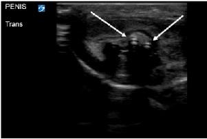
A 22-year-old male with bipolar disorder and depression presented to our ED after intentional insertion of a piece of plastic into his urethra. This was not his first presentation; on history, he noted this was a “coping mechanism.” He complained of penile pain, dysuria, and hematuria, although he was able to urinate. The patient’s vital signs were normal. Physical exam revealed an unremarkable abdominal exam. He had tenderness to palpation along the shaft of the penis
with a palpable foreign body, but nothing was visualized at the urethral meatus. Plain radiography was interpreted as normal with no visualized foreign body. Lab work and urinalysis were also normal. Clinicians using POCUS were able to visualize a linear hyperechoic object within the urethra (Image 1). Urology was consulted and took the patient to the operating room (OR) for removal with cystoscopy. In the OR a rolled-up piece of plastic was identified three centimeters into the urethra and was successfully removed. The patient was discharged without complication the following day.
A 60-year-old male with a history of depression, anxiety, post-traumatic stress disorder, insomnia, and self-harm with past foreign body insertion into the urethra presented for foreign body obstructing his urethra. Approximately nine hours prior to evaluation by emergency physicians, the patient described having an episode of anger, subsequently inserting five baby carrots into his penis. He stated that several of the baby carrots came out of the urethra but estimated two remained inserted. He attempted removal with chopsticks but was unsuccessful. Since inserting the foreign bodies, the patient noted pain and difficulty urinating. While
CPC-EM Capsule
What do we already know about this clinical entity?
Urethral foreign bodies rarely present to the ED but can result in significant morbidity. Diagnostic modalities include CT, plain radiography, and cystoscopy.
What makes this presentation of disease reportable?
Ultrasound is not the standard imaging modality to diagnose urethral foreign bodies. We report on its use to detect foreign bodies and help facilitate management.
What is the major learning point?
Point-of-care ultrasound provides a rapid means of evaluating patients with urethral foreign bodies.
How might this improve emergency medicine practice?
Use of ultrasound to diagnose urethral foreign bodies may result in less exposure to radiation, lower cost, and expedited care.
he had inserted foreign bodies into his urethra in the past, he had never required evaluation in the ED or urology consultation. In the ED, the patient was hypertensive to 180/96 millimeters of mercury; otherwise vital signs were stable. He was anxious. There were no findings on external genitourinary exam.
Bedside ultrasound identified three linear hyperechoic objects in the bladder (Image 2), with none seen in the urethra. This was followed by a CT, which confirmed the findings; three foreign bodies were retained in the bladder. Urology was consulted, and the patient was admitted to the hospital. Urology performed a cystoscopy with open cystotomy, removed the foreign bodies, and placed a Jackson-Pratt (JP) drain and Foley catheter. The hospital course was complicated by traumatic damage to the urethra, small extravasation from the bladder, and Enterococcus faecalis urinary tract infection. Infectious disease and psychiatry were consulted. The patient completed a course of antibiotics, had the JP drain and Foley catheter removed, and was discharged on hospital day 20.
An 11-year-old male presented with the chief complaint of foreign body inserted into his urethra. The patient had inserted
a braided USB-type cord without hub attachments into his urethra. He had similarly inserted foreign bodies into his urethra in the past but stated, “This is the first time I couldn’t get it out.” On arrival the patient was in no acute distress. Genitourinary exam was significant for a single, braided cord protruding from the urethra. Neither the patient nor the emergency physicians were able to extract the cord with gentle traction. Using POCUS, the clinicians identified the cord in the bladder, as well as evidence that the cord had looped within the urethra (Image 3). Urology was consulted, and a urologist evaluated the patient at bedside. The patient
underwent moderate sedation with ketamine. The cord had in fact looped once in the urethra. The cord was extracted by the urologist, with overall no complications besides the looping in the urethra. The patient had residual scant hematuria that resolved at the time of outpatient urology follow-up.
While self-insertion of urethral foreign bodies is a rare presentation to the ED, it is an issue requiring rapid assessment and intervention. A single-center study found that of almost 18,000 admitted patients in the six years prior to its publication, only 10 presented with urethral foreign bodies, representing 0.055% of the admitted population.2 Despite the low incidence of this presentation, there is significant morbidity including infection and loss of function, which makes prompt evaluation and care imperative for these patients.5 This is even more important when considering that many of these patients may present delayed due to embarrassment or concomitant psychiatric illness.1 While urethral foreign bodies can often be diagnosed with history and exam alone, CT and plain radiography are often used as well. This is particularly common when specific information such as location is unknown or if history and exam are inadequate. Cystoscopy is often necessary for removal of these objects, and less frequently open surgery, if the object cannot be removed endoscopically.
Point-of-care ultrasound provides multiple benefits while avoiding many of the drawbacks of more standard evaluation. Plain radiography can be helpful and performed at the bedside if needed; however, it is only useful if the object in question is radiopaque. As our case series demonstrates, POCUS is capable of viewing objects that are both radiopaque and radiolucent. Additionally, plain radiography shows a twodimensional picture while POCUS can be used to obtain views in multiple planes to better map out an object’s shape, location, and orientation. While CT provides a detailed, three-dimensional picture, it requires patient cooperation, as well as a substantial exposure to radiation. This is potentially exacerbated by the fact that some of these patients may have similar repeated presentations, especially if being driven by a behavioral problem or sexual gratification. Additional considerations are cost and time to obtain the imaging, often requiring transporting the patient to another area for imaging.
While POCUS does not provide the same level of detail as a CT, it does allow visualization of the object as well as provide information regarding shape, orientation, and location. Point-of-care ultrasound images can be obtained quickly, and the images are interpreted by the emergency physician, expediting imaging results. Additional benefits of ultrasound include avoiding radiation, lower cost, and not requiring the patient to remain still. Point-of-care ultrasound certainly has a role as a diagnostic tool for urethral foreign bodies, but it is rarely used in this capacity.
In this paper, we suggest a novel diagnostic approach
Use of Point-of-care Ultrasound for Detection of Urethral Foreign Bodies: A Case Series Tomasi et
using POCUS. Current barriers to using POCUS for this chief complaint likely include not realizing ultrasound can play a role, lack of confidence to obtain adequate images, and inability to interpret images. Incorporating additional training into existing ultrasound curricula would likely increase familiarity and comfort with this specific modality, in addition to reducing the risk of inappropriate interpretation of imaging. Medicolegal concerns for the incorporation of POCUS in such cases may be mitigated by establishing diagnostic protocols, as well as clear documentation of findings. Additional case series and studies focusing on this diagnostic approach could lead to POCUS as the standard of care in initial ED evaluations for urethral foreign bodies.
Urethral foreign bodies are an uncommon presentation to the emergency department, often requiring imaging modalities to provide necessary information to guide management. This paper describes three cases where POCUS was used in the diagnosis of urethral foreign bodies and helped facilitate management. Ultrasound has advantages over alternative imaging and, as demonstrated here, has proven to be useful. Patient care may benefit from more routine use of this tool in the evaluation of patients with urethral foreign bodies.
The authors attest that their institution requires neither Institutional Review Board approval, nor patient consent for publication of this case report. Documentation on file.
Address for Correspondence: Norah McIntyre, MD, Baystate Medical Center, Department of Emergency Medicine, 759 Chestnut Street, Springfield, MA, 01096. Email: norah. mcintyremd@baystatehealth.org.
Conflicts of Interest: By the CPC-EM article submission agreement, all authors are required to disclose all affiliations, funding sources and financial or management relationships that could be perceived as potential sources of bias. The authors disclosed none.
Copyright: © 2025 Tomasi et al. This is an open access article distributed in accordance with the terms of the Creative Commons Attribution (CC BY 4.0) License. See: http://creativecommons.org/ licenses/by/4.0/
1. John J and Kesner K. Urethral polyembolokoilamania: not a bread-and-butter issue. Ther Adv Urol. 2021;13:17562872211022866.
2. Mahadevappa N, Kochhar G, Vilvapathy KS, et al. Self-inflicted foreign bodies in lower genitourinary tract in males: our experience and review of literature. Urol Ann. 2016;8(3):338-42.
3. Tuncer H, Karacam H, Cam B. A self-inserted foreign body in the urinary bladder and urethra. Cureus. 2021;13(7):e16322.
4. Mori T, Ihara T, Nomura O. Detection of a Urethral Foreign Body in a Pediatric Patient: Another Useful Application of Point-of-Care Ultrasound. J Emerg Med. 2021;61(3):e26-e31.
5. Rafique M. Intravesical foreign bodies: review and current management strategies. Urol J. 2008;5(4):223-31.
Andrew Ryu, MD
Karizma Chhabra, MD
Thomas George, DO
Elizabeth Kasparov, MD
Mohamed Wali, MD
Christopher C. Lee, MD
Section Editor: Shadi Lahham, MD
South Shore University Hospital/Northwell Health, Department of Emergency Medicine, Bay Shore, New York
Submission history: Submitted June 2, 2025; Revision received July 13, 2025; Accepted July 25, 2025
Electronically published September 23, 2025
Full text available through open access at http://escholarship.org/uc/uciem_cpcem DOI: 10.5811/cpcem.48350
Introduction: A posterior circulation stroke at the level of the basilar artery can cause ischemia to the brainstem, cerebellum, and occipital lobes. Posterior circulation strokes are notoriously more difficult to clinically diagnose than anterior circulation strokes, with a variety of presenting symptoms including altered mental status, dizziness, vision changes, nausea, and vomiting. Anisocoria has been reported to occur in rare cases.
Case Report: We present two cases where patients had an acute episode of altered mental status with a key exam finding of anisocoria, or unequal pupil sizes. The combination of anisocoria and acute mental status decline are classically associated with traumatic brain injury, increased intracranial pressure, or both. In each of the two cases presented, acute basilar artery occlusion was seen on computed tomography with angiography.
Conclusion: When presented with acute decline in mental status and anisocoria, early clinical suspicion of an acute basilar artery occlusion is crucial in diagnosing and managing these patients with debilitating acute posterior stroke. Time-sensitive interventions such as thrombolytics and mechanical thrombectomy can be lifesaving. [Clin Pract Cases Emerg Med. 2025;9(4):369-372.]
Keywords: anisocoria; basilar artery occlusion; posterior stroke; altered mental status
INTRODUCTION
The posterior circulation of the brain refers to the vertebrobasilar vascular system, beginning with the vertebral arteries coming off the subclavian arteries that join to form the basilar artery. A posterior circulation stroke at the level of the basilar artery can cause ischemia to the brainstem, cerebellum, and occipital lobes, causing a wide range of symptoms including an acute change in mental status as well as vestibular and ocular symptoms.1 Anisocoria has been reported to occur in rare cases of posterior circulation strokes.2-4 Anisocoria is defined as unequal size of the pupils. Mydriasis is primarily mediated by the sympathetic neuronal input, while miosis is mediated by parasympathetic input. Anisocoria is
caused by disruption and mismatch of these neuronal inputs. Possible etiologies include physiologic anisocoria in up to 20% of the population, increased intracranial pressure and, in rare instances, posterior circulation strokes.5 We describe two cases of acute basilar artery occlusion in patients who had acute episodes of altered mental status with anisocoria on physical exam.
Patient 1
A 61-year-old female with past medical history of acute ischemic stroke and left atrial appendage thrombus treated with apixaban presented to the emergency department (ED)
after a mechanical fall. The patient’s son found her on the ground with baseline mental status after a fall from standing resulting in head trauma. Computed tomography (CT) of the head and cervical spine did not show any acute findings. The patient was admitted due to elevated troponin markers per cardiologist’s recommendations.
During her stay overnight, the rapid response team was activated for an acute mental status change, with last known well three hours prior. The patient was minimally responsive to pain with snoring respirations and a National Institute of Health Stroke Scale score of 22. On physical exam, the right pupil was 1 mm in diameter and reactive to light, and the left pupil was 4 mm and nonreactive. An emergent stroke workup was initiated, and CT angiography (CTA) of her head showed multifocal occlusions of the left posterior cerebral artery (PCA) (Image 1). The stroke neurologist communicated concerns for proximal basilar artery occlusion, and the patient was taken for emergent mechanical thrombectomy. Basilar tip occlusion was found. Thrombolysis in cerebral infarction grade 3 reperfusion was achieved after first pass, and the patient was admitted to the neurointensive care unit. The patient expired during her stay, which was complicated by development of aspiration pneumonia.
An 80-year-old female with past medical history of atrial fibrillation treated with rivaroxaban presented to the ED with syncope. The patient was in the kitchen when her family heard her fall, and she was minimally responsive afterward with blue lips and trouble breathing. On arrival, the patient was minimally responsive to pain and agitated. She was intubated for airway protection. On physical exam, her right pupil was 6
CPC-EM Capsule
What do we already know about this clinical entity?
An acute basilar artery occlusion can cause ischemia to the brainstem, cerebellum, and occipital lobes, causing a wide range of symptoms including ocular symptoms.
What makes this presentation of disease reportable?
No previous studies have described the prevalence of anisocoria and altered mental status as presenting symptoms in acute basilar artery occlusion.11
What is the major learning point?
Prompt consultation with the stroke radiologist or stroke neurologist may be warranted to communicate concerns for a possible acute posterior stroke.
How might this improve emergency medicine practice?
By creating more awareness, these stroke patients can be identified more quickly and potentially be candidates for thrombolytics or mechanical thrombectomy.
mm and nonreactive to light, and her left pupil was 4 mm and reactive. The patient had a trauma workup with CT head and cervical spine without contrast, CTA head and neck, CT chest, abdomen, and pelvis with intravenous (IV) contrast, which showed no acute traumatic findings.
The patient was admitted to the medical intensive care unit for further management. Repeat CT head without contrast was done a day after admission due to a decrease in responsiveness, showing acute infarcts in the bilateral cerebellar hemispheres and cerebellar vermis with mass effect on the fourth ventricle and rostral hydrocephalus. The initial CTA head result was addended at this time to show diminished flow in the distal basilar artery extending into the origins of the bilateral PCAs compatible with intraluminal thrombus (Image 2). Neurology and neurosurgery were consulted, and the patient was deemed not a candidate for advanced therapies or surgeries, given devastating neurological injury and poor prognosis. The decision was made with family to withdraw care, and the patient expired on the third day of admission.
Image 2. Computed tomography angiography coronal view (Patient 2) with arrow pointing to the site of basilar artery occlusion with bilateral posterior cerebral artery involvement.
Stroke is a critical condition in which time-sensitive interventions such as fibrinolytics or endovascular thrombectomy can be lifesaving for the patient . This case report shows that an acute basilar artery occlusion should be considered as a possible etiology for patients who present with acute change in mental status and anisocoria, even if there is a high clinical suspicion for acute traumatic brain injury as in the case of Patient 2. A stroke workup includes CTA of the brain and neck and CT perfusion, which are often read emergently by the stroke radiologist to aid in the timesensitive management of acute ischemic strokes. In the case of Patient 2, a trauma workup was pursued instead of a stroke workup to rule out traumatic intracranial hemorrhage. In the report for the CTA head ordered as part of the trauma workup, the basilar artery occlusion extending into the bilateral posterior cerebellar arteries was initially missed and then later included in the addendum.
Posterior circulation occlusions are known to be difficult to localize on CTA due to the anatomical and functional complexity of the posterior vasculature with high frequency of anatomical variants, such as hypoplastic arteries and congenital anomalies.6-7 Furthermore, since a stroke workup was not pursued in this case, a CT perfusion study was not ordered, which can greatly improve the diagnostic accuracy in acute posterior circulation strokes.8 In retrospect, it is not possible to know whether an emergent stroke workup may have led to earlier diagnosis of a basilar artery occlusion on the CTA. Posterior circulation strokes are notoriously more difficult to clinically diagnose than anterior circulation strokes, with a variety of presenting symptoms including altered mental status, dizziness, vision changes, nausea, and vomiting. Altered mental status has been reported to have been present in up to 25% of missed stroke cases.6, 9-10 This case demonstrates that prompt consultation with the stroke
radiologist or stroke neurologist may be warranted to communicate concerns for a possible acute posterior stroke in case presentations such as these.
While anisocoria is a known symptom of posterior circulation strokes, we did not find any previous studies that described the prevalence of anisocoria and altered mental status as presenting symptoms in acute basilar artery occlusion.11 By raising awareness of this symptomatology, these stroke patients can be identified more quickly and accurately in time-sensitive, emergent stroke evaluations and potentially be candidates for life-saving thrombolytics or mechanical thrombectomy.
The authors attest that their institution requires neither Institutional Review Board approval, nor patient consent for publication of this case report. Documentation on file.
Address for Correspondence: Andrew Ryu, MD, South Shore University Hospital/Northwell Health, Department of Emergency Medicine, 301 E Main St, Bay Shore, NY 11706. Email: aryu@ northwell.edu.
Conflicts of Interest: By the CPC-EM article submission agreement, all authors are required to disclose all affiliations, funding sources and financial or management relationships that could be perceived as potential sources of bias. The authors disclosed none.
Copyright: © 2025 Ryu et al. This is an open access article distributed in accordance with the terms of the Creative Commons Attribution (CC BY 4.0) License. See: http://creativecommons.org/ licenses/by/4.0/
1. Merwick Á and Werring D. Posterior circulation ischaemic stroke. BMJ. 2014;348:g3175.
2. Gurley K and Edlow J. Avoiding misdiagnosis in patients with posterior circulation ischemia: a narrative review. Acad Emerg Med. 2019;26(11):1273-84.
3. Schonewille WJ, Wijman CA, Michel P, et al. Treatment and outcomes of acute basilar artery occlusion in the Basilar Artery International Cooperation Study (BASICS): a prospective registry study. Lancet. 2009;8(8):724–30.
4. Mattle HP, Arnold M, Lindsberg PJ, et al. Basilar artery occlusion. Lancet Neurol. 2011;10(11):1002–14.
5. Antonio-Santos AA, Santo RN, Eggenberger ER. Pharmacological testing of anisocoria. Expert Opin Pharmacother. 2005;6(12):2007-13.
6. Hoyer C and Szabo K. Pitfalls in the diagnosis of posterior circulation stroke in the emergency setting. Front Neurol. 2021;12:682827.
7. Singh R, Kumar R, Kumar A. Vascular anomalies of posterior fossa
Report of Two Cases: Altered Mental Status and Anisocoria as Presenting Symptoms in ABAO Ryu et al. and their implications. J Craniofac Surg. 2017;28(8):2145-50.
8. Sporns P, Schmidt R, Minnerup J, et al. Computed tomography perfusion improves diagnostic accuracy in acute posterior circulation stroke. Cerebrovasc Dis. 2016;41(5-6):242-7.
9. Brandler ES, Sharma M, McCullough F, et al. Prehospital stroke identification: factors associated with diagnostic accuracy. J Stroke
Cerebrovasc Dis. 2015;24(9):2161-6.
10. Oostema JA, Konen J, Chassee T, et al. Clinical predictors of accurate prehospital stroke recognition. Stroke. 2015;46(6):1513-7.
11. Chang VA, Meyer DM, Meyer BC. Isolated anisocoria as a presenting stroke code symptom is unlikely to result in alteplase administration J Stroke Cerebrovasc Dis. 2019;28(1):163-6.
Madelyn Huttner, MD
Mitchell McMurray, MD
Martin Huecker, MD
Sohail Ikram, MD
Section Editor: Lev Libet, MD
University of Louisville, School of Medicine, Department of Emergency Medicine, Louisville, Kentucky
Submission history: Submitted October 20, 2024; Revision received November 27, 2024; Accepted May 24, 2025
Electronically published August 18, 2025
Full text available through open access at http://escholarship.org/uc/uciem_cpcem
DOI: 10.5811/cpcem.38069
Introduction: Coronary artery disease is uncommon in adults under the age of 35, and studies show a lower incidence in women of this age group. Physicians should suspect myocardial infarction in all patients who present with cardiac arrest and a shockable rhythm.
Case Report: We report a case of a 34-year-old female who presented after return of spontaneous circulation following both pulseless electrical activity and ventricular fibrillation. The initial emergency department 12-lead electrocardiogram (ECG) demonstrated ST-segment elevation in the anterior precordial leads. The second, more notable, ECG showed a unique ischemic pattern of ventricular bigeminy with each beat containing a different morphology of injury pattern. Emergent cardiac catheterization found a 100% occlusion of the proximal left anterior descending artery
Conclusion: Premature ventricular (or junctional) contractions can indicate ischemia when the morphology consists of excessive discordance between the QRS complex and the ST segment and T wave. This case illustrates the importance of scrutinizing each beat in every lead to increase sensitivity for ischemia. [Clin Pract Cases Emerg Med. 2025;9(4):373-375.]
Keywords: electrocardiogram; myocardial infarction; bigeminy; discordance.
Coronary artery disease is uncommon in adults under age 35, and studies show an even lower incidence in women of this age group.1 Myocardial infarction (MI) should be suspected in patients with prehospital ventricular fibrillation. The standard of care for patients with return of spontaneous circulation (ROSC) is to have electrocardiograms (ECG) performed as early as possible, understanding that falsepositive ST-segment elevation myocardial infarction (STEMI) can occur in ECGs obtained within eight minutes of ROSC.2
A 34-year-old female with a past medical history of type 2 diabetes, depression, anxiety, obesity, and tobacco use presented to the emergency department (ED) in cardiac arrest. Per emergency medical services (EMS), the patient had chest pain for one day and then collapsed in front of family. They
found the patient in pulseless electrical activity (PEA) that transitioned to ventricular fibrillation. En route, the EMS team defibrillated the patient twice and administered the following medications: 300 milligrams (mg) amiodarone; six mg total epinephrine, and two mg naloxone
Upon arrival to the ED the patient was intubated and noted to be in PEA. In the ED another 1 mg of epinephrine, one ampule of sodium bicarbonate, and two grams of magnesium sulfate intravenous were administered along with continued chest compressions. After >30 minutes without a pulse, ROSC was obtained, and the initial ECG obtained five minutes post-ROSC demonstrated ST-segment elevations in leads V1-V3 (Image 1). Lead V4 demonstrated hyperacute T waves. The ECG also showed a widened QRS complex with prominent T waves in aVL, V1, V2, V5 and V6. The precordial lead had an inconsistent R-wave progression, with R’ wave present in V1, no R wave (a QS wave) in V3, and no
Image 1. Electrocardiogram demonstrates ST-segment segment elevation in V1-V4 (arrows) with widened QRS complex in aVL, V1, V2, V5, and V6 (arrowheads) in a 34-yr-old female patient after cardiac arrest.
R wave (but a hyperacute T wave) in V4. Limb leads showed subtle ST-segment depression in II, III, and aVF that likely represented reciprocal changes.
An additional 150 mg amiodarone was administered for intermittent/non-sustained ventricular tachycardia. A chest radiograph demonstrated bilateral pulmonary edema, and a point-of-care cardiac ultrasound showed reduced ejection fraction. The medical intensive care unit and cardiology teams were consulted. Due to severe acidosis and hypoxemia, the cardiology team recommended medical stabilization before taking the patient emergently to cardiac catheterization.
The repeat ECG showed bigeminy, ventricular rate of 124 beats per minute, with the unique injury pattern (Image 2). Each of the bigeminy beats were consistent with infarction but in different morphologies. The first beat showed anterolateral
Image 2. Electrocardiogram demonstrates ST-segment elevation in V1-V6 and lead I with alternating QRS-complex morphologies (arrows) and reciprocal ST-segment depressions in leads II, III, aVF (arrowheads).
CPC-EM Capsule
What do we already know about this clinical entity?
False positive ST-segment elevation can occur in electrocardiograms obtained within eight minutes of return of spontaneous circulation (ROSC).
What makes this presentation of disease reportable?
The second ECG shows a unique ischemic pattern of ventricular bigeminy with each beat containing a different morphology of injury pattern.
How might this improve emergency medicine practice?
It is important to scrutinize each beat in every lead for ischemia and to evaluate for the persistence of post-ROSC ECG abnormalities with a repeat ECG in 10-20 minutes.
ST-segment elevation with QS waves in V1-V3. The second bigeminy beat showed apparent R waves in anteroseptal leads with a deep notch at the J point. It is unclear whether the beats represented premature ventricular contractions or junctional beats. For instance, the first of the two beats appeared to have a P wave preceding the QRS complex. The inferior limb leads showed reciprocal ST-segment depressions, but again with different morphologies.
The initial troponin level resulted at 131 nanograms per liter (ng/L) (reference range: 0-19 ng/L). Rectal aspirin was administered, and the patient was taken for emergent cardiac catheterization.
Coronary angiogram revealed a 100% occlusion of the proximal-mid segment of the left anterior descending artery. The occlusion was treated with a 3.5 × 28 millimeter Xience Skypoint stent (Abbott Laboratories, Abbott Park, IL). A repeat ECG after cardiac catheterization demonstrated sinus tachycardia with left anterior fascicular block and improved ST-segment elevations in V1-V3. The patient remained ventilated, on a heparin drip, and on vasopressors. On hospital day 8, she died peacefully with family at bedside.
Coronary artery disease is rare in adults under the age of 35.1 An observational study evaluating young adults with STEMI found common risk factors of male sex, hypertension, and obesity.1 Patients in this age group may experience a delay in diagnosis due to low suspicion for ischemia.3 Observational
Huttner et al. Bigeminy with Alternating Injury Pattern Morphologies in a Young Woman After Cardiac Arrest
data suggest that women are less likely than men to receive ECGs or fibrinolysis within benchmark timeframes (and less likely to receive percutaneous coronary intervention in general).4 Risk factors for MI in young female patients include stress, anxiety, and depression.5
Patients presenting in cardiac arrest with ventricular fibrillation should be presumed to have MI until proven otherwise. Electrocardiograms obtained within eight minutes of ROSC may show a pattern injury in the absence of coronary occlusion.2 Serial ECGs can help determine the persistence of concerning abnormalities as the cardiac membrane stabilizes. The initial ECG in this case led to concern for acute MI, with clear indication of active ischemia. Understanding that early post-ROSC ECGs can mislead, the ED obtained a second ECG (Image 2) and found the unique patterns of ischemia. Bundle branch blocks and paced rhythms can present challenges to detecting ischemia.
Our literature search found no published case with an alternating pattern of injury (Image 2). The first beat showed anterolateral ST-segment elevation, suggesting vessel occlusion. The second beat displays R waves in anteroseptal leads with a deep notch at the J point. As shown in Image 3 from an open-access ECG database, typical premature ventricular complexes (PVC) have a widened QRS complex but usually have an appropriately discordant ST-segment and T wave (Image 3).7
Physicians should scrutinize the 12-lead ECGs looking
closely at the morphology of all beats (including premature ventricular complexes and bundle branch blocks) to detect ischemia. To evaluate for the persistence of post-ROSC ECG abnormalities, physicians should obtain a repeat ECG within 10-20 minutes.
The authors attest that their institution requires neither Institutional Review Board approval, nor patient consent for publication of this case report. Documentation on file.
Address for Correspondence: Madelyn Huttner, MD, University of Louisville School of Medicine, Department of Emergency Medicine, 530 S. Jackson Street, Louisville, Kentucky 40202. Email: Madelyn.huttner@gmail.com.
Conflicts of Interest: By the CPC-EM article submission agreement, all authors are required to disclose all affiliations, funding sources and financial or management relationships that could be perceived as potential sources of bias. The authors disclosed none.
Copyright: © 2025 Huttner et al. This is an open access article distributed in accordance with the terms of the Creative Commons Attribution (CC BY 4.0) License. See: http://creativecommons.org/ licenses/by/4.0/
1. Ruiz Pizarro V, Palacios-Rubio J, Cruz-Utrilla A, et al. ST-elevation myocardial infarction in patients ≤35 years of age. AJC 2019;123(6):889-93
2. Baldi E, Schnaubelt S, Caputo ML, et al. Association of timing of electrocardiogram acquisition after return of spontaneous circulation with coronary angiography findings in patients with out-of-hospital cardiac arrest. JAMA Netw Open. 2021;4(1):e2032875.
3. Yandrapalli S, Nabors C, Goyal A, et al. Modifiable risk factors in young adults with first myocardial infarction. JACC. 2019;73(5):573-84.
4. Pelletier R, Humphries KH, Shimony A, et al. GENESIS-PRAXY Investigators. Sex-related differences in access to care among patients with premature acute coronary syndrome. CMAJ. 2014;186(7):497-504.
5. Chandrasekhar J, Gill A, Mehran R. Acute myocardial infarction in young women: current perspectives. Int J Womens Health 2018;10:267-84.
6. Smith, S. “Look at the PVCs!!” Dr. Smith’s ECG Blog, 22 Jan. 2014, hqmeded-ecg.blogspot.com/2014/01/look-at-pvcs.html. Accessed September 10, 2023
Image 3. Electrocardiogram demonstrates premature ventricular complexes in a bigeminy pattern with ST-segment depression in differing morphologies in multiple leads (arrows), along with STsegment elevation in leads I and avL (arrowheads).
7. Burns, E, Buttner. R. “Premature ventricular complex (PVC).” Life in the Fast Lane, 2 June 2021, litfl.com/premature-ventricular-complexpvc-ecg-library/. Accessed May 3, 2024
Tabitha Ranson, DO, MS*
Gregory Ruddy, DO, MS*
Zachary Ostapowicz, DO, MS*
Leah Joyner, MD*†
Section Editor: John Ashurst, DO
Kansas City University College of Medicine, Department of Emergency Medicine, Joplin, Missouri
Mercy Hospital, Department of Emergency Medicine, Joplin, Missouri
Submission history: Submitted August 13, 2024; Revision received February 27, 2025; Accepted February 27, 2025
Electronically published August 26, 2025
Full text available through open access at http://escholarship.org/uc/uciem_cpcem
DOI: 10.5811/cpcem.33528
Introduction: Wunderlich syndrome (WS) is a rare condition characterized by spontaneous, atraumatic renal hemorrhage. It often presents with non-specific symptoms and is typically diagnosed through computed tomography (CT). The most common presentation of WS includes the Lenk triad, which consists of flank pain, a palpable flank mass, and hypovolemic shock. If diagnosis and treatment are delayed, WS can rapidly progress and lead to unfavorable patient outcomes.
Case Report: A 65-year-old male presented to the emergency department with severe suddenonset left flank pain with subsequent CT angiogram demonstrating an actively bleeding left renal hematoma. The patient was managed conservatively with supportive care. His vitals remained stable, and he did not require any surgical or vascular interventions.
Conclusion: Wunderlich syndrome is a spontaneous renal or perinephric hemorrhage occurring in the absence of trauma; it is rarely included in the differential for patients with flank pain but can become life-threatening when not recognized. [Clin Pract Cases Emerg Med. 2025;9(4):376-379.]
Keywords: spontaneous renal hemorrhage; Wunderlich syndrome; case report; emergent causes of flank pain; atraumatic renal hemorrhage.
Atraumatic renal hemorrhage is a condition referred to as Wunderlich syndrome (WS) after it was first described by Carl Wunderlich in 1856.1 Acute onset of flank pain, a palpable mass, and hemodynamic compromise is the classic presentation; however, it is relatively uncommon to see all three signs at presentation.1-3 It is more common for patients to present with unilateral flank pain as the chief complaint and often the only symptom.3,4 Patients are typically diagnosed with computed tomography (CT) showing subcapsular or perirenal hemorrhage; however, ultrasound and magnetic resonance imaging (MRI) can also be useful in diagnosis.4,5 Treatment recommendations vary depending on hemodynamics and etiology. Hemodynamically stable patients do well when managed more conservatively, but those
exhibiting signs of hemodynamic compromise are usually managed with renal artery embolization.4 The majority of spontaneous renal hemorrhage (SRH) cases can be attributed to neoplasms, particularly angiomyolipoma and renal cell carcinoma.3,4 A smaller proportion of SRH is due to vasculitis or etiologies not otherwise categorized.4 Among the other causes contributing to the smallest proportion of cases, etiologies reported include renal artery aneurysms, arteriovenous malformations (AVM), infection, nephrolithiasis, ruptured renal cysts, and uncontrolled hypertension.4 Despite the aforementioned causes, occasionally even after thorough investigation there remains a small subset of SRH cases without underlying cause.4 The following case describes a 65-year-old male with idiopathic SRH who was successfully treated with conservative measures.
A 65-year-old male presented to the emergency department (ED) with severe sudden-onset left flank pain, reporting that it felt like a prior episode of nephrolithiasis. He denied fever, nausea, vomiting, abdominal pain, dysuria, or hematuria. Past medical history was positive for nephrolithiasis status post lithotripsy, hypertension, hyperlipidemia, type two diabetes mellitus, coronary artery disease status post two-vessel coronary artery bypass surgery, cerebrovascular accident, deep vein thrombosis, pulmonary embolism, and carotid artery stenosis. There was no use of anticoagulants at the time of the ED visit. Social history was positive for former cigarette use (cessation 40 years prior) but negative for alcohol and recreational drug use.
Vital signs on exam were within normal limits, showing blood pressure of 122/55 millimeters of mercury, heart rate 65 beats per minute, and oxygen saturation 100% on room air. Physical exam was significant for moderate to severe distress due to pain, non-tender abdomen, and left costovertebral angle tenderness. Lab work was significant for a hemoglobin of 11.2 grams per deciliter (g/dL) (reference range: 13.5-18.0 g/dL), hematocrit of 36.0% (42.0-52.0%), mean corpuscular volume of 90.9 femtoliters (fL) (78-100 fL), a white blood cell count of 15.0 thousand cells per microliter (k/μL) (4.0-11.0 k/μL), and an acute kidney injury with an elevated creatinine of 1.37 milligrams per dL (mg/dL) (0.67-1.17 mg/dL). Urinalysis was positive for 3+ protein (reference negative), trace ketones (reference negative), and red blood cells (reference negative).
Computed tomography without contrast was performed to evaluate for renal calculi and demonstrated an acute left subcapsular perirenal hematoma measuring up to 4.2 centimeters (cm) with surrounding inflammation and hemoperitoneum as well as hemorrhagic right renal cortical cysts (Image 1). Computed tomography angiogram chest, abdomen, and pelvis was performed to further evaluate the bleeding and demonstrated an actively bleeding left renal hematoma measuring 9.4 cm with fat stranding (Image 2).
The patient was admitted to the hospital for further management. During his hospital course, he began to receive packed red blood cells (PRBC) due to a drop in hemoglobin from 11.2 g/dL to 7.0 g/dL, but the transfusion was terminated prior to completion due to an acute febrile transfusion reaction. The patient was monitored with plans for embolization should he become unstable. He was also placed on strict bed rest with strict precautions to avoid any anticoagulant or antiplatelet drugs. His hemoglobin had initially improved from 7.0 g/dL to 8.0 g/dL but then dropped again to 7.3 g/dL on day six of hospitalization, and he required two units of PRBC, which he tolerated after premedication with diphenhydramine and dexamethasone. Ultimately, he required five units of PRBC prior to being discharged. The patient remained hemodynamically stable, and he was discharged home with outpatient urology follow-up.
The patient was seen as an outpatient by urology who were
What do we already know about this clinical entity?
Wunderlich syndrome (WS) is a rare condition typically characterized by flank pain, a palpable flank mass, and hypovolemic shock. It is typically diagnosed with computed tomography imaging.
What makes this presentation of disease reportable?
Wunderlich syndrome is rare, with few cases seen annually in the emergency department. Even without Lenk triad, it should remain in the differential for atraumatic flank pain to avoid missed diagnosis.
What is the major learning point?
This case emphasizes the importance of maintaining a broad differential in cases of vague abdominal symptoms. Point-of-care-ultrasound may be a useful tool in the workup of flank pain.
How might this improve emergency medicine practice?
Awareness of WS can improve early recognition, improving patient outcomes. Point-of-care ultrasound in cases of flank pain may improve diagnosis, reduce radiation, and enhance outcomes.
concerned about a possible underlying malignancy in light of the SRH. Repeat imaging four months after initial presentation demonstrated a significant improvement in the left subcapsular renal hematoma now measuring 4.3 x 2.6 cm without any contrast-enhancing masses identified. Additionally, there were several hypodense lesions in the lower pole of the left kidney likely consistent with hemorrhagic cysts. The patient was released from urology follow-up when a CT at seven months post renal hemorrhage was stable, negative for active bleeding, and negative for enhancing mass.
Kidney hemorrhage due to blunt trauma is a rare phenomenon, consisting of only 1-5% of all trauma patients.6 Wunderlich syndrome is a clinical syndrome characterized by acute atraumatic renal hemorrhage, which was first reported by Carl Wunderlich in 1856.1 Classic presentation of the Lenk triad (flank pain, flank mass, and hypovolemic shock) is seen in only 20% of patients with this type of hemorrhage.5 In a case series by Kim et al, the two most common symptoms and signs found in WS are acute-onset flank pain and microscopic hematuria found in 92% and 39% of total cases, respectively.3 Although vague symptoms such as flank pain are often associated with more common disorders such as pyelonephritis and nephrolithiasis, WS should remain on the differential particularly in patients with increased risk as it can quickly become life-threatening if not promptly diagnosed and treated. Patients at risk for WS include those with a history of diabetes, hypertension, pyelonephritis, renal cystic disease, and end-stage renal disease.7
While several causes of back pain at the costovertebral
angle may be treated presumptively without complications including musculoskeletal pain, nephrolithiasis, and uncomplicated pyelonephritis, the management of acute kidney hemorrhage varies greatly. Labs including complete blood count to evaluate for blood loss and infection, basic metabolic panel to assess kidney function, and urinalysis to assess for infection can support the diagnosis and rule out other disorders on the differential. However, an acute kidney hemorrhage can only be accurately diagnosed by imaging.5 A CT is currently the gold standard imaging technique in the diagnosis of WS.8 If CT is negative and suspicion is still high for WS, then an magnetic resonance imaging may be obtained.5
In this specific case, CT without contrast was the initial imaging ordered to rule out renal calculus, particularly with this patient’s history of prior renal calculi. However, the patient then required an additional CT angiogram to further characterize the bleed. Point-of-care ultrasound (POCUS) represents a quick, safe, and cost-effective method to determine possible hemoperitoneum. With the increasing use of POCUS, there have been cases of ultrasound being used to identify and monitor active kidney hemorrhage.4,5,9,10 To prevent unnecessary imaging, future standard of care for those with flank pain and risk factors for acute kidney hemorrhage could include POCUS prior to definitive CT to guide choice of imaging modality. If there is evidence of bleeding on POCUS, then CT angiogram can be performed and additional CT can be avoided.
Patients with WS are managed inpatient with renal artery embolization if the patient is unstable, or with supportive care and careful monitoring if the patient remains stable.4 The different pathways in managing these patients are important distinctions as renal artery embolization poses potential risk to the patient such as renal failure, hematoma formation at the site of the catheter, arterial hypertension and, lastly, postembolization syndrome.11 Thus, if it can be avoided then it is preferred to manage the patient conservatively.4
Once diagnosed with an acute renal hemorrhage and stabilized, further investigation typically in the outpatient setting is necessary to rule out secondary causes of WS including malignancy, AVMs, and vasculitides. Thorough examination of the medical history and patient risk factors followed by diagnostic imaging should be performed to evaluate the most likely cause. Exclusion of renal neoplasms via outpatient imaging once the acute bleed and inflammation has had time to resolve is important in the workup of someone with WS, as angiomyolipomas and clear cell renal cell carcinomas contribute to 60-65% of all cases.5 If there are no signs, symptoms, or other risk factors for malignancy but no cause has been identified, further workup may be warranted to evaluate for etiologies such as vasculitis or AVMs.2
Wunderlich syndrome can present with a wide variety of
Ranson et al. Idiopathic Atraumatic Renal Hemorrhage
symptoms that may be misdiagnosed as other common pathologies in the ED. Due to potential for rapid progression of WS and decompensation, it is important to maintain a high level of suspicion when patients with risk factors present with flank pain. Furthermore, when patients present with the classic Lenk triad (left flank pain, hypovolemic shock, and a palpable abdominal mass) interventions should be taken immediately to prevent mortality. While CT is the diagnostic imaging of choice, point-of-care ultrasound at presentation may be a useful adjunct to guide choice of imaging modality. Use of POCUS may assist physicians in ordering the appropriate imaging and decrease unnecessary imaging. Continuing research and clinical education on WS can help emergency physicians identify this disease process and provide patients with earlier interventions leading to a greater number of favorable outcomes.
The authors attest that their institution requires neither Institutional Review Board approval, nor patient consent for publication of this case report. Documentation on file.
Address for Correspondence: Leah Joyner, MD, Mercy Hospital, Department of Emergency Medicine, 100 Mercy Way, Joplin, MO 64804. Email: Leah.joyner@mercy.net.
Conflicts of Interest: By the CPC-EM article submission agreement, all authors are required to disclose all affiliations, funding sources and financial or management relationships that could be perceived as potential sources of bias. The authors disclosed none.
Copyright: © 2025 Ranson et al. This is an open access article distributed in accordance with the terms of the Creative Commons Attribution (CC BY 4.0) License. See: http://creativecommons.org/ licenses/by/4.0/
1. Albi G, del Campo L, Tagarro D. Wünderlich’s syndrome: causes, diagnosis and radiological management. Clin Radiol. 2002;57(9):840-5.
2. Grubb SM, Stuart JI, Harper HM. Sudden onset flank pain: spontaneous renal rupture. Am J Emerg Med. 2017;35(11):1787. e1-1787.e3.
3. Kim JW, Kim JY, Ahn ST, et al. Spontaneous perirenal hemorrhage (Wunderlich syndrome): an analysis of 28 cases. Am J Emerg Med 2019;37(1):45-7.
4. Ahn T, Roberts MJ, Navaratnam A, et al. Changing etiology and management patterns for spontaneous renal hemorrhage: a systematic review of contemporary series. Int Urol Nephrol 2017;49(11):1897-905.
5. Shah JN, Shah JN, Gandhi D, et al. Wunderlich syndrome: comprehensive review of diagnosis and management. Radiographics. 2023;43(6):e220172.
6. Singh S. Kidney trauma. StatPearls [Internet]. July 17, 2023. Accessed August 14, 2024.
7. Masino F, Montatore M, Panunzio A, et al. Bilateral renal hemorrhage in an anticoagulated patient: a rare case of Wunderlich syndrome. Radiol Case Rep. 2024;19(7):2859-63.
8. Wang BH, Pureza V, Wang H. A tale of Wünderlich syndrome. J Surg Case Rep. 2012;2012(11):rjs015.
9. McCall NN and Burgner A. Point of care ultrasound in monitoring of post-renal biopsy bleeding. POCUS J. 2022;7(Kidney):33-4.
10. Ishikawa E, Nomura S, Hamaguchi T, et al. Ultrasonography as a predictor of overt bleeding after renal biopsy. Clin Exp Nephrol 2009;13(4):325-31.
11. Irwine C, Kay D, Kirsch D, et al. Renal artery embolization for the treatment of renal artery pseudoaneurysm following partial nephrectomy. Ochsner J. 2013;13(2):259-63.
Irfan Husain, MD, MPH
Danielle Andrews, MD, MPH
Section Editor: Shadi Lahham, MD
Emory University School of Medicine, Department of Emergency Me dicine, Atlanta, Georgia
Submission history: Submitted January 12, 2025; Revision received February 25, 2025; Accepted April 11, 2025
Electronically published September 5, 2025
Full text available through open access at http://escholarship.org/uc/uciem_cpcem DOI: 10.5811/cpcem.35398
Introduction: Fat embolism syndrome (FES) is a rare, life-threatening condition most seen in traumatic orthopedic injuries, especially long bone fractures. Classically, FES presents with hypoxemia, neurological abnormalities, or a petechial rash; however, clinical findings can extend beyond this classic triad. Since FES is a clinical diagnosis, emergency physicians must recognize both classic and subtle presentations.
Case Report: A 22-year-old female presented as a transfer from an outside hospital for multiple long bone fractures secondary to gunshot wounds. Upon arrival, she was found to be hypoxic, despite no signs of thoracic injury on exam or initial imaging. Her presentation, laboratory findings, and repeat imaging were consistent with FES. She was given supportive care through supplemental oxygen and close monitoring. She improved with supportive care and was discharged home in stable condition.
Conclusion: Although there is no definitive treatment for fat embolism syndrome, prompt recognition of the various clinical findings associated with FES by emergency physicians can expedite supportive care, allow prompt admission to a critical care unit, and aid with monitoring for potential deterioration. [Clin Pract Cases Emerg Med. 2025;9(4):380-384.]
Keywords: case report; fat embolism syndrome; trauma; orthopedics; fracture.
Fat emboli occur when fat globules enter the circulation; however, the presence of these globules may or may not cause signs or symptoms. Fat emboli only become clinically significant when they progress to a rarer condition known as fat embolism syndrome (FES). Although not well defined, FES refers to the various clinical findings of end organ dysfunction that result from the dissemination of fat emboli into circulation.1 It is thought that as the fat emboli enter the microcirculation and disrupt the capillary bed, they can cause a systemic inflammatory reaction affecting multiple organ systems. Fat embolism syndrome can be challenging to diagnose as signs and symptoms can be non-specific, and there is no gold standard test. Thus, FES is a clinical diagnosis and a diagnosis of exclusion.1-3 The incidence of FES is dependent on the criteria used for diagnosis and has been estimated to be as high as 11% and as low as 1%.2
We report a case of a young female who was transferred from an outside hospital for multiple ballistic fractures to her left leg secondary to a high-powered rifle. Upon arrival to the trauma center nearly 12 hours from onset of injury, the patient was found to be hypoxic to mid 80% on room air. We review the patient’s clinical course and the findings that led to a diagnosis of FES. The case report highlights signs and symptoms, different diagnostic criteria, laboratory and imaging findings, and the management of FES. This case is unique in that it presents chest computed tomography (CT) from before and after the suspected development of FES, highlighting its characteristic features on imaging.
A 22-year-old female patient with no prior medical history presented to the emergency department (ED) of a Level I trauma center as a transfer. She had been shot multiple times
in the left lower extremity with a high-powered rifle. She was taken to a nearby hospital for stabilization. Labs, plain films, CT of the chest, abdomen and pelvis with intravenous (IV) contrast, and a CT angiogram of the lower extremities were completed. Plain films of the left lower extremity revealed a fracture of the lateral femoral condyle and non-displaced distal tibia and fibula fractures. The CT angiogram showed a lack of opacification of the left posterior tibial artery at the level of the ankle, with normal distal opacification and preservation of surrounding arteries of the foot. The radiology read suggested this could represent vasospasm, although vessel injury could not be excluded. The CT of the chest, abdomen and pelvis with IV contrast revealed no evidence of intrathoracic or intra-abdominal injuries. She was then transferred to the Level I trauma center for higher level of care.
The patient presented to our ED approximately 12 hours from the time of the incident. Her primary survey was intact, and she had a Glasgow Coma Score of 15. Her vitals were significant for an oxygen saturation in the mid 80% on room air and a heart rate in the low 100 beats per minute. Her remaining vitals were within normal limits. She was subsequently placed on four liters of oxygen via nasal cannula, which improved her oxygen saturation to within normal limits. There was no mention of hypoxia or an oxygen requirement in the outside hospital records. Her secondary survey was significant for large ballistic wounds to the anterolateral lower leg, the posteromedial lower leg, and the lateral knee. She had a palpable dorsalis pedis in her left foot with an ankle brachial index greater than 1. There were no signs of trauma to her torso.
The trauma surgeons reviewed the outside imaging and determined no arterial vascular injury. Orthopedic surgery was consulted, and she was placed in a long leg splint with anticipation for an open reduction internal fixation surgery. During her workup, the patient began to complain of increasing shortness of breath. A repeat chest radiograph done upon arrival showed no acute findings. She became increasingly hypoxic and tachypneic and eventually required high-flow nasal canula (HFNC). We began to suspect FES, given the hypoxia in the setting of a delayed presentation of long bone fractures. To further evaluate for FES, and to rule out a pulmonary embolism, a CT angiogram of the chest was performed. The CT showed scattered bilateral upper lobe peribronchovascular ground-glass opacities with trace nodular component, as well as smooth interlobular septal thickening concerning for pulmonary edema or FES. Image 1 shows a cross-sectional view of the unremarkable CT chest from the outside hospital. Image 2 shows the CT chest completed after the patient became hypoxic and demonstrates common findings seen in FES, such as ground-glass opacities and smooth interlobular septal thickening.
The patient’s B-type natriuretic peptide was within normal limits at 14 picograms per milliliter (pg/mL) (reference range: less than 79 pg/mL), and she showed no signs of volume
CPC-EM Capsule
What do we already know about this clinical entity?
Fat embolism syndrome is a rare complication of orthopedic injuries. It classically presents with hypoxemia, neurological changes, or petechial rash and management is supportive.
What makes this presentation of disease reportable?
This case provides pre- and post-hypoxia computed tomography imaging, highlighting common radiographic changes consistent with fat embolism syndrome.
What is the major learning point?
Delayed hypoxia in orthopedic trauma should prompt consideration of fat embolism syndrome, which ultimately is a clinical diagnosis.
How might this improve emergency medicine practice?
Improves early recognition of fat embolism syndrome by highlighting key clinical, laboratory, and imaging findings.
overload on exam. A reassessment of her physical exam just prior to admission showed a new finding of non-blanching petechiae to her left lateral upper thigh. The patient was ultimately admitted to trauma surgery.
The echocardiogram completed after admission showed normal systolic function with mild-to-moderate pulmonary hypertension and mild tricuspid regurgitation. During her
Image 2. Repeat computed tomography chest post hypoxia. Black arrows indicate ground-glass opacities. White arrows indicate smooth interlobular septal thickening.
evaluation of a patient’s signs and symptoms in combination with supporting lab and imaging findings. Fat embolism syndrome classically presents with respiratory, neurologic and skin abnormalities.5 Severe cases of FES can lead to heart failure, acute respiratory distress syndrome, cerebral edema, and shock.1 It is estimated that 10-44% of patients will require non-invasive or invasive ventilation.5
Currently, there are no validated or widely accepted diagnostic criteria for FES. However, over the years, several authors have proposed frameworks to aid in its identification. These include the Gurd and Wilson criteria, the Schonfeld Fat Embolism Index, and the Lindeque criteria, which emphasize common signs, symptoms, lab abnormalities, and imaging findings associated with FES.6-8 The table presents all three diagnostic criteria, outlining their respective features and scoring systems.
11-day hospital stay, the patient developed anemia and thrombocytopenia, both of which can develop in patients with FES. At the outside hospital, her initial hemoglobin was 12.8 grams per deciliter (gm/dL) (11.3-15.0 gm/dL), and her platelet count was 226,000/microliter (mcL) (150,000-450,000/mcL). Her hemoglobin dropped to 7.3 gm/dL (day five) and platelets to 98,000/mcL (day three), both improving without any blood products by time of discharge. During her stay the patient underwent operative fixation of her fractures and multiple irrigation and debridements with minimal blood loss documented. The inpatient team initially managed her hypoxia with HFNC but successfully weaned her to low-flow nasal cannula by day three, and eventually to room air before discharge.
Fat embolism syndrome can result from both traumatic and non-traumatic causes; non-traumatic causes are much rarer. Non-traumatic causes can include fatty liver, pancreatitis, bone marrow transplantation, and liposuction. Traumatic causes can include long bone fractures, pelvic fractures, intramedullary nailing, knee and pelvic arthroplasty, severe burns, and crush injuries.2 Fat embolism syndrome typically occurs anywhere from 12-72 hours after injury.1 Our case highlights a patient with suspected FES secondary to multiple long bone fractures. The presence of multiple long bone fractures further increased her risk for developing FES. A 2008 study of International Classification of Diseases, 9th Rev, codes from 1979–2005 in the National Hospital Discharge Survey found that the relative risk of FES in patients with multiple fractures (including the femur) compared with an isolated femur fracture was 2.35.4
Fat embolism syndrome can be difficult to identify given the wide array of clinical features and the lack of a gold standard test. It remains a clinical diagnosis that requires
Table. Criteria for diagnosis of fat embolism syndrome.
Criteria Features
Gurd and Wilson
FES = 1 major + 4 minor + fat macroglobulinemia
Major Criteria
Respiratory insufficiency
Cerebral involvement
Petechial rash
Minor Criteria
Pyrexia
Tachycardia
Retinal changes
Jaundice
Renal changes
Anemia
Thrombocytopenia
Elevated erythrocyte sedimentation rate
Fat macroglobulinemia
Schonfeld Fat Embolism Index
FES = Score > 5
Lindeque
FES = femur fracture ± tibia fracture + 1 feature
Petechial rash (5 points)
Diffuse alveolar infiltrates (4 points)
Hypoxemia - PaO2 < 70 mmHg (3 points)
Confusion (1 point)
Fever ≥ 38 °C (1 point)
Heart rate ≥ 120/min (1 point)
Respiratory rate ≥ 30/min (1 point)
PaO2 < 60 mmHg
PaCO2 > 55 mmHg
Respiratory rate > 35/min, despite sedation
Dyspnea, tachycardia, anxiety
FES, fat embolism syndrome; PaO2, partial pressure of oxygen in arterial blood; mmHg, millimeters of mercury; PaCO2, partial pressure of carbon dioxide in arterial blood.
The Lindeque criteria, which use respiratory symptoms and blood gas findings alone, are not as well accepted as the other two criteria.8 Ultimately, concern for FES should not rely strictly on diagnostic criteria but rather the emergency physician’s clinical suspicion and ability to rule out other potential causes.
We believe the clinical course supported a diagnosis of FES for our patient. Nearly 12 hours from the onset of injury she developed acute onset tachypnea and hypoxia requiring eventual HFNC. There was no blunt trauma or signs of injury to the chest. During her initial evaluation at the outside hospital, she had an unremarkable chest CT; however, when the CT was repeated nearly 12 hours later, she had developed the classic CT findings associated with FES. The most common pattern described on chest CT is patchy, groundglass opacities that are often associated with smooth interlobular septal thickening.3,9 The patient also had an echocardiogram that showed evidence of pulmonary hypertension and tricuspid regurgitation. These findings have been previously documented in cases of FES.10,11 A point-ofcare echocardiogram during the initial assessment may have been beneficial, possibly revealing small, floating, hyperechoic particles representing fat emboli in the inferior vena cava.12 The patient later developed supporting skin findings including petechia to the upper portion of her leg and supporting lab findings of an acute drop in her hemoglobin and platelets.
The mainstay treatment for FES is supportive care. This includes treating hypoxemia with supplemental oxygen, managing hypovolemia with fluid resuscitation or vasopressors, and addressing severe anemia with packed red blood cells. Extracorporeal membrane oxygenation should be considered for refractory hemodynamic instability. Close monitoring of neurologic status with frequent neuro checks is essential. Admission to the intensive care unit is strongly recommended for optimal management.1 For our patient, the inpatient team provided supportive care through supplemental oxygen via HFNC. Eventually the patient was weaned off oxygen and discharged in stable condition. Pharmacologic therapies have been studied, but none widely accepted. The most extensively studied is corticosteroids for purposes of FES prevention. A 2009 meta-analysis of seven, low-quality, randomized controlled trials showed a reduction in the risk for FES and resulting hypoxia in long bone fractures, but no mortality benefits.13 Therefore, the role of corticosteroids remains controversial.
Fat embolism syndrome is a rare condition that results from the release of fat emboli into circulation and can lead to various types of end-organ dysfunction. Fat embolism syndrome is commonly associated with traumatic orthopedic injuries and classically results in hypoxemia, neurologic dysfunction, or a petechial rash, among other symptoms. Fat embolism syndrome remains a clinical diagnosis, determined
by presentation, laboratory findings, and imaging. The mortality rate is estimated to be nearly 12%, and the mainstay treatment remains supportive care.14
The authors attest that their institution requires neither Institutional Review Board approval, nor patient consent for publication of this case report. Documentation on file.
Address for Correspondence: Irfan Husain, MD, MPH, Emory University School of Medicine, Department of Emergency Medicine, 1768 Tabor Drive, Marietta GA 30062. Email: irfan. husain@emory.edu.
Conflicts of Interest: By the CPC-EM article submission agreement, all authors are required to disclose all affiliations, funding sources and financial or management relationships that could be perceived as potential sources of bias. The authors disclosed none.
Copyright: © 2025 Husain et al. This is an open access article distributed in accordance with the terms of the Creative Commons Attribution (CC BY 4.0) License. See: http://creativecommons.org/ licenses/by/4.0/
1. Kosova E, Bergmark B, Piazza G. Fat embolism syndrome. Circulation. 2015;131(3):317-20.
2. Adeyinka A and Pierre L. Fat embolism. In: StatPearls [Internet]. Treasure Island, Florida: StatPearls Publishing; 2022 Oct 31. Available at: https://www.ncbi.nlm.nih.gov/books/NBK499885/. Accessed March 1, 2024.
3. Shaikh N, Mahmood Z, Ghuori SI, et al. Correlation of clinical parameters with imaging findings to confirm the diagnosis of fat embolism syndrome. Int J Burns Trauma. 2018;8(5):135-44.
4. Stein PD, Yaekoub AY, Matta F, et al. Fat embolism syndrome. Am J Med Sci. 2008;336(6):472-7.
5. Luff D and Hewson DW. Fat embolism syndrome. BJA Educ. 2021;21(9):322-8.
6. Gurd AR and Wilson RI. The fat embolism syndrome. J Bone Joint Surg Br. 1974;56-B(3):408-16.
7. Schonfeld SA, Ploysongsang Y, DiLisio R, et al. Fat embolism prophylaxis with corticosteroids. A prospective study in high-risk patients. Ann Intern Med. 1983;99(4):438-43.
8. Lindeque BG, Schoeman HS, Dommisse GF, et al. Fat embolism and the fat embolism syndrome. A double-blind therapeutic study. J Bone Joint Surg Br. 1987;69(1):128-31.
9. Newbigin K, Souza CA, Torres C, et al. Fat embolism syndrome: state-of-the-art review focused on pulmonary imaging findings. Respir Med. 2016;113:93-100.
10. Yonezaki S, Nagasaki K, Kobayashi H. Ultrasonographic findings in
Suspected Fat Embolism Syndrome in the Setting of Ballistic Long Bone Fractures: A Case Report
fat embolism syndrome. Clin Pract Cases Emerg Med 2021;5(2):263-4.
11. Schwalbach KT, Wade RC, Mkorombindo T, et al. Supportive care of right ventricular failure due to fat embolism syndrome. Respir Med Case Rep. 2021;34:101499.
12. Maghrebi S, Cheikhrouhou H, Triki Z, et al. Transthoracic echocardiography in fat embolism: a real-time diagnostic tool. J
Cardiothorac Vasc Anesth. 2017;31(3):e47-8.
13. Bederman SS, Bhandari M, McKee MD, et al. Do corticosteroids reduce the risk of fat embolism syndrome in patients with long-bone fractures? A meta-analysis. Can J Surg. 2009;52(5):386-93.
14. Tsai SHL, Chen CH, Tischler EH, et al. Fat embolism syndrome and in-hospital mortality rates according to patient age: a large nationwide retrospective study. Clin Epidemiol. 2022;14:985-96.
Terrence Habiyaremye, MD
Kelly Holz, MD
Jessica R. Jackson, MD
Section Editor: R. Gentry Wilkerson, MD
Lewis Katz School of Medicine at Temple University, Department of Emergency Medicine, Philadelphia, Pennsylvania
Submission history: Submitted January 31, 2025; Revision received April 1, 2025; Accepted April 1, 2025
Electronically published August 13, 2025
Full text available through open access at http://escholarship.org/uc/uciem_cpcem DOI: 10.5811/cpcem.42227
Introduction: Compartment syndrome is an emergent condition of increased pressure within a muscle compartment. The paraspinal region is a rare location for compartment syndrome and is typically secondary to exertion, trauma, or surgery.
Case report: We present a case of paraspinal compartment syndrome in a patient who presented after fentanyl overdose. This patient was likely exposed to xylazine, also known as “tranq,” which may have contributed to his presentation.
Conclusion: Emergency physicians must be aware of paraspinal compartment syndrome to facilitate rapid diagnosis and treatment and prevent associated morbidity and mortality. [Clin Pract Cases Emerg Med. 2025;9(4):385-388.]
Keywords: fentanyl, xylazine, paraspinal compartment syndrome
Compartment syndrome is an emergent condition of increased pressure within a muscle compartment; it most commonly results from trauma and typically affects the distal extremities. The paraspinal compartments are an uncommon anatomic location for this surgical emergency, for which the literature is limited. Delay in recognition of this condition and subsequent consultation for emergent surgical intervention carries the potential for ischemia, necrosis, and organ damage with ultimate poor health outcomes and quality of life. In existing case reports and reviews for acute paraspinal compartment syndrome, the etiology of this patient presentation is typically from exertion, trauma, or surgery.1 However, other recognized causes of compartment syndrome include prolonged immobilization,2 soft tissue infection,3 non-traumatic muscle injury,4 and spontaneous hemorrhage and hematoma.5 Our case report highlights a unique situation that proposes an additional risk factor: prolonged immobilization secondary to fentanyl use with likely xylazine adulteration.
A 39-year-old man with a past medical history of anxiety, depression, hepatitis C, prior deep vein thrombosis, and polysubstance use disorder presented to the emergency department (ED) with a hand laceration and head trauma. He was brought in by emergency medical services, who reported he fell while intoxicated, sustained a hand laceration, and was then punched in the face by an unknown bystander who reported he had been somnolent and bleeding on his property. The patient reported a relapse in which he used a bag of fentanyl by unknown route prior to these events. He denied any pain during nurse triage. On arrival to the ED, he was somnolent. He was noted to have dry mucous membranes, a small left thumb laceration, pinpoint pupils, scattered scalp ecchymoses, and a left knee abrasion. Thorough exam revealed no extremity deformity or tenderness. He awakened to command and answered basic questions but fell asleep quickly. Initial history was limited by opioid intoxication, but he denied any significant pain during history or physical exam. His initial vital signs were as follows: heart rate 126
beats per minute; blood pressure 93/51 millimeters of mercury, respirations 12 breaths per minute, temperature 98.2 oF (36.8 oC), and an oxygen saturation of 85% on room air. His glucose fingerstick was 116 milligrams per deciliter (mg/dL) (reference range: 60-100 mg/dL). The patient was placed on nasal cannula with prompt resolution of hypoxia, and his vital signs normalized without other intervention during a period of observation.
Approximately three and a half hours after arrival, the patient was fully alert and oriented, and he began complaining of pain to his left hip as well as his neck and waist. He was unable to stand upright to ambulate. His repeat physical exam was now notable for firm, tense thoracic and lumbar paraspinal musculature, worse on his left side. At this point because there was a clinical concern for the development of paraspinal compartment syndrome, additional labs and imaging were ordered. The initial labs were remarkable for leukocytosis of 14.5 x 10³ per cubic millimeter (K/mm3) (4.0-11.0 K/mm3), creatinine 2.50 mg/dL (0.80-1.30 mg/dL) (three months prior 1.11 mg/dL), potassium 3.0 millimoles per liter (mmol/L) (3.5-5.2 mmol/L), aspartate aminotransferase 236 units per liter (U/L) (0–34 U/L), alanine aminotransferase 65 U/L (0-44 U/L), anion gap 19 mmol/L (6-16 mmol/L), and creatinine kinase (CK) 19,323 U/L (49-174 U/L). Urine drug screen was positive for benzodiazepines, cocaine, opiates, and fentanyl. A computed tomography (CT) head and cervical spine showed severe sinusitis without acute trauma. Chest radiograph showed slight prominent interstitial markings with patchy areas of reticular nodular densities. A CT abdomen and pelvis showed ground-glass opacities at the lung bases without other acute abnormalities, and no findings were reported related to the paraspinal musculature.
The patient was transferred to the main academic ED for evaluation by the general surgery team, a specialty not available at the community site where the patient presented. He had a repeat CK level that was increasingly elevated at 29,025 U/L despite receiving two liters of normal saline. Based on the physical exam and rising CK, he was diagnosed with paraspinal compartment syndrome and taken to the operating room for bilateral paraspinal musculature fasciotomies. Compartment pressures were not measured as to not delay the necessary surgical intervention. In the operating room, the erector spinae were noted to be bulging, but reactive and viable. The CK peaked at 103,310 U/L and then began down-trending.
On hospital day five, the patient had hemodialysis for rising creatinine (10.10 mg/dL) and hyperkalemia (5.7 mmol/L). On hospital day eight, he underwent fasciotomy closure. He had improving kidney function and did not require further hemodialysis. He was discharged to a recovery house on hospital day 13. Creatinine at time of discharge was 1.53 mg/dL. After discharge, the patient had multiple outpatient follow-up visits with trauma surgery and sports medicine. He was noted to have persistent low back stiffness, but no issues with ambulation or functional limitations.
CPC-EM Capsule
What do we already know about this clinical entity?
Paraspinal is a rare location for compartment syndrome and is typically related to exercise. Delay in diagnosis can worsen patient clinical outcomes.
What makes this presentation of disease reportable?
This is a unique presentation of acute paraspinal compartment syndrome and is only the second case documented of paraspinal compartment syndrome related to opioid use.
What is the major learning point?
Clinicians should assess all major compartments in patients with history of illicit opioid use and have high index of suspicion for paraspinal compartment syndrome.
How might this improve emergency medicine practice?
Raising the index of suspicion for acute paraspinal compartment syndrome in patients using illicit opiates can help reduce the morbidity and mortality in a vulnerable population.
Functional groups of muscle fibers and their associated neurovasculature are organized and contained by layers of connective tissue known as fascia. Collectively, these units are known as compartments. Acute compartment syndrome is an emergent condition where the pressure within the muscle compartments is greater than the perfusion pressure of the tissue. It is most often identified as a condition of the extremities. Paraspinal compartment syndrome was first described in the literature in 1985.6 Along the dorsal aspect of the thorax are columns of muscle fibers contained by the thoracolumbar fascia. When localized inflammation occurs, muscle fibers swell within the immobile surrounding fascia. This restricted expansion leads to an increase in compartment pressures, which if not released leads to compartment syndrome and may lead to muscle ischemia, necrosis, and subsequent organ dysfunction such as renal failure.
A case report and review of the literature from 2022
Paraspinal Compartment Syndrome Associated with Opioid Overdose: A Case Report
found 37 cases of paraspinal compartment syndrome . Most cases (67.5%) were related to exertion or weightlifting. Another 16.2% were related to surgery. Of the 37 cases, 29 received magnetic resonance imaging (MRI). While MRI may show changes in the paraspinal muscles such as edema, decreased perfusion, or necrosis, it is not necessary for diagnosis and may further delay treatment. Patients who were treated operatively were significantly more likely to be pain-free or return to baseline activities compared to patients treated non-operatively.5
In 2020, a case of paraspinal compartment syndrome was reported in a patient with seizures and altered mental status. The patient’s urine drug screening was positive for cocaine and fentanyl. To our knowledge, this is the only other case described in the literature of paraspinal compartment syndrome related to fentanyl use.7
Rhabdomyolysis has been described in the context of heroin use previously with the suggestion that prolonged immobilization and pressure injuries while intoxicated are leading factors. Heroin-related compartment syndrome has been described with increasing frequency, secondary to tissue injury from prolonged immobilization in settings of intoxication and obtundation. The gluteal compartment has been the most common location identified in the literature.8 A phenomenon associated with fentanyl use specifically is wooden chest syndrome, a condition of chest wall and extremity muscle rigidity leading to rapid respiratory depression and obtundation. This syndrome has been uniquely identified with synthetic opiates such as fentanyl, is seen less with heroin and other opiates,9 and may contribute to the development of compartment syndrome.
Xylazine, commonly referred to as “tranq,” is an increasingly recognized adulterant in the illicit drug supply in Philadelphia. Xylazine is a veterinary anesthetic and analgesic and is an alpha-2 adrenergic agonist that has synergistic effects when used with fentanyl. It is known to cause profound sedation and prolong the effects of fentanyl. Ninety-one percent of fentanyl or heroin samples from 2021 contained xylazine.10 Testing from 2022 revealed increasing xylazine presence in the city drug supply and per the Philadelphia Department of Public Health, “people who use illicit opioids in Philadelphia are almost certainly being exposed to xylazine.” Fatal overdoses involving xylazine from 2015–2021 increased from 15 to 434.11 We theorize that the increased sedation and prolonging of fentanyl effects may also increase the risk for the development of compartment syndrome in the setting of prolonged immobilization.
Acute paraspinal compartment syndrome is an uncommon but high-stakes condition. This case demonstrates that paraspinal compartment syndrome may be associated with fentanyl and possibly xylazine use due to their profound and prolonged
sedative affects. With the increase in synthetic opioids and associated adulterants, patients who use these substances may be at increased risk for this condition. Given that patients may present acutely intoxicated, limiting a clinician’s ability to obtain suggestive history, it is imperative that the clinician recognize the signs of paraspinal compartment syndrome and maintain a high index of suspicion to initiate definitive care and reduce risk for acute and long-term complications.
The authors attest that their institution requires neither Institutional Review Board approval, nor patient consent for publication of this case report. Documentation on file.
Address for Correspondence: Jessica R. Jackson, MD 1316 West Ontario Street, Philadelphia, PA 19140. Email: jessica.jackson@ tuhs.temple.edu
Conflicts of Interest: By the CPC-EM article submission agreement, all authors are required to disclose all affiliations, funding sources and financial or management relationships that could be perceived as potential sources of bias. The authors disclosed none.
Copyright: © 2025 Habiyaremye et al. This is an open access article distributed in accordance with the terms of the Creative Commons Attribution (CC BY 4.0) License. See: http:// creativecommons.org/licenses/by/4.0/
1. Ilyas H, Fagan C, Roser F, et al. Lumbar paraspinal compartment syndrome: case report and critical evaluation of the literature. Clin Spine Surg. 2022;35(7):301-309.
2. Young Cho J, Lee JW, Jung Cho E, et al. Bilateral gluteal compartment syndrome complicated by rhabdomyolysis and acute kidney injury in a patient with alcohol intoxication. Kidney Res Clin Pract. 2012;31(4):246-8.
3. Taylor J and Wojcik A. Upper limb compartment syndrome secondary to streptococcus pyogenes (Group A streptococcus) infection. J Surg Case Rep. 2011;2011(3):3.
4. Rawson ES, Clarkson PM, Tarnopolsky MA. Perspectives on exertional rhabdomyolysis. Sports Med. 2017;47(Suppl 1):33-49.
5. Burnside J, Costello JM Jr, Angelastro NJ, et al. Forearm compartment syndrome following thrombolytic therapy for acute myocardial infarction. Clin Cardiol. 1994;17(6):345-7.
6. Carr D, Gilbertson L, Frymoyer J, et al. Lumbar paraspinal compartment syndrome. A case report with physiologic and anatomic studies. Spine (Phila Pa 1976). 1985;10(9):816-20.
7. Ahmed T, Safdar A, Ahmed T, et al. Acute paraspinal compartment syndrome in an unconscious patient. Cureus. 2020;12(3):e7216.
8. Benns M, Miller K, Harbrecht B, et al. Heroin-related compartment syndrome: an increasing problem for acute care surgeons. Am Surg 2017;83(9):962-965.
9. Rosal NR, Thelmo FL Jr, Tzarnas S, et al. Wooden chest syndrome: a case report of fentanyl-induced chest wall rigidity. J Investig Med High Impact Case Rep. 2021;9:23247096211034036.
10. Philadelphia Department of Public Health. Health Commissioner’s Office. Health Alert: Risks of Xylazine Use and Withdrawal in People Who Use Drugs in Philadelphia. 2022. Available at: https://hip.phila.
gov/document/2524/PDPHHAN_Alert_1_Xylazine_03.16.2022. pdf/#:~:text=March%2016%2C%202022&text=In%202021%2C%20 91%25%20of%20samples,in%20the%20presence%20of%20 fentanyl.&text=sedation%20in%20the%20presence%20of%20 normal%20respirations Accessed November 26, 2024.
11. Philadelphia Department of Public Health. Substance Use Prevention and Harm Reduction. Health Update: Xylazine (tranq) exposure among people who use substances in [City]. 2022. Available at: https://hip.phila.gov/document/3154/PDPH-HAN_Update_13_ Xylazine_12.08.2022.pdf/ Accessed November 26, 2024.
Lev Libet, MD
Jairo Garcia, MD
Lawrence Liu, MD
Section Editor: John Ashurst, MD
Submission history: Submitted February 23, 2025; Revision received April 23, 2025; Accepted May 30, 2025
Electronically published September 10, 2025
Full text available through open access at http://escholarship.org/uc/uciem_cpcem DOI: 10.5811/cpcem.42014
Introduction: Acute vision loss constitutes a true medical emergency, as a delay in diagnosis and treatment may lead to permanent visual impairment. Orbital compartment syndrome is most commonly associated with blunt trauma causing a retro-orbital hematoma and resulting compromise of the optic nerve. Orbital compartment syndrome, however, can occur in other scenarios including status post blepharoplasty.
Case Report: This is a case of a 67-year-old male who presented less than 24 hours after a bilateral upper blepharoplasty due to decreased visual acuity of his right eye. A lateral canthotomy was performed despite the absence of elevated intraocular pressures on tonometry. He regained visual acuity in his right eye shortly after the cantholysis.
Conclusion: It is vital to consider the range of entities that can cause orbital compartment syndrome, including blepharoplasty. Recognition and emergent intervention improved the visual acuity in this case. [Clin Pract Cases Emerg Med. 2025;X(X):2025;9(4):389-391.]
Keywords: lateral canthotomy; cantholysis; blepharoplasty; acute vision loss.
Visual complaints comprise approximately 0.7% of emergency department (ED) visits. Of these, 41.2% are categorized as emergent.1 The ocular nerve is susceptible to injury when pressure builds in the retro-orbital space. This classically occurs in the setting of blunt trauma that causes a retro-orbital hematoma. As the hematoma expands so does the pressure in the orbital compartment. The optic nerve is compressed leading to compromised vascular flow, which results in visual impairment. In orbital compartment syndrome, visual changes are often noted when the ocular pressure is greater than 40 millimeters of mercury (mmHg).2
Blepharoplasty is a common cosmetic outpatient procedure performed by ophthalmologists or plastic surgeons; on rare occasions it can result in orbital compartment syndrome. We present a case of acute vision loss status post blepharoplasty that improved after cantholysis.
A 67-year-old male presented to the ED with a complaint of bleeding and rapid swelling of his right lower eyelid. The patient had undergone bilateral upper blepharoplasty at an outpatient surgery center one day prior to presentation. After the procedure he felt well, was without complaints, and was released to recover at home. One hour prior to arrival to the ED, the patient noted significant bleeding from the right lower eyelid. He reported diplopia initially, which progressed to blurred vision on the right. By the time of his arrival to the ED, he reported seeing only shadows and movement and had a constant pressure sensation to his right eye. He was not taking anticoagulants, nor did he experience any preceding trauma.
Examination of the right eye was notable for right periorbital edema and ecchymosis predominantly of the lower lid, chemosis, and mild proptosis of the globe. Eyelid sutures were clean, intact, and without active hemorrhage. The right
Image 1. Computed tomography of the orbits with the axial view on the right and sagittal view on the left. The vertical yellow line delineates the sagittal plane shown on the left. Proptosis of the right eye can be seen on the axial view without evidence of retrobulbar hematoma or active hemorrhage.
pupil was fixed and dilated at 5 mm, and ophthalmoplegia was noted to the right eye in all directions. Intraocular pressures (IOP) were measured using an iCare™ IC100 tonometer (iCare USA, Inc, Raleigh, NC): oculus dexter (OD) 14 mmHg (reference range: 0-20 mmHg); oculus sinister (OS) 10 mmHg. Light perception was present only at the nasal upper quadrant. Due to the mismatch of examination and measured IOP, we ordered a computed tomography (CT) with intravenous contrast of the orbits. The CT demonstrated right eye proptosis with extraconal periorbital and infraorbital soft tissue swelling without demonstration of active hemorrhage or hematoma (Image 1).
Due to clinical findings of proptosis, vision loss, and ophthalmoplegia, the emergency medicine resident performed a lateral canthotomy followed by cantholysis of both the inferior and superior crus of the right eye. After cantholysis, the patient endorsed immediate improvement of the sensation of pressure and gradual return of visual acuity. The right pupil normalized to 3 mm in diameter, was reactive to light, and ophthalmoplegia resolved. Post cantholysis his visual acuity was OD 20/70, and OS 20/40. Ophthalmology was consulted and recommended that the patient be discharged back to his ophthalmologist for further evaluation.
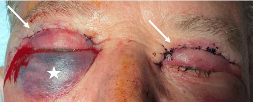
2.
CPC-EM Capsule
What do we already know about this clinical entity?
Orbital compartment syndrome is an expected complication of a retroorbital hemorrhage resulting from facial trauma.
What makes this presentation of disease reportable?
Orbital compartment syndrome as a result of a cosmetic surgery such as blepharoplasty is rarely seen in the emergency department.
What is the major learning point?
The clinical presentation in its totality is more important than a single data point, in this case the ocular pressure measurement.
How might this improve emergency medicine practice?
This case will hopefully increase awareness of the possibility of orbital compartment syndrome as a complication of blepharoplasty.
Blepharopasty is a commonly performed cosmetic procedure that entails removal of excess skin from either the upper or lower eyelid. The procedure is performed in the outpatient setting and is relatively safe. Some of the potential complications include lagophthalmos, which is failure of the upper lid to reach the lower lid, ectropion, or turning outward of the lower lid, and dry eyes. The complications that require emergent intervention are acute angle closure glaucoma and orbital compartment syndrome.3,4 Orbital compartment syndrome due to blepharoplasty procedure was first described in 1981.5 Our patient’s presentation was initially concerning for a retrobulbar hematoma secondary to postoperative bleeding causing acute orbital compartment syndrome. The presence of pain, a fixed mydriatic pupil, and vision loss made angle closure glaucoma a consideration but less likely, given the findings of chemosis and proptosis. The normal IOP did not support either etiology—orbital compartment syndrome or angle closure glaucoma.
side,
While retrobulbar hematoma is a rare complication, it accounts for 51% of cases of acute vision loss after blepharoplasty.3 Orbital compartment syndrome will usually present within 12 hours of the procedure but may occur up to nine days after blepharoplasty.6 A deep orbital hemorrhage is
Libet et al. Orbital Compartment Syndrome as a Complication of Blepharoplasty: A Case Report
the likely etiology in this case. This occurs as a complication in 0.05% of blepharoplasties and causes vision loss in 0.01%.7 When orbital compartment syndrome is diagnosed, patients who receive intervention within two hours have an increased likelihood of improved visual acuity compared with those delayed beyond that time frame.8 While the patient in the case presented had immediate resolution of visual symptoms, it is worth noting that improvement in visual acuity may take up to several weeks.8 The presentation aligns with orbital compartment syndrome despite the absence of a clear retrobulbar hematoma and the presence of a normal ocular pressure. It is possible that there was a calibration error of our tonometer, but otherwise it is unclear why the measured IOP was normal. This case highlights the importance of relying on the complete clinical picture and not on any single test.
The complication of orbital compartment syndrome can occur rarely in patients who have undergone recent blepharoplasty. The diagnosis requires emergent intervention as the duration of optic nerve compression is correlated with the outcome of visual acuity. In this case, the intraocular pressure was not elevated; however, the clinical picture dictated the need for emergent intervention. Cantholysis was performed by the emergency medicine service, and our patient had rapid improvement in his visual acuity.
The Institutional Review Board approval has been documented and filed for publication of this case report. Patient consent has been obtained and filed for the publication of this case report.
Address for Correspondence: Lev Libet, MD, Kern Medical, Department of Emergency Medicine, 1700 Mt Vernon Ave, Bakersfield CA 93306. Email: lev.libet@kernmedical.com
Conflicts of Interest: By the CPC-EM article submission agreement, all authors are required to disclose all affiliations, funding sources and financial or management relationships that could be perceived as potential sources of bias. The authors disclosed none.
Copyright: © 2025 Libet et al. This is an open access article distributed in accordance with the terms of the Creative Commons Attribution (CC BY 4.0) License. See: http://creativecommons.org/ licenses/by/4.0/
1. Channa R, Zafar SN, Canner JK, et al. Epidemiology of eyerelated emergency department visits. JAMA Ophthalmol. 2016;134(3):312–319.
2. Rowh AD, Ufberg JW, Chan TC, et al. Lateral canthotomy and cantholysis: emergency management of orbital compartment syndrome. J Emerg Med. 2015;48(3):325-330.
3. Mejia JD, Ergo FM, Nahai D. Visual loss after blepharoplasty: incidence, management, and preventive measures. Aesthetic Surg J 2011;31(1):21–29
4. Der Kelen LV and Mommaerts MY. Visual loss after cosmetic blepharoplasty using local anaesthesia containing epinephrine - a case series. Ann Maxillofac Surg. 2021;11(2):340-343.
5. Stasior OG. Blindness associated with cosmetic blepharoplasty. Clin Plast Surg. 1981;8(4):793-795.
6. Teng CC, Reddy S, Wong JJ, et al. Retrobulbar hemorrhage nine days after cosmetic blepharoplasty resulting in permanent visual loss. Ophthalmic Plast Reconstr Surg. 2006;22(5):388-389.
7. Hass AN, Penne RB, Stefanyszyn MA, et al. Incidence of post blepharoplasty orbital hemorrhage and associated visual loss. Ophthalmic Plast Reconstr Surg. 2004;20(6):426-432.
8. McCallum E, Keren S, Lapira M, et al. Orbital compartment syndrome: an update with review of the literature. Clin Ophthalmol 2019;13:2189-2194.
Jillian Rosenblum, MD
Section Editor: Austin Smith, MD
Submission history: Submitted March 23, 2025; Revision received June 2, 2025; Accepted June 7, 2025
Electronically published August 26, 2025
Full text available through open access at http://escholarship.org/uc/uciem_cpcem DOI: 10.5811/cpcem.46993
Introduction: Orbital compartment syndrome (OCS) is a rare but high-morbidity emergency requiring prompt recognition and management.
Case Report: We present a case of a man who developed OCS from external compression of the globe while lying in a prone position. Initially obtunded and unable to provide any history, the patient exhibited anisocoria, which later progressed to severe chemosis and proptosis. Intraocular pressure reached nearly 100 millimeters of mercury, improving immediately after emergent lateral canthotomy with cantholysis. His course was complicated by ipsilateral limb compartment syndrome and worsening renal failure requiring dialysis.
Conclusion: This case highlights the critical role emergency physicians play in the rapid diagnosis and treatment of orbital compartment syndrome. [Clin Pract Cases Emerg Med. 2025;9(4):392-394.]
Keywords: orbital compartment syndrome; Saturday night retinopathy; lateral canthotomy; case report.
Orbital compartment syndrome (OCS) is an ophthalmologic emergency in which a sudden increase in pressure within the orbit compromises blood flow to the optic nerve and retina, risking permanent vision loss. This case presents a rare etiology of OCS caused by external compression of the globe due to a prolonged, drug-induced downtime in the prone position. An ischemic reperfusion injury occurred once the patient remained in a supine position in the emergency department (ED). This differs from the typical cause of OCS, which is a retrobulbar hematoma following blunt facial trauma. The patient’s obtunded state, coupled with a lack of the typical finding of a retrobulbar hematoma on computed tomography (CT) of the brain, posed a diagnostic challenge. This case emphasizes the need for emergency physicians to remain vigilant in the rapid diagnosis and treatment of OCS, particularly in atraumatic cases and when the patient cannot report history or symptoms.
A 34-year-old man with a prior history of schizophrenia and substance use disorder presented to the ED with altered
mental status, found unresponsive on the sidewalk with unknown downtime. He was given naloxone in the field with no improvement in mental status. On arrival, he was moaning incomprehensibly and not answering questions or following commands. He was found to be hypothermic, tachycardic, tachypneic, and oxygen saturation was 72% on room air. Physical examination revealed anisocoria, with the right pupil two millimeters larger than the left and non-reactive to light. His point-of-care glucose revealed severe hypoglycemia. He was given dextrose with no improvement in mental status, followed by intubation and placement of warming blankets. Laboratory studies were notable for a leukocytosis with left shift, hyperkalemia with a potassium of 6.5 millimoles per liter (mmol/L) (reference range: 3.5-5.1 mmol/L), an elevated creatinine of 2.53 milligrams per deciliter (mg/dL) (0.50-1.30 mg/dL), elevated liver function tests with aspartate aminotransferase 805 units per liter (U/L) (0-37 U/L) and alanine transaminase 207 U/L (0-60 U/L), a high sensitivity troponin slightly over 2,000 nanograms (ng)/L (0-76 ng/L), a creatine kinase of 46,316 U/L (39-308 U/L), a lactic acid of 12.8 mmol/L (0.4-2.0 mmol/L), pH on arterial blood gas of 7.1 (7.35-7.45), negative serum alcohol level, and urine drug
screen positive for amphetamines, cannabinoids, and fentanyl. Electrocardiogram demonstrated no ST-segment elevation or arrhythmia. Initial imaging included a chest radiograph and computed tomography (CT) of the brain without contrast, which were significant for a right upper lobe infiltrate and a new, hypoattenuating lesion in the basal ganglia concerning for an acute infarction. He was promptly treated with sodium bicarbonate, calcium gluconate, lactated Ringer, broad spectrum intravenous antibiotics, and a norepinephrine infusion for mean arterial pressures consistently below 65 millimeters of mercury (mm Hg). He was placed on a propofol infusion for sedation.
While awaiting transportation to the intensive care unit, he developed new right-sided proptosis and chemosis. Radiology was called to re-evaluate the CT of the brain with special attention paid to his right orbit. It was then noticed that he had diffusely thickened right extraocular motor muscles (Image). Intraocular pressures were checked and found to be elevated to 97 mm Hg on the right and normal on the left (< 21 mm Hg). Emergent right lateral canthotomy with
CPC-EM Capsule
What do we already know about this clinical entity?
Orbital compartment syndrome (OCS) is a rare but vision-threatening emergency that usually occurs in the setting of blunt facial trauma.
What makes this presentation of disease reportable?
This case describes an atraumatic cause of OCS in a patient with a prolonged period of unconsciousness in the prone position.
What is the major learning point?
OCS can occur in the absence of trauma and must be considered in obtunded patients with proptosis or anisocoria.
How might this improve emergency medicine practice?
This case raises awareness of atypical OCS presentations, enabling clinicians to improve their diagnostic vigilance and ultimately prevent permanent vision loss.
cantholysis was performed with immediate improvement in right intraocular pressure.
The following day the patient’s clinical course was complicated by right lower extremity compartment syndrome requiring open fasciotomy of the upper and lower leg compartments. His renal function worsened with worsening rhabdomyolysis, hyperkalemia, and lactic acidosis requiring hemodialysis. By day 10, he was extubated and discharged in stable condition, with the need for ongoing hemodialysis as an outpatient and a poor prognosis for return of vision. He was instructed to follow up with plastic surgery for future skin grafting of his right leg, with cardiology for suspected druginduced cardiomyopathy, and with ophthalmology.
Orbital compartment syndrome is rare, occurring in less than 1% of ED presentations involving facial trauma.1 It most often occurs due to retrobulbar hemorrhage following blunt facial trauma.1 Prompt recognition and treatment are crucial to prevent rapid and irreversible vision loss. Making the diagnosis can be particularly challenging when a patient is obtunded and cannot provide history or report symptoms of eye pain or vision loss. Orbital compartment syndrome is a
clinical diagnosis made based on signs, symptoms, and intraocular pressure measurements.2 Clinical signs can include an afferent pupillary defect, proptosis, tense periorbital edema, limited extraocular movements, and a firm globe due to elevated intraocular pressure.3 Optimal outcomes are achieved if treatment is initiated within 90 minutes of onset.4
In our case, the presumed cause of OCS was external compression of the globe in the setting of prolonged, druginduced downtime in a prone position, which caused an ischemic reperfusion injury once the patient remained supine. This is a paradoxical phenomenon in which the restoration of blood flow to ischemic tissue triggers an inflammatory response that further exacerbates tissue damage. The ophthalmology literature refers to this entity as “Saturday night retinopathy.”5 No treatment will positively affect visual prognosis given that by the time of patient presentation, the ischemic insult from sustained external compression has usually already occurred.3 Vision loss is typically severe and permanent.
On the other hand, promptly performing a lateral canthotomy and cantholysis for traumatic cases of OCS is highly likely to be successful, with alleviation of pressure in the orbital compartment occurring between 68-79% of the time.6 The key is rapid diagnosis and initiation of management, as permanent vision loss can be prevented if performed within two hours of onset.7 If OCS is not relieved by canthotomy and cantholysis, the patient may need open orbitotomy and bony orbital decompression in the operating room.4 Systemic treatments, such as diuretics like acetazolamide and mannitol, have no role in management of OCS.4 Care measures do include adequate pain and blood pressure control, elevating the head of the bed, and minimizing Valsalva maneuver by giving antiemetics, decongestants, and stool softeners.4 Complications from a lateral canthotomy are relatively rare when performed correctly but can include iatrogenic injury to the globe, the lacrimal gland and artery, and the eyelids. Patients should be treated with aggressive eye lubrication until outpatient follow-up with an ophthalmologist, given the risk of exposure keratopathy from the lateral canthus deformity.4
This case underscores the emergency physician’s vital role in the rapid diagnosis and management of orbital compartment syndrome. Lateral canthotomy with cantholysis must be performed emergently, often before an ophthalmologist is available, to prevent permanent vision loss. Emergency physicians must recognize and treat OCS expeditiously, particularly in obtunded patients where clinical signs can be subtle.
The authors attest that their institution requires neither Institutional Review Board approval, nor patient consent for publication of this case report. Documentation on file.
Address for Correspondence: Jillian Rosenblum, MD, Alta Bates Summit Medical Center, Emergency Department, S 2450 Ashby Ave, Berkeley, CA 94705. Email: jrosenblum@bayem.org
Conflicts of Interest: By the CPC-EM article submission agreement, all authors are required to disclose all affiliations, funding sources and financial or management relationships that could be perceived as potential sources of bias. The authors disclosed none.
Copyright: © 2025 Rosenblum et al. This is an open access article distributed in accordance with the terms of the Creative Commons Attribution (CC BY 4.0) License. See: http://creativecommons.org/ licenses/by/4.0/
1. Voss JO, Hartwig S, Doll C, et al. The “tight orbit”: Incidence and management of the orbital compartment syndrome. J Craniomaxillofac Surg. 2016;44(8):1008-14.
2. Murali S, Davis C, McCrea MJ, et al. Orbital compartment syndrome: pearls and pitfalls for the emergency physician. JACEP Open 2021;2(2):e12372. Accessed March 5, 2025.
3. Saturday night retinopathy - EyeWiki. Eyewiki.org. Published March 2025. https://eyewiki.org/Saturday_Night_Retinopathy. Accessed February 1, 2025.
4. Engelmann A and Duncan K. Orbital compartment syndrome. American Academy of Ophthalmology. Published March 1, 2023. https://www.aao.org/eyenet/article/orbital-compartment-syndrome-2 Accessed March 5, 2025.
5. Malihi M, Turbin RE, Frohman LP. Saturday night retinopathy with ophthalmoplegia: a case series. J Neuroophthalmol. 2015;39(2):7782. Accessed on February 20, 2025
6. Scoville NM, Ding L, Stacey AW. Success rates of lateral canthotomy and cantholysis for treatment of orbital compartment syndrome. AJEM. 2023;70:140-3. Accessed on February 20, 2025
7. McCallum E, Keren S, Lapira M, et al. Orbital compartment syndrome: an update with review of the literature. Clin Ophthalmol 2019;13:2189-94.
Mark Ringer, MD*†
Tammy Phan*
Emmelyn J. Samones*
Brian Wolk, MD*
Section Editor: Melanie Heniff, MD, JD
Loma Linda University Medical Center, Department of Emergency Medicine, Loma Linda, California
Pomona Valley Hospital Medical Center, Department of Emergency Medicine, Pomona, California
Submission history: Submitted January 21, 2025; Revision received April 13, 2025; Accepted May 30, 2025
Electronically published August 18, 2025
Full text available through open access at http://escholarship.org/uc/uciem_cpcem
DOI: 10.5811/cpcem.41826
Introduction: Thyroid storm is a rare, life-threatening emergency with a 3.6-17% mortality rate despite proper management. Elevated levels of circulating thyroid hormones can increase metabolic demand, leading to adverse effects on multiple organ systems, particularly critical cardiovascular complications such as cardiomyopathy, myocardial infarction, ventricular arrhythmias, or coronary vasospasm. Precipitating factors can include infection, surgery, or trauma, with infection being the most common. The potential for cardiovascular mortality is high. While beta blockers are key for treatment, they can potentially reduce necessary cardiac output, risking hemodynamic collapse. Traditionally, propranolol has been recommended. We report a rare case of an adolescent experiencing cardiac arrest after propranolol administration in thyroid storm and acute appendicitis.
Case Report: A 17-year-old female with a history of Grave disease, non-adherent to methimazole, underwent evaluation and treatment of thyroid storm and concomitant acute appendicitis. Aggressive initial treatment measures were started, including intravenous and oral propranolol. She went into cardiac arrest approximately six hours after initial medication administration, with subsequent return of spontaneous circulation achieved. The patient underwent aggressive resuscitation and multidisciplinary management. She had a prolonged course in the intensive care unit and was ultimately discharged from the hospital approximately three weeks later
Conclusion: Beta-blocker use in the management of thyrotoxicosis can potentially cause cardiovascular collapse. We suggest consideration of shorter acting beta blockers, such as esmolol or landiolol. [Clin Pract Cases Emerg Med. 2025;9(4):395-399.]
Keywords: case report; propranolol; thyroid storm; appendicitis; beta blockers.
Thyroid storm is a rare but serious emergency. Even when managed appropriately, the mortality rate is 3.6-17%.1 Thyroid storm usually occurs in patients with underlying hyperthyroidism, such as Grave disease.1 Symptoms typically involve hyperthermia, tachycardia, central nervous system dysfunction, heart failure, and gastrointestinal symptoms such as nausea, vomiting, and diarrhea. Symptoms arise from a hypermetabolic state resulting from elevated levels of
circulating thyroid hormone.2,3 Typical precipitating factors include infection, cardiac ischemia, surgery, or trauma, with infection the most common.4 Concomitant pathology may also create challenges for identification and treatment of thyroid storm, such as occurred in our case.
Cardiovascular mortality is high in the acute phase of thyroid storm.5 Controlling tachycardia early in the disease course may prevent permanent cardiac damage.4 Beta blockers are a mainstay of treatment for thyrotoxicosis and thyroid
storm; however, in patients with heart failure, beta blockade can profoundly decrease cardiac output. Other reports document cardiovascular collapse after receipt of beta blockers.5-8 To date, the youngest patient reported in the literature with cardiovascular collapse after beta blocker administration for thyroid storm management was 32 years of age.6 We present a rare case of an adolescent patient who experienced cardiac arrest after propranolol administration in the setting of thyroid storm and concomitant acute appendicitis.
A 17-year-old non-pregnant female, with a history of Grave disease non-adherent on methimazole, presented to an outside hospital with concern for abdominal pain. The patient weighed 53 kilograms, with initial vital signs showing a blood pressure of 92/64 millimeters of mercury; heart rate (HR) 145 beats per minute (bpm); respiratory rate, 35 breaths per minute; and oxygen saturation of 97%, with a temperature of 37.1 °Celsius. The patient was noted to be in mild distress on initial examination but alert and oriented to person, place, and time. She also had right lower quadrant tenderness to palpation and was found to have acute appendicitis on computed tomography (CT).
The patient was tachycardic and mildly hypotensive, with thyroid-stimulating hormone less than 0.015 milliinternational units per milliliter (mIU/mL) (reference range: 0.465-4.680 mIU/mL), and free thyroxine (T4) greater than 6.99 nanograms per deciliter (ng/dL) (0.78-2.19 ng/dL), raising the concern for thyrotoxicosis. The patient was initially given 1.5 liters (L) of isotonic intravenous (IV) fluid, propranolol 0.5 milligrams (mg) IV push (IVP), hydrocortisone 300 mg IVP, and methimazole 20 mg by mouth. Later, the patient was given additional doses of propranolol: 1 mg IVP and 60 mg orally (PO). She also received potassium chloride 40 milliequivalents PO and calcium gluconate 1 gram (g) IV piggyback.
According to transfer records, the outside facility did not have potassium iodide available to administer. Due to concern for suspected thyroid storm and acute appendicitis, arrangements were made to transfer the patient to our tertiarycare center. The patient’s initial blood pressure improved and stabilized after IV fluid administration. However, prior to transfer, she developed worsening hypotension and lethargy, which did not improve with an additional 2 L of IV crystalloid. A norepinephrine infusion was started at 18 micrograms per hour (mcg/hr) and continued during transport, with an increased rate of 22 mcg/hr on arrival. The patient’s blood glucose was 137 mg/dL (74-106 mg/dL) at the transferring facility, just prior to transport.
On arrival at the tertiary center, the patient was initially awake and alert but was noted to be suddenly unresponsive 10 minutes later. She was found to be pulseless at that time, and cardiopulmonary resuscitation (CPR) was initiated. The norepinephrine drip was paused, and the patient was given a
CPC-EM Capsule
What do we already know about this clinical entity?
Thyroid storm is a life-threatening condition that requires early detection and prompt management. Beta blockers are a key component to the treatment regimen.
What makes this presentation of disease reportable?
We present a case of a pediatric patient that suffered cardiac arrest in the setting of thyroid storm, possibly secondary to propranolol toxicity.
What is the major learning point?
Judicious use of beta blockers in the management of thyroid storm is paramount, with consideration toward use of short-acting alternatives to propranolol.
How might this improve emergency medicine practice?
Careful and appropriate use of beta blockers in treatment of thyroid storm can potentially reduce complications and improve overall morbidity and mortality.
total of four doses of epinephrine 1 mg IVP during the code, along with one dose of atropine 1 mg IVP. The patient’s blood glucose was 27 mg/dL, and she received an initial dose of dextrose 25 g IVP with subsequent improvement of the blood glucose. She was intubated during the code and was noted to be in asystole on the first three rhythm checks. Return of spontaneous circulation was achieved after four rounds of CPR. The norepinephrine infusion was restarted, and an epinephrine infusion was added at 5 mcg/minute.
Central venous access and arterial line was placed for blood pressure monitoring. The patient developed profound tachycardia to 170 bpm with intermittent runs of ventricular tachycardia. Magnesium 2 g IV was given with improved HR and resolution of the ventricular tachycardia. Recurrent hypoglycemia was also noted, requiring multiple additional boluses of dextrose. Glucagon was administered to circumvent the possible effects of propranolol, given the patient’s cardiovascular collapse and recurrent hypoglycemia. Due to a hospital glucagon shortage, the patient received 1 mg IV bolus followed by 2 mg/hour for five hours only. Endocrinology was consulted for management, and the patient was given
et al. A Case Report of Thyroid Storm with Cardiovascular Collapse After Propranolol Administration
hydrocortisone 100 mg IVP. Potassium iodide and methimazole were also recommended; however, these were not given in the emergency department due to a delay in availability from the hospital pharmacy. Chest radiograph was notable for cardiomegaly, with possible mild pulmonary congestion (Image 1).
The patient was admitted to the pediatric intensive care unit and started on broad-spectrum IV antibiotics: ceftriaxone, metronidazole, and vancomycin. Toxicology was consulted due to concern for beta-blocker toxicity, and the patient was also given multiple doses of calcium. Milrinone was initiated for cardiovascular support, and high-dose insulin was considered but not given for treatment of beta-blocker toxicity. Echocardiogram noted an ejection fraction of 35%, with peak troponin T, high-sensitivity 42 ng/L (≤ 13 ng/L) and pro-Btype natriuretic peptide > 70,000 picograms (pg) per mL (30-125 pg/mL). The patient progressively improved with aggressive diuresis and continued cardiovascular support.
Repeat echocardiogram three days later showed normal systolic function. She was extubated on hospital day 5 and was able to tolerate a regular diet. Inotropes and pressors were also weaned and discontinued on day five. Given the patient’s overall instability upon admission, pediatric surgery recommended medical management of the patient’s appendicitis. A repeat CT of the abdomen and pelvis with IV contrast on hospital day 7 showed evidence of perforated appendicitis with multiple fluid collections concerning for abscess formation (Image 2).
Image 2. Computed tomography of the abdomen and pelvis with intravenous contrast showing perforated appendicitis with multiple peripherally enhancing fluid collections seen within the right paracolic gutter as well as the pelvis, the largest measuring up to 8.4 centimeters (red arrow). There is associated regional fat stranding and a small volume of free fluid.
Image 1. Anteroposterior chest radiograph taken in the supine position showing cardiomegaly (blue arrow) and perihilar airspace disease (red arrow), raising concern for pulmonary edema. The endotracheal tube is also seen post-intubation terminating 4.3 centimeters above the carina (green arrow).
Three intraperitoneal drains were placed by interventional radiology on hospital day 8, and the patient was continued on IV antibiotics after abscess fluid cultures grew Pseudomonas aeruginosa. She also required continuous renal replacement therapy for acute kidney injury, which was transitioned to hemodialysis. Per endocrinology recommendations, the patient was continued on stress-dose hydrocortisone, which was stopped on hospital day 9. Methimazole was held initially due to elevated liver enzymes and concern for acute liver injury but was started on hospital day 7 at the patient’s home dose of 5 mg PO daily. Potassium iodide was considered, but ultimately never given due to acute kidney injury and eventual improvement in thyroid labs. Cholestyramine was initiated on hospital day 3 and discontinued on hospital day 10. Three weeks after the patient was admitted, she was discharged home on oral antibiotics. She required continued hemodialysis in the outpatient setting for approximately six weeks.
Thyroid storm can be difficult to identify given the broad range of effects. End-organ damage in the setting of thyrotoxicosis is the key feature. The Burch-Wartofsky point scale can be used to gauge the severity of illness with a suspected thyrotoxic state. A score of < 25 indicates that thyroid storm is unlikely, 25-44 suggests impending thyroid storm, and > 45 indicates severe illness and suspected thyroid storm.9 According to transport records, the patient had an initial score of 70 prior to transfer.
The tenets of management are as follows: 1) giving supportive care and management of precipitating factors; 2) inhibiting the synthesis and release of thyroid hormone; and 3) controlling peripheral effects of the circulating hormones.4 First-line therapies typically involve thionamides such as propylthiouracil (PTU) or methimazole (MMI). Recent data suggest similar effectiveness and no difference in adverse events and in-hospital mortality between PTU and MMI.1 Iodine supplementation given after thionamide administration also inhibits thyroid hormone synthesis and is given as potassium iodide in either Lugol solution or saturated solution potassium iodide. Both glucocorticoids and beta blockers reduce the peripheral conversion of T4 to triiodothyronine (T3). Cholestyramine may improve gastrointestinal clearance of thyroid hormone. In severe refractory cases, plasmapheresis may be considered to remove circulating thyroid hormones from the plasma.1,3
Cardiopulmonary manifestations of thyroid storm are the most common cause of mortality. Ventricular arrhythmias are typically the proximate cause of cardiac arrest.2 Thyroid hormones increase protein synthesis, leading to an increased number of β-receptors, and a reduced myocardial refractory phase.10 In a healthy heart, this often manifests as high-output failure.6 However, individuals with pre-existing heart conditions are more likely to experience a low cardiac output state due to the hypermetabolic state created during thyroid storm.4
In our case, the patient received propranolol as part of the initial regimen used to treat the presentation of suspected thyroid storm. She experienced cardiovascular collapse multiple hours later and subsequent cardiac arrest. The exact etiology of the cardiac arrest is unclear and was most likely multifactorial. Case reports have described cardiac arrest in the setting of thyroid storm, possibly resulting from increased metabolic demand and subsequent heart failure.2,10 However, the exact mechanism of the precipitating events is often difficult to fully delineate. Several case reports have documented cardiovascular collapse after beta blocker administration, with a possible temporal association particularly with long-acting beta blockers (eg, propranolol). This systematic review only included patients ≥ 18 years of age.11
Thionamides and beta blockers have been used successfully within the pediatric population.12,13 Although beta blockers are often used to help reduce the peripheral effects of increased circulating thyroid hormone, they can be associated with cardiovascular collapse.6 Many patients with longstanding hyperthyroidism have some underlying thyrocardiac disease, even if not readily apparent.8 In previous cases of beta blocker-induced cardiovascular collapse, nearly all patients had some degree of thyrotoxic cardiomyopathy.7 In our case, the patient’s initial chest radiographs showed some mild to moderate cardiomegaly (Image 1). Unfortunately, there are no clear guidelines in this situation since most treatment guidelines for thyroid storm involve the use of beta blockade to reduce the symptoms and clinical manifestations
of circulating thyroid hormone.7
Beta-blocker toxicity typically presents with bradycardia, hypotension, altered mental status, and hypotension.14 In our case, the patient initially received 0.5 mg of IV propranolol approximately seven hours before the cardiac arrest, an additional 1 mg of IV propranolol two hours later, and then 60 mg of PO propranolol another hour later (~ 3.5 hours before the cardiac arrest). Intravenous propranolol has a peak onset of about five minutes, whereas immediate-release oral propranolol has a peak onset of 1-4 hours, according to the US Food and Drug Administration (FDA) product label. The half-life of propranolol is 3-6 hours. In our case, the patient was initially normoglycemic at the outside hospital and was persistently tachycardic, with HR ranging from 130-150 bpm on multiple re-evaluations. Approximately 2-3 hours after the subsequent doses of propranolol were administered, the patient developed worsening hypotension, which had not been noted previously during multiple hours of monitoring.
On arrival at the tertiary-care center, the patient had relative bradycardia at 65 bpm, along with new-onset hypoglycemia, which was noted in association with the patient’s acute change in mental status and subsequent cardiac arrest. Despite initially treating the hypoglycemia with initial improvement, there were multiple recurrences. This ultimately resolved after glucagon administration. The combination of hypoglycemia, bradycardia, hypotension, and altered mental status in our case raised the definite possibility of beta-blocker toxicity as the etiology for the patient’s cardiovascular collapse.
Multiple case reports demonstrate a potential relationship between propranolol administration and cardiovascular collapse.6-8,11 In the setting of potential cardiac instability, clinicians may consider use of short-acting beta blockers as suggested by Abubakar et al (2017), which have the benefit of being easily titratable, particularly in the instance of negative cardiovascular side effects.8 Increased metabolic demand and reliance on cardiac output in thyroid storm may increase the susceptibility for cardiovascular collapse secondary to beta blockade. Additionally, combining IV and PO doses of beta blockers or stacking doses at different time intervals may create unintended potentiating effects.
In management of thyroid storm, propranolol has been historically superior to other beta blockers due to its blockade of conversion of T4 to T3. However, esmolol is a short-acting IV beta-1 adrenergic blocker that is easily titratable and may serve as a reasonable alternative to managing thyrotoxicosis.6 The half-life of esmolol is nine minutes (2-4 minutes in children).15 Landiolol is an ultra-short-acting beta blocker that has also been studied in the management of thyroid storm. It is a beta-1 adrenergic blocker with high cardioselectivity and appears to be cardioprotective, even in situations of preexisting reduced ejection fraction heart failure.16 The half-life of landiolol is 4.5 minutes. This combination may make it a preferable alternative for management of thyroid storm, given the ability for rapid titration. Landiolol has recently received
Ringer et al. A Case Report of Thyroid Storm with Cardiovascular Collapse After Propranolol Administration
FDA approval for treatment of supraventricular tachycardias, without specific notation for use in thyrotoxicosis. Cardiac arrest has still been reported secondary to landiolol use in thyroid storm treatment, demonstrating the need for judicious use of beta blockers in thyroid storm.7
Thyroid storm poses several diagnostic and treatment challenges, with the potential for cardiovascular collapse. Beta blockers are usually indicated but may worsen the hemodynamic status of the patient during the acute phase of thyroid storm and, therefore, should be used with care. Further analysis of the potential use of short-acting and ultra-shortacting beta blockers in thyrotoxicosis is warranted.
The authors attest that their institution does not require Institutional Review Board approval. Patient consent has been obtained and filed for the publication of this case report.
Address for Correspondence: Emmelyn J. Samones, CCRP, Loma Linda University Medical Center, Department of Emergency Medicine,11234 Anderson St., Room A890A, Loma Linda, CA 92354. Email: Esamones@llu.edu
Conflicts of Interest: By the CPC-EM article submission agreement, all authors are required to disclose all affiliations, funding sources and financial or management relationships that could be perceived as potential sources of bias. The authors disclosed none.
Copyright: © 2025 Ringer et al. This is an open access article distributed in accordance with the terms of the Creative Commons Attribution (CC BY 4.0) License. See: http://creativecommons.org/ licenses/by/4.0/
1. Lee SY and Pearce EN. Hyperthyroidism: a review. JAMA 2023;330(15):1472-83.
2. Zayour M, Yasmin FA, Baydoun A, et al. Cardiac arrest as first presentation of thyroid storm. Cureus. 2023;15(4):e37057.
3. De Almeida R, McCalmon S, Cabandugama PK. Clinical review and update on the management of thyroid storm. Mo Med.
2022;119(4):366-71.
4. Nai Q, Ansari M, Pak S, et al. Cardiorespiratory failure in thyroid storm: case report and literature review. J Clin Med Res 2018;10(4):351-7.
5. Misumi K, Kodera S, Nagura F, et al. Cardiac arrest caused by landiolol in a patient in thyroid crisis. J Cardiol Cases. 2016;14(2):62-4.
6. Ngo AS and Lung Tan DC. Thyrotoxic heart disease. Resuscitation 2006;70(2):287-90.
7. Bokhari SFH, Sattar H, Abid S, et al. Cardiovascular collapse secondary to beta-blocker administration in a setting of coexisting thyroid storm and atrial fibrillation: a case report. Cureus 2022;14(9):e29321
8. Abubakar H, Singh V, Arora A, et al. Propranolol-induced circulatory collapse in a patient with thyroid crisis and underlying thyrocardiac disease: a word of caution. J Investig Med High Impact Case Rep. 2017;5(4).
9. Ross DS, Burch HB, Cooper DS, et al. 2016 American Thyroid Association guidelines for diagnosis and management of hyperthyroidism and other causes of thyrotoxicosis. Thyroid 2016;26(10):1343-1421. Erratum in: Thyroid. 2017;27(11):1462.
10. Nakashima Y, Kenzaka T, Okayama M, et al. A case of thyroid storm with cardiac arrest. Int Med Case Rep J. 2014;7:89-92.
11. Afifi S, Suryadevara V, Habab Y, et al (2023). Comparing the incidence of propranolol and esmolol-related cardiac arrest in patients with thyroid storm: a systematic literature review. Cureus 2023;15(9):e44655.
12. Aoki Y, Hanaki R, Toyoda H, et al. Case report: thyroid storm in a three-year-old girl presenting with febrile status epilepticus and hypoglycemia. Front Pediatr. 2023;11:1213040.
13. Bonfield A and Shenoy S. Thyrotoxic crisis as an acute clinical presentation in a child. BMJ Case Rep vol. 2018:bcr2017222850. 23 Mar 2018.
14. Goldfine CE, Troger A, Erickson TB, et al. Beta-blocker and calciumchannel blocker toxicity: current evidence on evaluation and management. Eur Heart J Acute Cardiovasc Care. 2024;13(2):247-53.
15. Yamashita Y, Iguchi M, Nakatani R, et al. Thyroid storm with heart failure treated with a short-acting beta-adrenoreceptor blocker, landiolol hydrochloride. Intern Med. 2015;54(13):1633-7.
16. Procaccini DE, Sawyer JE, Watt KM. Pharmacology of cardiovascular drugs. In: Ed(s): Ungerleider RM, Meliones JN, McMillan KN, Cooper DS, Jacobs JP. Critical Heart Disease in Infants and Children (3rd Ed), New York, NY: Elsevier, 2019 192-212.e6.
Diormi A. Rosario, DO
Stephanie Aronson, MD
Jessica Zerzan, MD
Section Editor: John Ashurst, DO
Maimonides Medical Center, Department of Pediatric Emergency Medicine, Brooklyn, New York
Submission history: Submitted January 31, 2025; Revision received May 1, 2025; Accepted June 2, 2025
Electronically published August 13, 2025
Full text available through open access at http://escholarship.org/uc/uciem_cpcem DOI: 10.5811/cpcem.42233
Introduction: Burkitt lymphoma is a highly aggressive subtype of non-Hodgkin lymphoma with varied clinical presentation, including in some cases involvement of the intraoral cavity. Early recognition of this malignancy is critical, as it typically responds well to prompt and intensive treatment. In this case report, we present a rare manifestation of Burkitt lymphoma presenting as an oropharyngeal mass.
Case Report: An eight-year-old male presented with tonsillar swelling and new-onset oral bleeding. A month earlier, he had been seen in the emergency department (ED) for similar swelling following a streptococcal infection. At that time, a needle aspiration for suspected peritonsillar abscess yielded no drainage, and he was treated with a week of clindamycin, resulting in brief symptom improvement. He subsequently developed difficulty breathing, a muffled voice, and oral bleeding, prompting a return to the ED. On evaluation, he was afebrile, well-appearing, and in no respiratory distress. Examination revealed significant left tonsillar swelling with uvular deviation but no active bleeding. Magnetic resonance imaging demonstrated a bulky left oropharyngeal mass with airway narrowing, raising suspicion for lymphoma. Laboratory results were unremarkable, and biopsy confirmed Burkitt lymphoma based on c-MYC positivity and the characteristic “starry sky” appearance, leading to the initiation of chemotherapy.
Discussion: Burkitt lymphoma is a high-grade lymphoma with a large tumor burden and, thus, high risk for tumor lysis syndrome. Fortunately, Burkitt lymphoma has superior survival outcomes in pediatrics with a two-year survival rate estimated to be 89% and requiring minimal cycles of chemotherapy. This case underscores the diverse presentations of Burkitt lymphoma and the importance of including it in the differential for all pediatric neck masses, regardless of demographics. [Clin Pract Cases Emerg Med. 2025;9(4):400-403.]
Keywords: Burkitt lymphoma; oropharyngeal mass; head and neck cancer; pediatric; lymphoma; oropharyngeal tumor.
Non-Hodgkin lymphoma is one of the most prevalent neoplasms seen worldwide.1 It can have many variants, including Burkitt lymphoma. Burkitt lymphoma is divided into three subgroups: sporadic, endemic, and immunodeficiency related.2 In the United States, the frequency
of non-Hodgkin lymphoma in the pediatric population is 0.5-1.2 per 100,000.3 The presentation of Burkitt lymphoma usually involves the abdomen, mandible, or maxilla; however, in rare instances it can involve the oral cavity. This case depicts a rare presentation of Burkitt lymphoma, arising as a peritonsillar mass.
An eight-year-old male presented with tonsillar swelling and new-onset oral bleeding. He had been seen in the emergency department (ED) one month earlier for similar symptoms and was treated with a seven-day course of clindamycin for suspected bacterial tonsillitis after a nonproductive needle aspiration. His symptoms initially improved but later progressed to include difficulty breathing and a muffled voice. On the day of presentation, oral bleeding developed, prompting return to the ED. The family denied fever, drooling, dyspnea, or weight loss at that time.
Upon arrival to the ED the patient was afebrile with heart rate 101 beats per minute, respiratory rate 17 breaths per minute, blood pressure 121/63 millimeters of mercury, and oxygen saturation 98% on room air. On examination the patient was well-appearing, in no respiratory distress without stridor, and with marked left tonsillar swelling with uvular deviation, and no active bleeding (Image 1). The ear, nose, and throat (ENT) physicians were contacted, who recommended a magnetic resonance imaging (MRI) of the neck. The patient received a dose of ampicillin-sulbactam and was admitted with plans for biopsy with ENT. Labs were obtained, which included complete blood count, basic metabolic panel, magnesium, phosphorus, lactate dehydrogenase, uric acid, and venous blood gas; all were unremarkable.
During the admission, the patient remained stable on room air. The MRI demonstrated a bulky well-circumscribed left oropharyngeal mass involving the tonsillar fossa, suspicious for lymphoma or other neoplasms, with narrowing of the oropharyngeal and nasopharyngeal airway (Images 2
CPC-EM Capsule
What do we already know about this clinical entity?
Burkitt lymphoma is a rapidly growing B-cell tumor which often presents with abdominal pain of mandibular masses.
What makes this presentation of disease reportable?
Burkitt lymphoma is known to mimic many common pediatric conditions which can sometimes delay the diagnosis of such a rapid growing pathology.
What is the major learning point?
Burkitt lymphoma can present subtly, therefore persistent symptoms warrant escalation to rule out malignancy.
How might this improve emergency medicine practice?
This case helps heighten awareness to include maligmancy in differential diagnosis for persistent abdominal pain or systemic symptoms in children.
and 3). Oncology was consulted, which recommended a biopsy. The ENT physician performed a biopsy of the left intraoral mass, debulking of mass, and endotracheal intubation. The biopsy was positive for c-MYC with “starry sky” appearance, all indicative of Burkitt lymphoma. The patient was then started on chemotherapy and was able to be extubated and discharged from the hospital.
Burkitt lymphoma is not often on the differential diagnosis of an otherwise healthy child presenting with an oropharyngeal mass. As in our patient’s case, a child would be evaluated primarily for a peritonsillar abscess, peritonsillar cellulitis, retropharyngeal abscess, or infectious mononucleosis.4 While Epstein-Barr virus causes infectious mononucleosis, the association of Epstein-Barr virus with Burkitt lymphoma is not usually considered in the United States. Epstein-Barr virus is more likely to be associated with the endemic form of Burkitt lymphoma, which frequently develops in African children and adolescents.5 The sporadic form of Burkitt lymphoma, which has no geographic focality, is more often found in adult patients,
Lymphoma Presentation with Oropharyngeal Mass of Tonsillar Fossa: A Case Report
large tumor burden and, thus, high risk for tumor lysis syndrome. Tumor lysis syndrome occurs when tumor cells rapidly break down and intracellular contents are released into circulation, causing major electrolyte disturbances (hyperkalemia, hyperphosphatemia, hypocalcemia, and hyperuricemia). The electrolyte changes may cause an array of symptoms, ranging from muscle cramps and acute kidney injury to potentially fatal cardiac arrhythmias and seizures.7 Due to the tumor burden and high speed of growth, Burkit lymphoma patients are at high risk of tumor lysis syndrome and must be started on appropriate fluids and hypouricemic agents as soon as possible. Despite our patient’s fast progression of the tumor (from ENT visit to ED), he did not develop tumor lysis syndrome, once again lowering clinical suspicion for Burkitt lymphoma.
Fortunately, Burkitt lymphoma has superior survival outcomes in pediatrics. Recent studies have shown the two-year survival rate to be 89%, and most patients are treated with only four cycles of chemotherapy.2
This case highlights the variability in initial presentation and serves as a reminder to include Burkitt lymphoma on the differential diagnosis for all pediatric neck masses regardless of the demographics.
often develops as an intra-abdominal mass rather than an oropharyngeal or peritonsillar mass.6 Our patient’s presentation was puzzling, in that he did have a muffled, “hot potato” voice in the setting of an oropharyngeal mass, seeming more like a peritonsillar abscess; however, he was otherwise well, afebrile, not in pain, and had no drainage on attempted incision and drainage by the ENT physician prior to ED presentation. Nonetheless, due to his demographics and presentation, Burkitt lymphoma was considered; however, it was low on our initial differential diagnosis.
Burkitt lymphoma is a high-grade lymphoma with a
Patient consent has been obtained and filed for the publication of this case report.
Address for Correspondence: 4802 10th Ave, Maimonides Medical Center, Brooklyn, NY 11219. Email: Diormi92@gmail.com.
Conflicts of Interest: By the CPC-EM article submission agreement, all authors are required to disclose all affiliations, funding sources and financial or management relationships that could be perceived as potential sources of bias. The authors disclosed none.
Copyright: © 2025 Rosario et al. This is an open access article distributed in accordance with the terms of the Creative Commons Attribution (CC BY 4.0) License. See: http://creativecommons.org/ licenses/by/4.0/
1. Patankar S. Burkitt’s lymphoma of maxillary gingiva: a case report. World J Clin Cases. 2015;3(12):1011.
2. Kali K, Alessandrino F, Beck R, et al. An update on Burkitt lymphoma: a review of pathogenesis and multimodality imaging assessment of disease presentation, treatment response, and recurrence. Insights Imaging. 2019;10(1):73.
3. Molyneux EM, Rochford R, Griffin B, et al. Burkitt’s lymphoma. Lancet. 2012;379(9822):1234-44.
Rosario et al. Burkitt Lymphoma Presentation with Oropharyngeal Mass of Tonsillar Fossa: A Case Report
4. Sung H, Ferlay J, Siegel RL, et al. Global cancer statistics 2020: GLOBOCAN estimates of incidence and mortality worldwide for 36 cancers in 185 countries. CA Cancer J Clin. 2021;71(3):209-49.
5. Balasubramaniam R, Goradia A, Turner LN, et al. Burkitt lymphoma of the oral cavity: an atypical presentation. Oral Surg Oral Med Oral Pathol Oral Radiol Endod. 2009;107(2):240-5.
6. Azim N, Razmara F, Derakhshan S, et al. Mandibular sporadic Burkitt lymphoma in an adult patient: a case report and review of the literature. Clin Case Rep. 2021;9(7):e4835.
7. Yuhan BT, Svider P, Mutchnick S, et al. Benign and malignant oral lesions in children and adolescents. Pediatr Clin North Am 2018;65(5):1033-50.
Cassidy T Yoshida*
Angela Vu*
Robert Lam, MD†
Sean Donahue, MD†
Section Editor: Joel Moll, MD
†
University of Colorado Anschutz Medical Center, School of Medicine, Aurora, Colorado Memorial Central Hospital, Department of Emergency Medicine, Colorado Springs, Colorado
Submission history: Submitted May 18. 2025; Revision received June 19. 2025; Accepted June 20, 2025
Electronically published August 26, 2025
Full text available through open access at http://escholarship.org/uc/uciem_cpcem
DOI: 10.5811/cpcem.47383
Introduction: Renal colic is a common reason for patients to present to the emergency department (ED). The most common reasons for this pain are usually renal in origin. Here we present the case of a 45-year-old woman with severe right-sided flank pain and associated hydronephrosis secondary to ureteral obstruction caused by the suction of a menstrual cup.
Case Report: A 45-year-old female presented to the ED with sudden severe right-sided flank pain. The patient endorsed nausea without vomiting, fever, chills, hematuria, or dysuria. She stated that she was currently having her menstrual period. On physical exam, the patient was in distress but had no tenderness with palpation of the flank or abdomen. A computed tomography of the kidneys, ureters, and bladder did not show renal or ureteral stones but demonstrated right-sided hydronephrosis secondary to an anatomical blockage of the ureter, which had been suctioned and involuted into a malpositioned menstrual cup. The patient removed her menstrual cup and had immediate relief of her symptoms. She was observed and remained completely asymptomatic upon reassessment two hours later.
Conclusion: Ureteral obstruction and hydronephrosis is a rare complication of menstrual cup use. As these devices become more common, emergency physicians must be aware of this complication as a cause of severe back pain in menstruating women. [Clin Pract Cases Emerg Med. 2025;9(4):404-406.]
Keywords: renal colic; menstrual cup; ureteral stricture; hydronephrosis; case report.
Renal colic is a common presentation for patients presenting to the emergency department (ED), with approximately 1-2 million cases annually.1 The most common cause of this pain is usually renal in origin, with kidney stones and associated pyelonephritis being the main culprits. Additional etiologies for renal colic include perinephric abscesses, mechanical obstruction due to a tumor, or extrinsic blockage of the ureter or bladder. In this case report we present a 45-year-old woman with severe right-sided flank pain and associated hydronephrosis secondary to the suction of a menstrual cup, causing a ureteral obstruction by
suctioning her vaginal wall, ureter, and adnexal vasculature into the cup.
A 45-year-old female with no past medical or surgical history presented to the ED with 10/10, sudden-onset, sharp, constant, and non-radiating right-sided flank pain that started three hours prior while she was driving. The patient endorsed nausea without vomiting but denied diarrhea, constipation, fever, chills, hematuria, or dysuria. She had not had pain like this before. The patient stated that she was currently having her menstrual period. She denied any history of kidney stones,
trauma, or injury. On physical exam, the patient was in distress due to pain but had no tenderness with palpation of the flank or abdomen.
The differential diagnosis included obstructing kidney stone, infected stone, pyelonephritis, urinary tract infection, and muscle strain. Her laboratory testing showed an unremarkable urinalysis aside from trace blood and a complete blood count with mild leukocytosis showing a white blood cell count of 15 x 109 per liter (L) (reference range: 4.0-11.1 x 10⁹/L). Her basic metabolic panel did not show elevation of creatinine. A computed tomography (CT) of the kidneys, ureters, and bladder did not show renal or ureteral stones but demonstrated right-sided hydronephrosis secondary to an anatomical blockage of the ureter, which had been suctioned and involuted into a malpositioned menstrual cup (Image).
Prior to the CT result, the patient was treated for assumed renal colic due to an obstructing stone. She received intravenous (IV) pain control and IV fluids with minimal relief. After reviewing the CT, we asked her to remove her menstrual cup, which resulted in immediate relief of her symptoms. She was observed and remained completely asymptomatic upon reassessment two hours later.
Image. Computed tomography of the kidneys, ureters, and bladder showed mild right hydronephrosis (black arrow) with a transition point at the lower pelvis secondary to a malpositioned menstrual cup with parts of the right vaginal wall, ureter, and adnexal vasculature insinuating into the cup (white arrow).
CPC-EM Capsule
What do we already know about this clinical entity?
Hydronephrosis causing renal colic is a rare complication of menstrual cup use, with only a few reported cases suggesting this possible but uncommon effect.
What makes this presentation of disease reportable?
Although a few cases link menstrual cups to severe renal colic, it is rare and no cases have been reported in any American journals previously.
What is the major learning point?
Consider menstrual cups as a rare cause of hydronephrosis in menstruating patients presenting with renal colic, especially when no other cause is found.
How might this improve emergency medicine practice?
Raises provider awareness to consider menstrual cup complications in the differential for renal colic in menstruating patients, aiding faster diagnosis and appropriate care.
Renal colic is a common condition that typically presents with severe sudden-onset unilateral flank pain. Initial evaluation of renal colic involves a thorough history, physical exam, and careful consideration of etiology. While nephrolithiasis is a common cause, other etiologies should be considered, particularly in patients with atypical presentations.
In recent years, many new menstrual products such as the menstrual cup or menstrual disc have become a more popular choice for menstruating women given their convenience, cost effectiveness, and decreased ecologic impact.2 A study by the Harvard School of Public Health found that 19% of women used menstrual cups.3 Menstrual cups are reusable, flexible, bell-shaped, self-retaining, silicone, intravaginal devices that collect menstrual fluid and rely on suction to the vaginal wall to prevent leakage.4 Correct placement takes practice while incorrect positioning can have unintended effects such as blood leakage or temporary pelvic discomfort. These symptoms usually resolve with correct placement of the menstrual cup or disc.5 Rarely, use of menstrual cups can also cause displacement of an intrauterine device, rashes, or vaginal wounds.5,6 If pain persists after placement or renal
Female Menstrual Cup Causing Renal Colic, Hydronephrosis, and Ureteral Stricture: A Case Report Yoshida et al.
colic ensues, ureteral entrapment may be the cause. Upon literature review, we found that menstrual cup-induced hydronephrosis is a rare complication not previously documented in a journal published in the United States.5-8
In this case, our patient presented with severe renal colic and associated hydronephrosis secondary to a misplaced menstrual cup. The suction mechanism of menstrual cups, combined with the vaginal wall’s elasticity and the ureters’ proximity, can increase the risk of ureteral obstruction when malpositioned.7 This subsequent blockage can cause severe flank pain secondary to unilateral ureteral obstruction and hydronephrosis, which is usually immediately relieved by removal of the menstrual cup.8 Given that renal colic is a common reason to visit the ED, menstrual cup entrapment causing hydronephrosis should be considered in menstruating females.
Ureteral obstruction and hydronephrosis is a rare side effect of menstrual cup use. As these devices become more common, emergency physicians must be aware of this complication as a cause of severe back pain in menstruating women.
The authors attest that their institution requires neither Institutional Review Board approval, nor patient consent for publication of this case report. Documentation on file.
Address for Correspondence: Cassidy T Yoshida, BA, University of Colorado Anschutz Medical Campus, School of Medicine, 13001 E 17th Place, Aurora, CO 80045. Email: Cassidy.yoshida@ cuanschutz.edu.
Conflicts of Interest: By the CPC-EM article submission agreement, all authors are required to disclose all affiliations, funding sources and financial or management relationships that could be perceived as potential sources of bias. The authors disclosed none.
Copyright: © 2025 Yoshida et al. This is an open access article distributed in accordance with the terms of the Creative Commons Attribution (CC BY 4.0) License. See: http://creativecommons.org/ licenses/by/4.0/
1. Giarrusso P, Raio C, Bhagavath A, et al. Do emergency department observation units help prevent revisits for patients with renal colic? Am J Emerg Med. 2024;89:182-6
2. Howard C, Rose CL, Trouton K, et al. FLOW (finding lasting options for women). Multicentre randomized controlled trial comparing tampons with menstrual cups. Can Fam Physician. 2011;57(6):e20815.
3. Wang Z, Peebles E, Baird DD, et al. Menstrual product use patterns in a large digital cohort in the United States: variations by sociodemographic, health, and menstrual characteristics. Am J Obstet Gynecol. Published online March 10, 2025.
4. Mouhanna JN, Simms-Cendan J, Pastor-Carvajal S. The menstrual cup: menstrual hygiene with less environmental impact. JAMA 2023;329(13):1114-5.
5. Hennegan J, Orozco A, Head A, et al. Menstrual cup acceptability and functionality in real-world use: a cross-sectional survey of young people in Australia. Aust N Z J Obstet Gynaecol. 2024;1-8.
6. van Eijk AM, Zulaika G, Lenchner M, et al. Menstrual cup use, leakage, acceptability, safety, and availability: a systematic review and meta-analysis. Lancet Public Health. 2019;4(8): e376-93.
7. Stolz A, Meuwly JY, Roussel A, et al. An improperly positioned menstrual cup complicated by hydronephrosis: a case report. Case Rep Womens Health. 2019;22:e00108.
8. Umaramanan T, Hjort-Pedersen K, Besenbruch A, et al. [Hydronephrosis caused by a menstrual cup]. Ugeskr Laeger 2019;181(45):V04190222
Eli Spevack*
Zachary M Weisner, DO, MSc†
Evgenia Nokovich, DO†
Michelle Joyner DO†
Lauren Exley, MD‡
Section Editor: John Ashurst, MD
Philadelphia College of Osteopathic Medicine, Philadelphia, Pennsylvania
Jefferson Northeast, Graduate Medical Education EM/IM Residency, Department of Internal Medicine, Department of Emergency Medicine, Philadelphia, Pennsylvania
Jefferson Northeast, Department of Emergency Medicine, Philadelphia, Pennsylvania
Submission history: Submitted May 7, 2025; Revision received June 16, 2025; Accepted June 16, 2025
Electronically published August 24, 2025
Full text available through open access at http://escholarship.org/uc/uciem_cpcem DOI: 10.5811/cpcem.47151
Introduction: Carotid artery dissections are uncommon but critical vascular injuries. They involve a tear to the intima, the innermost layer of the arterial wall, leading to formation of a false lumen. This false lumen can disrupt blood flow, weaken the wall, and lead to thrombus or rupture of the artery. Carotid artery dissections can occur spontaneously or in the setting of trauma. Traumatic carotid artery dissections (TCAD) are rare and occasionally present with third-order Horner syndrome, characterized by ipsilateral ptosis, miosis, and anhidrosis. The presence of subtle physical exam signs like Horner syndrome reinforces the importance of maintaining a high index of suspicion and obtaining vascular imaging in trauma-related cases. While there have been case reports of bilateral TCAD, these have been rarely reported in the literature.
Case Report: We present a case involving a 53-year-old female with no significant past medical history who presented to the emergency department after tripping and falling down a flight of stairs. Over three weeks, the patient had persistent tinnitus and right neck pain and, on the exam, was found to have right-sided miosis and ptosis. These exam findings led us to obtain a computed tomography (CT) angiogram of her neck, which revealed bilateral internal carotid artery dissections. The patient was taken for cerebral angiography, which confirmed the diagnosis. A stent was placed in the right internal carotid artery, and the patient was started on aspirin and clopidogrel. The patient was discharged without deficits three days later.
Conclusion: Traumatic internal carotid artery dissection can occasionally result in Horner syndrome and requires CT angiography of the neck and potentially a diagnostic cerebral angiogram to diagnose. This case adds to the limited literature on bilateral TCAD, particularly with a delayed and asymmetric presentation. Horner syndrome in the setting of trauma, while subtle, can suggest a carotid artery dissection. Awareness of such rare presentations is key to early diagnosis and treatment. Clinicians must maintain a high index of suspicion for underlying vascular injury in patients presenting with lesser mechanisms of injury. [Clin Pract Cases Emerg Med. 2025;9(4):407-410.]
Keywords: case report; trauma; carotid dissection; Horner syndrome.
Carotid artery dissections are uncommon but important vascular injuries. They involve a tear to the intima, the innermost layer of the arterial wall, leading to formation of a
false lumen. This false lumen can disrupt blood flow, weaken the wall, and lead to thrombus or rupture of the artery. Carotid artery dissections can occur spontaneously or in the setting of trauma. Traumatic carotid artery dissections (TCAD) are rare
Bilateral Carotid Artery Dissection After a Fall: A Case of Horner Syndrome Revealed on Examination
and occasionally present with third-order Horner syndrome, characterized by ipsilateral ptosis, miosis, and anhidrosis. The presence of subtle physical exam signs like Horner syndrome reinforces the importance of maintaining a high index of suspicion and obtaining vascular imaging in trauma-related cases. The annual incidence of CADs occurs at a rate of 2.6 per 100,000 people.1,2 Horner syndrome occurs in only 25% of these CAD cases. Among all CADs, internal carotid artery dissections (ICAD) are most associated with Horner syndrome. While there have been case reports of bilateral TCAD, rarely have they been reported in the literature.
We present a 53-year-old Spanish-speaking female with no significant past medical history who presented to the emergency department with right-sided neck pain after a fall. The patient reported falling down seven to eight stairs approximately three weeks earlier. Since that fall, she noticed she had issues with her right eye and had also been experiencing left-sided tinnitus. In addition, the patient had sustained a contusion to her left ankle but was able to ambulate without difficulty.
On physical exam, right-sided ptosis and miosis were evident. The pupils were unequal and reactive to light bilaterally. Visual acuity was intact, and extraocular eye motion testing was normal. A whisper test did not reveal any hearing deficits. Cranial nerve examination included assessment of facial sensation, motor function of the cheeks and jaw, and facial symmetry with smile, frown, eyebrow raise and tight eye closure. Gag reflex, swallowing ability, shoulder shrug, and tongue movement were also evaluated. All findings were within normal limits. Neck tenderness was noted from the third and fourth cervical midline and bilateral paraspinal areas.
With the presence of ptosis and miosis following trauma, there was concern for Horner syndrome. Due to the mechanism of injury and neurologic findings, a computed tomography (CT) angiography of the neck was obtained. Imaging revealed severe stenosis at the distal right internal carotid artery near the skull base, as well as severe stenosis of the mid- and distal left internal carotid artery, all concerning for dissection (Images 1 and 2).
Neurosurgery was consulted, and the patient was loaded with aspirin and clopidogrel before being transferred to our central hospital for further evaluation. Cerebral angiography confirmed bilateral dissections of the internal carotid arteries (Image 3). A stent was placed in the petrous segment of the right carotid artery, and the patient was admitted to the intensive care unit with a goal mean arterial pressure of < 100 millimeters of mercury. She was maintained on aspirin and clopidogrel. The patient was discharged home after three days of hospitalization without any neurological deficits.
This case contributes a valuable addition to our
CPC-EM Capsule
What do we already know about this clinical entity?
Carotid artery dissections may follow trauma and present with subtle findings like Horner syndrome or vague head and neck pain.
What makes this presentation of disease reportable?
This is a rare case of bilateral carotid artery dissection with a delayed, unilateral presentation of Horner syndrome after a minor trauma.
What is the major learning point?
Horner syndrome after trauma can indicate underlying carotid artery dissection. Maintain high suspicion for vascular injury, even with seemingly minor mechanisms.
How might this improve emergency medicine practice?
Raise awareness to consider carotid artery dissection in the setting of trauma and Horner syndrome, and obtain computed tomography angiography in patients with those signs.
understanding of bilateral TCAD. In one case of bilateral TCAD that occurred following a devastating motor vehicle crash, the patient presented with anisocoria after being evaluated in the intensive care unit.3 Other cases involve similarly high-caliber trauma mechanisms with varying lengths of time to identification of the CAD. Our patient faced a less severe mechanism of injury, and she did not present until three weeks later. In cases with more mild traumatic
mechanisms, unlikely conditions such as TCADs may be overlooked but still need to be considered.4-8
Horner syndrome is a condition that can result from a lesion occurring anywhere along the sympathetic pathway supplying the head, neck, and eyes.9 The path originates in the hypothalamus. Horner syndrome can be subcategorized as first, second, or third order depending on the location of the lesion.9 The first-order neuron descends to the eighth cervical to second thoracic spinal cord levels (C8-T2), where the first synapse occurs.9 The secondorder neuron travels from the sympathetic trunk, through the brachial plexus, to the superior cervical ganglion, located near the angle of the mandible and carotid artery bifurcation.9 The third-order neuron ascends within the internal carotid artery (ICA) adventitia through the cavernous sinus (closely related to cranial nerves VI).9 The triad of symptoms associated with Horner syndrome are miosis, ptosis, and anhidrosis.9
Coronary artery dissections involve a tear in the intimal layer of the artery, creating an intraluminal hematoma.10 In the event of an ICA dissection causing Horner syndrome, typically only miosis and ptosis are present, as facial sweat glands are innervated by sympathetic fibers surrounding the
external carotid artery.10 With ICADs, the presence of Horner syndrome symptoms is associated with a more favorable course, which includes fewer strokes and better outcomes.10
Patients with a traumatic mechanism who complain of any constellation of headache, facial or neck pain, and partial Horner syndrome should be imaged for underlying extracranial CAD. Intra-arterial angiography remains the gold standard for dissection diagnosis as it will demonstrate arterial lumens and vessel wall defects. Magnetic resonance angiographic or T1, T2, and proton density-weighted images are also valuable tools; however, time and accessibility can be a limiting factor. Ultrasound is a noninvasive option, allowing for direct visualization of vessel blood flow as well as true or false lumens.11
Treatment of CADs can vary depending on presenting symptoms. If a CAD is detected and the patient has an acute ischemic stroke, intravenous thrombolysis with either alteplase or tenecteplase is appropriate.12 In patients with CAD that meet criteria for acute large-vessel occlusion, mechanical thrombectomy should be performed.12 In the setting of a CAD causing stenosis or occlusion with hypoperfusion to distal territories, stenting the vessel remains beneficial. Stenting for CADs with the absence of hypoperfusion is controversial and should be considered on an individual basis.12 To decrease risk of secondary stroke in the setting of CAD after embolization or stenting, consider antithrombotic therapy.12 If there is no elevated bleeding risk and absence of high-risk features (no intraluminal thrombus, non-occlusive dissection), consider dual antiplatelet therapy or monotherapy with aspirin or a P2Y12 inhibitor.12 Presence of high-risk features would necessitate parenteral anticoagulation if low bleeding risk followed by oral anticoagulation, or dual/mono antiplatelet therapy for moderate bleeding risk.12 Patients should remain on antithrombotic therapy for three to six months.12 To avoid recurrent or worsening dissection, patients should avoid minor head/neck trauma in the months following. This includes neck manipulation, extreme sports, heavy lifting, hyperextension, and contact sports.12
In this patient, the presence of partial Horner syndrome played a critical role in raising early suspicion of an underlying CAD. This led to prompt imaging, identification, and dual antiplatelet therapy. However, if fewer or no neurological signs had been apparent, there may have been a significant delay in localizing the underlying problem. This patient still complained of neck pain, yet neck or head pain can mimic less severe conditions like musculoskeletal etiologies. An increased index of suspicion must always be present, especially in patients with a recent history of trauma, to avoid misdiagnoses. This risk will be mitigated by conducting a thorough history and a detailed physical exam and having a lower threshold for imaging in trauma patients.
Traumatic internal carotid artery dissection can occasionally result in Horner syndrome and requires CT angiography of the
Bilateral Carotid Artery Dissection After a Fall: A Case of Horner Syndrome Revealed on Examination Spevack et al.
neck and potentially a diagnostic cerebral angiogram to diagnose. This case adds to the limited literature on bilateral TCAD, particularly with a delayed and asymmetric presentation. Horner syndrome in the setting of trauma, while subtle, can suggest a carotid artery dissection. Awareness of such rare presentations is key to early diagnosis and treatment. Physicians must maintain a high index of suspicion for underlying vascular injury in patients presenting with lesser mechanisms of injury.
The authors attest that their institution requires neither Institutional Review Board approval, nor patient consent for publication of this case report. Documentation on file.
Address for Correspondence: Eli Spevack, MS3, Philadelphia College of Osteopathic Medicine, 4170 City Avenue Philadelphia, PA 19131, USA. Email: es3061@pcom.edu.
Conflicts of Interest: By the CPC-EM article submission agreement, all authors are required to disclose all affiliations, funding sources and financial or management relationships that could be perceived as potential sources of bias. The authors disclosed none.
Copyright: © 2025 Spevack et al. This is an open access article distributed in accordance with the terms of the Creative Commons Attribution (CC BY 4.0) License. See: http://creativecommons.org/ licenses/by/4.0/
1. Lee VH, Brown RD Jr, Mandrekar JN, et al. Incidence and outcome of cervical artery dissection: a population-based study. Neurology 2006;67(10):1809-12.
2. Schievink WI. Spontaneous dissection of the carotid and vertebral arteries. N Engl J Med. 2001;344(12):898-906.
3. Cronlein M, Sandmann GH, Beirer M, et al. Traumatic bilateral carotid artery dissection following severe blunt trauma: a case report on the difficulties in diagnosis and therapy of an often overlooked life-threatening injury. Eur J Med Res. 2015;20:62.
4. Agarwal A, Yadav D, Gupta A, et al. Delayed bilateral internal carotid artery dissection following motor vehicle accident: time to make its screening a part of trauma protocol? QJM. 2020;113(9):672-3.
5. Khormi YH, Darraj AI, Arishy A, et al. Bilateral blunt traumatic dissections of the extracranial internal carotid artery: a case report and literature review. Cureus. 2024;16(1):e53630.
6. Duncan MA, Dowd N, Rawluk D, et al. Traumatic bilateral internal carotid artery dissection following airbag deployment in a patient with fibromuscular dysplasia. Br J Anaesth. 2000;85(3):476-8.
7. Busch T, Aleksic I, Sirbu H, et al. Complex traumatic dissection of right vertebral and bilateral carotid arteries: a case report and literature review. Cardiovasc Surg. 2000;8(1):72-4.
8. Taoussi N, Alghamdi AJ, Bielewicz J, et al. Traumatic bilateral dissection of cervical internal carotid artery in the wake of a car accident: a case report. Neurol Neurochir Pol. 2017;51(4):432-438.
9. Zhan Z, Bollu PC. Horner Syndrome. In: StatPearls. Treasure Island (FL): StatPearls Publishing; April 10, 2023. Available at: https:// pubmed.ncbi.nlm.nih.gov/29763176/. Accessed June 09, 2025.
10. Lyrer PA, Brandt T, Metso TM, et al. Clinical import of Horner syndrome in internal carotid and vertebral artery dissection. Neurology. 2014;82(19):1653-9.
11. Flis CM, Jager HR, Sidhu PS, et al. Carotid and vertebral artery dissections: clinical aspects, imaging features and endovascular treatment. Eur Radiol. 2007;17(3):820-34.
12. Yaghi S, Engelter S, Del Brutto VJ, et al. Treatment and Outcomes of Cervical Artery Dissection in Adults: A Scientific Statement From the American Heart Association. Stroke. 2024;55(3):e91-e106.
Ossama Sayed, MD*
Samuel Garcia, MD*†
Benjamin J. Sandefur, MD, MHPE*
Editor:
R. Gentry Wilkerson, MD
Mayo Clinic College of Medicine and Science, Department of Emergency Medicine, Rochester, Minnesota
Mayo Clinic College of Medicine and Science, Division of Pulmonary and Critical Care Medicine, Rochester, Minnesota
Submission history: Submitted April 27, 2025; Revision received June 1, 2025; Accepted June 15, 2025
Electronically published September 10, 2025 Full text available through open access at http://escholarship.org/uc/uciem_cpcem DOI: 10.5811/cpcem.47280
Introduction: Foreign body airway obstruction is a high-stakes airway emergency that can rapidly become fatal without timely intervention.
Case Report: We present a case of a 65-year-old male in respiratory extremis due to aspiration of a chicken bone. Following double setup for rapid sequence intubation and cricothyrotomy, the foreign body was successfully removed using Magill forceps under video laryngoscopic guidance.
Conclusion: This case highlights the critical role of early recognition, team readiness, and familiarity with Magill forceps technique in managing foreign body airway obstruction in unstable patients. [Clin Pract Cases Emerg Med. 2025;9(4):411-415.]
Keywords: foreign body airway obstruction; Magill forceps; emergency airway management; video laryngoscopy; case report.
INTRODUCTION
Foreign body (FB) airway obstruction is an airway emergency responsible for 5,200 deaths in the United States annually.1 Foreign bodies may lodge in the pharynx, perilaryngeal space, esophagus, larynx, or tracheobronchial tree. Foreign-body impactions are most common in pediatric populations and specific adult groups, including the elderly, incarcerated individuals, and patients with psychiatric disorders, developmental delays, or alcohol intoxication.2 In adults, obstruction most commonly occurs from food, often fish or chicken bones.3 Complications from FBs in the upper aerodigestive tract are significant. Older patients are at the highest risk, particularly from sharp FBs like fish bones, which necessitates close observation and timely management, especially in the perioperative period.4
When caring for patients with a potential airway FB, it is imperative to identify signs and symptoms of partial or complete airway obstruction, such as cough, fever, breathlessness, and wheezing.5 These symptoms mimic many other medical conditions; therefore, a high index of suspicion is required when the history is not suggestive.6 A frequent
symptom in both adults and children is “penetration syndrome,” defined as the sudden onset of choking and intractable cough with or without vomiting.5 Given nonspecific signs and symptoms, FB airway obstruction may be attributed to other medical diagnoses, especially in the absence of a clear history of aspiration or ingestion. Prompt diagnosis and immediate intervention are required to avoid potentially fatal consequences. We present a case of critical FB airway obstruction that highlights the importance of prompt identification and management. We additionally review management options for upper airway FB removal using Magill forceps.
A 65-year-old male with a medical history of alcohol use disorder presented to the emergency department (ED) following suspected aspiration. He had been eating chicken wings when he appeared to choke and lose consciousness. Emergency medical services found the patient to be altered, stridulous, and in respiratory extremis, with oxygen saturation in the mid-80s. Paramedics made a single, unsuccessful attempt to view the
pharynx with a direct laryngoscope without medications. The patient was then transported to the ED with 15 liters per minute (L/min) oxygen delivered via a non-rebreather facemask.
The patient’s vital signs on arrival were heart rate 109 beats per minute, respiratory rate 28 breaths per minute, blood pressure 158/80 millimeters of mercury (mmHg), and oxygen saturation in the low 80s on 15 L/min oxygen. The patient was in a semi-Fowler position, exhibiting high-pitched stridor and markedly increased work of breathing. The patient was altered, with psychomotor agitation with uncoordinated movements, consistent with acute hypoxemia. Bilateral breath sounds were present and symmetric but diminished. Due to concern for FB airway obstruction, we proceeded with a double setup for rapid sequence intubation and cricothyrotomy, readying a C-MACTM videolaryngoscope (Karl Storz SE & Co. KG, Tuttlingen, Germany), Magill forceps, double suction, and a cricothyrotomy kit.
We emergently activated our airway backup protocols, mobilizing anesthesiology, otorhinolaryngology (ENT), and general surgery services. We determined that a “forced-to-act” scenario existed, mandating an initial best attempt at FB removal and intubation under neuromuscular blockade, with a plan for emergency cricothyrotomy should the first attempt fail. We passively preoxygenated the patient with 100% oxygen via an anesthesia bag and mask, achieving a saturation of 99%. We did not administer positive pressure ventilation due to concern that the FB could be pushed deeper into the airway. Using etomidate and succinylcholine in standard doses to achieve rapid sequence intubation conditions, we carefully introduced a Macintosh 4 videolaryngoscope into the mouth. Copious pharyngeal secretions were present, which we suctioned. We observed a white, glistening FB protruding from the glottic aperture. Using Magill forceps, we cautiously approached and grasped the FB, which was successfully removed (Image 1A). We intubated the trachea with a 7.5 endotracheal tube and confirmed placement with colorimetric capnography, bilateral breath sounds, and chest radiography. A chest radiograph revealed no radiopaque FBs. Propofol and fentanyl were used for post-intubation sedation.
The extracted FB measured 4 centimeters (cm) and was sharp on one end, identified as a partial bone from poultry (Image 1B). We administered empiric ceftriaxone, metronidazole, and dexamethasone, given concern for posterior trachea penetrating injury, and we admitted the patient to the medical intensive care unit, where he remained intubated overnight. On hospital day 2, the patient was extubated. The ENT subsequently performed a comprehensive upper airway exam using a flexible fiberoptic scope under topical anesthesia. They noted a hyperemic area on the posterior left vocal cord with a small amount of edema. The glottis and trachea were otherwise unremarkable. The patient was discharged home in stable condition with a five-day course of corticosteroids and a seven-day course of prophylactic amoxicillin-clavulanate.
CPC-EM Capsule
What do we already know about this clinical entity?
Foreign body airway obstruction is a highstakes emergency requiring rapid diagnosis and intervention to prevent fatal hypoxia.
What makes this presentation of disease reportable?
This case highlights successful Magill forceps extraction of a chicken bone in an unstable patient with impending complete obstruction.
What is the major learning point?
Magill forceps can be lifesaving in upper airway obstruction; proficiency requires both familiarity with the tool and practiced versatility in grip technique.
How might this improve emergency medicine practice?
Early recognition, team readiness, and Magill forceps proficiency are vital skills in emergent airway management.
The management of patients with partially or completely obstructing airway FBs can present a challenge for emergency physicians. Incomplete FB airway obstruction can rapidly degenerate into complete obstruction either spontaneously or inadvertently from airway management techniques, such as
Image 1A. Medical illustration showing the anatomical position of the foreign body lodged within the glottic opening. Image 1B. Extracted foreign body measuring approximately 4 cm in length, identified as a partial chicken bone with one sharp end, which was retrieved from the glottic aperture using Magill forceps.
Sayed et al.
Foreign Body Airway Obstruction with Magill Forceps: A Case Report
mask ventilation. While incomplete FB airway obstruction in stable patients is best managed with expedited specialty care in an operating room (OR) setting, incomplete obstruction in unstable patients (eg, severe hypoxemia, agitation, rapid deterioration) must be rapidly addressed in the ED. In the described case, an incomplete FB airway obstruction from a poultry bone was successfully relieved from a patient in extremis using Magill forceps. The Magill forceps is a stainless-steel surgical instrument with a long, angulated shaft and blunt, serrated jaws (Image 2).
The handle is designed to close the jaws when closing the handle, with the articulation midway along the length of the tool. Magill forceps are used to manipulate tracheal tubes, gastric tubes, and throat packs, although in the emergency setting, their primary utility is retrieval of upper airway FBs. The Magill forceps technique can be used for extraction of different types of FBs within reach of the forceps and in any age group. A 2020 systematic review by Couper et al identified Magill forceps as one of the three key interventions associated with FB airway obstruction survival with good neurological outcome.7 To clear a FB airway obstruction located in the oropharynx or hypopharynx involves several critical steps to ensure the safe and effective removal of the obstructing object. While we focus on the management of FB airway obstruction with Magill forceps, it is important to note that a wellconsidered and stepwise management approach to this type of obstruction is crucial but not covered in detail here. Detailed discussions of this topic for adult and pediatric patients can be found in dedicated emergency airway management texts.
Two broad patient populations present with FB airway obstructions: those requiring immediate intervention (eg,
complete obstruction or incomplete obstruction in extremis); and those who are stable and candidates for awake intervention. For those in extremis, a “forced-to-act” scenario often exists, and the clinician must determine whether a single best attempt with sedation and neuromuscular blockade is indicated, with a plan for immediate cricothyrotomy should the attempt fail. In the stable population, strong consideration should be given for a direct-to-OR approach. An awake procedure should be considered in patients who can maintain their own airway patency, spontaneously ventilate, follow commands, and who require no more than anxiolysis to cooperate.8 Intravenous access should be established. All patients should be placed on cardiac monitor and pulse oximetry and properly preoxygenated, avoiding positive pressure ventilation when possible, which may lead to abrupt complete FB airway obstruction. Below we outline and contrast the steps in managing FB airway obstruction with Magill forceps in a patient in extremis vs in the stable patient.
1. Preparing the Patient: We recommend meticulous passive preoxygenation, avoiding positive pressure ventilation, which could worsen the obstruction. For those in extremis, place the patient in a sniffing position. For an awake approach, the patient should be positioned as best tolerated; cooperation is vital to successful outcomes.
2. Sedating and Anesthetizing the Patient: For those who cannot be temporized and require immediate intervention due to suspected near complete obstruction or complete obstruction, a “forced-toact” scenario exists. In this extreme circumstance, we recommend proceeding with induction and complete neuromuscular blockade, allowing for a “best attempt” with a staged approach including FB extraction with Magill forceps or suction, pushing the FB distally into a mainstem bronchus, or cricothyrotomy, as the circumstances dictate. This contrasts with the awake patient who is actively maintaining airway patency, ideally cooperative, in whom a direct-to-OR plan or ED awake procedure should be considered. In this circumstance, we administer 0.3 mg of IV glycopyrrolate to reduce oral secretions and enhance topical anesthesia effectiveness, following which we atomize 4% lidocaine as a topical anesthetic.8 After appropriate topical analgesia, many patients may require little to no sedation to proceed with careful awake laryngoscopy. Sedative medication, if required, should be administered with a goal of decreasing periprocedural anxiety rather than sedation. We recommend using 1 mg boluses of midazolam for this purpose.8
3. Visualizing the Airway: We use a standard geometry videolaryngoscope to carefully visualize the
airway in both subsets of patients. When feasible, identification of the FB with endoscopic visualization (ie, nasopharyngoscopy) is useful for planning. While a hyperangulated geometry videolaryngoscope may allow for an easier view of the glottis or FB in either subset of patients, we favor standard geometry blades as it most closely approximates Magill forceps geometry. In all circumstances, measured and incremental movements toward the glottic structures are advised, to prevent inadvertent dislodgment, which could result in complete obstruction.
4. Holding the Magill Forceps: Two techniques for holding Magill forceps during FB extraction are demonstrated in Images 3A and 3B. In the overhand method (Image 3A), the forceps are held with the right thumb and middle finger, positioning the hand superior to the instrument in a handshake grip, with the angle of the forceps directed downward. This approach can help keep the operator’s hand out of the line of sight and may align more closely with the oral and pharyngeal axes. The traditional method (Image 3B), often used in posterior oropharyngeal procedures—such as tracheal tube or nasogastric tube advancement—positions the forceps at a different angle relative to the hand and is suited for varied procedural contexts. Both techniques may offer distinct ergonomic and visual advantages depending on the clinical scenario and the operator. Despite their critical utility, standardized education on Magill forceps technique is lacking, with recent work highlighting wide variability in hand positioning and significant differences in FB removal times depending on the grip used.9
5. Removing the Foreign Body with Magill Forceps: After incremental insertion of the laryngoscope,
Image 3A. Demonstration of the “overhand method” of holding Magill forceps, with the handle superior and jaws directed downward for ergonomic access to the oropharynx. Image 3B. Demonstration of the “traditional method” of holding Magill forceps, commonly used in posterior oropharyngeal procedures such as tracheal tube or nasogastric tube manipulation.
the FB is visualized, carefully grasped using Magill forceps, and then removed. Suction may be required to clear airway secretions for better visualization. For patients in extremis, secure the airway by passing the appropriately sized endotracheal tube into the trachea and confirm placement by colorimetric capnography, bilateral breath sounds, and chest radiography. Chest radiography is also an important modality for excluding additional aspirated FBs in some circumstances. For awake patients, securing the airway may not be necessary. In this circumstance, after the FB is removed, we recommend consideration of nasopharyngoscopy to ensure no residual FBs remain and to assess for injury.10
Foreign body airway obstruction is a potentially fatal airway emergency that demands rapid identification and management. In such scenarios, Magill forceps can be a lifesaving tool when used by skilled operators following a systematic approach. Although the choice of holding technique is less critical than tool familiarity and a structured approach, further study is needed to clarify best practice. Hands-on training to develop proficiency, such as on manikin or cadaveric models, will ensure the clinician can act confidently in emergencies, minimize risks, and improve patient outcomes.
The authors attest that their institution requires neither Institutional Review Board approval, nor patient consent for publication of this case report. Documentation on file.
Address for Correspondence: Ossama Sayed, MD, Mayo Clinic, Department of Emergency Medicine, 1216 2nd St SW Rochester, MN 55902. Email: sayed.ossama@mayo.edu
Conflicts of Interest: By the CPC-EM article submission agreement, all authors are required to disclose all affiliations, funding sources and financial or management relationships that could be perceived as potential sources of bias. The authors disclosed none.
Copyright: © 2025 Sayed et al. This is an open access article distributed in accordance with the terms of the Creative Commons Attribution (CC BY 4.0) License. See: http://creativecommons.org/ licenses/by/4.0/
1. National Safety Council. Preventable death and death rates per 100,000 population in the home and community by cause and age group, United States. Injury Facts. 2017. https://www.nsc.org/ community-safety/resources/injury-facts?gad_source=1 Accessed
January 23, 2025.
2. Nadir A, Sahin E, Nadir I, et al. Esophageal foreign bodies: 177 cases. Dis Esophagus. 2011;24(1):6-9.
Foreign Body Airway Obstruction with Magill Forceps: A Case Report
3. Devarajan K, Voigt S, Shroff S, et al. Diagnosing fish bone and chicken bone impactions in the emergency department setting: measuring the system utility of the plain film screen. Ann Otol Rhinol Laryngol. 2015;124(8):614-21.
4. Singh B, Kantu M, Har-El G, et al. Complications associated with 327 foreign bodies of the pharynx, larynx, and esophagus. Ann Otol Rhinol Laryngol. 1997;106(4):301-4.
5. Baharloo F, Veyckemans F, Francis C, et al. Tracheobronchial foreign bodies: presentation and management in children and adults. Chest 1999;115:1357-62.
6. Boyd M, Chatterjee A, Chiles C, et al. Tracheobronchial foreign body aspiration in adults. South Med J. 2009;102(2):171-4.
7. Couper K, Abu Hassan A, Ohri V, et al. Removal of foreign body airway obstruction: a systematic review of interventions. Resuscitation. 2020;156:174-81.
8. Sandefur BJ, Driver BE, Long B. Managing awake intubation. Ann Emerg Med. 2025;85(1):21-30.
9. EMS Airway. Grabbing life by the handles: optimal utilization of Magill forceps. Published May 31, 2022. https://emsairway.com/2022/05/31/ grabbing-life-by-the-handles-optimal-utilization-of-magillforceps/#gref. Accessed April 18, 2025.
10. Bajaj D, Sachdeva A, Deepak D. Foreign body aspiration. J Thorac Dis. 2021;13(8):5159-75.
Joseph P. O’Brien, MPH* Matthew Carvey, MD†
Section Editor: Steven Walsh, MD
Denver Health, Department of Emergency Medicine, Denver, Colorado MetroHealth Medical Center, Department of Emergency Medicine, Cleveland, Ohio
Submission history: Submitted March 27, 2025; Revision received May 28, 2025; Accepted June 16, 2025
Electronically published August 26, 2025
Full text available through open access at http://escholarship.org/uc/uciem_cpcem DOI: 10.5811/cpcem.47071
Introduction: Neuroleptic malignant syndrome (NMS) is a rare but life-threatening condition often associated with dopamine antagonist use. However, its overlap with other hyperthermic and toxidromic syndromes presents significant diagnostic challenges. We present the case of a 27-yearold man with severe hyperthermia, altered mental status, and diffuse rigidity, ultimately managed as possible NMS but with multiple differential diagnoses.
Case Report: We describe a diagnostically challenging case of a 27-year-old male with an unknown medical history presenting with altered mental status, absence of personal identification, severe hyperthermia, and positive systemic inflammatory response syndrome criteria. The patient initially presented with hyperthermia (42.1 °C), tachycardia, tachypnea, diaphoresis, agitation, and rigidity. Initial lab findings demonstrated leukocytosis, elevated creatine kinase, metabolic acidosis, and rhabdomyolysis. Computed tomography ruled out acute anatomical abnormalities, while urine toxicology returned positive for amphetamines and cocaine. The patient required sedation, rapid sequence intubation, and dantrolene administration, which rapidly resolved his rigidity and hyperthermia and stabilized his vital signs. He was admitted to the intensive care unit, where supportive care, including antipyretics, hydration, and muscle relaxants led to gradual improvement. When the patient became less altered, he admitted to a history of aripiprazole use for schizophrenia, as well as daily amphetamine and cocaine use.
Conclusion: This case underscores the importance of considering neuroleptic malignant syndrome in patients with atypical presentations, suspicion for comorbid psychiatric conditions, and substance use disorder. Timely diagnosis, discontinuation of the offending agent, and targeted therapies such as dantrolene are critical in preventing complications. We highlight the diagnostic challenges and management strategies for NMS in the context of a limited history and severe hyperthermia. [Clin Pract Cases Emerg Med. 2025;9(4):416-420.]
Keywords: neuroleptic malignant syndrome; serotonin syndrome; sympathomimetic toxicity; hyperthermia; altered mental status; case report.
Hyperthermia presenting with concomitant altered mental status and neuromuscular findings represents a high-stakes diagnostic challenge in emergency medicine. Among the potential causes are neuroleptic malignant syndrome (NMS), serotonin syndrome, sympathomimetic toxicity, and malignant hyperthermia, which share overlapping clinical features yet
differ markedly in their management. The stakes are high— failure to promptly identify and treat the correct syndrome can result in irreversible complications or death. Neuroleptic malignant syndrome is a rare and potentially life-threatening condition characterized by fever, muscular rigidity, altered mental status, and dysautonomia, primarily associated with exposure to dopamine-blocking agents.1 However, its
O’Brien et al. A Diagnostic Dilemma—Severe Hyperthermia and Rigidity in a Young Man with Polysubstance Use
presentation may be difficult to distinguish from other toxicologic and medical emergencies.
We report a case of an adult male initially presenting to the emergency department (ED) with severe hyperthermia, altered mental status, agitation, tremors, and diaphoresis with an unknown medical history. Given the diagnostic uncertainty and overlap with other hyperthermic syndromes, this case illustrates the critical importance of maintaining a broad differential and tailoring acute management based on syndrome recognition rather than premature diagnostic closure. We present this case not as a definitive diagnosis of NMS but as a clinical scenario highlighting the complexities of emergent toxicologic evaluation and the cognitive biases that can impact care. We discuss differential diagnoses, triage, and management strategies.
A 27-year-old male presented to the ED from the local jail for concern of acute agitation and tremors. Review of systems was limited due to the patient’s altered mental status and agitated state. On initial evaluation, he exhibited muscular rigidity, tachycardia, tachypnea, and hyperthermia. Initial vital signs were as follows: temperature, 42.1 °Celsius; blood pressure, 117/77 millimeters of mercury; heart rate, 163 beats per minute; respiratory rate, 30 breaths per minute; and oxygen saturation, 97% on room air.The initial physical examination was notable for diaphoresis, rigid extremities with fine persistent tremors, severe agitation, and altered mental status. The patient also exhibited Kussmaul respirations and mild occiput bleeding. Of note, the patient’s pupils were equal, round, and reactive to light at 4 millimeters bilaterally.
Because the patient presented with an unclear medical history, further investigation was necessary. An electrocardiogram showed sinus tachycardia without evidence of ST-elevation myocardial infarction, QT interval prolongation or QRS complex widening. Initial laboratory results are presented in Table 1. A venous blood gas was consistent with mixed metabolic and respiratory acidosis.
Urine toxicology was noted to be positive for tetrahydrocannabinol, amphetamines, and cocaine. The patient’s acetaminophen and aspirin levels were unremarkable. Due to the blood found on his head upon initial examination, computed tomography (CT) of the brain without contrast was conducted and found negative for acute processes. A CT of the chest, abdomen and pelvis with contrast showed lower lobe opacities and dilated bowel loops concerning for possible aspiration pneumonia and ileus.
Due to the patient’s agitation and suspicion for possible sympathomimetic overdose initially, 2-4 milligrams (mg) of lorazepam was serially administered totaling 22 mg intramuscularly and intravascularly. The patient underwent rapid sequence intubation due to loss of airway protection with 150 mg propofol, 70 mg rocuronium, and post-intubation
CPC-EM Capsule
What do we already know about this clinical entity?
Neuroleptic malignant syndrome, serotonin syndrome, sympathomimetic toxicity, and malignant hyperthermia share overlapping clinical features yet differ in their management.
What makes this presentation of disease reportable?
We offer insight into the cognitive challenges of managing critically ill, undifferentiated patients in the ED.
What is the major learning point?
This case underscores the complexity of assessing treatment response in critically ill patients where multiple interventions are administered concurrently.
How might this improve emergency medicine practice?
This case provides a framework for navigating diagnostic ambiguity when facing mixed toxidromes.
sedation with propofol and fentanyl. With a rectal temperature of 42.1 °C, the patient received 2.5 mg/kilogram dantrolene for concern of NMS or malignant hyperthermia. Within 1-2 minutes of administration, his vital signs improved, and the fine tremor and muscular rigidity resolved. The poison control center was consulted and recommended ongoing supportive care for suspicion of NMS.
The patient was accepted to the medical intensive care unit (MICU) and started on ceftriaxone and vancomycin for concern of a hip abscess found on physical exam, possible meningitis, or encephalitis. Blood cultures remained negative throughout his hospital stay, and antibiotics were discontinued. When the patient became less altered, his identification was obtained and revealed that he had presented to an outside hospital one day prior for a head injury, ultimately leaving against medical advice before a formal evaluation could be completed. The patient had also been seen two weeks earlier for right hip cellulitis and an abscess, for which he was prescribed doxycycline and keflex.
Additionally, he was evaluated by an outpatient psychiatrist two weeks prior for an underlying diagnosis of schizophrenia and a refill of his aripiprazole prescription, during which a history of amphetamine and cocaine use disorder was also noted. After consultation with psychiatry
Table 1. Lab values upon initial presentation of man with hyperthermia and altered mental status.
Laboratory value Result Reference range
White blood cell 13.5 x 10³/µL 4.5-11 x 103/µL
Hemoglobin 13.4 g/dL 12-16 g/dL
Platelets 499 x 103/µL 150-450 x 103/µL
Sodium 148 mEq/L 135-145 mEq/L
Potassium 5.7 mEq/L 3.4-5.0 mEq/L
Creatinine 1.53 mg/dL 0.5-1.1 mg/dL
Blood glucose 95 mg/dL 70-110 mg/dL
Phosphorus 6.7 mg/dL 2.5-4.5 mg/dL
Lactate >10 mmol/L 0.5-1.6 mmol/L
Bilirubin 0.9 mg/dL 0.3-1.0 mg/dL
Alkaline phosphatase 62 U/L 33-136 U/L
Aspartate aminotransferase 64 U/L 9-39 U/L
Alanine aminotransferase 92 U/L 10-52 U/L
Lipase 164 U/L 0-160 U/L
Creatine kinase 295 U/L 22-198 U/L
pH 7.13 7.35-7.45
pCO2 34.2 mm Hg 35-45 mm Hg
Bicarbonate 22 mEq/L 22-26 mEq/L
mmHg, millimeters of mercury; mg/dL, milligrams per deciliter; mEq/L, milliequivalents per liter; mmol/L, millimoles per liter; g/dL, grams per deciliter; pCO2, partial pressure of carbon dioxide; U/L, units per liter; µL, microliter.
diagnoses were considered. Serotonin syndrome, sympathomimetic toxicity, malignant hyperthermia, heat stroke, and sepsis all remained in the differential. The classic features, onset, triggers, and notable labs for these differential diagnoses are presented in Table 2.3-6 Notably, the rapidity of symptom onset and the presence of polysubstance use (amphetamines and cocaine) are more consistent with sympathomimetic or serotonergic toxidromes, which are known to cause hyperthermia, agitation, tremor, and autonomic instability.7 Unlike NMS, these syndromes may present within hours of exposure, while NMS classically evolves more gradually over 1-3 days.
on MICU admission day 3 the patient’s aripiprazole remained held, and a diagnosis of NMS was suggested by both the MICU and psychiatry services. The patient’s symptoms improved over eight days, and he was discharged to jail with police.
DISCUSSION
We describe a case of a 27-year-old man who presented with altered mental status, agitation, rigidity, hyperthermia, and unknown medical history. This case illustrates the diagnostic and therapeutic complexity of managing a critically ill patient in the absence of a clear history. The patient met many of the diagnostic criteria for NMS, including fever, severe muscle rigidity, diaphoresis, altered level of consciousness, tachycardia, elevated creatine kinase (CK), and leukocytosis.2 However, his rapid symptom onset and improvement following dantrolene administration occurred in the setting of multiple simultaneous interventions and is atypical for NMS, raising concern for premature diagnostic anchoring.
The differential diagnosis for altered mental status in the setting of dysautonomia is broad, and several alternative
Distinguishing between these conditions can be clinically challenging. Serotonin syndrome, characterized by altered mental status, autonomic disturbances, and motor symptoms due to serotonin excess, shares many features with NMS. However, it can typically be distinguished by its clinical history, the absence of leukocytosis and elevated CK levels, and the presence of gastrointestinal symptoms, such as nausea, vomiting, and diarrhea, along with motor findings like tremor, ataxia, myoclonus, and hyperreflexia rather than rigidity.5 Sympathomimetic toxicity presents with mydriasis, agitation, tachycardia, and diaphoresis but rarely the profound rigidity observed here. Neuroleptic malignant syndrome, in contrast, features “lead-pipe” rigidity, hyporeflexia, and bradykinesia, often with recent or ongoing dopamine antagonist exposure. In this case, multiple features from different syndromes overlapped. The initial presence of fine tremors and diaphoresis, coupled with the patient’s toxicology screen, pointed toward a sympathomimetic component, while the muscular rigidity, elevated CK, and dopamine antagonist history raised concern for NMS. Malignant hyperthermia was considered less likely given the lack of triggering anesthetic exposure.
Infectious etiologies include meningitis, encephalitis, rabies, sepsis, or a brain abscess, although these typically present with a history of a prodromal viral illness, headaches, and meningeal signs and have characteristic findings on brain imaging and cerebrospinal fluid studies.4 Although we initiated antibiotic coverage and obtained blood cultures after the patient met systemic inflammatory response syndrome criteria, our suspicion for an infectious etiology was lower given the profound muscle rigidity. Heat stroke was considered in the differential diagnosis due to the presence of hyperthermia and altered mental status; however, several key features made it less likely.
The patient presented with profuse diaphoresis, which is more consistent with exertional heat illness but less typical of classic heat stroke where anhidrosis is common. Additionally, there was no reported environmental exposure to high temperatures or recent strenuous physical activity, which are hallmark triggers of heat stroke. The presence of severe muscle rigidity and elevated CK also pointed toward a
Table 2. Differential diagnosis of hyperthermia with altered mental status and rigidity.
Syndrome Key Features Onset Typical Triggers
NMS
Serotonin syndrome
Sympathomimetic toxicity
Malignant hyperthermia
Sepsis / Encephalitis
Rigidity, hyporeflexia, AMS, autonomic instability
Tremor, clonus, hyperreflexia, GI symptoms
Agitation, mydriasis, diaphoresis, tachycardia
Rigidity, tachycardia, acidosis
Fever, hypotension, AMS, possible meningeal signs
Heat stroke Hyperthermia, AMS, dry skin (classic) or diaphoresis (exertional)
Gradual (1-3 days) Dopamine antagonists (eg, antipsychotics)
Rapid (within hours) SSRIs, MAOIs, serotonergic agents
Rapid (minutes to hours)
Immediate during anesthesia
Cocaine, amphetamines
Inhaled anesthetics, succinylcholine
Variable Infection (bacterial, viral)
Acute Environmental heat, exertion
Notable Labs Relevance to Case
↑ CK, ↑ WBC, mild ↑ LFTs
May see ↑ CK, no leukocytosis
Mild ↑ CK, metabolic acidosis
↑ CK, ↑ K⁺ , acidosis
↑ WBC, lactate, abnormal CSF
↑ CK, may see DIC
Aripiprazole exposure, rigidity, ↑ CK supports diagnosis
Tremor and agitation present but no clonus or GI symptoms
Positive for amphetamines/ cocaine, but rigidity atypical
No anesthetic exposure
Antibiotic prophylaxis for prior hip abscess, rigidity unusual
No known environmental exposure
AMS indicates altered mental status; CK, creatinine kinase; CSF, cerebrospinal fluid; DIC, disseminated intravascular coagulation; GI, gastrointestinal; LFT, liver function test; K, potassium; MAOI, monoamine oxidase inhibitor; NMS, neuroleptic malignant syndrome; SSRI, selective serotonin reuptake inhibitor; WBC, white blood cells.
neuroleptic or toxidromic etiology rather than thermoregulatory failure alone.4
Neuroleptic malignant syndrome has been primarily linked to first-generation antipsychotics such as haloperidol; however, second-generation agents including aripiprazole have also been implicated. although with lower incidence and severity.1 This case involved aripiprazole, a partial dopamine agonist with a lower affinity for D2 receptors compared to typical antipsychotics. The occurrence of NMS with aripiprazole supports existing literature indicating that any dopamine-modulating agent can precipitate this syndrome.1 Several case studies have reported NMS in patients with contributing factors such as dehydration, high-dose or rapidly escalating antipsychotic regimens, physical exhaustion, hyponatremia, iron deficiency, malnutrition, trauma, thyrotoxicosis, and comorbid substance use.8–10
This patient’s history of amphetamine and cocaine use may have exacerbated hyperthermia, tachycardia, and agitation, mimicking a sympathomimetic overdose. The distinction between these etiologies is critical, as management strategies differ. The cornerstone of NMS management is prompt discontinuation of the offending agent, supportive care, and targeted therapies such as dantrolene or bromocriptine. In this case, dantrolene administration rapidly resolved the patient’s rigidity and improved vital signs. However, it is important to note that use of dantrolene in NMS remains controversial, and multiple interventions were
initiated simultaneously, including sedation with benzodiazepines, neuromuscular blockade with rocuronium, and supportive care. These agents alone can significantly reduce muscle activity, mitigate hyperthermia, and improve vital signs in patients with severe agitation and rigidity, regardless of the underlying toxidrome.
Given that the patient was intubated and pharmacologically paralyzed prior to the observed improvement, we cannot definitively attribute the change in clinical status to dantrolene alone. Rather, the temporal relationship should be interpreted with caution, as improvement may have resulted from a combination of therapies. This case underscores the complexity of assessing treatment response in critically ill patients where multiple interventions are administered concurrently.
This case also serves as a reminder of the risks associated with anchoring bias in emergency toxicology. Faced with severe rigidity, hyperthermia, and a known antipsychotic exposure, the clinical team initially anchored on a diagnosis of NMS. However, this early diagnostic focus may have limited consideration of alternative or overlapping etiologies such as serotonin syndrome or sympathomimetic toxicity, both of which were supported by elements of the presentation including rapid onset and stimulant exposure. Anchoring can lead clinicians to filter new information through the lens of a premature diagnosis, potentially overlooking critical features that do not fit the presumed syndrome. Acknowledging and
challenging initial impressions is essential, especially in toxicologic cases with diagnostic ambiguity.
Neuroleptic malignant syndrome is a clinical diagnosis of exclusion, and in many cases a presumptive diagnosis must be made before all alternative explanations can be ruled out. This patient’s presentation and response to dantrolene suggest a possible overlap between syndromes, rather than a single causative pathology. The fact that his symptoms improved following dantrolene, in combination with propofol, lorazepam, intravenous fluids, and supportive measures, makes it difficult to attribute therapeutic success to any single intervention.
While this case does not present a novel toxidrome, its educational value lies in the structured exploration of diagnostic reasoning under uncertainty. The coexistence of both antipsychotic and stimulant exposures created a scenario where multiple life-threatening toxidromes—NMS, serotonin syndrome, and sympathomimetic toxicity—were plausible. Rather than highlighting a rare syndrome, this report offers insight into the cognitive challenges of managing critically ill, undifferentiated patients in the ED. It emphasizes the need to avoid cognitive pitfalls like anchoring bias and illustrates the difficulty of interpreting clinical responses to overlapping therapeutic interventions. For emergency physicians, this case provides a framework for navigating diagnostic ambiguity when facing mixed toxidromes, reinforcing the importance of maintaining diagnostic flexibility and focusing on syndromic management principles.
This case exemplifies the diagnostic uncertainty that often surrounds presentations of hyperthermia, rigidity, and altered mental status in the emergency department. While neuroleptic malignant syndrome was strongly considered, overlapping features with serotonin syndrome, sympathomimetic toxicity, and sepsis complicated the clinical picture. The patient’s rapid clinical improvement after dantrolene administration occurred in the context of simultaneous neuromuscular blockade and aggressive supportive care, making causality unclear. Rather than affirming a single diagnosis, this case highlights the importance of maintaining a broad differential, recognizing the limitations of clinical tools, and approaching such presentations as diagnostic dilemmas. Avoiding premature anchoring can help ensure that emergent management strategies are appropriately inclusive and responsive to evolving clinical data.
The authors attest that their institution requires neither Institutional Review Board approval, nor patient consent for publication of this case report. Documentation on file.
Address for Correspondence: Matthew Carvey, MetroHealth Medical Center, Department of Emergency Medicine, 2500 Metrohealth Dr, Cleveland, OH, 44109. Email: carveym@ccf.org
Conflicts of Interest: By the CPC-EM article submission agreement, all authors are required to disclose all affiliations, funding sources and financial or management relationships that could be perceived as potential sources of bias. The authors disclosed none.
Copyright: © 2025 O’Brien et al. This is an open access article distributed in accordance with the terms of the Creative Commons Attribution (CC BY 4.0) License. See: http://creativecommons.org/ licenses/by/4.0/
1. Wijdicks EFM and Ropper AH. Neuroleptic malignant syndrome. Hardin CC, ed. N Engl J Med. 2024;391(12):1130-8.
2. Strawn JR, Keck PE, Caroff SN. Neuroleptic malignant syndrome. Am J Psychiatry. 2007;164(6):870-6.
3. Modi S, Dharaiya D, Schultz L, et al. Neuroleptic malignant syndrome: complications, outcomes, and mortality. Neurocrit Care 2016;24(1):97-103.
4. Berman BD. Neuroleptic malignant syndrome: a review for neurohospitalists. Neurohospitalist. 2011;1(1):41.
5. Mikkelsen N, Damkier P, Pedersen SA. Serotonin syndrome-A focused review. Basic Clin Pharmacol Toxicol. 2023;133(2):124-9.
6. Rosenberg H, Pollock N, Schiemann A, et al. Malignant hyperthermia: a review. Orphanet J Rare Dis. 2015;10(1):1-19.
7. Brown H and Pollard KA. Drugs of abuse: sympathomimetics. Crit Care Clin. 2021;37(3):487-99.
8. Trollor JN, Chen X, Chitty K, et al. Comparison of neuroleptic malignant syndrome induced by first- and second-generation antipsychotics. Br J Psychiatry. 2012;201(1):52-6.
9. Szota AM, Radajewska I, Araszkiewicz AS. Atypical neuroleptic malignant syndrome: case reports and diagnostic challenges. J Psychoactive Drugs. 2022;54(3):284-93.
10. Berardi D, Amore M, Keck PE, et al. Clinical and pharmacologic risk factors for neuroleptic malignant syndrome: a case-control study Biol Psychiatry. 1998;44(8):748-54.
11. Rosenberg MR and Green M. Neuroleptic malignant syndrome. Review of response to therapy. Arch Intern Med 1989;149(9):1927-31.
12. Laughon SL, Sowa NA, Gala GJ. Complexities of diagnosing neuroleptic malignant syndrome in a patient with burn injury: Could stimulant abuse be a risk factor? Psychosomatics. 2016;57(5):534-9.
Andrea Molin, MD*
Molly Crowe, MD†
Brionna Matt, DO*
Jessica R. Jackson, MD†
Editor: R. Gentry Wilkerson, MD
Lewis Katz School of Medicine at Temple University, Department of Infectious Diseases, Philadelphia, Pennsylvania
Lewis Katz School of Medicine at Temple University, Department of Emergency Medicine, Philadelphia, Pennsylvania
Submission history: Submitted April 7, 2025; Revision received May 31, 2025; Accepted June 15, 2025
Electronically published August 26, 2025
Full text available through open access at http://escholarship.org/uc/uciem_cpcem
DOI: 10.5811/cpcem.47139
Introduction: Cold agglutinin hemolytic anemia is a rare but serious complication of infections, including Mycoplasma pneumoniae. This case highlights the importance of considering infectious causes in patients with unexplained hemolysis.
Case Report: A 62-year-old previously healthy male developed jaundice, dyspnea, and fatigue three weeks after returning from South America. Labs showed hemolysis with agglutination, a positive direct Coombs test, and elevated cold agglutinin titers. M pneumoniae was identified via polymerase chain reaction, confirming the diagnosis. He required uncrossmatched blood transfusion and was treated with doxycycline, with clinical improvement over four days.
Conclusion: This case underscores the need for emergency physicians to recognize M pneumoniae-induced hemolysis during periods of increased incidence and seasonal activity. Early diagnosis, targeted testing, and awareness of macrolide resistance are critical for timely intervention and improved outcomes. [Clin Pract Cases Emerg Med. 2025;9(4):421-424.]
Keywords: hemolytic anemia; cold agglutinin disease; Mycoplasma pneumoniae
INTRODUCTION
Cold agglutinin disease is a rare form of autoimmune hemolytic anemia that is often triggered by infections, with Mycoplasma pneumoniae being one cause. The immune system produces cold agglutinins, typically immunoglobulin M antibodies that bind to red blood cells at low temperatures, leading to complement-mediated hemolysis. This case highlights a classic presentation of M pneumoniae infection, complicated by cold agglutinin hemolytic anemia in a previously healthy individual. It underscores the importance of considering infectious triggers in patients presenting with hemolysis. Early recognition is essential for emergency physicians to initiate prompt treatment of Mycoplasma infection and associated complications.
A 62-year-old man with no significant past medical
history presented to the emergency department (ED) with yellowing of his skin and sclera. His symptoms also included progressively worsening fatigue, dyspnea, fevers, and a productive cough. He first noticed these symptoms three weeks prior, during his flight home from South America. The patient had recently completed a two-week trip to South America, visiting Buenos Aires, Argentina; Montevideo, Uruguay; Santiago, Chile; and Viña del Mar, Chile. He stayed in urban environments and upscale hotels, where he consumed most of his meals. He denied insect bites or significant animal exposures. He did not take malaria prophylaxis, as it was not recommended for his destinations. Since returning to Philadelphia, he had not traveled outside the city. His only recent medication was occasional use of an over-the-counter cough medicine containing dextromethorphan. He denied tobacco, alcohol, recreational drug use, or new sexual partners.
On arrival, his vital signs were significant for tachycardia with a heart rate in the 130s and tachypnea with a respiratory rate in the mid-20s. Initial laboratory findings revealed a total bilirubin of 2.8 milligrams per deciliter (mg/ dL) (reference range: 0.2-1.1 mg/dL), alkaline phosphatase of 103 units per liter (U/L) (34-104 U/L), alanine aminotransferase of 30 U/L (0-44 U/L), aspartate aminotransferase of 31 U/L (0-34 U/L), creatinine of 1.00 mg/dL (0.80-1.30 mg/dL), lactate dehydrogenase of 696 U/L (140-271 U/L), and haptoglobin <10 mg/dL (44-215 mg/dL), suggesting hemolysis. A complete blood count (CBC) could not be processed initially due to hemolysis of the sample. Urinalysis showed moderate bilirubin and elevated urobilinogen. Computed tomography of the chest, abdomen, and pelvis demonstrated diffuse tree-in-bud opacities in the mid to lower lung fields and hepatic steatosis, without biliary ductal dilatation or gallbladder abnormalities.
Two subsequent CBC samples were repeatedly hemolyzed. A third sample, warmed prior to testing, yielded a hemoglobin of 5.5 grams (g)/dL (14-17.5 g/dL), white blood cell count of 24.5 x 10³ per cubic millimeter (K/mm³) (4.011.0 K/mm³), and platelets of 739 K/mm³ (150-450 K/mm³). A peripheral blood smear showed significant red blood cell clumping and agglutination, which markedly decreased when the sample was warmed (Image).
A direct Coombs test returned positive, and a cold agglutinin titer was elevated at 1:320 (Figure 1). The
CPC-EM Capsule
What do we already know about this clinical entity?
Mycoplasma pneumoniae can trigger cold agglutinin-mediated hemolytic anemia, which may progress to life-threatening complications and requires early intervention.
What makes this presentation of disease reportable?
This case highlights the diagnostic hurdles and empiric management of M pneumonia-associated hemolytic anemia from an emergency medicine perspective.
What is the major learning point?
In patients with respiratory symptoms and hemolysis, consider M pneumoniae. Polymerase chain reaction for the bacteria and cold agglutinin testing enable prompt diagnosis and treatment.
How might this improve emergency medicine practice?
Underscores importance of broad hemolysis workup; if M pneumoniae is confirmed, begin early atypical antimicrobials and use warmed blood products if needed.
patient’s blood was unable to be crossmatched due to agglutination. He was ultimately transfused one unit of un-crossmatched blood.
Following these findings, a respiratory pathogen panel by polymerase chain reaction (PCR) tested positive for Mycoplasma pneumoniae, confirming the diagnosis of cold agglutinin hemolytic anemia precipitated by M pneumoniae infection. The patient’s laboratory results were notable for a negative Epstein-Barr virus (EBV) plasma deoxyribonucleic acid (DNA) PCR, ruling out active EBV viremia. Additionally, leptospira DNA PCR was negative, as were three consecutive blood parasite smears (Table 1).
Infectious disease and hematology services were consulted by the ED and followed the patient during his inpatient stay. He was treated with supportive blood transfusion and a 14-day course of doxycycline 100 mg every 12 hours. His hepatic function tests and blood counts gradually improved during his four-day hospital stay (Table 2). He was discharged with instructions for close follow-up with his primary care physician.
While cold antibody autoimmune hemolytic anemia secondary to M pneumoniae is a well-documented phenomenon in infectious diseases, this case offers unique
Table 1. Specialty laboratory testing in patient who tested positive for Mycoplasma pneumoniae.
Laboratory Test Result
Blood culture No growth at five days
Cold Agglutinin Titer 1:320
EBV DNA Quantitative PCR (IU/mL) Target not detected
EBV IgG Positive
EBV IgM Negative
Fourth Generation HIV Screen Negative
Hepatitis C antibody Positive
Hepatitis C RNA PCR (IU/mL) Target not detected
Leptospirosis DNA PCR Qualitative Target not detected
Mononucleosis screen Negative
Blood parasite smear (hospital day 0)
No blood parasites seen
Blood parasite smear (hospital day 1) No blood parasites seen
Blood parasite smear (hospital day 2) No blood parasites seen
Respiratory pathogen panel by PCR Positive for Mycoplasma pneumoniae; negative for all other organisms tested
DNA indicates deoxyribonucleic acid; EBV, Epstein Barr virus; IgG, immunoglobulin G; IgM, immunoglobulin M; IU/mL, international unit per milliliter; PCR, polymerase chain reaction; RNA, ribonucleic acid.
insights from an emergency medicine perspective, particularly with the broad differential diagnosis that was considered and the acuity of the hemolytic anemia.
The potential causes of hemolytic anemia in an adult with a history of recent travel are numerous. While noninfectious causes such as congenital or inherited disorders, malignancies, systemic autoimmune diseases, and druginduced conditions should be considered, the presence of hemolytic anemia in a returning traveler is particularly concerning for acute infections. In this context, vector-borne illnesses such as malaria, Carrion disease, babesiosis, and leptospirosis, as well as infection-associated hemolytic uremic syndrome, should be prioritized in the differential diagnosis.1 However, infectious causes of hemolytic anemia in the setting of cold agglutination most commonly involve M pneumoniae or Epstein-Barr virus. Although rare, M pneumoniae infections can progress to life-threatening extrapulmonary complications, in addition to severe hemolytic anemia including neurologic, mucocutaneous, cardiac and vascular complications, and renal failure.2-4
Furthermore, this case highlights the critical role of emergency physicians in evaluating and managing M pneumoniae-associated hemolytic anemia. Difficulty in obtaining accurate lab results due to hemolysis and agglutination may hinder diagnosis and treatment, particularly if transfusion is needed. Early initiation of targeted laboratory testing such as direct Coombs test and cold agglutinin titers facilitates the prompt identification of hemolytic anemia secondary to M pneumoniae infection. Empiric antibiotic treatment for M pneumoniae typically involves a macrolide or tetracycline. Interestingly, macrolide resistance in M pneumoniae has been gradually increasing since the early 2000s. The prevalence of
Abbreviations: ALP, alkaline phosphatase; ALT, alanine aminotransferase; AST, aspartate aminotransferase; Cr, creatinine; Db, direct bilirubin; g, gram; HgB, hemoglobin; LDH, lactate dehydrogenase; mg/dL, milligrams per deciliter; Plt, platelets; K/mm3, thousands per cubic millimeter; Tb, total bilirubin; U/L, units per liter; WBC, white blood count.
Jaundice in a Returning Traveler—A Rare Manifestation of Mycoplasma pneumoniae Infection: Case Report Molin et al.
resistance varies by region, with a global rate of 28% and a lower rate of 10% in the United States.5
If required, it is imperative to recognize that transfused blood should be warmed to 37° Celsius, and the number of transfusions should be minimized to prevent exacerbating ongoing hemolysis. Additional early interventions to consider may include folate supplementation and maintaining warmth in the extremities, nose, and ears to reduce cold-induced hemolysis.6
In 2023, M pneumoniae infections began to resurge globally after a period of relative inactivity during the COVID-19 pandemic. By early spring and late summer 2024, the number of cases surged, leading to a significant rise in ED visits with diagnoses of M pneumoniae. Although most cases were among children, adults were also notably affected. Data quantifying the percentage of individuals impacted by severe hemolytic anemia remain limited.5
This case illustrates the complexities of diagnosing and managing M pneumoniae-associated hemolytic anemia in the emergency department, emphasizing the importance of a broad differential diagnosis, particularly in returning travelers. Early recognition and targeted lab testing are crucial for timely intervention and improved patient outcomes. With increasing macrolide resistance and a rising incidence of M pneumoniae infections, clinicians must remain vigilant for its potential life-threatening complications. Prompt consultation with hematology and infectious disease specialists can help mitigate morbidity and optimize management in complex cases.
The authors attest that their institution requires neither Institutional Review Board approval, nor patient consent for publication of this case report. Documentation on file.
Address for Correspondence: Jessica R. Jackson, MD, Lewis Katz School of Medicine at Temple University, Department of Emergency Medicine, 1316 West Ontario Street, Philadelphia, PA 19140. Email: Jessica.jackson@tuhs.temple.edu.
Conflicts of Interest: By the CPC-EM article submission agreement, all authors are required to disclose all affiliations, funding sources and financial or management relationships that could be perceived as potential sources of bias. The authors disclosed none.
Copyright: © 2025 Molin et al. This is an open access article distributed in accordance with the terms of the Creative Commons Attribution (CC BY 4.0) License. See: http://creativecommons.org/ licenses/by/4.0/
1. Centers for Disease Control and Prevention. CDC Health Information for International Travel 2024. Walker ED, ed. Oxford University Press; 2023. https://wwwnc.cdc.gov/travel/page/yellowbook-home Accessed June 11, 2025.
2. Centers for Disease Control and Prevention. Mycoplasma pneumoniae infections have been increasing. CDC. Updated October 18, 2024. https://www.cdc.gov/ncird/whats-new/mycoplasma-pneumoniaeinfections-have-been-increasing.html. Accessed February 27, 2025.
3. Georgakopoulou VE, Lempesis IG, Sklapani P, et al. Exploring the pathogenetic mechanisms of Mycoplasma pneumoniae (Review). Exp Ther Med. 2024;28(1):271.
4. Paz A and Potasman I. Mycoplasma-associated carditis. Case reports and review. Cardiology. 2002;97(2):83-8.
5. Centers for Disease Control and Prevention. Mycoplasma pneumoniae Infection Surveillance and Trends. Updated November 14, 2024. https://www.cdc.gov/mycoplasma/php/surveillance/index. html. Accessed March 27, 2025.
6. Despotovic JM and Kim TO. Cold AIHA and the best treatment strategies. Hematology Am Soc Hematol Educ Program. 2022;2022(1):90-5.
Alexander Adler, MD*
Samy
Shelbaya, MS†
*
Sean McCormick, MD
Section Editor: Steven Walsh, MD
Wayne State University, Department of Emergency Medicine, Detroit, Michigan Wayne State University, School of Medicine, Detroit, Michigan
Submission history: Submitted April 21, 2025; Revision received June 4, 2025; Accepted June 25, 2025
Electronically published September 15, 2025
Full text available through open access at http://escholarship.org/uc/uciem_cpcem DOI: 10.5811/cpcem.47252
Introduction: Paralysis from hypokalemia commonly presents with generalized weakness; however, in rare cases it may present with unilateral or focal symptoms. Unilateral paralysis in hypokalemia is particularly challenging due to its mimicry of central nervous system (CNS) disorders such as ischemic stroke. Patients often undergo extensive and costly neuroimaging before a metabolic etiology is recognized.
Case Report: A 19-year-old male presented to the emergency department reporting an abrupt onset of inability to hold things in his right hand. He denied any precipitating factors but did note that he was fasting for the Muslim holy month of Ramadan. On exam, the patient was seen to have absent grip strength in the right hand. The patient’s metabolic panel showed hypokalemia with a potassium of 2.4 millimoles per liter (mmol/L) (reference range: 3.5 to 5.2 mmol/L). Following neurology consultation, we determined that the patient’s focal weakness was secondary to hypokalemia, possibly triggered by his fasting. The patient was given potassium chloride 120 milliequivalents by mouth, and repeat potassium had increased to 3.2 mmol/L. The patient was re-evaluated and reported that his symptoms had completely resolved.
Conclusion: Cases of focal weakness due to hypokalemia can occur. Primary CNS causes should be ruled out prior to making the diagnosis. Treatment should be focused on potassium repletion and avoidance of triggers. If hypokalemic periodic paralysis is a concern, neurology follow-up should be arranged for definitive diagnosis with electromyography. [Clin Pract Cases Emerg Med. 2025;9(4):425-428.]
Keywords: case report; hypokalemia; paralysis; unilateral.
Hypokalemia, defined as a serum potassium level below 3.5 millimoles per liter (mmol/L), is a frequent electrolyte disturbance in clinical practice with diverse etiologies and clinical manifestations. Hypokalemia-induced paralysis is attributed to hyperpolarization of skeletal muscle cell membranes, rendering the muscle fibers electrically unexcitable and leading to failure of conduction of action potentials. Common presentations include generalized muscle weakness and arrhythmias. However, hypokalemia may present as unilateral paralysis posing unique diagnostic
challenges. Instances of unilateral paralysis secondary to hypokalemia are rare. Physiologically, asymmetric or focal presentations of hypokalemic periodic paralysis may involve subclinical corticospinal pathway damage or pre-existing structural abnormalities unmasked by potassium depletion.1
The etiologies of hypokalemia include renal losses due to distal renal tubular acidosis and primary hyperaldosteronism, gastrointestinal losses due to diarrhea or vomiting, and intracellular shifts caused by insulin administration or betaadrenergic activity. Unilateral paralysis in hypokalemia is particularly challenging due to its mimicry of central nervous
system (CNS) disorders such as ischemic stroke.2 This emphasizes the need for timely neural electrophysiological examinations and potassium supplementation if an initial CNS etiology is not discovered.3 Furthermore, malnutrition-induced hypokalemia can result in focal neurological deficits, underscoring the importance of nutritional history in diagnostic workups.4 Assessing a patient with unilateral symptoms requires a high index of suspicion for accurate diagnosis and timely management.
A 19-year-old right-hand dominant male presented to the emergency department (ED) during an overnight shift complaining of right-hand weakness that started approximately 12 hours prior to presentation. He denied any significant past medical history or previous episodes. He reported an abrupt onset of symptoms, finding it difficult to hold things in his right hand. He denied any precipitating factors but did note that he was fasting for the Muslim holy month of Ramadan. His symptoms seemed to be localized to the right hand only. He denied any slurred speech, facial weakness, or involvement of his other extremities.
At triage, the patient’s blood pressure was 106/62 millimeters of mercury, heart rate 85 beats per minute, respiration rate 18 breaths per minute, temperature 36.7°C, and pulse oximetry was 97% on room air. On physical examination, the patient was well appearing, in no acute distress. His cranial nerves were intact. He had full sensory and motor function of the left upper extremity and lower extremities bilaterally. However, the patient had absent grip strength (0/5) in the right hand. The patient appeared to have full strength in the right wrist, elbow and remainder of the right upper extremity.
Considering the patient’s sudden-onset, unilateral, focal neurological symptoms, neurology was consulted. Given the patient’s current religious fasting, there was concern for an electrolyte disturbance. The patient’s complete blood count showed mild anemia with a hemoglobin of 12.1 grams per deciliter (g/dL) (reference range: 13.6-17.2 g/dL) but otherwise was within normal limits. The patient’s complete metabolic panel showed hypokalemia with a potassium of 2.4 millimoles per liter (mmol/L) (3.5-5.2 mmol/L), hypocalcemia with a calcium of 7.1 milligrams per deciliter (mg/dL) (8.510.2 mg/dL), and hypomagnesemia with a magnesium of 1.4 mg/dL (1.6-2.2 mg/dL). With neurology’s assistance we diagnosed the patient with muscle weakness secondary to hypokalemia. Later, additional history was obtained that the patient had an episode of generalized weakness four months earlier that had resolved spontaneously. There was concern the patient may also have had hypokalemic periodic paralysis that was triggered by his current fasting.
Neurology did recommend potassium repletion to a level greater than 4 mmol/L and re-evaluation. The patient was given potassium chloride 40 milliequivalents (mEq) by mouth. The patient was given three doses for a total of 120 mEq of
CPC-EM Capsule
What do we already know about this clinical entity?
Hypokalemia can present as a central nervous system disorder mimic, usually with symptoms of generalized weakness.
What makes this presentation of disease reportable?
This is the first known case of focal weakness secondary to hypokalemia due to religious fasting followed by a large meal.
What is the major learning point?
In some instances, hypokalemia-induced paralysis may present with unilateral symptoms and should be considered in the differential diagnosis for acute stroke.
How might this improve emergency medicine practice?
A thorough dietary and nutritional history may aid in the timely diagnosis and treatment of electrolyte disturbances as the cause of muscle weakness
potassium. Repeat laboratory studies were sent, and repeat potassium had increased to 3.2 mmol/L.
The patient was re-evaluated. He reported that his symptoms had completely resolved. Despite being informed of the neurology team recommendation to replete potassium to a level ≥ 4 mmol/L, at this point the patient declined further electrolyte replacement from the ED. He reported that since it was almost sunrise, his window to eat was closing and that he needed to leave the ED or he would not be able to eat again until sunset. Therefore, the patient was discharged.
This case represents a rare manifestation of hypokalemia resulting in focal weakness. Typically, most cases of hypokalemic periodic paralysis result in paralysis that symmetrically affects skeletal muscle cells in those patients with the requisite mutations to be susceptible to this condition. These involve mutations in CACNA1S (a voltage-gated calcium channel found in the transverse tubules of skeletal muscle cells), SCN4A (a voltage- gated sodium channel found in the neuromuscular junction), and KCNJ2 (an inward rectifier potassium channel).5 These mutations are loss of function mutations, impeding the affected channel’s ability to
Unilateral Upper Extremity Paralysis Secondary to Hypokalemia and Fasting: A Case Report
function normally. In patients with mutations in CACNA1S and SCN4A, the outcome involves reduced excitability and a reduced ability to depolarize the skeletal muscle cell. This interferes with its ability to contract, resulting in paralysis. Mutations in KCNJ2 will result in an inability to repolarize the skeletal muscle cell, resulting in a similar clinical manifestation and the additional association of cardiac arrhythmias resulting in Andersen-Tawil syndrome. This is a form of long QT syndrome resulting in a prolonged QT interval, ventricular ectopy, and ventricular tachycardia.6
These mutations precipitate muscle weakness due to hypokalemia in several ways. First, low extracellular potassium concentration will cause the skeletal muscle cell to repolarize to its resting potential more quickly. This is due to an increased chemical gradient between the intracellular concentration of potassium and the extracellular concentration of potassium. This results in less sustained depolarization, which makes it more difficult for the skeletal muscle cell to reach the threshold by which it can contract. Additionally, it also results in premature muscle relaxation.7 In patients with hypokalemic periodic paralysis, an existing channelopathy in CACNA1S and SCN4A will exacerbate the normal effect of hypokalemia, resulting in insufficient depolarization to the threshold potential to initiate muscle contraction. In patients with mutations in KCNJ2, there is impeded repolarization back to the resting potential, resulting in a decreased electrical gradient for depolarization. This leads to a less excitable membrane and less forceful muscle contraction.8
In our patient, the most likely factor for his symptoms was his dietary habits during Ramadan. An undiagnosed mutation in one of the previously described channels could have also been a factor. During Ramadan, people who observe the Muslim holy month will fast from dawn to sunset and can eat between sunset and dawn. This patient presented to the ED several hours after sunset, after eating a large meal following a prolonged period of fasting. This sort of dietary pattern will often result in hypokalemia due to significant amounts of insulin being secreted in response to the large increase in blood glucose. Insulin is thought to decrease extracellular potassium concentration by increasing the translocation of the sodium-potassium pump to the surface of skeletal muscle cells.9 This patient possibly also had one of the above mutations, which resulted in the clinical manifestation of right upper-extremity focal weakness.
Although most cases of hypokalemic periodic paralysis present with generalized paralysis, case reports of focal paralysis have also been discussed in the literature. Ma et al describes eight patients with focal paralysis in the setting of hypokalemia, which subsequently resolved following potassium repletion as was the case in the patient described here.3 Negrotto et al describe case reports of unilateral weakness due to Sjogren disease and hyperaldosteronism. Further triggers of stroke-like symptoms have been reported from anti-hypertensive use and a nutrient-poor diet of
primarily ramen noodles.2,4 The underlying mechanism resulting in focal paralysis as opposed to generalized paralysis is not completely understood. Some studies indicate that sodium-potassium pump activity may be asymmetrically distributed in skeletal muscle cells in response to corticospinal tract involvement.10 It should also be noted that patients with focal paralysis are at risk for developing generalized paralysis; thus, prompt diagnosis and management is necessary to prevent further complications.2
This case illustrates the importance of including hypokalemia on the differential diagnosis for unilateral focal weakness, especially in the setting of common triggers such as high carbohydrate meals, meals with high sodium content, rest after strenuous exercise, and sudden changes in temperature. Thus, a thorough history must be taken; in addition, other etiologies involving the CNS should be ruled out. Although the condition can be diagnosed clinically, the gold standard for diagnostic testing is through an electromyographic exercise test.11 Patients diagnosed with this condition clinically should be referred to a neurologist for outpatient follow-up and so that electromyographic testing can be conducted to confirm the diagnosis.
Treatment of the condition involves resolving acute symptoms and preventing further attacks. The importance of diagnosing the disorder early is paramount since future attacks can be more severe and potentially life-threatening, since they have the potential to cause cardiac arrhythmias and respiratory depression. Potassium repletion is generally performed orally, unless the patient is unable to tolerate oral intake. Repletion with 10 mEq of potassium is typically expected to raise serum potassium by 0.1 mEq/L immediately after administration. Daily potassium supplementation is often required, with 100-150 mEq of potassium often needed to manage daily fluctuations in muscle strength and function.11 Additionally, the patient should be informed of common triggers and how to avoid them. Acetazolamide and spironolactone can also be prescribed to prevent future attacks.12 If there is strong family history, disorders can be diagnosed using gene- targeted testing (multigene panel), whereas those in whom the diagnosis of hypokalemic periodic paralysis has not been considered are more likely to be diagnosed using genomic testing (exome sequencing and genome sequencing).13
In patients who are susceptible to hypokalemia, cases of focal weakness can occur and should be considered in the differential diagnosis. Primary central nervous system causes should be ruled out prior to making the diagnosis. Treatment should be focused on potassium repletion to resolve the acute symptoms, and preventative measures should be taken to avoid triggers for future attacks. Outpatient follow-up with neurology should be arranged prior to discharge so that the disorder can be definitively diagnosed with electromyographic testing. Genetic testing for ion channel mutations should also
Unilateral Upper Extremity Paralysis Secondary to Hypokalemia and Fasting: A Case Report
be considered in patients with a strong family history of concerning symptoms.
The authors attest that their institution requires neither Institutional Review Board approval, nor patient consent for publication of this case report. Documentation on file.
Address for Correspondence: Sean McCormick, MD, Wayne State University, Emergency Department, 4201 St Antoine St, Suite 3R, Detroit, MI 48201. Email: ed4232@wayne.edu.
Conflicts of Interest: By the CPC-EM article submission agreement, all authors are required to disclose all affiliations, funding sources and financial or management relationships that could be perceived as potential sources of bias. The authors disclosed none.
Copyright: © 2025 Adler et al. This is an open access article distributed in accordance with the terms of the Creative Commons Attribution (CC BY 4.0) License. See: http://creativecommons.org/ licenses/by/4.0/
1. Negrotto L and Barroso FA. Focal hypokalemic paralysis: report of 2 cases and review of the literature. J Clin Neuromusc Dis. 2012;14:2127.
2. Adjei P, Amponsah GM, Atenebano M. An unusual stroke mimic: a case report. SAGE Open Med Case Rep. 2023;11:1-5.
3. Ma G, Ma G, He J, et al. Hypokalemic periodic paralysis presenting as asymmetric focal flaccid paralysis: a case report and literature review. Heliyon. 2023;9:e14988.
4. Lalley A, Bawa S, Harmouche E. Ramen noodle neuropathy: an atypical case of partial paralysis from malnutrition. Am J Emerg Med 2024;75:198.e7-198.e10.
Adler et al.
5. Jurkat-Rott K, Lehmann-Horn F. Genotype-phenotype correlation and therapeutic rationale in hyperkalemic periodic paralysis. Neurotherapeutics. 2007;4(2):216-24.
6. Veerapandiyan A, Statland JM, Tawil R. Andersen-Tawil syndrome. 2004 Nov 22 [updated 2018 Jun 7]. GeneReviews [Internet]. Seattle (WA): University of Washington, Seattle; 1993–2025. Available at: https://www.ncbi.nlm.nih.gov/books/NBK1264/. Accessed on May 1, 2025.
7. Rüdel R, Lehmann-Horn F, Ricker K, Küther G. Hypokalemic periodic paralysis: in vitro investigation of muscle fiber membrane parameters. Muscle Nerve. 1984;7(2):110-20.
8. Nguyen HL, Pieper GH, Wilders R. Andersen-Tawil syndrome: clinical and molecular aspects. Int J Cardiol. 2013;170(1):1-16.
9. Benziane B, Chibalin AV. Frontiers: skeletal muscle sodium pump regulation: a translocation paradigm. Am J Physiol Endocrinol Metab 2008;295(3):E553-8.
10. Huang F, Rabson D, Chen W. Distribution of the NA/K pumps’ turnover rates as a function of membrane potential, temperature, and ion concentration gradients and effect of fluctuations. J Phys Chem B. 2009;113(23):8096-102.
11. Negrotto L, Barroso FA. Focal hypokalemic paralysis: report of 2 cases and review of the literature. J Clin Neuromuscul Dis 2012;14(1):21-7.
12. Statland JM, Fontaine B, Hanna MG, et al. Review of the diagnosis and treatment of periodic paralysis. Muscle Nerve. 2018;57(4):522530.
13. Kim JB, Kim MH, Lee SJ, et al. The genotype and clinical phenotype of Korean patients with familial hypokalemic periodic paralysis. J Korean Med Sci. 2007;22(6):946-51.
14. Weber F, Lehmann-Horn F. Hypokalemic periodic paralysis. 2002 Apr 30 [updated 2018 Jul 26]. GeneReviewsx [Internet]. Seattle (WA): University of Washington, Seattle; 1993–2025. Available at: https:// https://pubmed.ncbi.nlm.nih.gov/20301512/. Accessed on May 1, 2025.
Lucas Delicio, MD
Adam Pearl, MD
Vu Huy Tran, MD
Section Editor: Shadi Lahham, MD
Submission history: Submitted March 23, 2025; Revision received May 28, 2025; Accepted June 25, 2025
Electronically published August 24, 2025
Full text available through open access at http://escholarship.org/uc/uciem_cpcem DOI: 10.5811/cpcem.43504
Introduction: Idiopathic intracranial hypertension is a disorder typically affecting females with common complaints of headaches and visual disturbances. Diagnostic criteria have been described with clinical findings, high opening pressures in lumbar punctures, and magnetic resonance imaging (MRI) findings.
Case Report: A 36-year-old female presented with double vision and headaches. Point-of-care ultrasound demonstrated tortuosity of the optic nerve, a finding previously described in MRI studies, which may serve as an additional marker for ideopathic intracranial hypertension.
Conclusion: This case highlights the potential of point-of-care ultrasound to detect tortuous optic nerves, which may help in the early diagnosis of ideopathic intracranial hypertension, facilitating more timely and effective management. [Clin Pract Cases Emerg Med. 2025;9(4):429-431.]
Key Words: idiopathic intracranial hypertension; point-of-care ultrasound; optic nerve tortuosity; magnetic resonance imaging.
Idiopathic intracranial hypertension (IIH), or pseudotumor cerebri, primarily affects females 15-44 years of age and is characterized by increased intracranial pressure without identifiable structural causes.1 Symptoms include headaches, visual disturbances and, occasionally, double vision.1 While IIH diagnosis typically relies on clinical criteria, point-of-care ultrasound (POCUS) is emerging as a valuable bedside diagnostic tool in detecting increased intracranial pressure. This case introduces the novel finding of a tortuous optic nerve on POCUS, which could serve as an additional marker for IIH.
A 36-year-old female with a medical history of Chiari malformation, iron deficiency, and uterine fibroids presented to the emergency department (ED) with complaints of headaches, double vision, and elevated blood pressure for the prior two days. The patient also noted intermittent episodes of dizziness over the preceding three weeks. On physical examination, the patient
exhibited normal cranial nerve function, intact motor strength, and absence of nystagmus. The reflexes were 2+, and her gait was normal. The remainder of the exam was unremarkable. Laboratory results were non-contributory, and computed tomography imaging of the brain, along with venography, revealed tonsillar ectopy.
Ocular POCUS was performed, revealing a dilated optic nerve sheath bilaterally with elevation of the left optic disk, consistent with papilledema. Notably, the left optic nerve exhibited a tortuous, rather than linear, appearance (Image 1). Based on these findings, the patient was admitted for further evaluation by neurology to exclude IIH.
The patient underwent magnetic resonance imaging (MRI), which showed slit-like ventricles and tortuosity of the left optic nerve (Image 2). These findings, along with the clinical presentation, supported the diagnosis of IIH. The patient was started on acetazolamide and discharged with close neurology follow-up.
Idiopathic intracranial hypertension, also known as pseudotumor cerebri, is a rare but well-known condition typically affecting women 15-44 years of age, with a strong association to obesity.1 Symptoms can include blurred vision, neck pain, dizziness, double vision, headaches, and pulsatile tinnitus. The pathophysiology of IIH is still unknown, as it is not due to fluid buildup like other common causes of increased intracranial pressure (eg, hemorrhage). Although the precise pathophysiology remains unclear, proposed mechanisms of IIH include CSF flow diversion and venous sinus stenting.2 A diagnosis is typically made after exclusions of other causes of intracranial hypertension are ruled out. The Modified Dandy Criteria are frequently used to aid in diagnosis, incorporating symptoms of increased ICP, no localizing neurological findings, lumbar puncture opening pressure of greater than 25 centimeters of water with negative CSF analysis, absence of abnormalities with the ventricular system, and no discernable etiology for increased ICP.3 During evaluation, imaging is commonly obtained to rule out secondary causes of increased ICP, with MRI findings often aiding in characterization. Over
CPC-EM Capsule
What do we already know about this clinical entity?
Idiopathic intracranial hypertension presents with well described symptoms and exam findings previously described in magnetic resononance imaging studies.
What makes this presentation of disease reportable?
A point-of-care ultrasound demonstrated a tortuous optic nerve, expanding ultrasound’s role in identifying subtle neurological findings.
What is the major learning point?
Ultrasound can potentially identify tortuous nerves, highlighting its potential as a quick, accessible diagnostic tool in the emergency department.
How might this improve emergency medicine practice?
Ultrasound could aid in identifying subtle neurological findings at bedside.
the last few decades MRI findings have been shown to increase specification of IIH with signs including empty turcica, venous sinus stenosis, posterior globe flattening, optic nerve sheath dilation, and slit-like ventricles.4
A systematic review by Kwee et al highlighted optic nerve tortuosity on MRI as having low sensitivity (36.9%) but high specificity (88.4%) for diagnosing IIH across seven pooled studies.5-6 Although not commonly detected, this feature can be useful in cases where other diagnostic signs are inconclusive; it has been used to aid in diagnosis of patients who have common symptoms associated with IIH.5-6 Our patient’s MRI demonstrated nearly all findings consistent with her end diagnosis, IIH. Unique to our case was the use of POCUS to discern a torturous optic nerve before the MRI imaging was done, which raised our suspicion for IIH and necessary neurology evaluation.
Increased optic nerve sheath diameter and optic disc elevation seen on POCUS has been well described as a finding associated with IIH.7 Optic nerve elevation refers to a hyperechoic prominence that extends into the vitreous. The height of the prominence has been shown to correlate with severity of the increased intracranial pressure. Optic nerve elevation measurement cutoff has been proposed to be 0.3-1 millimeter (mm), with a sensitivity of 70-90% and specificity of 69-100%.8 The optic nerve sheath diameter has been studied and shown to correlate with increased intracranial
Delicio et al. Sonographic Visualization of a Tortuous Optic Nerve: Case Report of a Novel Finding on POCUS
pressure. An optic nerve sheath diameter greater than 5.6 mm was found to have a sensitivity of 93.75% and a specificity of 86.67% for increased intracranial pressure in Korean adults.9 This proves to be useful in a setting where early diagnosis of increased intracranial pressure improves patient outcomes.
The torturous optic nerve route on POCUS has not been previously well established in the literature. Given our patient’s MRI findings and similar findings on POCUS, we believe that this finding may aid in the diagnosis of increased intracranial pressure and possibly IIH. It Is important to note that other conditions may cause a tortuous optic nerve, such as neurofibromatosis, connective tissue disease, optic gliomas, and glaucoma.10-11 In the ED, POCUS can be a valuable tool for primary screening. The presence of a tortuous optic nerve on ultrasound may serve as a useful diagnostic clue, raising suspicion for increased intracranial pressure and potentially indicating conditions such as IIH.
This case underscores the potential of point-of-care ultrasound in detecting tortuous optic nerves, which may serve as a novel marker for increased intracranial pressure and idiopathic intracranial hypertension. Further research is needed to validate this finding and establish it as a reliable diagnostic clue in clinical practice. In the meantime, POCUS remains a promising tool for early diagnosis, potentially leading to better outcomes in patients with idiopathic intracranial hypertension.
Patient consent has been obtained and filed for the publication of this case report.
Address for Correspondence: Lucas Delicio, MD, HCA Aventura Hospital, Emergency Department, 20900 Biscayne Blvd, Aventura, FL 33180. Email: Lucas.Delicio@hcahealthcare.com.
Conflicts of Interest: By the CPC-EM article submission agreement, all authors are required to disclose all affiliations, funding sources and financial or management relationships that could be perceived as potential sources of bias. The authors disclosed none.
Copyright: © 2025 Delicio et al. This is an open access article distributed in accordance with the terms of the Creative Commons Attribution (CC BY 4.0) License. See: http://creativecommons.org/ licenses/by/4.0/
1. Friedman DI. The pseudotumor cerebri syndrome. Neurol Clin. 2014;32(2):363-96.
2. Giridharan N, Patel SK, Ojugbeli A, et al. Understanding the complex pathophysiology of idiopathic intracranial hypertension and the evolving role of venous sinus stenting: a comprehensive review. Neurosurg Focus. 2018;45(1):E10.
3. Dandy WE. Intracranial pressure without brain tumor: diagnosis and treatment. Ann Surg. 1937;106(4):492-513.
4. Barkatullah AF, Leishangthem L, Moss HE. MRI findings as markers of idiopathic intracranial hypertension. Curr Opin Neurol. 2021;34(1):75-83.
5. Hieda S, Yasumoto T, Kokudai Y, et al. Optic nerve tortuosity in idiopathic intracranial hypertension. Intern Med 2020;59(20):2635.
6. Kwee RM and Kwee TC. Systematic review and meta-analysis of MRI signs for diagnosis of idiopathic intracranial hypertension. Eur J Radiol. 2019;116:106-115.
7. Lau T, Ahn JS, Manji R, et al. A narrative review of point of care ultrasound assessment of the optic nerve in emergency medicine. Life. 2023;13(2):531.
8. Ghanem AG, Haase D, Brzezinski A, et al. Ultrasound-detected increase in optic disk height to identify elevated intracranial pressure: a systematic review. Ultrasound J. 2023;15(1):26.
9. Jeon JP, Lee SU, Kim SE, et al. Correlation of optic nerve sheath diameter with directly measured intracranial pressure in Korean adults using bedside ultrasonography. PLoS One. 2017;12(9):e0183170.
10. Ji J, Shimony J, Gao F, et al. Optic nerve tortuosity in children with neurofibromatosis type 1. Pediatr Radiol. 2013;43(10):1336-43.
11. Scott RA, Tarver WJ, Brunstetter TJ. Optic nerve tortuosity on earth and in space. Aerosp Med Hum Perform. 2020;91(2):91-97.
Richard Fisher, MD*‡
Cyrus E. Kuschner, MD*†
Michael A. Goldstein, DO*‡
Soha Jhaveri‡
Sanjay Mohan, MD§
Payal Sud, MD*‡
Section Editor: Steven Walsh, MD
Northwell Health, Division of Medical Toxicology, Department of Emergency Medicine, New Hyde Park, New York
Feinstein Institutes for Medical Research, Northwell Health, New Hyde Park, New York
Northwell Health, Department of Emergency Medicine, New Hyde Park, New York
NYU Grossman Long Island School of Medicine, Department of Emergency Medicine, Mineola, New York
Submission history: Submitted March 8, 2025; Revision received May 28, 2025; Accepted July 1, 2025
Electronically published September 23, 2025
Full text available through open access at http://escholarship.org/uc/uciem_cpcem DOI: 10.5811/cpcem.43950
Introduction: Chlorine and chloramine gases are pulmonary irritants that can cause pulmonary edema and acute respiratory distress syndrome (ARDS). We present two cases that show effective treatment with noninvasive positive pressure ventilation (NIPPV).
Case Reports: Case 1. A 9-year-old male developed chloramine pneumonitis and ARDS with hypoxia to 78% on room air after urinating in a bucket of sodium hypochlorite. He was placed on NIPPV with improvement in symptoms and discharged on day four. Case 2. A 58-year-old male developed chlorine gas pneumonitis with hypoxia to 85% on room air. Point-of-care ultrasound of this patient demonstrated greater than three B-lines in bilateral lower lung fields, which resolved after initiating NIPPV. He ultimately left against medical advice.
Conclusion: Noninvasive positive pressure ventilation can be an effective treatment modality for severe lung injury secondary to chlorine or chloramine exposure. [Clin Pract Cases Emerg Med. 2025;9(4):432-435.]
Keywords: chlorine; chloramine; respiratory distress; case report.
Chlorine and chloramine gases are common pulmonary irritants that can lead to a spectrum of clinical manifestations, from coughing to bronchospasm to pulmonary edema and acute respiratory distress syndrome (ARDS). The most common setting of exposure is the mixture of household cleaning solutions due to the reaction of sodium hypochlorite (commonly referred to as bleach) with ammonia or a strong acid.1 This has been particularly noticeable since the coronavirus disease 2019 pandemic, during which a sharp rise in chlorine and chloramine exposure was noted due to increased disinfectant use.2 Historically, severe toxicity leading to fatality has occurred in the context of mass casualty events after chemical warfare or occupational exposures, such as the 2009 train derailment in South Carolina.3,4 There is also
the potential for exposure of chloramine development from a mixture of urine and bleach, given that the ammonia in urine can react with the sodium hypochlorite. While media reports advise not to urinate in a solution of bleach, no documented cases of severe health effects have been reported in the literature to date.
Chlorine gas is known to cause dose-dependent ARDS.3,4 Initial symptoms after exposure include olfactory and pulmonary effects, such as coughing and nasopharyngeal irritation, with more water-soluble chemicals causing quicker onset of action of symptoms and, thus, more immediate recognition potential to limit severity of exposure.5 However, larger exposures to chlorine gas or chemicals with intermediate- or low-water solubility, such as heavier chloramine compounds, are able to penetrate deeper into the
respiratory system. This leads to delayed symptom onset, delayed recognition of exposure, and higher severity of presentation.
Treatment of chlorine-based chemical irritants focuses on symptomatic management. For most patients this will be bronchospasm, particularly in patients with reactive airway disease. However, in those with signs of pulmonary edema, further interventions will be needed. To date, there are few reported cases detailing the use of noninvasive positive pressure ventilation (NIPPV) in chlorine gas exposure, with no cases regarding chloramine toxicity.6,7
We present two cases, the first of which is an uncommon exposure to chloramine. The cases demonstrate that NIPPV can effectively assist in the treatment of chlorine- and chloramine gas-associated pulmonary toxicity.
Case 1: Chloramine
A nine-year-old male with no past medical history presented with dyspnea and hypoxia after urinating in a bucket of sodium hypochlorite. Within minutes after the incident, he developed chest discomfort associated with three episodes of emesis. Four hours later he experienced new-onset difficulty breathing, prompting evaluation in the emergency department (ED).
On arrival, vital signs were significant for hypoxia to 78% on room air, tachypnea of 32 breaths per minute, and tachycardia of 112 beats per minute. On exam, the patient was speaking in short sentences with diffuse wheezing bilaterally. Initial chest radiograph showed hazy and nodular opacities throughout both lungs (Image 1). He was initially given three nebulizer treatments of 2.5 mg albuterol and 0.5 mg ipratropium, 60 mg of prednisolone, 1000 mL normal saline, and 2 mg intravenous midazolam for agitation. Despite initial treatment, the patient’s tachypnea continued to worsen to greater than 40 breaths per minute, and he was placed on NIPPV at 10 cm of water (cmH2O)/5 cmH2O, fraction of
CPC-EM Capsule
What do we already know about this clinical entity?
Chlorine and chloramine gas are pulmonary irritants that can range in severity of disease presentation.
What makes this presentation of disease reportable?
Two cases were treated with non-invasive ventilation; one involved an uncommon chloramine exposure after urinating in a bleachfilled bucket.
What is the major learning point?
The pathophysiology and management of chlorine and chloramine gas injury, with the potential role of point-of-care ultrasound.
How might this improve emergency medicine practice?
Consider non-invasive positive pressure ventilation early for chlorine/chloramine lung injury. Remain cognizant of common household items leading to exposure.
inspired oxygen (FiO2) 25%. He was then transferred to the pediatric intensive care unit.
Repeat chest radiograph showed worsening opacities, requiring an increase in NIPPV settings to 16 cmH2O/8 cmH2O, FiO2 30%, along with treatment with 114 mg methylprednisolone (2 mg/kg), continuous albuterol nebulization, and one dose of aerosolized 4.2% sodium bicarbonate (Image 1). The patient was weaned off NIPPV and albuterol nebulization after three days with improvement in his oxygenation and tachypnea. He was ultimately discharged on hospital day four.
A 58-year-old male with a history of asthma and uncontrolled diabetes presented to the ED with difficulty breathing after mixing sodium hypochlorite (~1-5%, exact concentration unknown) and hydrochloric acid (31.45%) in an enclosed bathroom while attempting to clean mold in his shower. Within minutes of the exposure, the patient developed dyspnea with no improvement after self-treatment with ipratropium bromide.
On arrival 45 minutes after the onset of symptoms, vital signs were the following: blood pressure 174/114 mm Hg,
Chloramine/Chlorine Injury Treated with NIPPV: A Report of Two Cases
heart rate 125 beats per minute, respiratory rate 32 breaths per minute, and oxygen saturation of 85% on room air. Oxygen saturation improved to 98% with 15 L/minute oxygen via a non-rebreather mask with limited improvement in tachypnea. Pertinent findings on physical exam were diffuse wheezing in all lung fields. Point-of-care ultrasound (POCUS) revealed greater than three B-lines throughout the bilateral lower lung fields, sparing the upper and middle lung fields (Image 2).
Minimal improvement was found after treatment with nebulized albuterol/ipratropium and intravenous methylprednisolone, with persistent hypertension (205/104 mm Hg) and tachypnea to greater than 30 breaths per minite. Non-invasive positive pressure ventilation was initiated at 10 cmH2O/5 cmH2O, FiO2 30%, respiratory rate of 16 breaths/ minute with immediate relief and improved vital signs (blood pressure 160/98 mm Hg, heart rate 125 beats per minute, and respiratory rate 16 breaths per minute). Non-invasive positive pressure ventilation was implemented for 2.5 hours before transitioning to nasal cannula 2 liters. Repeat POCUS demonstrated resolving B-lines after NIPPV initiation. The patient ultimately was recommended admission to continue monitoring for delayed pulmonary edema but left against medical advice. Vital signs normalized prior to leaving seven hours after initial presentation and with follow-up 24 hours later demonstrating no symptoms or difficulty breathing.
Chloramine, which includes monochloramine, dichloramine, and trichloramine, is produced from a combination of free chlorine and nitrogen-containing compounds. The aqueous solubility of the chloramines decreases as the size of the compound increases. Monochloramine (51 grams/mole [g/mol]) is easily soluble and stable in an aqueous solution.8 Dichloramine (85.92 g/ mol) is also soluble but easily degrades through hydrolysis and base catalyzed reactions.9 Lastly, trichloramine (120.365 g/mol) is the least soluble in aqueous solutions and has a high vapor pressure, making inhalation injury more likely for
trichloramine compared to mono- and dichloramine.8 In the first case, urine mixed with bleach produced chloramine through multiple possible chemical reactions. The most common includes ammonia in urine reacting with the sodium hypochlorite in bleach to produce chloramine gas and sodium hydroxide. The presence of urea can also form chlorurea, which through further chemical reactions will produce chloramine (Figure).
Urine as the source of nitrogen for the formation of chloramine exposure has been previously reported at an outbreak from an indoor swimming pool.10 However, adverse health effects from directly urinating in household bleach have not been noted in the literature. While pneumonitis may initially develop, persistent and worsening symptoms and imaging findings of pulmonary edema are consistent with ARDS.
Chlorine gas typically accumulates in low-lying areas since it is heavier than air with a molecular weight of 70.9 g/ mol.11 Traditionally most injury is at the level of the larynx and segmental bronchi, as approximately 95% of chlorine gas is scrubbed by the upper respiratory tract.11,12 However, significant exposures appear to facilitate deeper penetration to the alveoli, leading to pneumonitis and, if untreated, severe ARDS. Our second case, with overt lower lung injury patterns on ultrasonography accompanied by significant hypoxemia, supports the finding that chlorine gas is associated with a predominate lower lung injury pattern on initial lung POCUS.
Initial treatment for both chloramine and chlorine includes aerosolized beta-2 agonists, aerosolized anticholinergics, and steroids to improve bronchospasm that can develop. Aerosolized sodium bicarbonate can also be used as an adjunct therapy if given early in disease course for acid neutralization. While sodium bicarbonate has shown to be beneficial in the acute phase of chlorine gas toxicity, the benefit in chloramine toxicity is not as well known. However, it is still a safe treatment option.13,14
These cases support several essential findings for emergency physicians. First, noninvasive positive pressure ventilation can effectively assist in the treatment of chlorine gas pneumonitis and chloramine ARDS, in this case facilitating pediatric respiratory support without requiring intubation. Noninvasive positive pressure ventilation should represent an essential first-line treatment for emergency
Figure. Simplified reaction of urea with sodium hypochlorite. Abbreviations: H2O, water; (NH2)2CO, urea; NaOCl, sodium hypochlorite; NH2Cl, monochloramine; NaCl, sodium chloride.
Fisher et al.
Chloramine/Chlorine Injury Treated with NIPPV: A Report of Two Cases
physicians treating patients with significant tachypnea or hypoxemia secondary to chlorine and chloramine gases. Second, our findings suggest that initial ultrasonographic response to NIPPV may differentiate irritant gas pneumonitis from ARDS, with reversible pneumonitis carrying resolving B-lines after NIPPV initiation while ARDS may have persistent and diffuse B-lines. Further research is required for initial and continued ultrasonographic monitoring of patients with exposure to chlorine gas agents.
The authors attest that their institution requires neither Institutional Review Board approval, nor patient consent for publication of this case report. Documentation on file.
Address for Correspondence: Richard Fisher, MD, Northwell Health, Division of Medical Toxicology, Department of Emergency Medicine, 300 Community Dr. Manhasset, NY 11030. Email: rfisher7@northwell.edu.
Conflicts of Interest: By the CPC-EM article submission agreement, all authors are required to disclose all affiliations, funding sources and financial or management relationships that could be perceived as potential sources of bias. The authors disclosed none.
Copyright: © 2025 Fisher et al. This is an open access article distributed in accordance with the terms of the Creative Commons Attribution (CC BY 4.0) License. See: http://creativecommons.org/ licenses/by/4.0/
1. Reisz GR and Gammon RS. Toxic pneumonitis from mixing household cleaners. Chest. 1986;89(1):49-52.
2. Atalla A, Shulman J, Rose J, et al. Trends in chlorine and chloramine gas exposures reported to United States poison centers. Clin Toxicol (Phila). 2024;62(9):589-95.
3. Van Sickle D, Wenck MA, Belflower A, et al. Acute health effects after exposure to chlorine gas released after a train derailment. Am J Emerg Med. 2009;27(1):1-7.
4. Achanta S and Jordt SE. Toxic effects of chlorine gas and potential treatments: a literature review. Toxicol Mech Methods 2021;31(4):244-56.
5. Brüning T, Bartsch R, Bolt HM, et al. Sensory irritation as a basis for setting occupational exposure limits. Arch Toxicol. 2014;88(10):185579.
6. Matos AM, Oliveira RR, Lippi MM, et al. Use of noninvasive ventilation in severe acute respiratory distress syndrome due to accidental chlorine inhalation: a case report. Rev Bras Ter Intensiva 2017;29(1):105-10.
7. Ejaz T, Saadia S, Akhlaq S, et al. Clinical features and outcomes of acute chlorine gas inhalation; a brief report. Arch Acad Emerg Med 2022;10(1):e15.
8. Wastensson G and Eriksson K. Inorganic chloramines: a critical review of the toxicological and epidemiological evidence as a basis for occupational exposure limit setting. Crit Rev Toxicol 2020;50(3):219-71.
9. IARC Working Group on the Evaluation of Carcinogenic Risks to Humans. Some drinking-water disinfectants and contaminants, including arsenic. IARC Monogr Eval Carcinog Risks Hum 2004;84:1-477.
10. Kaydos-Daniels SC, Beach MJ, Shwe T, et al. Health effects associated with indoor swimming pools: a suspected toxic chloramine exposure. Public Health. 2008;122(2):195-200.
11. Summerhill EM, Hoyle GW, Jordt SE, et al. An official American Thoracic Society Workshop report: Chemical inhalational disasters. biology of lung injury, development of novel therapeutics, and medical preparedness. Ann Am Thorac Soc. 2017;14(6):1060-72.
12. Agency for Toxic Substances and Disease Registry (ATSDR). 2010. Toxicological Profile for Chlorine. Atlanta, GA: U.S. Department of Health and Human Services, Public Health Service. https://www. atsdr.cdc.gov/toxprofiles/tp172-p.pdf. Date Accessed: January 2025
13. Nelson LS and Odujebe OA. Simple asphyxiants and pulmonary irritants. In: Nelson LS, Howland M, Lewin NA, et al. Goldfrank’s Toxicologic Emergencies, 11e. New York, NY: McGraw-Hill Education; 2019.
14. Huynh Tuong A, Despréaux T, Loeb T, et al. Emergency management of chlorine gas exposure - a systematic review. Clin Toxicol (Phila) 2019;57(2):77-98.
Meghan Warner, BS*
Nicole Villa, DO*
Jordan Winebrenner, DO*
Steven Lewis, MD*†
Lindsay Tjiattas-Saleski, DO, MBA*‡
Section Editor: Jacqueline Le, MD
Edward Via College of Osteopathic Medicine - Carolinas Campus, Spartanburg, South Carolina
Spartanburg Regional Hospital System, Department of Obstetrics and Gynecology, Spartanburg, South Carolina
Prisma Health, Department of Emergency Medicine, Greenville, South Carolina
Submission history: Submitted April 22, 2025; Revision received June 12, 2025; Accepted July 9, 2025
Electronically published September 23, 2025
Full text available through open access at http://escholarship.org/uc/uciem_cpcem DOI: 10.5811/cpcem.47264
Introduction: Determining a mother’s Rhesus (Rh) antigen status is a critical component of prenatal care, guiding the administration of Rh immunoglobulin (RhIG) to prevent Rh alloimmunization, a condition that can lead to hemolytic disease of the newborn. Hemolytic disease of the newborn is a blood disorder where the blood types of a mother and fetus are incompatible and causes hemolysis of the fetus’ erythrocytes, a major cause of fetal death. Rh immunoglobulin is commonly administered to Rh-negative (Rh-) women as a prophylactic measure. However, categorizing a patient’s Rh status is not always straightforward as individuals can exhibit weakly Rh+ or formerly Rh+ phenotypes, complicating clinical management.
Case Report: We present a case of a 28-year-old gravida three para two woman whose Rh status has varied across multiple pregnancies, who presented to the emergency department (ED) with an active first trimester miscarriage requesting a dose of RhIG. Her blood typing indicated O+ status, which conflicted with her previous history of O-.
Conclusion: Most women in the United States are Rh+, which eliminates the need for RhIG during pregnancy. Nevertheless, approximately 550,000 women annually are categorized as Rh-, and 16,700 of these cases may represent weak Rh positivity.1 Identifying weakly Rh+ individuals holds potential to reallocate scarce RhIG resources to those who require them.2 In this report we explore the clinical implications of weak Rh positivity, emphasizing maternal-fetal health considerations and the nuanced approach required to manage such cases effectively in the ED. [Clin Pract Cases Emerg Med. 2025;9(4):436-438.]
Keywords: Anti-D-immunoglobulin; Rh-alloimmunization; Rhesus Ag; Rh immune globulin; RHOgam; case report.
INTRODUCTION
The rhesus (Rh) antigen system is a cornerstone of transfusion medicine and prenatal care, comprising over 50 distinct antigens. The most clinically significant antigen within this system is the RhD antigen, which determines Rh positivity or negativity. Approximately 15-17% of White individuals are Rh-, while this figure is lower, at 3-8%, among those of African and Asian descent.4 Rhesus alloimmunization
occurs when an Rh- individual is exposed to Rh+ erythrocytes, prompting the production of anti-D antibodies. These antibodies can cross the placenta during subsequent pregnancies, targeting the fetal erythrocytes and leading to hemolytic disease of the newborn.
To mitigate this risk, the American College of Obstetricians and Gynecologists (ACOG) recommends blood typing for all pregnant women > 12 weeks’ gestation, with
prophylactic administration of Rh immunoglobulin (RhIG) at 28 weeks’ gestation and again postpartum if the neonate is Rh+.3 Since its implementation in the 1970s, RhIG has reduced alloimmunization rates by 80-90%.4 However, current guidelines primarily address individuals with clear Rh+ or Rh- status, leaving a gap in management strategies for those with weak Rh positivity (also known as weak D phenotype). Adding to this complexity is Rh mosaicism, where erythrocytes express varying levels of Rh antigen, resulting in conflicting test results. This phenomenon complicates diagnosis and necessitates careful clinical evaluation.
A 28-year-old gravida three para two woman presented to the emergency department (ED) with vaginal bleeding. The patient stated that the first day of her last menstrual period was about six weeks prior to presentation. Her beta-human chorionic gonadotropin level in the ED was 27 milliinternational units per milliliter, and ultrasound showed no evidence of intrauterine or extrauterine pregnancy. These findings led to the conclusion that the bleeding she was experiencing was likely sequelae of a complete and spontaneous first trimester miscarriage. The patient’s primary concern was the administration of RhIG, given her history of receiving the treatment during her prior pregnancies. During the current visit, blood indicated O+ status which conflicted with her previous history of being O-.
A thorough review of her medical history uncovered a series of inconsistencies in blood typing results across multiple pregnancies (Table). These conflicting results called attention to the challenge of accurately determining her Rh status and the potential implications for managing subsequent pregnancies.
Table. Summary of patient’s rhesus antigen (Rh) blood type results and corresponding Rh immunoglobulin (RhIG) administration across four pregnancies, demonstrating how initial and repeat blood typing, including identification of weak D variants, guided clinical decisions to administer RhIG for alloimmunization prevention.
Gestation Initial Blood Result On Re-check RhIG Administered*
*The first + symbol represents the first dose of anti-Rh immunoglobulin; second + symbol represents the second dose; the - symbol indicates no dose; / indicates not applicable.
CPC-EM Capsule
What do we already know about this clinical entity?
Weak D phenotypes can produce conflicting rhesus antigen results, complicating rhesus (Rh) immunoglobulin use in miscarriage care.
What makes this presentation of disease reportable?
This case highlights how inconsistent Rh typing creates real-time challenges for emergency department management of miscarriage.
What is the major learning point?
Emergency physicians should recognize weak D variants to guide appropriate RhIG use during miscarriage care.
How might this improve emergency medicine practice?
This report promotes awareness of weak D testing, supporting safe RhIG use and conserving resources in miscarriage care.
In consultation with obstetrics and gynecology (OB/GYN), the decision was made to administer RhIG prophylactically during this visit to safeguard against future complications. The patient was discharged with instructions for follow-up testing to definitively determine her Rh phenotype.
Advanced serological testing during the patient’s fourth pregnancy classified the patient as weakly Rh+. Weak D phenotypes, found in up to 1% of White women, are characterized by reduced expression of the RhD antigen on erythrocytes. Approximately 80% of individuals with this phenotype possess genotypes unlikely to cause Rh alloimmunization, while the remaining 20% harbor genotypes that could place them at risk.1 Although not pursued in this case, genetic testing is available as a definitive diagnostic tool. After the patient consulted with her OB/GYN and hematology, it was decided that she continue to be managed as Rh- as a prophylactic approach.
The management of weak Rh+ individuals during pregnancy remains controversial. While most weak D phenotypes do not necessitate RhIG, exceptions exist where alloimmunization occurs. Treating all weak Rh+ individuals as Rh- is a cautious but potentially resource-intensive approach.
Literature suggests that most weak Rh+ women with common genotypes do not require RhIG.1 Nonetheless, rare weak D genotypes can elicit anti-D antibody production, justifying the prophylactic treatment.
An estimated 13,000 pregnant women annually in the United States are weak Rh+ but receive RhIG unnecessarily, consuming approximately 24,000 doses of RhIG.1 Genetic testing could refine patient management by distinguishing those who truly require RhIG; however, the cost effectiveness and logistical feasibility of universal genetic testing for weak D phenotypes is prohibitive. Moreover, the growing scarcity of RhIG underscores the urgency of optimizing its use.
Early pregnancy loss or bleeding in early pregnancy account for 2.7% (900,000) of visits annually to the ED for women of reproductive age.6 As the ED does not always have access to old records or records from other facilities, it is not atypical for patients with these concerns to have a new blood type drawn. Given the ongoing RhIG shortage, ensuring appropriate use of RhIG is increasingly critical to preserve limited resources through prioritization and conservation while maintaining safety.2 The most recent ACOG guidelines recommend forgoing Rh testing and RhIG administration in patients experiencing pregnancy loss < 12 weeks’ gestation.3 However, the implications extend beyond obstetrics to involve blood compatibility during a transfusion. Accurate identification of weak D phenotypes also impacts transfusion medicine, particularly as blood product shortages become more pronounced. Depending on the specific weak D phenotype, some patients can be safely classified as Rh+ and receive Rh+ blood, while others should be managed as Rh- and require Rh- blood. For example, common weak D phenotypes such as 1, 2, 3, 4.0, 4.1, and 5 are generally considered Rh(D)-positive and can be transfused with Rh+ blood without increased risk of alloimmunization. In contrast, less common weak D phenotypes, including 4.2-11 and type 15 are more likely to provoke an immune response and should be treated as Rh(D)-negative and receive Rh- blood.7 Further research and policy development are essential to establish evidence-based guidelines for managing weak Rh+ individuals.
This case highlights the challenges of managing weak Rh+ patients and the broader implications for maternal-fetal health and resource allocation. In both obstetrics and emergency settings, weak Rh+ patients may require careful consideration when it comes to RhIG administration, blood transfusions, and other interventions to prevent hemolytic complications. As genetic testing becomes more accessible, it may play a pivotal role in guiding the management of weak Rh+ individuals, balancing patient safety with judicious use of RhIG. In transfusion medicine, accurate determination of Rh status is essential to prevent adverse reactions, especially
during trauma or critical care. Collaborative efforts are needed to address these complexities, optimize resource allocation, and improve outcomes for patients and their families across diverse clinical settings.
The authors attest that their institution does not require Institutional Review Board approval. Patient consent has been obtained and filed for the publication of this case report.
Address for Correspondence: Dr. Lindsay Tjiattas-Saleski, DO, Edward Via College of Osteopathic Medicine – Carolinas Campus, 350 Howard Street, Spartanburg, SC 29303. Email: ltjiattassaleski@carolinas.vcom.edu.
Conflicts of Interest: By the CPC-EM article submission agreement, all authors are required to disclose all affiliations, funding sources and financial or management relationships that could be perceived as potential sources of bias. The authors disclosed none.
Copyright: © 2025 Warner et al. This is an open access article distributed in accordance with the terms of the Creative Commons Attribution (CC BY 4.0) License. See: http://creativecommons.org/ licenses/by/4.0/
1. Sandler SG, Flegel WA, Westhoff CM, et al. It’s time to phase in RHD genotyping for patients with a serologic weak D phenotype. Transfusion. 2015;55(3):680-9.
2. American College of Obstetricians and Gynecologists. Rho(D) immune globulin shortages. ACOG Practice Advisory. Published March 2024. Accessed June 19, 2025. https://www.acog.org/clinical/ clinical-guidance/practice-advisory/articles/2024/03/rhod-immuneglobulin-shortages
3. Dean L. Blood Groups and Red Cell Antigens. National Center for Biotechnology Information (US); 2005. Accessed June 19, 2025. https://www.ncbi.nlm.nih.gov/books/NBK2269/
4. ACOG Clinical Practice Update: Rh D immune globulin administration after abortion or pregnancy loss at less than 12 weeks of gestation. Obstet Gynecol. 2024;144(6):e140-e143.
5. American College of Obstetricians and Gynecologists. Practice Bulletin No. 181: Prevention of Rh D alloimmunization. Obstet Gynecol. 2017;130(2):e57-e70. Accessed June 19, 2025. https:// www.mhanet.com/mhaimages/Prevention_RhD_Alloimmunization.pdf
6. Benson LS, Holt SK, Gore JL, et al. Early pregnancy loss management in the emergency department vs outpatient setting. JAMA Netw Open. 2023;6(3):e232639.
7. Brar RK, Shaiji PS, Sehgal S. Testing for weak D antigen: spectrum and its applied role in rhesus-negative transfusions in Andaman and Nicobar Islands. Tzu Chi Med J. 2020;32(2):167-170.
Janan Niknam, DO Candidate*
Sarah Al-Zaher, DO*
Sivarma K. Kotikalapudi, MD†
Editor: R Gentry Wilkerson, MD
William Carey University College of Osteopathic Medicine, Hatti esburg, Mississippi
†
Southern Star Medical Group, Hattiesburg, Mississippi
Submission history: Submitted March 13, 2025; Revision received June 25, 2025; Accepted July 8, 2025
Electronically published October 2, 2025
Full text available through open access at http://escholarship.org/uc/uciem_cpcem DOI: 10.5811/cpcem.46593
Introduction: Dysarthria-clumsy hand syndrome (DCHS) is a rare finding reported in lacunar strokes. Lesions in various anatomic locations have been reported. While the association of DCHS with a caudate nucleus lesion has been documented, such reports remain infrequent.
Case Report: In this case we present a 52-year-old male who presented with DCHS following a stroke affecting the caudate nucleus. Neurological examination revealed left-sided motor deficits. Magnetic resonance imaging confirmed an isolated infarct in the right caudate nucleus.
Conclusion: This case report describes a patient with dysarthria-clumsy hand syndrome, due to a lesion in the caudate nucleus and the internal capsule. [Clin Pract Cases Emerg Med. 2025;9(4):439-442.]
Keywords: dysarthria-clumsy hand syndrome; caudate nucleus; lacunar stroke; hypertension; case report.
Stroke has been one of the leading causes of disability in the United States in recent years.1 Among the two major subtypes of stroke, ischemic and hemorrhagic stroke, ischemic strokes are much more commonly seen. Lacunar strokes are a somewhat rare subtype of ischemic strokes, accounting for 20-30% of cases.2 It has previously been shown that most cases of lacunar strokes are caused by occlusive lesions affecting small, perforating branches of large cerebral arteries that supply deep subcortical structures in the brain.3 The pathology has been described as fibrinoid necrosis of the vessel walls, as well as segmental arteriolar disorganization, happening as a result of uncontrolled hypertension.3 Therefore, although traditional risk factors for stroke can predispose individuals to lacunar strokes, hypertension has been found to be a more powerful risk factor for lacunar strokes compared to other types of ischemic strokes.4
Multiple clinical syndromes have been described in patients as a result of lacunar strokes. These syndromes
include pure motor hemiparesis, ataxic hemiparesis, pure sensory stroke, sensorimotor stroke, and dysarthria-clumsy hand syndrome (DCHS).5 Among these, DCHS is the least common and poorly studied syndrome. Typical symptoms observed in DCHS include unilateral facial weakness, moderate dysarthria, slight weakness observed in the arms and legs, and slowing of rapid repetitive movement of the hand and foot.6 Dysarthria-clumsy hand syndrome is an exceedingly rare syndrome, accounting for only 4.6% of 283 possible lacunar strokes and 7.1% of 210 probable lacunar strokes in the North American Symptomatic Carotid Endarterectomy Trial.7 We present the case of a 52-year-old male presenting with DCHS following a lacunar stroke affecting the caudate nucleus.
A 52-year-old, right-handed male truck driver with a past medical history of poorly controlled hypertension and type II diabetes mellitus presented to the emergency department with
new-onset slurred speech, left facial droop, and diaphoresis. He denied dizziness, dysphagia, vision changes, and palpitations. The patient’s last known normal at 5:15 am, making the total symptom duration at least 10 hours and 10 minutes. Upon examination, the patient was alert and oriented. On admission, his vital signs were as follows: temperature, 98.6 °F; heart rate, 88 beats per minute; respiratory rate, 19 breaths per minute; blood pressure, 174/88 millimeters of mercury; and oxygen saturation, 98% on room air. His physical exam confirmed a left-sided facial droop and slurred speech. The physical exam also showed motor weakness of 4/5 in the left upper and lower extremities. The remainder of the physical exam was unremarkable. His National Institute of Health Stroke Scale (NIHSS) score was determined to be two.
On admission, the patient’s hemoglobin A1c was measured at 12% (reference range: 4-5.6%), further indicating inadequate control of his diabetes mellitus. A complete metabolic panel indicated a potassium level of 3.0 millimoles per liter (3.5-5 mmol/L), thought to be partly due to his use of lisinopril. The remainder of his laboratory results, including his estimated glomerular filtration rate, blood urea nitrogen, and creatinine were unremarkable. A computed tomography with angiography (CTA) of the neck showed no obvious proximal stenosis; however, the reported findings were limited due to motion artifact and contrast bolus timing issues. A CT of the head showed evidence of subacute ischemia in the white matter adjacent to the caudate nucleus on the right. At the time of presentation, the therapeutic window for thrombolytic therapy with tenecteplase had passed. The patient’s home dose of lisinopril was held while at the hospital to allow permissive hypertension.
The patient was admitted for stroke. The workup included a carotid Doppler ultrasound, a transthoracic echocardiogram (TTE), and a magnetic resonance imaging (MRI) of the brain without contrast (Image). The carotid Doppler ultrasound
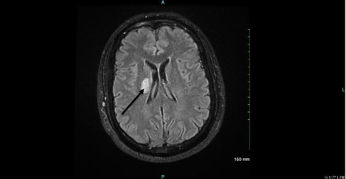
CPC-EM Capsule
What do we already know about this clinical entity?
Dysarthria-clumsy hand syndrome (DCHS) is a rare lacunar stroke syndrome presenting with unilateral facial weakness, dysarthria, and motor weakness. It is one of the least common syndromes.
What makes this presentation of disease reportable?
This case reports DCHS from a lacunar infarct involving both the caudate nucleus and posterior limb of the internal capsule, a presentation not commonly documented.
What is the major learning point?
The major learning point is that DCHS can result from lacunar infarcts involving both the caudate nucleus and internal capsule.
How might this improve emergency medicine practice?
This case can help emergency medicine physicians consider DCHS as a possible diagnosis in a patient presenting with a lacunar stroke affecting the caudate nucleus.
revealed no limiting lesions in the right or left carotid arteries. Similarly, the TTE revealed no abnormalities in the patient’s heart. The brain MRI indicated a lesion in the right caudate nucleus, extending into the posterior limb of the internal capsule on the right.
The patient’s slurred speech had resolved spontaneously by his discharge date, two days after admission. Some presenting symptoms, however, were found to persist. These symptoms included a decreased nasolabial fold on the left side, deviation of the tongue and the uvula to the right side, and left-sided motor deficits. The patient was started on atorvastatin as well as dual antiplatelet therapy, consisting of aspirin 81 mg and clopidogrel 75 mg. He was instructed to continue the dual antiplatelet therapy for 21 days.
The patient was seen two weeks later for a follow-up internal medicine appointment. He complained of difficulty maintaining focus while at work and diaphoresis. He also complained of dysarthria, possibly due to the persistence of his
Niknam et al.
Dysarthria-Clumsy Hand Syndrome in a Patient with a Caudate Nucleus Stroke
left-sided facial droop and weakness of the left hand. Physical exam indicated right tongue and uvula deviation, as well as decreased strength in the left hand. Motor strength was 4/5 in the left upper extremity, unchanged compared to strength upon discharge from the hospital; the left lower extremity strength, however, was 5/5. The remainder of the physical exam yielded no significant findings. During this appointment, the patient provided informed consent for the use of his medical information in the writing of this case report.
We describe a case of DCHS following a stroke in the caudate nucleus, with the lesion extending into the posterior limb of the internal capsule in a patient with poorly controlled hypertension and diabetes mellitus. Dysarthria-clumsy hand syndrome is the most infrequent syndrome caused by a lacunar stroke. The syndrome can consist of dysarthria and dysphagia as well as weakness of one hand. A range of lesions have been previously associated with DCHS. One study showed that most patients with DCHS have pontine infarctions.8 In contrast, a more recent study implicated lesions in the anterior limb of the internal capsule in the development of DCHS.9 Wouter et al (1999) reported two patients with DCHS, with one patient having a lesion in the putamen and the other in the caudate nucleus. Both patients in that study also exhibited involvement of white matter tracts.10 Our patient reported symptoms and observable signs aligning with previous descriptions of DCHS. However, the clinical presentation observed in the patient, in conjunction with the involvement of the caudate and the posterior limb of the internal capsule, offers valuable clinical insight into the course and spectrum of manifestation of DCHS in the literature.
The caudate nucleus, a part of the basal ganglia, is a potentially vulnerable region to ischemia and damage secondary to disconnection due to damage to white matter following a cerebrovascular accident.11. Various consequences of caudate nucleus strokes have been described in the literature. For instance, subacute strokes in the caudate region have been shown to be associated with preservation, independent of the presence of hemi-neglect.12 Lesions involving the caudate have also been shown to be associated with post-stroke dysphagia.13 Studies have also shown lesions involving the posterior limb of the internal capsule to be correlated with more severe motor deficits after a stroke.14 The present case demonstrated a lacunar infarction involving the caudate nucleus, extending into the posterior limb of the internal capsule.
Lacunar infarcts can present with a variety of symptoms that can be diverse and subtle at times. The present report showcases a rare presentation associated with lacunar infarcts due to a lesion in the caudate nucleus and the internal capsule.
This report presents important insights for the management and prognosis of patients with lacunar infarcts affecting these anatomical regions.
Patient consent has been obtained and filed for the publication of this case report.
Address for Correspondence: Janan Niknam, DO, William Carey University College of Osteopathic Medicine, 904- 1710 Bayshore Dr. Vancouver, BC. V6G3G4 Canada. Email: JNiknam560785@ student.wmcarey.edu
Conflicts of Interest: By the CPC-EM article submission agreement, all authors are required to disclose all affiliations, funding sources and financial or management relationships that could be perceived as potential sources of bias. The authors disclosed none.
Copyright: © 2025 Niknam et al. This is an open access article distributed in accordance with the terms of the Creative Commons Attribution (CC BY 4.0) License. See: http://creativecommons.org/ licenses/by/4.0/
1. Murray CJL and Lopez AD. Measuring the global burden of disease. N Engl J Med. 2013;369(5):448-57.
2. Caplan LR. Lacunar infarction and small vessel disease: pathology and pathophysiology. J Stroke. 2015;17(1):2.
3. Fisher CM. The arterial lesions underlying lacunes. Acta Neuropathol. 1968;12(1):1–15.
4. You R, McNeil JJ, O’Malley HM, et al. Risk factors for lacunar infarction syndromes. Neurology. 1995;45(8):1483–7.
5. Donnan GA and Norrving B. Lacunes and lacunar syndromes. Handb Clin Neurol. 2009;93:559–75.
6. Fisher CM. A lacunar stroke. The dysarthria-clumsy hand syndrome. Neurology. 1967;17(6):614–7.
7. Inzitari D, Eliasziw M, Sharpe BL, et al. Risk factors and outcome of patients with carotid artery stenosis presenting with lacunar stroke. North American Symptomatic Carotid Endarterectomy Trial Group. Neurology, 2000;54(3),660–6
8. Glass JD, Levey AI, Rothstein JD. The dysarthria-clumsy hand syndrome: a distinct clinical entity related to pontine infarction. Ann Neurol. 1990;27(5):487–94.
9. Arboix A, Bell Y, García-Eroles L, et al. Clinical study of 35 patients with dysarthria-clumsy hand syndrome. J Neurol Neurosurg Psychiatry. 2004;75(2):231–4.
10. Schonewille WJ, Tuhrim S, Singer MB, et al. Diffusion-weighted MRI in acute lacunar syndromes. Stroke. 1999;30(10):2066-9
11. Looi JCL, Tatham V, Kumar R, et al. Caudate nucleus volumes in
Dysarthria-Clumsy Hand Syndrome in a Patient with a Caudate Nucleus Stroke
stroke and vascular dementia. Psychiatry Res Neuroimaging. 2009;174(1):67–75.
12. Nys GMS, van Zandvoort MJE, van der Worp HB, et al.
Neuropsychological and neuroanatomical correlates of perseverative responses in subacute stroke. Brain. 2006;129(8):2148–57.
13. Im I, Jun JP, Hwang S, et al. Swallowing outcomes in patients with
subcortical stroke associated with lesions of the caudate nucleus and insula. J Int Med Res. 2018;46(9):3552
14. Puig J, Pedraza S, Blasco G, et al. Acute damage to the posterior limb of the internal capsule on diffusion tensor tractography as an early imaging predictor of motor outcome after stroke. AJNR Am J Neuroradiol. 2011;32(5):857–63.
William Osae, MD
Kevin Gurysh, MD
Section Editor: Shadi Lahham, MD
Duke University Hospital, Department of Emergency Medicine, Durham, North Carolina
Submission history: Submitted June 9, 2025; Revision received July 12, 2025; Accepted July 21, 2025
Electronically published September 23, 2025
Full text available through open access at http://escholarship.org/uc/uciem_cpcem DOI: 10.5811/cpcem.48486
Introduction: Extracorporeal membrane oxygenation (ECMO) is a life-saving intervention that has become more prevalent in the emergency department (ED) for patients with potentially reversible cardiac or pulmonary failure.
Case Report: We report a case of a young male patient who presented in septic shock and ultimately suffered a cardiac arrest in the ED. Extracorporeal membrane oxygenation was initiated after multiple rounds of cardiopulmonary resuscitation proved futile. Transthoracic echocardiography (TTE) was employed in the ED to guide ECMO cannulation, and the patient was able to make a full recovery after a one-month admission in the intensive care unit.
Conclusion: Transesophageal echocardiography and fluoroscopy are often favored over TTE for ECMO cannulation due to greater resolution of the former modalities. Transesophageal echocardiography is invasive, less accessible, and requires greater expertise. Fluoroscopy requires patients to be moved to a catheterization suite and comes with a risk of extra radiation and contrastinduced nephropathy. While the concept of TTE-guided ECMO cannulation is not especially novel, few case reports exist on its emergent deployment in the ED. Here, we discuss a unique case in which TTE proved effective for timely ECMO deployment for a critically ill ED patient. [Clin Pract Cases Emerg Med. 2025;9(4):443-446.]
Keywords: extracorporeal cardiopulmonary resuscitation; extracorporeal membrane oxygenation; transthoracic echocardiography; extracorporeal life support; case report.
Since its first successful use in 1972, extracorporeal life support (ECLS) has become increasingly common on a global scale. When used to circumvent the heart-lung circulation, such as during a coronary artery bypass graft, it is referred to as a cardiopulmonary bypass. When deployed in the emergency department (ED) or intensive care unit (ICU) to facilitate cardiac output, ventilation or oxygenation, ECLS then becomes extracorporeal membrane oxygenation (ECMO). Due to technological advancements over the last few decades, ECMO deployment has expanded from the pediatric population to adults.1 A 2015 study by Sauer et al revealed a 400% increase in ECMO use among adults from 2006 to 2011.1
Given its efficacy in stabilizing patients with reversible causes of cardiopulmonary failure, the use of ECMO has become more prevalent in EDs and even in the prehospital setting.2 When ECMO is used to augment conventional cardiopulmonary resuscitation, it is referred to as extracorporeal cardiopulmonary resuscitation (ECPR). Recent data have shown substantial benefit when ECPR is deployed early in patients with cardiac arrest.3
The most common form of ECMO cannulation is dual cannulation such as veno-venous cannulation (VV ECMO) and veno-arterial cannulation (VA ECMO). Veno-venous ECMO primarily provides pulmonary support in patients with respiratory failure such as in acute respiratory distress syndrome. Veno-arterial ECMO introduces a large right-to-left
Transthoracic Echocardiography-guided ECMO Cannulation in the ED: A Case Report
shunt and mainly assists with hemodynamic support. The most common modes of cannulation include transthoracic echocardiography (TTE), transesophageal echocardiography (TEE), and fluoroscopy. Cannulation is typically performed percutaneously, especially in the emergency setting.4 Currently, there are no agreed-upon guidelines for venous ECMO cannulation, and there are no studies demonstrating definitively that one method is superior. However, TEE and fluoroscopy are often favored over TTE for ECMO cannulation due to greater resolution of the former modalities.5
Transthoracic echocardiography can be used at the bedside to estimate cardiac ejection fraction, assess volume status by looking at the inferior vena cava, determine right ventricular strain, and assess for pericardial effusion. Transthoracic echocardiography provides a fast, noninvasive, and accessible way of locating the guidewire and cannula during ECMO, especially in the hemodynamically unstable patient undergoing ECPR. Previous studies have discussed TEE as a feasible method of ECMO cannulation in the ED, but TEE is more invasive, less accessible, and requires greater expertise.5,6 Fluoroscopy also has its drawbacks, including the need to move patients to the catheterization suite, the risk of contrast-induced nephropathy, and added radiation. Such a modality would not be ideal in the unstable patient. In this case report, we discuss a unique case in which TTE proved effective for timely ECMO deployment for a critically ill ED patient.
A 24-year-old male patient with no significant past medical history presented to the ED with altered mental status, hypotension, hypoxia, and tachycardia. The ED team began intravenous (IV) fluid resuscitation and prepared for intubation after poor response to high-flow nasal cannula. During setup for intubation, the patient went into pulseless electrical activity and then pulseless ventricular tachycardia necessitating initiation of CPR with administration of IV epinephrine. Intubation was then completed after CPR had begun. Return of spontaneous circulation was achieved after five minutes. However, the patient remained unstable, and intermittently required epinephrine to maintain blood pressure. Broad spectrum antibiotics were administered after blood cultures were obtained.
The ECMO team was contacted and arrived promptly to the ED. Given the urgency of the patient’s clinical situation, the cardiothoracic surgery and ED teams agreed to initiate VV ECMO using TTE for guidance in venous cannulation. Chest radiograph revealed bilateral pulmonary edema. The patient had frothy secretions pooling in the endotracheal tube, and TTE showed a plethoric inferior vena cava (IVC) with a dilated and hypokinetic left ventricle. The aortic outflow tract also appeared narrowed, with echogenic material visualized on the aortic valve suspicious for vegetation.
A phased-array ultrasound probe was placed in the
CPC-EM Capsule
What do we already know about this clinical entity?
Extracorporeal membrane oxygenation (ECMO) is a life-saving intervention for emergency department (ED) patients with reversible cardiac or pulmonary failure.
What makes this presentation of disease reportable?
Transthoracic echocardiography was successfully utilized to facilitate ECMO cannulation in a critically ill ED patient.
What is the major learning point?
Transthoracic echocardiography is a safe and fast method for ECMO cannulation in the emergency department.
How might this improve emergency medicine practice?
Transthoracic echocardiography is a valuable tool for ED physicians to provide timely care for critically ill patients who need ECMO deployment in the ED.
subxiphoid position to identify the IVC emptying into the right atrium. The surgeons at bedside in the ED accessed the bilateral femoral veins after making an open incision and using a seeker needle. Guidewires were subsequently placed through the seeker needles and the tract was serially dilated. The probe was maintained in the subxiphoid position as two guidewires were placed into the bilateral common femoral veins and advanced into the IVC. Ultrasound was used to confirm correct placement of two guidewires close to the caval-atrial junction from the left and right common femoral veins. The image below shows correct placement of two guidewires into the IVC.
After VV ECMO was fully deployed, the patient’s oxygen saturation improved significantly, and he was transferred to the cardiothoracic intensive care unit (ICU). Transesophageal echocardiography performed in the ICU showed that the patient had wide open aortic insufficiency secondary to multiple aortic vegetations concerning for acute endocarditis. He was taken to the operating room for vegetation removal and repair of his aortic valve and root. Although the patient sustained a large-territory unilateral middle cerebral artery infarct, his neurological status improved over the course of his hospitalization with IV
Osae et al. Transthoracic Echocardiography-guided ECMO Cannulation in the ED: A Case Report
invasive, and accessible method to guide venous cannulation during ECPR without compromising effective chest compression. Transthoracic echocardiography is not only useful for ECMO cannulation but is also reliable in patient selection for ECMO and cardiac activity detection during CPR.5 Plain radiography to confirm the tip of the guidewire has been shown to be inferior to echocardiography.9 Plain radiography provides a static view and is especially limited in patients with loss of traditional landmarks due to distorted anatomy.9
Image. Visualization of two guidewires (large arrows) in the inferior vena cava (IVC) during cannula placement for veno-venous extracorporeal membrane oxygenation.
Percutaneous extracorporeal membrane oxygenation has been used for management of persistent shock and cardiac arrest for over three decades.10 One meta-analysis showed that survival to hospital discharge was 44.9% of 675 cardiac arrest patients.11 Extracorporeal membrane oxygenation not only improves perfusion in such patients but has been shown to be superior in achieving therapeutic hypothermia in post-anoxic encephalopathy patients.12 Time to initiation of ECMO is vital in ECPR cases. The average reported time from hospital arrival to ECMO deployment ranges from 19-40 minutes, and the survival rate was about 20% when time from cardiac arrest to ECMO was less than 60 minutes.13,14
antibiotics, supportive care, and physical/occupational therapy. He was discharged home after four weeks.
This case report demonstrates the successful use of TTE for ECMO guidewire placement in a critically ill ED patient. Established modes of guiding ECMO cannulation include TEE, fluoroscopy, and plain radiograph to confirm location of guidewire tip. Transesophageal echocardiography can provide a three-dimensional view of the heart chambers and is especially beneficial when large dual-vessel cannulation is being performed at the level of the superior vena cava. Fair et al discussed TEE as a feasible method of ECMO cannulation during chest compressions in the ED.7 However, TEE requires a much higher level of expertise, is less accessible, and is more invasive compared to TTE. Fluoroscopy has also been shown to be an effective way of facilitating ECMO cannulation especially when combined with ultrasound.8 Kashiura et al reported lower rates of complications such as cannula malposition and vessel injury when fluoroscopy was combined with ultrasound as opposed to ultrasound alone.8 While fluoroscopy offers a safe way to securely dilate the cannula tract and confirm correct cannula flow, it is not without disadvantages, especially in the ECPR setting. Fluoroscopy requires transport to the catheterization suite, a time-consuming process that may not be favorable to the unstable patient. It also comes with an inherent risk of increased radiation and contrast-induced nephropathy, with the latter complication posing greater risk to the ECPR patient with poor end-organ perfusion.
Transthoracic echocardiography provides a timely, less
Transthoracic echocardiography has been widely adopted in everyday clinical practice by emergency physicians, hospitalists, and intensivists. Given its ease of accessibility compared to TEE and fluoroscopy, it serves as a powerful tool in real-time guidance for ECMO cannulation in the ED. In this case report, we describe how ECPR in the ED was used to rescue a young patient who had cardiac arrest in the setting of suspected sepsis. Time was of the essence for this patient, and TTE served as a fast and safe method to provide lifesaving care. We acknowledge that TEE is superior for cannulation at the superior vena cava level for visualizing arterial inflow. Nevertheless, in VV ECMO cannulation at the femoral vessel, TTE can be safely used in guiding venous guidewire placement into the IVC using the subxiphoid view.15
Transthoracic echocardiography is not without limitations for venous ECMO cannulation. First, the physician’s clinical competency in TTE will primarily determine which imaging modality is best for cannulation. Additionally, depending on the patient’s body habitus and anatomy, TTE may not provide the spatial resolution needed to facilitate ECMO initiation. Furthermore, assessment of the depth of the guidewire into the IVC beyond the right atrium is limited with TTE.5 In this case report, TTE was used to identify only the guidewires and not the cannula. Despite these limitations, TTE is a cost-effective and efficient tool in guiding cannulation during ECPR. Further prospective study is needed to elucidate how TTE compares with other techniques for ECMO cannulation in the ED.
Extracorporeal cardiopulmonary resuscitation in the ED has been shown to reduce mortality especially when time to initiation is within 60 minutes. This case report discusses how ECPR guided by point-of-care ultrasound (via transthoracic
Transthoracic Echocardiography-guided ECMO Cannulation in the ED: A Case Report
echocardiography) was used to rescue a young, critically ill patient in the ED. Transesophageal echocardiography and fluoroscopy are often favored over TTE for extracorporeal membrane oxygenation cannulation due to greater resolution of the former modalities. Currently, there are no agreed-upon guidelines for ECMO cannulation, and there are no studies demonstrating definitively that one method is superior. While the concept of TTE-guided ECMO cannulation is not especially novel, few case reports exist on its emergent deployment in the ED. We support the routine use of TTE in guiding timely ECMO cannulation in the ED.
The authors attest that their institution requires neither Institutional Review Board approval, nor patient consent for publication of this case report. Documentation on file.
Address for Correspondence: William Osae, MD, Duke University Hospital, Department of Emergency Medicine, 2301 Erwin Rd, Durham NC 27710. Email: wao6@duke.edu.
Conflicts of Interest: By the CPC-EM article submission agreement, all authors are required to disclose all affiliations, funding sources and financial or management relationships that could be perceived as potential sources of bias. The authors disclosed none.
Copyright: © 2025 Osae et al. This is an open access article distributed in accordance with the terms of the Creative Commons Attribution (CC BY 4.0) License. See: http://creativecommons.org/ licenses/by/4.0/
1. Sauer CM, Yuh DD, Bonde P. Extracorporeal membrane oxygenation use has Increased by 433% in adults in the United States from 2006 to 2011. ASAIO Journal. 2015;61(1):31.
2. Mosier JM, Kelsey M, Raz Y, et al. Extracorporeal membrane oxygenation (ECMO) for critically ill adults in the emergency department: history, current applications, and future directions. Crit Care. 2015;19(1):431.
3. Sakamoto T, Morimura N, Nagao K, et al. Extracorporeal cardiopulmonary resuscitation versus conventional cardiopulmonary resuscitation in adults with out-of-hospital cardiac arrest: a prospective observational study. Resuscitation. 2014;85(6):762-8.
Osae et al.
4. Rupprecht L, Lunz D, Philipp A, et al. Pitfalls in percutaneous ECMO cannulation. Heart Lung Vessel. 2015;7(4):320-326.
5. Platts DG, Sedgwick JF, Burstow DJ, et al. The role of echocardiography in the management of patients supported by extracorporeal membrane oxygenation. J Am Soc Echocardiogr. 2012;25(2):131-41.
6. Sidebotham D, Allen SJ, McGeorge A, et al. Venovenous extracorporeal membrane oxygenation in adults: practical aspects of circuits, cannulae, and procedures. J Cardiothorac Vasc Anesth. 2012;26(5):893-909.
7. Fair J, Tonna J, Ockerse P, et al. Emergency physician-performed transesophageal echocardiography for extracoporeal life support vascular cannula placement. Am J Emerg Med. 2016;34(8):1637-9.
8. Kashiura M, Sugiyama K, Tanabe T, et al. Effect of ultrasonography and fluoroscopic guidance on the incidence of complications of cannulation in extracorporeal cardiopulmonary resuscitation in out-of-hospital cardiac arrest: a retrospective observational study BMC Anesthesiol. 2017;17(1):4.
9. Thomas TH, Price R, Ramaciotti C, et al. Echocardiography, not chest radiography, for evaluation of cannula placement during pediatric extracorporeal membrane oxygenation. Pediatr Crit Care Med. 2009;10(1):56-59.
10. Mattox KL and Beall Jr AC. Resuscitation of the moribund patient using portable cardiopulmonary bypass. Ann Thorac Surg. 1976;22(5):436-42.
11. Nichol G, Karmy-Jones R, Salerno C, et al. Systematic review of percutaneous cardiopulmonary bypass for cardiac arrest or cardiogenic shock states. Resuscitation. 2006;70(3):381-94.
12. Nagao K, Hayashi N, Kanmatsuse K, et al. Cardiopulmonary cerebral resuscitation using emergency cardiopulmonary bypass, coronary reperfusion therapy and mild hypothermia in patients with cardiac arrest outside the hospital. J Am Coll Cardiol. 2000;36(3):776-83.
13. Kagawa E, Inoue I, Kawagoe T, et al. Assessment of outcomes and differences between in-and-out-of-hospital cardiac arrest patients treated with cardiopulmonary resuscitation using extracorporeal life support. Resuscitation. 2010;81(8):968-73.
14. Kim SJ, Jung JS, Park JH, et al. An optimal transition time in extracorporeal cardiopulmonary resuscitation for predicting good neurological outcome in patients with out-of-hospital cardiac arrest: a propensity-matched study. Crit Care. 2014;18(5):535.
15. De Lorenzo RA, Morris MJ, William JB, et al. Does a simple beside sonographic assessment measurement of the inferior vena cava correlate to central venous pressure? J Emerg Med 2012;42(4):429-36.
Vahe Zograbyan, MD
Andrea Hladik, MD
Lysdie Espinoza,
Manuel
Cruz, BS
BS
Section Editor: Christopher Sampson, MD
Submission history: Submitted February 9, 2025; Revision received June 24, 2025; Accepted August 7, 2025
Electronically published October 22, 2025
Full text available through open access at http://escholarship.org/uc/uciem_cpcem
DOI: 10.5811/cpcem.42509
Introduction: Ruptured aneurysms are associated with significant mortality limiting a patient’s chances of survival, making early and accurate diagnoses crucial. A commonly overlooked cause is the hepatic artery aneurysm, where most patients exhibit no distinct symptoms and detection typically occurs only after the aneurysm has ruptured. Hepatic artery aneurysms are linked with high rupturing rates resulting in substantial mortality when compared to other splanchnic artery aneurysms. Enhancing recognition and consideration of splanchnic artery aneurysms, including hepatic artery aneurysms, will increase a patient’s odds of a successful recovery. The following case report illustrates the critical nature of these cases and highlights how important early diagnosis and aggressive intervention are to prevent death once rupture of the hepatic artery aneurysm has occurred.
Case Report: A 57-year-old female presented to the emergency department brought in by helicopter for generalized chest and abdominal pain. A computed tomography angiography of the chest, abdomen, and pelvis was performed and revealed a saccular aneurysm exhibiting multiple lobes in the left hepatic artery accompanied by hemoperitoneum confirming a spontaneous rupture. As a result of the ruptured aneurysm, it was decided an immediate coil embolization was necessary. Ultimately the patient underwent a successful coil embolization and was transferred to a facility with hepatobiliary and transplant surgery capabilities. She remained stable, was extubated the following day, and did not require any additional surgeries.
Conclusion: By encompassing hepatic and associated splanchnic artery aneurysms in the diagnosis of patients with abdominal pain and signs of hemodynamic instability, physicians can improve early identification, facilitating early endovascular repair and improved patient outcomes. It is a rare diagnosis that can present with a wide range of symptoms. Currently, endovascular approaches for ruptured hepatic artery aneurysms are preferred over open surgery. [Clin Pract Cases Emerg Med. 2025;9(4):447-450.]
Keywords: hepatic artery aneurysm; coil embolization; hepatobiliary; abdominal pain; case report.
Most intra-abdominal aneurysmal ruptures have catastrophic consequences; as a result, emergency physicians often have a heightened suspicion for rupture in patients who present hypotensive with abdominal pain. A rapid but detailed
history and thorough physical exam are necessary in pointing the practitioner in the direction of expedient diagnosis and subsequent definitive treatment. While repair is clearly mandated in patients with a symptomatic aneurysm or contained rupture, asymptomatic lesions also warrant
Spontaneous Rupture of a Hepatic Artery Aneurysm: A Case Report, Against the Odds
intervention. Splanchnic artery aneurysms often have no specific symptoms and are found incidentally, making the decisions regarding treatment difficult.1 The natural clinical course of these aneurysms has not been well defined.2 Although hepatic artery aneurysms are rare, they have an overall reported rupture rate of 44%.3 Because of the high mortality associated with emergent repair, aggressive treatment is necessary in symptomatic patients. The following case report focuses on a case in which rapid detection and aggressive intervention saved the patient’s life.
A 57-year-old female with unknown past medical history presented to the emergency department (ED) via helicopter air ambulance with complaints of chest and epigastric pain since the previous night. Per emergency medical services on scene, the patient was found to be tachycardic, diaphoretic, illappearing, and somnolent; given the extended ground transport times, air ambulance transport was called. En route, given the complaint of chest pain since the previous night, there were concerns for possible cardiac etiology of the patient’s condition and she was given aspirin, nitroglycerin, and fentanyl, although there were no acute ischemic changes noted on her electrocardiogram (ECG). On arrival to the ED, the patient was pale, cool, diaphoretic, tachycardic with a heart rate of 123 beats per minute, hypotensive with a blood pressure reading of 92/75 millimeters of mercury, and oriented only to person and time. Her exam findings included midrange and reactive pupils, clear lungs, normal heart sounds, and a soft abdomen.
Bilateral, large-bore peripheral intravenous lines were established, and laboratory studies including type and screen, basic chemistries, complete blood cell count, high sensitivity troponins, lactic acid, blood cultures, arterial blood gas, pregnancy test, lipase, and urinalysis were collected. Crystalloid fluid boluses were initiated. Point-of-care blood glucose demonstrated euglycemia, and a 12-lead ECG was unremarkable for any acute ischemic changes. Bedside extended focused assessment with sonography for trauma demonstrated good lung sliding bilaterally, no cardiac tamponade, and no appreciable free fluid in the abdomen. The patient then underwent a non-contrast computed tomography (CT) of the head and CT angiography of the chest, abdomen, and pelvis to help identify the etiology of her symptoms. Differential diagnoses included aortic dissection, ruptured abdominal aortic aneurysm, massive pulmonary embolism, and septic shock. The patient was found to have a bilobed multilobulated saccular aneurysm arising from the left hepatic artery measuring 1.7 x 1.6 x 2.1 cm and 1.1 x 1.0 x 1.9 cm with hemoperitoneum consistent with rupture (Image 1).
The patient’s initial hemoglobin was measured at 11 grams/deciliter (g/dL (reference range: 12-16 g/dL); however, given hemoperitoneum and signs of shock, a single
CPC-EM Capsule
What do we already know about this clinical entity?
Splanchnic artery aneurysms tend to be asymptomatic until time of rupture and have a high mortality rate.
What makes this presentation of disease reportable?
We report the case of a patient with early signs of shock and ruptured hepatic aneurysm, whose diagnosis was made rapidly and led to emergent intervention.
What is the major learning point?
Rapid diagnosis and early resuscitation is critical. Although the patient ultimately did well, more aggressive treatment could have been initiated in this case.
How might this improve emergency medicine practice?
Given the high mortality rate, aggressive and expedient resuscitation, such as transfusion, should be considered in the treatment of splanchnic artery aneurysms.
lumen 9 French central line was established, and the patient was started on massive transfusion protocol. After consultation with interventional radiology, vascular surgery, and general surgery, the patient was taken emergently to the interventional radiology (IR) suite for angiography (Image 2) and coil embolization.
Coiling was completed with 11 coils (Image 3) to the left hepatic artery aneurysm, which appeared to control the bleeding.
However, during the IR procedure, the patient became profoundly hypotensive, necessitating treatment by the ED team with a push dose of 100 micrograms (mcg) of epinephrine to stabilize her blood pressure prior to transfusion while transporting her back to the ED. During transport her blood pressure continued to be in the range of 60/42 mm Hg with a heart rate of 147 bpm, and it was decided to give a second emergent push dose of epinephrine (100 mcg) until the blood transfusion could be started. She developed a worsening of her mental status with stupor and was eventually intubated for airway protection upon return to the ED. Given the location of the aneurysm and the lack of availability of hepatobiliary or transplant surgery specialties at our facility, the patient, once
Spontaneous Rupture of a Hepatic Artery Aneurysm: A Case Report, Against the
Image 1. Computed tomography angiography of the chest, abdomen, and pelvis of a patient with abdominal pain and hypotension, showing a bilobed, multilobulated saccular left hepatic artery aneurysm (yellow arrow) and hemoperitoneum (red arrow).
2. Angiography of the multilobulated hepatic
(red arrow) seen under fluoroscopy in a
with abdominal pain and hypotension.
stabilized, was ultimately transferred to a facility with such capabilities. If she had developed further bleeding, liver mobilization with transection of the hepatic artery would had to have been considered. However, after transfer the patient remained stable, with no further bleeding, and was extubated the following day. She did not require any further surgeries as the coils successfully embolized the ruptured hepatic artery aneurysm.
Splanchnic artery aneurysms are more often found in autopsy studies as compared with abdominal aortic aneurysms, yet aortic aneurysms are repaired much more frequently. Up to 10% of autopsy reports find splanchnic artery aneurysms, whereas only 0.5% find abdominal aortic aneurysms.4 Early recognition of splanchnic artery aneurysms is imperative as nearly one in four may be complicated by rupture with mortality approaching 80-100% after rupture.5 Among splanchnic arteries, aneurysms of the splenic artery are the most common, followed by the hepatic artery. Hepatic
artery aneurysms are found in .02-0.4% of the population and are rarely symptomatic; however, hepatic artery aneurysms have the highest rate of spontaneous rupture,6 and rupture can be catastrophic. Most of these aneurysms are found incidentally on CT; they are commonly seen in male patients in their sixties who have atherosclerosis.2
Other risk factors include connective tissue disorders, fibromuscular dysplasia, multiparity, transplant of an abdominal organ, and portal hypertension. Pseudoaneurysms have also been reported in splanchnic arteries, but they are usually secondary to trauma, infection, inflammatory changes, or chronic pancreatitis. Symptomatic hepatic artery aneurysms that are > 2 cm, expand by more than 0.5 cm in a year, or those that are found in patients with a history of vasculopathy should be considered for repair.7 Techniques for repair range from open surgical intervention to endovascular techniques with IR.1,8 While open surgical repair had been considered the gold standard, recent advances in endovascular techniques and technology in conjunction with reported faster recovery, lower cost, and shorter length of stay, the endovascular approach is now recommended by the Society of Vascular Surgery as the primary intervention. Our patient received successful endovascular coil embolization. Hepatic and other splanchnic artery aneurysms are not commonly included in the differential of abdominal pain, but it should be considered.3 Rupture of such aneurysms should be considered in all those with concomitant hemodynamic instability.
This case highlights a rare cause of abdominal pain associated with hemodynamic instability. The differential is
Spontaneous Rupture of a Hepatic Artery Aneurysm: A Case Report, Against the Odds Zograbyan et al.
Address for Correspondence: Andrea Hladik, MD, Eisenhower Emergency Medicine Residency, Eisenhower Health, 39000 Bob Hope Dr., Rancho Mirage, CA, 92270. Email: ahladikpotz@ eisenhowerhealth.org.
Conflicts of Interest: By the CPC-EM article submission agreement, all authors are required to disclose all affiliations, funding sources and financial or management relationships that could be perceived as potential sources of bias. The authors disclosed none.
Copyright: © 2025 Zograbyan et al. This is an open access article distributed in accordance with the terms of the Creative Commons Attribution (CC BY 4.0) License. See: http://creativecommons.org/ licenses/by/4.0/
1. Rosenberg A, Trebska-McGowan K, Reichman T, et al. Management of hepatic artery aneurysm: a case series. Ann Hepatobiliary Pancreat Surg. 2020;24(3):333-338.
Image 3. Embolization of the multilobulated hepatic artery aneurysm with 11 coils (red arrow) placed by interventional radiology, as seen under fluoroscopy.
wide including intra-abdominal infection and sepsis, abdominal aortic aneurysm rupture, traumatic organ damage, and aortic dissection. It is imperative to keep the differential broad and to include vascular etiologies, even those not commonly seen. Although point-of-care ultrasound was used initially in the evaluation of this patient, no intra-abdominal fluid was noted (which emphasizes the importance of operator expertise). While early recognition of hemoperitoneum may have prompted quicker evaluation by CT angiography, it would not have precluded it; the localization of the bleeding site remained imperative. The importance of interventional radiology and embolization as a stabilizing measure is paramount, especially in facilities that may lack surgical subspecialties capable of treating patients with splanchnic artery aneurysms.
The authors attest that their institution requires neither Institutional Review Board approval, nor patient consent for publication of this case report. Documentation on file.
2. Berceli SA. Hepatic and splenic artery aneurysms. Semin Vasc Surg 2005;18(4):196-201.
3. Haghighatkhah H, Sanei Taheri M, Kharazi SM, et al. Hepatic artery aneurysms as a rare but important cause of abdominal pain; a case series. Arch Acad Emerg Med. 2019;7(1):e25.
4. Pasha SF, Gloviczki P, Stanson AW, et al. Splanchnic artery aneurysms. Mayo Clin Proc. 2007;82(4):472-9.
5. Dougherty MJ, Gloviczki P, Cherry KJ Jr, et al. Hepatic artery aneurysms: evaluation and current management. Int Angiol. 1993;12(2):178-84.
6. Bueschel P, Meyer F, Weber M, et al. Rare aneurysm of the hepatic artery with overlap to the gastroduodenal artery in very uncommon coincidence with occurrence of hepatomesenteric trunk. Wien Klin Wochenschr. 2013;125(3-4):111-4. Epub 2013 Feb 19.
7. Chaer RA, Abularrage CJ, Coleman DM, et al. The Society for Vascular Surgery clinical practice guidelines on the management of visceral aneurysms. J Vasc Surg. 2020;72(1S):3S-39S. Epub 2020 Mar 20.
8. Graham I, Kanitra J, Berg R, et al. Management of a common and proper hepatic artery aneurysm. J Vasc Surg Cases Innov Tech 2021;7(2):283-285.
9. Janata F, Fezoulidis N, Barachini O, et al. Common hepatic artery aneurysm detected by 18F-FDG PET/CT imaging. Radiol Case Rep. 2021;16(11):3157-3161.
Sophia Mounce, BS*
Sharon H. Kim, PhD†
James Waymack, MD, MBA†
Section Editor: Lev Libet, MD
Southern Illinois University, School of Medicine, Springfield, Illinois
Southern Illinois University, School of Medicine, Department of Emergency Medicine, Springfield, Illinois
Submission history: Submitted May 12, 2025; Revision received July 8, 2025; Accepted August 7, 2025
Electronically published October 22, 2025
Full text available through open access at http://escholarship.org/uc/uciem_cpcem DOI: 10.5811/cpcem.47381
Introduction: Ogilvie syndrome is described as the dilation of the colon without a clear mechanical obstruction. One predisposing factor to Ogilvie syndrome is hypothyroidism. The hypothyroid state can cause decreased gastrointestinal motility; however, hypothyroidism resulting in Ogilvie syndrome is a rare complication and is referred to as myxedema ileus. A review of literature shows limited reports of this specific process and none in the emergency medicine literature.
Case Report: A 54-year-old woman with a history of hypothyroidism presented to the emergency department with three days of fatigue, generalized weakness, chills, diarrhea, and shortness of breath without chest pain or cough. Lab work showed high levels of ultra thyroid-stimulating hormone and decreased thyroid hormone levels. A computed tomography angiography of the chest, abdomen and pelvis showed multiple dilated loops of large bowel. Ultimately, she was diagnosed with pseudoobstruction (Ogilvie syndrome) secondary to myxedema ileus.
Conclusion: Ogilvie syndrome in the setting of myxedema ileus is a serious complication that may occur in patients who are in a severe hypothyroid state. It is important for emergency physicians to consider hypothyroidism as a potential cause of intestinal pseudo-obstruction. [Clin Pract Cases Emerg Med. 2025;9(4):451-453.]
Keywords: Ogilvie syndrome; myxedema ileus; hypothyroidism; case report.
Colonic pseudo-obstruction, also known as Ogilvie syndrome, is dilation of the colon without a clear mechanical obstruction that was first described in 1948 by Sir Heneage Ogilvie. The exact pathophysiology of this condition is not fully understood; however, it is thought to be due to imbalance in autonomic innervation.1 There are many potential causes and predisposing factors to Ogilvie syndrome. In a retrospective study of 400 patients with Ogilvie syndrome, the predisposing factors identified included infection, trauma, surgery, and miscellaneous medical conditions (metabolic, cancer, respiratory failure).2 Another potential metabolic predisposing factor includes hypothyroidism. Severe hypothyroidism can cause decreased gastrointestinal motility resulting in Ogilvie syndrome, which is a rare complication
and is sometimes referred to as myxedema ileus.3 We report a case of Ogilvie syndrome secondary to myxedema ileus in a 54-year-old female with history of hypothyroidism. A review of the literature shows few case reports of this specific process and none in the emergency medicine literature.
A 54-year-old woman with a history of hypothyroidism, diabetes mellitus type two, chronic obstructive pulmonary disease, asthma, anxiety, and depression presented to the emergency department (ED) via emergency medical services with three days of fatigue, generalized weakness, chills, and shortness of breath without chest pain or cough. Her symptoms had been progressively worsening, and she stated that she felt as if she could not move her body. She also noted diarrhea without
abdominal pain, melena, or hematochezia. Prior to arrival, the patient’s daughter noted that the patient appeared paler and had dyspnea and a syncopal episode. The patient reported two missed doses of levothyroxine. On examination, her temperature was 36.4 °C, blood pressure 106/64 millimeters of mercury (mm Hg), heart rate 62 beats per minute, and respiratory rate 16 breaths per minute with oxygen saturation of 96% on room air.
On physical examination, the patient appeared drowsy. She was sitting with her eyes closed and was slow to answer questions, without any obvious distress. Her abdomen was soft, non-distended, and with normal bowel sounds. She noted diffuse abdominal discomfort to palpation; however, she stated this was chronic for her. She was oriented to person, place, time, and situation without any focal neurological deficits. Her strength was 3/5 throughout all extremities. The extremities were without edema.
Laboratory evaluation by point-of-care venous blood gas showed a pH of 7.28 (reference range: 7.35-7.45), partial pressure of carbon dioxide level of 60 mm Hg (41-51 mm Hg), bicarbonate level of 28.2 millimoles per liter (mmol/L) (24.0-28.0 mmol/L). Complete blood cell count and comprehensive metabolic profile were grossly unremarkable, and creatine kinase level was 333 international units per liter (IU/L) (30-223 IU/L). Thyroid studies showed an elevated thyroid-stimulating hormone (TSH) of 196.80 IU/mL (0.45-5.33 IU/mL). Free triiodothyronine (T3)/thyroxine (T4) and total T3/T4 were not resulted during the patient’s ED stay but were found to be significantly decreased upon admission: free T3 1.2 picograms per milliliter (pg/mL) (2.4-4.4 pg/mL), total T3 < 25 nanograms per deciliter (ng/ dL) (87-178 ng/dL), free T4 0.2 ng/dL (refe0.5-1.3 ng/dL) and total T4 < 1.0 micrograms/dL (mcg/dL) (6.1-12.2 mcg/ dL). Cortisol level was 1.7 mcg/dL (8.7-22.4 mcg/dL).
A computed tomography angiography (CTA) of the chest with routine abdomen and pelvis showed multiple dilated loops of large bowel and a few loops of distal small bowel with air-fluid levels (Images 1 and 2). Given this patient’s history, TSH level, and dilated bowel loops of large bowel on the CTA without a transition point, a diagnosis of pseudoobstruction (Ogilvie syndrome) secondary to myxedema ileus was made. The few loops of dilated small bowel present were presumed to be sequelae of the large bowel dilatation.
The patient remained stable in the ED and was admitted to the intensive care unit (ICU). Initial treatment in the ICU included 100 mg of intravenous (IV) hydrocortisone and 200 mcg of IV levothyroxine with 12.5 mcg of liothyronine. Nasogastric tube placement was deferred due to lack of vomiting, and the patient was started on a liquid diet while her hypothyroid condition was treated. The following day her symptoms and mental status improved, and she was downgraded to the intermediate care unit. At that time, she was started on a maintenance weight-based dose of IV
CPC-EM Capsule
What do we already know about this clinical entity?
Colonic pseudo-obstruction (Ogilvie syndrome) is colon dilation without mechanical obstruction, often after surgery, trauma, or severe illness.
What makes this presentation of disease reportable?
Hypothyroidism leading to Ogilvie syndrome is a rare complication referred to as myxedema ileus.
What is the major learning point?
Myxedema ileus should be suspected in patients presenting with Ogilvie syndrome and signs of severe hypothyroidism.
How might this improve emergency medicine practice?
Early recognition of myxedema ileus allows prompt initiation of thyroid hormone therapy, which can reverse colonic pseudo-obstruction and avoid complications.
Image 1. Computed tomography of the abdomen and pelvis (axial) demonstrating dilated large bowel/ascending colon (white arrow) with air-fluid levels (yellow arrow).
Image 2. Computed tomography of the abdomen and pelvis (coronal) demonstrating dilated, fluid-filled large bowel/ ascending colon (arrow) without obvious obstruction or transition point.
Syndrome in the Setting of
as abdominal compartment syndrome and bowel perforation.4,7 This case highlights the importance for emergency physicians to consider myxedema ileus as a potential diagnosis in patients with Ogilvie syndrome or a history of hypothyroidism.
Myxedema ileus, pseudo-obstruction (Ogilvie syndrome), is a serious complication that may occur in patients who are in a hypothyroid state. It is important for emergency physicians to consider hypothyroidism as a potential cause of intestinal pseudo-obstruction. The treatment for these patients includes correction of the underlying endocrine pathophysiology and supportive care.
The authors attest that their institution requires neither Institutional Review Board approval, nor patient consent for publication of this case report. Documentation on file.
Address for Correspondence: James Waymack, MD, MBA, Southern Illinois University, School of Medicine, Department of Emergency Medicine, 701 North First Street, Springfield, IL 62781. E-mail: jwaymack@siumed.edu
Conflicts of Interest: By the CPC-EM article submission agreement, all authors are required to disclose all affiliations, funding sources and financial or management relationships that could be perceived as potential sources of bias. The authors disclosed none.
Ileus: A Case Report levothyroxine. On day three of the patient’s hospitalization, her diet was advanced, and she was transitioned to oral medications. She was discharged one day later.
Copyright: © 2025 Mounce et al. This is an open access article distributed in accordance with the terms of the Creative Commons Attribution (CC BY 4.0) License. See: http://creativecommons.org/ licenses/by/4.0/
Myxedema ileus due to hypothyroidism is a rare but serious cause of Ogilvie syndrome. There are few case reports in the literature.4-8 Classically, Ogilvie syndrome presents with marked constipation or obstipation. We believe the patient’s diarrhea was related to overflow in the setting of the acute colonic pseudo-obstruction as evidenced by CT findings of large-bowel dilatation without transition point or other signs of obstruction. While our patient stated the abdominal tenderness found on exam was chronic, it is likely her pain was related to her diagnosis of Ogilvie syndrome.
The current standard of care for myxedema ileus is conservative management with nasogastric tube decompression, bowel rest, and resumption or commencement of levothyroxine. Surgical intervention should be considered in cases with cecal distention of more than 12 cm, bowel ischemia, or perforation.4 While this is an uncommon complication, it should be considered as a differential diagnosis in patients with hypothyroidism who are presenting with symptoms consistent with an ileus. If myxedema ileus is left untreated, it can lead to more serious consequences such
1. Saunders MD. Acute colonic pseudo-obstruction. Best Pract Res Clin Gastroenterol. 2007;21(4):671–687.
2. Vanek VW and Al-Salti M. Acute pseudo-obstruction of the colon (Ogilvie’s syndrome). Dis Colon Rectum. 1986;29(3):203-210.
3. Moss AA and Goldberg HI. Intestinal pseudo-obstruction. CRC Crit Rev Radiol Sci.1972;3(3):363–387.
4. Thakur V, Gupta JK, Gupta A. Abdominal compartment syndrome secondary to myxedema ileus. Tzu Chi Med J. 2019;32(2):219-221.
5. Peña-Vélez R, Reynoso-Castorena JM, Espinosa-Flores L, et al. Intestinal pseudo-obstruction: a rare presentation of congenital hypothyroidism. Rev Gastroenterol Mex (Engl Ed). 2022;87(4):499-501.
6. Shera IA, Vyas A, Bhat MS, et al. Unusual case of Hashimoto’s encephalopathy and pseudo-obstruction in a patient with undiagnosed hypothyroidism: a case report. J Med Case Rep. 2014;8:296.
7. Zachariah SK and Raja N. Spontaneous perforation of the colon and hypothyroidism: report of a case and review of literature. Gastroenterology Res. 2010;3(3):147-149.
Matthew E Mollman, MD
Lauren Mays, MD
Section Editor: Lev Libet, MD
Saint Louis University School of Medicine, Department of Surgery, Division of Emergency Medicine, St. Louis, Missouri
Submission history: Submitted April 11, 2025; Revision received June 11, 2025; Accepted July 28, 2025
Electronically published October 22, 2025
Full text available through open access at http://escholarship.org/uc/uciem_cpcem DOI: 10.5811/cpcem.47181
Introduction: Subclavian artery pseudoaneurysms are a rare complication of blunt thoracic trauma with high mortality and incidence of long-term disability.
Case Report: We describe a 49-year-old female who suffered a midshaft clavicle fracture after a motorcycle collision who presented five weeks later with right arm weakness, paresthesias, and persistent clavicle pain and swelling. She was diagnosed with a subclavian artery pseudoaneurysm on point-of-care ultrasound performed in the emergency department, which was confirmed with computed tomography angiography. She underwent endovascular stenting but continued to suffer from long-term neurologic deficits related to her condition.
Conclusion: This case underscores that the diagnosis of subclavian artery pseudoaneurysm requires a high index of suspicion. In addition, the case also highlights the utility of point-of-care ultrasound as a modality that can assist in arriving at the diagnosis. [Clin Pract Cases Emerg Med. 2025;9(4):454-457.]
Keywords: subclavian artery pseudoaneurysm; point-of-care ultrasound; computed tomography angiography; blunt trauma; case report.
Traumatic injuries account for approximately 17% of all emergency department (ED) visits annually and are the third leading cause of death when combined for all ages.1,2 It is estimated that up to 12.8% of these patients suffer a posttraumatic complication, representing significant morbidity.3 Subclavian artery pseudoaneurysm is rare, occurring in approximately 1 in 12,500 admitted trauma patients.4 This injury is more commonly a result of penetrating trauma; only 25% are secondary to blunt trauma.5 Despite successful treatment of the subclavian artery pseudoaneurysm, 50% of patients will continue to experience long-term neurologic deficits.6 The diagnosis of SAP is most often made either intraoperatively or with computed tomography angiography; rarely is it first discovered on ultrasound. We report the case of a blunt traumatic subclavian artery pseudoaneurysm with delayed presentation that was initially identified by point-ofcare ultrasound (POCUS) in the ED.
A 49-year-old female with a history of polysubstance use disorder, including intravenous heroin, presented to the ED five weeks after a motorcycle accident where she had sustained a direct blow to her right shoulder. She complained of right upper extremity weakness, numbness, and pain. She also noted swelling around her right clavicle. She was not seen at the time of the accident; however, she presented to an outside ED one week after the injury. At the outside facility, radiographs were performed of her chest and shoulder, and she was diagnosed with right 4th-8th rib fractures and a midshaft right clavicle fracture. No other imaging was performed at that time, and she was discharged with a sling and provided pain medication. When she presented to our ED for ongoing pain in her upper chest/right shoulder with radiation down the arm and progressive swelling over the clavicle, she also had weakness to her right upper extremity and tingling in her fingers. She denied any subsequent trauma but admitted to not wearing her sling as instructed.
et al. Delayed Presentation of Subclavian Artery Pseudoaneurysm Following Blunt Thoracic Trauma
On examination, the patient appeared underweight but in no acute distress. There was tenderness and swelling over the right mid clavicle without overlying skin changes. The right upper extremity showed no deformities or external signs of injury and had tenderness to palpation from the upper arm down through the hand. Pulses were 2+ and symmetric in the radial and ulnar arteries bilaterally, but capillary refill was delayed in the right hand compared to the left. Wrist flexion and extension and hand grip strength were 4/5 on the right compared to 5/5 on the left, and sensation to light touch along the right posterior forearm, palm, and palmar aspect of all five digits and fingertips was diminished. Radiographs were repeated and redemonstrated her previously identified fractures.
A POCUS of the clavicle swelling revealed a circular mass inferior to the clavicle with surrounding Doppler flow, raising the likelihood of a vascular etiology (Image 1).
Computed tomography angiography (CTA) of the chest and neck confirmed and better characterized the pseudoaneurysm: a 5.5-cm partially thrombosed right subclavian artery pseudoaneurysm with extension above the clavicle (Image 2).
After consultation with vascular surgery, the patient underwent endovascular repair with stent placement. Postoperatively, she reported improvement in her paresthesias and regained full strength in her right arm and hand. The patient was discharged the following day on dual-antiplatelet therapy with aspirin and clopidogrel.
CPC-EM Capsule
What do we already know about this clinical entity?
Traumatic subclavian pseudoaneurysms are potentially life-threatening injuries that may cause long-term neurovascular complications.
What makes this presentation of disease reportable?
The delayed presentation in this case with soft findings of vascular injury highlights the importance of maintaining a high level of suspicion.
What is the major learning point?
Point-of-care ultrasound is a quick, non-invasive diagnostic tool that can aid in the detection of a traumatic subclavian pseudoaneurysm.
How might this improve emergency medicine practice?
This case highlights a rare but serious complication of a common thoracic injury and the use of point-ofcare ultrasound to aid in the diagnosis.
We present a rare case of a blunt trauma subclavian pseudoaneurysm with delayed presentation. The diagnosis of a subclavian pseudoaneurysm is time sensitive due to the risk of rupture that may result in catastrophic hemorrhage and death. Rupture and hemorrhage occurs in approximately 10% of untreated subclavian artery pseudoaneurysms.7 Other potential complications arising from delayed diagnosis include thromboembolic events, limb ischemia, and neurologic deficits.8 Although blunt subclavian artery injuries are rare, accounting for only 2% of all blunt vascular injuries, the mortality is high.8,9 Only about 15% of patients with blunt subclavian artery injuries arrive alive to the hospital, and of those the mortality remains high at up to 30%.5,9 As with other subclavian artery injuries, mortality from subclavian artery pseudoaneurysms is due to exsanguination. Hard signs of vascular injury such as absent pulses, a rapidly expanding hematoma, palpable thrill, or active pulsatile bleeding are likely to prompt vascular imaging. This imaging often aids in the rapid diagnosis of a subclavian artery injury. However, soft signs such as a peripheral nerve deficit or a small nonpulsatile hematoma over an artery, must be considered as well.
In our case, the patient did not demonstrate hard signs of vascular injury but did have soft signs including a new
neurologic deficit in her right upper extremity and a small nonpulsatile hematoma over the site of her arterial injury. The delayed presentation of a subclavian pseudoaneurysm secondary to blunt trauma is even more uncommon and thus requires a high index of suspicion. Presenting symptoms often involve swelling and pain at the site of injury, chest pain and/or referred pain to the ipsilateral shoulder. As in this case, during the patient’s first presentation, those symptoms may be initially falsely attributed solely to bony injuries to the overlying clavicle and rib fractures if radiograph is the only imaging modality used. In blunt chest trauma presenting several days after the injury, clinicians may assume that radiograph imaging is sufficient to identify significant non-bony traumatic injuries of the chest such as pneumothorax, hemothorax, or pulmonary contusions. This case demonstrates the importance of considering less obvious traumatic thoracic injuries, especially in the context of a high-risk mechanism, and obtaining advanced imaging such as CTA.
Other symptoms of a subclavian artery pseudoaneurysm may include upper extremity paresthesias, motor weakness, and even paralysis secondary to either claudication or compression of the brachial plexus. These symptoms could be erroneously attributed to either a cervical spine or cerebral process prompting CT of the head/neck that might fail to diagnose the true pathology. The diagnosis is typically made by CTA of the chest due to its wide availability and accuracy. This case is the first reported use of POCUS to diagnose a subclavian artery pseudoaneurysm in the ED. The findings on
POCUS prompted the decision to obtain a CTA of the chest to further evaluate the patient’s symptoms. Ultrasound can be performed immediately at the bedside, is non-invasive, and can serve to expedite further diagnostic studies and subsequent consultation with vascular surgery.
Endovascular repair with stent placement is now the preferred treatment option. Open vascular repair is less favorable as access to the subclavian artery is difficult. Despite successful treatment, many patients suffer long-term neurologic symptoms caused by brachial plexus compression by the pseudoaneurysm.9 Nerve damage tends to be worse in those who have a delay in diagnosis and emphasizes the need for prompt diagnosis.
Blunt traumatic subclavian artery pseudoaneurysms carry a high rate of mortality and morbidity and require a high index of suspicion in the setting of significant blunt chest trauma. Point-of-care ultrasound can provide rapid, non-invasive identification of subclavian artery injury and pseudoaneurysm formation. Timely diagnosis is critical to reducing the associated morbidity. Despite treatment, some patients will suffer chronic neurologic sequelae resulting from secondary brachial plexus injury.
The authors attest that their institution requires neither Institutional Review Board approval, nor patient consent for publication of this case report. Documentation on file.
Address for Correspondence: Matthew E Mollman, MD, Saint Louis University School of Medicine, Department of Surgery, Division of Emergency Medicine, 3691 Rutger St. St. Louis, MO 63110. Email: matt.mollman@ssmhealth.com
Conflicts of Interest: By the CPC-EM article submission agreement, all authors are required to disclose all affiliations, funding sources and financial or management relationships that could be perceived as potential sources of bias. The authors disclosed none.
Copyright: © 2025 Mollman et al. This is an open access article distributed in accordance with the terms of the Creative Commons Attribution (CC BY 4.0) License. See: http://creativecommons.org/ licenses/by/4.0/
1. Weiss AJ, Reid LD, Barrett ML. Overview of Emergency Department Visits Related to Injuries, by Cause of Injury, 2017. Accessed March 2, 2025. https://hcup-us.ahrq.gov/reports/statbriefs/sb266-InjuriesCauses-ED-Visits-2017.pdf
2. Murphy SL, Kochanek KD, Xu JQ, et al. Mortality in the United States, 2023 Key Findings Data from the National Vital Statistics
Mollman et al. Delayed Presentation of Subclavian Artery Pseudoaneurysm Following Blunt Thoracic Trauma System; 2024 Accessed March 2, 2025. https://www.cdc.gov/nchs/ data/databriefs/db521.pdf
3. Jakobsen RK, Bonde A, Sillesen M. Assessment of post-trauma complications in eight million trauma cases over a decade in the USA. Trauma Surg Acute Care Open. 2021;6(1):e000667.
4. Wojtyś, M.E, Skórka, P, Kordykiewicz, D, et al. Post-traumatic left subclavian artery pseudoaneurysm secondary to clavicular fracture: a case report and literature review. Biomedicines. 2025;13(1):187-187.
5. Francis D, Kumar M, Singh M, et al. Endovascular management of traumatic pseudoaneurysm of left subclavian artery: a case report. Radiol Case Rep. 2023;5;18(11):4066-4070.
6. Shaw AD, Milne AA, Christie J, et al. Vascular trauma of the upper limb and associated nerve injuries. Injury. 1995;26(8):515-8.
7. Jaiswal LS, Prasad JN, Maharjan R, Pandit N. Giant pseudoaneurysm of subclavian artery after blunt chest trauma. J Vasc Surg Cases Innov Tech. 2018;4(3):220-222.
8. Moey MYY, Prabhudesai V, Greco E, et al. Angio-seal closure for traumatic left subclavian artery pseudoaneurysm: a case report. J Soc Cardiovasc Angiogr Interv. 2024;23;3(6):102021.
9. Elkbuli A, Shaikh S, McKenney M, et al. Subclavian artery avulsion following blunt trauma: a case report and literature review. Int J Surg Case Rep. 2019;61:157-160.
Vijay Chandramaniya, MRCEM*
Sanjay Mehta, MD*
Nandkishore Kapadia, MCh†‡ Harikrishnan Chandramohanan, DNB§ Jasmin Custodio, MD¶
Kokilaben Dhirubhai Ambani Hospital and Medical Research Institute, Department of Emergency Medicine, Mumbai, India
Criticare Hospital, Department of Cardiovascular and Thoracic Surgery, Mumbai, India
Kiran Hospital, Heart-Lung Transplant Unit, Surat, Gujarat, India
Krishna Vishwa Vidyapeeth, Department of Emergency Medicine, Karad, Maharashtra, India
Kern Medical, Department of Emergency Medicine, Bakersfield, California
Section Editor: Steven Walsh, MD
Submission history: Submitted June 12, 2025; Revision received July 10, 2025; Accepted July 28, 2025
Electronically published October 21, 2025
Full text available through open access at http://escholarship.org/uc/uciem_cpcem DOI: 10.5811/cpcem.47929
Introduction: This case is unique in that it documents isolated, painless vaginal bleeding as the sole presenting symptom of a Stanford type A aortic dissection (STAAD), a presentation not previously reported. It adds to the literature by expanding the spectrum of atypical aortic dissection presentations and underscores the need to consider this diagnosis in elderly patients with vascular risk factors, even when they present with non-classical symptoms such as unexplained bleeding.
Case Report: We present a novel case of STAAD in a 72-year-old woman with a history of hypertension, dyslipidaemia, prior hysterectomy, and cholecystectomy. Her primary complaint was a single, transient episode of painless vaginal bleeding. Notable clinical findings included a diminished right radial pulse, a significant inter-arm blood pressure discrepancy, and unremarkable systemic and vaginal examinations. Given these findings, further evaluation was pursued. Computed tomography aortography revealed a STAAD extending from the aortic arch to the bifurcation, involving the left internal iliac artery and a vaginal arterial branch. The patient underwent emergent surgical repair and was discharged in good condition on hospital day 11. At her most recent follow-up, three years postevent, she remained clinically stable with no recurrence.
Conclusion: Isolated painless vaginal bleeding, although uncommon, may indicate lifethreatening pathology. Subtle signs, such as inter-arm blood pressure discrepancy, can offer critical diagnostic clues, underscoring the importance of comprehensive evaluation in atypical emergency presentations. [Clin Pract Cases Emerg Med. 2025;9(4):458-462.]
Keywords: Stanford type A aortic dissection; aortic dissection; vaginal bleeding; case report
Acute aortic dissection remains a diagnostic challenge in emergency medicine, particularly when presentations deviate from classical patterns. While sudden, severe chest, back, or abdominal pain is the hallmark symptom, up to 6% of patients—more often older women—present painlessly, leading to delays in recognition and intervention.¹⁻³ Known atypical symptoms include neurologic deficits, syncope, and
even gastrointestinal complaints, but isolated gynaecologic bleeding has not been previously documented. This diagnostic gap has implications for population-level outcomes, as early surgical management significantly reduces mortality.⁴
We present a novel case of Stanford type A aortic dissection (STAAD) in a 72-year-old woman whose only symptom was a single episode of painless vaginal bleeding. No previous reports in the literature describe this form of presentation. The case
underscores how sex- and age-related atypical symptomatology can obscure time-critical diagnoses. It further highlights the importance of integrating subtle clinical cues, such as pulse deficits or blood pressure differentials, into a broad diagnostic framework, especially for high-risk patients. Recognizing such outliers is vital to ensuring equitable, life-saving care across diverse patient populations.
A 72-year-old, moderately built female presented to the emergency department (ED) with a single, transient episode of painless vaginal bleeding that had occurred earlier that morning while bathing. She appeared mildly anxious, as she had not experienced any vaginal bleeding since undergoing a hysterectomy several years prior. She denied chest pain, back pain, abdominal or pelvic pain, as well as any history of coagulopathy, substance use, malignancy, or recent unintentional weight loss. She also reported no fever, urinary symptoms, vaginal rash or discharge, recent trauma, or haemorrhoids. Her medical history was significant for hypertension and dyslipidaemia. Surgical history included a hysterectomy for uterine fibroids and a cholecystectomy.
On physical examination, the patient was hemodynamically stable, with a respiratory rate of 16 breaths per minute and an oxygen saturation of 96% on room air. Her heart rate was 102 beats per minute. Cardiac examination revealed normal heart sounds without murmurs. Respiratory, neurological, abdominal, and pelvic examinations were unremarkable. All extremities were warm and well-perfused, with no evidence of cyanosis. Peripheral pulses were well-felt and symmetrical, except for a comparatively diminished right radial pulse, which was identified during routine pulse assessment performed on all patients in our ED, regardless of presenting complaint. Blood pressure in the right arm was 86/60 millimetres of mercury (mm Hg), a finding not explained by the patient’s single, transient episode of minimal vaginal bleeding. Given the unexplained hypotension and the subtle discrepancy in pulse volume between the arms, blood pressure was also measured in the left arm. A significant inter-arm blood pressure difference (reference range: <10 mm Hg) was noted: 86/60 mm Hg in the right arm and 160/80 mm Hg in the left—an abnormality that had not been previously reported or known to the patient. A vaginal examination performed by the gynaecologist was unremarkable and did not reveal any identifiable source of bleeding.
The diagnostic assessment began with a physical examination, which revealed a diminished right radial pulse and a significant inter-arm blood pressure discrepancy. The hypotension in the right arm was disproportionate to the transient, minimal vaginal bleeding and raised concern for an underlying alternative pathology. Despite the absence of chest, back, or abdominal pain, the findings raised concern for a vascular aetiology, particularly involving the aorta or peripheral vessels. Laboratory tests were unremarkable and did not suggest
CPC-EM Capsule
What do we already know about this clinical entity?
Aortic dissection may present without pain; atypical cases in elderly women often face delayed diagnosis and high mortality.
What makes this presentation of disease reportable?
Isolated painless vaginal bleeding as the sole manifestation of Stanford type A aortic dissection is novel and unreported in the medical literature.
What is the major learning point?
Subtle findings such as inter-arm blood pressure difference can uncover life-threatening vascular disease, even without classic symptoms.
How might this improve emergency medicine practice?
Maintaining a high suspicion for aortic dissection in atypical cases can expedite diagnosis and enable timely, life-saving treatment.
infection or coagulopathy. The patient’s medical history, along with a non-contributory vaginal examination performed by the gynaecologist, made local gynaecologic causes of vaginal bleeding less likely. Given the atypical presentation—painless vaginal bleeding in an elderly woman with cardiovascular risk factors—combined with abnormal vital signs, advanced imaging was pursued in consultation with vascular surgery. Computed tomography (CT) aortography confirmed a STAAD extending from the aortic arch to the aortic bifurcation, with further extension into the left internal iliac artery and one of the vaginal arterial branches (Image).
The primary diagnostic challenge was the absence of classic symptoms of aortic dissection, a factor that can contribute to delayed diagnosis, particularly in older women. The source of bleeding was also highly atypical, initially suggesting differential diagnoses such as gynaecologic bleeding of local origin (eg, vaginal cuff granulation or neoplasia), coagulopathy, or vascular malformations. However, these possibilities were inconsistent with the patient’s medical history, stable laboratory results, and normal physical examination findings. In contrast, careful attention to the vascular exam guided the diagnostic approach toward the correct diagnosis. Following early recognition, the prognosis improved significantly with prompt surgical intervention.
Image. Axial (A), sagittal (B), and coronal (C, D) computed tomography aortography views demonstrating Stanford type A aortic dissection. Panel A shows a dissection flap (arrow) in the ascending aorta and aortic arch. Panel B reveals the dissection flap (arrow) extending through the thoracic aorta in the sagittal view Panel C shows involvement of the abdominal aorta (arrow), with propagation of the dissection flap inferiorly into the left common iliac artery. Panel D shows the origin of the dissection flap (arrow) in the proximal ascending aorta in coronal reconstruction.
blood pressure discrepancy—combined with the lack of an identifiable alternative source for the vaginal bleeding— prompted a high index of suspicion and led to appropriate diagnostic imaging. (We were unable to definitively confirm the vascular source of the vaginal bleeding due to the urgency of surgical intervention and the absence of tissue-level vascular mapping.)
Aortic dissection is a time-sensitive vascular emergency with high early mortality. Without treatment, AAD carries an estimated mortality rate of approximately 50% within the first 48 hours.1 However, timely surgical intervention significantly reduces this risk. Data from the International Registry of Acute Aortic Dissection (IRAD) demonstrated a 48-hour mortality of only 4.4% with prompt surgery.4 Historically, atypical presentations of STAAD have contributed to delayed diagnoses. According to Harris et al, AAD is less frequently observed in older females and is more likely to present with nonspecific or neurological symptoms.5
Aortic dissection, particularly type A dissections, carries a high mortality rate if left untreated, but early diagnosis and repair substantially improve outcomes.
The patient underwent emergent surgical repair, including reconstruction of the sinus of Valsalva, ascending aorta replacement, and hemi-arch repair. Her postoperative course was notable for a left-sided pneumothorax on day 3, which was promptly managed with intercostal drain placement and resolved without further complication. She was discharged in stable condition on postoperative day 11. Twenty days after discharge, she was hospitalized for new-onset atrial fibrillation, which was managed conservatively with oral anti-arrhythmic medications and sinus rhythm maintained without recurrence. At her most recent follow-up, three years after the event, the patient remained clinically stable without new symptoms, complications, or recurrence.
The timely recognition of an atypical presentation of STAAD enabled life-saving intervention. Despite the absence of classic symptoms, the presence of a significant inter-arm
Our case contributes to the expanding literature on unusual manifestations of STAAD. While previous reports have described atypical presentations of AAD—including painless paralysis,8 sore throat,9 testicular pain,11 symptoms mimicking ureteral calculus,10 pulmonary embolism12 or bowel obstruction,14 and an elderly, hypertensive female presenting with profound weakness and hypotension15—vaginal bleeding has not been previously documented as a presenting feature. In this case, CT aortography revealed dissection extending into the left internal iliac artery and a vaginal arterial branch, offering a plausible anatomical basis for the bleeding (Figure). Although a significant systolic blood pressure differential between arms is a known sign of AAD, its diagnostic accuracy is limited.6 Nonetheless, in our patient, this finding prompted further imaging that confirmed the diagnosis. As IRAD data and subsequent studies have shown, early recognition and intervention can drastically alter the prognosis of STAAD.3,4,7
In this case presentation, the aortic dissection was the likely cause of the patient’s vaginal bleeding, rather than a coincidental finding, based on several key factors. First, a thorough pelvic examination performed by a gynaecologist was entirely normal, with a recommendation to investigate causes beyond the local genital tract. Second, the patient had no prior medical history suggestive of a cause for vaginal bleeding, and her laboratory parameters were within normal limits. Third, a newly identified significant inter-arm blood pressure discrepancy, in the context of long-standing hypertension, raised suspicion for a vascular aetiology. The aortic dissection may have initiated atypically and progressed in a manner that involved extension of the intimal flap into a branch supplying the vaginal vasculature. This could have led to a minor arterial or pseudo-aneurysmal rupture, followed by localized thrombosis, resulting in a single episode of minimal vaginal bleeding.
et al. A Novel Presentation of Stanford Type A Aortic Dissection with Vaginal Bleeding: A Case Report
Patient consent has been obtained and filed for the publication of this case report.
Address for Correspondence: Chandramaniya VD, MRCEM, Consultant, Department of Emergency Medicine Kokilaben Dhirubhai Ambani Hospital and Medical Research Institute, Mumbai, Maharashtra 400053, India. Email: Vijay. Chandramaniya@kokilabenhospitals.com.
Conflicts of Interest: By the CPC-EM article submission agreement, all authors are required to disclose all affiliations, funding sources and financial or management relationships that could be perceived as potential sources of bias. The authors disclosed none.
Copyright: © 2025 Chandramaniya et al. This is an open access article distributed in accordance with the terms of the Creative Commons Attribution (CC BY 4.0) License. See: http:// creativecommons.org/licenses/by/4.0/
1. Levy D, Sharma S, Grigorova Y, et al. Aortic dissection. In: StatPearls [Internet]. Treasure Island, FL: StatPearls Publishing; January 2025–. Updated October 6, 2024. Available from: https://www.ncbi.nlm.nih. gov/books/NBK441989/. Accessed January 1, 2025.
Figure. Focused illustration of vaginal vascular anatomy. The diagram illustrates the descending aorta branching into the common iliac, internal iliac, and subsequently the vaginal artery, which supplies the vaginal canal. This schematic illustrates the anatomical context of potential vascular involvement in cases of aortic dissection presenting with vaginal bleeding.
The patient expressed gratitude for the timely and thorough care she received. She was surprised that a serious cardiac condition could present as vaginal bleeding but felt reassured by the emergency team’s evaluation. Despite a challenging recovery, she remains committed to ongoing treatment and follow up.
The authors thank Dr. Vaishali Joshi for her patient evaluation and expert gynaecology consultation, and Dr. Raghuram Sekhar for his guidance on selecting appropriate radiologic imaging to assess the vascular aetiology. We also acknowledge the radiology and cardiothoracic surgery teams at Kokilaben Dhirubhai Ambani Hospital for their timely expertise and collaborative care in managing this patient.
2. Fatima S and Sharma K. Painless aortic dissection—diagnostic dilemma with fatal outcomes: What do we learn? J Investig Med High Impact Case Rep. 2017;5(3):2324709617721252.
3. Pape LA, Awais M, Woznicki EM, et al. Presentation, diagnosis, and outcomes of acute aortic dissection: 17-year trends from the International Registry of Acute Aortic Dissection. J Am Coll Cardiol. 2015;66(4):350–8.
4. Harris KM, Nienaber CA, Peterson MD, et al. Early mortality in type A acute aortic dissection: insights from the International Registry of Acute Aortic Dissection. JAMA Cardiol. 2022;7(10):1009–15.
5. Nienaber CA, Fattori R, Mehta RH, et al. Gender-related differences in acute aortic dissection: the International Registry of Acute Aortic Dissection. Circulation. 2004;109(24):3014–21.
6. Um SW, Ohle R, Perry JJ. Bilateral blood pressure differential as a clinical marker for acute aortic dissection in the emergency department. Emerg Med J. 2018;35(9):556–8.
7. Evangelista A, Isselbacher EM, Bossone E, et al. Insights from the International Registry of Acute Aortic Dissection: a 20-year experience of collaborative clinical research. Circulation. 2018;137(17):1846–60.
8. Joo JB and Cummings AJ. Acute thoracoabdominal aortic dissection presenting as painless, transient paralysis of the lower extremities: a case report. J Emerg Med. 2000;19(4):333–7.
9. Liu WP and Ng KC. Acute thoracic aortic dissection presenting as sore throat: report of a case. Yale J Biol Med. 2004;77(3-4):53–8.
10. Tai H and Chen W. Acute aortic dissection mimicking as ureteral
A Novel Presentation of Stanford Type A Aortic Dissection with Vaginal Bleeding: A Case Report Chandramaniya et al.
calculus. J Acute Med. 2016;6(3):61–63.
11. Chan-Tack KM. Aortic dissection presenting as bilateral testicular pain. N Engl J Med. 2000;343(16):1199.
12. Lee SH, Hong JH, Kim C. Atypical presentation of DeBakey type I aortic dissection mimicking pulmonary embolism in a pregnant patient: a case report. J Yeungnam Med Sci. 2024;41(2):128–133.
13. Bozorgi A, Khoshnevis M, Mehrabi Nasab E, et al. Report of aortic
dissection without risk factor and typical symptoms in a 25-year-old man. Open Public Health J. 2022;15:2205270.
14. Mbennah N, Andrade K, Ang G, et al. A silent killer: a case of aortic dissection presenting with atypical symptoms. Chest. 2023;164(4):A2693–4.
15. Rao BVN. Aortic dissection: case series. Int J Res Med Sci. 2016;4(4):1268–71.
Jesse Ewaldt, MD
James Waymack, MD, MBA
Sharon Kim, PhD
Section Editor: Joel Moll, MD
Southern Illinois University School of Medicine, Department of Emergency Medicine, Springfield, Illinois
Submission history: Submitted April 14, 2025; Revision received June 13, 2025; Accepted July 11, 2025
Electronically published October 21, 2025
Full text available through open access at http://escholarship.org/uc/uciem_cpcem DOI: 10.5811/cpcem.47218
Introduction: Discovery of pearl-like, radiopaque foreign bodies is not widely documented in the literature. In this report, we describe an unusual radiological finding of bubble tea pearls (small, chewy spheres derived from cassava starch) on computed tomography (CT) from an increasingly popular drink among adolescents.
Case Report: An 11-year-old female presented to the emergency department with severe abdominal pain. Physical examination revealed generalized abdominal tenderness, with increased pain in the right lower quadrant. The patient’s history was concerning for acute appendicitis. Laboratory results were unremarkable, and ultrasound was inconclusive for suspected appendicitis. A contrastenhanced CT of the abdomen found several ingested radiopaque densities within the stomach. Further toxicology testing was negative or within normal limits. It was later found that the patient had consumed bubble tea earlier in the day. The patient was admitted for monitoring, and symptoms resolved spontaneously the following morning.
Conclusion: When pearl-like, radiopaque densities are found in the abdomen, bubble tea could be considered as a possible etiology to prevent unnecessary workup and exposure to radiation for pediatric patients. [Clin Pract Cases Emerg Med. 2025;9(4):463-466.]
Keywords: bubble tea; boba tea; computed tomography (CT); case report.
Using computed tomography (CT) to evaluate pediatric patients for abdominal pain in the emergency department (ED) is not routinely recommended due to the increased risk of radiation exposure.1 However, CT may be necessary in the evaluation of certain diagnoses such as appendicitis following appropriate clinical decision- making and considering resource availability. Here we discuss an incidental finding of ingested foreign bodies on CT, later identified as bubble tea pearls, in a patient presenting with abdominal pain.
Bubble tea, also known as boba tea or pearl milk tea, is a popular Taiwanese beverage that traditionally consists of milk, tea, and tapioca balls (boba pearls). This drink is highly customizable, contributing to its rise in popularity. Tapioca is a starch that is extracted from cassava roots and often mixed
with sweet potato starch to give the final bubble pearls a more durable and chewy structure. Despite being part of a beverage, the bubble pearls themselves are meant to be chewed before swallowing to properly digest their dense, gelatinous nature. Bubble tea pearls being identified on abdominal CT is not well documented in the literature. Upon review, only two such articles were found of an abdominal CT demonstrating bubble tea pearls.2,3 This finding may have implications on patient care, and thus may be important for emergency physicians to recognize.
An 11-year-old female patient with no past medical history presented to the ED with eight hours of abdominal pain rated 10/10 in severity. The pain was described as sharp
Pediatric Abdominal Pain: Boba Tea and Computed Tomography Findings: Case Report
in nature, intermittent, and worse in the right lower quadrant with radiation to the left lower quadrant and epigastrium. The patient denied fever, vomiting, and diarrhea but did endorse nausea with her symptoms. There were no alleviating or aggravating factors. Presenting vital signs were within expected range for age: blood pressure 119/73 millimeters of mercury, heart rate 72 beats per minute, respiratory rate 18 breaths per minute, oral temperature 36.6 °Celsius, oxygen saturation 100% on room air, and weight 47 kilograms.
The patient’s physical exam was significant for abdominal tenderness that was generalized but subjectively worse in the right lower quadrant. There was no rebound, guarding or Rosving’s sign present. The patient did have pain when jumping at bedside in the right lower quadrant. The patient underwent evaluation for appendicitis including laboratory blood testing and an ultrasound of the appendix. Initial laboratory values are listed in the Table. The patient’s urine was collected via clean catch which was negative for glucose, ketones, blood, nitrites, leukocytes, and bacteria. The urine sample had less than one white blood cell per high power field (wbc/hpf) (reference range: 0-6 wbc/hpf). Abdominal ultrasound was significant for a non-visualized appendix. The
Table. Initial laboratory and comprehensive metabolic panel values.
White
13.5 cells/µL Hemoglobin
Platelet count 371 150 - 400 ×10³/µL
Sedimentation rate
C-reactive protein
mm/hr
<0.8 mg/dL Lipase 26 13-75 U/L Sodium 141 136-145 mEq/L
mEq/L
21-32 mEq/L
Anion gap 3.6 5-15 mEq/L
Blood Urea Nitrogen 8 7-18 mg/dL
Creatinine 0.68 0.55-1.02 mg/dL
Alkaline Phosphatase 158 178-526 U/L
Aspartate transaminase 13 15-37 U/L
Alanine transaminase 17 13-56 U/L
Total bilirubin 0.8 0.2-1 mg/dL
Abbreviations: cells/µL, cells per microliter; g/dL, grams (g) per deciliter (dL); x103/L, platelets per µL; mm/hr, millimeters per hour; mg/dL, milligrams (mg) per dL; U/L, units (U) per liter (L); mEq/L, milliequivalents per liter.
CPC-EM Capsule
What do we already know about this clinical entity?
Computed tomography (CT) is a frequently used diagnostic modality. Bubble tea is an increasingly popular consumed food item.
What makes this presentation of disease reportable?
The radiographic appearance of bubble tea is not well documented. This case shows radiopaque densities on CT after bubble tea ingestion, mimicking pathology.
What is the major learning point?
Consumption of bubble tea without properly chewing or digesting may lead to pearl-like radiopaque densities appearing on CT.
How might this improve emergency medicine practice?
Emergency medicine physicians should consider bubble tea as a possible etiology of radiopaque foreign bodies to prevent unnecessary workup or misdiagnosis.
patient continued to have persistent abdominal pain in the right lower quadrant and acute appendicitis or other concerning abdominal pathology were still strongly considered. While magnetic resonance imaging may have been the test of choice in this clinical situation, that imaging modality was unavailable. Following local practice guidelines and resource availability, a CT scan of the abdomen with intravenous contrast was obtained. The CT was significant for a fluid distended stomach with several ingested radiopaque densities within the stomach and a non-visualized appendix (Image 1 and Image 2).
A consideration for possible ingestion or overdose was suggested by the radiologist. Workup was amended to include an acetaminophen, salicylate, iron levels, and urine drug screen. Toxicology testing returned negative or within normal limits. Upon re-evaluation of the patient’s history, she denied intentional ingestion of pills or foreign objects. Her mother endorsed that the patient had ingested bubble tea while shopping earlier that evening. The patient was admitted to the pediatric hospitalist service for monitoring
and serial abdominal examinations. The patient’s symptoms resolved spontaneously without intervention by the following morning, and she was discharged home with primary follow-up.
This case highlights the finding of bubble (“boba”) tea as a possible cause of radiopaque foreign bodies within the stomach. This can be an important historical finding as it may lead to unnecessary workup, procedures, hospitalization or medication administration that would not be beneficial or could even be harmful to patients. Therefore, it is important that emergency medicine physicians be aware of such historical ingestions when considering the differential diagnosis for acute abdominal pain. As bubble tea has become more popular in recent years, this may be a more common incidental finding in the future.4
Bubble tea is comprised of pearls or ‘boba’ that are made from a collection of starches, brown sugar and water that are rolled together in small balls and boiled into a gummy and chewy substance for ingestion. The main starch is tapioca, derived from the cassava root, which is sometimes mixed with potato starch to provide a firm finish.5 It is not well understood how this food item shows up as radiopaque foreign bodies within the stomach. Radiopacity is an intrinsic feature of an object that depends on its ability to absorb (attenuate) or scatter X-ray photons.6 Further complicating this topic, radiographic visibility of an object can depend not only on its size and radiopacity but also on its anatomic location, the patient’s body habitus, and the surrounding anatomic structures.7 Literature review demonstrates only two documented cases of similar occurrence on CT scan.2,3
The presence of unexpected intraluminal hyperdensities can potentially cause erroneous interpretation of images and consideration for other differential diagnoses such as foreign body or toxicologic ingestion.6 The mechanism by which bubble tea forms a hyperdense body may not be obvious but could be explained by general radiologic terms. It may be beneficial for radiologists as well as emergency providers to be aware of this finding as it may change management of patient care. In the case presented, additional laboratory testing and an unnecessary hospital admission.
Bubble tea pearls may be seen as radiopaque foreign bodies on abdominal CT imaging. Physicians should be aware of this potential finding when evaluating patients for abdominal pain or gastrointestinal complaints to help determine if further workup is necessary.
The authors attest that their institution requires neither Institutional Review Board approval, nor patient consent for publication of this case report. Documentation on file.
Pediatric Abdominal Pain: Boba Tea and Computed Tomography Findings: Case Report
Address for Correspondence: Sharon Kim, PhD, Southern Illinois University School of Medicine, Department of Emergency Medicine, 701 North First Street, Springfield, IL 62781. Email: skim35@siumed.edu
Conflicts of Interest: By the CPC-EM article submission agreement, all authors are required to disclose all affiliations, funding sources and financial or management relationships that could be perceived as potential sources of bias. The authors disclosed none.
Copyright: © 2025 Ewaldt et al. This is an open access article distributed in accordance with the terms of the Creative Commons Attribution (CC BY 4.0) License. See: http://creativecommons.org/ licenses/by/4.0/
1. American Academy of Family Physicians. CT scans are not necessary in the routine evaluation of abdominal pain. Am Fam Physician. December 5, 2013. Accessed April 1, 2025. https://www.
aafp.org/pubs/afp/collections/choosing-wisely/21.html
Ewaldt et al.
2. Kao CL, Hung SE, Lu CH, et al. Pearl-like lesions in the guts: Bubble tea in a non-contrasted computed tomography. J Acute Med 2017;7(3):110-2.
3. Yang TY, Chen KC, Chong CF. Adolescent male with abdominal pain. Ann Emerg Med. 2021;77(2):232.
4. Qin H. Boba’s boom: Reshaping the U.S. beverage landscape. Mich J Econ. Published December 4, 2023. Accessed April 1, 2025. https:// sites.lsa.umich.edu/mje/2023/12/04/bobas-boom-reshaping-the-u-sbeverage-landscape/
5. Wei C. Origins and cultural impact of Boba Tea, Taiwan’s iconic drink. National Geographic. Accessed April 1, 2025. https://www. nationalgeographic.com/travel/article/what-is-boba-bubble-tea-taiwan
6. Sin FN, Tsang JP, Siu KL, et al. Medications as causes of intraluminal hyperdensities: what radiologists need to know. Eur J Radiol 2012;81(7):1652-6.
7. Tseng H, Hanna T, Shuaib W, et al. Imaging foreign bodies: ingested, aspirated, and inserted. Ann Emerg Med. 2015;66(6):570-582.e5.
Aileen Virella, DO
Stephanie Jose, DO
Joseph Mirro, PA-C
Allison Cohen, MD
Nicholas Bielawa, PA-C
Mathew Nelson, DO
Section Editor: John Ashurst, MD
North Shore University Hospital, Department of Emergency Medicine, Manhasset, New York
Submission history: Submitted April 11, 2025; Revision received June 16, 2025; Accepted August 8, 2025
Electronically published October 22, 2025
Full text available through open access at http://escholarship.org/uc/uciem_cpcem DOI: 10.5811/cpcem.47179
Introduction: Patients with symptoms suggestive of acute coronary syndromes account for up to 10% of emergency department (ED) visits, and of those visits 2% are diagnosed with takotsubo syndrome. Takotsubo syndrome associated with left ventricular outflow tract (LVOT) obstruction is an important but uncommon cause of chest pain and syncope in patients presenting with ST- segment elevations. Although rare, this variant is associated with worse clinical outcomes. Early recognition of LVOT obstruction in these patients is important to help guide proper management.
Case Report: We report a case of a 66-year-old female presenting to the ED after a syncopal episode with ST-segment elevations on the electrocardiogram. Point-of-care ultrasound revealed apical hypokinesis, thickened basal septum with LVOT obstruction and systolic anterior motion.
Conclusion: Point-of-care ultrasound can help quickly diagnose takotsubo cardiomyopathy and its complications, providing guidance to accurate management. [Clin Pract Cases Emerg Med. 2025;9(4):467-470.]
Keywords: syncope; takotsubo syndrome; left ventricular outflow tract obstruction; point-of-care ultrasound; case report.
Syncope is a common cause of emergency department (ED) visits. While the underlying etiology is often benign, approximately 10% of these patients have a more serious underlying condition requiring further work-up and diagnostic evaluation.1 Potentially life-threatening cardiac causes of syncope, including arrhythmias, structural abnormalities, and ischemia, are often diagnosed in the ED.2 Among patients presenting with syncope, those with abnormal electrocardiograms (ECG) and symptoms suggestive of acute coronary syndrome, may actually be caused by takotsubo syndrome with left ventricular outflow tract (LVOT) obstruction. Takotsubo syndrome, also known as broken heart syndrome, is a form of cardiomyopathy that can resemble a
myocardial infarction, presenting with similar clinical symptoms, ECG changes, and echocardiogram findings.3 Although a rare presentation, patients presenting with syncope due to dynamic LVOT obstruction require unique management considerations. We discuss a case that highlights the critical role of point-of-care ultrasound (POCUS), specifically echocardiogram, in the early diagnosis of takotsubo syndrome in the ED to further guide management.
A 66-year-old female with a medical history of hypertension and hyperlipidemia presented to the ED following a syncopal episode during exercise. The patient was participating in a Zumba class when she experienced
lightheadedness, followed by a syncopal event, resulting in trauma to the right side of her face and right upper arm. Emergency medical services (EMS) reported an abnormal ECG with ST-segment changes. She was also noted by EMS to be hypotensive and received a small volume of intravenous fluids. The patient denied chest pain or shortness of breath but reported experiencing intermittent lightheadedness over the prior one to two weeks while walking, which resolved with rest. Upon arrival, the patient was awake, alert, and responsive to commands, with vital signs within normal limits. The physical examination was notable for a grade 3 harsh systolic murmur heard best at the left sternal border, while the remainder of her exam was unremarkable. A POCUS was then performed to further evaluate the myocardial function. The ultrasound revealed anterior and apical hypokinesis with ballooning of the apex and thickened basal septum with LVOT obstruction (Image 1) and systolic anterior motion (SAM) (Image 2).
Laboratory findings showed a significant elevation in troponin T from 22 to 222 nanograms per liter (ng/L) (reference range: 0-51 ng/L) within one hour and a creatine kinase MB of 8.2 ng/mL (0.0-3.8.ng/mL). Initial ECG demonstrated a normal sinus rhythm at a rate of 68 beats per minute with diffuse ST elevations and PR depressions (Image 3). Cardiology was consulted out of concern for an STelevation myocardial infarction.
CPC-EM Capsule
What do we already know about this clinical entity?
Takotsubo syndrome is a reversible cardiomyopathy but can sometimes cause left ventricular outflow tract (LVOT) obstruction, a complication associated with worse outcomes.
What makes this presentation of disease reportable?
Point-of-care ultrasound (POCUS) helped in diagnosing takotsubo syndrome with LVOT obstruction in a patient with syncope and electrocardiogram changes.
What is the major learning point?
Takotsubo syndrome can lead to cardiogenic shock and arrythmia. POCUS is key to making the diagnosis and evaluating a patient presenting with syncope.
How might this improve emergency medicine practice?
POCUS is highly useful in diagnosing the cause of life-threatening syncope.
After reviewing the POCUS, the decision was made to take the patient to the catheterization lab, due to anterior apical wall motion abnormalities after computed tomography of the head was performed to rule out traumatic injury. Findings from the catheterization revealed no significant coronary artery disease. She was subsequently admitted to the cardiac intensive care unit, where she required a phenylephrine drip for persistent hypotension secondary to takotsubo cardiomyopathy with LVOT obstruction and SAM of the mitral valve. The patient was eventually weaned off vasopressors with fluid therapy and discharged home on a beta blocker after a four-day hospital stay.
Takotsubo syndrome is a well-recognized acute, reversible myocardial injury characterized by transient regional cardiac dysfunction, believed to result from either increased catecholamine-induced myocyte toxicity and ischemia, or in patients with hypertrophic cardiomyopathy due to latent LVOT obstruction.5 Takotsubo syndrome exhibits distinct features that differentiate it from other acute cardiac emergencies. It is defined by non-obstructed coronary arteries and a distinct anteroseptal-apical dyskinetic ballooning of the left ventricle, accompanied by hyperkinetic basal segments. This characteristic shape resembles an inverted vase, like the traditional pots used by Japanese fishermen to trap octopuses, which inspired the syndrome’s name.6 Initially, it was considered a rare event; however, with increasing physician awareness the incidence of takotsubo syndrome is estimated to
affect approximately 2% of patients being evaluated for acute coronary syndrome.6
Risk factors for the syndrome include diabetes, postmenopausal status, cannabis use disorder, and recent asthma exacerbation.7 Although often considered a benign and often self-limiting syndrome, takotsubo syndrome can frequently lead to complications such as arrythmias, cardiogenic shock, heart failure, and valvulopathies including mitral regurgitation from SAM of the mitral leaflet, making early identification important.2,5 Patients with LVOT obstruction and SAM represent the most severe form of takotsubo syndrome and often present the greatest challenges in management and treatment.7
Systolic anterior motion refers to the displacement of the distal anterior mitral leaflet toward the interventricular septum during ventricular systole, causing obstruction. Due to the characteristic pattern of regional wall motion abnormalities in
takotsubo syndrome, as previously mentioned, the anterior mitral leaflet is subjected to the Venturi effect. This phenomenon occurs when drag forces pull the leaflet forward, leading to contact with the septum, subaortic obstruction, and posteriorly directed mitral regurgitation into the left atrium.8
Point-of-care echocardiography plays a crucial role in diagnosing SAM, with M-mode providing direct visualization of the anterior displacement of the mitral valve during systole.10 The primary approach to managing SAM involves medical therapy, particularly the use of negative inotropic agents such as non-vasodilating beta blockers to alleviate outflow obstruction.9 Additional strategies include volume resuscitation to improve preload and ventricular filling and peripheral alpha-adrenergic stimulation (eg, phenylephrine).10 Inotropes should be avoided or discontinued, as they can exacerbate dynamic LVOT obstruction.9
Clinically, the initial presentation and ECG changes in these patients mimic a myocardial infarction, leading to the initiation of heparin and, in some cases, inotropes to manage associated low blood pressure. Additionally, patients presenting with syncope caused by cardiac arrythmia, ischemia, or structural abnormalities face a higher risk of adverse outcomes, making earlier identification critical for improving care.11 Although patients with takotsubo syndrome are less likely to present with syncope, it is estimated that 7-25% of patients of these patients display LVOT obstruction.4 Therefore, recognizing the distinctive features of takotsubo syndrome via point-of-care echocardiogram in the ED is crucial for accurate diagnosis and effective management, allowing for prevention of hemodynamic compromise and cardiogenic shock. Early identification also helps clinicians avoid treatments commonly used for acute coronary syndrome such as nitrates, afterload-reducing agents, and inotropes, which can worsen LVOT obstruction.12,13
Rapid identification of life-threatening causes of syncope, including cardiac arrythmias, ischemia, or cardiac structural abnormalities, is crucial for emergency physicians. One known cause of syncope is left ventricular outflow tract obstruction, which can result from takotsubo syndrome or hypertrophic cardiomyopathy. Although uncommon, LVOT obstruction is a potentially serious complication of takotsubo syndrome, often presenting with abnormal ECG findings and hemodynamic instability. Emergency physicians should consider this complication when evaluating patients presenting after syncope with an abnormal ECG and a concerning history, especially when point-of-care ultrasound reveals characteristics such as apical ballooning, hypokinesis, and evidence of LVOT obstruction. This case further highlights the value of POCUS in expanding differential diagnoses and detecting abnormalities, ultimately leading to accurate patient management and treatment, as well as improved patient outcomes.
Not Just Another Broken Heart: A Case Report of Takotsubo Cardiomyopathy Causing Syncope Virella et al.
The authors attest that their institution requires neither Institutional Review Board approval, nor patient consent for publication of this case report. Documentation on file.
Address for Correspondence: Email: Aileen Virella, DO, North Shore University Hospital, Department of Emergency Medicine, 300 Community Drive, Manhasset, NY 11030. Email: Avirella1@ northwell.edu
Conflicts of Interest: By the CPC-EM article submission agreement, all authors are required to disclose all affiliations, funding sources and financial or management relationships that could be perceived as potential sources of bias. The authors disclosed none.
Copyright: © 2025 Virella et al. This is an open access article distributed in accordance with the terms of the Creative Commons Attribution (CC BY 4.0) License. See: http://creativecommons.org/ licenses/by/4.0/
1. Wakai A, Sinert R, Zehtabchi S, et al. Risk-stratification tools for emergency department patient with syncope: a systematic review and meta-analysis of direct evidence for SAEM GRACE. Acad Emerg Med. 2025;32(1):72-86.
2. Khoo C, Chakrabarti S, Arbour L, et al. Recognizing life-threatening causes of syncope. Cardiol Clin. 2013;31(1):51-66.
3. Goodacre S, Cross E, Arnold J, et al. The health care burden of acute chest pain. Heart. 2005;91(2):229-230.
4. Lee L, Khawcharoenporn T, Chokrungvaranon N, et al. Takotsubo cardiomyopathy associated with syncope. Heart Lung 2009;38(2):163-166.
5. Citro R, Bellino M, Merli E, et al. Obstructive hypertrophic cardiomyopathy and takotsubo syndrome: how to deal with left ventricular ballooning. J Am Heart Assoc. 2023;12(21):e032028.
6. Singh T, Khan H, Gamble, D, et al. Takotsubo syndrome: pathophysiology, emerging concepts, and clinical implications. Circulation. 2022;145(13):1002-1019.
7. Medina de Chazal H, Del Buono MG, Keyser-Marcus L, et al. Stress cardiomyopathy diagnosis and treatment: JACC state-of-the-art review. J Am Coll Cardiol. 2018;72(16):1955-1971
8. Wigle D, Rakowski H, Kimball P, et al. Hypertrophic cardiomyopathy. Clinical spectrum and treatment. Circulation. 1995;92(7):1680-1692.
9. Veselka J, Anavekar NS, Charron P. Hypertrophic obstructive cardiomyopathy. Lancet 2017;389(10075):1253-1267.
10. Loulmet F, Yaffee W, Ursomanno A, et al. Systolic anterior motion of the mitral valve: a 30-year perspective. J Thorac Cardiovasc Surgy 2014;148(6):2787–2793.
11. Puppala VK, Dickinson O, Benditt DG. Syncope: classification and risk stratification. J Cardiol. 2014;63(3):171-177.
12. Haley JH, Sinak LJ, Tajik AJ, et al. Dynamic left ventricular outflow tract obstruction in acute coronary syndromes: an important cause of new systolic murmur and cardiogenic shock. Mayo Clin Proc 1999;74(9):901-906.
13. Luria D, Klutstein MW, Rosenmann D, et al. Prevalence and significance of left ventricular outflow gradient during dobutamine echocardiography. Eur Heart J. 1999;20:386–392.
Adiba M. Matin, MD*
Jacob A. Klinger*
Timothy T. Xu, MD†
James L. Homme, MD*
Section Editor: Shadi Laham, MD
* † Mayo Clinic, Department of Emergency Medicine, Rochester, Minnesota Mayo Clinic, Department of Ophthalmology, Rochester, Minnesota
Submission history: Submitted July 24, 2024; Revision received October 19, 2024; Accepted December 6, 2024
Electronically published September 5, 2025
Full text available through open access at http://escholarship.org/uc/uciem_cpcem
DOI: 10.5811/cpcem.25339
Case Presentation: An otherwise healthy, 30-year-old female was referred to the emergency department by a local optometrist after having flashers and blurry vision for two weeks. Point-ofcare ultrasound revealed partial retinal detachment with underlying mass, and dilated fundoscopic examination suggested hyperpigmented lesions. Ophthalmology was consulted, and the diagnosis of amelanotic choroidal melanoma was confirmed.
Discussion: Choroidal melanoma is the most common primary malignant tumor in the eye, but its diagnosis is often delayed due to non-specific symptoms. Early identification is crucial given relatively high rates of metastasis. This case highlights how a tentative diagnosis, made using pointof-care ultrasound and funduscopic examination, can drive timely referral to ophthalmology. [Clin Pract Cases Emerg Med. 2025;9(4):471-473.]
Keywords: choroidal melanoma; flashers; malignancy; point-of-care ultrasound; uveal.
A 30-year-old, otherwise healthy female presented to an optometrist after having “flashers” in the upper quadrant of her left eye for two weeks and blurring of the medial vision of the eye. She had bifrontal headaches for one week. She was referred from there to the emergency department (ED) where a point-of-care ultrasound and dilated funduscopic exam with image acquisition were performed. Point-of-care ultrasound demonstrated a partial retinal detachment with underlying mass (Image 1). Dilated funduscopic exam revealed hyperpigmented lesions (Image 2). Ophthalmology was consulted and evaluated the patient where a diagnosis of an amelanotic left-eye choroidal melanoma was confirmed by non-invasive techniques (Image 3).
She was discharged from the ED in stable condition with expedited follow-up in clinic. Further outpatient evaluation identified pulmonary nodules, a thyroid mass, and liver nodules that were concerning for metastasis. She ultimately underwent fine needle aspiration and plaque radiotherapy.
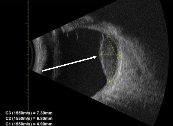
Ocular and orbital melanomas are rare and lifethreatening, malignant etiologies most commonly observed in individuals whose race is White. Uveal melanoma includes the choroid, ciliary body, and iris. However, of these, choroidal melanoma makes up >90% of primary malignant tumors in the eye.1 It is the second most common primary tumor seen in
CPC-EM Capsule
What do we already know about this clinical entity?
Choroidal melanoma is the most common ocular malignancy, often associated with visual symptoms, but may also present asymptomatic.
What is the major impact of the image(s)?
Point-of-care ultrasound can identify choroidal mass, guiding fundoscopy when available or referral in resource-limited settings.
How might this improve emergency medicine practice?
Emergency physicians should consider bedside ultrasound as a readily accessible, low-cost tool for rare but life-threatening conditions like choroidal melanoma.
malignant melanoma. Most commonly, presentation can be characterized by flashers, floaters, visual field loss, visual acuity loss. However, patients may also present asymptomatic.2 Diagnosis is generally made by indirect ophthalmoscopy, A- and B-ultrasonography, fundus fluorescein angiography, and transillumination.3
Biopsies may not always be required for diagnosis and, as a result, ultrasonography is an important diagnostic tool along with fundoscopy. While ultrasonography is commonly used in diagnosing choroidal melanoma, this case highlights how a tentative, timely diagnosis of choroidal melanoma can be made in the ED using ultrasound and funduscopic examination. The utility of ultrasonography for this purpose in the ED has been under-reported in the literature. Characteristics of a mass that raises concern for melanoma include hollowness and absence of halo.2 Size of the mass is the most important prognostic factor for uveal melanoma and predictor of metastasis.3 Prompt arrangement of outpatient follow-up with ophthalmology is important in getting a definitive diagnosis and prompt oncologic care.
The authors attest that their institution requires neither Institutional Review Board approval, nor patient consent for publication of this case report. Documentation on file.
Address for Correspondence: James L. Homme M.D., Mayo Clinic, 200 1st Street SW, Rochester, MN, United States 55902. E-mail: homme.james@mayo.edu.
Conflicts of Interest: By the CPC-EM article submission agreement, all authors are required to disclose all affiliations, funding sources and financial or management relationships that could be perceived as potential sources of bias. The authors disclosed none.
Copyright: © 2025 Matin et al. This is an open access article distributed in accordance with the terms of the Creative Commons Attribution (CC BY 4.0) License. See: http://creativecommons.org/ licenses/by/4.0/
1. Valasapalli S and Guddati AK. Nation-wide trends in incidence-based mortality of patients with ocular melanoma in USA: 2000 to 2018. Int J Gen Med. 2021;14:4171-4176.
2. Shields CL, Furuta M, Berman EL, et al. Choroidal nevus transformation into melanoma: analysis of 2514 consecutive cases. Arch Ophthalmol. 2009;127(8):981-987.
3. Singh P and Singh A. Choroidal melanoma. Oman J Ophthalmol 2012;5(1):3-9.
Matthias Barden, MD
Marissa Benbassat, MD
Emilio Benbassat, MD
Section Editor: Austin Smith, MD
Submission history: Submitted April 11, 2024; Revision received June 1, 2025; Accepted June 11, 2025
Electronically published October 24, 2025
Full text available through open access at http://escholarship.org/uc/uciem_cpcem DOI: 10.5811/cpcem.47211
Case Presentation: A 62-year-old man was brought into the emergency department by ambulance with right hip pain and deformity following a suspected hip dislocation. A plain film radiograph confirmed the diagnosis. He was sedated for closed reduction at the bedside. Despite apparently successful reduction, his case was complicated by persistent intraprosthetic dislocation of a polyurethane liner component of his prosthetic joint articulation. Computed tomography confirmed displacement of the liner, which required operative intervention.
Discussion: Intraprosthetic dislocation is a complication specific to dual-mobility hip prosthetics, characterized by displacement of the polyurethane liner unique to this type of device. This liner was designed to offer some benefits over other types of hip prosthetics, including improved biomechanics and lower risk of dislocation. The liner can become dislodged during a hip dislocation and remain displaced despite successful reduction of the metallic prosthetic components. Due to its radiolucency, diagnosis can be challenging on plain radiographs, often requiring advanced imaging. This case highlights the importance of recognizing this potential complication in patients with dualmobility prostheses. [Clin Pract Cases Emerg Med. 2025;9(4):474-476.]
Keywords: hip dislocation; dual-mobility total hip arthroplasty; intraprosthetic dislocation; orthopedics; images in emergency medicine.
A 62-year-old male with history of avascular necrosis of the femoral head status-post total hip arthroplasty presented to the emergency department via ambulance with concern for left hip dislocation. His initial total hip prosthesis was performed nine years prior and was complicated by loosening of the acetabular cup component. He underwent revision a year or two later with placement of a dual-mobility prosthetic C-STIM ATM (DePuy Synthes, RaynHam, MA). The dualmobility class of total hip prosthesis indicates the presence of a polyurethane liner positioned between the metallic femoral head and acetabular cup components.1, 2
He had done well after the revision until the day of the visit. He had been bent over gardening when he felt the joint become suddenly unstable and experienced immediate pain
about the hip joint. Plain film radiography confirmed prosthetic hip dislocation. Noted retrospectively, this initial radiograph does reveal a subtle “bubble” or “halo” sign, suggestive of concomitant intraprosthetic liner dislodgement (Image 1).3
Following closed reduction under sedation, computed tomography confirmed reduction of the metallic portions of the prosthetic but also demonstrated dislodgement of the polyurethane liner component into the soft tissue about the hip, indicating intraprosthetic dislocation (Image 2).
The on-call orthopedic surgeon was contacted and took the patient to the operating room, where the acetabular liner was successfully replaced back into appropriate positioning within the prosthesis. The patient has done well since surgery without any further prosthesis complications on follow-up. He was advised to avoid further similar bending positions in the future.
Dislocation remains a common major complication of total hip arthroplasty and a leading cause of revision surgery.4 Dual-mobility prostheses are designed with a polyurethane liner at the point of articulation between the femoral and acetabular metallic components. This design offers better joint mobility and lower rates of dislocation, particularly in highrisk patients.5 However, this design introduces the potential for intraprosthetic dislocation, a unique complication involving dislodgment of the polyurethane liner, which necessitates operative correction.3 While intraprosthetic dislocation was more prevalent in earlier dual-mobility designs, it remains a potential complication with contemporary prostheses and should be considered in any patient presenting with a dislocated dual-mobility total hip arthroplasty.1
The radiolucent polyurethane liner may manifest as a subtle “bubble” or “halo” sign on plain radiographs.3
Awareness of dual-mobility hip prosthetics and the potential for dislodgement of the intraprosthetic liner should prompt careful evaluation of plain films and consideration of axial imaging in cases of dislocation. Missed intraprosthetic dislocations will result in poor functional status, damage to metallic or ceramic components of the prosthesis, and higher
CPC-EM Capsule
What do we already know about this clinical entity?
Dual-mobility hip prostheses reduce dislocation rates but can develop intraprosthetic dislocation where the polyurethane liner becomes dislodged.
What is the major impact of the image(s)?
Images demonstrate the subtle ‘bubble’ sign on X-ray and CT confirmation of liner dislodgement, helping emergency physicians recognize this complication.
How might this improve emergency medicine practice?
Recognition of the ‘halo’ sign prompts CT imaging and early orthopedic consultation, preventing missed diagnosis and prosthetic component damage.
Intraprosthetic Dislocation Following Reduction of Dual-mobility Total Hip Arthroplasty
risk for the need to replace the entire prosthesis.2 Open reduction or revision is likely to be required in cases of intraprosthetic dislocation; therefore, early orthopedic consultation should be considered.3
The authors attest that their institution requires neither Institutional Review Board approval, nor patient consent for publication of this case report. Documentation on file.
Address for Correspondence: Emilio Benbassat, MD, Eisenhower Health, Department of Emergency Medicine, 39000 Bob Hope Dr. Rancho Mirage, CA 92270. Email: emiliobenbassat@gmail.com
Conflicts of Interest: By the CPC-EM article submission agreement, all authors are required to disclose all affiliations, funding sources and financial or management relationships that could be perceived as potential sources of bias. The authors disclosed none.
Copyright: © 2025 Barden et al. This is an open access article distributed in accordance with the terms of the Creative Commons Attribution (CC BY 4.0) License. See: http://creativecommons.org/ licenses/by/4.0/
Barden et al.
1. Patil N, Deshmane P, Deshmukh A, Mow C. Dual mobility in total hip arthroplasty: biomechanics, indications and complications-current concepts. Indian J Orthop. 2021 Oct 13;55(5):1202-1207
2. Waddell BS, De Martino I, Sculco T, Sculco P. Total hip arthroplasty dislocations are more complex than they appear: a case report of intraprosthetic dislocation of an anatomic dualmobility implant after closed reduction. Ochsner J. 2016 Summer;16(2):185-90.
3. Mallett KE, Taunton MJ, Abdel MP, et al. Dislocated and dissociated dual-mobility components are easily missed and more than half fail closed reduction: six tips to aid management. JB JS Open Access 2023 Jul 17;8(3):e22.00108
4. Pai FY, Ma HH, Chou TA, Huang TW, Huang KC, Tsai SW, Chen CF, Chen WM. Risk factors and modes of failure in the modern dual mobility implant. a systematic review and meta-analysis. BMC Musculoskelet Disord. 2021 Jun 14;22(1):541.
5. Harwin SF, Sodhi N, Ehiorobo J, Khlopas A, Sultan AA, Mont MA. Outcomes of dual mobility acetabular cups in total hip arthroplasty patients. Surg Technol Int. 2019 May 15;34:367-370.

- Western Journal of Emergency Medicine Subscription
- CAL/AAEM News Service email updates
- Discounted AAEM pre-conference fees
- And more!
CAL/AAEM NEWS SERVICE
- Healthcare industry news
- Public policy - Government issues - Legal cases and court decisions