
Volume 9, Number 3, August
Volume 9, Number 3, August

Clinicopathological Cases From The University Of Maryland
248 51-Year-Old Male with Back Pain, Groin Pain, and a Rash
L Mhonda, B Lowie, LJ Bontempo, A Windsor
Medical Legal Case Report
255 Medical and Legal Risks in Tibial Plateau Fractures
R Lindor, S Ghaith, J Newberry, A Thomas
Case Series
259 Wellens Syndrome Corollaries: A Call for Definition with a Case Series
AR Sparks, PM Bruss
264 Intravenous Low-dose Buprenorphine for Acute Pain Management in the Emergency Department: A Case Series
J Lee, N Ashenburg, J Park, T Ahern
268 The “Unlinkables”: A Case Series of Overcoming Social Determinants of Health for Successful Linkage to Care for HIV from the ED
P Moschella, MA Gormley, K Faryar
Case Reports
274 Case Report: Lurasidone-Induced Type 2 Brugada Pattern in a Pediatric Patient
E Start, A Enabore
278 Delirious Hyperactivity and Agitation in a Young Male Unveiling an Intriguing Underlying Diagnosis: Case Report
M Garey, J McLaughlin, H Kaur, J Graf, J Garcia, M De Kok, AJ Scumpia
282 A Rare Case Report of Contrast Media-induced Sympathetic Crashing Acute Pulmonary Edema
CP Adams, CI Wade


About Us: Penn State Health is a multi-hospital health system serving patients and communities across central Pennsylvania. We are the only medical facility in Pennsylvania to be accredited as a Level I pediatric trauma center and Level I adult trauma center. The system includes Penn State Health Milton S. Hershey Medical Center, Penn State Health Children’s Hospital and Penn State Cancer Institute based in Hershey, Pa.; Penn State Health Hampden Medical Center in Enola, Pa.; Penn State Health Holy Spirit Medical Center in Camp Hill, Pa.; Penn State Health Lancaster Medical Center in Lancaster, Pa.; Penn State Health St. Joseph Medical Center in Reading, Pa.; Pennsylvania Psychiatric Institute, a specialty provider of inpatient and outpatient behavioral health services, in Harrisburg, Pa.; and 2,450+ physicians and direct care providers at 225 outpatient practices. Additionally, the system jointly operates various healthcare providers, including Penn State Health Rehabilitation Hospital, Hershey Outpatient Surgery Center and Hershey Endoscopy Center.
We foster a collaborative environment rich with diversity, share a passion for patient care, and have a space for those who share our spark of innovative research interests. Our health system is expanding and we have opportunities in both academic hospital as well community hospital settings.
Benefit highlights include:
•Competitive salary with sign-on bonus
•Comprehensive benefits and retirement package
•Relocation assistance & CME allowance
•Attractive neighborhoods in scenic central Pennsylvania

FOR MORE INFORMATION PLEASE CONTACT:
Heather Peffley, PHR CPRP
Penn State Health Lead Physician Recruiter hpeffley@pennstatehealth.psu.edu


Indexed in PubMed and full text in PubMed Central
Rick A. McPheeters, DO, Editor-in-Chief Kern Medical/UCLA- Bakersfield, California
R. Gentry Wilkerson, MD, Deputy Editor University of Maryland School of Medicine
Mark I. Langdorf, MD, MHPE, Senior Associate Editor University of California, Irvine School of Medicine- Irvine, California
Shahram Lotfipour, MD, MPH, Senior Associate Editor University of California, Irvine School of Medicine- Irvine, California
Shadi Lahham, MD, MS, Associate Editor Kaiser Permanente- Orange County, California
John Ashurst, DO, Decision Editor/ ACOEP Guest Editor Kingman Regional Health Network, Arizona
Anna McFarlin, MD, Decision Editor Louisiana State University Health Science Center- New Orleans, Louisiana
Lev Libet, MD, Decision Editor Kern Medical/UCLA- Bakersfield, California
Amin A. Kazzi, MD, MAAEM
The American University of Beirut, Beirut, Lebanon
Anwar Al-Awadhi, MD
Mubarak Al-Kabeer Hospital, Jabriya, Kuwait
Arif A. Cevik, MD United Arab Emirates University College of Medicine and Health Sciences, Al Ain, United Arab Emirates
Abhinandan A.Desai, MD University of Bombay Grant Medical College, Bombay, India
Bandr Mzahim, MD
King Fahad Medical City, Riyadh, Saudi Arabia
Barry E. Brenner, MD, MPH Case Western Reserve University
Brent King, MD, MMM University of Texas, Houston
Daniel J. Dire, MD University of Texas Health Sciences Center San Antonio
David F.M. Brown, MD Massachusetts General Hospital/Harvard Medical School
Edward Michelson, MD Texas Tech University
Edward Panacek, MD, MPH University of South Alabama
Erik D. Barton, MD, MBA Icahn School of Medicine, Mount Sinai, New York
Francesco Dellacorte, MD
Azienda Ospedaliera Universitaria “Maggiore della Carità,” Novara, Italy
Francis Counselman, MD Eastern Virginia Medical School
Gayle Galleta, MD
Sørlandet Sykehus HF, Akershus Universitetssykehus, Lorenskog, Norway
Hjalti Björnsson, MD Icelandic Society of Emergency Medicine
Jacob (Kobi) Peleg, PhD, MPH Tel-Aviv University, Tel-Aviv, Israel
Jonathan Olshaker, MD Boston University
Katsuhiro Kanemaru, MD University of Miyazaki Hospital, Miyazaki, Japan
Amal Khalil, MBA UC Irvine Health School of Medicine
Elena Lopez-Gusman, JD
California ACEP
American College of Emergency Physicians
DeAnna McNett, CAE
American College of Osteopathic Emergency Physicians
John B. Christensen, MD California Chapter Division of AAEM
Randy Young, MD
California ACEP
American College of Emergency Physicians
Mark I. Langdorf, MD, MHPE UC Irvine Health School of Medicine
Jorge Fernandez, MD
California ACEP
American College of Emergency Physicians University of California, San Diego
Peter A. Bell, DO, MBA
American College of Osteopathic Emergency Physicians
Baptist Health Science University
Robert Suter, DO, MHA
American College of Osteopathic Emergency Physicians UT Southwestern Medical Center
Shahram Lotfipour, MD, MPH UC Irvine Health School of Medicine
Brian Potts, MD, MBA
California Chapter Division of AAEM Alta Bates Summit-Berkeley Campus
Christopher Sampson, MD, Decision Editor University of Missouri- Columbia, Missouri
Joel Moll, MD, Decision Editor
Virginia Commonwealth University School of Medicine- Richmond, Virginia
Steven Walsh, MD, Decision Editor Einstein Medical Center Philadelphia-Philadelphia, Pennsylvania
Melanie Heniff, MD, JD, Decision Editor University of Indiana School of Medicine- Indianapolis, Indiana
Austin Smith, MD, Decision Editor Vanderbilt University Medical Center-Nashville, Tennessee
Rachel A. Lindor, MD, JD, Decision Editor Mayo Clinic College of Medicine and Science
Jacqueline K. Le, MD, Decision Editor Desert Regional Medical Center
Christopher San Miguel, MD, Decision Editor Ohio State Univesity Wexner Medical Center
Khrongwong Musikatavorn, MD King Chulalongkorn Memorial Hospital, Chulalongkorn University, Bangkok, Thailand
Leslie Zun, MD, MBA Chicago Medical School
Linda S. Murphy, MLIS University of California, Irvine School of Medicine Librarian
Nadeem Qureshi, MD
St. Louis University, USA Emirates Society of Emergency Medicine, United Arab Emirates
Niels K. Rathlev, MD Tufts University School of Medicine
Pablo Aguilera Fuenzalida, MD Pontificia Universidad Catolica de Chile, Región Metropolitana, Chile
Peter A. Bell, DO, MBA Baptist Health Science University
Peter Sokolove, MD University of California, San Francisco
Robert M. Rodriguez, MD University of California, San Francisco
Robert Suter, DO, MHA UT Southwestern Medical Center
Robert W. Derlet, MD University of California, Davis
Rosidah Ibrahim, MD
Hospital Serdang, Selangor, Malaysia
Samuel J. Stratton, MD, MPH Orange County, CA, EMS Agency
Scott Rudkin, MD, MBA University of California, Irvine
Scott Zeller, MD University of California, Riverside
Steven Gabaeff, MD Clinical Forensic Medicine
Steven H. Lim, MD Changi General Hospital, Simei, Singapore
Terry Mulligan, DO, MPH, FIFEM ACEP Ambassador to the Netherlands Society of Emergency Physicians
Vijay Gautam, MBBS University of London, London, England
Wirachin Hoonpongsimanont, MD, MSBATS Siriraj Hospital, Mahidol University, Bangkok, Thailand
Ian Oliffe, BS Executive Editorial Director
Sheyda Aquino, BS WestJEM Editorial Director
Tran Nguyen, BS CPC-EM Editorial Director
Stephanie Burmeister, MLIS WestJEM Staff Liaison
Cassandra Saucedo, MS Executive Publishing Director
Isabelle Kawaguchi, BS WestJEM Publishing Director
Alyson Tsai, BS CPC-EM Publishing Director
June Casey, BA Copy Editor
Official Journal of the California Chapter of the American College of Emergency Physicians, the America College of Osteopathic Emergency Physicians, and the California Chapter of the American Academy of Emergency Medicine


Available in MEDLINE, PubMed, PubMed Central, Google Scholar, eScholarship, DOAJ, and OASPA.

Editorial and Publishing Office: WestJEM/Depatment of Emergency Medicine, UC Irvine Health, 3800 W. Chapman Ave. Suite 3200, Orange, CA 92868, USA Office: 1-714-456-6389; Email: Editor@westjem.org
Volume 9, no. 3: August 2025 i Clinical Practice and Cases in Emergency
Indexed in PubMed and full text in PubMed Central
This open access publication would not be possible without the generous and continual financial support of our society sponsors, department and chapter subscribers.
Professional Society Sponsors
American College of Osteopathic Emergency Physicians
California ACEP
Academic Department of Emergency Medicine Subscribers
Albany Medical College Albany, NY
American University of Beirut Beirut, Lebanon
Arrowhead Regional Medical Center Colton, CA
Augusta University Augusta GA
Baystate Medical Center Springfield, MA
Beaumont Hospital Royal Oak, MI
Beth Israel Deaconess Medical Center Boston, MA
Boston Medical Center Boston, MA
Brigham and Women’s Hospital Boston, MA
Brown University Providence, RI
Carl R. Darnall Army Medical Center Fort Hood, TX
Conemaugh Memorial Medical Center Johnstown, PA
Desert Regional Medical Center Palm Springs, CA
Doctors Hospital/Ohio Health Columbus, OH
Eastern Virginia Medical School Norfolk, VA
Einstein Healthcare Network Philadelphia, PA
Emory University Atlanta, GA
Genesys Regional Medical Center Grand Blanc, Michigan
Hartford Hospital Hartford, CT
Hennepin County Medical Center Minneapolis, MN
Henry Ford Hospital Detroit, MI
State Chapter Subscribers
Arizona Chapter Division of the American Academy of Emergency Medicine
California Chapter Division of the American Academy of Emergency Medicine
Florida Chapter Division of the American Academy of Emergency Medicine
International Society Partners
INTEGRIS Health
Oklahoma City, OK
Kaweah Delta Health Care District Visalia, CA
Kennedy University Hospitals Turnersville, NJ
Kern Medical Bakersfield, CA
Lakeland HealthCare
St. Joseph, MI
Lehigh Valley Hospital and Health Network Allentown, PA
Loma Linda University Medical Center Loma Linda, CA
Louisiana State University Health Sciences Center New Orleans, LA
Madigan Army Medical Center Tacoma, WA
Maimonides Medical Center Brooklyn, NY
Maricopa Medical Center Phoenix, AZ
Massachusetts General Hospital Boston, MA
Mayo Clinic College of Medicine Rochester, MN
Mt. Sinai Medical Center Miami Beach, FL
North Shore University Hospital Manhasset, NY
Northwestern Medical Group Chicago, IL
Ohio State University Medical Center Columbus, OH
Ohio Valley Medical Center Wheeling, WV
Oregon Health and Science University Portland, OR
Penn State Milton S. Hershey Medical Center Hershey, PA
Presence Resurrection Medical Center Chicago, IL
California Chapter Division of AmericanAcademy of Emergency Medicine
Robert Wood Johnson University Hospital New Brunswick, NJ
Rush University Medical Center Chicago, IL
Southern Illinois University Carbondale, IL
St. Luke’s University Health Network Bethlehem, PA
Stanford/Kaiser Emergency Medicine Residency Program Stanford, CA
Staten Island University Hospital Staten Island, NY
SUNY Upstate Medical University Syracuse, NY
Temple University Philadelphia, PA
Texas Tech University Health Sciences Center El Paso, TX
University of Alabama, Birmingham Birmingham, AL
University of Arkansas for Medical Sciences Little Rock, AR
University of California, Davis Medical Center Sacramento, CA
University of California Irvine Orange, CA
University of California, Los Angeles Los Angeles, CA
University of California, San Diego La Jolla, CA
University of California, San Francisco San Francisco, CA
UCSF Fresno Center Fresno, CA
University of Chicago, Chicago, IL
University of Colorado, Denver Denver, CO
University of Florida Gainesville, FL
University of Florida, Jacksonville Jacksonville, FL
University of Illinois at Chicago Chicago, IL
University of Illinois College of Medicine Peoria, IL
University of Iowa Iowa City, IA
University of Louisville Louisville, KY
University of Maryland Baltimore, MD
University of Michigan Ann Arbor, MI
University of Missouri, Columbia Columbia, MO
University of Nebraska Medical Center Omaha, NE
University of South Alabama Mobile, AL
University of Southern California/Keck School of Medicine Los Angeles, CA
University of Tennessee, Memphis Memphis, TN
University of Texas, Houston Houston, TX
University of Texas Health San Antonio, TX
University of Warwick Library Coventry, United Kingdom
University of Washington Seattle, WA
University of Wisconsin Hospitals and Clinics Madison, WI
Wake Forest University Winston-Salem, NC
Wright State University Dayton, OH
Uniformed Services Chapter Division of the American Academy of Emergency Medicine
Virginia Chapter Division of the American Academy of Emergency Medicine
To become a WestJEM departmental sponsor, waive article processing fee, receive print and copies for all faculty and electronic for faculty/residents, and free CME and faculty/fellow position advertisement space, please go to http://westjem.com/subscribe or contact: Emergency Medicine Association of Turkey Lebanese Academy of Emergency Medicine MediterraneanAcademyofEmergencyMedicine
Stephanie Burmeister
WestJEM Staff Liaison
Phone: 1-800-884-2236
Email: sales@westjem.org
Sociedad Chileno Medicina Urgencia ThaiAssociationforEmergencyMedicine
Indexed in PubMed and full text in PubMed Central
Clinical Practice and Cases in Emergency Medicine (CPC-EM) is a MEDLINE-indexed internationally recognized journal affiliated with the Western Journal of Emergency Medicine (WestJEM). It offers the latest in patient care case reports, images in the field of emergency medicine and state of the art clinicopathological and medicolegal cases. CPC-EM is fully open-access, peer reviewed, well indexed and available anywhere with an internet connection. CPC-EM encourages submissions from junior authors, established faculty, and residents of established and developing emergency medicine programs throughout the world.
285 A Case Report of Delayed, Severe, Paroxysmal Muscle Cramping After Chilean Rose Tarantula (Grammostola rosea) Envenomation
B Gooley, K Hughes, M Gooley, D Keyler, R Vetter, J Cole
289 Emergency Management of Post-Pancreatectomy Hemorrhage Secondary to a Ruptured Common Hepatic Artery Pseudoaneurysm: A Case Report
A Banks-McClelland, T Jackson, NA Royall
294 Presentation of Renal Cell Carcinoma Invading into the Pulmonary Artery in the Emergency Department: Case Report
S Yang, C Jewell
297 Second Scope, New Findings: Pediatric Stridor Is Not Always Due to Croup or Laryngomalacia: A Case Report
S Ghaith, D Hsu, W Dixon
302 Skeletal Fluorosis: A Case Report of Rare Diagnosis of Computer-cleaner Toxicosis
T Patriarca, JR Pescatore, W Rushton, E Sochovka, J Brown
310 Delayed Presentation of Congenital Diaphragmatic Hernia in the Emergency Department: Case Report
M Rayyan, R Reece
314 Abortion, Anemia, and an Account of Idiopathic Intracranial Hypertension: A Case Report
C Miller, R Sherak
318 A Case of Atraumatic and Non-obstetric Vulvar Hematoma from Contralateral Internal Iliac Artery Rupture
R Raveiro, M Bengio, J Sharp, G Lindblad, D Mir, S Serio
322 The Trigeminocardiac Reflex? Severe Bradycardia Secondary to Facial Trauma: A Case Report
B Penev, H Hughes, K Scarpino, DJ Ritter
326 Hypokalemia-induced Type 1 Brugada Reveals Type 3 Brugada Pattern with Repletion: Case Report
C Cantwell, MI Langdorf
329 Case Report: Early Valvular Repair of Rothia mucilaginosa Endocarditis with Intraparenchymal Hemorrhage from Septic Emboli
E Alley, K Holecko
Policies for peer review, author instructions, conflicts of interest and human and animal subjects protections can be found online at www.cpcem.org.
Indexed in PubMed and full text in PubMed Central
Clinical Practice and Cases in Emergency Medicine (CPC-EM) is a MEDLINE-indexed internationally recognized journal affiliated with the Western Journal of Emergency Medicine (WestJEM). It offers the latest in patient care case reports, images in the field of emergency medicine and state of the art clinicopathological and medicolegal cases. CPC-EM is fully open-access, peer reviewed, well indexed and available anywhere with an internet connection. CPC-EM encourages submissions from junior authors, established faculty, and residents of established and developing emergency medicine programs throughout the world.
334 Thoracic Paravertebral Block for Tube Thoracostomy Analgesia in the Emergency Department: A Case Report
MT Reeves
340 An Unexpected Cause of Shock in a Trauma Patient with Hemodynamic Instability: A Case Report
NA Jansen, C Fritz
345 A Pediatric Case Report of Acute Torticollis Secondary to Atraumatic Cerebellar Hemorrhage
JA Enabore, R Vezzetti, G Hill
Images in Emergency Medicine
349 Wrong Tube: Tracheal Obstruction from Megaesophagus
A Pearl, A Roka
352 Point-of-care Ultrasound Clarified the Diagnosis of an Occipital Artery Pseudoaneurysm After Blunt Trauma
K Nix, S Johnson, D Perling, B Parkinson, H Studebaker, B Foster
355 Point-of-care Ultrasound Diagnosis of Cardiac Myxoma
J Brutico, D Kreider
358 A Woman with Abdominal Pain
C Conrad, R Alouidor, C Allison
Policies for peer review, author instructions, conflicts of interest and human and animal subjects protections can be found online at www.cpcem.org.

Lorado Mhonda, MD*
Bobbi-Jo Lowie, MD†
Laura J. Bontempo, MD, MEd†
T.Andrew Windsor, MD†
Section Editor: Joel Moll, MD
University of Maryland Medical Center, Department of Emergency Medicine, Baltimore, Maryland
University of Maryland School of Medicine, Department of Emergency Medicine, Baltimore, Maryland
Submission history: Submitted December 24, 2024; Revision received February 24, 2025; Accepted April 10, 2025
Electronically published July 14, 2025
Full text available through open access at http://escholarship.org/uc/uciem_cpcem DOI: 10.5811/cpcem.41530
A 51-year-old male presented to the emergency department with back pain, bilateral groin pain, and bilateral leg numbness for four days. He was hypothermic, tachycardic, tachypneic, and hypotensive on presentation. A diffuse purpuric rash with bullae and desquamation was noted on exam. This case explores the differential diagnosis and evaluation of an ill patient who presented with an impressive rash. [Clin Pract Cases Emerg Med. 2025;9(3):248-254.]
CASE PRESENTATION (DR. MHONDA)
A 51-year-old male presented to the emergency department (ED) with complaint of back pain, bilateral groin pain, and bilateral leg numbness. The patient reported that his back pain started four days after lifting his father. He described the pain as a constant burning sensation and muscle pain that radiated to his bilateral lower extremities and rated it a 10/10 in severity. He also reported a loss of balance, inability to urinate, fecal incontinence, fevers, and a dark-colored rash with blisters on his legs and groin. He reported that his legs felt cold. He denied any headache, dizziness, or neck pain.
On chart review of a prior healthcare visit from several years prior, the patient’s past medical history was significant for HIV, although he reported being unaware of this diagnosis. The patient’s medications were listed as darunavir, elvitegravir/cobicistat/emtricitabine/tenofovir, and maraviroc, but he reported he was not taking any of those medications. He denied any history of cancer or recent infections. His surgical history was significant for an exploratory laparotomy for an abdominal gunshot wound, and his social history included frequent inhaled marijuana and phencyclidine (PCP) use, but he denied any history of intravenous (IV) drug use. No prior laboratory tests were available in the electronic health record to review. He denied any known allergies.
The patient’s vitals were significant for hypothermia at 92.8 °Fahrenheit/33.8 °Celsius, tachypnea at 28 breaths per minute, tachycardia at 117 beats per minute, and hypotension at 90/62 millimeters of mercury. His oxygen saturation was 98% on room air. He weighed 73.4 kilograms with a body mass index of
29 (normal 18.5-25). He appeared ill and in acute distress. He was awake, alert, and oriented to person, place, and time with no cranial nerve deficits. His head was normocephalic and atraumatic. His neck was non-tender with full range of motion and no stiffness or rigidity. Pupils were equal, round, and reactive to light, and extraocular movements were intact. His oropharynx and ears were without erythema, edema, or lesions. On auscultation of the heart, the patient was noted to have a tachycardic regular rhythm with no rubs, murmurs, or gallops. His peripheral pulses were 2+ bilaterally. Lungs were clear to auscultation with no wheezing or rales noted and the patient was tachypneic but not in respiratory distress.
The patient’s abdomen was diffusely tender on palpation with voluntary guarding and no rebound or distention. He did not have any costovertebral angle tenderness. He was tender to palpation in the bilateral groin and scrotal regions with purpura, desquamation, warmth, and edema. No lymphadenopathy was noted. His rectal tone was intact with no evidence of urinary retention on bladder ultrasound. His bilateral lower extremities and back were tender on palpation with full range of motion, 5/5 strength, and no signs of trauma or edema. Sensation was diffusely decreased in the bilateral lower extremities. Nonblanching purpura was noted on his lower extremities and dorsal aspect of his hands. Bullae and desquamation were also present on his lower extremities with a positive Asboe-Hansen sign and negative Nikolsky sign (Image 1). There was no rash involvement of his face, torso, or mucosal membrane.
Laboratory studies (Table), blood cultures, and an electrocardiogram (ECG) (Image 2) were obtained. Chest
1. Rash on a 51-year-old male who presented with back pain, lower extremity numbness, and a progressive purpuric rash.
(A) Desquamation (white arrow), non-blanching purpura and hemorrhagic bullae (black arrow) on the bilateral lower extremities.
(B) Non-blanching purpura (black arrows) on the bilateral dorsal hands and bilateral legs.
An extensive workup was initiated by the team including blood, urine, and multiple imaging studies. Some key findings from this workup included elevated blood urea nitrogen and creatinine, elevated creatine kinase levels, a bandemia, thrombocytopenia, and a D-dimer level that was greater than the upper limit that the laboratory could report. The ECG showed sinus tachycardia but was otherwise non-diagnostic. He had a chest radiograph, which did not reveal any infiltrates, pneumothorax, or effusions. Computed tomography was apparently not revealing of surgical pathology, and the normal ABI testing lessened the likelihood of acute vascular compromise.
radiography (Image 3) and computed tomography (CT) with contrast of the cervical, thoracic, and lumbar spine, and of the chest, abdomen, and pelvis were also completed but did not demonstrate any acute abnormalities to explain the patient’s symptoms. Bilateral lower extremity ankle brachial indices (ABI) were normal. Ultimately, a diagnostic test was performed that confirmed the diagnosis.
This is a case of a 51-year-old male who presented to the ED with an initial chief complaint of back pain, groin pain, and bilateral leg numbness. His back pain had started four days prior to his presentation, was initially attributed to lifting his father, and described as a burning sensation that radiated to his groin and legs bilaterally. He went on to report numbness in his legs, loss of balance, urinary retention, and fecal incontinence. Notably, he had also reported fevers and a dark rash with blisters on his legs and groin. He had a documented history of HIV but had never started treatment as he was apparently not aware of this diagnosis. He denied any history of IV drug use, nicotine use, or alcohol use, but frequently used marijuana and PCP. His triage vitals were almost all abnormal as he was tachycardic, hypothermic, borderline hypotensive, and tachypneic. His examination revealed many abnormalities, and he was noted to be in distress with diffuse abdominal guarding and a desquamating purpuric rash on the bilateral groin, scrotum, and legs with a positive Asboe-Hansen sign. He also had purpura of the bilateral dorsal hands. Interestingly, while the patient reported decreased sensation to touch of his extremities, he did not have any focal neurologic deficits; he had normal rectal tone and no urinary retention on point-of-care ultrasound. Some pertinent negatives from his examination were that he had no lesions in the oropharynx and a negative Nikolsky sign.
The differential diagnosis for this patient is quite broad given the many initial presenting complaints, which seem to be pulling in many different directions. Is this a case where Occam’s razor applies and all symptoms can be explained by one diagnosis, or Hickam’s dictum, where many different diseases may be coinciding all at once? Perhaps there are some red herrings here distracting from the true problem. At first glance, the chief complaint seems to be pointing toward spinal cord pathology or a primary neurologic complaint given the back pain, numbness, bladder and bowel complaints. However, we quickly learn of this patient’s normal motor examination followed by the finding of purpura, which cannot be ignored.
Purpura is a tangible starting point when attempting to determine this patient’s diagnosis. The broad categories where purpura can be found include trauma, infections, vasculitis, drug-induced, vitamin deficiencies, collagen disorders, pigmented purpuric dermatosis, and disorders of hemostasis.1 Trauma can easily be ruled out here as the patient had no reports or evidence of trauma, and the examination also supports no findings of traumatic injury. Next, we can easily eliminate drug-induced purpura as the patient does not seem to be taking any medications, and marijuana and PCP are not considered culprits of drug-induced purpura. Vitamin C deficiency is also unlikely in this patient as this is exceedingly rare in developed countries, and the patient has systemic illness not generally seen in vitamin C deficiency. Collagen disorders like Ehlers-Danlos syndrome include joint hypermobility and hyperextensibility, which are not present in this patient. Pigmented purpuric dermatosis is a capillaritis that can cause purpura localized to the lower extremities; however, it is usually non-painful and not associated with systemic illness.
While it seems that we have eliminated many diagnoses quickly, there are still more broad categories to discuss, including disorders of hemostasis. This includes the subcategories of thrombocytopenia, platelet function abnormalities, clotting factor deficiencies, and disseminated intravascular coagulopathy (DIC). The patient does have profound thrombocytopenia, and any hereditary cause of thrombocytopenia could be easily eliminated as most should have manifested before the patient reached adulthood. Similarly, vitamin deficiences induced by use of drugs or
51-Year-Old Male with Back Pain, Groin Pain, and a Rash
Table. Initial laboratory results in a 51-year-old male with back pain, groin pain, and a rash. Test Name
Complete Metabolic Panel
Complete Blood Count
alcohol could again be eliminated from the differential. You may see thrombocytopenia in patients with chronic alcohol use, but the patient has no history of alcohol use. He does report frequent marijuana and PCP use; however, neither marijuana nor PCP are known to result in thrombocytopenia. Some notable diagnoses that require further thought include immune thrombocytopenia (ITP), thrombotic thrombocytopenic purpura (TTP), paroxysmal nocturnal hemoglobinuria, and hemolytic uremic syndrome (HUS). While ITP is common in children, it can also occur in patients with HIV. It classically causes isolated thrombocytopenia, which we do see in this patient, but it does not explain the renal dysfunction and what
appears to be systemic illness. Both TTP and HUS cause thrombocytopenia and renal failure but should also result in hemolysis and anemia, which is not seen in this patient. Paroxysmal nocturnal hemoglobinuria can cause thrombocytopenia with renal dysfunction but also causes anemia, which again, the patient does not have. Platelet function abnormalities and clotting factor deficiencies could also result in purpura; however, they are often inherited disorders or secondary to drugs or other underlying processes such as infection, trauma, or uremia. Alone, these do not explain this patient’s presentation. The same goes for DIC, which is often seen because of an underlying process and is not, in fact, a diagnosis itself.
Mhonda et al. 51-Year-Old Male with Back Pain, Groin Pain, and a Rash
Table. Continued
Test Name Patient Value Reference Range
Urinalysis
Color Dark Yellow Yellow
Appearance Turbid Clear
Specific gravity >= 1.030 1.002 - 1.030
pH 5 5.0 - 8.0
Glucose Negative Negative
Bilirubin 1+ Negative
Urobilinogen 1.0 EU/dL 0.2 EU/dL
Ketones Trace Negative
Blood 1+ Negative
Protein 3+ Negative
Nitrite Negative Negative
Leukocyte esterase Trace Negative
White blood cells Too numerous to count 0-5/hpf
Red blood cells 11 – 20/hpf 0-5/hpf
Squam epithelial cells Too numerous to count 0-2/hpf
Hyaline casts Too numerous to count 0-2/hpf
Bacteria Negative Negative
Additional Labs
HIV antigen/antibody Reactive Non-Reactive
HAV immunoglobulin M Non-Reactive Non-Reactive
HCV antibody Non-Reactive Non-Reactive
HBV surface antigen Non-Reactive Non-Reactive
HBV core immunoglobulin M Non-Reactive Non-Reactive
Rapid plasma reagin Non-Reactive Non-Reactive
SARS-CoV-2 (COVID-19 PCR) RNA Not Detected Not Detected
Influenza A Not Detected Not Detected
Influenza B Not Detected Not Detected
Respiratory syncytial virus Not Detected Not Detected
Folate
Vitamin B12
5.2 ng/mL
533 pg/mL
Lactate 5.8 mmol/L
Creatine kinase
4.8 - 20.0 ng/mL
211 - 946 pg/mL
- 2.2 mmol/L
unit/L 39 - 308 unit/L
Myoglobin 1834 mg/mL 28 - 72 mg/mL
Abbreviations: PT, prothrombin time; PTT, partial thromboplastin time; INR, international normalized ratio; HIV, human immunodeficiency virus; HAV, hepatitis A virus; HBV, hepatitis B virus; SARS-CoV-2, severe acute respiratory syndrome coronavirus 2; COVID-19, coronavirus disease 2019; RNA, ribonucleic acid; K, thousands; mcL, microliter; g, grams; dL, deciliter; mmol, millimole; L, liter; mg, milligram; ng, nanogram; pg, picogram; hpf, high powered field; sec, seconds; EU, Ehrlich unit; FEU, fibrinogen equivalent units.
Additionally, the laboratory workup is not consistent with a diagnosis of DIC due to the normal fibrinogen and partial thromboplastic time values, both of which are typically low.
Next, we should consider vasculitis as a cause of purpura, which entails another long list of diagnoses. The broad categories of vasculitis include small, medium, and large vessel vasculitis, immune complex, variable vessel, and single-organ
vasculitis. The medium and large vessel vasculitides including Kawasaki disease, Takayasu arteritis, giant cell arteritis (GCA), and polyarteritis nodosa (PAN) can be eliminated: Takayasu usually occurs before the age of 30; Kawasaki is seen in children; GCA usually results in headache and jaw claudication; and PAN results in erythematous nodules, different from the purpura seen in this patient. Of the small vessel vasculitides, a
few need to be considered carefully. Microscopic polyangiitis and granulomatosis with polyangiitis are interesting to consider in this patient as they can result in renal dysfunction as well as arthralgias and paresthesias of the hands and feet, in addition to the skin manifestations of purpura. However, they generally include ear, nose, throat, and even pulmonary findings which the patient did not have.
Immunoglobulin A (IgA) vasculitis, formerly known as Henoch-Schonlein purpura, also results in purpura of the lower extremities with renal dysfunction and can also have gastrointestinal manifestations causing abdominal pain. However, one key feature making this diagnosis less likely is that the purpura in IgA vasculitis is often not painful. Finally, cryoglobulinemia can similarly be considered given his paresthesias, purpura, and history of HIV, but as the findings did not worsen with cold temperatures and there was no foot or wrist drop on exam, this diagnosis is also unlikely.
This patient has a history of HIV that has gone untreated for an unknown length of time, putting him at risk for certain types of malignancy including Kaposi sarcoma and nonHodgkin lymphoma. Kaposi sarcoma is defined by a purplishbrown lesion on the skin, but it also includes the mucosal surfaces. No mucosal findings are reported in this patient and, additionally, the physical exam revealed the lesions to be Absoe-Hansen positive, which would not be the case in Kaposi sarcoma. Lymphoma is also very unlikely as this patient’s underlying diagnosis as the presentation was acute, and there was no reported lymphadenopathy or symptoms reported that were classic to this presentation.
Finally, we must think about infection as the cause of purpura and this patient’s underlying presentation, especially with his untreated HIV. He presented with many vital sign abnormalities concerning for infection. When it comes to infection in this patient, he is absolutely at risk both for opportunistic infections and severe or disseminated infection involving common pathogens. When considering the patient’s rash and overall presentation, purpura fulminans (PF) is fitting; it is usually a result of underlying severe infection or sepsis in adults. Purpura fulminans is a syndrome of microvascular thrombosis that results from an acquired protein-C deficiency that causes skin necrosis. It can be associated with end-organ damage and lab results often include the renal dysfunction also seen in this patient, as PF can act as a smallmedium vessel vasculitis. Additionally, the lab results support this diagnosis including thrombocytopenia and a significantly elevated D-dimer. Coagulation studies can be normal or elevated and, if done, a protein-C level will be low. Interestingly, fibrinogen levels (normal in this patient) can be normal because while infection can increase levels, the microthrombosis may lower it. Infection is also supported by the bandemia seen on the automated differential. While bandemia is not always a result of infection, a bandemia of 14% is certainly concerning for infection.
At this point a decision must be made on what is the most likely underlying pathogen. There is an extensive list of possible infections. Looking at the data on PF, one of the most common culprits is Neisseria meningitidis. In fact, up to 20% of patients with N meningitidis develop PF.2 The patient is not presenting with typical signs and symptoms of meningitis in this case but rather of sepsis and meningococcemia. Therefore, after careful consideration of the facts of this case, my test of choice would be a blood culture revealing my final diagnosis of meningococcemia, resulting in PF.
Blood cultures obtained in the ED demonstrated N meningitidis bacteremia. Dermatology was consulted for the extensive purpuric rash on his bilateral lower extremities and groin, concerning for PF. Punch biopsy was completed, and it showed extensive epidermal necrosis suggestive of thrombotic vasculopathy. The bullae continued to worsen with skin denuding and sloughing in the bilateral lower extremities, requiring aggressive rehydration and skin care. During his
51-Year-Old Male with Back Pain, Groin Pain, and a Rash admission, the patient developed worsening abdominal pain and distension. A repeat CT of the abdomen and pelvis was completed, and it showed findings suggestive of spontaneous bacterial peritonitis. The patient also spontaneously developed dry gangrene of multiple toes in his bilateral feet. Vascular surgery recommended no acute intervention in the setting of intact pedal pulses and an unremarkable ABI and felt this was due to small vessel ischemia. During his admission, he was treated with broad spectrum antibiotics, narrowed based on culture and susceptibilities. He was also restarted on his HIV antiretroviral medication leading to improvement in his cluster of differentiation 4 (CD4) cells and viral load and fortunately was able to be discharged after 23 days of hospitalization with primary care, wound care, and podiatry outpatient follow-up.
Meningococcal septicemia is a bloodstream infection caused by N meningitidis bacteria, which is an encapsulated Gramnegative diplococcus transmitted through respiratory droplets or secretions. Initial infection results from direct contact with respiratory secretions. The bacteria colonizes the respiratory tract and invades the nasopharyngeal epithelium. The meningococcal bacteria successfully adheres, invades, and proliferates due to its structural components that protect against phagocytosis and lysis. The adhesion of the bacteria to the epithelial and endothelial cells activates the innate immune system.3
The activation of the immune system leads to the release of multiple inflammatory mediators. These mediators activate multiple pathways including the coagulation cascade, the leukotriene, prostaglandin, and complement pathways. These subsequently lead to increased capillary permeability, pathologic vasoconstriction and vasodilation, coagulopathy and severe myocardial dysfunction. These series of events are responsible for the development of shock and end-organ failure.3
Risk factors for meningococcal septicemia include conditions that weaken the immune system’s response to the bacteria. These include complement deficiencies and inhibitors, HIV, and functional or anatomic asplenia.4 Individuals who are younger than one year of age are at increased risk as they have not developed an appropriate immune system to adequately fight against the bacteria.4 Smoking is another risk factor, as it results in the destruction of the initial nasopharyngeal epithelium protective barrier against bacterial invasion.5 Individuals living in crowded conditions, including college students, military recruits, those of low socioeconomic status, and travelers to the “meningitis belt” in Sub-Saharan Africa, are also at an increased risk.6
Meningococcal septicemia initially presents as high fevers with shaking chills, severe myalgias, tachycardia, normotension, and cold extremities. The patient then develops petechiae, which transforms into diffuse PF and ultimately necrosis with gangrene. The changes can occur within hours. The vascular
damage leads to hypotension, adrenal hemorrhage also known as Waterhouse-Friderichsen syndrome, cardiac failure, renal failure, and acute respiratory distress syndrome. 7
Neisseria meningitidis is diagnosed through clinical presentation, Gram stain, cultures or antigen detection, with the gold standard being cultures. Gram staining will show Gram-negative diplococci. Cultures can be obtained from the mucosa, cerebrospinal fluid (CSF), and blood but have variable sensitivity.8 Cerebrospinal fluid cultures have a 90% sensitivity, blood cultures a 40-75% sensitivity, and a combination of both has a 94% sensitivity.3 Antigen detection through deoxyribonucleic acid polymerase chain reaction of the CSF, plasma, or serum can also be completed with a sensitivity and specificity greater than 90% and has the advantage of being able to rapidly detect the organism.3
Electrolyte and metabolic derangements can also be present including hypoglycemia, hypokalemia, hypocalcemia, hypomagnesemia, hypophosphatemia, and metabolic acidosis. Patients can also be hematologically unstable with anemia and decreased protein C, fibrinogen, prothrombin, and coagulation factors (V, VII, and X).2 Primary treatment for meningococcal septicemia is intravenous (IV) ceftriaxone or cefotaxime.8 Alternative therapy includes IV penicillin G or IV ampicillin.8,9 Ideally, culture data should be obtained prior to initiating penicillin due to increased resistance. Patients can also be treated with chloramphenicol, but it is less effective when compared to the other antibiotics. Patients with severe allergies and unavailable antibiotic sensitivity information can be treated with IV meropenem instead.9 Chemoprophylaxis should be administered to close contacts of the patient. Prophylactic treatments include rifampin, ceftriaxone and ciprofloxacin.8
• Purpura fulminans in adults is most commonly due to severe infection.
• Meningococcal septicemia can rapidly progress from seemingly benign flu-like symptoms to multiorgan failure.
• Empiric treatment with IV antibiotics should be started as soon as meningococcal infection is suspected, even before confirmation, due to the rapid and life-threatening nature of the disease.
The authors attest that their institution requires neither Institutional Review Board approval, nor patient consent for publication of this case report. Documentation on file.
51-Year-Old Male with Back Pain, Groin Pain, and a Rash Mhonda et al.
Address for Correspondence: T. Andrew Windsor, MD, University of Maryland School of Medicine, Department of Emergency Medicine, 110 S Paca Street, 6th Floor, Suite 200, Baltimore, MD 21201. Email: awindsor@som.umaryland.edu.
Conflicts of Interest: By the CPC-EM article submission agreement, all authors are required to disclose all affiliations, funding sources and financial or management relationships that could be perceived as potential sources of bias. The authors disclosed none.
Copyright: © 2025 Mhonda et al. This is an open access article distributed in accordance with the terms of the Creative Commons Attribution (CC BY 4.0) License. See: http://creativecommons.org/ licenses/by/4.0/
1. Georgesen C, Fox LP, Harp J. Retiform purpura: a diagnostic approach. J Am Acad Dermatol. 2020;82(4):783-96.
2. Perera TB and Murphy-Lavoie HM. Purpura fulminans. [Updated 2023 Jul 17]. In: StatPearls [Internet]. Treasure Island, FL: StatPearls Publishing; 2024 Jan. Available from: https://www.ncbi.nlm.nih.gov/ books/NBK532865/. Accessed March 29, 2024.
3. Batista RS, Gomes AP, Dutra Gazineo JL, et al. Meningococcal disease, a clinical and epidemiological review. Asian Pac J Trop Med 2017;10(11):1019-29.
4. Mbaeyi S, Duffy J, McNamara LA. Chapter 14: Meningococcal disease. In: Epidemiology and Prevention of Vaccine-Preventable Diseases. 14th ed. Centers for Disease Control and Prevention; 2021. Updated June 20, 2023. Available at: https://www.cdc.gov/ pinkbook/hcp/table-of-contents/chapter-14-meningococcal-disease. html. Accessed March 30, 2024.
5. Pace D and Pollard AJ. Meningococcal disease: clinical presentation and sequelae. Vaccine. 2012;30 Suppl 2:B3-B9.
6. Virji M. Pathogenic Neisseriae: surface modulation, pathogenesis and infection control. Nat Rev Microbiol. 2009;7(4):274-86.
7. Batista RS, Gomes AP, Dutra Gazineo JL, et al. Meningococcal disease, a clinical and epidemiological review. Asian Pac J Trop Med. 2017;10(11):1019-29.
8. Bosis S, Mayer A, Esposito S. Meningococcal disease in childhood: epidemiology, clinical features and prevention. J Prev Med Hyg 2015;56(3):E121-4.
9. Meningitis, Neisseria meningitidis. Sanford Guide to Antimicrobial Therapy. Antimicrobial Therapy, Inc.; August 5, 2023. Available at: https://www.sanfordguide.com Accessed April 2, 2024.
10. Vaz LE. Meningococcal disease. Pediatr Rev. 2017;38(4):158-69.
Rachel Lindor, MD, JD*
Summer Ghaith, JD†
Jennifer Newberry, MD, JD‡
Aaron Thomas, MD§
Section Editor: Melanie Heniff, MD, JD
Mayo Clinic, Department of Emergency Medicine, Rochester, MN
Mayo Clinic Alix School of Medicine, Mayo Clinic Phoenix, AZ
Stanford Health Care, Department of Emergency Medicine, Palo Alto, CA
Mayo Clinic, Department of Emergency Medicine, Phoenix, AZ
Submission history: Submitted November 6, 2024; Revision received February 8, 2025; Accepted March 11, 2025
Electronically published July 14, 2025
Full text available through open access at http://escholarship.org/uc/uciem_cpcem DOI: 10.5811/cpcem.38452
Introduction: Tibial plateau fractures, which comprise about 1% of all fractures, can be challenging to diagnose in the emergency department setting. Missed and delayed diagnoses can result in poor outcomes for patients and legal risks for clinicians, necessitating a high level of vigilance.
Case Series: In this article we review three malpractice cases related to tibial plateau fractures. Key issues included missed or delayed diagnosis, mismanagement of associated complications, inadequate discharge instructions, and lack of documentation.
Conclusion: Tibial plateau fractures can be challenging to identify, heightening the risk of downstream complications. As a result, emergency physicians must remain vigilant in assessing patients who are at increased risk for these injuries and document their efforts to both evaluate for and communicate these risks to patients. [Clin Pract Cases Emerg Med. 2025;9(3):255-258.]
Keywords: tibial plateau fracture; malpractice; lawsuit.
Tibial plateau fractures account for 1% of all fractures, including 8% of fractures in patients ≥60 years of age.1,2 In younger populations, the most common mechanisms include motor vehicle collisions, sporting injuries, and high energy falls, while in the older population the most common mechanism is a ground level fall.3,4 Ultimately these forces result in articular depression and malalignment.5 Plain radiographs fail to identify approximately 20% of tibial plateau fractures, with complications of missed diagnoses or mismanagement ranging from chronic pain and disability to acute compartment syndrome and amputation.6
The low sensitivity associated with plain radiographs and the potential poor outcomes associated with tibial plateau fractures combine to make this a high-risk injury in the emergency department (ED) setting from both a medical and legal perspective.7 However, increased availability of computed tomography (CT) has improved detection when clinical suspicion remains high. Here, we examine three
malpractice cases involving tibial plateau fractures, highlighting the key factors considered during litigation.
Case 1: Sullivan, California
A 27-year-old police officer presented to the ED after a high-speed pursuit of a suspect ended in a motor vehicle accident. Initial radiographs of his knee did not definitively identify a fracture, although it did show a fat-fluid level within the joint. Neither the treating emergency physician (EP) nor the consulting orthopedist ordered further testing. The patient was diagnosed with a knee sprain and instructed to bear weight as tolerated. Weeks later, due to persistent pain, he underwent repeat radiographs that revealed a significant fracture of the tibial plateau. The patient sued both the EP and the orthopedist for failing to diagnose his fracture on the radiographs and for allowing him to bear weight on the injury for so long, resulting in permanent pain and disability. This case was settled for $59,998 in 1986 (~$170,000 adjusted for inflation).8
A 13-year-old boy presented to the hospital with severe knee pain after falling from an 18-foot cliff and was found to have a tibial plateau fracture. Shortly afterward, his nurse reported to his physician that he had worsening lower leg pain, numbness, and a cold foot, but the physician did not return to re-evaluate him. Instead, he reportedly encountered an orthopedist at the elevator, verbally asked him to see the patient but did not document this request. The orthopedist did not recall the conversation, and the patient was not evaluated by either physician for the remainder of the night. By the time the patient was seen the next day, he was noted to have necrosis of a significant amount of his leg from a vascular injury and compartment syndrome, necessitating an amputation several days later. The patient alleged that the defendant physicians failed to diagnose this known complication, while the defendants contended that this was exceptionally unusual and that no intervention would have changed the outcome. This case went to trial and resulted in a verdict for the patient of $1,270,000 in 1984 (~$3.65 million adjusted for inflation).9
A 35-year-old male presented to the ED after a fall and was diagnosed with a tibial plateau fracture by a radiograph. A long-leg splint was applied, and the patient was given morphine, diazepam, and ketorolac for pain. The defendant EP initially recommended that the patient be transferred to an in-network county hospital for evaluation of his knee. However, the patient refused transfer preferring instead to transfer to a private hospital and was ultimately discharged home with a referral for a next-day orthopedics follow-up appointment. The patient did not attend that appointment and four days later returned to the ED with worsening pain, where he was found to have compartment syndrome in his lower leg, necessitating multiple surgeries and resulting in permanent disability. The patient alleged that signs and symptoms of compartment syndrome were present at the time of his initial evaluation, that he should have been admitted for observation given this is a well-established complication, and that orthopedics should have been consulted.10 The initial trial resulted in a hung jury, and a second trial—more than four years after the incident—resulted in a verdict in favor of the physician, largely due to the patient’s refusal of transfer and failure to return.
Fractures: Clinical Pearls
Tibial plateau fractures can be difficult to diagnose in the ED setting and require a careful and well documented clinical approach. First, because they tend to occur in the setting of high energy trauma, the component of knee pain may be overlooked as secondary to other injuries. However, there are
CPC-EM Capsule
What do we already know about this clinical entity?
Up to 20% of tibial plateau fractures are missed on initial imaging, creating the risk of serious long-term outcomes like chronic pain, compartment syndrome, or even amputation.
What makes this presentation of disease reportable?
These cases demonstrate how inadequate diagnosis, management, and documentation strategies have been tied to malpractice risks in previous cases of tibial plateau fractures.
What is the major learning point?
Tibial plateau fractures are easy to miss in the emergency department setting, requiring a high degree of clinical suspicion; understanding the clinical presentation, documenting an appropriate exam, and providing adequate follow-up care are essential to reducing the risk of bad outcomes.
How might this improve emergency medicine practice?
Improved recognition of tibial plateau fracture challenges can enhance patient care and potentially reduce clinicians’ liability risks.
multiple exam findings that should increase suspicion of intra-articular pathology, such as tibial plateau fracture.5 Joint effusion, inability to fully extend the leg, or ecchymosis with obvious deformity when compared to the unaffected side are possible findings.5 Other indicators include inability to bear weight, difficulty raising the straight leg against gravity, and limitation of flexion-extension mechanism.5
Second, because these fractures are often caused by torsion or impaction of the knee joint and present without classic overt signs of trauma, physicians often forego radiographs of the knee. In one study, researchers found that over half of patients with a tibial plateau fracture did not receive a knee radiograph in the ED.1 Use of a clinical decision rule among these patients would have significantly increased the frequency of plain radiography and identification of fractures.1 The Pittsburgh Knee Rule, used in this study, recommends radiographs following blunt trauma or fall in patients <12 or >50 years of age or in patients unable to walk
four steps while weight-bearing. While the Pittsburgh Knee Rule has been demonstrated to have a sensitivity of 99% and specificity of 60% for identifying patients with fractures11,12 and these types of rules have been touted as a way to decrease unnecessary radiography, in the case of tibial plateau fractures, use of a decision rule may prevent physicians from missing these occasionally subtle fracture presentations.
An additional clinical challenge posed by tibial plateau fractures is the difficulty in visualizing these injuries on standard radiographs. Sensitivity of standard radiographs for these fractures is generally estimated to be around 80%, increasing to 85% with the addition of oblique views.13 However, in at least one study, fractures were missed in almost 40% of patients, possibly due to difficulty positioning patients appropriately for imaging.1 Many patients have subtle signs of fracture on radiographs, including non-alignment of the femoral condyles, presence of a joint effusion, or a fat/fluid level (lipohemarthrosis).1,15 Ultimately, in cases where radiographs do not reveal a tibial plateau fracture but clinical suspicion remains high, a CT or magnetic resonance imaging (MRI) should be considered as the next step in the diagnostic evaluation. Over the past several decades, MRI has emerged as the preferred imaging modality due to its ability to better characterize the fracture patterns and associated soft-tissue injuries, which aid in surgical planning. However, CT remains a viable option when MRI is unavailable, with both considered definitive tests for identifying significant fractures.14
Finally, disposition of patients with tibial plateau fractures poses risks, given the potential for downstream complications. In addition to associated compartment syndrome, these patients are at high risk of surgical complications as a result of associated meniscal and ligamentous injuries.15
Tibial Plateau Fractures: Documentation Pearls
I. Discharge Instructions
Cases 1 and 3 highlight the importance of documenting clear discharge instructions. This is true for patients in whom tibial plateau fractures are confirmed, or even suspected, if definitive imaging is not available. Physicians are expected to communicate clearly with patients about the results of their testing, what changes would warrant a return visit or another evaluation, and how to manage their symptoms in the interim. In the first case, the patient was not told that it was a possibility that he had a knee fracture, and he was allowed to walk on his leg with the belief that it would improve with time, contributing to the development of his chronic symptoms. Failure of his treating physicians to recognize the poor sensitivity of radiographs for diagnosing tibial plateau fractures, to limit his weight-bearing, and to communicate and document this possibility of an occult fracture, likely contributed to the decision to settle this case out of court, as
they could not argue that they met the standard of care. In contrast, in the third case the patient was diagnosed with a tibial plateau fracture but had a delayed presentation of compartment syndrome, also resulting in chronic symptoms; however, the physicians in his case had arranged for him to have follow-up the next day, had documented this plan, and were able to show that they had met the standard of care regarding return precautions; ultimately it was determined to be the patient who failed to follow the recommendations.
Although physicians often emphasize documenting the details of clinical encounters, effective communication with patients during and after discharge is also crucial for reducing liability risks. Legal actions in this area may arise from unclear referrals, inadequate discharge instructions, insufficient return precautions, or a lack of follow-up on pending test results.16 Investing time in discussing and documenting post-discharge care recommendations may help reduce physicians’ exposure to these types of lawsuits.17. In patients with diagnosed or suspected tibial plateau fractures, discharge instructions should include non-weight bearing with the use of assistive devices such as crutches, walkers, or wheelchairs. Splinting or supportive braces may be prescribed for comfort. Close orthopedic follow-up is of utmost importance. Lastly, discussion of the signs of compartment syndrome must be discussed, among them skin color changes, loss of sensation, increase in pain, and loss of distal pulses.
Case 2 highlights the importance of appropriately documenting formal consultations. The EP in this case contended that he abided by the standard of care by consulting an orthopedist to assist in the patient’s care when he was alerted to developing compartment syndrome, but he did not document this consult and there was no record that this ever occurred. Malpractice cases for EPs related to consultations can be mitigated by adhering to a well-defined protocol for formal consultations, including documenting the consultant’s name, the time and relevant details of the discussion, and ensuring that consultants understand that their recommendations will be used for patient care.16 Of course, in this case, the treating physician’s failure to return to the bedside highlighted his inattention to the patient and prevented him from recognizing that the consult did not happen. Taking continued responsibility for patients regardless of how many people have been consulted on their behalf is also key to reducing liability exposure.
Our review of three malpractice cases associated with tibial plateau fractures reveals insights into the challenges of timely and accurate diagnosis and the medico-legal risks associated with this diagnosis. Better recognizing the clinical
Medical and Legal Risks in Tibial Plateau Fractures
challenges associated with tibial plateau fracture diagnosis and management can help clinicians improve patient care, communicate more clearly with patients and consultants, and mitigate their liability risks.
The authors attest that their institution requires neither Institutional Review Board approval, nor patient consent for publication of this case report. Documentation on file.
Address for Correspondence: Rachel A. Lindor, MD, JD, Mayo Clinic, Department of Emergency Medicine, 200 1st St SW, Rochester, MN 55905. Email: Lindor.Rachel@mayo.edu.
Conflicts of Interest: By the CPC-EM article submission agreement, all authors are required to disclose all affiliations, funding sources and financial or management relationships that could be perceived as potential sources of bias. The authors disclosed none.
Copyright: © 2025 Lindor et al. This is an open access article distributed in accordance with the terms of the Creative Commons Attribution (CC BY 4.0) License. See: http://creativecommons.org/ licenses/by/4.0/
1. Kiel CM, Mikkelsen KL, Krogsgaard MR. Why tibial plateau fractures are overlooked. BMC Musculoskelet Disord. 2018;19(1):244.
2. Bormann M, Neidlein C, Gassner C, et al. Changing patterns in the epidemiology of tibial plateau fractures: a 10-year review at a level-I trauma center. Eur J Trauma Emerg Surg. 2023;49(1):401-9
3. Donovan RL, Smith JRA, Yeomans D, et al. Epidemiology and outcomes of tibial plateau fractures in adults aged 60 and over treated in the United Kingdom [published correction appears in Injury. 2023 Mar;54(3):1023. Injury. 2022;53(6):2219-25.
4. Hua K, Jiang X, Zha Y, et al Retrospective analysis of 514 cases of tibial plateau fractures based on morphology and injury mechanism. J
Orthop Surg Res. 2019;14(267): 1-10.
5. McBrien B. Assessment and management of patients with tibial plateau fractures in emergency departments. Emerg Nurse 2019;27(6):26-30.
6. Malik S, Herron T, Mabrouk A, Rosenberg N. Tibial Plateau Fractures. In: StatPearls. Treasure Island (FL): StatPearls Publishing; April 22, 2023. Available at: https://pubmed.ncbi.nlm.nih.gov/29261932/ Accessed February 27, 2025.
7. Mthethwa J and Chikate A. A review of the management of tibial plateau fractures. Musculoskelet Surg. 2018;102(2):119-127.
8. Sullivan v. St. Francis Memorial Hospital, 38 Trials Digest (TD) 09446, 1986 WL 795083 (Cal.Super.) (Verdict and Settlement Summary)
9. Reager v. Anderson, JVR No. 5655, 1984 WL 317775 (W.Va.Cir.Ct.) (Verdict and Settlement Summary)
10. Colchado v. Rouzier, 26 Trials Digest 2d 47, 1996 WL 763139 (Cal. Super.) (Verdict and Settlement Summary)
11. Seaberg DC and Jackson R. Clinical decision rule for knee radiographs. Am J Emerg Med. 1994;12(5):541-3.
12. Seaberg DC, Yealy DM, Lukens T, et al. Multicenter comparison of two clinical decision rules for the use of radiography in acute, high-risk knee injuries. Ann Emerg Med. 1998;32(1):8-13.
13. Gray SD, Kaplan PA, Dussault RG, et al. Acute knee trauma: How many plain film views are necessary for the initial examination? Skelet Radiol. 1997;26(5):298–302.
14. Liu X, Wang H, Zhang T, et al. Comparison between computed tomography and magnetic resonance imaging in clinical diagnosis and treatment of tibial platform fractures. World J Clin Cases. 2020 26;8(18):4067–74
15. Khatri K, Sharma V, Goyal D, et al. Complications in the management of closed high-energy proximal tibial plateau fractures. Chin J Traumatol. 2016;19(6):342-7.
16. Ghaith S, Moore GP, Colbenson KM, et al. Charting practices to protect against malpractice: case reviews and learning points. West J Emerg Med. 2022;23(3):412-7.
Addison R. Sparks, MS* Patrick M. Bruss, MD†
The University of Toledo College of Medicine & Life Sciences, Department of Emergency Medicine, Toledo, Ohio
Promedica Monroe Emergency Medicine Residency, Department of Emergency Medicine, Monroe, Michigan
Section Editor: Joel Moll, MD
Submission history: Submitted October 7, 2024; Revision received December 27, 2024; Accepted February 16, 2025
Electronically published June 8, 2025
Full text available through open access at http://escholarship.org/uc/uciem_cpcem
DOI: 10.5811/cpcem.35877
Introduction: First described in 1982, the Wellens wave is an electrocardiographic (ECG) finding indicative of a critical lesion of the left anterior descending artery. These T-wave findings are classically found in ECG leads V2 and V3, although they may extend into the lateral leads V4-V6.
Case Series: We present three cases of patients with Wellens waves that were found only in leads V3 and V4 and did not include V2.
Conclusion: We suggest that the classical definition of T-waves in leads V2 and V3 is not the only manifestation of Wellens waves to indicate pathology. Wellens waves found in two contiguous leads in leads V1-V6 can be considered Wellens corollaries, thereby requiring the same emergent treatment as classical Wellens syndrome. We also recognize the need for a consensus on the inclusion criteria of Wellens syndrome, particularly the laboratory and ECG findings that define the disease. [Clin Pract Cases Emerg Med. 2025;9(3):259-263.]
Keyword: Wellens; electrocardiogram; ischemia; left anterior descending artery; acute coronary syndrome.
First described by Dutch cardiologist Hein JJ Wellens and colleagues in 1982,1 Wellens waves are subtle, difficultto-detect changes present on electrocardiogram (ECG) as T-wave abnormalities during ventricular repolarization. They are classified as Wellens type A or B (1 or 2) according to the T-wave morphology.2 Type A (or 1) Wellens is less common and involves biphasic T-waves in the early precordial leads.2,3 Wellens syndrome more often presents with deeply inverted T-waves in these precordial leads, which is classified as Wellens type B (or 2).2,3 These T-waves can be present for as long as several weeks.4 The current definition requires that T-wave abnormalities be noted in precordial leads V2 and V3 but may also occur in V1, V4, V5, and V6.1,2,5 The depth of T-wave inversions required for diagnosis has not been defined in the literature. The finding of Wellens waves is a highly specific indication for a pre-infarction, proximal occlusion of the left anterior descending (LAD)
coronary artery, which causes an acute infarction and life-threatening dysfunction of the left ventricle, necessitating urgent cardiac catheterization.1,2
It is important to note that ECGs are measured at one specific time point and can change over time. A Wellensappearing ECG, as a pre-infarction state, is dynamic and can later instead meet ST-elevation myocardial infarction (STEMI) criteria. Interestingly, one cross-sectional study and one systematic review have noted the presence of Wellens changes in the ECGs of patients who did not have an LAD occlusion or had multivessel disease.6,7 Those studies found that several patients met the criteria of Wellens syndrome but did not have an LAD occlusion and instead had occlusions of the right coronary artery, left coronary artery, or left circumflex artery.6,7
Diagnosis of Wellens syndrome also includes recognition of a lack of serum marker abnormalities (from some sources), a lack of pathologic Q-waves, presence of a normal R-wave progression, and an ST-segment that is not highly elevated.4,5,8
It is noteworthy that there is not a consensus on whether cardiac biomarkers must be within normal limits to make the diagnosis, and the original description of Wellens syndrome did not comment on whether serum chemistry changes were a diagnostic requirement.1 However, troponin levels were not measured in the 1980s as they are today. Wellens and colleagues relied on creatinine phosphokinase, lactate dehydrogenase, and glutamic oxaloacetic transaminase.1 The criteria for diagnosing Wellens syndrome has evolved since that time, and consensus is now needed. Some sources say these biomarkers, such as troponin, can be minimally elevated in Wellens syndrome.4,5 A suitable threshold for cardiac biomarkers needs to be defined; otherwise, it is difficult to separate Wellens syndrome as an independent pathology from a non-STEMI.
Additionally, Wellens syndrome must not be confused with left ventricular hypertrophy or right bundle branch blocks, which can include repolarization abnormalities such as T-wave inversions.4 Poor R-wave progression is also an exclusion criterion of Wellens.4,5 It is also critical to note that Wellens changes have been reported as “Wellens variants” on an ECG secondary to coronary vasospasm, such as vasospastic angina or cocaine-induced vasospasm.7 In fact, the initial description of Wellens syndrome included one patient with Prinzmetal angina and initial Wellens ECG changes that normalized after treatment with a calcium antagonist.1 One report presented a case of potential Wellens syndrome in the setting of a known left-septal fascicular block, which is supplied by the LAD.9 However, the patient in that specific case was experiencing chest pain at the time of the ECG recording, which by definition excluded the definition of Wellens syndrome.
It is possible to have a history of anginal pain, but these ECG findings must be obtained from a pain-free episode to be considered Wellens syndrome.4,10 The initial description of Wellens syndrome included patients who had previously experienced chest pain but whose characteristic ECG changes were obtained during pain-free episodes.1 Electrocardiogram changes characteristic of Wellens syndrome can appear even after anginal pain resolution.11
The risk factors for Wellens reflect what is expected when discussing acute coronary syndromes; hypertension, diabetes, and family history were more prevalent in the Wellens cases.6 Interestingly, smoking and hyperlipidemia were less prevalent at 14.2% and 22.5%, respectively.6 However, it is difficult to draw conclusions from one small study, highlighting the need to investigate Wellens-specific risk factors. Another study found that Wellens patients were less likely to have a previous history of heart disease or vessel occlusion, extrapolating that Wellens is more prevalent as an initial presentation of cardiac disease.12
When this condition was first described, it was found that 75% of patients with this wave pattern who did not undergo surgical treatment developed an infarction of the anterior heart
CPC-EM Capsule
What do we already know about this clinical entity?
We know the typical presentation of Wellens syndrome, including T-wave abnormalities in electrocardiogram leads V2 and V3 and lack of chest pain or cardiac biomarker elevation.
What makes this presentation of disease reportable?
The presence of T-wave abnormalities in leads V3 and V4, excluding V2, in patients that had nearcomplete left anterior descending artery blockage is an unusual presentation of Wellens syndrome..
What is the major learning point?
Wellens’ syndrome corollaries can be present and excellent clinical acumen is needed to diagnose. There is a need for clarification of Wellens’ diagnostic criteria.
How might this improve emergency medicine practice?
This may expand consideration of Wellens’ syndrome in forming a differential diagnosis and call for clarification and definition of the diagnostic criteria.
wall within a few weeks.1 These patients had non-diagnostic serum biomarkers and were initially treated with nitroglycerin and a calcium-channel blocker as their ECGs normalized over several days; unfortunately, they died later due to vessel occlusion. One study found that there was no significant difference in 24-month survival of Wellens patients compared to patients with non-Wellens acute coronary syndrome.12 Despite its critical implications, there is no consensus in the literature as to whether Wellens is considered a true STEMI or a “STEMI equivalent.”
While involvement of leads V2 and V3 is the classically accepted presentation of Wellens syndrome, we discuss below three cases of “Wellens corollaries” to consider Wellens waves in leads V3 and V4 that excluded V2. In each of the three cases, the patient with biphasic T-waves in leads V3 and V4 without V2 underwent cardiac catheterization, and prominent LAD stenosis was discovered.
Case One
A 45-year-old male presented to the emergency
department (ED) with the chief complaint of back pain. The patient reported acute onset of pain while moving a water heater up a flight of stairs. He described the pain as dull in nature, constant, radiating through to the center of his chest, worse with exertion, and accompanied by nausea and shortness of breath. He had no past medical history and took no medications, nor did he have a family history of coronary artery disease.
Physical exam revealed an inability to reproduce the pain with palpation. Workup included complete blood count (CBC), basic metabolic panel (BMP), troponin, D-dimer, and chest radiograph, which were all unremarkable. The patient had no pain in the ED and no symptoms while the ECG was conducted. The ECG was concerning for subtle biphasic T-waves in leads V3 and V4, with an unremarkable V2 (Image 1). This biphasic wave is initially positive and then trends negative. The absence of pain, presence of normal cardiac biomarkers, and ECG changes excluded STEMI or nonSTEMI but fit the classic definition of Wellens syndrome. The patient was admitted to the hospital and had a stress test with positive results. He then underwent cardiac catheterization that revealed 99% stenosis of the LAD.
A 79-year-old female presented to the ED with a chief complaint of fatigue. She had multiple risk factors including hypertension, hyperlipidemia, and tobacco use. Her physical exam was unremarkable, and she denied any pain. Diagnostic workup included CBC, BMP, and chest radiograph, which were all within normal limits. The troponin was slightly elevated at 0.07 nanograms per milliliter (ng/mL) (reference range ≤0.04 mg/mL). The absence of ST-segment elevations excluded the diagnosis of STEMI; however, the elevated biomarker and lack of pain did not clearly indicate nonSTEMI. The ECG was concerning for the presence of biphasic T-waves, isolated to V3 and V4 (Image 2). These T-wave inversions were subtle compared to lead V2. The patient was admitted to the hospital and underwent cardiac catheterization, which showed 98% stenosis of the LAD.
A 54-year-old male presented to the ED with a chief complaint of chest pressure. He had a family history of coronary artery disease, hypertension, and hyperlipidemia. He stated that the pain woke him up in the middle of the night but
Image 1. Concerning biphasic T-waves in leads V3 and V4, but not in V2 (arrows), which were present in a 45-year-old male with acute onset of back pain. Cardiac catheterization revealed 99% stenosis of the left anterior descending artery.
Image 2. Biphasic T-waves in leads V3 and V4, but not in V2 (arrows), which were present in a 79-year-old female with a chief complaint of fatigue and several risk factors. Cardiac catheterization revealed 98% stenosis of the left anterior descending artery.
Image 3. Electrocardiogram (ECG) leads V3 and V4 with biphasic T-waves are absent in V2 (arrows) in the ECG of a 54-year-old male with a chief complaint of chest pressure. The T-waves were initially positive and then trended negative. Cardiac catheterization revealed a 99% stenosis of the left anterior descending artery.
had resolved spontaneously prior to arrival. Physical exam was unremarkable, and he experienced no symptoms while the ECG was performed. Diagnostic CBC, BMP, troponin, and chest radiograph were all within normal limits. However, the ECG was concerning for biphasic T-waves in leads V3 and V4 that were absent from V2 (Image 3).
The different appearance of the T-waves in leads V2 and V3 were subtle but pertinent to the discussion. Importantly, the R-wave progression did not meet the classic definition of poor R-wave progression (R wave ≤3 millimeters), but the R-wave progression in this ECG appeared atypical and should have hinted at anterior infarction. Other features that possibly indicated infarction included the mild ST-elevation in lead III, ST-segment and T-wave deviation in the same direction in leads V3 and V4, and questionable ST-segment depression in the lateral leads. The absence of pain, the presence of normal cardiac biomarkers, and the ECG changes excluded STEMI or non-STEMI but did fit the classic definition of Wellens syndrome. The patient was admitted to the hospital, where serial troponin values were elevated. Catheterization discovered a 99% stenosis of the LAD.
The classic definition of Wellens syndrome involves biphasic T-waves in ECG leads V2 and V3, while sometimes extending into leads V4, V5, and V6.2 This ECG finding is specific for stenosis of the proximal LAD.2 From the three cases presented, we propose considering Wellens corollaries when Wellens waves are noted in leads V3 and V4, even when not seen in lead V2. Future studies should evaluate whether these changes in any two contiguous leads could be considered Wellens syndrome. In the three cases discussed here each
patient presented with biphasic T-waves in ECG leads V3 and V4, with a monophasic T-wave in V2. This falls outside the classical definition of Wellens syndrome; however, all three cases still necessitated emergency cardiac catheterization that found a dangerously stenotic LAD.
Subsequent ECGs were not available for the cases discussed above. It is also important to note that there are numerous findings on the ECG in Case 3 that should prompt suspicion for infarction.
The three patients discussed above presented to the ED with various symptoms and histories. Electrocardiogram revealed biphasic T-waves (Wellens waves) in leads V3 and V4 but, notably, not in V2. While this presentation differs from the accepted definition of Wellens waves, these patients still qualified for cardiac catheterization and had significant stenosis of the left anterior descending artery. Therefore, we propose expanding the definition of Wellens wave/Wellens syndrome to include biphasic or inverted T-waves present in any two adjacent leads of V1-V6. We also recognize the need for clarification of the definition of Wellens criteria, particularly with respect to cardiac biomarkers and diseased vessel territory. There is also a need for further investigation into Wellens-specific risk factors and survival.
The authors attest that their institution requires neither Institutional Review Board approval, nor patient consent for publication of this case series. Documentation on file.
Sparks et al. Wellens Syndrome Corollaries: A Call for Definition with a Case Series
Address for Correspondence: Addison Sparks, MS, The University of Toledo College of Medicine & Life Sciences, Department of Medical Education, 3000 Arlington Ave. Toledo, OH 43614. Email: asparks4@rockets.utoledo.edu.
Conflicts of Interest: By the CPC-EM article submission agreement, all authors are required to disclose all affiliations, funding sources and financial or management relationships that could be perceived as potential sources of bias. The authors disclosed none.
Copyright: © 2025 Sparks et al. This is an open access article distributed in accordance with the terms of the Creative Commons Attribution (CC BY 4.0) License. See: http://creativecommons.org/ licenses/by/4.0/
1. de Zwaan C, Bär FWHM, Wellens HJJ. Characteristic electrocardiographic pattern indicating a critical stenosis high in left anterior descending coronary artery in patients admitted because of impending myocardial infarction. Am Heart J. 1982;103(4, Part 2):730-6.
2. Miner B, Grigg WS, Hart EH. Wellens syndrome. In: StatPearls StatPearls Publishing; 2021. http://www.ncbi.nlm.nih.gov/books/ NBK482490/. Accessed October 21, 2021.
3. Alexander J and Rizzolo D. Wellens syndrome: an important consideration in patients with chest pain. JAAPA. 2023;36(2):25.
4. Rhinehardt J, Brady WJ, Perron AD, et al. Electrocardiographic manifestations of Wellens’ syndrome. Am J Emerg Med 2002;20(7):638-43.
5. de Zwaan C, Bär FW, Janssen JHA, et al. Angiographic and clinical characteristics of patients with unstable angina showing an ECG pattern indicating critical narrowing of the proximal LAD coronary artery. Am Heart J. 1989;117(3):657-65.
6. Mohamed S and Abdelaziz S. Wellens syndrome: prevalence, risk factors and coronary angiographic variation. A cross-sectional study. BMC Cardiovasc Disord. 2024;24(1):84.
7. Obi MF, Namireddy V, Noel C, et al. The comparative assessment of Wellens’ syndrome with proximal left anterior descending artery (lad) stenosis versus right coronary artery (RCA) or circumflex coronary artery stenosis and its prevalence: a systematic review. Cureus 2023;15(4):e37991.
8. Tandy TK, Bottomy DP, Lewis JG. Wellens’ syndrome. Ann Emerg Med. 1999;33(3):347-51.
9. Riera AR, Ferreira C, Ferreira Filho C, et al. Wellens syndrome associated with prominent anterior QRS forces: an expression of left septal fascicular block? J Electrocardiol. 2008;41(6):671-4.
10. Masuda M, Sakamoto T, Shimizu T. Wellens’ syndrome. Am J Med Sci. 2021;362(4):e37-8.
11. Parikh KS, Agarwal R, Mehrotra AK, et al. Wellens syndrome: a life-saving diagnosis. Am J Emerg Med. 2012;30(1):255.e3-255.e5.
12. Zhou L, Gong X, Dong T, et al. Wellens’ syndrome: incidence, characteristics, and long-term clinical outcomes. BMC Cardiovasc Disord. 2022;22(1):176.
Jonathan Lee, MD*†
Nicholas Ashenburg, MD†
Joshua Park, PharmD‡
Terence Ahern, MD†
Editor: R. Gentry Wilkerson, MD
Stanford University School of Medicine, Department of Anesthesiology, Perioperative, and Pain Medicine, Palo Alto, California
Stanford University School of Medicine, Department of Emergency Medicine, Palo Alto, California
Stanford Health Care, Department of Pharmacy, Palo Alto, California
Submission history: Submitted February 23, 2025; Revision received March 25, 2025; Accepted April 11, 2025
Electronically published July 14, 2025
Full text available through open access at http://escholarship.org/uc/uciem_cpcem DOI: 10.5811/cpcem.43494
Introduction: Buprenorphine is used for treating opioid use disorder, but its role as an analgesic in the emergency department (ED) is frequently overlooked. Emerging evidence indicates that, at low doses, it can be used safely and advantageously as an alternative to full-agonist opioids for treating acute pain.
Case Series: This case series examines the novel use of intravenous (IV) low-dose buprenorphine for acute pain management in the ED for five patients presenting with diverse past medical history and varied painful indications.
Conclusion: Intravenous low-dose buprenorphine may represent an important new tool in our ED armamentarium, and research into its role in emergency pain management is warranted. Further work is needed to determine optimal dosing strategies and identify which patients will be most likely to benefit from IV low-dosebuprenorphine in the ED. [Clin Pract Cases Emerg Med. 2025;9(3):264267.]
Keywords: pain; analgesia; buprenorphine; opioids; pain management.
INTRODUCTION
Buprenorphine is a high-affinity partial μ opioid receptor agonist with unique and complex pharmacology, commonly used for managing pain across various medical specialties.1-3 When administered at doses lower than those used for opioid use disorder, emerging evidence supports its safety and efficacy as an alternative to full-agonist opioids for treating acute pain.4-5 Currently, research on low-dose buprenorphine in the emergency department (ED) is limited, and existing studies have primarily focused on sublingual dosing. This case series highlights the novel use of intravenous (IV) low-dose buprenorphine as an alternative to full-agonist opioids for the treatment of acute undifferentiated pain in the ED.
A 71-year-old male with a history of metastatic pancreatic cancer presented with abdominal pain and elevated liver function tests. On arrival, his vital signs were within normal limits, and he reported severe abdominal pain rated as 10/10 on the numeric rating scale that was intractable to home doses of 5 milligrams (mg) oxycodone. The patient reported using oxycodone intermittently at home for several months but found its pain relief too short-lasting and experienced intolerable gastrointestinal side effects, primarily constipation. He was administered 150 micrograms (µg) of IV low-dose buprenorphine and 10 mg of IV ketorolac while awaiting computed
tomography (CT). Labs were notable for leukocytosis of 14.4 x 10³ per microliter (K/µL) (reference range: 4.0-11.0 K/ µL), hyperbilirubinemia of total bilirubin 2.9 mg/dl (<1.2 mg/ dL), and elevated alkaline phosphatase of 693 Units per liter (U/L) (4-30 U/L). The patient experienced significant pain relief, with a reduction of more than five points on the numerical rating scale and no reported side effects on reassessment one hour later. He expressed satisfaction with the treatment. Subsequently, he was found to have pancreatic duct stenosis and was admitted to the oncology service, where IV low-dose buprenorphine doses were continued for pain management.
A 68-year-old female with a history of multiple surgeries and small bowel obstruction presented with abdominal pain and bloating, rated as 10/10 on the numeric rating scale. Her vital signs, complete blood count, comprehensive metabolic pain, and lactic acid level were within normal limits. Due to her documented intolerance to many full-agonist opioids, she was administered 150 µg IV low-dose buprenorphine and 1000 mg IV acetaminophen while awaiting CT. The patient experienced significant pain relief, reported no side effects, and expressed high satisfaction with the medication on reassessment one hour later. She was diagnosed with a small bowel obstruction and admitted to the surgical service for further management, where her pain regimen was transitioned to full-agonist opioids without issue.
A 60-year-old female with history of end-stage renal disease and congestive heart failure presented for right hip pain and headache after a ground-level fall. On arrival, her blood pressure was 154/89 millimeters of mercury (mm Hg); otherwise vital signs were within normal limits. Lab testing was unremarkable apart from her baseline chronic kidney disease (creatinine 6.8 mg/dL, blood urea nitrogen 47 mg/dL). She reported severe pain. Given her advanced renal disease, high suspicion for hip fracture, and our preference for a potent, longer acting analgesic, she received 150 µg IV low-dose buprenorphine and 1000 mg IV acetaminophen while awaiting imaging results. The patient experienced significant pain relief, reported no side effects, and expressed high satisfaction with the medication. She was diagnosed with a right femoral neck fracture and admitted to the medicine service for further management, including orthopedic surgery consultation, regional anesthesia consultation, and initiation of an opioidsparing pain protocol.
A 44-year-old male with a history of insulin-dependent type 2 diabetes, hypertension, latent syphilis, and prior opioid use disorder presented with multiple abscesses and left facial swelling. He met sepsis criteria, with a temperature of 99.8 ºF, blood pressure of 174/89 mm Hg, respiratory rate of 25
CPC-EM Capsule
What do we already know about this clinical entity?
IV buprenorphine is an μ-opioid partial agonist with potent analgesic effects, FDA-approved for acute pain.
What makes this presentation of disease reportable?
IV buprenorphine has been studied extensively perioperatively. This is the first described use in the ED for pain management.
What is the major learning point?
(Emergency physicians can use 0.15mg IV buprenorphine to provide safe and efficacious analgesia for acute pain.
How might this improve emergency medicine practice?
IV buprenorphine offers a promising alternative to full-agonist opioids, offering another tool for pain management in the ED.
breaths per minute, and heart rate of 99 beats per minute (bpm). He reported facial and neck pain rated as “20 of 10” on the numerical rating scale. Labs were notable for leukocytosis (18.5 K/µL), hyperglycemia (glucose 491 mg/dL), and normal anion gap, renal function, and lactic acid. Due to the patient’s concern regarding his addiction to opioids, he was administered 150 µg IV low-dose buprenorphine and 1000 mg of oral acetaminophen for pain control. The patient experienced significant pain relief with only minor nausea, which was treated with 4 mg of IV ondansetron. He expressed satisfaction with the medication. He was diagnosed with hyperglycemia, sepsis, facial cellulitis, and phlegmon and was admitted to the medical service for management, including broad-spectrum antibiotics and otorhinolaryngology surgery consultation, which resulted in operative management and placement of Penrose drains to treat the infection.
A 23-year-old male with a history of chronic back pain following a traumatic injury at age 16 presented with acute worsening of low back pain and bilateral radicular symptoms. On arrival, his vital signs were notable for heart rate of 102 bpm. He reported severe, debilitating pain, 10/10 on the numerical rating scale, and inability to ambulate despite home treatment with nonsteroidal anti-inflammatory medica-
Intravenous Low-dose Buprenorphine for Acute Pain Management in the ED: A Case Series
tions and tramadol. He was initially treated with 10 mg of IV ketorolac, 1000 mg of IV acetaminophen, 10 mg of oral cyclobenzaprine, and a 5% lidocaine patch. On reassessment 30 minutes later, this regimen had failed to adequately control his pain, and he was subsequently administered 150 µg IV lowdose buprenorphine, which provided excellent relief for the next several hours. The patient reported prior history of opioid use disorder and thus preferred to avoid full-agonist opioid medications, but he was amendable to using low-dose buprenorphine. The patient denied any side effects and expressed high satisfaction with the medication. Labs were unremarkable apart from leukocytosis (white blood cell count 16 K/µL), and magnetic resonance imaging did not show a significant pathologic cause for his pain. He was observed overnight in the clinical decision unit for physical therapy and continued opioid-sparing pain management.
Buprenorphine was approved in the United States in 2002 for the treatment of opioid dependence, significantly improving the safe and effective management of opioid use disorder. However, because of its association with opioid use disorder and classification as a partial μ-opioid receptor agonist, buprenorphine is often misunderstood as a partial analgesic. Its pharmacology is complex and extends beyond its interaction with the μ-opioid receptor; at low-dose ranges, an analgesic ceiling is not known to exist.9,10 Several studies now suggest that buprenorphine provides analgesia comparable to full-agonist opioids, such as morphine, and offers multiple potential advantages. These include potent analgesia with less euphoria, a ceiling for respiratory depression, lower rates of constipation and pruritus, effective anti-hyperalgesia, reduced immune suppression, and a long duration of action (6-12 hours with a single dose).8-11 Given these advantages, numerous specialty groups support the use and continued study of buprenorphine for acute pain.12-15
Low-dose buprenorphine for acute pain involves doses much lower than those used for opioid use disorder. Given the novelty of use in the ED, there is no universally agreed-upon definition for appropriate dose ranges. We recommend IV doses of 150-300 µg. At these doses, low-dose buprenorphine provides potent analgesia with minimal side effects.5 To date, very few studies have investigated low-dose buprenorphine in the ED setting. The existing studies assessed sublingual lowdose buprenorphine, comparing it to IV morphine or ketorolac in small groups of patients with specific painful conditions, such as fractures and renal colic.6-8 These studies found buprenorphine to be an effective analgesic option, although there were slightly higher rates of dizziness and nausea compared to the control treatments when using a 2 mg sublingual dose. This dose equates to approximately 60 morphine milligram equivalents (MME), and we believe it is too high for opioidnaïve or opioid-intolerant patients. Dosing with sublingual formulations comes with challenges, given the lowest dose of
generic buprenorphine is 2 mg in the US. Administering this as a half tablet or even quarter tablet is a possibility, but the tablets are small, and because they are not scored for cutting, it could lead to inaccuracies in dosing. Furthermore, this formulation was approved for opioid use disorder, while use for acute pain indication is considered off label.
The IV route of administration for low-dose buprenorphine has not been described in the ED literature, but it may offer an essential new tool in pain management. Intravenous buprenorphine has been approved by the US Food and Drug Administration for use as an analgesic since 1981 and is now available in a generic formulation.15 The cost difference of IV buprenorphine compared to IV morphine per MME is marginal. It is available in 150 µg (15 MME) and 300 µg (30 MME) doses, with a rapid onset within minutes and a duration of action up to 6-12 hours. For comparison, a dose of 150 µg is equivalent to 5 mg IV morphine in terms of MME. This makes it titratable and particularly valuable for treating painful conditions expected to last for extended periods.
The patients included in this case series presented with common ED complaints, including abdominal pain, fractures, abscess with cellulitis, and chronic back pain. They may represent ideal candidates for treatment with IV low-dose buprenorphine. Evidence from other specialties suggests that low-dose buprenorphine may be particularly beneficial for acute pain associated with oncologic diseases, renal colic, long bone fractures, neuropathic pain, and in patients with a history of (or active) substance use disorders. However, further research is needed to determine optimal dosing strategies and to identify which patients would benefit from low-dose buprenorphine in the ED.
Our ED has adopted a set of conservative recommendations for the use of low-dose buprenorphine. We recommend against its use in patients on chronic full-agonist opioid therapy. While the risk of precipitated withdrawal is much lower with low-dose buprenorphine, it remains a possibility. Patients who have received a single dose of full agonists (rather than chronic daily use) will not experience precipitated withdrawal with subsequent administration of buprenorphine. However, due to buprenorphine’s high affinity for the μ-opioid receptor, it may displace other full agonists, leading to variable analgesic effects depending on the timing, dose, and potency of the prior opioid. Furthermore, we urge caution in patients with known opioid sensitivity or allergies, as buprenorphine’s potent μ-opioid receptor activity increases the likelihood of cross-reactivity. As research on low-dose buprenorphine evolves, so too will our understanding and its clinical application.
This case series highlights the innovative use of IV lowdose buprenorphine as an alternative to full-agonist opioids for acute pain treatment. Intravenous low-dose buprenorphine may represent an important new tool in our ED armamentari-
Lee et al.
Intravenous Low-dose Buprenorphine for Acute Pain Management in the ED: A Case Series
um, and research into its role in emergency pain management is warranted. Further work is needed to determine optimal dosing strategies and identify which patients would be most likely to benefit from IV low-dose buprenorphine in the ED.
The authors attest that their institution requires neither Institutional Review Board approval, nor patient consent for publication of this case report. Documentation on file.
Address for Correspondence: Jonathan Lee, MD, Stanford University School of Medicine, Department of Emergency Medicine, 600 Welch Road, Suite 350, Palo Alto, CA 94304. Email: jonblee@ stanford.edu
Conflicts of Interest: By the CPC-EM article submission agreement, all authors are required to disclose all affiliations, funding sources and financial or management relationships that could be perceived as potential sources of bias. The authors disclosed none.
Copyright: © 2025 Lee et al. This is an open access article distributed in accordance with the terms of the Creative Commons Attribution (CC BY 4.0) License. See: http://creativecommons.org/ licenses/by/4.0/
in older adults: a systematic review and meta-analysis. Pain Med 2024:pnae064.
5. Hickey TR, Costa GPA, Oliveira D, et al. Buprenorphine versus full agonist opioids for acute postoperative pain management: a systematic review and meta-analysis of randomized controlled trials. Reg Anesth Pain Med. 2025:rapm-2024-106014. Epub ahead of print.
6. Jalili M, Fathi M, Moradi-Lakeh M, et al. Sublingual buprenorphine in acute pain management: a double-blinded randomized clinical trial. Ann Emerg Med. 2012;50(4):276-80.
7. Payandemehr P, Jalili M, Davani BM, et al. Sublingual buprenorphine for acute renal colic pain management: a double-blind, randomized controlled trial. Int J Emerg Med. 2014;7(1):1-5.
8. Mozafari J, Masoami K, Farouzan A, et al. Sublingual buprenorphine efficacy in renal colic pain relief: a randomized placebo-controlled clinical trial. Pain Ther. 2017;6(2):227-34.
9. Dahan A, Yassen A, Romberg R, et al. Buprenorphine induces ceiling in respiratory depression but not in analgesia. Br J Anaesth 2006;96(5):627-32.
10. Pergolizzi J, Aloisi AM, Dahan A, et al. Current knowledge of buprenorphine and its unique pharmacological profile. Pain Pract 2010;10(5):428-50.
11. Franchi S, Panerai AE, Sacerdote P. Buprenorphine ameliorates the effect of surgery on hypothalamus-pituitary-adrenal axis, natural killer cell activity and metastatic colonization in rats in comparison with morphine or fentanyl treatment. Brain Behav Immun. 2007;21(6):767-74.
REFERENCES
1. Poliwoda S, Noor N, Jenkins JS, et al. Buprenorphine and its formulations: a comprehensive review. Health Psychol Res 2022;10(3):37517.
2. Patanwala A, Moran B, Johnstone C, et al. Crit Care Med 2023;51(12):1650-58.
3. Powell VD, Rosenberg JM, Yaganti A, et al. Evaluation of buprenorphine rotation in patients receiving long-term opioids for chronic pain: a systematic review. JAMA Netw Open. 2021;4(9): e2124152.
4. Selman K, Perlman S, Blatcher C, et al. Buprenorphine for acute pain
12. Nwanaji J, Rivera Blanco L, Kiernan E, et al. Revisiting emergency department use of buprenorphine as a primary analgesic in nonopioid use disorder patients post–X-waiver. Acad Emerg Med 2023;30:1272-1274.
13. Davis M. Twelve reasons for considering buprenorphine as a frontline analgesic in the management of pain. J Support Oncol 2012;10(6):209-19.
14. Hale M, Garofoli M, Raffa R, et al. Benefit-risk analysis of buprenorphine for pain management. J Pain Res. 2021;14:1359-69.
15. Johnson R, Fudula P, Payne R. Buprenorphine: considerations for pain management. J Pain Symptom Manage. 2005;29(3):297-326.
Phillip Moschella, MD, PhD*†
Mirinda Ann Gormley, PhD, MSPH*†
Kiran Faryar, MD, MPH‡
Section Editor: R. Gentry Wilkerson, MD
Prisma Health, University of South Carolina, School of Medicine Greenville, Department of Emergency Medicine, Greenville, South Carolina Clemson University School of Health Research, Clemson, South Carolina University Hospitals Cleveland Medical Center, Case Western Reserve University School of Medicine, Department of Emergency Medicine, Cleveland, Ohio
Submission history: Submitted November 7, 2024; Revision received December 26, 2024; Accepted March 11, 2025
Electronically published July 29, 2025
Full text available through open access at http://escholarship.org/uc/uciem_cpcem
DOI: 10.5811/cpcem.39649
Introduction: Despite the success of emergency department (ED)-based universal HIV screening programs in select cities, widespread integration of similar programs across the United States has not followed. Within the US Centers for Disease Control and Prevention (CDC)-designated “Ending the HIV Epidemic (EHE)” areas, ED-based HIV screening is low. This case series highlights successful strategies for notification and linkage to care of patients with various challenging social determinates of health (SDoH). The goal is to inspire more EDs to offer universal HIV screening by providing insight into these challenging SDoH and successful strategies to overcome them.
Case Series: We describe four cases, two from a site in upstate South Carolina and two from Cuyahoga County in Ohio, that highlight successful notification and linkage to care of these perceived “worst-case” scenarios. Both ED-based programs are located in CDC-designated EHE areas. We discuss ED screening opportunities and successful linkage for these minority patients (2136 years of age), and highlight the concomitant and challenging mental health and substance use disorders, and SDoH that were overcome. All four of these patients are currently receiving treatment for HIV and 3 of the 4 have reached viral suppression.
Conclusion: Despite challenging SDoH including unstable housing and lack of transportation, phone, and even legal identification documentation, these ED-identified patients with HIV were successfully notified of their disease status and linked to care. The patient navigators used perseverance, connections to local community resources, and leveraged family support to achieve linkage success. The cases serve as both a roadmap and source of inspiration to other EDs in priority EHE areas to begin ED HIV screening programs. [Clin Pract Cases Emerg Med. 2025;9(3):268-273.]
Keywords: HIV; linkage to care; emergency department; social determinants of health.
INTRODUCTION
Over a decade of literature has demonstrated that emergency departments (ED) can systematically identify and successfully link patients with HIV to long-term care, yet few articles have described the coexisting mental health and substance use disorders, and social determinants of health
(SDoH) barriers that make successful linkage to care difficult for patients with either newly or previously diagnosed HIV who present to an ED.1,2 This case series demonstrates how these coexisting disorders and SDoH posed challenges to linkage to HIV treatment for four patients from two different healthcare systems. Patients were identified as having experi-
enced the most barriers to linkage to care by research coordinators within each program. These programs were located within priority US Centers for Disease Control and Prevention (CDC)-designated “Ending the HIV Epidemic” (EHE) jurisdictions (Cuyahoga County, Ohio and Greenville County, South Carolina).
The EHE initiative is a nationwide program coordinated by the US Department of Health and Human Services working with the CDC on community-specific programs and collaborations to reduce new HIV infections and support treatment and prevention. The EHE initiative has identified 50 local areas and seven entire states as priority jurisdictions where more than half of new HIV diagnoses occur.3 We highlight the following complicating medical comorbidities:
1) mental health disorders such as depression, anxiety, bipolar disorder, schizophrenia, or antisocial personality disorder; and
2) substance use disorder including use of opioids, stimulants, alcohol, hallucinogens, or any other abused prescriptions or illicit substances. We also highlight various SDoH that complicate linkage including transportation issues, lack of reliable contact information, current or prior incarceration, and housing insecurity.
This case series highlights the challenges surrounding several medical comorbidities and the SDoH that complicated or delayed successful linkage to care across two separate healthcare systems, each located in an EHE priority jurisdiction, to emphasize the unique and shared challenges faced across the US (Table 1). These ED patients represent some of the most challenging and medically and socially complex patients with HIV to provide insight and inspiration for other EDs that successful linkage can be obtained.
Site 1 is in an urban, academic Level I trauma center in Greenville County, South Carolina. Since August 2019, Site 1 has implemented a universal, ED-based opt-out HIV- screening program with linkage to care for eligible adults (≥18 years of age). Adults are eligible for screening if they have no recorded history of HIV infection and have not been screened within the prior 12 months. The program uses a serum HIV 1/2 antigen and antibody (Ag/Ab) immunoassay with HIV 1/2 Ab differentiation and RNA quantitation as confirmatory tests for HIV diagnosis. People with HIV not in care may be identified through this program. These individuals may have a prior positive HIV Ag/Ab screen and confirmatory test or HIV RNA viral load and no documented antiretroviral medication listed in our electronic health record (EHR) and are not currently in care. All those testing positive for HIV through this program are contacted and confirmed in treatment by full-time patient navigators.
Those newly diagnosed or patients already diagnosed with HIV but not in care are linked to an infectious disease clinic within the hospital system or one of two non-profit clinics,
CPC-EM Capsule
What do we already know about this clinical entity?
The HIV epidemic continues across the US. The ED represents a unique and critical venue for HIV screening and linkage to care.
What makes this presentation of disease reportable?
Many EDs providing HIV screening may not be aware of barriers posed by social determinants of health (SDo,) which prevent patients with HIV from accessing treatment.
What is the major learning point?
Although ED patients may face SDoH barriers to accessing HIV treatment, overcoming barriers is possible using ED adjunct staff and a sustained linkage-to-care approach.
How might this improve emergency medicine practice?
Emergency clinicians and staff can assist in identifying and overcoming patient SDoH barriers to successfully link patients to HIV treatment.
which partner with the program. Between January 1, 2023 December 31, 2024, this program performed a total of 50,438 HIV tests (2023: 20,866 tests; 2024: 29,572 tests), approximately 2,100 per month, with a positivity rate of 0.33%. Most of those who screened positive for HIV were non-Hispanic Black (54.3%) and male (79.8%) with an average age of 43 years. Of the 164 individuals who tested positive, 54 (32%) were confirmed to already be in care. Of the remaining 110 HIV-positive individuals, 95 (86.4%) were linked to treatment, 3 (2.7%) were deceased at time of follow-up, and only 12 (10.9%) are currently lost to follow-up. Of the 95 individuals who were linked to treatment, 71 (74.7%) were newly diagnosed and 24 (24.3%) were previously diagnosed but not in care.
Site 2 is in an urban, academic Level I trauma center in Cuyahoga County, Ohio. Since August 2022, Site 2 has implemented an opt-out universal HIV screening program using clinician-, patient navigator-, and EHR-initiated approaches. The primary ED at Site 2 uses a serum HIV 1/2 Ag/Ab immunoassay with HIV 1/2 Ab differentiation confirmatory test for HIV diagnosis. Patients with HIV who are not in care are identified by a prior positive HIV Ag/Ab screen and confirmatory test or HIV RNA viral load and no documented antiret-
Table. Summary of
Number of ED visits 12 months prior to HIV screening test
Medical comorbidities
Mental Health
Substance Use Disorder
SDoH variables
Transportation
Schizophrenia Depression, anxiety, posttraumatic stress disorder
Alcohol, cocaine None
Schizophrenia, mood disorder, substance-induced psychosis
ADHD, anxiety, bipolar 1 disorder, depression, schizophrenia
Methamphetamine Tobacco, marijuana
Abbreviations: ADHD, attention-deficit/hyperactivity disorder; ART, antiretroviral therapy; ED, emergency department; SDoH, social determinants of health
roviral therapy listed in the EHR or by self-report. Site 2 has an associated infectious disease clinic specifically for patients with HIV; those newly diagnosed with HIV as well as those with HIV not in care are referred to the clinic via warm handoff by two full-time patient navigators. Of 1,960 HIV tests performed between 2022–2024, 64 (3.3%) were positive. Of those who screened positive for HIV, most were non-Hispanic Black 52 (81.3%) and male 38 (59.4%). Of the 64 individuals who tested positive, 17 were identified as newly diagnosed active infections, and 12 (70.6%) of those newly diagnosed were linked to treatment.
Case 1
A 36-year-old non-Hispanic Black male presented to the ED at Site 1 10 times between January 2022–April 2024 for various chief complaints. He screened positive for HIV in January 2022 and was ultimately linked to HIV care in May 2023. As a known individual with HIV out of care who
frequented this ED, across his various ED visits, repeat HIV screening was not obtained. The time from the first ED patient engagement with our patient navigator (September 2022) to a successful follow-up appointment with initiation of antiretroviral therapy was 235 days. The patient had multiple medical comorbidities that complicated his successful linkage including schizophrenia, and alcohol and cocaine use disorders. These comorbidities contributed to various complicating SDoH including unstable housing, multiple episodes of incarceration in 2018 and 2024, and his lack of transportation and phone.
The navigators specifically highlighted lack of transportation, chronic uncontrolled pain due to medication noncompliance, and “couch surfing” between family members as the main barriers to linkage. The navigators used various strategies including repeated re-engagement during subsequent ED visits, appointment reminder calls 1-2 days before his scheduled appointments, and finally arranging unfunded transportation using his family and social network. He is currently in
Moschella et al. Overcoming Social Determinants of Health for Successful Linkage to Care for HIV from
care but has not yet reached viral suppression.
A 21-year-old non-Hispanic Black male presented to the ED at Site 1 in August 2023 with concerns for a sexually transmitted infection (STI). HIV testing was included in his STI workup. His HIV screening was positive, but confirmation testing was not completed secondary to a lack of blood sample. Patient navigators successfully contacted the patient, and he returned to the ED for a second visit where his confirmation testing and HIV post-test counseling were completed. The time from ED diagnosis and engagement to successful outpatient linkage and initiation of antiretroviral therapy was 20 days. He had multiple comorbidities including depression, anxiety, and post-traumatic stress disorder secondary to past physical abuse from various family members and domestic partners. His SDoH barriers included unstable housing, food insecurity, and lack of transportation. His linkage was complicated most by his housing insecurity, specifically needing to leave his current partner’s house due to domestic violence. Throughout 2023 and early 2024, navigators in both the ED and infectious disease clinic remained in close contact with the patient and connected him to community resources including housing and electricity support to facilitate his safe transition to living independently. Despite his challenges with his living situation, he has been compliant with treatment and his HIV viral load was undetectable as of January 2024.
A 35-year-old non-Hispanic Black male presented to and subsequently eloped from the ED at Site 2 in early September 2023. Blood work was obtained prior to elopement that showed both lymphopenia and thrombocytopenia. The patient was unable to be contacted to discuss results, but he returned to the Site 2 ED later that month and was diagnosed with methamphetamine-induced psychosis and held for psychiatric evaluation. During this visit, based on his previous lab work and as part of the medical evaluation for new-onset psychosis, he tested positive for HIV. The patient was transported to a psychiatric facility with post-test counseling completed prior to his transfer. Subsequent HIV ribonucleic acid (RNA) confirmatory testing was delayed secondary to multiple factors but was obtained in November 2023 and he was successfully linked to care in December 2023. The time from ED diagnosis and engagement to successful outpatient linkage and initiation of antiretroviral therapy was 77 days.
The patient had multiple comorbidities including schizophrenia, methamphetamine-induced psychosis, and methamphetamine use disorder. He had multiple admissions to various substance use disorder- and psychiatric treatment facilities across the state that ultimately delayed his linkage to HIV care. The patient navigators noted several key barriers surrounding SDoH including unemployment, lack of health
insurance, and transient housing. Despite these barriers, he had strong family connections and requested that family be informed of his diagnosis to help with linkage to care. The navigators coordinated follow-up appointments and transportation through his family members, whom he lived with intermittently. After successful linkage to HIV care, the patient is now living with one family member who helps with transportation. He has been compliant with treatment and reached viral suppression in March 2023.
A 33-year-old non-Hispanic Black male presented to the ED at Site 2 in September 2023 with a chief complaint of “anxiety.” As part of our ED-based HIV screening program, he was screened and found positive for HIV with reflex confirmation testing. The patient eloped from the ED before post-test counseling could be completed. He was unable to be contacted upon discharge but returned to the same ED in October 2023. At that visit, both his post-test counseling and linkage to care were completed, and he was seen the same day at a clinic across the street from the ED. His time from HIV diagnosis to initiation of treatment was 20 days.
The patient has multiple comorbidities including attention-deficit/hyperactivity disorder, anxiety, bipolar 1 disorder, depression, and schizophrenia. He had been incarcerated several times and had multiple SDoH that complicated his linkage to care to outpatient office visits. Specifically, the patient navigators noted his lack of housing compounded by a lack of a legal identification documentation, which prevented him access to local shelters. The patient did begin living in a tent behind a fast-food restaurant and could be contacted there intermittently using a Wi-Fi messaging service on a shared phone and through intermittent direct contact over a meal at the restaurant. He has been compliant with treatment and reached viral suppression in February 2024.
This case series is the first to discuss the pathway to successful linkage to care for four patients with HIV from the ED with various complicating mental health or substance use disorder comorbidities and SDoH. The intersection of these complicating factors often dissuades clinicians from offering HIV screening. We highlight the journey within priority jurisdictions of the EHE campaign representing areas of the country that are driving the current HIV epidemic. These selected patients all had significant medical comorbidities or SDoH barriers that prevented linkage to care. All identified patients were non-Hispanic Black males with an average age of 37.
Each patient had either a mental health or substance use disorder, with three of the four patients having both, in addition to at least one SDoH barrier. Despite this, our EDs used both ED HIV program staff and other ED wrap-around services (including social work, case management, and financial aid counselors) to successfully link these patients to HIV care.
Viral suppression was confirmed in three of the four patients.
While prior case series surrounding ED patients with HIV highlight specific comorbid diseases including COVID-19, pediatric arthropathy, horizontal HIV transmission, encephalitis, and others,4-8 literature is sparse characterizing the prevalence or discussion of comorbid mental health and substance use disorders, and other SDoH.2 This is despite a surge in HIV infections as part of a parallel outbreaks in intravenous drug use.9-14 In only one study evaluating HIV screening in a Midwestern urban ED was problematic alcohol or other substance use and mental health disorders reported in 58.5% and 47% of patients, respectively.2
A brief internal analysis of these two study sites demonstrated that at Site 1 over 60% of patients with HIV had a mental health or substance use disorder, over 40% reported unstable housing, and over 25% endorsed a lack of reliable contact information with or without current or prior incarceration. At Site 2, over 38% reported some substance use and 21% had unstable housing. The co-localization and intersection of the HIV and opioid epidemics is highlighted by the increasing prevalence of substance use disorder among patients with HIV. Despite identifying this association within regional outbreaks, the long-term HIV care successes and challenges of people who inject drugs, who have concomitant mental health or substance use disorders alongside various SDoH have not been described in any detail.
While many articles have cited the SDoH barriers that plague overall ED patient populations, few articles highlight specific barriers in patients with HIV who seek care from the ED.15 The HIV epidemic is far from over, and the coexisting opioid epidemic only complicates the issue. This case series highlights several unique cases that discuss at least one SDoH critically impeding successful linkage. We also describe the intersection of other mental health and substance use disorders that affect patients’ ability to initiate HIV treatment. This case series highlights some of our self-identified “toughest cases” to demonstrate how these barriers were mitigated to provide successful linkage to care.
Despite challenging mental health and substance use disorders, and SDoH barriers, these ED-identified, patients with HIV were notified of their disease status and successfully linked to care. The patient navigators used perseverance, connections to local community resources, and leveraged family support to achieve linkage success. As we better understand the full scope of these co-localized patient factors, we will gain a better understanding of the necessary resources for successful linkage and treatment. A deeper understanding of the interaction of HIV with substance use and mental health disorders and SDoH must be prioritized to better facilitate linkage for our most medically and socially complex patients.
The authors are incredibly thankful for the hardworking individuals at both hospitals who work to link individuals living with HIV to treatment. The authors would further like to recognize Sarah Feingold and José Correa Ibarra for their work identifying and addressing social determinants of health in this population, and Katiey Kitzmiller, who works tirelessly to help patients overcome barriers and be linked to HIV treatment.
This project was deemed not humans subject research by the PRISMA Health IRB.
Address for Correspondence: Phillip Moschella, MD, PhD, Prisma Health, University of South Carolina, School of Medicine Greenville (USCSOMG), Department of Emergency Medicine, Prisma Health, 701 Grove Road, Greenville SC 29605. Email: Phillip.Moschella@prsimahealth.org.
Conflicts of Interest: By the CPC-EM article submission agreement, all authors are required to disclose all affiliations, funding sources and financial or management relationships that could be perceived as potential sources of bias. The authors disclosed none.
Copyright: © 2025 Moschella et al. This is an open access article distributed in accordance with the terms of the Creative Commons Attribution (CC BY 4.0) License. See: http://creativecommons.org/ licenses/by/4.0/
1. Menon AA, Nganga-Good C, Martis M, et al. Linkage-to-care methods and rates in U.S. emergency department-based HIV testing programs: a systematic literature review brief report. Acad Emerg Med. 2016;23(7):835-42.
2. Brown JL, Gause NK, Braun R, et al. Substance use and mental health screening within an emergency department-based HIV screening program: outcomes from 1 year of implementation. Health Promot Pract. 2024;25(6):1049-57.
3. Pitasi MA, Delaney KP, Brooks JT, et al. HIV testing in 50 local jurisdictions accounting for the majority of new HIV diagnoses and seven states with disproportionate occurrence of HIV in rural areas, 2016-2017. Morb Mortal Wkly Rep. 2019;68(25):561-567.
4. Swaminathan N, Moussa P, Mody N, et al. COVID-19 in HIV-infected patients: a case series and literature review. J Med Virol
Moschella et al. Overcoming Social Determinants of Health for Successful Linkage to Care for HIV from the ED 2021;93(5):2557-63.
5. Harrison MJ, Brice N, Scott C. Clinical features of HIV arthropathy in children: a case series and literature review. Front Immunol 2021;12:677984.
6. Myburgh D, Rabie H, Slogrove AL, et al. Horizontal HIV transmission to children of HIV-uninfected mothers: a case series and review of the global literature. Int J Infect Dis. 2020;98:315-20.
7. Sathirapanya P, Fujitnirun C, Setthawatcharawanich S, et al. Peripheral facial paralysis associated with HIV infection: a case series and literature review. Clin Neurol Neurosurg. 2018;172:124-9.
8. Lucas SB, Wong KT, Nightingale S, et al. HIV-associated CD8 encephalitis: a UK case series and review of histopathologically confirmed cases. Front Neurol. 2021;12:628296.
9. Conrad C, Bradley HM, Broz D, et al. Community outbreak of HIV infection linked to injection drug use of oxymorphone--Indiana, 2015. Morb Mortal Wkly Rep. 2015;64(16):443-4.
10. Golden MR, Lechtenberg R, Glick SN, et al. Outbreak of human immunodeficiency virus infection among heterosexual persons who are living homeless and inject drugs - Seattle, Washington, 2018. Morb Mortal Wkly Rep. 2019;68(15):344-9.
11. Alpren C, Dawson EL, John B, et al. Opioid use fueling HIV transmission in an urban setting: an outbreak of HIV infection among people who inject drugs-Massachusetts, 2015-2018. Am J Public Health. 2020;110(1):37-44.
12. Philadelphia Department of Health. Health advisory: increase in new HIV diagnoses among people who inject drugs, October 16, 2019. https://hip.phila.gov/Portals/_default/HIP/HealthAlerts/2019/ PDPH-HAN_Advisory_12_HIVAmongPWID_10-16-2019.pdf. Accessed: October 5, 2024.
13. Furukawa NW, Blau EF, Reau Z, et al. Missed opportunities for human immunodeficiency virus (HIV) testing during injection drug use-related healthcare encounters among a cohort of persons who inject drugs with HIV diagnosed during an outbreak-Cincinnati/Northern Kentucky, 2017-2018. Clin Infect Dis. 2021;72(11):1961-1967.
14. Faryar KA, Ancona RM, Reau Z, et al. HIV detection by an emergency department HIV screening program during a regional outbreak among people who inject drugs. PLoS One. 2021;16(5):e0251756.
15. Hong R, Baumann BM, Boudreaux ED. The emergency department for routine healthcare: race/ethnicity, socioeconomic status, and perceptual factors. J Emerg Med. 2007;32(2):149-58.
Ethan
Start,
MD* Aldrin Enabore, MD
†
Section Editor: Melanie Heniff, MD, JD
University of Florida – Jacksonville, Department of Emergency Medicine, Jacksonville, Florida
Prisma Health - Upstate, Department of Emergency Medicine, Greenville, South Carolina
Submission history: Submitted November 30, 2023; Revision received February 18, 2024; Accepted June 20, 2024
Electronically published May 28, 2025
Full text available through open access at http://escholarship.org/uc/uciem_cpcem DOI: 10.5811/cpcem.1573
Introduction: Brugada syndrome, a cardiac channelopathy, manifests with ventricular arrhythmia. Diagnosis relies on a type 1 Brugada electrocardiogram (ECG) pattern, while type 2 and type 3 patterns may necessitate electrophysiologic testing to uncover an underlying type 1 Brugada pattern. Differentiation between these patterns is important, as type 1 patterns pose a significantly greater risk of arrhythmia relative to types 2 and 3 counterparts.
Case Report: A 14-year-old male with autism presented after a syncopal episode following a lurasidone dosage increase. His ECG revealed a type 2 Brugada pattern. He was monitored overnight in the pediatric intensive care unit, where he remained asymptomatic. After being discharged with a Holter monitor, a quaternary hospital’s procainamide challenge test weeks later contradicted an official diagnosis of Brugada syndrome, as dictated by elucidation of a type 1 Brugada pattern. After reverting to the initial lurasidone dose, a follow-up ECG after two months showed no Brugada pattern.
Conclusion: In syncope cases, an ECG is crucial for identifying arrhythmogenic causes, including Brugada syndrome. This case highlights an ECG suggestive of Brugada syndrome with negative pharmacological tests and resolution post-discontinuation of the offending agent. Emergency physicians should be vigilant for Brugada and long QT syndromes in patients on antipsychotic medications. [Clin Pract Cases Emerg Med. 2025;9(3):274-277.]
Keywords: Brugada; pediatrics; case report; lurasidone; anti-psychotic.
Brugada syndrome, an autosomal dominant heart disorder with variable expression, poses a risk of ventricular arrhythmia and sudden cardiac death in individuals with structurally normal hearts, especially among the young. Three distinct Brugada electrocardiogram (ECG) patterns, illustrated in Image 1, have been identified. Of these, only type 1 is considered potentially diagnostic for Brugada syndrome. Type 1 patterns pose a 0.4% yearly risk of arrhythmia, while type 2 and 3 patterns pose a risk at a lesser rate of 0.03%.1 Due to the increased risk in patients with definitive type 1 patterns either spontaneously or provocatively with electrophysiologic testing, type 2 and 3 patterns necessitate additional workup to
uncover an underlying type 1 Brugada pattern.
The type 1 Brugada pattern is characterized by a coved elevation of at least 2 millimeters (mm) in the ST-segment and a negative T-wave in leads V1-V3. In contrast, the type 2 ECG pattern exhibits a “saddleback” shape, featuring a gradually descending ST-segment elevation greater than 2 mm followed by a positive T-wave. Lastly, type 3 can manifest with either morphology, albeit with less than 2 mm of ST-segment elevation.2
Lurasidone hydrochloride is an atypical antipsychotic primarily used as a mood stabilizer in those with behavioral and psychiatric disorders. Common side effects of lurasidone include hypertriglyceridemia, hypercholesterolemia,
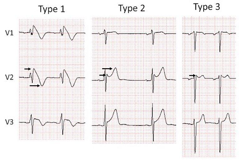
Image 1. Type 1, 2, 3 Brugada electrocardiogram patterns (left to right). Only type 1 is diagnostic of Brugada syndrome. Type 1 has a coved-shaped ST-segment elevation greater than 2 millimeters (mm) (short arrow), followed by a negative T-wave (long arrow). Type 2 has a saddleback ST-segment elevation greater than 2 mm (short arrow), followed by a positive T-wave (long arrow). Type 3 can resemble either morphology but with less than 2 mm of ST-segment elevation (arrow).
Published from the 2016 J-waves syndromes expert consensus conference report.3 Reprinted with permission.
CPC-EM Capsule
What do we already know about this clinical entity?
Brugada syndrome is a channelopathy associated with electrocardiogram (ECG) abnormalities and sudden cardiac death. Some medications may unmask it.
What makes this presentation of disease reportable?
This presentation is reportable because lurasidone has not been previously associated with Brugada patterns.
What is the major learning point?
Lurasidone and many other antipsychotics can cause ECG abnormalities and may unmask Brugada patterns.
How might this improve emergency medicine practice?
Physicians should be vigilant for Brugada patterns and other ECG findings in syncope patients, especially those on multiple psychotropic medications. hyperglycemia, nausea, and extrapyramidal reactions.4 Notably, there is no documented association between lurasidone and Brugada syndrome.
A 14-year-old male previously diagnosed with autism, who was taking lurasidone as a behavioral suppressant, presented to the emergency department (ED) after a reported syncopal episode in the bathroom. The patient, along with his mother, denied any previous history of syncopal episodes. In the week leading up to the incident, the patient’s pediatrician had escalated his lurasidone dosage from 20 milligrams (mg) to 40 mg daily due to increased behavioral challenges at school. Aside from this adjustment, the patient reported no recent illnesses or heightened stressors and denied any history of smoking, alcohol use, or illicit drug consumption.
In the ED, his vital signs were as follows: temperature 98.1 °F (36.7 °Celsius), heart rate 70-90 beats per minute, respiratory rate 16-20 breaths per minute, and blood pressure 138/71 millimeters of mercury. Orthostatic vitals were unremarkable for any change in heart rate or blood pressure. Physical examination revealed an alert and oriented Black male in no acute distress. There were no findings concerning for significant trauma or clinical dehydration. He was without any respiratory distress, with lungs clear to auscultation bilaterally. On cardiac auscultation, there was a regular rate and rhythm with no appreciable murmurs. His abdomen was soft, non-tender, and non-distended. He had an appropriate range of all his extremities and ambulated without difficulty.
He had an appropriate mood and affect, had no neurological deficits, and was overall asymptomatic.
In the ED, an ECG revealed a Type 2 Brugada pattern (Image 2). No prior documented ECG was available for comparison. Following the exclusion of other correctable causes of syncope, pediatric cardiology was consulted, and the attending physician concurred with the diagnosis of a type 2 Brugada pattern. Subsequently, the patient was admitted to the pediatric intensive care unit for continuous telemetry and observation. Throughout the night, the patient remained asymptomatic, and the ECG continued to display the type 2 Brugada pattern the following morning. Cardiology devised a treatment plan consisting of a 24-hour Holter monitor, an outpatient procainamide challenge test at a quaternary-care center, and a reduction of the patient’s lurasidone medication to 20 mg.
Several days later, the patient followed up with his pediatrician for re-evaluation and Holter monitor interrogation. The patient’s initial ECG revealed a type 2 Brugada pattern. A saddleback, 2 mm ST-segment elevation was observed in leads V2 and V3, followed by a positive T-wave. Per the cardiology report:
[T]here is minimal heart rate variability, and only variable results include one episode of sinus
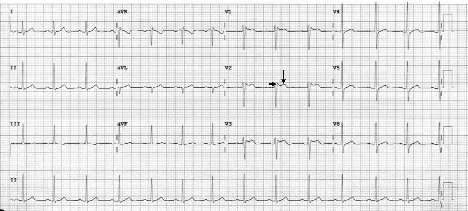
Image 2. The patient’s initial electrocardiogram reveals a type 2 Brugada pattern. A saddleback ST-segment elevation of 2 millimeters is visible in leads V2 and V3 (short arrow), followed by a positive T-wave (long arrow).
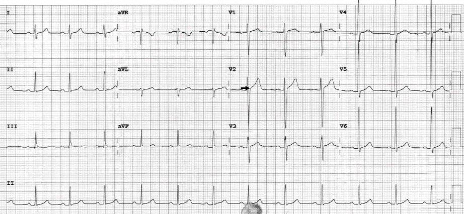
Image 3. The subsequent electrocardiogram demonstrates the resolution of the Brugada pattern in this patient. Leads V2 and V3 no longer exhibit the coved or saddleback ST-segment elevation (arrow).
tachycardia, and two isolated premature beats likely supraventricular ectopic beats. There is no evidence of tachyarrhythmias or conduction abnormalities or sinus pauses.”
The patient was awaiting a procainamide study scheduled for one week later at a quaternary-care hospital, which was ultimately negative. Throughout this period, the patient remained asymptomatic and exhibited controlled behavior on his initial dose of lurasidone 20 mg.
One month later, the patient followed up with his primary pediatric cardiologist. His mood was stable on lurasidone 20 mg, and a repeat ECG showed sinus rhythm without any findings suggesting a Brugada pattern (Image 3). During a subsequent phone call with the patient’s family, it was noted that there were no further episodes of syncope, and no planned follow-up appointments with pediatric cardiology were required. There were no additional ED visits.
Brugada syndrome, an autosomal dominant heart disorder with variable expression, poses a risk of ventricular arrhythmia and sudden cardiac death, particularly in young individuals with structurally normal hearts. Men face an elevated risk, and the average age of cardiac arrest is approximately 45 years. Symptoms typically manifest between 20-65 years of age and, although rare, there are documented cases of sudden death due to Brugada syndrome in children.4,5 The primary therapeutic approach involves implantable cardioverter-defibrillator (ICD) placement. In some cases, patients may receive supplemental quinidine or amiodarone therapy either as a bridge to ICD or to diminish the frequency of ICD shocks.4,5
Diagnosis of Brugada syndrome hinges on the presence of the type 1 Brugada ECG pattern. According to the 2013 consensus statement by the Heart Rhythm Society, European Heart Rhythm Association, and Asia Pacific Heart Rhythm Society, a definitive diagnosis occurs when a type 1 Brugada
ECG pattern is observed spontaneously or following provocative drug testing. For symptomatic patients presenting with a type 2 or type 3 pattern, provocative drug testing with a sodium channel blocker is indicated.3 In patients with type 1 Brugada ECG pattern, workup to exclude structural abnormalities may be indicated, as Brugada patterns have been observed as precursor findings of arrhythmogenic right ventricular cardiomyopathy.2
Nineteen genes encoding sodium, calcium, and potassium channels have been associated with Brugada syndrome. The most commonly mutated gene is the sodium voltage-gated alpha subunit 5 (seen in ~20-30% of patients). These mutations produce a reduced inward sodium or calcium current or an increased outward potassium current.4 These mutations result in both normal and abnormal channels within the epicardium. This results in adjacent myocytes with different refractory periods hypothesized to produce sustained arrhythmias via re-entry during repolarization or abnormal conduction during depolarization.3,4
Several classes of medications have been shown to induce the Brugada pattern. Sodium channel-blocking medications, such as procainamide, ajmaline, or flecainide, can unmask the Brugada pattern and are sometimes intentionally administered for diagnostic purposes.5 The BrugadaDrugs.org registry contains a collection of case reports and mechanistic studies of medications precipitating a Brugada pattern. Per this registry, tricyclic antidepressants, several selective serotonin reuptake inhibitors, several typical antipsychotics, lamotrigine, lithium, and propofol have been listed as drugs that can induce the Brugada ECG pattern. Therefore, caution is recommended in administering these drugs to individuals with Brugada syndrome.6
To date, few studies have demonstrated Brugada syndrome due to atypical antipsychotics.7 Three case reports have documented the emergence of a type 1 Brugada pattern following clozapine administration, with resolution upon discontinuation.8-10 Another case involved a 25-year-old male, treated with risperidone for schizophrenia, who developed a
Start et al. Lurasidone-Induced Type 2 Brugada Pattern in a Pediatric Patient
type 1 Brugada pattern that spontaneously resolved after discontinuation.11 The typical antipsychotics loxapine, trifluoperazine, perphenazine, and thioridazine have also been associated with Brugada syndrome due to the blockade of fast sodium channels.3 Intriguingly, patients undergoing treatment for schizophrenia exhibit a higher prevalence of Brugada syndrome; however, whether this is linked to antipsychotic side effects or a genetically related channelopathy remains unknown.12 Antipsychotic therapy more commonly is associated with other causes of sudden cardiac death, particularly QT prolongation and subsequent torsades de pointes.3
The prognosis of Brugada syndrome is highly variable based on initial presentation. In a study of 1,029 patients diagnosed with Brugada syndrome by type 1 ECG, patients presenting in cardiac arrest had a 35% incidence of ventricular tachyarrhythmia at four years. Six percent of Brugada syndrome patients presenting with syncope had an arrhythmic event at four years.13 This massive difference in mortality is likely due to difficulty distinguishing arrhythmic syncope from vasovagal syncope in this cohort. A separate longitudinal study of 1,149 patients demonstrated a 0.4% yearly risk of arrhythmia with spontaneous type 1 Brugada patterns relative to those with provocatively induced type 1 patterns, who carry a significantly lower risk of 0.03%.1 In contrast, type 2 and type 3 Brugada patterns that do not convert to a type 1 pattern with electrophysiologic testing are considered non-diagnostic of Brugada syndrome. In a longitudinal study of 18 Finnish patients with type 2 and type 3 ECG patterns, no lifethreatening ventricular arrhythmias occurred during a followup period of 10-21 years.14
In all patients presenting with syncope, an ECG is warranted to identify underlying arrhythmogenic etiologies, including Brugada syndrome. Patients with Brugada syndrome, as diagnosed by a type 1 ECG, are treated with an implantable cardioverter-defibrillator. In contrast, those with lower ventricular arrhythmia risk type 2 and type 3 ECG patterns require additional electrophysiological workup. This case illustrates a transient mimic of a Brugada pattern, characterized by negative results in pharmacological induction tests and ECG resolution after discontinuing the initial causative agent.
Patient consent has been obtained and filed for the publication of this case report.
1. Gaita F, Cerrato N, Giustetto C, et al. Asymptomatic patients with Brugada ECG pattern: long-term prognosis from a large prospective study. Circulation. 2023;148(20):1543-1555.
2. Priori SG, Wilde AA, Horie M, et al. HRS/EHRA/APHRS expert consensus statement on the diagnosis and management of patients
Address for Correspondence: Aldrin Enabore, MD, Dell Children’s Medical Center, Department of Emergency Medicine, 701 Grove Rd, Greenville, SC 29605. Email: enabore2019@gmail.com
Conflicts of Interest: By the CPC-EM article submission agreement, all authors are required to disclose all affiliations, funding sources and financial or management relationships that could be perceived as potential sources of bias. The authors disclosed none.
Copyright: © 2025 Start et al. This is an open access article distributed in accordance with the terms of the Creative Commons Attribution (CC BY 4.0) License. See: http://creativecommons.org/ licenses/by/4.0/
with inherited primary arrhythmia syndromes. Heart Rhythm 2013;10(12):1932-1963.
3. Antzelevitch C, Yan GX, Ackerman MJ, et al. J-wave syndromes expert consensus conference report: emerging concepts and gaps in knowledge. Heart Rhythm. 2016;13(10):E295-E324.
4. Sicouri S, Antzelevitch C. Mechanisms underlying the actions of antidepressant and antipsychotic drugs that cause sudden cardiac arrest. Arrhythm Electrophysiol Rev. 2018;7(3):199-209.
5. Probst V, Denjoy I, Meregalli PG, et al. Clinical aspects and prognosis of Brugada syndrome in children. Circulation 2007;115(15):2042-2048.
6. Postema PG, Wolpert C, Amin AS, et al. Drugs and Brugada syndrome patients: review of the literature, recommendations, and an up-to-date website (www.brugadadrugs.org). Heart Rhythm 2009;6(9):1335-1341.
7. Rastogi A, Viani-Walsh D, Akbari S, et al. Pathogenesis and management of Brugada syndrome in schizophrenia: a scoping review. Gen Hosp Psychiatry. 2020;67:83-91.
8. Sawyer M, Goodison G, Smith L, et al. Brugada pattern associated with clozapine initiation in a man with schizophrenia. Intern Medicine J. 2017;47(7):831-833.
9. Ukaigwe A, Oluwasanjo A, Donato AA. Clozapine and Brugada –unlikely but true. J Gen Intern Med. 2013;28:S302-S302.
10. Chong JHE, F. Clozapine-induced reversible Brugada syndrome. GSTF J Adv Med Res. 2015;1(3):18-20.
11. Laboudi F, Slimani G, Gourani ME, et al. Neuroleptic-induced Brugada syndrome – a case report. Int J Med Surg. 2018;5(1):189. Available at https://mbmj.org/index.php/ijms/article/view/92/283. Accessed May 1, 2025.
12. Blom MT, Cohen D, Seldenrijk A, et al. Brugada syndrome ECG Is highly prevalent in schizophrenia. Circ Arrhythm Electrophysiol 2014;7(3):384-391.
13. Probst V, Veltmann C, Eckardt L, et al. Long-term prognosis of patients diagnosed with Brugada syndrome: results from the FINGER Brugada registry. Circulation. 2009;30:886-886.
14. Junttila MJ, Raatikainen MJP, Karjalainen J, et al. Prevalence and prognosis of subjects with Brugada-type ECG pattern in a young and middle-aged Finnish population. Eur Heart J. 2004;25(10):874878.
Mitch Garey, DO*
Joy McLaughlin, MD*
Harman Kaur, DO*
Jason Graf, MD*
Jacklyn Garcia, MD†
Megan De Kok, MSc‡
Alexander John Scumpia, DO, MHA, MSc*§
Section Editor: Christopher Sampson, MD
HCA FL Aventura Hospital, Department of Emergency Medicine, Florida
HCA FL Aventura Hospital, Department of Radiology, Florida
Rocky Vista University College of Osteopathic Medicine, Parker, Colorado
Lakeside Medical Center, Department of Emergency Medicine, Belle Glade, Florida
Submission history: Submitted September 20, 2024; Revision received December 4, 2024; Accepted January 17, 2025
Electronically published May 10, 2025
Full text available through open access at http://escholarship.org/uc/uciem_cpcem DOI: 10.5811/cpcem.33483
Introduction: Altered mental status presentations are commonplace in the emergency department (ED), but not all are due to psychiatric etiologies, even if the patient has had a previous psychiatric diagnosis. It is critical to evaluate for organic causes of a patient’s altered presentation. This case highlights the necessity of a broad workup to correctly diagnose an altered patient.
Case Report: A 23-year-old Haitian male with a past medical history of bipolar 1 disorder, seizure disorder, and developmental delay presented to a critical access ED for altered mental status. The patient was given 300 milligrams of ketamine for delirious hyperactivity and agitation by emergency medical services in the field. On physical examination, the patient was in acute respiratory distress, hypoxic, not tolerating secretions, tachycardic, lethargic, and was subsequently intubated for airway protection. Computed tomography (CT) of the brain without contrast was obtained and revealed findings consistent with Fahr disease.
Conclusion: Fahr disease is a rare neurodegenerative condition that causes accumulation of calcium deposits in the basal ganglia as demonstrated on CT.1 Phenotypes can be variable, including symptoms such as parkinsonism, chorea, dystonia, cognitive impairment, and ataxia.2 This case illustrates the importance of a broad differential diagnosis and emergent medical interventions for emergency physicians practicing in critical access facilities. [Clin Pract Cases Emerg Med. 2025;9(3):278-281.]
Keywords: delirious hyperactivity; agitation; basal ganglia; Fahr disease; intracranial calcifications.
Karl Theodor Fahr first described calcifications of the basal ganglia with associated dementia nearly a century ago, contributing to prior postmortem case descriptions by mid-19th century physicians Delacour and Bamberger.1 Fahr disease (FD) is a rare and progressive neurological pathology characterized by idiopathic, bilateral basal ganglia calcifications. It is known to be insidious and degenerative, presenting with extensive phenotypic diversity. Fahr syndrome is differentiated from FD by having an identified secondary
cause of calcification and expands the location of calcification beyond the basal ganglia.2 To further complicate understanding of these pathologies, close to 30 different names including bilateral strio-pallido-dentate calcinosis and calcinosis nucleurum have historically been used.2,3 Most recently, primary bilateral brain calcification was introduced as an alternative to FD, encompassing both hereditary and idiopathic etiologies.1,2
Clinical symptoms of basal ganglia calcification can be vast, encompassing asymptomatic presentations, mood
disorders, and extrapyramidal movement disorders. The increasing variety of presenting symptoms in case reports coupled with the interchangeability of terms used to describe intracranial calcification makes FD both an epidemiologic and diagnostic challenge. Bilateral calcification of the basal ganglia identified on computed tomography is considered a diagnostic hallmark; however, it is not specific and estimated to be found in up to 20% of asymptomatic patients over 50 years of age.4 Diagnosis of FD should be especially considered in patients with evidence of genetic involvement, which can be autosomal dominant or recessive. Seven distinct genetic mutations have been associated with primary familial brain calcification, four of which are dominant. Concerns continue to arise in using this data, as roughly half of cases lack specific genetic findings.5 The case we describe is one of an acute psychiatric presentation that is notable for the young age of the patient and the diagnostic course, which revealed evidence of FD in a critical access rural emergency department (ED).
A 23-year-old Haitian male with a past medical history of bipolar 1 disorder, seizure disorder, and developmental delay presented to a critical access ED for altered mental status. The patient was given 300 milligrams (mg) of ketamine for delirious hyperactivity and agitation by emergency medical services in the field. Aside from the diagnoses, no additional medical history or situational context prior leading to the patient’s altered mental status was available upon the patient’s arrival to the ED; specifically, there were no reports of known recreational drug use or specific concerns regarding a toxicologic exposure. His initial vital signs upon ED arrival were as follows: temperature, 37.2 °Celsius; respiratory rate, 42 breaths per minute; blood pressure, 245/170 millimeters of mercury; heart rate ,144 beats per minute; and oxygen saturation of 72% on 4 liters nasal cannula.
On physical examination the patient was in acute respiratory distress, hypoxic, tachycardic, lethargic, and was not tolerating oral secretions. Pupils were equal, round, and sluggish to light bilaterally. His neurological and psychiatric examinations were limited secondary to his clinical presentation. The patient was not moving his extremities nor tolerating oral secretions and was immediately intubated via video laryngoscopy with rapid sequence intubation (etomidate, rocuronium, respectively). Propofol and fentanyl were used for sedation as well as a nicardipine drip for blood pressure control and levetiracetam for seizure prophylaxis.
The patient received an extensive workup including comprehensive metabolic panel, complete blood count with differential, arterial blood gas, lactic acid, blood cultures, coagulation profiles, type and screen, urine analysis, creatine phosphokinase, drug screen, salicylate, acetaminophen, and ethanol levels, and influenza and coronavirus disease 2019 testing. Laboratory analysis was significant for hyperglycemia at 172 mg/deciliter (dL) (reference range: 70-100 mg/dL),
CPC-EM Capsule
What do we already know about this clinical entity?
Fahr disease is a neurodegenerative disorder causing basal ganglia calcifications, with diverse symptoms including movement disorders and cognitive decline.
What makes this presentation of disease reportable?
The young age at presentation, severe psychiatric symptoms, and discovery in a critical access ED highlight the need for a broad differential diagnosis.
What is the major learning point?
A thorough workup is essential for altered mental status to uncover rare conditions such as Fahr disease, which may mimic psychiatric disorders.
How might this improve emergency medicine practice?
Early imaging and broad diagnostic considerations can enhance detection of organic causes in psychiatric presentations, leading to better patient outcomes.
hypokalemia at 2.9 milliequivalents per liter (mEq/L) (3.5-5.0 mEq/L), elevated creatinine at 1.86 mg/dL (0.7-1.3mg/dL), hypocalcemia at <5 mg/dL (9-10.5mg/dL), elevated lactic acid at 3.9 millimoles (mmol)/L (0.67-1.8 mmol/L), elevated creatine phosphokinase at 2,284 units (U)/L (30-170 U/L), and a positive COVID-19 test, but no single value was ultimately deemed contributory to the patient’s agitation. All other laboratory studies were grossly unremarkable or within normal limits.
Electrocardiogram, chest radiography, and computed tomography (CT) angiography of the chest were obtained and revealed a right lower lobe pneumonia but were negative for other pathology such as pulmonary embolus or aortic dissection. In addition to the medications, the patient was given piperacillin/tazobactam and vancomycin to cover empirically for aspiration pneumonia and/or sepsis. A 2-liter bolus of lactated Ringer solution was also administered. Computed tomography without contrast of the head was obtained (Image), which revealed findings consistent with FD.
Of note, upon discussion with the patient’s mother, who arrived in the ED during the patient’s initial workup and stabilization, the patient had never previously received CT
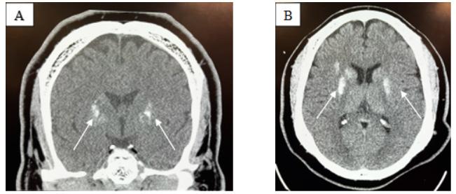
imaging of the brain prior to this presentation, despite multiple prior presentations of agitation that were attributed to psychiatric disorders. Additionally, the patient’s father presented to the ED with the patient’s mother and stated he had Parkinson disease. The patient’s father was noted at that time to exhibit gross neurologic deficits atypical of Parkinson disease at the father’s age.
The patient was ultimately stabilized and transferred to a tertiary-care hospital where he underwent further diagnostics and management by a multidisciplinary team including an intensivist, a neurologist, and a medical geneticist. Laboratory analysis at the tertiary-care hospital revealed a normal parathyroid hormone level. The patient was found to have a genetic cause of his FD. Both the patient and his father had further genetic testing performed, which revealed an autosomal dominant trait in both, confirming our initial diagnosis of FD.
While adult-onset FD classically presents in the fourth and fifth decades of life, this case demonstrates clinical manifestations in a 23-year-old, who began showing symptoms of cognitive impairment in his early teens. Jaworski et al delineate sub-types of Fahr presentations by age of onset, with childhood onset characterized by developmental delay, early onset (third decade) characterized by psychiatric symptoms, and late onset (fifth decade) characterized by progressive dementia and movement disorders.5 Fahr disease has previously been reported to demonstrate an anticipatory genetic effect, wherein subsequent affected generations present symptoms at progressively younger ages, often with increased severity.6,12 The severity of Fahr symptoms has been correlated to the extent of intracranial calcification, highlighting the importance of early diagnosis and prompt management, especially when due to secondary causes with distinct treatments.5
Our patient’s comorbid diagnoses of severe developmental delay, mood disorder, and seizure disorder likely contributed to his delayed diagnosis of basal ganglia
calcification. Naqvi et al recently described a case of bilateral calcification of the basal ganglia in a 21-year-old male discovered on CT during evaluation of acute exacerbation of schizophrenia.9 That patient had been diagnosed with schizophrenia three years prior and only received CT imaging after having a seizure.9 Nearly 40% of FD patients present with only psychiatric symptoms, making the diagnosis challenging.8 The utility of imaging is exemplified by Nicolas et al who describe a case in which a patient presenting with psychosis in the fourth decade of life was found to have basal ganglia calcification on imaging.10 In that case, the the CTs of both the patient’s parents revealed similar calcifications; however, both of her parents were asymptomatic.10
Our patient’s severe hypocalcemia and calcified basal ganglia, as well as his psychiatric and seizure history are highly suggestive of FD. His age and mood symptoms align with bipolar disorder; however, a more thorough evaluation should have included organic causes of neuropsychiatric symptoms, especially considering his seizure history and developmental delay. Additionally, the patient’s father also displayed signs of a neurocognitive pathology, which further suggested a genetic factor. Early diagnosis of this disorder has implications for management and, importantly, for genetic counseling. Cassamina et al described a case of FD with bipolar disorder refractory to pharmaceutical intervention in which electroconvulsive therapy provided complete resolution of mood, cognitive, and behavioral symptoms for several years.11
Limitations in this case largely result from the patient arriving in acute respiratory distress.
While intramuscular ketamine is a known sedative agent in acute psychiatric agitation, the patient’s presentation was not consistent with respiratory depression but rather severely altered mental status resulting in the inability to protect his airway. Additionally, the patient’s known history of seizure disorder, which may have caused this patient to present postictal, could have explained his initial symptoms. Moreover, genetic studies would have been beneficial, although the acuity of the patient’s
Garey et al.
Delirious Hyperactivity and Agitation Unveiling an Intriguing Underlying Diagnosis
deterioration upon arrival to the ED limited such analyses. Ultimately, it is impossible to conclude definitively what caused this patient’s altered mental status before and upon arrival to the ED. Despite this, the intended objective of bringing this case to light is not to retrospectively explain one event of altered mental status but rather demonstrate the need for a broad differential in such cases, as this patient had a profound and previously unrecognized pathology that had affected him throughout his life.
For patients with newly diagnosed psychiatric disorders accompanied by neurologic symptoms such as seizure activity and/or family history of psychiatric or neurologic deterioration, imaging is a vital element of the diagnostic workup. In this report we did not aim to challenge the current multifactorial understanding of psychiatric disorders but rather to highlight calls for increased use of imaging of new psychiatric and neurologic diagnoses to better characterize the highly variable presentations associated with intracranial calcification. Treatment course varies depending on underlying etiology, which makes early diagnosis vitally important for optimizing patient outcomes.
The genetic anticipation of Fahr disease further illustrates this point, as early genetic counseling can be provided. Further research and systematic review of case presentations are needed to better characterize FD and cement the naming scheme of the pathology. Distinct classification of symptoms, quantitative imaging findings, and genetic analyses can create definitive diagnostic criteria, improving time to diagnosis and thus intervention for these patients.
Address for Correspondence: Alexander J. Scumpia, DO, MHA, MSc, Lakeside Medical Center, Department of Emergency Medicine, 39200 Hooker Highway, FL 33430. Email: ascumpia@ yahoo.com
Conflicts of Interest: By the CPC-EM article submission agreement, all authors are required to disclose all affiliations, funding sources and financial or management relationships that could be perceived as potential sources of bias. The authors disclosed none.
Copyright: © 2025 Garey et al. This is an open access article distributed in accordance with the terms of the Creative Commons Attribution (CC BY 4.0) License. See: http://creativecommons.org/ licenses/by/4.0/
The authors attest that their institution does not require Institutional Review Board approval of this case report. Patient consent was obtained. Documentation on file.
1. Donzuso G, Mostile G, Nicoletti A, et al. Basal ganglia calcifications (Fahr’s syndrome): related conditions and clinical features. Neurol Sci. 2019;40(11):2251-2263.
2. Saleem S, Aslam H, Anwar M, et al. Fahr’s syndrome: literature review of current evidence. Orphanet J Rare Dis. 2013;8(1):156.
3. Manyam BV. What is and what is not “Fahr’s disease.” Park Rel Disord. 2005;11(2):73-80.
4. Golüke NMS, Meijer E, van Maren EA, et al. Amount and Distribution of Intracranial Calcification in Symptomatic and Asymptomatic Primary Familial Brain Calcification. Neurol Clin Pract. 2023;13(4):e200163.
5. Jaworski K, Styczyńska M, Mandecka M, et al. Fahr syndrome – an important piece of a puzzle in the differential diagnosis of many diseases. Pol J Radiol. 2017;82:490-3
6. Chen SY, Ho CJ, Lu YT, et al. The Genetics of Primary Familial Brain Calcification: A Literature Review. Int J Mol Sci. 2023;24(13):10886.
7. Goyal D, Khan M, Qureshi B, et al. Would you recognize Fahr’s disease if you saw it? Inn Clin Neurosci. 2014;11(1-2):26-28.
8. Gülsün M, Baykız AF, Kabataş S, et al. Fahr syndrome: three cases presenting with psychiatric signs. Eur J Gen Med. 2006;3(1):35-40.
9. Naqvi S, Arshad S, Hanif R, et al. Fahr’s syndrome misdiagnosed as schizophrenia: a case report. Cureus. Published online March 2, 2017. Accessed November 5, 2024.
10. Nicolas G, Guillin O, Borden A, et al. Psychosis revealing familial idiopathic basal ganglia calcification. Gen Hosp Psychiatry. 2013;35(5):575.e3-575.e575005.
11. Casamassima F, Lattanzi L, Perlis RH, et al. Efficacy of electroconvulsive therapy in Fahr disease associated with bipolar psychotic disorder. J ECT. 2009;25(3):213-5.
12. Maeda K, Idehara R, Nakamura H, et al. Anticipation of familial idiopathic basal ganglia calcification? Intern Med J 2012;51(8):987.
13. Teplin LA, Abram KM, Luna MJ. Racial and ethnic biases and psychiatric misdiagnoses: toward more equitable diagnosis and treatment. Amer J Psychiatry. 2023;180(6):402-3.
Clates P. Adams, DO*
Christian I Wade, MD†
Section Editor: Lev Libet, MD
Madigan Army Medical Center, Department of Emergency Medicine, Tacoma, Washington
Brooke Army Medical Center, The San Antonio Uniformed Service Health Education Consortium, Department of Emergency Medicine, San Antonio, Texas
Submission history: Submitted October 27, 2024; Revision received January 17, 2025; Accepted February 19, 2025
Electronically published May 6, 2025
Full text available through open access at http://escholarship.org/uc/uciem_cpcem DOI: 10.5811/cpcem.38444
Introduction: Sympathetic crashing acute pulmonary edema (SCAPE), also known as flash pulmonary edema or hypertensive acute heart failure, is a critical condition characterized by a rapid escalation of sympathetic outflow, excessive afterload, and worsening heart failure. Although rare, contrast media-induced pulmonary edema is a severe adverse reaction, occurring in 0.001-0.008% of patients receiving intravenous contrast and accounting for 10-20% of lethal contrast reactions.
Case Report: A 70-year-old male developed acute respiratory distress shortly after undergoing an outpatient, contrast-enhanced computed tomography. Despite treatment for suspected anaphylaxis, the patient’s condition continued to deteriorate until a diagnosis of SCAPE was ultimately recognized. Treatment with high-dose nitroglycerin, non-invasive positive pressure ventilation (NIPPV), and eventual intubation resulted in the patient’s full recovery.
Conclusion: This report highlights the importance of recognizing SCAPE in patients presenting with sudden dyspnea after contrast administration and emphasizes the need for early intervention with NIPPV and vasodilators to reduce morbidity and mortality. [Clin Pract Cases Emerg Med. 2025;9(3): 282-284.]
Keywords: SCAPE; pulmonary edema; hypoxia; contrast induced; case report.
INTRODUCTION
Sympathetic crashing acute pulmonary edema (SCAPE), also known as flash pulmonary edema or hypertensive acute heart failure, is a critical and rapidly progressing condition. It is characterized by an acute increase in sympathetic outflow, excessive afterload, and worsening heart failure. Although SCAPE is rare, contrast media-induced SCAPE occurs in only 0.001-0.008% of all cases of SCAPE but accounts for 10-20% of lethal contrast reactions. 1-3
Sympathetic acute crashing pulmonary edema is more frequently seen in patients with chronic left ventricular dysfunction, particularly those with coexisting hypertension and renal artery stenosis. Additionally, factors that increase sympathetic tone and catecholamine release can precipitate SCAPE, creating a vicious cycle of dyspnea, anxiety, and worsening of the patient’s presentation.4,5 Early recognition
and appropriate management are crucial to improving patient outcomes in such cases.
A 70-year-old male with a history of coronary artery disease status post two stents in the right coronary artery, atrial fibrillation, hypertension, obstructive sleep apnea, and eosinophilic esophagitis presented to the emergency department (ED) in acute respiratory failure. Approximately 20 minutes before his presentation to the ED, the patient had received intravenous (IV) contrast for a routine outpatient computed tomography (CT) hematuria protocol. While undergoing the study, he experienced sudden-onset nausea, tachypnea, tachycardia, and respiratory distress.
Upon arrival to the ED, his first set of vitals were as follows: systolic blood pressure (BP), 151 millimeters of
mercury (mm Hg); heart rate, 112 beats per minute; oxygen saturation, 74% on room air; and respiration rate, 30 breaths per minute. Physical examination was significant for an acutely distressed, diaphoretic male with increased work of breathing, diffuse rales in all lung fields, and tachycardia without murmurs, gallops, or rubs. There was no evidence of lower extremity edema, and no evidence of urticaria. He was immediately placed on nasal cannula. Given the recency of the contrasted CT study and his clinical presentation, an anaphylactic reaction was initially suspected. He was treated with three successive doses of intramuscular epinephrine (0.3 milligram [mg] per dose), nebulized ipratropium-albuterol (3 milliliters [mL]), diphenhydramine (50 mg), famotidine (20 mg), magnesium (2 grams), and methylprednisolone (125 mg).
Despite these interventions, the patient’s respiratory failure persisted, leading to the initiation of an epinephrine drip and the application of a non-rebreather mask, although without improvement to his oxygen saturation.
Due to the patient’s worsening condition, which at that point was characterized by severe hypertension (systolic BP>200 mm Hg, partially secondary to our aforementioned interventions of epinephrine), rhonchorous breath sounds, and lack of wheezing, lack of urticaria, absence of mucosal edema, and lack of response to epinephrine, SCAPE was suspected. An electrocardiogram (ECG) and chest radiograph (CXR) were obtained. The ECG showed no signs of ischemia, while the CXR revealed significant right-sided pulmonary edema (Image). The epinephrine drip was discontinued, and the patient was administered IV furosemide (20 mg), followed by a nitroglycerin drip (starting at 5 micrograms per minute [mcg/min]), and placed on continuous positive airway pressure (CPAP).
CPC-EM Capsule
What do we already know about this clinical entity?
Sympathetic Crashing Acute Pulmonary Edema (SCAPE) is a critical condition seen in emergency departments, characterized by rapid escalation of sympathetic outflow, excessive afterload, and worsening heart failure that requires prompt recognition and treatment.
What makes this presentation of disease reportable?
This presentation is reportable given the use of contrast-media, in which this presentation occurs in 0.001 - 0.008% of all patients receiving intravenous contrast, but accounts for 10-20% of lethal contrast reactions.
What is the major learning point?
The major learning point from this condition is early recognition of SCAPE, and prompt treatment with high-dose nitroglycerin and noninvasive positive pressure ventilation (NIPPV).
How might this improve emergency medicine practice?
This case may improve emergency medicine practice to broaden the differential diagnosis of patients suffering from adverse contrast reactions, from the more common anaphylaxis.
To improve CPAP tolerance, the patient received lorazepam (1 mg), which temporarily stabilized oxygen saturation to the low 90s. However, the patient eventually became increasingly somnolent, with oxygenation saturations dropping to the mid-80s to low 90s. A venous blood gas revealed acute hypercapnic acidosis (pH 7.15, reference range: 7.29-7.45) and partial pressure of carbon dioxide 67 mm Hg (30.0-68.0 mm Hg), prompting the decision to intubate the patient. After successful intubation, the patient was placed on a ventilator, administered propofol, and admitted to the intensive care unit. There, a point-of-care echocardiogram demonstrated a normal left ventricular ejection fraction (>60%), a small-to-moderate pericardial effusion, and mild tricuspid regurgitation. The patient was eventually discharged home seven days after presentation.
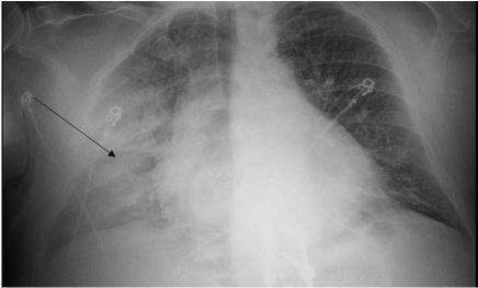
. Initial emergency department chest radiograph with evidence of acute right-sided “flash” pulmonary edema (arrow).
Sympathetic acute crashing pulmonary edema is classified as a non-cardiogenic pulmonary edema and is thought to result from four primary mechanisms: catecholamine release, increased left atrial pressure leading to distension of small pulmonary capillaries, vascular endothelial cell damage
Media-induced Sympathetic Crashing Acute Pulmonary Edema
causing interstitial edema, and increased microvascular permeability.2,4,6 The elevated catecholamine levels result in an increased heart rate, reduced diastolic time, and activation of the renin-angiotensin-aldosterone system, exacerbating diastolic stiffening and increasing diastolic and mean arterial pressures, thus contributing to pulmonary edema. Additionally, heightened sympathetic tone adversely affects pulmonary circulation by increasing permeability, provoking pulmonary capillary failure and causing splanchnic venoconstriction, further worsening dyspnea and sympathetic activation.4,5,7
The administration of contrast media triggers the release of inflammatory mediators and complement activation, leading to endothelial damage. This damage increases microvascular permeability, resulting in fluid accumulation in the lungs. The leakage of fluids from the circulation raises hemoglobin concentration and packed-cell volume, along with increased left atrial pressure. Furthermore, the complement system releases vasodilatory substances, increases vascular permeability and edema, promotes smooth muscle cell contraction, precipitates bronchospasm, and increases mucus secretion in the airways.3
Contrast-induced pulmonary edema is an exceedingly rare but highly lethal phenomenon, occurring in 0.001-0.008% of patients receiving IV contrast, with a mortality rate of 1020%.1-3 Given the high frequency of contrast-enhanced studies performed in the ED, SCAPE should be a key differential diagnosis in patients presenting with any combination of sudden dyspnea, tachypnea, hypoxemia, rales, or hypertension. The treatment of SCAPE centers on oxygen administration via non-invasive positive pressure ventilation (NIPPV) or invasive ventilation with positive end-expiratory pressure and the use of high-dose vasodilators (such as nitroglycerin). Volume removal (diuresis or dialysis) may be indicated based on clinical judgement.2,3,6,8
Non-invasive positive pressure ventilation reduces the work of breathing, decreases preload and afterload, and has proven effective in preventing intubation.8 Studies indicate no significant difference in outcomes between bilevel positive airway pressure and continuous positive airway pressure.8 Vasodilators, such as nitroglycerin, relieve pulmonary congestion by reducing preload and afterload.8 The recommended initial dose is a bolus of 1,000-2,000 mcg over two minutes, followed by an infusion of 50-300 mcg/min, with rapid titration up to 800 mcg/min if needed, targeting a BP of less than 140 mm Hg.5,8 In cases of refractory hypertension, clevidipine (preferred) or nicardipine should be considered.5
Sympathetic crashing acute pulmonary edema is a critical condition driven by a vicious cycle of increasing sympathetic outflow, excessive afterload, and worsening heart failure.5 When evaluating a patient for SCAPE, it is essential to differentiate it from angioedema and anaphylaxis, as the
treatments differ significantly. Early recognition and prompt intervention, including the use of NIPPV and high-dose nitroglycerin, are crucial to improving patient outcomes and avoiding intubation and death.
The authors attest that their institution requires neither Institutional Review Board approval, nor patient consent for publication of this case report. Documentation on file.
Address for Correspondence: Clates P. Adams, DO, Madigan Army Medical Center, Department of Emergency Medicine, 9040 Jackson Ave, Tacoma, WA 98341.Email: clatesadams@gmail.com.
Conflicts of Interest: By the CPC-EM article submission agreement, all authors are required to disclose all affiliations, funding sources and financial or management relationships that could be perceived as potential sources of bias. The authors disclosed none. The views expressed in this publication are those of the author(s) and do not reflect the official policy or position of Madigan Army Medical Center, Brooke Army Medical Center, the San Antonio Uniformed Services Health Education Consortium, the Department of the Army, the Department of Defense, the Defense Health Agency, or the US Government.
Copyright: © 2025 Adams et al. This is an open access article distributed in accordance with the terms of the Creative Commons Attribution (CC BY 4.0) License. See: http://creativecommons.org/ licenses/by/4.0/
1. Iqbal A, Mahmood R, Jasti M, et al. Water lung: an interesting case of non-cardiogenic pulmonary edema in a heart failure patient. Cureus 2023;15(11):e48814.
2. Pincet L and Lecca G. Acute pulmonary edema induced by non-ionic low-osmolar radiographic contrast media. Open Access Emerg Med 2018;10:75-9.
3. Yi KM and Li X. Fatal noncardiogenic pulmonary edema related to nonionic, iso-osmolar iodine contrast medium: one case report. BMC Pulm Med. 2022;22(1):118.
4. Agrawal N, Kumar A, Aggarwal P, et al. Sympathetic crashing acute pulmonary edema. Indian J Crit Care Med. 2016;20(12):719-23.
5. Farkas, J. Sympathetic crashing acute pulmonary edema (SCAPE). In: The Internet Book of Critical Care. 2025. Available at: https:// emcrit.org/ibcc/scape/. Accessed February 1, 2025.
6. Nagao G, Masaki K, Kawada I, et al. Non-cardiogenic pulmonary oedema caused by iodine contrast medium. BMJ Case Rep. 2020;13(12):e239016.
7. Siddiqua N, Bhaskararayuni J, Sahu AK, et al. Taming the tiger: ultra high dose nitroglycerin in managing sympathetic crashing acute pulmonary edema patient. Am J Emerg Med. 2023;67:194.e1-194.e5
8. Mathew R, Kumar A, Sahu A, et al. High-dose nitroglycerin bolus for sympathetic crashing acute pulmonary edema: a prospective observational pilot study. J Emerg Med. 2021;61(3):271-7.
Brian Gooley, MD*
Kirk Hughes, RN*
Mark Gooley, PharmD†
Daniel Keyler, PharmD‡
Richard Vetter, MS§
Jon Cole, MD*
Section Editor: Lev Libet, MD
Minnesota Poison Control System, Minneapolis, Minnesota
Mayo Clinic, Rochester, Minnesota
University of Minnesota, Minneapolis, Minnesota
University of California Riverside, Riverside, California
Submission history: Submitted June 24, 2024; Revision received October 23, 2024; Accepted November 19, 2024
Electronically published May 28, 2025
Full text available through open access at http://escholarship.org/uc/uciem_cpcem
DOI: 10.5811/cpcem.24973
Introduction: Grammostola rosea (Chilean rose tarantula) is a common exotic pet belonging to the Theraphosidae (tarantula) family. Case reports of theraphosid bites in adults commonly describe local tissue damage and local pain. Muscle spasms have also been described as a result of the bites but are rarer. We present a case of severe and persistent muscle spasms after a G rosea bite, which is uncommonly reported in the literature.
Case Report: A 42-year-old woman was holding a G rosea tarantula when she was bit on the forearm. Within hours, severe local muscle cramping occurred. Due to worsening cramping, she initially presented to the emergency department the day after the bite, and again on the following day. She was admitted on her second visit and treated with diazepam, cephalexin, diphenhydramine, baclofen, cefpodoxime, doxycycline, prednisone, and topical hydrocortisone. Her laboratory testing was unremarkable, and while medical management may have mildly improved her symptoms, painful cramping persisted. After discharge, her paroxysmal muscle cramping continued for four weeks before completely resolving.
Conclusion: While local tissue damage and pain are common, G rosea bites may lead to severe muscle cramping that persists for weeks. Standard laboratory testing may be completely normal in these cases. Muscle cramps may be persistent and are difficult to manage. [Clin Pract Cases Emerg Med. 2025;9(3):285-288.]
Key Words: tarantula; Grammostola rosea; envenomation; muscle cramp.
INTRODUCTION
Theraphosidae (tarantulas) are popular exotic pets1 and the most common culprit of pet arachnid envenomation.2 Tarantulas, particularly the “New World” species of North, Central and South America, typically cause injuries to humans via their urticating hairs, which can penetrate into skin and mucous membranes leading to localized tissue
reactions.3 Bites from “New World” theraphosid spiders, although uncommon, can be fatal to small animals such as household pets and are generally described as benign in humans.4 Nevertheless, rare cases of severe, delayed, prolonged, diffuse muscle cramps have been reported.5 Grammostola rosea is a medium-sized tarantula native to Chile, Bolivia, and Argentina. Urticating hairs of G rosea
appear rose-colored; hence, it is commonly known as the Chilean rose or rose hair tarantula. Known for their generally docile nature, G rosea are common among hobbyists and as a result are frequently sold around the world;3 over 600,000 were traded in 2020 alone.1 Like Theraphosidae in general, bites from G rosea are rare and poorly described in the medical literature. Here we report a case of G rosea envenomation resulting in severe, painful muscle cramping that lasted for several weeks.
A 42-year-old woman was bitten by a G rosea while at an exotic animal birthday party with her child. When her child was startled by the tarantula’s behavior, the patient took the tarantula from the child (Image 1) and was subsequently bitten on the forearm.
The patient estimated the tarantula had its fangs inserted for 20-30 seconds and that it was making a “pulsing” motion with its body as she walked to the tarantula’s handler to ask for aid in removing it. The patient described the sensation as very painful with a feeling around the bite site as “scratchy, pinchy, and needles” around the two puncture wounds made by the bite (Image 2). Her arm then began to burn and ache, which persisted overnight.
The day after the bite her symptoms persisted. She called the exotic animal handler that morning and was recommended to treat the area with ice and ibuprofen, as would be done for a bee sting. Shortly after, while on a morning walk, the patient experienced severe, painful cramping in her feet that she rated as “10 of 10 pain” (Image 2). She had no history of muscle dystrophy or cramping. The patient is right-handed, and her past medical history was remarkable only for panic attacks, for which she was prescribed lorazepam as needed, but which she rarely used.
She returned home and drank an electrolyte solution with no improvement. Dorsiflexing her great toe seemed to help
CPC-EM Capsule
What do we already know about this clinical entity?
Effects beyond local tissue damage from tarantula bites are rare, and the management of these cases is mostly supportive.
What makes this presentation of disease reportable?
This is a case of prolonged muscle cramping that affected muscles far away from the site of envenomation. The cramping lasted for over a month.
What is the major learning point?
Tarantula envenomation may lead to debilitating muscle cramping that is difficult to manage with conventional medication for muscle cramps.
How might this improve emergency medicine practice?
Realizing that tarantula envenomation can lead to such prolonged symptoms will improve recognition of this rare condition and stimulate further study.
relieve the pain, while turning or twisting the leg exacerbated the cramps. Over the next few hours, she noticed that the cramps progressed proximally from her right ankle to her right thigh and hip flexors. The patient went to the local emergency department (ED), whereupon the regional poison center was consulted and recommended supportive care for muscle symptoms and local wound care. She was discharged after lab workup that included a normal basic metabolic panel and complete blood count. She was prescribed diazepam 2 milligrams (mg) every eight hours as needed and cephalexin 500 mg every six hours for seven days for muscle cramping and concern for local wound infection.
Over the subsequent hours the patient developed worsening muscle spasms despite taking 0.5 mg of her lorazepam. The cramping progressed to her abdomen and prompted her return to the ED. She did not take the diazepam but did take the cephalexin as prescribed. The spasms were becoming more frequent with 1-3 minute paroxysms occurring in 15-20 minute intervals. During this ED visit, the erythema around the bite was noticed to be increasing in diameter, and she was admitted to internal medicine for pain control, intravenous fluids, and antibiotics.
On days 2-5 the patient was hospitalized with cramping
occurring approximately every 30 minutes. Cramping frequency decreased on day 3 post-envenomation. She received diphenhydramine and baclofen during this hospital stay, which subjectively decreased the frequency of cramping. Throughout her stay she had been able to tolerate food and oral medications without difficulty. No further specialist consultations occurred during her hospital stay. She was discharged with baclofen, cefpodoxime, diphenhydramine, doxycycline, prednisone, and topical hydrocortisone, reporting that the severity had also decreased at this point.
On day 6 her cramping worsened again, accompanied by hand cramping that was exacerbated by use, similar to what she had experienced in her legs. The patient continued to take diazepam, diphenhydramine, and baclofen; however, unlike during hospitalization, these medications did not help her symptoms. Lying down also seemed to exacerbate spasms. On day 7 she started to notice myalgias.
Over the next two weeks the spasms continued but seemed to decrease in frequency. She noticed a burning pain in her legs in multiple locations and described other sensations such as neuropathic pain in her feet that made it painful to walk. She noticed muscle cramping in her face two weeks after the bite, which recurred on two other occasions. By week four her symptoms had completely improved, and no further cramping occurred.
While local wound reactions are the most common result in human tarantula exposures, systemic and regional symptoms do occur with tarantula envenomation.6 These symptoms can occur after envenomation from a wide variety of tarantula species. Symptoms may be delayed by hours and last for several weeks, similar to the case we present here.7 Burning, pain, swelling, and localized muscle cramping are
common after these bites. One study suggests that 23% of patients may develop muscle cramps and 12.7% of bites can lead to pain affecting several parts of the body.2 Because there is a paucity of confirmed cases in the medical literature, guidance from medical professionals is heavily dependent upon sources such as internet forums and other non-traditional resources in addition to the few published cases.
Our patient had no obvious serum electrolyte derangements that would have contributed to dysfunctional nerve conduction or muscle contraction. There was no evidence of rhabdomyolysis, which has also been absent in other case reports.8 In other case reports of envenomation by Lampropelma nigerrimum (Sangihe Island tarantula), severe, spreading muscle spasms and trismus occurred. In contrast to our case, elevations in creatine kinase were seen; however, as in our case electrolytes were normal.8 In those cases, one patient had symptoms for seven days and the others were lost to follow-up. Although literature is limited, it appears muscle spasms can
Table. Chart of laboratory values during patient ED visits and hospital admission.
Abbreviations: Na, sodium; K, potassium; Cl, chloride; HCO3, bicarbonate; BUN, blood urea nitrogen; ALT, alanine aminotransferase; AST, aspartate aminotransferase; CK, total creatine kinase; ESR, erythrocyte sedimentation rate; CRP, C-reactive protein; WBC, white blood cell count.
occur for days to weeks after tarantula envenomation.
The mechanism of muscle-spasm toxicity from theraphosid envenomation is not well understood, but it is theorized that a direct toxic effect from the venom is the etiology. Tarantula venom in general is composed of organic peptides and molecules, none of which have been studied to completely explain the effects of muscle cramping in humans. The GTx1-15 toxin is an inhibitor cystine knot peptide found in G rosea venom and has been shown to inhibit both lowvoltage activated Cav3.1 calcium channels and sodium channels of Na v 1.3 and Nav1.7 subtypes.9 Furthermore, the degradation of this toxin is prolonged, showing little degradation at 24 hours in the same study. Both sodium- and calcium-channel dysfunction could affect muscle contractions and potentially lead to spontaneous or prolonged depolarization. Difficulties identifying the exact mechanism and cause of severe muscle cramping is likely related to varying amounts of venom quantity and composition based on individual spider species, diet, and geographic distribution. In this case, our patient felt very little relief from the various medications administered for treatment of muscle spasms. Patients in other published cases have been treated with benzodiazepines, magnesium, and calcium; however, the effectiveness of these treatments has been variable and unproven.8 In a mouse study assessing venom-induced toxicity from tarantulas of the Poecilotheria genus, mice were injected with sub-lethal doses of venom and were split into groups that received atropine, chlorpromazine, chloropyramine, diazepam, ethanol, flupirtine, haloperidol, ketotifen, lamotrigine, oxcarbazepine, tolperisone, xylazine, and calcium chloride via various administration routes; chlorpromazine was the only medication found to reduce muscle cramping following administration of venom.10 Further research to delineate the most effective treatments for Theraphosidae envenomation is needed.
Envenomation from Grammostola rosea may lead to significant and prolonged muscle cramping. The mechanism of toxicity is poorly understood, and no specific therapy has been established as effective. Further research is needed into medical treatments to effectively manage these venominduced muscle spasms. Patients should be counseled that delayed muscle cramps may occur in a delayed fashion and can potentially last for weeks.
Patient consent has been obtained and filed for the publication of this case report.
Address for Correspondence: Brian Gooley, MD, Minnesota Poison Control System, 730 S 8th St, Minneapolis, MN 55415. Email: brian.gooley@eccemergency.com.
Conflicts of Interest: By the CPC-EM article submission agreement, all authors are required to disclose all affiliations, funding sources and financial or management relationships that could be perceived as potential sources of bias. The authors disclosed none.
Copyright: © 2025 Gooley et al. This is an open access article distributed in accordance with the terms of the Creative Commons Attribution (CC BY 4.0) License. See: http://creativecommons.org/ licenses/by/4.0/
1. Marshall BM, Strine CT, Fukushima CS, et al. Searching the web builds fuller picture of arachnid trade. Commun Biol. 2022;5(1):448.
2. Hauke TJ and Herzig V. Love bites–Do venomous arachnids make safe pets? Toxicon. 2021;190:65-72.
3. Shirey K and Rayburn J. Old World vs. New World: a preliminary comparison of the developmental toxicity of venom from two tarantula species, Grammostola rosea and Haplopelma lividum, using frog embryos from Xenopus laevis Bios. 2013;84(3):127-35.
4. Isbister GK, Seymour JE, Gray MR, et al. Bites by spiders of the family Theraphosidae in humans and canines. Toxicon 2003;41(4):519-24.
5. Kong EL and Hart KK. Tarantula Spider Toxicity. In: StatPearls Treasure Island (FL): StatPearls Publishing; 2023. [cited 4/21/24]. Available from: https://europepmc.org/article/nbk/nbk557667. Accessed June 20, 2024.
6. Hauke TJ and Herzig V. Dangerous arachnids—fake news or reality? Toxicon. 2017;138:173-83.
7. Hauke TJ and Herzig V. Muscle spasms - A common symptom following theraphosid spider bites? Toxicon. 2021;192:74-7.
8. Ahmed N, Pinkham M, Warrell DA. Symptom in search of a toxin: muscle spasms following bites by Old World tarantula spiders (Lampropelma nigerrimum, Pterinochilus murinus, Poecilotheria regalis) with review. QJM. 2009;102:851–7.
9. Kimura T. Stability and Safety of Inhibitor Cystine Knot Peptide, GTx1-15, from the Tarantula Spider Grammostola rosea. Toxins [Internet]. 2021;13. Available from: http://dx.doi.org/10.3390/ toxins13090621. Accessed June 20, 2024.
10. Andreev-Andrievskiy A, Popova A, Lagereva E, et al. Pharmacological analysis of Poecilotheria spider venoms in mice provides clues for human treatment. Toxicon. 2017;138:59-67.
Abigail Banks-McClelland, MD*
Terence Jackson, MD†
Nelson A. Royall, MD‡
Northeast Georgia Medical Center, Emergency Medicine Residency Program, Department of Graduate Medical Education, Gainesville, Georgia
Northeast Georgia Medical Center Hepato-Pancreato-Biliary Surgery, Department of Surgery, Gainesville, Georgia
Northeast Georgia Medical Center, Hepato-Pancreato-Biliary Surgery, Department of Surgery, Department of Graduate Medical Education, Gainesville, Georgia
Editor: R. Gentry Wilkerson, MD
Submission history: Submitted November 8, 2024; Revision received February 12, 2025; Accepted March 11, 2025
Electronically published May 28, 2025
Full text available through open access at http://escholarship.org/uc/uciem_cpcem DOI: 10.5811/cpcem.39652
Introduction: Post-pancreatectomy hemorrhage (PPH) is a deadly complication of pancreatectomy procedures. Rapid identification of these life-threatening complications is crucial to mitigating associated morbidity and mortality rates. Delayed PPH is managed similarly to aortoenteric fistulas with damage control resuscitation and emergent endovascular interventions such as embolization and stent placement.
Case Report: Here, we present the case of a delayed PPH presenting to the emergency department as a sentinel bleed secondary to a ruptured common hepatic artery pseudoaneurysm following a pancreatoduodenectomy.
Conclusion: With hepatobiliary procedures being performed with more frequency, emergency physicians must be aware of the deadly postoperative complications such as post-pancreatectomy hemorrhage, their presentations, and their treatments. [Clin Pract Cases Emerg Med. 2025;9(3):289-293.]
Keywords: pancreatoduodenectomy/Whipple procedure; post-pancreatectomy hemorrhage (PPH); sentinel bleed; postoperative pancreatic fistula (POPF)
Pancreatoduodenectomies are the primary treatment of periampullary masses and pancreatic malignancies, with a five-year overall survival rate of 50-70%.1 The pancreatoduodenectomy (also known as a Whipple procedure) removes the head and neck of the pancreas, duodenum (partial or complete), gallbladder, distal extrahepatic common bile duct, and associated lymphovascular tissues. The resection involves multiple vascular structures including the gastroduodenal and superior and inferior pancreaticoduodenal arteries. An anastomosis is created between the proximal jejunum and the body of the pancreas, proximal extrahepatic bile duct, and gastric antrum or proximal duodenum, with
noted variations depending on surgeon preference (Figure).
Pancreatoduodenectomies, among other pancreatectomy procedures, have morbidity rates of up to 60% and mortality rates of up to 3% in high-volume centers.2 Postoperative pancreatic fistula (POPF) is a common postoperative complication in which pancreatic ductal fluid (and potentially enteric contents) leak from the pancreatojejunostomy anastomosis or pancreatic parenchyma, leading to profound local and systemic inflammation with occasional tissue necrosis.1-6 The POPF occurs in 10-40% of pancreatectomy procedures and has been identified in at least 50% of patients who develop delayed post-pancreatectomy hemorrhage (PPH).1 Post-pancreatectomy hemorrhage is believed to be due to
Figure. The pancreatoduodenectomy (Whipple procedure) involves the resection of the duodenum (partial or complete), pancreatic head and neck, extrahepatic bile duct (and gallbladder when present). The pancreatic neck, proximal extrahepatic bile duct, and proximal duodenum (or stomach) are anastomosed to the jejunum as shown. Medical infographic of Whipple procedure pancreaticoduodenectomy with gastrojejunostomy. Surgery operation in treatment of pancreatic cancer. By Aqua Art under Adobe standard license.
pancreatic enzyme leakage creating ulceration of vascular walls or destruction of suture materials along ligated vascular structures such as the gastroduodenal or pancreaticoduodenal arteries.1-6 Although PPH occurs with an incidence of 3-4% of all pancreatectomies. it is responsible for 15-60% of postoperative mortalities in pancreatomy procedures.3-5
The International Study Group for Pancreatic Surgery has classified PPH based on timing in relation to the primary operation, location, and hemorrhage severity (Table 1). Early hemorrhage is within 24 hours of the procedure, while delayed hemorrhage is defined as any bleeding occurring more than 24
What do we already know about this clinical entity?
Post-pancreatectomy hemorrhage may present with an intraabdominal or gastrointestinal sentinel bleed. These patients can quickly decompensate into profound hemorrhagic shock.
What makes this presentation of disease reportable?
This case highlights the importance of a high index of suspicion for and rapid identification of hemorrhagic complications in post-pancreatectomy patients, even those who are well-appearing.
What is the major learning point?
Post-pancreatectomy hemorrhage is ideally identified on early computized tomography angiography and should be treated using damage control resuscitation and emergency endovascular intervention.
How might this improve emergency medicine practice?
Greater awareness of post-pancreatectomy hemorrhage among emergency physicians may lead to more rapid identification and earlier implementation of life-saving intervention.
Table 1: Definition of post-pancreatectomy hemorrhage (PPH) and the classification of PPH as described by the International Study Group for Pancreatic Surgery.3
et al.
PPH Secondary to a Ruptured Common Hepatic Artery Pseudoaneurysm
hours postoperatively.3 Hemorrhage can be intraluminal presenting as gastrointestinal (GI) bleeding, or extraluminal presenting as intra-abdominal bleeding.3 Mild PPH is defined as non-clinically significant bleeding that does not alter clinical course, while severe PPH is defined by the requirement of at least four units of packed red blood cell transfusion or a decrease in hemoglobin of at least 4 grams per deciliter.3 Grade A PPH occurs within 24 hours and does not change clinical course, while Grade C PPH is a late severe bleed that results in life-threatening hemorrhage and end organ damage (Table 1).3
Intra-abdominal PPH most frequently occurs along the gastroduodenal artery stump (16.7% of PPH), while bleeding from the common hepatic artery/proper hepatic artery not associated with other identified branches is less common (9% of PPH).4 Patients with delayed PPH have a significantly higher 90-day mortality rate when compared to patients without PPH (30.6% vs 6%, respectively).4 Half of delayed PPHs present with a sentinel bleed, which is a small bleed, either upper GI or intra-abdominal, that may herald an impending uncontrolled hemorrhage.5 The presence of a sentinel bleed alone is associated with a 57% mortality in pancreatectomy procedures.5 Here, we present the case of severe PPH secondary to a delayed ruptured common hepatic artery pseudoaneurysm following a pancreaticoduodenectomy complicated by POPF.
This is the case of a 59-year-old male who presented to
the emergency department (ED) nine days after an open pancreatoduodenectomy with distal gastrectomy and pancreaticojejunostomy. This patient’s immediate postoperative course was complicated by post-pancreatectomy pancreatitis and POPF with resultant delayed gastric emptying, which were significant predictors of delayed PPH. He had previously been diagnosed with a resectable, moderately differentiated pancreatic ductal adenocarcinoma of the head and completed 12 cycles of chemotherapy. He had undergone an endoscopic ultrasound with biopsy and endoscopic retrograde cholangiopancreatography (ERCP) with metal endobiliary stent placement complicated by post-ERCP pancreatitis prior to systemic therapy.
He was awoken the morning of the ninth postoperative day with severe exacerbation of epigastric abdominal pain despite home oral oxycodone. He presented emergently to the medical center where he was noted to have normal blood pressure and heart rate, but 75 milliliters of frank blood was noted in the abdominal drain. On abdominal exam, the patient did not present with any obvious peritonitis, only some epigastric discomfort. The lack of severe pain may have been secondary to the patient’s recent opioid treatment. The patient had adequate distal perfusion suggesting no active hemorrhage. Without signs of impending decompensation, the hepatobiliary service was consulted, and imaging was ordered while the patient received additional opioid medications to control the pain. Throughout the initial examination, he
1. Axial computed tomography image with late arterial phase demonstrates a pseudoaneurysm (double yellow arrow) originating from the common hepatic artery (single white arrow). There is regional fat stranding related to pancreatoduodenectomy complicated by post-pancreatectomy pancreatitis.
2. Axial computed tomography image in early arterial phase following angiography with endovascular coil embolization of the proximal common hepatic artery (single yellow arrow). There is noted absent arterial flow within the liver due to lack of replaced hepatic arterial flow. Previous extravasated contrast within the peritoneal cavity is shown (single white arrow) from the preceding angiography study.
remained alert, oriented, and appropriate.
Computed tomography (CT) of the abdomen and pelvis with intravenous (IV) contrast demonstrated a pseudoaneurysm along the common hepatic artery not adjacent to the gastroduodenal artery stump without active extravasation (Image 1). Laboratory workup revealed improving leukocytosis and hemoglobin from prior studies without other significant abnormalities.
During his evaluation in the ED, the patient developed sudden-onset tachycardia, hypotension, and altered mental status two hours after arrival. Repeat examination revealed severely decreased peripheral perfusion, pallor, and vital sign abnormalities suggestive of shock. At this time, the patient had rigidity and guarding in the abdomen, indicating peritonitis. Given the new accumulation of blood in the abdominal drain on presentation, shock was presumed to be secondary to hemorrhage. A second, large-bore peripheral IV access was obtained as the patient received two units of emergency release whole blood. He was initiated on norepinephrine with a goal systolic arterial pressure of 90 millimeters of mercury (mm Hg) in accordance with permissive hypotension. He was transferred emergently to the endovascular suite operating room for intervention.
During angiography he was found to have ruptured the common hepatic artery pseudoaneurysm with inability to place a covered metal endovascular stent or landing zone for embolization materials. Therefore, he underwent endovascular embolization of the common hepatic artery as a salvage therapy (Image 2). He was admitted to the surgical intensive care unit with ongoing post-hemorrhage resuscitation complicated by post-embolization acute liver failure, acute renal failure, acute respiratory failure, ventilator-associated pneumonia, pancreatic remnant necrosis, and atrial fibrillation. He unfortunately died seven weeks later following recurrent septic shock related to ischemic cholangitis.
In this case, the patient presented to the ED with a sentinel extraluminal bleed identified by blood noted in the abdominal drain. He decompensated within hours of arrival when the common hepatic artery pseudoaneurysm ruptured. Follow-up imaging showed massive hemoperitoneum and angiography identified a likely source in the common hepatic artery. Unfortunately, the only means of hemorrhage control was to embolize the entire common hepatic artery. Due to rapidly increasing rates of pancreatic malignancies and improved rates of pancreatomy procedures being performed in the United States across academic and large community centers, it is essential for emergency physicians to be able to identify life-threatening complications of these procedures. This is not unlike the development of a secondary aortoenteric fistula
(AEF) following aortic aneurysm repair, which frequently presents with a sentinel bleed in the form of GI bleeding. Severe delayed PPH is associated with sentinel bleeds 30100% of the time.3,7 Sentinel bleeds can present as either GI bleeding (indicating likely intralumenal source) or increased drain output (indicating likely extraluminal source).5
Similar to the workup for a possible AEF, vascular imaging is required for diagnosis. While an AEF is best visualized with CT angiography of the abdomen, the imaging modality of choice in post-pancreatectomy patients is a CT pancreas protocol, which includes a non-contrast phase, an arterial phase, and a portal venous phase axial and coronal reconstructions to enable evaluation for pseudoaneurysms and portal venous thromboses or fluid collections.7,8 If vascular abnormalities or active bleeding is identified in delayed PPH, management involves emergent endovascular embolization/ stenting (80% success rate).5 For patients exclusively diagnosed with early PPH, re-laparotomy is preferred with a 76% success rate.5 If a patient presents in hemorrhagic shock, he should undergo standard hemorrhagic shock interventions including whole blood transfusion, tranexamic acid, and thromboelastogram-based coagulopathy transfusion. The hemodynamic goals include permissive hypotension with a systolic pressure target of 90 mm Hg until vascular control has been achieved.5,9,10
Due to the frequent association of delayed gastric emptying with significant aspiration risk, early nasogastric decompression and endotracheal intubation should be considered. Emergent CT imaging if feasible is preferred, although in patients with persistent hemodynamic instability after initial transfusion upfront, angiography with intervention is required. Hemodynamic instability and end-organ damage associated with PPH indicates a grade C bleed, which has a mortality rate of 28.5%.1 Post-stabilization transfer of care to a high-volume pancreatectomy center is associated with improved survival for pancreatectomy patients due to significant risks for subsequent complications and failure to rescue.
Post-pancreatectomy hemorrhage is a severe and lifethreatening complication of pancreatic surgeries. Much like an aortoenteric fistula, delayed hemorrhage frequently presents with a sentinel bleed prior to massive hemorrhage with limited time frame for emergency endovascular interventions prior to hemodynamic collapse. Emergency physicians should be aware of the complications of pancreatic surgery and be prepared to promptly evaluate and mobilize resources. They should have a high suspicion of delayed PPH if a patient has had postpancreatectomy pancreatitis, any documented post-operative pancreatic fistula or prolonged abdominal drainage symptoms of pancreatic or biliary juice leakage (worsening pain, delayed
Banks-McClelland et al.
PPH Secondary to a Ruptured Common Hepatic Artery Pseudoaneurysm
gastric emptying, or sentinel bleeding. Early CT pancreas protocol imaging and endovascular intervention to diagnose these patients can improve survival.
Key Takeaways
• Post-pancreatectomy hemorrhage is a life-threatening complication of pancreatic surgery.
• Severe delayed PPH is the most common PPH sub-type presenting with a sentinel bleed.
• Treatment involves damage control resuscitation with blood products and emergent endovascular or surgical intervention.
The authors attest that their institution does not require Institutional Review Board approval. Patient consent has been obtained and filed for the publication of this case report.
1. Maccabe TA, Robertson HF, Skipworth J, et al. A systematic review of post-pancreatectomy haemorrhage management stratified according to ISGPS grading. HPB. 2022;24(7):1110-1118.
2. Simon R. Complications after pancreaticoduodenectomy. Surg Clin North Am. 2021;101(5):865-74.
3. Wente MN, Veit JA, Bassi C, et al. Postpancreatectomy hemorrhage (PPH)–an International Study Group of Pancreatic Surgery (ISGPS) definition. Surgery. 2007;142(1):20-5.
4. Preston WA, Collins ML, Gönen M, et al. Hemorrhage sites and mitigation strategies after pancreaticoduodenectomy. JAMA Surgery 2024;159(8):891-9.
5. Roulin D, Cerantola Y, Demartines N, et al. Systematic review of delayed postoperative hemorrhage after pancreatic resection. Gastrointest Surg. 2011;15(6):1055-62.
6. Mirrielees JA, Weber SM, Abbott DE, et al. Pancreatic fistula and delayed gastric emptying are the highest-impact complications after Whipple. J Surg Res. 2020;250:80-7.
Address for Correspondence: Abigail Banks-McClelland, MD, Northeast Georgia Medical Center, Emergency Medicine Residency Program, Department of Graduate Medical Education, 743 Spring St NE Suite 710, Gainesville, GA 30501. Email: Abigail.Banks-McClelland@nghs.com.
Conflicts of Interest: By the CPC-EM article submission agreement, all authors are required to disclose all affiliations, funding sources and financial or management relationships that could be perceived as potential sources of bias. The authors disclosed none.
Copyright: © 2025 Banks-McClelland et al. This is an open access article distributed in accordance with the terms of the Creative Commons Attribution (CC BY 4.0) License. See: http:// creativecommons.org/licenses/by/4.0/
7. Dorosh J and Lin JC. Aortoenteric Fistula. In: StatPearls. Treasure Island (FL): StatPearls Publishing; September 26, 2022. Available at: https://pubmed.ncbi.nlm.nih.gov/33620806/ Accessed November 9, 2022
8. Smith SL, Hampson F, Duxbury M, et al. Computed tomography after radical pancreaticoduodenectomy (Whipple’s procedure). Clin Rad 2008;63(8):921-8.
9. Morrison CA, Carrick MM, Norman MA, et al. Hypotensive resuscitation strategy reduces transfusion requirements and severe postoperative coagulopathy in trauma patients with hemorrhagic shock: preliminary results of a randomized controlled trial. J Trauma 2011;70(3):652-63.
10. Woodward L and Alsabri M. Permissive hypotension vs. conventional resuscitation in patients with trauma or hemorrhagic shock: a review. Cureus. 2021;13(7):e16487.
Sumin Yang, BS
Corlin Jewell, MD
Section Editor: Steven Walsh, MD
University of Wisconsin School of Medicine and Public Health, Berbee Walsh Department of Emergency Medicine, Madison, Wisconsin
Submission history: Submitted January 25, 2025; Revision received February 27, 2025; Accepted March 11, 2025
Electronically published June 15, 2025
Full text available through open access at http://escholarship.org/uc/uciem_cpcem DOI: 10.5811/cpcem.41985
Introduction: We present a case of a renal tumor infiltrating the pulmonary arteries that was diagnosed after using point-of-care ultrasound in the emergency department (ED).
Case Report: A 78-year-old female presented with non-specific symptoms of heart failure. Efficient diagnosis and management were possible after using imaging in the ED that showed renal tumor extension from her left kidney to pulmonary arteries.
Conclusion: This is the first case report to our knowledge on diagnosing and managing a newly discovered renal mass in the emergency setting. For non-specific symptoms of heart failure, one should consider obtaining a point-of-care ultrasound in the ED. [Clin Pract Cases Emerg Med. 2025;9(3):294-296.]
Keywords: case report, renal cell carcinoma, point-of-care ultrasound, emergency department.
Renal cell carcinoma (RCC) is the most common type of urogenital cancer and accounts for 90% of renal malignancies.1 It is also more lethal compared to other cancers of that anatomical region such as bladder or prostate cancer.2 When a patient presents to the emergency department (ED) with RCC, symptoms are often non-specific. These symptoms can include hematuria, abdominal pain, and weight loss, which can vary depending on the anatomical region infiltrated by the mass.3 Right heart involvement of RCC affects 1% of patients with the diagnosis.4 We present a patient who presented to the ED with right-sided heart failure due to a renal mass suspicious for RCC infiltrating into heart and pulmonary arteries. Given the rarity of such a presentation, we hope this case report adds to existing literature with the goal of expanding on how an advanced case of RCC can present subtly and how it can be managed in the ED.
We present a 78-year-old female with past medical history of hypertension, hyperlipidemia, and 40-pack-year smoking history who was sent to the ED after an outpatient
transthoracic echocardiogram (TTE) detected a previously unknown mass in her right ventricle. In the ED, the patient noted some dyspnea, weakness, and fatigue for the prior two months. She also had endorsed some constipation, hemorrhoids, cough, poor sleep, cold intolerance, and worsening bilateral lower leg edema. Her edema and dyspnea were refractory after a five-day trial of furosemide, which prompted her primary care physician to obtain an outpatient TTE. The echocardiogram report noted a significant mass in the right atrium that protruded into the right ventricle and prompted a referral to the ED the same day.
The patient was alert, in no acute distress, and hemodynamically stable during her ED stay. Her physical examination was largely benign including having normal heart sounds and breathing comfortably on room air. The only remarkable physical exam finding was 2+ bilateral lower extremity pitting edema extending to her knees. Her initial lab workup was overall unremarkable with mildly elevated B-natriuretic peptide (BNP) level of 815 picograms per milliliter (pg/mL) (reference range: <450 pg/mL) and a mildly elevated creatinine of 1.04 milligrams per deciliter (mg/dL) (0.55-1.02 mg/dL). Her electrocardiogram (ECG) showed
right axis deviation, but there were no previous ECGs available for comparison.
A point-of-care cardiac ultrasound performed in the ED by a medical student under the resident and attending physician’s supervision showed concentric dilation of the right ventricle and a suspicious mass extending from the right atrium into the right ventricle (Image 1). Computed tomography (CT) angiogram of the chest and abdomen obtained in the ED revealed a large, heterogeneous, infiltrative mass that originated in the superior pole of the left kidney that extended into the inferior vena cava and the right atrium and ventricle, as well as into the pulmonary artery (Image 2). Maximum dimensions of the mass estimated in the right atrium were 94 millimeters (mm) in length and 37 mm in width.
Initially, the differential included acute heart failure, deep vein thrombosis, and pulmonary embolism. However, the clinical picture and imaging interpretation by a radiologist suggested a primary renal cell carcinoma with a hypervascularized tumor. The patient remained hemodynamically stable while in the ED. She was admitted to the hospitalist team for further evaluation and treatment. Interventions presented to the patient included a potential biopsy for tumor genetics to guide immunotherapy and a radical nephrectomy with thrombectomy for tumor removal. After a lengthy discussion with multiple consult teams regarding possible medical and surgical interventions, the patient decided to forgo invasive treatment and was discharged to home hospice.
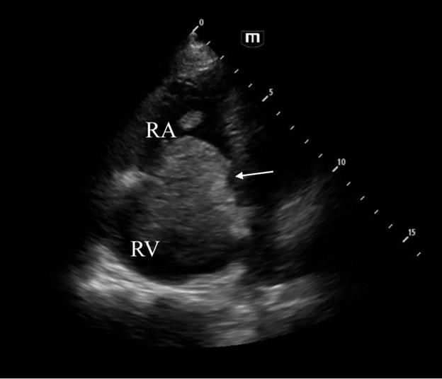
1. Point-of-care ultrasound image in apical 4-chamber view demonstrating dilated right ventricle (RV) with a large mass (indicated by arrow) extending from the right atrium (RA) into the right ventricle through the tricuspid valves.
CPC-EM Capsule
What do we already know about this clinical entity?
Renal cell carcinoma presents with nonspecific and variable symptoms, and in rare instances, it can invade into the heart.
What makes this presentation of disease reportable?
A new diagnosis and initial management of renal cell carcinoma invading the pulmonary arteries in the emergency department (ED) has never been reported.
What is the major learning point?
Renal cell carcinoma infiltrating the pulmonary arteries was identified using point-of-care ultrasound in the ED.
How might this improve emergency medicine practice?
Utilizing point-of-care cardiac ultrasound can deliver better patient care in the ED in patients with nonspecific cardiopulmonary symptoms.
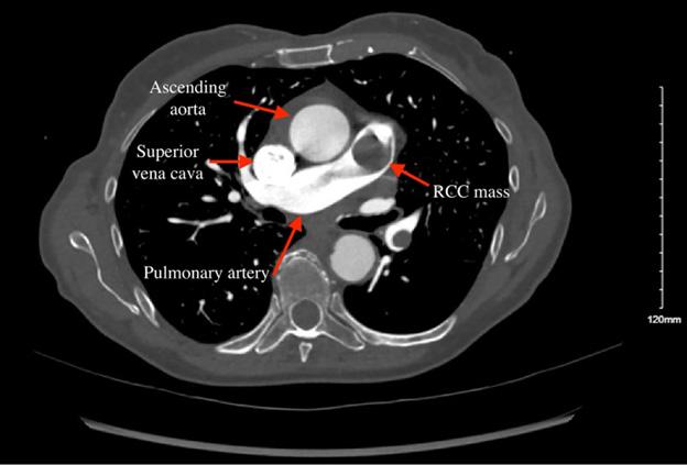
To our knowledge, this is the first case report on ED management and diagnosis of a rare presentation of renal tumor extension into the pulmonary artery. The tumor infiltration into the right ventricle explains the patient’s lack of response to diuretics due to the sheer size of the mass obstructing blood flow. Although a confirmatory biopsy was not completed for our patient, this was most likely a metastasized case of RCC given the tumor origin and presentation. Most often, RCC metastasizes to the lungs, bones, liver, adrenal glands, and lymph nodes. Prognosis of RCC has been shown to be poor for lymphatic and perinephric involvement of the metastasis rather than the size of vascular extension into the renal vein or inferior vena cava.5 Extension of renal tumors into the pulmonary arteries is even more rare with only two other case reports published in the literature.6,7 While this makes treatment even more challenging, it can be curable with surgical management of the primary tumor.8,9
This case highlights the critical role of point-of-care ultrasound (POCUS) in the emergency setting in diagnosis and treatment of heart failure that turned out to be a rare presentation of RCC. Relying on less time-consuming modalities for diagnosis such as laboratory markers can be enticing in the busy emergency setting. As demonstrated in our case, however, point-of-care echocardiography has been demonstrated to be more useful than BNP levels for diagnosing heart failure in the ED.10 In our case, it also allowed for rapid escalation of assessment with CT to further characterize the cause of the patient’s acute heart failure, which showed the hypervascularity of the tumor in detail and the physical extent of the tumor. Even though the patient decided to forgo invasive management, the rapid diagnosis made in the ED with POCUS allowed the shared decisionmaking process to occur without delay.
This is the first case report that presents a rare ED presentation of a renal cell cancer mass extending into the pulmonary artery. When a patient presents with non-specific symptoms of heart failure, it is important to maintain a wide differential including new malignancy and consider obtaining a point-of-care ultrasound to guide further diagnostic and therapeutic management of patients.
The authors attest that their institution requires neither Institutional Review Board approval, nor patient consent for publication of this case report. Documentation on file.
Address for Correspondence: Corlin Jewell, MD, University of Wisconsin School of Medicine and Public Health, Berbee Walsh Department of Emergency Medicine, 300-32 Madison, WI 53705. Email: cmjewell@medicine.wisc.edu
Conflicts of Interest: By the CPC-EM article submission agreement, all authors are required to disclose all affiliations, funding sources and financial or management relationships that could be perceived as potential sources of bias. The authors disclosed none.
Copyright: © 2025 Yang et al. This is an open access article distributed in accordance with the terms of the Creative Commons Attribution (CC BY 4.0) License. See: http://creativecommons.org/ licenses/by/4.0/
1. Hsieh JJ, Purdue MP, Signoretti S, et al. Renal cell carcinoma. Nat Rev Dis Primers. 2017;3(1):17009.
2. Jemal A, Tiwari RC, Murray T, et al. Cancer statistics, 2004. CA Cancer J Clin. 2004;54(1):8-29.
3. Skinner DG, Colvin RB, Vermillion CD, et al. Diagnosis and management of renal cell carcinoma. a clinical and pathologic study of 309 cases. Cancer. 1971;28(5):1165-77.
4. Chiappini B, Savini C, Marinelli G, et al. Cavoatrial tumor thrombus: single-stage surgical approach with profound hypothermia and circulatory arrest, including a review of the literature. J Thorac Cardiovasc Surg. 2002;124(4):684-8.
5. Tongaonkar HB, Dandekar NP, Dalal A V., et al. Renal cell carcinoma extending to the renal vein and inferior vena cava: results of surgical treatment and prognostic factors. J Surg Oncol. 1995;59(2):94-100.
6. Wieder JA, Laks H, Freitas D, et al. Renal cell carcinoma with tumor thrombus extension into the proximal pulmonary artery. J Urol 2003;169(6):2296-97.
7. Laguna G, Di Stefano S, Melero JM, et al. Giant clear cell renal carcinoma extended from the kidney until the pulmonary artery. Eur Heart J. 2013;34(40):3108.
8. Vogt PR, Ensner R, Prêtre R, et al. Less invasive surgical treatment of renal cell carcinomas extending into the right heart and pulmonary arteries: surgery for renal cell carcinoma. J Card Surg. 1999;14(5):330-3.
9. Manoharan A, Lugo-Baruqui A, Ciancio G. Radical nephrectomy and pulmonary lobectomy for renal cell carcinoma with tumor thrombus extension into the inferior vena cava and pulmonary arteries. Anticancer Res. 2020;40(10):5837-44.
10. Martindale JL, Wakai A, Collins SP, et al. Diagnosing acute heart failure in the emergency department: a systematic review and meta‐analysis. Acad Emerg Med. 2016;23(3):223-42.
Summer Ghaith, MD, JD*
Deborah Hsu, MD, MEd†
William Dixon, MD, MEd‡
Mayo Clinic Alix School of Medicine, Mayo Clinic, Phoenix, Arizona
Stanford University School of Medicine, Department of Emergency Medicine, Division of Pediatric Emergency Medicine, Palo Alto, California
Stanford University School of Medicine, Department of Emergency Medicine, Palo Alto, California
Section Editor: Anna McFarlin, MD
Submission history: Submitted October 29, 2024; Revision received January 27, 2025; Accepted March 11, 2025
Electronically published June 19, 2025
Full text available through open access at http://escholarship.org/uc/uciem_cpcem DOI: 10.5811/cpcem.38443
Introduction: Infantile subglottic hemangioma is a rare and serious condition characterized by stridor, respiratory distress, and a barking cough. This condition poses a significant risk as it can lead to life-threatening airway obstruction.
Case Report: We present a five-week-old patient who was diagnosed in the emergency department (ED) with moderate laryngomalacia via laryngoscopy by otolaryngology and discharged; he returned to the ED the next day with worsening symptoms of recurrent stridor, difficulty feeding, and worsening respiratory distress. A second laryngoscopic exam performed on the return ED visit revealed a subglottic mass that was later identified as a left-sided subglottic hemangioma via bronchoscopy and magnetic resonance imaging. The patient was treated with propranolol and discharged from the inpatient unit with dermatology and otolaryngology follow-up.
Conclusion: Infantile subglottic hemangioma is a rare but serious cause of respiratory distress in infants, posing a risk of airway obstruction. This diagnosis should be considered in the ED, particularly for patients under two years of age, who present with recurrent stridor and respiratory distress and do not respond to standard treatments for croup. [Clin Pract Cases Emerg Med. 2025;9(3):297-301.]
Key words: infantile subglottic hemangioma; subglottic stenosis; stridor; respiratory distress; case report.
Infantile subglottic hemangioma is a rare and serious condition that is potentially life-threatening.1 It is a benign vascular tumor of infancy that, if large enough, could obstruct the airway. A mortality rate of up to 50% has been reported in this circumstance.2,3 Infantile hemangiomas have an incidence of 4%-5%, but involvement of the subglottic region is much more rare.1 Subglottic hemangioma accounts for approximately 1.5% of congenital laryngeal abnormalities.1 Most patients will present with sudden-onset symptoms including dyspnea, stridor, barking cough, hoarseness, respiratory distress, feeding difficulty, and cyanosis.1,4 Given the nature and presentation of these symptoms, it is commonly
confused with croup. We present a case of a patient diagnosed with a left-sided subglottic hemangioma on a subsequent visit to the emergency department (ED) after an initial diagnosis of moderate laryngomalacia.
A five-week-old male patient born at 35 weeks gestational age, presented to the pediatric ED for evaluation of difficulty breathing and stridor. Parents noted he had never experienced stridulous breathing previously. He had been well the night before the onset of acute symptoms, and his birth history was unremarkable. On the first visit, his vital signs were heart rate 190 beats per minute (bpm), respiratory rate 56 breaths per
minute, temperature 37.4 ºCelsius, and oxygen saturation 99% on room air. Review of symptoms was unremarkable. An extended viral panel was performed, which included influenza A, influenza B, respiratory syncytial virus, coronavirus disease 2019, parainfluenza, metapneumovirus, rhinovirus, enterovirus, chlamydia pneumoniae, and mycoplasma pneumoniae. These were all negative. He had some but not complete symptomatic improvement with racemic epinephrine and oral dexamethasone.
Otolaryngology was consulted, and a laryngoscopic exam was performed in the ED. The exam demonstrated moderate laryngomalacia, but prior to this diagnosis he required two additional doses of racemic epinephrine during his ED stay. He was admitted to the acute care pediatric unit overnight for observation. He required no further interventions during the hospital stay and was discharged within 12 hours of admission. On discharge, his vital signs were heart rate 156 bpm, respiratory rate 21 breaths per minute, temperature 36.8 ºCelsius, and oxygen saturation 99% on room air. Twelve hours after discharge from the hospital, the patient developed worsening inspiratory and expiratory stridor with feeds and crying, increased fussiness, and could not finish his feeding.
The patient’s presenting vital signs on his second ED visit included temperature 36.9 ºC, blood pressure 105/71 millimeters of mercury, heart rate 196 bpm, respiratory rate 26 breaths per minute, and oxygen saturation 100% on room air. The initial physical examination was notable for a crying, mottled infant in respiratory distress. Nasal congestion was present. There was increased work of breathing, as well as non-positional inspiratory stridor that worsened with crying or agitation. Grunting and suprasternal retractions were present. Biphasic stridor was not seen in the ED, but parents reported biphasic stridor at home. The rest of his physical exam was normal, including a skin examination that was negative for other cutaneous hemangiomas.
High-flow nasal cannula oxygen was started. Racemic epinephrine and dexamethasone were immediately started with some but not complete improvement of symptoms. The patient had decreased respiratory distress and improved color and overall condition after ED interventions. Labs were obtained. A complete blood count and comprehensive metabolic panel were unremarkable. Lactate was elevated to 2.73 millimoles per liter (mmol/L) (referencee range: 0.5 to 2.2 mmol/L). A chest radiograph (CXR) was performed, which showed diffuse bronchial cuffing. This was the same finding as the CXR from the first ED visit the day prior.
A repeat laryngoscopic exam was performed in the ED by an otolaryngologist to explore other etiologies of the patient’s symptoms. Moderate laryngomalacia was again seen, but this time a mild post-cricoid edema (concerning for subglottic stenosis) was also found (Image 1).
Neck and chest magnetic resonance imaging (MRI) was performed during the patient’s hospital admission. A well-circumscribed sub-centimeter enhancing lesion in the
CPC-EM Capsule
What do we already know about this clinical entity?
Infantile subglottic hemangioma is a rare, lifethreatening airway lesion causing sudden stridor, dyspnea, barking cough, hoarseness, distress, and cyanosis.
What makes this presentation of disease reportable?
A patient was diagnosed with a left-sided subglottic hemangioma on a return emergency department visit after initially being diagnosed with moderate laryngomalacia.
What is the major learning point?
Consider infantile subglottic hemangioma in children under two with persistent stridor or poor response to airway treatments; propranolol is first-line therapy.
How might this improve emergency medicine practice?
Point-of-care laryngoscopy may expedite diagnosis and guide management in pediatric patients with upper airway obstruction, improving emergency care outcomes.
left subglottic larynx suggestive of a subglottic hemangioma was seen causing moderate narrowing of the subglottic airway (Image 2).
The patient was admitted from the ED to the pediatric intensive care unit (PICU) for close monitoring of his airway with a plan to go to the operating room (OR) the following morning for bronchoscopy to assess the subglottic hemangioma as well as a possible supraglottoplasty for the patient’s diagnosed laryngomalacia. Upon admission to the PICU, his vital signs were heart rate 152 bpm, respiratory rate 39 breaths per minute, and oxygen saturation 100% on 3L/21% high-flow nasal cannula. After administration of racemic epinephrine and dexamethasone, high-flow nasal cannula oxygen was started in the ED for respiratory support and continued in the PICU. Heliox was also used in the PICU
early in his hospitalization.
In the OR, the patient underwent bronchoscopy, which showed a narrowed subglottis due to a reddish mass emanating from the left subglottis, consistent with subglottic hemangioma (Image 3).
Dermatology was consulted for the patient’s new diagnosis of left-sided subglottic hemangioma. Infectious etiology of symptoms was deemed less likely than this diagnosis, and the patient was started on propranolol. He was scheduled for follow-up with pediatric otolaryngology and dermatology clinics for outpatient monitoring of his subglottic hemangioma.
We present a case of a patient diagnosed with a leftsided subglottic hemangioma on a subsequent visit to the ED after an initial diagnosis of moderate laryngomalacia. Risk factors for infantile subglottic hemangiomas include factors such as female sex, prematurity, low birth weight, and family history of hemangioma.5 Drug exposures can increase the risk as well, including maternal beta-blockers, progesterone, and illicit drugs.5
Differential diagnoses for pediatric stridor can be organized by the patient’s age. In patients under six months of age, emergency physicians should consider laryngotracheomalacia, vocal cord paralysis, subglottic stenosis, airway hemangiomas, and vascular rings. In patients greater than six months of age, physicians should consider croup, epiglottitis, bacterial tracheitis, foreign body aspiration, and retropharyngeal abscess. In our case, our patient was
Second Scope, New Findings: Pediatric Stridor Is Not Always Due to Croup or Laryngomalacia Ghaith
initially diagnosed with laryngomalacia. Laryngomalacia is a common cause of stridor that presents with inspiratory stridor that worsens when feeding or when the patient is supine. This is a self-limiting disease and usually resolves within 12-24 months of age.6 Moreover, subglottic hemangioma is commonly misdiagnosed as croup. Airway problems should be considered in patients who are being evaluated for croup in cases of recurrent (two or more episodes per year) or prolonged symptoms.7 Additionally, close attention should be paid to infants younger than 12 months, infants with a history of intubation, and premature infants.7
Typically, infantile subglottic hemangiomas are not present at birth and develop within the first few months of life, followed by a proliferation phase of about 6-9 months, and subsequently a spontaneous regression phase that occurs over years.2 During the proliferative phase, the risk for airway obstruction is at its highest.2 Patients typically present during this proliferative phase with symptoms of stridor, feeding difficulties, and respiratory distress, as in our case. Stridor in these patients is characterized as a biphasic stridor associated with a barky cough that develops as the hemangioma enlarges. Additionally, subglottic hemangiomas are commonly associated with cutaneous findings of cutaneous hemangioma and segmental hemangiomas in a “beard distribution.”8 Diagnosis of infantile subglottic hemangioma is usually established with endoscopy.3 Imaging such as computed tomography (CT) and MRI can be used to determine the depth of the lesions or to exclude other etiologies.3
First-line treatment of subglottic hemangioma is propanolol.3 Propranolol is the drug of choice for subglottic hemangiomas as it has been shown to significantly reduce the size of hemangiomas and alleviate the symptoms of stridor and respiratory distress. Propranolol is a non-selective betablocker that causes capillary vasoconstriction, decreased expression of vascular endothelial growth factors to inhibit angiogenesis, apoptosis of capillary endothelial cells, and inhibition of nitric oxide production, causing the hemangioma to shrink.8 Dosage of propranolol is 2-3 milligrams per kilogram per day.8 One study found that stridor was eliminated with use of propranolol within 24 hours or less in 85% of patients.9 The recommended duration of treatment with propranolol is at least six months; however, continuing treatment until at least 12 months of age may reduce the risk of rebound growth.8 As dermatologists are typically the primary specialists involved in the treatment of cutaneous hemangiomas with propranolol, they are well-positioned to manage the cases associated with airway hemangiomas comprehensively. However, a multidisciplinary approach with pediatricians and otolaryngologists is warranted. Other treatment modalities include steroids, both systemic and intralesional, alpha-interferon, vincristine, bleomycin, laser, tracheostomy, and surgical excision.1,10-12 However, propranolol is associated with a high rate of hemangioma clearance with an expected clearance of 95%, which is superior to other
treatments and usually avoids the need for surgery.8
Infantile subglottic hemangioma is a critical pediatric emergency. An emergency physician should consider intubating an infant with subglottic hemangioma if the infant presents with severe respiratory distress, signs of impending respiratory failure, or if there is a rapid deterioration in the clinical condition.8 It is important to use a smaller endotracheal tube in a stridulous infant because the subglottic space is already narrowed due to the hemangioma.13 A smaller tube can help pass through the stenotic area while reducing the risk of trauma to the airway.13 As emergency physicians increasingly expand their scope of practice, developing competency in diagnostic laryngoscopy may be useful. The ability to promptly diagnose conditions such as infantile subglottic hemangioma has the potential to impact patient outcomes through timely and appropriate interventions.
Infantile subglottic hemangioma is a rare cause of pediatric respiratory distress, but it is an important condition to consider when formulating differential diagnoses for pediatric patients with signs of upper airway obstruction. Specific patient populations in which to consider this diagnosis are children less two years of age who have recurrent or worsening stridor and other respiratory symptoms, and/or those who do not respond to standard treatment for upper and lower airway diseases. Propranolol remains firstline treatment for infantile subglottic hemangioma. This case illustrates the role point-of-care diagnostic laryngoscopy may have in shortening the time to diagnosis and informing management decisions of patients with signs of upper airway obstruction.
The authors attest that their institution requires neither Institutional Review Board approval, nor patient consent for publication of this case report. Documentation on file.
Address for Correspondence:Summer Ghaith, Mayo Clinic Alix School of Medicine, 5777 E. M ayo Blvd., Phoenix, AZ 85054. Email: Ghaith.Summer@mayo.edu.
Conflicts of Interest: By the CPC-EM article submission agreement, all authors are required to disclose all affiliations, funding sources and financial or management relationships that could be perceived as potential sources of bias. The authors disclosed none.
Copyright: © 2025 First Author last name et al. This is an open access article distributed in accordance with the terms of the Creative Commons Attribution (CC BY 4.0) License. See: http:// creativecommons.org/licenses/by/4.0/
Ghaith et al. Second Scope, New Findings: Pediatric Stridor Is Not Always Due to Croup or Laryngomalacia
1. Chen W, Zhu P, Xu M, et al. Diagnosis of infantile subglottic hemangioma and the effect of oral propranolol. Am J Otolaryngol 2022;43(6):103610.
2. McCormick AA, Tarchichi T, Azbell C, et al. Subglottic hemangioma: understanding the association with facial segmental hemangioma in a beard distribution. Int J Pediatr Otorhinolaryngol. 2018;113:34-7.
3. Kumar P, Kaushal D, Garg PK, et al. Subglottic hemangioma masquerading as croup and treated successfully with oral propranolol. Lung India. 2019;36(3):233-5.
4. Appiah-Thompson P, Hanson NAM, Quansah K, et al. Congenital laryngeal hemangioma: a case report of a rare presentation. Cureus 2023;15(2):e34814.
5. Sun Y, Zhao J, Meng Y, et al. The prevalence, complications, and risk factors for infantile hemangioma: a systematic review and metaanalysis. Int J Dermatol. 2024;63(6):737-46.
6. Katwala A, Anderson C, Thayer E, et al. Predominantly unilateral laryngomalacia in infants with unilateral vocal fold paralysis. Int J Pediatr Otorhinolaryngol. 2024;179:111922.
7. Hiebert JC, Zhao YD, Willis EB. Bronchoscopy findings in recurrent
croup: a systematic review and meta-analysis. Int J Pediatr Otorhinolaryngol. 2016;90:86-90.
8. Krowchuk DP, Frieden IJ, Mancini AJ, et al. Clinical practice guideline for the management of infantile hemangiomas. Pediatrics 2019;143(1):e20183475.
9. Elluru RG, Friess MR, Richter GT, et al. Multicenter Evaluation of the Effectiveness of Systemic Propranolol in the Treatment of Airway Hemangiomas [published correction appears in Otolaryngol Head Neck Surg. 2016;154(6):1164.]. Otolaryngol Head Neck Surg. 2015;153(3):452-460.
10. Schwartz T, Faria J, Pawar S, et al. Efficacy and rebound rates in propranolol-treated subglottic hemangioma: a literature review. Laryngoscope. 2017;127(11):2665-72.
11. Hardison S, Wan W, Dodson KM. The use of propranolol in the treatment of subglottic hemangiomas: a literature review and meta-analysis. Int J Pediatr Otorhinolaryngol. 2016;90:175-80.
12. Darrow DH. Management of infantile hemangiomas of the airway. Otolaryngol Clin North Am. 2018;51(1):133-46.
13. Strutt JR, Thompson NR, Stotesbery JL, et al. Emergency endotracheal intubation with a rigid stylet of an infant with severe subglottic stenosis. J Emerg Med. 2020;58(3):e157-60.
Tiana Patriarca, PharmD*
Jessica Rivera Pescatore, PharmD, DABAT*‡
William Rushton, MD*‡
Emily Sochovka, PharmD†
Julie Brown, MD‡
Section Editor: Lev Libet, MD
Alabama Poison Information Center, Children’s of Alabama, Birmingham, Alabama
University of Alabama Birmingham, Department of Pharmacy, Birmingham, Alabama
University of Alabama at Birmingham, Department of Emergency Medicine, Birmingham, Alabama
Submission history: Submitted January 25, 2025; Revision received March 13, 2025; Accepted April 1, 2025
Electronically published June 8, 2025
Full text available through open access at http://escholarship.org/uc/uciem_cpcem DOI: 10.5811/cpcem.42020
Introduction: Skeletal fluorosis is a complication of excess fluoride, which may be associated with chronic inhalation or “huffing” of compressed air cleaners for keyboards and electronics. The rare presentation of this condition can lead to a missed diagnosis and lack of appropriate intervention. Clinicians should be aware of the potential development of fluorosis in patients reporting a history of inhalant abuse.
Case Report: We present a case of skeletal fluorosis in a 46-year-old female patient with a fourmonth history of daily inhalant use of computer cleaner containing difluoroethane (DFE). She presented to the emergency department after developing myalgias for approximately four months. The pain was alleviated by rest, heat therapy, and pain medication. She was noted to have diffuse bilateral swelling to upper and lower extremities, as well as interphalangeal joint swelling and nonmobile lesions to bilateral hands and left forearm on physical exam. Radiography revealed diffuse periosteal reaction throughout the hand and forearm suggestive of fluorosis. She was counseled to cease inhalant use.
Conclusion: Skeletal fluorosis is a rare and painful condition that can have prolonged adverse effects and a lasting impact on quality of life. Patients who report regular inhalant use should be counseled on the potential toxicities of these products and encouraged to discontinue use of DFE. Those presenting with diffuse skeletal findings and reported DFE use should be evaluated for skeletal fluorosis. [Clin Pract Cases Emerg Med. 2025;9(3):302-309]
Keywords: skeletal fluorosis; difluoroethane; inhalants; case report.
INTRODUCTION
Inhalants are common substances of misuse, which are widely available and used primarily for their euphoric effects. They consist of hydrocarbon compounds found in a variety of products such as cleaners, dusters, paints, and fuels. These compounds produce their central nervous system effects via gamma-aminobutyric acid receptors similar to ethanol, which itself is an alcohol hydrocarbon derivative. In addition to their
euphoric and intoxicating properties, inhalants have also been implicated in cases of cardiovascular toxicities including ventricular dysrhythmias, which likely lead to “sudden sniffing death.” Adolescents and children are particularly susceptible to inhalant misuse due to ease of access and relative affordability.
Halogenated hydrocarbons are hydrocarbon derivates containing an atom from group 17 of the periodic table,
typically chlorine or fluorine. 1,1-diflouroethane (DFE) is a fluorinated hydrocarbon, commonly found in compressed air cleaners for keyboards and electronics. Chronic DFE inhalation can lead to toxicities such as acute kidney injury, hepatotoxicity, neurologic deficits, and cardiotoxicity.1 A rare consequence of chronic fluorinated hydrocarbon inhalation is a metabolic bone disorder known as skeletal fluorosis, which is characterized by osteosclerosis of the axial skeletal system, formation of osteophytes at joints and distal extremities, and ligament ossification.2 We present a case of skeletal fluorosis that developed within four months of chronic DFE inhalant use.
A 46-year-old female with a past medical history of anxiety, depression, and tobacco use presented to the emergency department (ED) with complaints of diffuse body swelling for approximately four months. She described pain in various joints that coincided with new-onset upper and lower extremity swelling. Three weeks prior, the patient had been evaluated at a separate ED and was diagnosed with skeletal fluorosis related to inhalation of compressed air cleaner for computer dust. She reported daily inhalant use of the cleaner for the previous four months, which she ceased using following her diagnosis at the outside hospital weeks prior. Initial vital signs were heart rate 75 beats per minute, blood pressure 144/82 millimeters of mercury, temperature 98.6 ºFahrenheit, and oxygen saturation 100% on room air. Physical examination was remarkable for diffuse swelling to the bilateral upper and lower extremities, with right lower extremity swelling greater than that of the left lower extremity, interphalangeal joint swelling in bilateral hands (Image 1), and multiple non-mobile lesions on bilateral hands and left forearm (Image 2). Initial laboratory values from complete blood count, basic metabolic panel, and hepatic function panel were remarkable only for elevated
CPC-EM Capsule
What do we already know about this clinical entity?
Skeletal fluorosis is a complication of excess fluoride which may be associated with chronic inhalation or “huffing” of compressed air cleaners.
What makes this presentation of disease reportable?
We present an uncommon and clinically noteworthy case of non-endemic skeletal fluorosis characterized by unique radiographic findings.
What is the major learning point?
Skeletal fluorosis is a rare and painful condition, which can have prolonged adverse effects and a lasting impact on quality of life.
How might this improve emergency medicine practice?
Increased awareness improves early recognition of skeletal fluorosis and emphasizes the importance of appropriate counseling on the risks of inhalant abuse.
alkaline phosphatase of 442 units per liter (L) (reference range: 44-147 units/L). Electrocardiogram showed sinus rhythm without evidence of ischemia.
Radiography revealed diffuse periosteal reaction throughout the hand (Image 1) and forearm (Image 2) suggestive of fluorosis. Chest radiograph and Doppler ultrasound of the right lower extremity showed no acute findings. Patient history, physical examination, and imaging results were highly consistent with a diagnosis of skeletal fluorosis. The patient was advised to continue to abstain from inhalant use and was scheduled for outpatient follow-up for continued monitoring and pain management.
At follow-up approximately two weeks later, she reported no improvement in symptoms since the ED visit. Pain in ankles, knees, and hands was described as sharp and constant, aggravated by movement and palpation and alleviated by rest, heat therapy, and pain medication. Swelling persisted to upper and lower extremities, which reportedly improved with rest. She was prescribed celecoxib for pain management and referred to rheumatology for elevated inflammatory markers and long-term management of skeletal fluorosis.
Skeletal fluorosis from inhalant abuse occurs when DFE is metabolized to free fluoride ions, which replace the hydroxyl ions in hydroxyapatite, converting it to fluorapatite in the bone.2 This leads to decreased bone turnover with increased osteoblast activity and greater resistance to breakdown by parathyroid hormone.2-3 Bones and bony protrusions become more brittle and susceptible to fracture, despite an overall normal or increased bone mineral density (BMD).4 Clinical manifestations of skeletal fluorosis may initially include joint and back pain or stiffness, eventually leading to loss of mobility and range of motion.2 Osteosclerosis, osteophytosis, and ligament ossifications are hallmark findings on skeletal fluorosis imaging.2 Furthermore, patients may develop secondary hyperparathyroidism with vitamin D and C deficiencies, which can be supplemented in treatment. Additional supplementation with vitamin E and methionine may also be considered to reduce skeletal fluoride accumulation.5
Skeletal fluorosis has historically been considered an endemic condition, occurring primarily in areas with fluorinated well water contaminated from nearby volcanic rock or industrial sources, and developing in patients over decades of exposure.2 Other causes of skeletal fluorosis have included exorbitant and chronic ingestions of fluorinated products such as teas made from Camellia sensis, toothpastes, mouthwashes, and drugs such as voriconazole.1,6 These non-endemic fluoride sources can have more rapid onset of the skeletal deformities and manifestations of fluorosis, developing over the course of months to years, likely due to higher fluoride concentrations.6
A review of non-endemic skeletal fluorosis case reports by
Cook et al revealed that of patients with a known duration of use, five patients with tea exposures ingested anywhere from 14-74 mg of fluoride per day. These patients reported durations of use of anywhere from 17-37 years prior to initial presentation.7 One patient with an estimated daily toothpaste ingestion of 66 mg of fluoride presented after at least five years of exposure.7 In a systematic review of voriconazoleinduced periostitis, it was found that skeletal symptoms developed after as little as six weeks to eight years of voriconazole treatment, with most reported voriconazole doses being 400 mg daily, an equivalent fluorine amount of 65 mg per day.8
Interestingly, inhalant use with DFE has also been linked with rapid onset of skeletal fluorosis in over a dozen cases (Table).1,3-7,9-14 Reports of use duration range anywhere from six months to five years in frequencies ranging from 2-7 cans weekly to 20-25 cans daily.5,9-10 Quantification of daily fluoride intake from DFE is difficult to determine due to varying product sizes and limited data on systemic absorption of the inhalant. Peicher et al and subsequently Chen et al estimated that exposure to 1-7 cans weekly for three years and 3-4 cans daily for 10-11 months resulted in total fluoride exposures of thousands of grams and approximately 147 kilograms, respectively.9,11
Our patient presented four months after initiation of daily DFE inhalation and described her symptom onset as coinciding around the same time. Her case emphasizes the potential for rapid development of skeletal fluorosis from daily DFE use and encourages suspicion of fluorosis in patients with a history of DFE inhalation and signs of skeletal pain, swelling, or deformities. Additional diagnoses to consider may include myelofibrosis, osteoblastic metastasis, renal osteodystrophy, ankylosing spondylitis, and Paget disease.2
Treatment of skeletal fluorosis is primarily supportive with physical therapy, minimizing fracture risk, and ceasing use of the offending product. In the case of abrupt DFE cessation, observation and benzodiazepines may be warranted for inhalant withdrawal.10 Once there is no further fluoride exposure, skeletal fluorosis improves slowly over the course of years due to the long skeletal fluoride half-life of seven years.2 Tucci et al described a 28-year-old male who had been huffing keyboard duster with DFE for about 3-4 years, with elevated urine and serum fluoride levels, progressive bony deformities on both hands, and loss of mobility in several joints. Three years after cessation of DFE, he had continued elevations in fluoride levels and BMD, with improvements in walking and mobility six months after a left hip arthrotomy for ankylosis, which included extensive osteochondroplasty of the femoral head and neck and prophylactic pinning of the femoral neck.6 Suwak et al described a 56-year-old male who had been huffing three cans of dust cleaner daily for about two years, in addition to previous occupational exposure to chlorofluorocarbon solvent cleaners. One year after he stopped
Skeletal Fluorosis: A Case Report of Rare Diagnosis of Computer-cleaner
Table. Reported cases of skeletal fluorosis associated with chronic inhalation keyboard dust cleaners.
Citation Age (yrs) Sex Frequency and Duration of DFE Use Clinical Presentation
Peicher 2017 33 M 2-7 cans weekly for 3 years Progressive back pain over 3 years with loss of lumbar lordosis and tenderness of lumbar spine
Tucci 2017
28 M Unknown for 3-4 years Difficulty walking, abnormal gait, anterior left hip pain, loss of movement in right forearm and wrist, and progressive deformities in both hands with limited motion
2-3 years Uniform osteosclerosis in the long bones, entire spine, rib cage, and pelvic bones
2 years Multiple exostoses, ossification of the left hip, and multiple surface lesions along the radius and about the left elbow; periostitis deformans in hands and high bone density in spine
Ponce 2018
27 M 9-11 cans daily for 11 months
Presented for frostbite from prolonged contact with DFE container; also had hard, nonpainful growths on right hand, hypertrophic nodule on right elbow, and hard anterior nodules on right tibia
5 months Periosteal bone formation on right tibia, right phalanges, right radius and ulna, and a focal nodule of bone on distal right humerus
Not reported
Serum 25(OH) D improved to 38 ng/mL after supplementation with vitamin D. Six months after cessation of DFE, plasma F- level was still elevated at 0.16 mg/L and urine F- level was 18.9 mg/L. About 1-2 years after DFE cessation patient underwent left hip arthrotomy for ankylosis, and he had improved hip function with near normal gait. About 3 years after cessation BMD was not significantly changed
Counseling on cessation of inhalant use and prescribed oral vitamin D; lost to follow-up
Continued
Citation
Custer 2020
Age (yrs) Sex Frequency and Duration of DFE Use Clinical Presentation
39 M 20-25 cans daily for 6 months
Presented after abrupt cessation of DFE use 6 days prior with signs of withdrawal and bony deformities on hands
Time from Skeletal Symptom Onset to Presentation Radiology Findings
Unknown Diffuse bilateral periosteal reaction in the phalanges and distal ulnas
Cook 2021
51 M Unknown for 2-3 years
Musculoskeletal pain, opiate use, hypocalcemia, secondary hyperparathyroidism, long recurrent bone fractures
2 years Diffuse osteosclerosis of spine and pelvis, cortical thickening and new bone formation in tubular bones, muscle and ligament ossification, possibly including the external occipital protuberance
Suwalk 2021 56 M 3 cans daily for 2 years + previous occupational CFC exposure
Progressively worsening lumbar and bilateral ankle pain with bony prominences over the bilateral tibial crests and tenderness of left distal fibula and medial malleolus
5 years Diffuse periosteal reactions with osteosclerotic bone formation of multiple long bones, sclerosis of the axial skeleton, relative osteopenia and periosteal bone formation of the appendicular skeleton, and ossification of multiple ligaments
Withdrawal symptoms of irritability and agitation resolved after 72 hours with persisting psychosis and hallucinations; long term followup not reported
Ceased “huffing,” prescribed oral calcium and Vit D3; 3 months later 25(OH)D was WNL, alk phos remained elevated, and BMD had not changed significantly; 19 months later PTH normalized, plasma and urine F- levels decreased, although still elevated, and pain continued
Counseling on cessation of inhalant use and prescribed vitamin A, E, and D supplementation; 1 year later presented with mild back pain, posterior left leg pain, and bony projections on digits and anterior tibia similar to previous visits. Serum fluoride and alkaline phosphatase levels decreased to 0.26 mg/L and 128 U/L, respectively, and calcium levels remained within normal limits
Table. Continued
Citation
Age (yrs) Sex Frequency and Duration of DFE Use Clinical Presentation
Fikse 2022 33 M Unknown for 5 years Presented following inhalation of 4 cans of DFE in suicide attempt; also had significant bilateral interphalangeal joint swelling of hands and multiple lesions of forearms and lower extremities
Mayer 2022
37 M Unknown within past year
Neck pain with reduced range of motion of the cervical spine
Time from Skeletal Symptom Onset to Presentation Radiology Findings
6 months Diffuse periosteal new bone formation and sclerosis on radiology of bilateral hands, forearms, lower extremities, and chest
Counseling on cessation of inhalant use and prescribed vitamin C, E, and D supplementation; lost to follow-up
Mohideen 2022 41 M Up to 10 cans daily for 1 year 50 pound weight loss in 6 months with diffuse joint pain, bony nodules throughout upper and lower extremities, diffuse tenderness of anterior and posterior ribs, and restricted shoulder and lumbar spine mobility due to pain
1 year Diffuse osteosclerosis of the pelvis, left forearm, and distal right leg and ankle; ossification and calcification of ligaments
2 months Numerous foci of abnormal bone activity in the skull, long bones, ribs, pelvis, and spine
Patient underwent laparoscopic sigmoid colectomy for adenocarcinoma; Counseling on cessation of inhalant use and prescribed oral vitamin D supplementation; follow-up not reported
Table. Continued
Citation
Neto 2022
Age (yrs) Sex Frequency and Duration of DFE Use Clinical Presentation
27 F Up to 16 cans daily from ages 18-22; resumed 6 months prior
Features suspicious for hypophosphatasia with arthropathy and elevated BMD, maxillary exostoses, periosteal excrescences, osteosclerosis, and periarticular calcifications
Time from Skeletal Symptom Onset to Presentation Radiology Findings
Multiple skeletal manifestations over the course of 5 years
Chen 2023
Chen 2024
26 M >10 bottles daily for 2 years
Presented following assault with diffuse bone pain
Calcifications around hips and right sacrotuberous ligament, focal periosteal excrescences at femurs, knees, tibias, and fibulas, diffuse osteosclerosis of spine and pelvis, and soft tissue calcifications around shoulders
Unknown Diffuse sclerosis of axial skeleton and bilateral lower extremities, and cortical expansion of all visualized bones
30 M 3-4 cans daily for 10-11 months
Sudden right leg pain with a hard mass above the right ankle with night sweats and weight loss
Recent and sudden
Periosteal reaction of right distal tibia and fibula, soft tissue extension at the distal lateral surface of the fibula, diffuse periosteal reactions and hyperostosis of the cortices, osteosclerosis of the trabecular areas, and periosteal reaction of the right foot
Modest improvement in mobility and pain after 4 months of “huffing” cessation; later resumed inhalant use and suffered left distal femur fracture following a fall
Counseling on cessation of inhalant use and prescribed oral vitamin D; 3 months later patient had not ceased DFE use
Counseling on cessation of inhalant use and prescribed oral vitamin D and calcium; 6 months later urine F- decreased to 12.14 mg/L and bone specific alkaline phosphatase from 156 to 57 ug/dL
Abbreviations: DFE, 1,1-difluorethane; Alk phos, alkaline phosphatase; PTH, serum parathyroid hormone; 25-(OH)D, serum 25-hydroxyvitamin D; Ca, calcium or corrected calcium; mg/L, milligrams per liter; U/L, units per liter; pg/mL, picograms per milliliter; ng/mL, nanograms per milliliter; M, male; F, female; mg/dL, milligrams per deciliter; F-, fluoride; BMD, bone mineral density; X ULN, times upper limit of normal; WNL, within normal limits; CFC, chlorofluorocarbon; umol/L, micromoles per liter.
Patriarca et al.
Skeletal Fluorosis: A Case Report of Rare Diagnosis of Computer-cleaner Toxicosis
huffing, the patient had decreased, although still elevated, serum fluoride levels, and he had continued bony protrusions on his digits and anterior tibia.12
Our patient had continued symptoms and pain after five weeks of DFE cessation, and reported alleviation of symptoms with rest, heat therapy and pain medication. Our case is limited by the lack of serum or urine fluoride concentrations. However, the patient presented to our ED three weeks after reportedly discontinued use, making serum concentrations less useful, except for confirming exposure. Additionally, radiologic examination, which is considered the best method of diagnosis, was highly consistent with the patient’s reported history and clinical course.
Skeletal fluorosis is a rare and painful condition that can have prolonged adverse effects and lasting impacts on quality of life. Patients presenting to the emergency department who report regular inhalant use should be counseled on the potential toxicities of these products and encouraged to cease use. Those presenting with diffuse skeletal findings and reported DFE use should be evaluated for skeletal fluorosis.
Patient consent has been obtained and filed for the publication of this case report.
1. Salles Rosa Neto N, Englert D, McAlister WH, et al. Periarticular calcifications containing giant pseudo-crystals of francolite in skeletal fluorosis from 1,1-difluoroethane “huffing.” Bone. 2022;160:116421.
2. Sellami M, Riahi H, Maatallah K, et al. Skeletal fluorosis: Don’t miss the diagnosis! Skeletal Radiol. 2020;49(3):345-57.
3. Ponce A, Oakes JA, Eggleston W. Acute skeletal fluorosis in the setting of 1,1-difluoroethane abuse. Clin Toxicol (Phila). 2019;57(5):374-5.
4. Mohideen H, Dahiya DS, Parsons D, et al. Skeletal fluorosis: a case of inhalant abuse leading to a diagnosis of colon cancer. J Investig Med High Impact Case Rep. 2022;10:23247096221084919.
5. Fikse DJ, Le D, Surmaitis RM. Skeletal fluorosis secondary to harmful inhalant use. Clin Toxicol (Phila). 2022;60(12):1376-8
6. Tucci JR, Whitford GM, McAlister WH, et al. Skeletal fluorosis due to inhalation abuse of a difluoroethane-containing computer cleaner. J Bone Miner Res. 2017;32(1):188-95.
7. Cook FJ, Seagrove-Guffey M, Mumm S, et al. Non-endemic skeletal fluorosis: causes and associated secondary hyperparathyroidism (case report and literature review). Bone. 2021;145:115839.
8. Adwan MH. Voriconazole-induced periostitis: a new rheumatic disorder. Clin Rheumatol. 2017;36(3):609-615.
Address for Correspondence: Email: Tiana Patriarca, PharmD, Alabama Poison Information Center, Children’s of Alabama, 1600 7th Ave S Birmingham, AL 35233. Email: tiana.patriarca@ childrensal.org
Conflicts of Interest: By the CPC-EM article submission agreement, all authors are required to disclose all affiliations, funding sources and financial or management relationships that could be perceived as potential sources of bias. The authors disclosed none.
Copyright: © 2025 Patriarca et al. This is an open access article distributed in accordance with the terms of the Creative Commons Attribution (CC BY 4.0) License. See: http://creativecommons.org/ licenses/by/4.0/
9. Peicher K and Maalouf NM. Skeletal fluorosis due to fluorocarbon inhalation from an air dust cleaner. Calcif Tissue Int 2017;101(5):545-548.
10. Custer A, Corse A, Vazirani S. Difluoroethane inhalant abuse, skeletal fluorosis, and withdrawal. Fed Pract. 2020;37(6):288-289.
11. Chen E, Jayman J, Bedrossian N. Skeletal fluorosis in a patient with computer cleaner inhalant abuse. Cureus. 2023;15(4):e37229.
12. Suwak P, Van Dyke JC, Robin KJ, et al. Rare case of diffuse skeletal fluorosis due to inhalant abuse of difluoroethane. J Am Acad Orthop Surg Glob Res Rev. 2021;5(10):e21.00077.
13. Chen Z, Akanbi F, Lucas DR, et al. A skeleton in a huff: insights in etiologies of osteosclerosis. J Bone Miner Res. 2024;39(2):79-84.
14. Mayer MJ, Gliedt JA, Shaker JA, et al. Case 298: skeletal fluorosis secondary to huffing. Radiology. 2022;302(2):484-488.
Morsi Rayyan, MD*
Ryan Reece, MD†
University of Michigan, Department of Emergency Medicine, Ann Arbor, Michigan University of Michigan, Hurley Medical Center, Department of Emergency Medicine, Flint, Michigan
Section Editor: Anna McFarlin, MD
Submission history: Submitted January 1, 2025; Revision received March 28, 2025; Accepted March 31, 2025
Electronically published June 19, 2025
Full text available through open access at http://escholarship.org/uc/uciem_cpcem DOI: 10.5811/cpcem.41565
Introduction: Congenital diaphragmatic hernia (CDH) is an embryological defect of the diaphragm that typically presents in the neonatal period with respiratory distress. However, delayed presentations do occur rarely and can pose diagnostic and therapeutic challenges.
Case Report: We describe the case of a 9-month-old male who presented to the emergency department (ED) with respiratory distress and was subsequently diagnosed with congenital diaphragmatic hernia.
Conclusion: This case underscores the importance of considering CDH in the differential diagnosis of pediatric patients presenting to the ED with unexplained respiratory or gastrointestinal symptoms, even beyond the neonatal period. [Clin Pract Cases Emerg Med. 2025;9(3):310-313.]
Keywords: delayed presentation; congenital diaphragmatic hernia; respiratory distress; case report.
Congenital diaphragmatic hernia (CDH) occurs due to a developmental defect in the diaphragm, allowing abdominal contents to herniate into the thoracic cavity. This condition predominantly presents in neonates with severe respiratory distress immediately after birth. Delayed presentation beyond the neonatal period is rare and often misdiagnosed due to the non-specific nature of symptoms. Early recognition and appropriate management are crucial to improving outcomes. In this case report we aimed to increase awareness among emergency physicians about the potential for delayed CDH presentation and highlight the importance of thorough clinical evaluation and timely intervention in pediatric patients with persistent respiratory distress.
A nine-month-old male presented to the emergency department (ED) with respiratory distress and cyanosis. The patient’s parent reported that the patient began crying and his lips appeared blue. Emergency medical services who
responded noted that the patient had wheezing bilaterally with retractions and an oxygen saturation of 86%. He was given nebulized ipratropium-albuterol and blow-by oxygen, and transported to the ED. Of note, he had been hospitalized at three weeks of age for acute hypoxic respiratory failure in the setting of respiratory syncytial virus and human rhinovirusenterovirus infection requiring pediatric intensive care and high-flow oxygen. He had a chest radiograph (CXR) at the time, which showed bilateral hazy opacities suggesting viral process or small airway disease. His parents reported intermittent episodes of coughing and wheezing since then that had worsened over the previous two days. His immunizations were up to date, and he had no history of other infectious or gastrointestinal (GI) symptoms, or trauma. He had been born at term following an uncomplicated pregnancy. Of note, the patient was at the third percentile for weight.
On arrival to the ED, the patient was found to have a blood pressure of 77/54 millimeters of mercury (mm Hg), heart rate 183 beats per minute, respiratory rate 28 breaths per minute, oxygen saturation 99% on blow-by oxygen, and oral
et al. Delayed Presentation of Congenital Diaphragmatic Hernia in the Emergency Department: Case Report
temperature of 37.3 ºCelsius. Physical exam revealed increased work of breathing with intercostal retractions and diminished air entry bilaterally with faint expiratory wheezing. Initial labs were significant for a venous blood gas with pH 7.33 (reference range: 7.30-7.40), partial pressure of carbon dioxide 49 mm Hg (33-46 mm Hg) and a metabolic panel significant for bicarbonate of 22 milliequivalents per liter (mEq/L) (23-29 mEq/L). A CXR showed bubbly lucencies in the left hemithorax with rightward mediastinal shift and adjacent compressive atelectasis of the right lung, suspicious for large diaphragmatic hernia with bowel throughout the left hemithorax (Image).
The patient was initially treated with additional nebulized ipratropium-albuterol and four milligrams dexamethasone for wheezing while diagnostics were being performed, with concern for reactive airway disease. His work of breathing did not improve considerably. After the CXR was available, a nasogastric tube was considered for gastric decompression, but appropriately sized pediatric tubes were unavailable. The patient’s case was discussed with a pediatric tertiary-care center, and he was transferred. There, he underwent left open CDH repair.
CPC-EM Capsule
What do we already know about this clinical entity?
Congenital diaphragmatic hernia (CDH) usually presents neonatally with respiratory distress; delayed presentations are rare but can occur.
What makes this presentation of disease reportable?
It highlights a rare delayed CDH presentation diagnosed in the emergency department, missed on prior imaging, emphasizing diagnostic vigilance.
What is the major learning point?
CDH should be considered in pediatric patients with recurrent respiratory symptoms unresponsive to standard treatments.
How might this improve emergency medicine practice?
Raises awareness of delayed CDH presentation and reviews diagnostic and management strategies for emergency physicians.
The patient had a postoperative course briefly complicated by an expected pneumothorax and desaturations that improved with blow-by oxygen. He was discharged on postoperative day three. At his six- week follow-up, he was noted to be doing well with improved respiratory symptoms and oral intake. His weight had improved to the 38th percentile.
Congenital diaphragmatic hernia is thought to be caused by multiple factors including genetics, environmental exposures, and nutritional deficiencies, all resulting in incomplete diaphragmatic development. The diaphragmatic defect allows intra-abdominal organs to migrate into the thoracic cavity, which can impede normal lung development and function. Congenital diaphragmatic hernia has an incidence ranging from 0.8-5/10,000 live births and is most commonly diagnosed prenatally or immediately postnatally.1 Most CDH cases present in neonates with severe respiratory distress necessitating immediate intervention. Delayed presentations, such as in the case we describe, are rare and account for an estimated 10-13% of all CDH cases. These cases often present with non-specific symptoms including recurrent respiratory infections, GI symptoms, and failure to thrive.2
In older infants and children, the delayed presentation of CDH can mimic common pediatric conditions such as asthma,
Delayed Presentation of Congenital Diaphragmatic Hernia in the Emergency Department: Case Report Rayyan
pneumonia, or gastroesophageal reflux, leading to misdiagnosis and delayed treatment. Key clinical features that should raise suspicion for CDH in the differential diagnosis include the following: persistent or recurrent respiratory symptoms unresponsive to standard medical treatments; unilateral decreased breath sounds or abnormal chest auscultation findings; GI symptoms such as vomiting, feeding difficulties, or failure to thrive; and abnormal CXR findings including air-fluid levels or mediastinal shift.3-5
Diagnosis of CDH in delayed presentations often relies on imaging studies. A CXR is typically the initial imaging modality and may reveal bowel loops in the thoracic cavity, an elevated hemidiaphragm, or mediastinal shift. Chest radiograph is warranted even if the patient has had a prior radiograph that did not show CDH, as in the presented patient. In cases of diagnostic uncertainty, a nasogastric tube can be placed with subsequent contrast administration and follow-up CXR to aid in diagnosis. Additionally, abdominal radiographs, small bowel follow-through, and abdominal and thoracic ultrasound may lead to CDH diagnosis. Further imaging with contrast-enhanced computed tomography, the most sensitive modality, can provide detailed anatomy, confirm the diagnosis, and assist in preoperative planning by delineating the size and contents of the hernia.6
Definitive management of CDH presenting beyond the neonatal period involves surgical correction of the hernia and repair of the diaphragmatic defect. Preoperative stabilization is often necessary in the ED, including respiratory support and nasogastric decompression, especially in cases with significant respiratory compromise. Emergency physicians should be conscious of the potential for positive pressure ventilation, such as that provided by a bag valve mask, to cause gastric distention and worsen respiratory instability. They should perform endotracheal intubation in severe respiratory compromise or if prolonged resuscitation is required, similar to the suggested management in neonatal CDH.1 Additionally, the presented case highlights the importance of stocking appropriately sized pediatric equipment as nasogastric decompression could not be performed in a timely manner and could have led to decompensation prior to definitive management. In the event of failed nasogastric decompression leading to refractory respiratory failure, cases have been described using bedside percutaneous gastric puncture, emergent thoracotomy, and initiation of extracorporeal membrane oxygenation, although we recommend considering these approaches only in consultation with a pediatric surgeon.7-9
The prognosis for patients with delayed presentation of CDH is favorable if timely diagnosis and surgical intervention are achieved. However, the severity of pulmonary hypoplasia and any associated congenital anomalies can influence outcomes. Long-term follow-up is recommended to monitor for respiratory function, growth, and development as well as to identify and address any late complications.2,3,10
Congenital diaphragmatic hernia is a rare but important differential diagnosis for pediatric patients presenting with unexplained respiratory distress, particularly when symptoms are refractory to standard treatments. This case report highlights the necessity for emergency physicians to maintain a high index of suspicion for CDH in older infants and children with persistent respiratory and gastrointestinal symptoms. Early recognition and appropriate referral for surgical management are critical to improving outcomes in these patients.
The authors attest that their institution requires neither Institutional Review Board approval, nor patient consent for publication of this case report. Documentation on file.
Address for Correspondence: Ryan Reece, MD, Assistant Professor of Emergency Medicine, Hurley Medical Center Division, University of Michigan, 1 Hurley Plaza Flint, MI 48503. Email: rreece2@hurleymc.com.
Conflicts of Interest: By the CPC-EM article submission agreement, all authors are required to disclose all affiliations, funding sources and financial or management relationships that could be perceived as potential sources of bias. The authors disclosed none.
Copyright: © 2025 Rayyan et al. This is an open access article distributed in accordance with the terms of the Creative Commons Attribution (CC BY 4.0) License. See: http://creativecommons.org/ licenses/by/4.0/
1. Chandrasekharan PK, Rawat M, Madappa R, et al. Congenital diaphragmatic hernia – a review. Matern Health Neonatol Perinatol 2017;3(6):1-16.
2. Bagłaj M. Late-presenting congenital diaphragmatic hernia in children: a clinical spectrum. Pediatr Surg Int. 2004;20(9):658-669.
3. Congenital Diaphragmatic Hernia Study Group. Late-presenting congenital diaphragmatic hernia. J Pediatr Surg 2005;40(12):1839-1843.
4. Vinton V, Posa M, Kelly MN, et al. Beyond the neonate: a delayed presentation of congenital diaphragmatic hernia in a 17‐year‐old. Case Rep Pediatr. 2024;2024(1):1-3.
5. Zaleska-Dorobisz U, Bagłaj M, Sokołowska B, et al. Late presenting diaphragmatic hernia: clinical and diagnostic aspects. Med Sci Monit 2007;13 Suppl 1:137-46.
6. Flausino F, Manara LM, Camacho JG, et al. Delayed presentation of congenital diaphragmatic hernia with strangulated bowel. J Pediatr Surg Case Rep. 2022;76:102109.
7. Yuan M, Li F, Xu C, et al. Emergency management of acute late-presenting congenital diaphragmatic hernia in infants and
Rayyan et al. Delayed Presentation of Congenital Diaphragmatic Hernia in the Emergency Department: Case Report children. Pediatr Emerg Care. 2019;37(7):357-359.
8. Guo R, Zhang L, Zhang S, et al. Case report: emergency treatment of late-presenting congenital diaphragmatic hernia with tension gastrothorax in three Chinese children. Front Pediatr. 2023;11:1-6.
9. Hon KL, Fung RC, Leung AK. Delayed presentation of congenital diaphragmatic hernia with acute respiratory distress: challenges in diagnosis and management. Case Rep Pediatr. 2020;2020:6109487.
10. Hosgor M, Karaca I, Karkıner A, et al. Associated malformations in delayed presentation of congenital diaphragmatic hernia. J Pediatr Surg. 2004;39(7):1073-1076.
Chelsey Miller, MD, MPH
Raphael Sherak, MD, MPH
Editor: R. Gentry Wilkerson, MD
Submission history: Submitted January 21, 2025; Revision received March 9, 2025; Accepted April 3, 2025
Electronically published June 8, 2025
Full text available through open access at http://escholarship.org/uc/uciem_cpcem DOI: 10.5811/cpcem.39958
Introduction: Idiopathic intracranial hypertension is a rare but serious cause of headache. Prompt diagnosis and treatment is needed to prevent permanent neurologic sequelae.
Case Report: We present a case of a 32-year-old female with multiple emergency department visits for a headache after having a medical termination of pregnancy. She was found to have severe anemia, retained products of conception, and radiographic findings suggestive of idiopathic intracranial hypertension, which was confirmed by an elevated opening pressure on lumbar puncture. Her symptoms improved after transfusion of packed red blood cells, initiation of acetazolamide and corticosteroids, and manual uterine evacuation. She was ultimately discharged without any neurologic deficits.
Conclusion: Idiopathic intracranial hypertension is a rare but serious cause of headache, and anemia is an underappreciated precipitating factor. The hormonal changes associated with pregnancy may further predispose patients to this rare medical condition, especially in the setting of vaginal bleeding. [Clin Pract Cases Emerg Med. 2025;9(3):314-317.]
Keywords: idiopathic intracranial hypertension; anemia; headache; abortion.
Idiopathic intracranial hypertension (IIH) is a rare cause of headache (1-2 cases per 100,000 people) that can cause disabling pain and the risk of severe vision loss.1,2 Given that it is a diagnosis of exclusion with a poorly understood pathogenesis, it often carries a prolonged diagnostic course. Below we present a case of IIH in the setting of anemia due to blood loss from a recent medical abortion.
A 32-year-old female with a medical history of asthma, herpes simplex virus-2, and a body mass index (BMI) of 33 originally presented to the emergency department (ED) with a severe headache that had developed the day prior. She notably had no history of migraines, and her symptoms were refractory to home management with both ibuprofen and acetaminophen. She had no associated nausea, vomiting, or vision changes. Vital signs and neurologic exam were normal. The headache was thought to be a primary headache of benign
etiology, and symptomatic treatment consisting of 15 milligrams (mg) of ketorolac, 10 mg of prochlorperazine, 975 mg of acetaminophen, and 1 liter of intravenous (IV) normal saline were ordered. Prior to administering the medications, a urine pregnancy test was ordered and resulted positive.
The patient subsequently disclosed she had had an elective termination of pregnancy with misoprostol 800 mg vaginally and mifepristone 200 mg orally two weeks prior to her presentation to the ED. A serum beta-human chorionic gonadotropin (β-hCG) resulted at 2,263 milli-international units per milliliter (mIU/mL) (reference range: <5 mIU/mL). She denied abdominal pain or vaginal bleeding. Her headache subsequently resolved after receiving the medications, and she was discharged to follow-up with primary care and obstetrics and gynecology (Ob/Gyn) for repeat β-hCG.
Eight days later, the patient re-presented to the ED complaining of an ongoing frontal headache with associated nausea since her previous visit, which had been refractory to at-home management. She again denied vision changes,
vomiting, fevers, or neck pain. In the interim, she had experienced daily vaginal bleeding including passage of clots and had been using four to five sanitary pads a day. She had been in contact with her Ob/Gyn but had yet to schedule a repeat β-hCG and transvaginal ultrasound (TVUS). The patient was also on a waiting list for an outpatient neurology appointment. During this ED visit she again had a normal neurological exam, and her symptoms improved with a combination of ketorolac, metoclopramide, and an IV normal saline bolus. Her blood work during the visit consisted of a β-hCG of 488 mIU/mL, and point-of-care lab testing was normal aside from a hematocrit of 20% that was not addressed. After her symptoms remained improved, she was again discharged to follow-up with both Ob/Gyn and neurology.
Three days later, the patient had an outpatient ophthalmology visit where the ophthalmologist noted bilateral retinal hemorrhages and optic disc edema on exam. The patient was instructed to go directly to the ED due to concerns for elevated intracranial pressure (ICP). At triage the patient complained of ongoing headache, binocular blurred vision, and generalized weakness as well as persistent vaginal bleeding. Her vital signs were all within reference range for her age and sex, and her neurologic exam had no focal findings including fully intact visual fields. Her labs showed normal electrolytes, liver function, and thyroid tests but a hemoglobin of 5.9 grams per deciliter (g/dL) (11.7-15.5 g/dL) with a mean corpuscular volume of 93.8 femtoliters (fL) (80.0-100.0 fL). Her lethargy improved after a transfusion of one unit of packed red blood cells.
Neurology was promptly consulted, and she had a computed tomography (CT) angiography and computed tomography venography (CTV) of her head and neck as well as magnetic resonance imaging (MRI) of her brain that showed, “Findings compatible with chronic/idiopathic intracranial hypertension, including partially empty sella, narrow transverse sinuses, prominence of the optic nerve sheath complexes, and bilateral papilledema” (Image) No thromboembolism was found on imaging. The patient underwent a lumbar puncture (LP) that showed an opening pressure greater than 55 centimeters of water (cm H2O), and she had 20 mL of clear cerebral spinal fluid (CSF) removed with an improvement to a closing pressure of 19 cm H2O. Infectious testing was negative. The patient was not on retinoids, antibiotics, or oral contraceptives, and her only additional risk factor for IIH was an elevated BMI.
The patient was admitted to the neurology stepdown floor for a neurologic check every two hours and started on acetazolamide 500 mg twice daily for a presumptive diagnosis of IIH. On admission, she had a TVUS showing retained products of conception. Obstetrics/gynecology was promptly consulted, and she had a dilation and curettage performed on hospital day two. Following the procedure, she complained of ongoing headache, and she was treated with IV corticosteroids as well as migraine
CPC-EM Capsule
What do we already know about this clinical entity?
Anemia can precipitate idiopathic intracranial hypertension (IIH). IIH is a rare cause of headache and if untreated can cause visual loss.
What makes this presentation of disease reportable?
This is a case of delayed diagnosis of retained products of conception causing severe anemia leading to IIH, highlighting a rare but critical linkage.
What is the major learning point?
Consider severe anemia as a risk factor for IIH in the differential diagnosis of headaches, especially in patients who are or recently were pregnant.
How might this improve emergency medicine practice?
Increased awareness of IIH, a time sensitive diagnosis, as a cause of headache in patients who were recently pregnant with anemia.
medications including metoclopramide and rizatriptan with good response. On the day of discharge (hospital day four), she reported a mild headache; however, visual acuity and fields remained normal. She was discharged home on acetazolamide, a solumedrol dose pack, and rizatriptan with plans to follow up with general neurology as well as neuro-ophthalmology.
Idiopathic intracranial hypertension is defined by the modified Dandy criteria: signs and symptoms of increased ICP (such as headache, vision loss, papilledema); normal neurologic exam; and an elevated ICP (>25 cm H2O on lumbar puncture) with normal CSF composition and no other primary cause of elevated ICP.1,3 The pathophysiology of IIH remains poorly understood but is speculated to involve increased CSF production via the choroid plexus and decreased CSF drainage through arachnoid granulations.4 Obesity is also a proposed risk factor attributed to increased production of leptin, 11β-hydroxysteroid dehydrogenase, or both, although the exact link is not fully understood.1,2
The relationship between severe anemia and IIH is controversial but hypothesized to involve a hyperviscous state leading to increased venous pressure. Another theory is that
Image. Axial T1 gadolinium enhanced magnetic resonance image of patient’s brain. Patient was found to have multiple findings consistent with idiopathic intracranial hypertension including partially empty sella (white arrow) and prominent optic nerve sheath and papilledema (black arrow).
80%), followed by visual changes (approximately 70%), dizziness, tinnitus, photophobia, and neck pain. Risk factors include pregnancy, rapid weight gain, and anemia as well as the use of contraceptives, fluoroquinolones, tetracycline, and vitamin A derivatives.1 When diagnosing IIH, a physical exam should include serial vital sign measurements, visual acuity and fundoscopic exam (or surrogate such as an ocular ultrasound for nerve sheath diameter), and comprehensive neurologic exam.2,3
The preferred imaging modality is MRI and magnetic resonance venography to assess for parenchymal lesion or meningeal process. In EDs where MRI is not readily available, second-line CT/CTV is acceptable particularly to rule out venous sinus thrombosis. As in our patient, neuroimaging may show signs of increased ICP such as empty sella, tortuous optic nerve, and enlarged nerve sheaths.1–3
To confirm a diagnosis of IIH, a lumbar puncture must be performed in the left lateral decubitus position. An opening pressure over 25 cm H2O is diagnostic.1–3 For significantly elevated pressures, clinicians should consider draining 20-30 mL of CSF and measuring the closing pressure. Just as in all routine LPs, glucose, protein, cell count, and cultures should be sent to rule out alternative etiologies.
the reduced oxygen-carrying capacity of the blood results in cerebral hypoxia and edema precipitating headache, optic disc edema, and ultimately vision loss.4–8 It is curious that in the case presented above, the patient had significant papilledema and elevated ICP but no visual field deficits or changes to her visual acuity. Of the two cases presented in the literature of IIH following a medical abortion complicated by blood loss anemia, both had headache and an abnormal visual exam and only one had signs of elevated ICP.9,10 We were unable to find any evidence that misoprostol or mifepristone is associated with elevated ICP or IIH, and the association in this patient’s case is likely not causal. We believe it most likely that the vaginal bleeding from the incomplete medically assisted termination of pregnancy worsened the patient’s underlying anemia and subsequently precipitated fulminant IIH.
A thorough history and physical exam are crucial for diagnosing IIH. Headache is the most common symptom (over
The mainstay in IIH management is preserving vision along with alleviating symptoms. Both medical and surgical treatments exist (with varying degrees of efficacy) and highly depend on the severity of visual impairment. The first line of medical management for IIH is the use of 250-500 mg acetazolamide orally every 12 hours, a carbonic anhydrase inhibitor that decreases CSF production. This dose can be increased up to 4 g/ day for severe vision loss or high-grade papilledema. Furosemide is an adjunctive medication that is sometimes used for long-term management, based on small-animal studies showing a marginal decrease in CSF production. Topiramate, also a carbonic anhydrase inhibitor, is another option for patients whose symptoms are refractory to acetazolamide as it has been shown to decrease CSF, but it also lacks robust data supporting its efficacy.1,3 For patients with proposed anemia-associated IIH, it is imperative that blood resuscitation occur. Given that this is an association without robust publication data, there is no consensus regarding target hemoglobin, but rapid resuscitation to “normal” values seems to improve outcomes.4
There are several surgical options for treatment of IIH: optic nerve fenestration; lumboperitoneal shunting; ventriculoperitoneal shunting; venous sinus stenting; and bariatric surgery. Serial LPs may be performed inpatient for patients with resistant symptoms but are not recommended on a routine basis. As was demonstrated in our case, medical and surgical therapies may abate symptoms, but often these patients continue to have headaches throughout their lives. Preventative measures such as weight loss, salt- and fluid- restriction dieting, correction of metabolic abnormalities (such as anemia), and discontinuation of any possible inciting medication are paramount.
Idiopathic intracranial hypertension is a difficult diagnosis to make because it is a rare diagnosis of exclusion. However, clinicians should have a high index of suspicion for this diagnosis, particularly in overweight women of child-bearing age. While the incidence of IIH is rising and speculated to be due to the obesity epidemic, this is certainly not the only risk factor for the condition. Here we report a case of IIH associated with blood loss anemia and medical abortion. Anemia and pregnancy are established secondary causes of this disease, and more research is needed to understand the role that abortive agents such as misoprostol and mifepristone may play. A complete blood count should always be obtained if there is suspicion for IIH, even in the absence of an identifiable risk factor for anemia.
We can improve the outcomes of these patients by understanding the signs and symptoms of increased intracranial pressure, identifying which patients are most at risk, and performing thorough neuro-ophthalmologic examinations. Early diagnosis and interdisciplinary care with radiology, neurology, and ophthalmology to correct anemia and urgently lower ICP will prevent vision loss and result in the best outcomes for these patients.
The authors attest that their institution requires neither Institutional Review Board approval, nor patient consent for publication of this case report. Documentation on file.
1. Markey KA, Mollan SP, Jensen RH, et al. Understanding idiopathic intracranial hypertension: mechanisms, management, and future directions. Lancet Neurol. 2016;15(1):78-91.
2. Olaru C, Langberg S, McCoin NS. A review of the clinical presentation, causes, and diagnostic evaluation of increased intracranial pressure in the emergency department. West J Emerg Med. 2024;25(6):1003-10.
3. Mollan SP, Davies B, Silver NC, et al. Idiopathic intracranial hypertension: consensus guidelines on management. J Neurol Neurosurg Psychiatry. 2018;89(10):1088-100.
4. Yu CW, Waisberg E, Kwok JM, et al. Anemia and idiopathic intracranial hypertension: a systematic review and meta-analysis. J Neuro Ophthalmol. 2022;42(1):e78-86.
5. Biousse V, Rucker JC, Vignal C, et al. Anemia and papilledema. Am J Ophthalmol. 2003;135(4):437-46.
6. Lin WV, Berry S, Nakawah MO, et al. Idiopathic intracranial hypertension and anemia: a matched case-control study [published correction appears in J Neuroophthalmol. 2021;41(2):e276. J Neuroophthalmol. 2020;40(2):163-8.
Address for Correspondence: Raphael Sherak, MD, MPH, Yale University Department of Emergency Medicine, 464 Congress Ave, New Haven, CT 06519. Email: chelsey.miller@yale.edu.
Conflicts of Interest: By the CPC-EM article submission agreement, all authors are required to disclose all affiliations, funding sources and financial or management relationships that could be perceived as potential sources of bias. The authors disclosed none.
Copyright: © 2025 Miller et al. This is an open access article distributed in accordance with the terms of the Creative Commons Attribution (CC BY 4.0) License. See: http://creativecommons.org/ licenses/by/4.0/
7. Waisberg E, Yu CW, Sverdlichenko I, et al. New onset severe anemia and fulminant idiopathic intracranial hypertension. Can J Neurol Sci 2022;49(5):713-5.
8. Sverdlichenko I, Yu CW, Margolin E, et al. Anemia and idiopathic intracranial hypertension: a case-control study. J Neuro-Ophthalmol 2023;43(1):63-8.
9. Selvaraj J, Veeranki V, Kommaraju SY, et al. Abortion and fulminant idiopathic intracranial hypertension. Cureus. 2021;13(2):e13501
10. Jha S and Ghosh S. Medical termination of.pregnancy-precipitated idiopathic intracranial hypertension. Ann Indian Acad Neurol 2023;26(6):1042-4.
Roger Raveiro, MD*
Moshe Bengio, DO, MEd*†‡
Justin Sharp, DO*
Geoffrey Lindblad, DO§
Danial Mir, MD§
Sean Serio, MD*†
Section Editor: Joel Moll, MD
HCA Florida Aventura Hospital, Department of Emergency Medicine, Aventura, Florida
Baptist Health South Florida, Department of Emergency Medicine, Coral Gables, Florida Florida Atlantic University, Charles E. Schmidt College of Medicine, Department of Emergency Medical Services, Boca Raton, Florida
HCA Florida Aventura Hospital, Department of Radiology, Aventura, Florida
Submission history: Submitted January 27, 2025; Revision received March 2, 2025; Accepted May 14, 2025
Electronically published July 24, 2025
Full text available through open access at http://escholarship.org/uc/uciem_cpcem DOI: 10.5811/cpcem.42023
Case Presentation: An 18-year-old female, gravida 0, para 0, with no significant past medical history presented with spontaneous left vulvar hematoma that started two hours prior to arrival. History also revealed amenorrhea for the past nine months, menorrhagia three days ago, and oral contraceptive use. Her vitals demonstrated tachycardia to 130s beats per minute but otherwise were normal, consistent with an early stage of hemorrhagic shock. Physical exam was remarkable for significant left labia majora hematoma with active hemorrhage on computed tomography from the right internal iliac artery. She underwent emergent gelfoam embolization with interventional radiology and subsequent hematoma evacuation with an obstetrician gynecologist.
Discussion: Etiologies of vulvar hematomas fall within two categories: obstetric or non-obstetric. In rare circumstances, hematomas that lack evidence of obstetric or traumatic events are presumed to be of spontaneous artery rupture origin. Vulvar hematomas are a clinical diagnosis but can be challenging. The hallmark symptom is moderate to severe pain that is usually in the perineum but can be in the groin, abdominal and/or buttock regions depending on the size and location of the hematoma. A proper history and physical exam are essential to rule out the differential diagnoses such as vulvar varicosities, folliculitis, Bartholin gland cysts/abscesses or vulvar cancer. Management of vulvar hematomas is not well defined. Ultimately, clinical decision should be based on degree of hemodynamic stability, size of the hematoma, rate of expansion, risk or presence of pressure necrosis, urologic symptoms and presence of unremitting pain. To date, there are three reported spontaneous vulvar hematomas due to pudendal artery rupture and one due to internal iliac artery rupture. To the best of our knowledge, our case represents the second reported account of non-obstetric, non-traumatic spontaneous vulvar hematoma due to internal iliac artery rupture and the first reported account where the resulting hematoma was contralateral to the affected artery [Clin Pract Cases Emerg Med. 2025;9(3):318-321.]
Keywords: Hematoma; Vulva injuries; Shock; Hemorrhagic; Arterial Rupture; Iliac Artery / injuries; Embolization; Therapeutic; Radiology; Interventional; Emergency Medicine; Gynecology
An 18-year-old female, gravida 0, para 0, with no significant past medical history presented with spontaneous left vulvar hematoma that started two hours prior to arrival. She reported amenorrhea for the past nine months, however,
began experiencing menorrhagia three days ago. She reported being on an oral contraceptive, uncertain of the type. She denied recent trauma or sexual activity.
Initial vitals were as follows: temperature 36.7 oC, pulse ox 98% O2 on room air, blood pressure 126/91 millimeters of
mercury (mm Hg), heart rate 138 beats per minute, and respiratory rate 19 breaths per minute, consistent with an early stage of hemorrhagic shock, according to the American College of Surgeons Advanced Trauma Life Support hemorrhagic shock classification system. Physical exam was remarkable for significant left labia majora hematoma without active external hemorrhage. Right vulva was normal in appearance. Complete blood count, comprehensive metabolic panel, and coagulation studies were unremarkable. Computed tomography of the pelvis with intravenous (IV) contrast showed left vulva hematoma measuring 12.4 cm x 6.8 cm x 7.0 cm with active arterial hemorrhage from the right internal iliac artery (image 1).
While pending interventional radiology recommendations, repeat examination revealed the hematoma with area of skin breakdown and external hemorrhage (image 2). Patient’s tachycardia remained unchanged; mean arterial pressure dropped from 103 mm Hg to 72 mm Hg. Repeat labs showed hemoglobin dropped from 14.3 grams per deciliter (g/dL) to 11.9 g/dL (reference range: 11.2 – 15.7 g/ dL) (likely secondary to hemorrhage and/or dilutional effect from the one liter normal saline IV bolus). One unit of uncrossmatched packed red blood cells and 1 g tranexamic acid IV administered while in the emergency department.
The patient was taken for emergent right internal iliac gelfoam embolization (image 3). Obstetrics and gynecology was consulted for right vulvar hematoma evacuation, which occurred five hours post embolization. The patient had resolution of hemorrhage and her hemoglobin was approximately 10 g/dL post-embolization and mid 9s g/dL on next morning labs with an upward trend thereafter. She was discharged home on post operative day five after achieving tolerable pain control.
The vulva is the external part of the female genitalia consisting of the labia, clitoris and vestibule. It is located in the anterior half of the perineum, also known as the urogenital
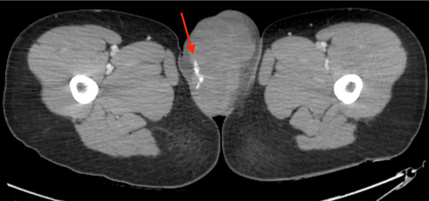
CPC-EM Capsule
What do we already know about this clinical entity?
Vulvar hematomas are generally due to obstetric or traumatic causes, but, rarely, spontaneous local artery rupture can occur.
What makes this presentation of disease reportable?
This presentation is the only reported case of a spontaneous vulvar hematoma secondary to a contralateral internal iliac artery rupture.
What is the major learning point?
Vulvar hematomas are a clinical diagnosis but can be challenging. A proper history and physical exam are essential. Management is not well defined.
How might this improve emergency medicine practice?
Exposure to rare cases like this one allows for emergency medicine physicians to broaden their knowledge base and differential diagnoses.
triangle. Arterial supply to this region is primarily carried out by terminal branches of the internal pudendal artery which comes off the anterior division of the internal iliac artery. Vulvar hematomas most commonly arise from insult to these terminal branches, frequently the posterior labial branch, but can rarely arise from the pudendal artery as well as the internal iliac artery as in our case. The superficial perineal pouch, the potential space formed by the perineal membrane (formerly urogenital diaphragm) and Colles fascia, allows for isolated vulvar hematomas to collect as fluctuant masses that expand outwardly towards the skin and can reach sizes greater than 15 cm.1,2
Etiologies of vulvar hematomas fall within two categories: obstetric or non-obstetric. Obstetric related hematomas are either due to direct injury during labor such as from instrumentation use, laceration repairs and episiotomies, or indirect injury such as from excessive stretching of birth canal during vaginal deliveries. Risk factors include primiparity, coagulopathies, use of anticoagulants, vulvovaginal varicosities, macrosomia, hypertensive disorders of pregnancy and prolonged second stage of labor.3-5
Non-obstetric related hematomas are mostly attributed to trauma to the perineum. Insults include but are not limited to vulva surgery, foreign body insertion, saddle injury, sexual
assault as well as consensual coitus.6,7 Age is a significant risk factor for trauma-related vulvar hematomas. Pre-pubertal children and adolescents have decreased vulval fat, notably in the labia majora, which normally acts as a protective barrier for the underlying structures in healthy adult women. Similarly, hypoestrogenic postmenopausal women are more
susceptible to arterial injury from loss of vulvar elasticity and vaginal atrophy.2
In rare circumstances, hematomas that lack evidence of obstetric or traumatic events are presumed to be of spontaneous artery rupture origin, mainly the internal pudendal or the internal iliac arteries. Rupture of iliac artery is most associated with aneurysms secondary to atherosclerotic disease but can also be caused by coagulopathies. Rarely the aneurysms can be due to connective tissue disease or infections. In cases where traumatic events do not result in immediate arterial ruptures, pseudoaneurysms can form that may lead to spontaneous vulvar hematomas on a later date. This is most associated with the pudendal artery rather than the internal iliac artery.1,8
Vulvar hematomas are a clinical diagnosis but can be challenging. The hallmark symptom is moderate to severe pain that is usually in the perineum but can be in the groin, abdominal and/or buttock regions depending on the size and location of the hematoma. Additionally, neurologic and urologic symptoms, from compression of the nerve roots and bladder or urethra respectively, can be observed. Complications can include infection, pressure necrosis and hemodynamic instability.1,8,9 A proper history and physical exam are essential to rule out the differential diagnoses such as vulvar varicosities, folliculitis, Bartholin gland cysts/abscesses or vulvar cancer.
Image 2. Lithotomy view demonstrates large, slowly extravasating vulvar hematoma with vulvar ecchymosis. This figure clarifies that this is a left vulvar hematoma with deviation of the vulva to the right. Not visible is four cm skin breakdown of the left vaginal wall.
Management of vulvar hematomas is not well defined. Small and stable hematomas can be treated expectantly and conservatively with local compression, ice packs and analgesics, as most will resolve on their own. The exact point at which a hematoma would benefit from operative or selective artery embolization management has not been established. Among the reported cases where invasive treatment was pursued, hematoma sizes varied greatly ranging anywhere from 7 cm to 15 cm and were present with other serious complications such as hemodynamic instability and/or pressure necrosis.9-11 Ultimately, clinical decision should be based on degree of hemodynamic stability, size of the
Image 3. Left: Digital subtraction angiogram (DSA) of normal right iliac artery for reference: red = internal iliac anterior division, blue = internal iliac posterior division, green = iliolumbar artery, orange = lateral sacral artery, black = superior gluteal artery. Middle: right internal iliac DSA with focal extravasation: Red arrow points to a subselective catheterization of right internal iliac artery anterior division with a small focus of extravasation. Right: right internal iliac DSA post embolization: Red arrow points to selective catheterization of right internal iliac artery with DSA following gel foam embolization demonstrating truncation and pruning of vessel.
Raveiro et al. A Case of Atraumatic and Non-obstetric Vulvar Hematoma from Contralateral Internal Iliac Artery Rupture
hematoma, rate of expansion, risk or presence of pressure necrosis, urologic symptoms and presence of unremitting pain.2,9 Important to note, a retrospective study found that conservative treatment increased risks of complications leading to longer hospital stays, infections and need for transfusions when compared to operative management.12
Although a definitive explanation as to how a bleed originating from the right internal iliac artery resulted in a left vulvar hematoma could not be established, we surmise the complex network of vascular anastomoses within the pelvic cavity, in conjunction with the continuity of the fascial planes in this region, allowed for extravasated blood to cross the midline via flow through established vascular networks.13-15
The majority of vulvar hematomas are obstetric in nature. Non-obstetric hematomas have an incidence of 3.7% and make up 0.8% of all gynecological emergencies. Of these, even a smaller percentage constitutes spontaneous etiology.16 To date, there are three reported spontaneous vulvar hematomas due to pudendal artery rupture and one due to internal iliac artery rupture.1,10,17,18 To the best of our knowledge, our case represents the second reported account of non-obstetric, non-traumatic spontaneous vulvar hematoma due to internal iliac artery rupture and the first reported account where the resulting hematoma was contralateral to the affected artery.
Patient consent has been obtained and filed for the publication of this case report.
Address for Correspondence: Moshe Bengio. DO, Med, HCA Florida Aventura Hospital, Department of Emergency Medicine, 3330 N 65th Ave, Hollywood, FL 33024, Email: Moshebengio1@ gmail.com.
Conflicts of Interest: By the CPC-EM article submission agreement, all authors are required to disclose all affiliations, funding sources and financial or management relationships that could be perceived as potential sources of bias. The authors disclosed none.
Copyright: © 2025 Raveiro et al. This is an open access article distributed in accordance with the terms of the Creative Commons Attribution (CC BY 4.0) License. See: http://creativecommons.org/ licenses/by/4.0/
1. Egan E, Dundee P, Lawrentschuk N. Vulvar hematoma secondary to spontaneous rupture of the internal iliac artery: clinical review. Am J Obstet Gynecol. 2009;200(1):e17-8.
2. Lapresa Alcalde MV, Hernandez E, Bustillo Alfonso S, et al. Non-
obstetric traumatic vulvar hematoma: Conservative or surgical approach? A case report. Case Rep Womens Health 2019;22:e00109.
3. Iskender C, Topcu HO, Timur H, et al. Evaluation of risk factors in women with puerperal genital hematomas. J Matern Fetal Neonatal Med. 2016;29(9):1435-9.
4. Rani S, Verma M, Pandher DK, et al. Risk Factors and Incidence of Puerperal Genital Haematomas. J Clin Diagn Res. 2017;11(5):QC01-3.
5. Sheikh GN. Perinatal genital hematomas. Obstet Gynecol 1971;38(4):571-5.
6. Jones IS and O’Connor A. Non-obstetric vulval trauma. Emerg Med Australas. 2013;25(1):36-9.
7. Sherer DM, Stimphil R, Hellmann M, et al. Transperineal sonography of a large vulvar hematoma following blunt perineal trauma. J Clin Ultrasound. 2006;34(6):309-12.
8. Dix FP, Titi M, Al-Khaffaf H. The isolated internal iliac artery aneurysm--a review. Eur J Vasc Endovasc Surg. 2005;30(2):119-29.
9. Mangwi AA, Ebasone PV, Aroke D et al. Non-obstetric vulva haematomas in a low resource setting: two case reports. Pan Afr Med J. 2019;33:314.
10. Hong HR, Hwang KR, Kim SA, et al. A case of vulvar hematoma with rupture of pseudoaneurysm of pudendal artery. Obstet Gynecol Sci 2014;57(2):168-71.
11. Yadav GS and Marashi A. Evacuation of a large traumatic vulvar haematoma with an intravaginal cosmetic approach. BMJ Case Rep 2019;12(5):e228535
12. Benrubi G, Neuman C, Nuss RC, et al. Vulvar and vaginal hematomas: a retrospective study of conservative versus operative management. South Med J. 1987;80(8):991-4.
13. Gore RM, Balfe DM, Aizenstein RI, et al. The great escape: interfascial decompression planes of the retroperitoneum. AJR Am J Roentgenol. 2000;175(2):363-70.
14. Lee MJ, Wright A, Cline M, et al. Pelvic Fractures and Associated Genitourinary and Vascular Injuries: A Multisystem Review of Pelvic Trauma. AJR Am J Roentgenol. 2019;213(6):1297-306.
15. Zhou Z, Li B, Zhou J, et al. Anatomical investigation of the pelvic urogenital fascia in 10 formalin-fixed female cadavers: novel insights into the laparoscopic total mesometrial resection. BMC Surg 2023;23(1):329.
16. Papoutsis D and Haefner HK. Large Vulvar Haematoma of Traumatic Origin. J Clin Diagn Res. 2017;11(9):QJ01-2.
17. Eze IO, Anyanyu N, Nzeribe-Abangwu U, et al. A rare case of life threatening non-obstetric-non-traumatic vulvar-vaginal haematoma secondary to suspected spontaneous rupture of the right pudendal artery: surgical management in a reference hospital in Southern Nigeria. Int J Reprod Contracept Obstet Gynecol. 2023;12:3646-50.
18. Mann D, Satin R, Gordon PH. Neurologic sequelae following transcatheter embolization to control massive perineal hemorrhage. Dis Colon Rectum. Mar 1984;27(3):190-2.
Boris Penev, MD*
Hallmon Hughes, MHS†
Katherine Scarpino, DO*
Daniel
J.
Ritter, MD*
Section Editor: Anna McFarlin, MD
Medical University of South Carolina, Department of Emergency Medicine, Charleston, South Carolina
Medical University of South Carolina, College of Medicine, Charleston, South Carolina
Submission history: Submitted February 17, 2025; Revision received April 4, 2025; Accepted May 9, 2025
Electronically published July 8, 2025
Full text available through open access at http://escholarship.org/uc/uciem_cpcem
DOI: 10.5811/cpcem.43035
Introduction: The trigeminocardiac reflex (TCR), a physiologic response to irritation of the branches of the trigeminal nerve, was first described in humans in 1870. Gastric hypermotility, hypotension, bradycardia, and even asystole have been reported in response to surgical manipulation of the trigeminal nerve and its branches, but literature is limited in patients not undergoing surgery. Although effects are generally transient and benign, TCR can present a significant diagnostic and therapeutic challenge in patients undergoing surgical manipulation of the trigeminal nerve and its branches.
Case Report: We describe a case of severe bradycardia secondary to facial trauma causing hemodynamic compromise and diagnostic uncertainty.
Conclusion: This case highlights a possible case of TCR, as well as therapeutic considerations, in a patient presenting to the emergency department with severe facial trauma. [Clin Pract Cases Emerg Med. 2025;9(3):322-325.]
Keywords: case report; trigeminocardiac reflex; facial trauma; bradycardia.
The trigeminocardiac reflex (TCR) is defined as the sudden onset of parasympathetic dysrhythmia, hypotension, apnea, or gastric hypermotility during stimulation of the sensory fibers of the trigeminal nerve. It manifests during craniofacial surgeries or secondary to trauma and can present as an imbalance between sympathetic and parasympathetics nervous systems.1 This neurologic response to stimulation of the trigeminal nerve was first recognized in 1870, and the term “trigeminocardiac reflex” was coined in 1999 by Schaller et al.2 The exact mechanism, although unclear, is thought to be a protective one that aims at reducing cerebral blood flow and intracranial pressure. There are different clinical subtypes of TCR that present with various cardiorespiratory manifestations such as bradycardia, hypotension, apnea, and sometimes even the opposite sympathetic effects, depending
on where along the reflex arc the nerves are stimulated.1-3 The oculocardiac reflex, a subtype of the TCR, has been described in the emergency medicine literature.4 The trigeminocardiac reflex has been documented in instances of surgical manipulation, but there is a paucity of data with regard to traumatic injuries not undergoing surgical intervention. We report a case of a 57-year-old male exhibiting severe bradycardia after presenting with significant facial trauma from a syncopal episode, prompting the need for vigilant monitoring and ultimately resulting in an intensive care unit (ICU) admission.
A 57-year-old male with past medical history significant for nonischemic cardiomyopathy, coronary artery disease, HIV, and hypertension presented to a Level I trauma center emergency department (ED) for a syncopal episode resulting
Penev et al. The Trigeminocardiac Reflex? Severe Bradycardia Secondary to Facial Trauma: A Case Report
in a ground-level fall while at a gas station. He experienced facial trauma from a syncopal event after prolonged bicycling on a hot day. On arrival to the ED, triage vital signs were notable for a heart rate of 49 beats per minute (bpm) and blood pressure of 142/95 millimeters of mercury (mm Hg). He was noted to be mildly confused and had significant facial trauma with a left-sided mandible deformity, intra-oral bleeding, chin and lip lacerations, and instability to his upper and lower teeth. Analgesia, tetanus prophylaxis, and fluid resuscitation were initiated. An electrocardiogram (ECG) (Image 1) demonstrated sinus bradycardia with a rate of 45 bpm and inferolateral T-wave inversions. Labs were notable for a lactate of 4.7 millimoles per liter (mmol/L) (reference range: 0.7-2.1 mmol/L) and creatinine 2.3 milligrams per deciliter (mg/dL) (0.7-1.3 mg/dL) with a baseline creatinine of 1.1 mg/dL. Electrolytes and troponin were within normal lab ranges. Approximately two hours into the patient’s ED stay, while awaiting computed tomography (CT), he was noted to have episodes of symptomatic bradycardia to as low as 35 bpm with associated respiratory distress and nausea. These episodes were particularly pronounced with supine positioning, improving with upright positioning and suctioning of secretions. Given a high clinical concern for intracranial and/or maxillofacial injuries, it was decided to attempt CT while the ED staff closely monitored him. While in the CT scanner, he had two more episodes of bradycardia with heart rates of 20-30 bpm upon lying flat that resolved with upright positioning and suctioning. Given concerns for impending hemodynamic collapse and lack of airway protection, the patient was taken to a resuscitation bay and evaluated with the trauma service. A vagal-mediated bradycardia was suspected, and the patient was administered 1 mg of intravenous atropine with resolution of bradycardia. Non-contrast CT of the head revealed no intracranial injuries, and non-contrast CT of the facial bones (Image 2) demonstrated bilateral subcondylar fractures, comminuted symphysis fracture, fracture of anterior maxilla with
Image 1. Electrocardiogram demonstrating sinus bradycardia and inferolateral T-wave inversions (arrows).
CPC-EM Capsule
What do we already know about this clinical entity?
The trigeminocardiac reflex (TCR) is a physiologic response to irritation of branches of the trigeminal nerve and has been described in craniofacial surgery literature.
What makes this presentation of disease reportable?
To date, TCR has not been described in response to maxillofacial trauma independent of surgical manipulation.
What is the major learning point?
Clinicians should keep TCR in their differential diagnosis when faced with characteristic hemodynamic instability in patients with facial trauma.
How might this improve emergency medicine practice?
Awareness of TCR will aid emergency physicians in recognizing and managing this potentially lifethreatening neurologic response.
intrusion of teeth #7 - 10, and multiple skin lacerations. The patient was admitted to the trauma ICU for airway and cardiac monitoring with plans for open reduction and internal fixation of his injuries. During his admission, he continued to suffer from episodes of severe sinus bradycardia. The patient was evaluated by cardiology for possible pacemaker placement in the setting of bradycardia. Per chart review, the patient’s baseline heart rate was 60-75 bpm (no prior ECGs are available), and he was not prescribed atrioventricular (AV) nodal blocking agents. Ultimately it was determined that the patient’s bradycardia was mediated by increased vagal tone secondary to facial trauma. On hospital day 2, he underwent surgical fixation of his injuries with no reported significant intraoperative events. With surgical fixation on hospital day 2 the patient had no further episodes of bradycardia and was uneventfully discharged on hospital day 5.
The trigeminocardiac reflex is a rare phenomenon that has been described during maxillofacial surgery. Stimulation of the trigeminal nerve sends an afferent signal to the brainstem, which manifests in efferent vagal nerve-mediated modulation
The Trigeminocardiac Reflex? Severe Bradycardia Secondary to Facial Trauma: A Case Report
supine positioning. The bradycardic episodes were >20% decrease from his baseline. Of note, the patient had no significant recorded hypotension. Our suspicions were further supported by the improvement of his bradycardia with atropine and its resolution with surgery. While improvement with atropine is not unique to TCR, it does suggest a vagally mediated bradycardia. The lack of bradycardic episodes following surgery thus gave us high suspicion that the TCR was the cause of his bradycardia. Finally, the patient was evaluated by the electrophysiology service, which concluded he most likely suffered bradycardic episodes related to heightened vagal tone consistent with the TCR.
of blood pressure, inducing bradycardia. Much of the current literature on the topic describes its occurrence and management in maxillofacial surgery. Although the oculocardiac reflex has been described in relation to ocular trauma, this case provides a unique addition to the literature, as our patient did not sustain ocular trauma. If our case was indeed a TCR-mediated bradycardia, it would provide valuable insight into expansion of the current definition of TCR. The incidence of TCR during maxillofacial and cranial surgery is estimated to be 11-18%.5,6 Clinicians must be aware of this phenomenon as it can occur in the absence of known cardiac conduction disease (eg, sinus node dysfunction, AV block), and close monitoring should be provided.
Suspicion of TCR in this patient was largely based on elimination of other causes for his bradycardia and symptoms. Proposed diagnostic criteria for TCR include the following: characteristic changes in heart rate (10-20% drop) and blood pressure; plausibility of a relationship to irritation of trigeminal nerve; and repeatability.2 The patient was not taking any AV nodal blocking and did not have a known history of bradycardia. He sustained no other significant trauma beside his mandibular and maxillary fractures that did not involve the orbit. The bradycardic episodes were also repeatable with
While published data on the treatment of TCR specifically is sparse, much can be applied from the treatment of other bradycardias, specifically vagal-mediated bradycardias. The surgery literature suggests consideration of prophylactic treatment with atropine or epinephrine.6 The use of hemodynamic-altering medication, however, may cloud the clinical picture in the ED. Initial management includes ensuring airway patency, oxygenation, and establishing IV access. Noxious stimuli and repositioning can improve the heart rate, as was seen in our patient. External cardiac-pacing pads should be at the bedside, ready to be applied. The primary pharmacological treatment of TCR is atropine, administered as 1 mg intravenously every 3-5 minutes, up to a total dose of 3 mg. Epinephrine and transcutaneous or transvenous pacing can also be considered as additional therapy in cases refractory to other treatments.7 Finally, we recommend expert consultation with the trauma and cardiology services in severe or refractory cases.
While this case report provides valuable insights into the potential for TCR to occur in instances beyond surgical manipulation, it is important to acknowledge its limitations. Most of the supporting literature discusses this reflex primarily in the context of surgical manipulation, with limited reference to facial trauma. Variability in injury severity, patient physiology, and underlying electrophysiologic conditions may limit the generalizability of our conclusions. Despite these limitations, we suggest that clinicians consider TCR in the context of facial trauma, where mechanical stimulation of the trigeminal nerve can lead to potentially life-threatening bradycardia, hypotension, or cardiac arrest.
The trigeminocardiac reflex is a brainstem-mediated response triggered by stimulation of the trigeminal nerve or its branches, leading to parasympathetic activation. This reflex arc can result in bradycardia, hypotension, and even asystole. It is particularly common during manipulation of orbital or maxillofacial structures in surgery, but as reported here, it may be seen in maxillofacial trauma. Management of TCR-induced bradycardia involves a systematic approach to stabilize the
Penev et al. The Trigeminocardiac Reflex? Severe Bradycardia Secondary to Facial Trauma: A Case Report
patient and address the underlying cause. We suggest that clinicians become aware of TCR and its treatment and to remain vigilant that it may present outside the surgical theater.
The authors attest that their institution requires neither Institutional Review Board approval, nor patient consent for publication of this case report. Documentation on file.
Address for Correspondence: Daniel J. Ritter, MD, Assistant Professor, Medical Univeristy of South Carolina Department of Emergency Medicine, 169 Asley Ave. MSC 300 Charleston SC 29425. Email: ritterda@musc.edu.
Conflicts of Interest: By the CPC-EM article submission agreement, all authors are required to disclose all affiliations, funding sources and financial or management relationships that could be perceived as potential sources of bias. The authors disclosed none.
Copyright: © 2025 Penev et al. This is an open access article distributed in accordance with the terms of the Creative Commons Attribution (CC BY 4.0) License. See: http://creativecommons.org/ licenses/by/4.0/
1. Singh G and Chowdhury T. Brain and heart connections: the trigeminocardiac reflex! J Neuroanaesth Crit Care. 2017;04(02):071-7.
2. Meuwly C, Chowdhury T, Sandu N, et al. Definition and Diagnosis of the Trigeminocardiac Reflex: A Grounded Theory Approach for an Update. Front Neurol. 2017;8:533.
3. Chowdhury T, Mendelowith D, Golanov E, et al. Trigeminocardiac reflex: the current clinical and physiological knowledge. J Neurosurg Anesthesiol. 2015;27(2):136-47.
4. Fahling JM and McKenzie LK. Oculocardiac reflex as a result of intraorbital trauma. J Emerg Med. 2017;52(4):557-558.
5. Schaller B, Probst R, Strebel S, et al. Trigeminocardiac reflex during surgery in the cerebellopontine angle. J Neurosurg. 1999;90(2):21520.
6. Arasho B, Sandu N, Spiriev T, et al. Management of the trigeminocardiac reflex: facts and own experience. Neurol India 2009;57(4):375-80.
7. Farkas J. Bradycardia. EMCrit Project. January 4, 2025. Available at: https://emcrit.org/ibcc/bradycardia/#atropine_&_glycopyrrolate. Accessed February 20, 2025.
Christina Cantwell, MD, MS
Mark I Langdorf, MD, MHPE
Section Editor: Steven Walsh, MD
University of California, Irvine, Medical Center, Department of Emergency Medicine, Orange, California
Submission history: Submitted March 17, 2025; Revision received April 30, 2025; Accepted May 20, 2025
Electronically publishedJuly 14, 2025
Full text available throu gh open access at http://escholarship.org/uc/uciem_cpcem DOI: 10.5811/cpcem.47005
Introduction: Brugada syndrome is a ventricular arrhythmia and type of sodium channelopathy that can be seen in the absence of structural heart disease. Recognition of this pattern on electrocardiogram (ECG) is important for stabilization and correction of underlying triggers that can be addressed in the emergency department (ED).
Case report: We describe a case of a 58-year-old male who presented with chest pain and was found to have type 1 Brugada pattern in the setting of severe hypokalemia. Repletion of potassium later revealed type 3 Brugada pattern followed by resolution on repeat ECG.
Conclusion: Rapid identification of underlying metabolic derangements that can trigger Brugada syndrome is important in the ED setting. Correction of the underlying abnormality can reveal a type 3 pattern with subsequent resolution of the pattern if well-controlled. [Clin Pract Cases Emerg Med. 2025;9(3):326-328.]
Keywords: hypokalemia; Brugada; case report; type 1 Brugada; type 3 Brugada.
We describe a case of a 58-year-old male who presented with chest pain and was found to have type 1 Brugada pattern in the setting of severe hypokalemia. Repletion of potassium later revealed type 3 Brugada pattern. Previous case reports have described hypokalemia unmasking type 1 pattern that resolved with electrolyte repletion; here we report type 1 pattern converting to type 3 pattern with normalization of potassium which, to the best of our knowledge, has not yet been reported.
A 58-year-old male of Asian descent with a history of Cushing disease from a pituitary tumor, hypokalemia, and hypertension presented to the emergency department (ED) with one day of chest pain and shortness of breath. His medications included spironolactone, furosemide, metoprolol, potassium supplementation, and amlodipine. There was no known reported family history of cardiac disease.
Upon arrival the patient was initially an ST-elevation
myocardial infarction (STEMI) activation from triage. Initial vitals were temperature 37.1 °C, heart rate 93 beats per minute, blood pressure 158/94 millimeters of mercury, and oxygen saturation 100% on room air with respiratory rate of 18 breaths per minute. He was alert and in no acute distress. Cardiac exam was significant for regular rate and rhythm with equal bilateral peripheral pulses and no audible murmur. Lungs were clear to auscultation bilaterally. There was symmetric 3+ pitting edema to the knees bilaterally.
Differential diagnosis included acute coronary syndrome, arrhythmia, Brugada syndrome, electrolyte derangement, aortic dissection, pulmonary embolism, and pneumonia. Workup initially included complete blood count, comprehensive metabolic panel, thyroid panel, serum troponin I, D-dimer, magnesium level, B-type natriuretic peptide, prothrombin time/ international normalized ratio/partial thromboplastin time, hemoglobin A1c, venous blood gas, repeat electrocardiogram, portable one-view chest radiograph, and cardiology consult. Labs were significant for hypokalemia of 2.5 millimoles per liter (mmol/L) (reference range: 3.5-5.1 mmol/L), troponin of
et al. Hypokalemia-induced Type 1 Brugada Reveals Type 3 Brugada Pattern with Repletion: Case Report
40 nanograms (ng)/L (0-20 ng/L), glucose of 356 milligrams per deciliter (mg/dL) (70-115 mg/dL). Electrocardiogram (ECG) showed type 1 Brugada pattern (Image 1) with an incomplete right bundle branch block pattern in V1 and V2 followed by straightening of the ST segment from the top of the QRS complex leading to an inverted T wave. Chest radiograph revealed a widened mediastinum, and computed tomography angiogram of the whole aorta ruled out aortic dissection. Diagnostic challenges included hypokalemia precluding the patient from being optimized for potential cardiac catheterization. However, when the type 1 Brugada pattern was recognized in the ED, the STEMI activation was cancelled.
Potassium repletion initiated in the ED consisted of 40 milliequivalents (mEq) oral and 40 mEq intravenous potassium chloride. Chest pain improved with two sublingual sprays of nitroglycerin, 400 micrograms each, and with potassium repletion.
After cardiology consultation, the patient was admitted to the internal medicine service for further workup and cardiac monitoring. Troponin downtrended to 33 ng/L on repeat check one hour later. Upon repletion of potassium, repeat ECG 24 and 48 hours after initial presentation revealed type 3 Brugada patterns (Image 2).
During his hospital course, the patient had difficult-tocontrol hypokalemia and hyperglycemia concerning for a hypercortisol state with an unclear source despite unremarkable magnetic resonance imaging and positron emission tomography without identified tumor or mass, ultimately requiring initiation of insulin and aggressive potassium supplementation with potassium chloride 40 mEq three times daily. Forty-eight hours and six weeks after initial presentation, his ECG showed normal sinus rhythm without Brugada pattern (Image 3). (Six-week ECG is not shown).
Image 1. Initial electrocardiogram on arrival showing type 1 Brugada pattern in a patient presenting with chest pain. Note the straightening of the ST segment from the QRS complex to the inverted T wave (arrows).
CPC-EM Capsule
What do we already know about this clinical entity?
Brugada syndrome is a sodium channelopathy that can predispose to life-threatening arrhythmias if left untreated.
What makes this presentation of disease reportable?
In a patient with severe hypokalemia, Brugada pattern is seen to transform from type 1 to type 3 pattern and ultimately resolve with electrolyte repletion.
What is the major learning point?
A hypokalemic state can cause a hyperpolarization of the cardiac membrane and slow the action potential resulting in a reversible Brugada pattern when corrected.
How might this improve emergency medicine practice?
Identification and correction of underlying potassium abnormality can reveal a type 3 pattern with subsequent resolution of the Brugada pattern if well-controlled.
Image 2. Second electrocardiogram 24 hours after arrival showing type 3 Brugada pattern after correcting severe hypokalemia. Note the ST segment in leads V1 and V2 are now less than two millimeters (arrows).
Hypokalemia-induced Type 1 Brugada Reveals Type 3 Brugada Pattern with Repletion: Case Report Cantwell et al.
Brugada syndrome has been reported as a type of ventricular arrhythmia identified in patients in the absence of known structural heart disease.1 This pattern was initially characterized by a right bundle branch block and ST- segment elevation in V1, V2, and V3 leads, which our patient had on arrival. The first study by Brugada and Brugada in 1992 found this pattern in the absence of electrolyte derangement.1 It is now known that Brugada syndrome is a sodium channelopathy caused by a genetic mutation in the sodium channel gene.2 Factors that can unmask a Brugada pattern include fever, certain drugs, hypokalemia, hyperkalemia, and hypothermia.2 Type 1 describes an incomplete right bundle branch block pattern in V1 and V2 followed by straightening of the ST segment from the top of the QRS complex leading to an inverted T wave. Type 2 describes a saddleback ST-segment pattern, and type 3 describes type 1 or type 2 morphology but with < 2 mm of ST- segment elevation. While there have been case reports of a type 1 pattern being seen with hypokalemia that resolves with repletion,3,4,5 in this case a type 3 pattern was revealed as potassium normalized. In the cardiac myocyte, a hypokalemic state can cause hyperpolarization of the membrane and inhibit the Na+-K+ ATPase, resulting overall in increased intracellular sodium and calcium and a slowed action potential.6 While types 2 and 3 patterns are suggestive of Brugada disease, the type 1 pattern is diagnostic.7 In this case, normalization of serum potassium levels reduced and ultimately resolved the ST-segment Brugada patterns. The type 3 pattern may be more favorable physiologically compared to type 1 given that the ST-segment elevation is less pronounced, representing a less delayed action potential.
Brugada pattern can be seen in severe underlying metabolic derangement—in this case, associated with hypokalemia. It is
important to identify and address any triggers such as metabolic derangement and infection and to avoid drugs that affect serum potassium levels. Correction of the underlying abnormality can reveal a type 3 pattern with subsequent resolution of the pattern if well-controlled. While our patient did not have any resultant arrhythmias characteristic of Brugada syndrome, he did have the electrocardiographic changes consistent with Brugada pattern.
Patient consent has been obtained and filed for the publication of this case report. The authors attest that their institution does not require Institutional Review Board approval for publication of this case report. Documentation on file.
Address for Correspondence: Mark I. Langdorf, MD, MHPE, University of California, Irvine, Department of Emergency Medicine, 3800 W Chapman Avenue, Suite 3200, Orange, CA 92868. Email: milangdo@uci.edu
Conflicts of Interest: By the CPC-EM article submission agreement, all authors are required to disclose all affiliations, funding sources and financial or management relationships that could be perceived as potential sources of bias. The authors disclosed none.
Copyright: © 2025 Cantwell et al. This is an open access article distributed in accordance with the terms of the Creative Commons Attribution (CC BY 4.0) License. See: http://creativecommons.org/ licenses/by/4.0/
1. Brugada P and Brugada J. Right bundle branch block, persistent ST segment elevation and sudden cardiac death: a distinct clinical and electrocardiographic syndrome. a multicenter report. J Am Coll Cardiol. 1992;20(6):1391-6.
2. Larkin J, Cadogan M, Buttner R. Brugada syndrome. Life in the fast lane. October 22, 2024. Accessed November 6, 2024. Available at: https://litfl.com/brugada-syndrome-ecg-library/
3. Swe T and Dogar MH. Type 1 Brugada pattern electrocardiogram induced by hypokalemia. J Family Med Prim Care 2016;5(3):709-11.
4. Maturi B, Crawford W, Sharma A, et al. Brugada syndrome unmasked by severe hypokalemia from alcoholism. J Am Coll Cardiol 2022;79(9S):3359.
5. Kutsuzawa D, Arimoto T, Watanabe T, et al. Persistent abnormal value of late potential in Brugada syndrome associated with hypokalemia. Ann Noninvasive Electrocardiol. 2011;16(1):104-6.
6. Weiss JN, Qu Z, Shivkumar K. The electrophysiology of hypo- and hyperkalemia. Circ Arrhythm Electrophysiol. 2017;10(3):e004667
Image 3. Electrocardiogram 48 hours later showing normal sinus rhythm and no indication of Brugada pattern after achieving normalization of electrolytes.
7. Antzelevitch C, Brugada P, Borggrefe M, et al. Brugada syndrome: report of the second consensus conference: endorsed by the Heart Rhythm Society and the European Heart Rhythm Association [published correction appears in Circulation. 2005 Jul 26;112(4):e74]. Circulation. 2005;111(5):659-670.
Emma Alley, DO, MPH, MS
Kristy Holecko, MD
Section Editor: Anna McFarlin, MD
Submission history: Submitted December 29, 2024; Revision received April 22, 2025; Accepted May 18, 2025
Electronically published July 8, 2025
Full text available through open access at http://escholarship.org/uc/uciem_cpcem
DOI: 10.5811/cpcem.41539
Introduction: Rothia mucilaginosa is a rare cause of endocarditis, typically seen in intravenous (IV) drug users who use needles contaminated with saliva. However, it is rare in individuals who are immunocompetent, have no history of valvular disease, or have not undergone valvular repair.
Definitive management of R mucilaginosa endocarditis is valvular repair, but this procedure can be delayed in the setting of intracranial hemorrhage.
Case Report: We document the case of a 35-year-old male IV drug user who developed R mucilaginosa endocarditis, resulting in severe neurologic sequelae due to septic emboli. The patient presented to the emergency department (ED) where work-up revealed a clinical presentation consistent with endocarditis resulting in septic emboli. He was later admitted to the neurosurgical and cardiac intensive care units, where he underwent thrombectomy, monitoring of his intraparenchymal hemorrhage (IPH), and mitral valve repair. This case highlights the patient’s functional neurologic outcome following delayed mitral valve repair due to IPH.
Conclusion: This case report highlights a rare form of R mucilaginosa endocarditis recognized in the ED, with a hospital course including thrombectomy, IPH monitoring, and mitral valve repair. The patient had progressive neurologic sequelae given delayed mitral valve repair due to concerns that procedural heparinization would worsen his IPH. Given functional decline, the patient underwent mitral valve repair on hospital day six without worsening of his IPH, demonstrating that current guidelines to delay mitral valve repair by four weeks in the setting of intracranial hemorrhage may be too strict for patients who are high risk for continued showering of septic emboli. [Clin Pract Cases Emerg Med. 2025;9(3):329-333.]
Keywords: endocarditis; septic emboli; Rothia mucilaginosa; IV drug; case report.
Rothia mucilaginosa is a rare cause of disease and endocarditis. The bacteria exist as a normal part of the flora within the human oral cavity and respiratory tracts. The pathogen is opportunistic and mostly associated with infection in the setting of immunocompromised patients or patients who have implantable devices, such as prosthetic heart valves.1,2 Rothia mucilaginosa infection has also been seen in intravenous (IV) drug users.3 In addition to endocarditis, R
mucilaginosa has been linked with bacteremia, pneumonia, catheter-associated bloodstream infections, meningitis, peritonitis, osteomyelitis, and soft tissue infections.1,2
The patient, a 35-year-old male with a medical history of IV drug use, active fentanyl use, cured hepatitis C postantiviral therapy, and hypertension presented to the emergency department with left-sided facial and upper extremity numbness that began upon awakening on the day of
presentation. The patient also reported right peripheral vision loss the day prior, which he thought was a complication of welding. His visual loss had not resolved by the time of presentation. He denied chest pain, shortness of breath, abdominal pain, and nausea. The patient endorsed using IV fentanyl at least three times a week and stated he reused his own needles but would never share needles.
The patient was afebrile on arrival, and all vitals were within normal limits for his age. Physical examination revealed no abnormalities in the respiratory and abdominal systems. The cardiac exam showed a 2/6 systolic murmur appreciated at the cardiac apex. Neurologic examination revealed normal sensorium and subjective left facial numbness. The remaining cranial nerves were intact, with no facial droop observed. Speech was normal, and there was no weakness in the arms or legs. Pronator drift was absent. Ophthalmologic exam was pertinent for a temporal visual field defect in the right eye with retinal exam showing hemorrhage. Dermatological exam was significant for several erythematous lesions along the hypothenar eminence of the left hand as well as on the palm of the right hand, consistent with Janeway lesions (Image 1).
Initial laboratory workup revealed mild leukocytosis of 12 x 103 cells per microliter (K/µL) (reference range: 4-11 K/µL), elevated C-reactive protein at 170 milligrams per liter (mg/L) (< 5 mg/L), and erythrocyte sedimentation rate of 56 millimeters per hour (mm/hr) (<15 mm/hr). Two sets of blood cultures were obtained, eventually growing R mucilaginosa after admission. Computed tomography angiography (CTA) of the head and neck showed a new focal area of hypodensity in the right frontal lobe white matter and new small transcortical infarct in the left occipital lobe. Based on the CTA findings along with the physical exam and laboratory findings, the patient was admitted to the hospital for further evaluation of endocarditis with potential septic emboli. He was started on vancomycin and ceftriaxone.
Additional imaging studies were ordered. Magnetic resonance imaging (MRI) of the brain, with and without contrast, revealed multiple parenchymal signal abnormalities, including foci of diffusion restriction throughout the bilateral cerebral hemispheres and the right superior cerebellum, consistent with multifocal infarcts. Additionally, an intraparenchymal hemorrhage (IPH) within the right frontal lobe measuring a centimeter (cm) was observed. Transthoracic echocardiogram in the ED showed evidence of a mitral valve vegetation with regurgitation. Transesophageal echo ordered inpatient showed a 1.5 cm x 1.1 cm homogenous, mobile mass attached to the anterior mitral valve leaflet that represented a vegetation (Image 2). An anterior leaflet perforation was also appreciated, with severe mitral regurgitation present. No additional vegetations were observed on the other valves.
With visualization of vegetations on the mitral valve, the patient was primarily diagnosed with mitral valve endocarditis as a result of R mucilaginosa infection. Given the primary
CPC-EM Capsule
What do we already know about this clinical entity?
Rothia mucilaginosa is a rare form of infectious endocarditis typically seen in IV drug users.
What makes this presentation of disease reportable?
After Rothia mucilaginosa was identified in the ED, the patient underwent valvular repair within four weeks of intracranial hemorrhage.
What is the major learning point?
Patient care can be improved by rapid identification and risk/benefit analysis, although guidelines may not fit every case.
How might this improve emergency medicine practice?
This case highlights the need for emergency physicians to identify and advocate for patients at high risk of worsening neurologic function in the setting of continued septic emboli.
diagnosis, the new multifocal infarcts throughout the cerebral hemispheres and the right superior cerebellum were attributed to septic emboli. Cardiothoracic surgery was consulted and recommended mitral valve repair, but deferred surgery for at least four weeks given the presence of IPH on MRI.
Four days after admission, the patient developed sudden aphasia and right-sided arm weakness. He was taken urgently for thrombectomy after computed tomography (CT) head showed new right frontal infarct with hemorrhage.
Neurosurgery found a left superior division M2 occlusion during thrombectomy. The septic emboli were successfully removed, and the left middle cerebral artery superior division of M2 was revascularized with a thrombolysis in cerebral infarction score of three. He was subsequently transferred to the neurosurgical intensive care unit (ICU) after thrombectomy, and repeat MRI was performed (Image 3). Due to the high risk of continued ischemic events from septic emboli, the decision was made to expedite the patient’s mitral valve repair, which was performed six days after presentation.
He underwent mitral valve repair with P1 triangular resection, using the semi-rigid #34 Colvin-Galloway Future Band annuloplasty ring and band (Medtronic, PLC, Minneapolis, MN). After surgery, the patient was transferred to the cardiac ICU for further care. His CT head imaging
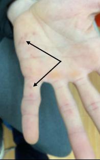
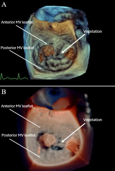
Image 2. The image depicts two three-dimensional reconstructions of the transesophageal echocardiogram labeled A and B. These images show a 1.5 cm x 1.1cm homogenous, mobile mass attached to the anterior mitral valve leaflet that represents a vegetation. There is anterior leaflet perforation.
remained stable after mitral valve repair and annuloplasty. He was discharged on hospital day 15 with a peripherally inserted central catheter for continued antibiotic therapy. At the time of discharge, he still exhibited right-sided weakness and aphasia, with minimal improvement.
Intravenous drug users are at increased risk of Rothia species endocarditis.3 Rothia mucilaginosa exists most commonly within the flora of the oral cavity, and it is believed that the bacteria is introduced into the blood stream of IV drug users when they lick their needles prior to injection or reuse dirty needles contaminated with saliva.4 Furthermore, IV drug users constitute a significant portion of endocarditis cases,
often resulting in damage to native or prosthetic valves from previous episodes of endocarditis. Damaged valves and prosthetic valves provide a habitable environment for opportunistic pathogens such as R mucilaginosa to grow.5 Intravenous drug users are also at increased risk of becoming immunocompromised in the setting of HIV infections, active hepatitis infection, or cirrhosis in the setting of polysubstance abuse and co-abuse with alcohol.6 Our patient had no history of valvular disease but admitted to reusing dirty needles. He had a prior hepatitis C infection, which was cured with treatment before he developed endocarditis.
Rothia mucilaginosa endocarditis has been associated with episodes of septic emboli. In 2020 Song et al documented a case of a 65-year-old male with diabetes mellitus and
Image 3. Images from a FLAIR MRI of the brain without contrast: A1 and A2 from MRI on hospital day two, show multiple parenchymal signal abnormalities throughout the cerebral hemispheres bilaterally compatible with multifocal infarcts from septic emboli (see arrows). A focus of intraparenchymal hemorrhage within the right frontal lobe measuring 1 cm is seen in A1 and redemonstrated in B1 with stable appearance (*). B1 and B2 are from MRI on hospital day 4 with new, left middle cerebral artery distribution infarcts involving the left basal ganglia, insula, corona radiata, and parietal lobe seen in B2 that were not seen prior when compared to A2. New infarcts are labeled with arrows in B1 and B2.
MRI, magnetic resonance imaging.
alcoholic liver cirrhosis who had a history of multiple episodes of endocarditis and had a prosthetic valve placed only to subsequently develop R mucilaginosa endocarditis. His course was complicated by septic emboli that resulted in multiple microhemorrhages. Song and colleagues also reviewed 39 cases of intracranial complications due to Rothia species endocarditis and noted that 37.5% sustained intracranial hemorrhage, 25% septic emboli, and 18.7% cerebral infarction, all of which were complications in our patient. It is important to note that only 15% of the cases of Rothia species reviewed in their paper were R mucilaginosa.7
In our case, the patient’s neurologic sequelae were likely exacerbated by the delay in valve repair, as he continued to experience septic emboli. The Society of Thoracic Surgeons recommends delaying valvular repair in the setting of infective
endocarditis by four weeks when an ICH is present, given concerns that heparinization during the procedure could worsen the ICH. However, the risk of deterioration or worsening of ICH after valvular surgery within a four-week period has not been well studied. In a similar case to ours, the authors reported valvular repair on hospital day 10 after discovery of subarachnoid hemorrhages due to septic emboli, Haddad et al describe an 80-year-old male who had a Rothia species aortic valve endocarditis with aortic root abscess. He underwent aortic debridement, pericardial patch placement, aortic valve repair, and mitral valve repair. No worsening of the patient’s subarachnoid hemorrhages or neurologic function was reported after repair.9
Yoshioka et al performed a retrospective study to investigate stratified risk in patients who had preoperative ICH prior to valvular surgery in the setting of infective endocarditis. They examined 30 patients with valvular repair ranging from within seven days of ICH onset up to >29 days of onset. They found that none of the 30 patients had worsening of their ICH after the procedure. Two patients developed new asymptomatic ICH after the procedure, one who was eight days out and the second 81 days out from onset of their preoperative ICH.10 Our patient ultimately underwent valvular repair approximately six days after the onset of IPH, without any complications of worsening ICH.
The findings from Yoshioka et al and the Haddad et al case report, along with our case, present an interesting contrast to the recommendations provided by the Society of Thoracic Surgeons. While R mucilaginosa is susceptible to beta-lactams and vancomycin, studies have shown that early valvular repair has been linked with improved mortality outcomes in patients with infective endocarditis over treatment with antibiotics alone.9-11 Understanding that functional outcomes for infective endocarditis are improved by early valvular repair highlights two important needs for better outcomes of patients with intracranial hemorrhage from septic emboli: 1) early identification of endocarditis, which can occur within the ED; a thorough history to identify risk factors, an in-depth physical exam, and use of diagnostic imaging, such as CT, MRI, or ultrasound to identify infective endocarditis and septic emboli before admission, as was seen in our case; and 2) further investigation into whether ICH from septic emboli should be listed as a relative vs absolute contraindication to valvular repair within four weeks, as early repair may help prevent the worsening of neurologic sequelae from continued septic emboli as we reported.
The authors attest that their institution requires neither Institutional Review Board approval, nor patient consent for publication of this case report. Documentation on file.
Alley et al.
Early Valvular Repair of Rothia mucilaginosa Endocarditis with IPH from Septic Emboli
Address for Correspondence: Emma Alley, DO, MPH, MS, Geisinger Medical Center, Emergency Department, 100 N Academy Ave., Danville, PA 17822. Email: eealley1@geisinger. edu
Conflicts of Interest: By the CPC-EM article submission agreement, all authors are required to disclose all affiliations, funding sources and financial or management relationships that could be perceived as potential sources of bias. The authors disclosed none.
Copyright: © 2025 Alley et al. This is an open access article distributed in accordance with the terms of the Creative Commons Attribution (CC BY 4.0) License. See: http://creativecommons.org/ licenses/by/4.0/
1. Ekkelenkamp MB, Rooijakkers SHM, Bonten MJM. Staphylococci and micrococci. In: Infectious Diseases 3rd Edition. Vol 2. Elsevier Ltd ; 2010:1632-44. Available at: https://doi.org/10.1016/b978-0-32304579-7.00165-9. Accessed May 19, 2025.
2. Ramanan P, Barreto JN, Osmon DR, et al. A 10-year experience at Mayo Clinic, Rochester, Minnesota. Diekema DJ, ed. J Clin Microbiol 2014;52(9):3184-9.
3. Bruminhent J, Tokarczyk MJ, Jungkind D, et al. Rothia mucilaginosa prosthetic device infections: a case of prosthetic valve endocarditis. J Clin Microbiol. 2013;51(5):1629–32.
4. Franconieri F, Join-Lambert O, Creveuil C, et al. Rothia spp. infective endocarditis: a systematic literature review. Infect Dis Now 2021;51(3):228-35.
5. Abdelmaseih R, Abdelmasih R, Faluk M, et al. Uncommon pathogen in an unexpected host: a rare case of Rothia mucilaginosa infective endocarditis in an immunocompetent patient without an underlying valvular disease. Cureus. 2021;13(6):e15458.
6. Deren S, Cleland CM, Lee H, et al. Brief report: the relationship between injection drug use risk behaviors and markers of immune activation. J Acquir Immune Defic Syndr. 2017;75(1):e8-12.
7. Song YJ, Kim B, Kim Y, et al. A Case of prosthetic valve endocarditis with cerebral hemorrhage caused by Rothia mucilaginosa Ann Clin Microbiol. 2020;23(4):211-6.
8. Byrne JG, Rezai K, Sanchez JA, et al. Surgical Management of Endocarditis: The Society of Thoracic Surgeons Clinical Practice Guideline. Ann Thorac Surg 2011;91(6):2012-9.
9. Haddad S, Saade Y, Ramlawi B, et al. Native valve endocarditis complicated by abscess formation caused by Rothia mucilaginosa IDCases. 2021;26:e01348.
10. Yoshioka D, Toda K, Sakaguchi T, et al. Valve surgery in active endocarditis patients complicated by intracranial haemorrhage: the influence of the timing of surgery on neurological outcomes. Eur J Cardiothoracic Surg. 2014;45(6):1082-8.
11. Netzer R, Altwegg S, Zollinger E, et al. Infective endocarditis: determinants of long term outcome. Heart. 2002;88(1):61-6.
M. Townsend Reeves, MD
Editor: R. Gentry Wilkerson, MD
Atrium Health Carolinas Medical Center, Department of Emergency Medicine, Charlotte, North Carolina
Submission history: Submitted March 30, 2025; Revision received May 19, 2025; Accepted May 27, 2025
Electronically published July 9, 2025
Full text available through open access at http://escholarship.org/uc/uciem_cpcem DOI: 10.5811/cpcem.47070
Introduction: Tube thoracostomy is a common yet exceedingly painful emergency department (ED) procedure, primarily used for pneumothorax evacuation. To facilitate safe performance, stable patients generally receive intravenous anxiolytic or opioid premedication, or perhaps even procedural sedation, in combination with local anesthetic infiltration of the insertion tract. However, more advanced pain management strategies, such as ultrasound-guided truncal blocks, offer a targeted and effective analgesic alternative without the risks or side effect profile inherent to sedation and opioid administration. Herein, a case is presented of painless ED tube thoracostomy following an ultrasound-guided thoracic paravertebral block (TPVB).
Case Report: A 74-year-old female presented to the ED with chest pain and dyspnea from a recurrent, large right-sided spontaneous pneumothorax. An ultrasound-guided thoracic paravertebral block was performed for full-thickness chest wall analgesia prior to tube thoracostomy. A pigtail catheter was inserted painlessly into the pleural space without need for rescue analgesia or procedural sedation, and the pneumothorax was successfully evacuated.
Conclusion: Ultrasound-guided regional anesthesia is increasingly employed in the emergency care setting as part of an opioid-sparing, multimodal analgesia strategy to manage acute pain. For chest tube insertion, the ultrasound-guided thoracic paravertebral block provides potent, long-lasting, and non-euphorigenic, hemithoracic analgesia across multiple contiguous dermatomes from skin to parietal pleura, reducing the need for procedural sedation and opioid therapy while avoiding the incomplete chest wall blockade often associated with other truncal blocks. It is a valuable addition to the analgesic armamentarium of the emergency physician, enabling more comprehensive pain control prior to tube thoracostomy. [Clin Pract Cases Emerg Med. 2025;9(3):334-339.]
Keywords: thoracic paravertebral block; tube thoracostomy; pneumothorax; ultrasound-guided regional anesthesia; case report.
Pneumothorax is the abnormal accumulation of air in the pleural cavity, the potential space between the parietal and visceral pleura lining the inner chest wall and lung, respectively. It is typically classified as traumatic or spontaneous in nature, with the latter further characterized as primary (occurring in absence of underlying lung disease) or secondary (occurring in presence of existing lung disease). Pneumothorax is commonly managed in the emergency department (ED) with tube
thoracostomy to evacuate air from the pleural cavity, allowing lung re-expansion and restoration of normal cardiopulmonary function.1 While more recent guidelines also support needle aspiration as a less invasive alternative treatment for spontaneous pneumothorax, immediate success rates are lower compared to tube thoracostomy, with the latter more commonly performed.2,3 Despite its efficacy, tube thoracostomy is a notoriously painful procedure, with 50% of patients reporting severe intraprocedural pain levels of 9-10/10 on a numerical rating scale.4
Excluding cases of tension pneumothorax, where expeditious chest tube insertion is prioritized over aggressive pain management to prevent respiratory failure, hemodynamic collapse, and death, emergency physicians traditionally provide intravenous anxiolytic or opioid premedication, combined with local anesthetic infiltration of the insertion tract prior to tube thoracostomy. Some may even use procedural sedation in stable patients needing large-bore (≥ 20 French [F]) chest tube insertion for viscous fluid (pyothorax, hemothorax) evacuation. However, these analgesic strategies are not ideal in many facets. Procedural sedation is time-consuming, resourceintensive, and carries risk of serious complications (eg, apnea, hypoxia, and hypotension), while opioids are habit-forming with a plethora of adverse side effects (eg, nausea/vomiting, pruritus, and delirium). Moreover, insufficient local anesthetic deposition along the chest tube’s tract commonly results in significant insertional and postprocedural pain. The success of local anesthetic infiltration is ultimately predicated on creating a regional field block through large-volume, wide-spread deposition. Unsurprisingly, this method results in significant variability of chest wall anesthesia, with an unpredictable blockade of the intercostal nerve running caudal to each rib between the innermost and internal intercostal muscles. These nerves innervate the parietal pleura, ribs, and intercostal musculature, with their lateral cutaneous branches supplying the overlying skin and subcutaneous tissue of the lateral thorax (Figure).5 Intersegmental anastomoses are also common between adjacent intercostal nerves, resulting in variable overlap of classically described dermatomes, further complicating attempts at adequate anesthetic coverage with local infiltration.6 Lastly, inadvertent misplacement of the chest tube outside the locally anesthetized tract, even by one intercostal space, will result in block failure.
Ultrasound-guided truncal blocks can provide targeted, reliable, and long-lasting analgesia of the ipsilateral chest wall by blocking hemi-thoracic nerves, either peripherally or at their central paravertebral origins. This makes them a promising analgesic alternative to facilitate safe ED tube thoracostomy. Since the thoracic wall from the superior thoracic aperture to xiphoid process derives its innervation primarily from the spinal nerves of T2-T6, the thoracic paravertebral block (TPVB) is particularly suited out of all truncal blocks to provide complete chest wall analgesia.5 By anesthetizing the spinal nerves and dorsal/ventral rami within the thoracic paravertebral space (TPVS), the TPVB inhibits all downstream nociceptive input arising from penetrated chest wall layers (skin, subcutaneous tissue, intercostal muscles, and parietal pleura) (Figure). While the serratus anterior muscle is not anesthetized, the long thoracic nerve (C5-C7) responsible for its innervation is primarily a motor nerve, making its role in pain transmission negligible. In contrast to other truncal blocks (i.e. erector spinae and serratus plane blocks), the TPVB ensures both anterior and posterior hemithorax blockade, as well as reliable anesthesia of the deeper chest wall structures (intercostal musculature, ribs,
CPC-EM Capsule
What do we already know about this clinical entity?
Tube thoracostomy is a common yet painful emergency department procedure requiring potent analgesia for safe performance.
What makes this presentation of disease reportable?
The thoracic paravertebral block (TPVB) facilitated painless tube thoracostomy in the ED without need for rescue analgesia or procedural sedation.
What is the major learning point?
Unlike the serratus and erector spinae plane blocks, the TPVB provides potent, full-thickness analgesia of the anterior and posterior hemithorax across multiple thoracic dermatomes.
How might this improve emergency medicine practice?
Emergency physicians who are adept at ultrasoundguided regional anesthesia can safely perform the TPVB for acute thoracic pain conditions.
and parietal pleura) predominantly responsible for the severe pain experienced by patients undergoing tube thoracostomy. Additionally, local anesthetic spread cephalad and caudad within the TPVS ensures multi-level block coverage, allowing for imprecise chest tube delivery above or below the target rib space without risk of block failure.
Herein, the first case of an emergency physician performed ultrasound-guided TPVB is reported, facilitating painless ED tube thoracostomy for spontaneous pneumothorax evacuation.
A 74-year-old female with past medical history of ovarian cancer with lung metastases, complicated by recurrent right-sided spontaneous pneumothorax, presented to the ED for evaluation of gradually worsening chest pain and dyspnea over the past several days. She reported feeling like her “lung had dropped again.” She was mildly tachypneic though hemodynamically stable with normal oxygen saturation. Physical examination revealed a frail appearing elderly female in mild respiratory distress with absent right-sided breath sounds. A lung point-of-care ultrasound demonstrated absent right-sided lung sliding, and chest radiography confirmed a large right-sided pneumothorax without significant mediastinal or tracheal deviation.
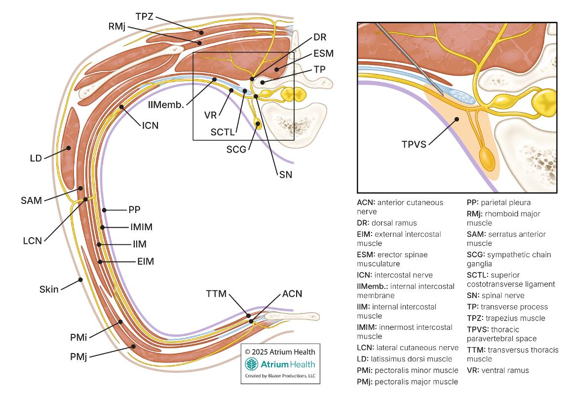
Figure. Thoracic wall innervation and paravertebral space anatomy. Each thoracic spinal nerve exits its intervertebral foramen into the paravertebral space, which is bounded medially by the spinal column, anterolaterally by the parietal pleura, and posteriorly by the internal intercostal membrane and superior costotransverse ligament (see inset). Within this wedge-shaped space, spinal nerves communicate with the sympathetic chain anteriorly and divide into dorsal/ventral rami. The ventral rami continue laterally below their corresponding rib as intercostal nerves, initially traveling between the internal intercostal membrane and the parietal pleura, caudal to their accompanying intercostal vein and artery (not shown). Immediately lateral to the angle of the rib, the intercostal nerves and vessels enter the intercostal space between the innermost and internal intercostal muscles for the remainder of their anterior course. Each intercostal nerve sends branches along its trajectory to innervate structures of the anterolateral chest wall including the ribs, intercostal muscles, and parietal pleura. At the midaxillary line, the intercostal nerves give rise to lateral cutaneous branches that pierce the overlying intercostal and serratus anterior muscles to provide sensory innervation to the skin and subcutaneous tissue of the lateral thorax. The intercostal nerves terminate as anterior cutaneous branches that ascend parasternally to innervate the anteromedial chest and midline. In performing an in-plane, transverseoblique approach to the thoracic paravertebral block, the needle is inserted lateral to medial through the paraspinal musculature, with the tip positioned just anterior to the internal intercostal membrane within the apex of the paravertebral space (see inset).
Considering the patient’s history of severe pain from prior tube thoracostomies, and the absence of tension physiology in this case, a tailored analgesic strategy was sought to facilitate pigtail catheter placement. Ultimately, an ultrasound-guided TPVB was performed to provide full-thickness chest wall analgesia prior to catheter insertion (Image, Video).
Informed consent for ultrasound-guided TPVB performance was obtained. The patient was placed on continuous monitoring with intravenous access established. She was positioned sitting upright and a pre-block scan was performed to identify the relevant TPVB sonoanatomy. A 21-gauge, 100-millimeter echogenic block needle was inserted in-plane from lateral-to-medial and into the TPVS apex at the T4 (4th intercostal space) level. The block was completed with 12.5 milliliters (mL) of 0.5% ropivacaine and repeated at the T6 level for a total volume of 25 mL administered. Given the
patient weighed 62 kilograms (kg), the total ropivacaine dose (125 milligrams [mg]) was well below the patient’s weightbased maximum (3 mg/kg) for avoidance of local anesthetic systemic toxicity. A total of 10 mg of preservative-free dexamethasone was used as a local anesthetic additive to prolong the block duration.7
Approximately thirty minutes post-block performance, the patient reported significantly diminished sensation of the right hemithorax spanning a T2-T7 dermatomal distribution. A lateral approach pigtail tube thoracostomy was performed without need for rescue analgesia or procedural sedation, and the pneumothorax was successfully evacuated. The patient reported feeling pressure but 0/10 intraprocedural pain on a numerical rating scale. She was subsequently admitted for cardiothoracic surgery evaluation. The TPVB lasted approximately 8-10 hours, with the patient reporting minimal (3/10)
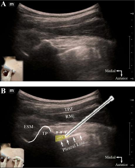
Image Ultrasound-guided thoracic paravertebral block, in-plane transverse-oblique approach, unlabeled (A) and labeled (B) sonoanatomy. Lateral to the transverse process, the apex of the thoracic paravertebral space (yellow triangle) is seen interposed between the internal intercostal membrane posteriorly (arrowheads) and parietal pleura (arrows) anterolaterally. The block needle is inserted in-plane with the transducer, in a lateralto-medial direction (see inset, Figure 1B), with the aim of placing the needle tip within the apical part of the thoracic paravertebral space.
Abbreviations: ESM, erector spinae musculature; RMj, rhomboid major muscle; TP, transverse process; TPVS, thoracic paravertebral space; TPZ, trapezius muscle.
non-insertion site chest pain managed adequately with a multimodal analgesic regimen during hospitalization. The chest tube was removed on day five, and the patient discharged home in stable condition.
The TPVS runs the length of the thoracic vertebral column bilaterally, and communicates medially with the epidural space and laterally with the intercostal space.8,9 This allows for multisegmental, epidural, and intercostal spread of local anesthetic following a single level TPVB injection. By anesthetizing the thoracic spinal nerves/rami and sympathetic chain ganglia,
Video. Ultrasound-guided thoracic paravertebral block performance, in-plane transverse-oblique approach. The block needle tip (arrowhead) is visualized within the apex of the thoracic paravertebral space. Injection confirms proper needle-tip positioning, with expansion of the thoracic paravertebral space and depression (anterior displacement) of the pleural line.
ESM, erector spinae musculature; IIMemb, internal intercostal membrane, RMj, rhomboid major muscle; TP, transverse process; TPZ, trapezius muscle.
the TPVB provides somatic and sympathetic blockade of the ipsilateral hemithorax across multiple contiguous thoracic dermatomes (Figure).8,9
While there are numerous variations in ultrasound-guided TPVB technique described in the literature, the choice of approach is generally a matter of proceduralist preference and experience.8,9 In this case, a transverse-oblique, in-plane approach was performed at the level of the transverse process. Compared to parasagittal approaches, this technique provides the advantage of a shallower needle trajectory and enhanced visualization in relation to the parietal pleura, which may reduce the risk of inadvertent pleural puncture.8
Patients can be positioned sitting, lateral decubitus or prone for TPVB performance.9 After proper patient positioning, the ribs are counted down posteriorly under dynamic ultrasound guidance till the target injection level is reached. A high-frequency linear array or curvilinear probe (for larger habitus patients) is positioned just lateral to midline in a transverse-oblique orientation, parallel to the underlying rib course. Initially, the hyperechoic transverse process and articulating rib are identified with their confluent acoustic shadows. The probe is then translated slightly caudad into the intercostal space till the TPVS is revealed just lateral to the “thumb-like” contour of the transverse process and between the parietal pleura and internal intercostal membrane (Image).8 An 80 – 100-millimeter block needle is inserted in-plane from lateral-to-medial into the apex of the TPVS. Proper needle tip positioning is confirmed by widening of the TPVS and depression (anterior displacement) of the pleural line with test injection (Video).
A multi-level (T4 and T6) ultrasound-guided TPVB was performed using 25 mL of 0.5% ropivacaine with 10 mg dexamethasone as an additive, injecting 12.5 mL per level. However, a clinical study by Uppal et al. demonstrated no difference in dermatomal coverage following a single (T3-T4) versus multiple injection (T1-T5, 5 mL per level) ultrasoundguided TPVB using 25 mL of 0.5% ropivacaine, with both groups resulting in a median of five dermatomes blocked.10 Despite being quicker to perform, the single-injection TPVB is more dependent on local anesthetic spread for efficacy, and clinically results in an analgesia level block compared to the
Thoracic Paravertebral Block for Tube Thoracostomy Analgesia in the Emergency Department
surgical anesthesia produced with a multi-injection approach. For ED tube thoracostomy, a single-injection TPVB at the level of chest tube insertion (T4/T5 [4th/5th intercostal space]) is likely sufficient.
TPVB Contraindications
Major contraindications to TPVB performance include local anesthetic allergy, coagulopathy or systemic anticoagulation, overlying cellulitis, empyema, or paravertebral spaceoccupying tumor.
TPVB Potential Complications
Complications after TPVB are relatively uncommon, especially when performed under ultrasound-guidance. Given the proximity of the injection site to the parietal pleura, neuraxis, and intercostal vasculature, potential needle-related complications include pneumothorax (< 1%), nerve damage (< 1%), and vascular puncture (< 1%).9 For the purpose of facilitating ED tube thoracostomy, iatrogenic pneumothorax is not of concern when it is already present. Because the TPVS is a non-compressible space, the TPVB should be avoided in patients on anticoagulation to minimize risk of serious bleeding, particularly epidural hematoma. Other reported block complications related to local anesthetic injection/spread include hypotension (from sympathectomy), transient Horner syndrome, and local anesthetic systemic toxicity.11 As with all ultrasound-guided nerve blocks, patient monitoring and clear needle tip visualization is paramount to mitigating risk. Emergency physicians must be cognizant of local anesthetic systemic toxicity and adhere to maximum, ideal weight-based dosing of local anesthetic to minimize the occurrence of this potentially fatal clinical entity.
TPVB Distribution of Anesthesia
Two clinical studies using the transverse-oblique, in-plane approach to the TPVB demonstrated sensory blockade over a median of 4 or 6 dermatomes (range: 3 to 7) following a single 20 mL injection of 0.75% ropivacaine.12,13 Similarly, a cadaver study observed the spread of 20 mL injected dye over 3 to 4 paravertebral spaces (range: 1 to 10).14
TPVB Limitations
Though generally safe and effective, the ultrasound-guided TPVB is an expert level block by nature of its paravertebral injection location near critical structures. It should not be performed by novices of ultrasound-guided regional anesthesia. Emergency physicians should demonstrate mastery of more readily performed “gateway” nerve blocks (eg, erector spinae and fascia iliaca blocks) prior to attempting TPVB performance. Performance may also be limited in certain patient populations, particularly those with substantial paraspinal tissue or who are morbidly obese, intolerant of repositioning, or needle averse.
Alternative ultrasound-guided truncal blocks are more commonly performed in the ED for thoracic analgesia. The serratus plane block, by targeting the lateral cutaneous branches of the intercostal nerves, anesthetizes the skin and subcutaneous tissue of the lateral thorax.15 However, it does not anesthetize deeper chest wall structures (intercostal musculature, ribs, parietal pleura) involved in tube thoracostomy. In fact, local anesthetic spread has been demonstrated to not reach the intercostal nerves unless concomitant rib fractures are present to disrupt myofascial tissue planes.16 In contrast, the erector spinae plane block targets the dorsal rami of the thoracic spinal nerves as they traverse the erector spinae plane. Anterior penetration of local anesthetic into the TPVS does occur though is variable, resulting in unpredictable anterolateral chest wall coverage.17 By nature of its proximal paraspinal injection location, the TPVB eliminates the shortcomings of the serratus and erector spinae plane blocks, providing reliable full-thickness coverage across both the anterior and posterior hemithorax.
Given its comprehensive ipsilateral chest wall coverage, the TPVB has significant analgesic potential for other commonly encountered acute thoracic pain conditions in the ED, such as chest wall burns, herpetic neuralgia, and rib fractures.
Tube thoracostomy is an exceedingly painful ED procedure necessitating effective analgesia to facilitate safe performance. By providing safe and effective hemithorax analgesia or anesthesia, the ultrasound-guided TPVB has potential to reshape established pain management paradigms for tube thoracostomy. Future research should focus on ED utilization of the TPVB for tube thoracostomy in addition to other acute thoracic pain conditions.
The authors attest that their institution requires neither Institutional Review Board approval, nor patient consent for publication of this case report. Documentation on file.
Address for Correspondence: M. Townsend Reeves, MD, Department of Emergency Medicine, Atrium Health Carolinas Medical Center, 1000 Blythe Blvd., Medical Education Bldg., 3rd Floor, Charlotte, NC 28203. Email: Matthew.Reeves@ atriumhealth.org
Conflicts of Interest: By the CPC-EM article submission agreement, all authors are required to disclose all affiliations, funding sources and financial or management relationships that could be perceived as potential sources of bias. The authors disclosed none.
Reeves Thoracic Paravertebral Block for Tube Thoracostomy Analgesia in the Emergency Department
Copyright: © 2025 Reeves. This is an open access article distributed in accordance with the terms of the Creative Commons Attribution (CC BY 4.0) License. See: http://creativecommons.org/ licenses/by/4.0/
1. Anderson D, Chen SA, Godoy LA, et al. Comprehensive review of chest tube management: a review. JAMA Surg. 2022;157(3):269-74.
2. Roberts ME, Rahman NM, Maskell NA, et al. British Thoracic Society Guideline for pleural disease. Thorax. 2023;78(11):1143-1156..
3. Carson-Chahhoud KV, Wakai A, van Agteren JE, et al. Simple aspiration versus intercostal tube drainage for primary spontaneous pneumothorax in adults. Cochrane Database Syst Rev 2017;9(9):CD004479.
4. Luketich JD, Kiss M, Hershey J, et al. Chest tube insertion: a prospective evaluation of pain management. Clin J Pain 1998;14(2):152-4.
5. Chin KJ, Versyck B, Elsharkawy H, et al. Anatomical basis of fascial plane blocks. Reg Anesth Pain Med. 2021;46(7):581-99.
6. Sakamoto H, Akita K, Sato T. An anatomical analysis of the relationships between the intercostal nerves and the thoracic and abdominal muscles in man. I. Ramification of the intercostal nerves. Acta Anat (Basel). 1996;156(2):132-42.
7. Pehora C, Pearson AM, Kaushal A, et al. Dexamethasone as an adjuvant to peripheral nerve block. Cochrane Database Syst Rev 2017;11(11):CD011770.
8. Krediet AC, Moayeri N, van Geffen GJ, et al. Different approaches to
ultrasound-guided thoracic paravertebral block: an illustrated review. Anesthesiology. 2015;123(2):459-74.
9. Pawa A., Wojcikiewicz T, Barron A, et al. Paravertebral blocks: anatomical, practical, and future concepts. Curr Anesthesiol Rep. 2019;9(3):263-70.
10. Uppal V, Sondekoppam RV, Sodhi P, et al. Ganapathy S. Singleinjection versus multiple-injection technique of ultrasound-guided paravertebral blocks: a randomized controlled study comparing dermatomal spread. Reg Anesth Pain Med. 2017;42(5):575-81.
11. Ardon AE, Lee J, Franco CD, et al. Paravertebral block: anatomy and relevant safety issues. Korean J Anesthesiol. 2020;73(5):394400.
12. Renes SH, Bruhn J, Gielen MJ, et al. In-plane ultrasound-guided thoracic paravertebral block: a preliminary report of 36 cases with radiologic confirmation of catheter position. Reg Anesth Pain Med 2010;35(2):212-6.
13. Bouzinac A, Delbos A, Mazières M, et al. [Application of ultrasound in the realization of thoracic paravertebral block in breast surgery]. Ann Fr Anesth Reanim. 2011;30(5):453-5.
14. Cowie B, McGlade D, Ivanusic J, et al. Ultrasound-guided thoracic paravertebral blockade: a cadaveric study. Anesth Analg 2010;110(6):1735-9.
15. Blanco R, Parras T, McDonnell JG, et al. Serratus plane block: a novel ultrasound-guided thoracic wall nerve block. Anaesthesia. 2013;68(11):1107-13.
16. Johnston DF, Black ND, O’Halloran R, et al. Cadaveric findings of the effect of rib fractures on spread of serratus plane injections. Can J Anaesth. 2019;66(6):738-9.
17. Chin KJ, Adhikary SD, Forero, M. Erector spinae plane (ESP) block: a new paradigm in regional anesthesia and analgesia. Curr Anesthesiol Rep. 2019;9(3):271-80.
Natalie A. Jansen, MD, PhD
Christie Fritz, MD, MS
Editor: R. Gentry Wilkerson, MD
Beth Israel Deaconess Medical Center, Department of Emergency Medicine, Boston, Massachusetts
Submission history: Submitted February 17, 2025; Revision received May 19, 2025; Accepted June 3, 2025
Electronically published July 29, 2025
Full text available through open access at http://escholarship.org/uc/uciem_cpcem DOI: 10.5811/cpcem.43502
Introduction: Traumatic injury is the leading cause of death in individuals under 45 years of age, and point-of-care ultrasound (POCUS) has become an essential component of the initial trauma evaluation. However, positive findings on the extended focused assessment with sonography in trauma (eFAST) may be misinterpreted as evidence of an acute surgical emergency, particularly in the context of blunt trauma, underscoring the need for careful clinical correlation.
Case Report: We present a case in which a hemodynamically unstable patient had significant free abdominal fluid on eFAST after a fall from standing height. She was ultimately diagnosed with a highrisk pulmonary embolism as the cause of her hemodynamic instability, while the free abdominal fluid was identified as originating both from a ruptured ovarian cyst and from moderate-volume ascites.
Conclusion: The eFAST exam is a valuable tool in rapidly identifying intra-abdominal injuries following blunt trauma. However, the presence of free fluid on eFAST may result from causes other than acute intra-abdominal injury requiring surgical intervention. Therefore, emergency physicians should interpret positive findings with clinical judgment and consider the broader clinical context.
[Clin Pract Cases Emerg Med. 2025;9(3):340-344.]
Keywords: case report; eFAST; trauma; pulmonary embolism.
Traumatic injury is the leading cause of death in individuals under 45 years of age in the United States.1 Blunt abdominal injuries may cause significant bleeding and hemodynamic instability, and quickly identifying life-threatening injuries is a crucial component of the initial trauma evaluation. Adoption of point-of-care ultrasound on initial trauma evaluation has led to significant reductions in time to operative management and in overall medical costs.2 Extended focused assessment with sonography in trauma (eFAST) can helpfully rule in, but not rule out, free intra-abdominal fluid in hypotensive adults with a sensitivity of 74% and specificity of 95%.3,4 Fluid accumulation becomes visible in the hepatorenal or splenorenal spaces when it exceeds 500 milliliters (mL), whereas as little as 150 mL can be detected in the pelvic view.5 The presence of fluid without evidence of intra-abdominal injury may arise from causes such as ovarian cyst rupture (traumatic or atraumatic), ascites,
peritoneal dialysate,6 or physiologic fluid (up to 50 mL)5 in women of childbearing age.7 In this case, we describe a highrisk pulmonary embolism (PE) in an otherwise healthy female who presented for trauma evaluation after a fall from standing height and was found to have a grossly positive eFAST exam.
An 18-year-old female without significant past medical history presented as a transfer from an outside hospital with hypotension and a positive eFAST exam after a fall from standing height. While walking outside, the patient slipped on a patch of ice and fell. She had immediate abdominal and lower back pain. At the outside hospital, the patient was found to be tachycardic and hypotensive with a systolic blood pressure of 80/40 millimeters of mercury (mm Hg). Her right upper quadrant view on eFAST was positive for free abdominal fluid. Her initial hemoglobin and hematocrit were
Jansen et al. An Unexpected Cause of Shock in a Trauma Patient with Hemodynamic Instability: A Case Report
8.3 grams per deciliter (g/dL) and 32.7%, respectively. She was started on two units of packed red blood cells, 1 g of tranexamic acid, and 1 liter (L) of intravenous (IV) fluids and was transferred for further trauma evaluation. On arrival, the patient denied recent illnesses or influenza-like symptoms. She reported possible dizziness or lightheadedness before the fall but was uncertain whether any symptoms truly preceded it. She denied abdominal pain or back pain prior to the fall. The patient denied current chest pain or shortness of breath.
Her initial vitals showed a heart rate of 139 beats per minute (min), blood pressure of 113/65 mm Hg, respiratory rate of 25 breaths per min, and oxygen saturation of 100% on four L/min of oxygen via nasal canula. Notably, all subsequent systolic blood pressures were in the 60s-70s. She was alert, oriented, answering all questions appropriately, and following commands with a Glasgow Coma Scale of 15. On exam, she was in significant pain and appeared pale. Her lungs were clear to auscultation with equal breath sounds bilaterally. She was tachycardic with a regular heart rhythm. Her abdomen was soft and diffusely tender to palpation with voluntary guarding. She did not have midline spinal tenderness or paraspinal tenderness. She had palpable radial and dorsalis pedis pulses bilaterally. Notable laboratory results are shown in the Table. A basic metabolic panel was within normal limits (not shown). Because point-of-care testing was not available, we had no lab results e during initial management.
Her eFAST exam was remarkable for free fluid in the right upper quadrant (Figure 1), left upper quadrant, and suprapubic views. There was no pericardial effusion on subxiphoid view. We did not obtain additional cardiac views or views of the inferior vena cava on initial presentation. Right heart strain was not evaluated for on initial eFAST exam.
We established additional IV access and an arterial line for accurate blood pressure monitoring. As repeat lab results were not available, and in the absence of other causes of the the patient’s hemodynamic stability, we initiated massive transfusion protocol for presumed hemorrhagic shock. Given relative stability and appropriate mentation after initial resuscitative measures, in conjunction with our trauma surgery team, we opted to pursue cross-sectional imaging with computed tomography (CT) prior to mobilizing the patient to the operating room for exploratory laparotomy. Her CT was remarkable for bilateral PE with a large thrombus in the right main pulmonary artery with enlargement of the right atrium and ventricle demonstrating evidence of right heart strain (Figures 2 and 3).
She also had a non-occlusive thrombus within the inferior vena cava. The CT also demonstrated a large volume of free fluid within the pelvis, characterized as slightly complex in the posterior cul-de-sac and adjacent to the right ovary, although predominantly hypoattenuating. A 1.4 centimeter (cm) corpus luteal cyst was noted within the right ovary. The liver exhibited a nutmeg appearance with associated mild-tomoderate periportal edema and moderate ascites. No evidence
CPC-EM Capsule
What do we already know about this clinical entity?
Extended Focused Assessment with Sonography for Trauma (EFAST) examinations are a vital source of information in unstable trauma patients and may help guide further treatment and interventions.
What makes this presentation of disease reportable?
Unstable trauma patient with positive intraabdominal free fluid found to have ruptured ovarian cyst/ascites and massive pulmonary embolism (PE).
What is the major learning point?
EFAST examinations have high specificity, but there are important false positive causes that can lead to additional interventions which providers should be aware of.
How might this improve emergency medicine practice?
Emergency physicians should be aware of limitations and false positive causes of intraabdominal free fluid in EFAST exams.
of traumatic injury was identified.
Given uncertainty about the origins of the abdominal fluid, we deferred administering tissue plasminogen activator. A heparin drip was started, and the patient was admitted to the intensive care unit in stable condition. Shortly after admission she was taken emergently to the catheterization lab for aspiration thrombectomy with interventional radiology. The procedure was unsuccessful, and catheter-directed thrombolytic therapy was performed. Her inpatient course was complicated by a right groin hematoma and acute blood loss anemia requiring transfusion, as well as significantly elevated pulmonary artery pressures. She was discharged after an 18-day hospital stay on apixaban and sildenafil for chronic thromboembolic pulmonary hypertension. Extensive outpatient workup did not identify a cause of her PE.
Prompt identification of the etiology of shock is essential in preventing its sequelae including multiorgan failure and death. Even in cases where there appears to be a clear cause of shock, maintaining a high index of suspicion for mixed shock
Table. Notable laboratory results on initial presentation to the emergency department.
Complete Blood Count
WBC 8.0 × 10³ cells/µL 4.0–10.0 × 10³ cells/µL
50 × 10³/µL 150–400 × 10³/µL
Coagulation Panel
Venous Blood Gas
mm Hg
mm Hg
mm Hg
mm Hg Lactate 5.0 mmol/L 0.5–2.0 mmol/L
Cardiac Biomarkers* Troponin 0.22 ng/mL 0–0.01 ng/mL
197 pg/mL 0–178 pg/mL
*Cardiac biomarkers were not included in the initial order set. Troponin and proBNP were sent after computed tomography demonstrated pulmonary embolism with associated right heart strain. Abbreviations: ED, emergency department; WBC, white blood cells; µL, microliters; Hgb, hemoglobin; dL, deciliters; Hct, hematocrit; Plt, platelets; PT, prothrombin time; PTT, partial thromboplastin time; s, seconds; INR, international normalized ratio; pO₂, partial pressure of oxygen; pCO₂, partial pressure of carbon dioxide; ng, nanograms; pg, picograms; proBNP, pro B-type natriuretic peptide.
prior to her arrival. Information received pre-arrival led the emergency department (ED) and trauma teams to frame her case as one of hemorrhagic shock secondary to a fall. In her case, however, the patient’s positive eFAST findings were likely instead related to rapid accumulation of ascites and periportal edema consistent with acute right heart failure and Lab Test Result
or alternative causes can prevent premature closure. The incidence of PE in ED patients under the age of 21 is approximately 2.1 per 100,000 visits,8 and it is even lower in patients who do not have any discernable risk factors, making our patient’s ultimate diagnosis an unusual and unexpected cause of shock.
Although blunt trauma is a common cause of hemorrhagic shock,9 falls from standing height rarely result in intraabdominal injury significant enough to require procedural intervention in this patient age.10 Otherwise healthy young adults who sustain injuries from falling from standing height most commonly experience orthopedic injuries11 and largely do not require admission.12 Women often attribute falls to external factors and may downplay symptoms suggestive of syncope.13 In the case of our patient, she attributed her fall to slipping on ice and only later posited that she may have felt dizzy preceding her fall.
The patient’s initial presentation was as a transfer from another hospital, and a prehospital trauma alert was placed
hepatic congestion14 and were likely exacerbated by the aggressive administration of IV fluids and blood products both in the prehospital setting and upon arrival.
Although a corpus luteal cyst with a small amount of complex fluid in the posterior cul-de-sac was identified, it is unlikely that a ruptured hemorrhagic cyst alone accounted for the grossly positive eFAST exam. One study reported an average volume of 66 mL for hemorrhagic ovarian cysts,15 typically insufficient to result in such significant free fluid. Interestingly, a framing effect may have led to early diagnostic closure, particularly when her eFAST exam provided positive results. Given the patient’s initial insistence that the fall was not preceded by prodromal symptoms, and the absence of reported right heart strain on the initial eFAST performed by the trauma team, early testing for syncope-related causes was not pursued despite the patient requiring 4 L/min oxygen via nasal cannula to maintain adequate oxygenation.
Often intertwined with the framing effect, anchoring bias involves an over-reliance on initial information during diagnostic reasoning and may contribute to premature closure,16 such as attributing hypotension solely to hemorrhagic shock from trauma without fully considering alternative causes. A recent study found that patients presenting with shortness of breath and triaged with congestive heart failure (CHF) were less likely to be evaluated for PE compared to those without a specific diagnosis mentioned, despite similar PE rates. This may reflect anchoring or framing bias related to known CHF history.16 In our patient’s case, prehospital information suggesting a traumatic mechanism may have misdirected her initial
diagnostic workup and management. The eFAST was approached primarily as an assessment for acute traumatic injuries – especially in light of her report of trauma and positive intra-abdominal fluid seen on outside hospital ultrasound and full cardiac evaluation was not performed on initial examination. This narrow focus, coupled with premature closure on the diagnosis of hemorrhagic shock both at the outside hospital and upon arrival to our ED, may have led to rapid volume expansion that further impaired right ventricular function and reduced cardiac output.14,17
The extended focused assessment with sonography in trauma is a critical aspect of the initial trauma evaluation in unstable patients who present to the ED. However, in the case of a positive eFAST exam in a patient who falls from standing height, contextualizing risks of intra-abdominal trauma is critical to interpreting the eFAST. Indeed, abdominal trauma requiring intervention in individuals who fall from standing height is extremely uncommon.10 It is essential to maintain a high index of suspicion for alternative causes of both the fall and the shock in unstable patients presenting after a fall from standing height, while also critically reflecting on potential biases that may influence the interpretation of the patient’s presentation.
The authors attest that their institution requires neither Institutional Review Board approval, nor patient consent for publication of this case report. Documentation on file.
Address for Correspondence: Natalie Jansen, MD, PhD, Beth Israel Deaconess Medical Center, Department of Emergency Medicine, 1 Deaconess Road, Boston, MA 02215. Email: njansen@bidmc.harvard.edu.
Conflicts of Interest: By the CPC-EM article submission agreement, all authors are required to disclose all affiliations, funding sources and financial or management relationships that could be perceived as potential sources of bias. The authors disclosed none.
Copyright: © 2025 Jansen et al. This is an open access article distributed in accordance with the terms of the Creative Commons Attribution (CC BY 4.0) License. See: http://creativecommons.org/ licenses/by/4.0/
1. Heron M. Deaths: leading causes for 2018. Natl Vital Stat Rep 2021;70(4):1-115.
2. Melniker LA, Leibner E, McKenney MG, et al. Randomized controlled clinical trial of point-of-care, limited ultrasonography for trauma in the emergency department: the first sonography outcomes assessment program trial. Ann Emerg Med. 2006;48(3):227-235.
3. Lobo V, Hunter-Behrend M, Cullnan E, et al. Caudal edge of the liver in the right upper quadrant (RUQ) view is the most sensitive area for free fluid on the FAST exam. West J Emerg Med. 2017;18(2):270.
4. Netherton S, Milenkovic V, Taylor M, et al. Diagnostic accuracy of eFAST in the trauma patient: a systematic review and meta-analysis. Can J Emerg Med. 2019;21(6):727-738.
5. Bella FM, Bonfichi A, Esposito C, et al. Extended focused assessment with sonography for trauma in the emergency department: a comprehensive review. J Clin Med. 2025;14(10):3457.
6. Bloom BA and Gibbons RC. Focused assessment with sonography for trauma. In: StatPearls. Treasure Island (FL): StatPearls Publishing; July 24, 2023. Available at: https://pubmed.ncbi.nlm.nih. gov/29261902/ Accessed January 12, 2025.
7. Khosravian K, Boniface K, Dearing A, et al. EFAST exam errors at a Level 1 trauma center: a retrospective cohort study. Am J Emerg Med. 2021;49:393-398.
8. Agha BS, Sturm JJ, Simon HK, et al. Pulmonary embolism in the pediatric emergency department. Pediatrics. 2013;132(4):663-667.
9. Simon LV, Lopez RA, King KC. Blunt force trauma. In: StatPearls [Internet]. Treasure Island (FL): StatPearls Publishing; 2023. Updated August 7, 2023. Available from: https://www.ncbi.nlm.nih.gov/books/ NBK470338/ Accessed December 16, 2024.
10. Hagan NE, Berdel HO, Tefft A, et al. Torso injuries after fall from standing—empiric abdominal or thoracic CT imaging is not indicated. Injury. 2020;51(1):20-25.
11. Talbot LA, Musiol RJ, Witham EK, et al. Falls in young, middle-aged and older community dwelling adults: perceived cause, environmental factors and injury. BMC Public Health. 2005;5:1-9.
12. Brazell CJ, Carry PM, Holmes KS, et al. Pediatric and adult fracture incidence: a decreasing trend with increasing hospital admissions. Orthopedics. 2023;46(6):e369-e375.
13. Williamson TM, Runte M, Runte T, et al. A qualitative study to identify factors that influence patients’ decisions to call emergency medical services for syncope. Can J Emerg Med. 2021;23:195-205.
14. Houston BA, Brittain EL, Tedford RJ. Right ventricular failure. N Engl J Med. 2023;388(12):1111-1125.
15. Abbas AM, Amin MT, Tolba SM, et al. Hemorrhagic ovarian cysts: clinical and sonographic correlation with the management options. Middle East Fertil Soc J. 2016;21(1):41-45.
16. Ly DP, Shekelle PG, Song Z. Evidence for anchoring bias during physician decision-making. JAMA Intern Med. 2023;183(8):818-823.
17. Petit M and Vieillard-Baron A. Ventricular interdependence in critically ill patients: from physiology to bedside. Front Physiol. 2023;14:1232340.
JanAldrinEnabore,MD*
RobertVezzetti,MD*†
GuyonHill,MD*†
Section Editor: R. Gentry Wilkerson, MD
*DellChildren’sMedicalCenter,DepartmentofEmergencyMedicine,Austin,Texas † UniversityofTexasDellMedicalSchool,Austin,Texas
Submission history: Submitted July 31, 2023; Revision received November 27, 2023; Accepted November 28, 2023
Electronically published February 15, 2025
Full text available through open access at http://escholarship.org/uc/uciem_cpcem DOI: 10.5811/cpcem.1574
Introduction: There exists a wide differential of etiologies for pediatric torticollis that extends beyond musculoskeletal factors.
Case Report: We present a novel case of a pediatric patient with an acute atraumatic hemorrhage of the left cerebellum presenting with gradual worsening torticollis. Upon further diagnostic workup, he was found to have an intracerebral hemorrhage due to a cerebellar cavernous malformation. Although the hemorrhage boundaries were extensive, the patient had only exhibited transient dysmetria and facial weakness, with ultimate resolution of torticollis and these neurological symptoms after several days.
Conclusion: This case demonstrates the importance of maintaining a broad differential in the workup of acute pediatric torticollis. [Clin Pract Cases Emerg Med. 2025;9(3)345-348.]
Keywords: torticollis; pediatric; cerebellar hemorrhage; cavernoma; case report.
Theetiologyofacutepediatrictorticollisismostoften eithertraumatic,infectious,ormedicationinduced.1 However,keepingabroaddifferentialinevaluatingthese childrenisalwaysimportant,astheetiologiescanbevastand includepotentiallylife-threateningcauses.Alackof diagnosisandfailureoftherapeuticinterventionsshould alwaysspurfurtherworkup.Wepresentanovelcaseofa pediatricpatientwithanacuteatraumaticcerebellar hemorrhagesecondarytocavernousmalformation presentingwithisolatedtorticollis.
Aneight-year-oldmalewithoutchronicmedicalproblems presentedtotheemergencydepartment(ED)afterwakingup withposteriorleftneckpainandaninabilitytomovehisneck totheleftside.Themotherbelievedhehadsleptinan uncomfortablepositionthepreviouseveninganddeniedany historyofrecenttrauma.Thepatienthadnohistoryoffevers andnopriorsimilarsymptomatology.Intheprecedingweek, thepatienthadepisodesofnausea,vomiting,andheadache,
butallthesesymptomshadsinceresolvedseveraldaysprior. Atthetimeofpresentation,therewerenocomplaintsof significantheadache,nausea,gastrointestinalsymptoms, visionchanges,focalweakness,ordifficultyambulating. Norecentmedicationswerestarted.
Theinitialvitalswereasfollows:temperature37.6° Celsius,bloodpressure102/72millimetersofmercury (mmHg),heartrate98beatsperminute,respiratoryrate24 breathsperminute,andoxygensaturation98%onroomair. Onphysicalexamination,thechildwascalmandinno significantdistress.Heheldhisheadandchindeviatedtothe rightandinslightcervicalextension.Hehadleftlateralneck tendernessoverthesternocleidomastoidregionbutno posteriormidlinetenderness.Therewerenoidentified concernsforinfection,mass,orlymphadenopathy.Hehad baselinerighteyestrabismus,butextraocularmuscles wereintactbilaterallywithoutvisual fielddeficits.An oropharyngealexamrevealedmildbilateraltonsillar swelling;however,therewasnoevidenceofuvuladeviation, peritonsillarabscess,sublingualtenderness,submandibular swelling,ordentalinfection.Hecouldambulateandfollow
all commands but did so with an unchanged head and neck position. He also had equal strength and sensation in all extremities. There were no other physical exam abnormalities identified.
Laboratory findings for complete blood count, chemistry, C-reactive protein, and creatine kinase were within normal limits. However, his erythrocyte sedimentation rate was elevated: 38 millimeters per hour (mm/hr) (reference range: 0–20 mm/hr). A computed tomography (CT) with intravenous (IV) contrast of his neck showed only mild maxillary sinus disease and no other documented abnormalities (Image 1). Based on these findings, a trial of supportive treatment was undertaken consisting of oral acetaminophen, IV ketorolac, and two doses of IV diazepam.
Given that there was no significant improvement, magnetic resonance imaging (MRI) of the cervical spine was obtained and revealed an epidural spinal hemorrhage and a partially visualized left cerebellar hemorrhage. Subsequent imaging included an MRI brain with contrast and a CT angiogram that showed a left cerebellar hemorrhage from a cavernous malformation without evidence of an intracranial mass (Images 2, 3). The patient remained in the intensive care unit for five days, where he transiently developed left facial weakness and left upper extremity dysmetria. He received a course of dexamethasone and pain medications during this time. Pediatric neurosurgery discussed surgical resection options; however, the family deferred in favor of close neurosurgical follow-up. His torticollis and neurological deficits were resolved by the time of discharge on day five of hospitalization.
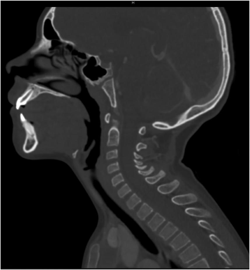
Image1. Sagittalviewofcomputedtomographysofttissueneck withcontrastwithevidenceofmaxillarysinusdiseaseandwithout evidenceofhemorrhageorotherinfectiousetiologyfortorticollis.
PopulationHealthResearchCapsule
Whatdowealreadyknowaboutthis clinicalentity?
Acutepediatrictorticollisisattributedto trauma,infection,ormedication.Many cerebrovascularmalformationsare incidentallydiscoveredand remainasymptomatic.
Whatmakesthispresentationof diseasereportable?
Thisrarepresentationofisolatedtorticollis resultedfromanatraumatic cerebellarhemorrhagesecondaryto cerebrovascularmalformation.
Whatisthemajorlearningpoint?
Itisimportanttocomprehensivelyinvestigate unresolvedpediatrictorticollis.
Howmightthisimproveemergency medicinepractice?
Emergencyphysiciansshouldmaintaina broaddifferentialandconsideradvanced imaging,suchasmagneticresonance imaging,wheninitialevaluations areinconclusive.
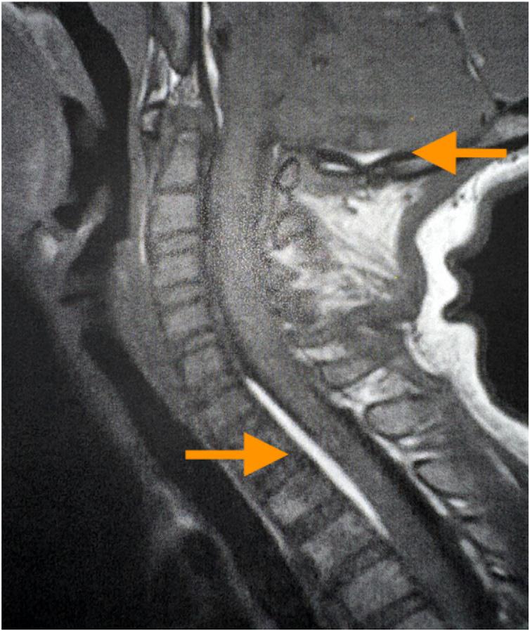
Image2. Sagittalviewofmagneticresonanceimagingoftheneck withcontrastdemonstratingleftcerebellarhemorrhage(upper arrow)with fifthcervicaltosecondthoracicspinalextension (lowerarrow).
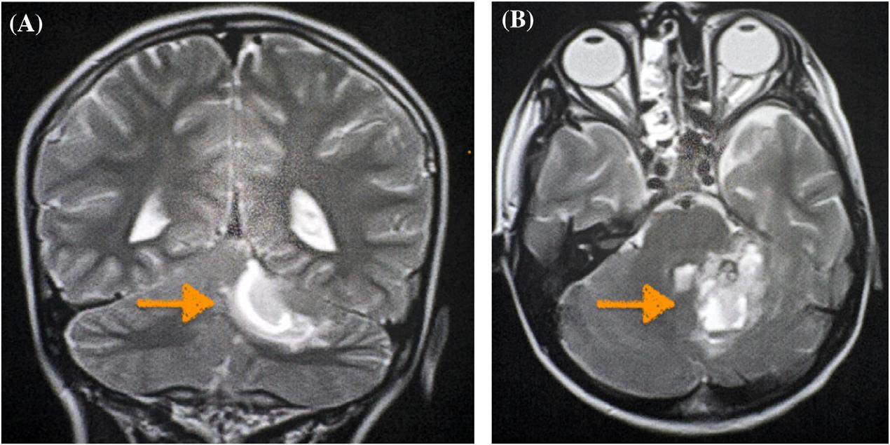
TorticollisarisesfromtheLatinrootsof “torus” and “collum,” meaningtwistedandneck,respectively.Itis sometimesalsoreferredtoascervicaldystonia.Thisisa commondisorderthatcanoccuratallages.Themost commoncauseoftorticollisiscongenitalmuscular torticollis,typicallycausedbyahematomaor fibromaonthe sternocleidomastoidmusclethatregressesbyeightmonthsof age. 2 However,thecausesofacquiredtorticollisarevast. Traumatic,infectious,andmedication-inducedtorticollis arethethreemostcommoncausesofacutetorticollis. Commoninfectiousetiologiesincluderetropharyngeal andperitonsillarabscessesandcervicallymphadenitis. Commonmedicationculpritsincludecholinergicand antipsychoticmedications.1
Athoroughclinicalhistoryandphysicalexaminationcan frequentlyleadtoaconciseworkupforacutecervical torticollisinthepediatricpatient.Intraumaticcaseswithout neurological findings,plain-filmanterior-posteriorand lateralcervicalspineradiographsareappropriateandcanbe followedbyCTwhennon-diagnostic.3 Whensuperficial massesarepresentinthecervicalregion,ultrasoundcanbe usedtolimitexposuretoionizingradiation.Whenclinical signsandsymptomsofinfectionarepresent,aCTwithIV contrastistheappropriatetest.1,4 Thiscandeterminethe extentandlocationoftheinfection,suchasincases ofretropharyngealabscess,peritonsillarabscess, lymphadenitis,orsimilarskinandsofttissueinfections intheparacervicalandperioralregions.
Thispatient’spresentationdidnotyieldadiagnosisinthe ED,nordidherespondasexpectedtoanalgesiaandmuscle relaxants.ThepatientwasadmittedtoobtainanMRI,which showedanacutecerebellarhemorrhagewithipsilateral cervicalextension.Thisledtothediscoveryofacerebral cavernousmalformation(CCM)inthecerebellum. Cavernousmalformationsareaconglomerationof abnormal,intermixedsmallandlargebloodvesselsandare alsoreferredtoascavernomas,cavernousangiomas,
cavernoushemangiomas,orintracranialvascular malformations.Theetiologyofthesemalformationsis unknown,butitissuspectedthatthereislikelyagenetic component.5 Magneticresonanceimaginghasnearly100% sensitivityfordetectingaCCM,andmanyareincidentally discovered.6 ThedecisionforsurgicalresectionofaCCM dependsonsize,location,andsymptomatology.5 Ultimately, thispatientwastreatedwithsteroidsandanalgesicswithan improvementofhistorticollisandwasdischarged fivedays aftertheinitialdiagnosis.
Briefly,thiscaseservesasagreatexampleofpursuing continuedworkupofacute,unresolvedtorticollis,especially whenthemostcommonculpritsoftrauma,infection,and medication-inducedcauseshavebeenruledout.Thereare twosimilarcasereportsofisolatedtorticollisinthepediatric population:anine-year-oldafterfallingoffherbicycle withbrainstemcavernomahemorrhageandan eight-month-oldwithalargecerebellar-pontineangle arachnoidcyst.7,8 However,toourknowledge,thisis the firstdocumentedcaseofisolatedtorticollis secondarytoatraumaticcerebellarhemorrhagefroma cavernousmalformation.
Thispediatriccasereporthighlightstheimportanceofa comprehensiveapproachinassessingacutetorticollisin children.Whilefactorsliketrauma,infection,and medication-relatedissuesarefrequentlyencountered,the presentedcaseunveilsanatraumaticcerebellarhemorrhage secondarytoacavernousmalformationastherootcause. Despitetheextensivehemorrhagicinvolvement,thepatient’ s symptomswereinitiallyconfinedtotorticollis,later accompaniedbytransientneurologicalmanifestationsthat ultimatelyresolvedthroughconservativemanagement. Thiscaseemphasizestheongoingneedforathorough investigationofunresolvedtorticollis,emphasizingthe potentialforlife-threateningcauseswithinthediverse spectrumofpediatricpresentations.
Patientconsenthasbeenobtainedand filedforthepublicationofthis casereport.
AddressforCorrespondence:JanAldrinEnabore,MD,Dell Children’sMedicalCenter,DepartmentofEmergencyMedicine, 4900MuellerBlvd,Austin,Texas78723.Email: Enabore2019@ gmail.com
ConflictsofInterest:Bythe CPC-EM articlesubmissionagreement, allauthorsarerequiredtodiscloseallaffiliations,fundingsources and financialormanagementrelationshipsthatcouldbeperceived aspotentialsourcesofbias.Theauthorsdisclosednone.
Copyright:©2024Enaboreetal.Thisisanopenaccessarticle distributedinaccordancewiththetermsoftheCreativeCommons Attribution(CCBY4.0)License.See: http://creativecommons.org/ licenses/by/4.0/
1.TzimenatosL,VanceCW,KuppermannN.Neckstiffness.In:ShawKN, BachurRG,ChamberlainJM,etal.(Eds.), Fleisher&Ludwig’sTextbook
ofPediatricEmergencyMedicine, 8thed(324–32).Philadelphia,PA Austin,Texas,USA:WoltersKluwer,2021.
2.TomczakKKandRosmanNP.Torticollis. JChildNeurol 2013;28(3):365–78.
3.AnSandHyunSJ.Pediatricspinetrauma. JKoreanNeurosurgSoc 2022;65(3):361–9.
4.HaqueS,BilalShafi BB,KaleemM.Imagingoftorticollisinchildren. Radiographics.2012;32(2):557–71.
5.BatraS,LinD,RecinosPF,etal.Cavernousmalformations:natural history,diagnosisandtreatment. NatRevNeurol.2009;5(12):659–70.
6.CatonMTandShenoyVS.CerebralCavernousMalformations.In: StatPearls.TreasureIsland(FL):StatPearlsPublishing;April10,2023. AccessedNovember1,2023.
7.AgrawalA,CincuR,JoharapurkarSR,etal.Hemorrhageinbrain stemcavernomapresentingwithtorticollis. PediatrNeurosurg 2009;45(1):49–52.
8.YueJK,OhT,HanKJ,etal.Acaseoftorticollisinan8-month-oldinfant causedbyposteriorfossaarachnoidcyst:animportantentityfor differentialdiagnosis. PediatrRep.2021;13(2):197–202.
Adam Pearl, MD
Abishek Roka, MD
Section Editor: Austin Smith, MD
HCA Health Care Aventura Hospital, Aventura, Florida
Submission history: Submitted September 30, 2024; Revision received November 26, 2024; Accepted December 9, 2024
Electronically published May 28, 2025
Full text available through open access at http://escholarship.org/uc/uciem_cpcem DOI: 10.5811/cpcem.35603
Case Presentation: Our patient presented in respiratory distress with stridor with the chief complaint of “inhaled a piece of pizza.” Foreign body airway obstruction algorithmic evaluation was followed but revealed megaesophagus compressing the trachea.
Discussion: Megaesophagus is a disorder characterized by diffuse dilation with decreased peristalsis. It is commonly divided into congenital and acquired etiologies. Most documented cases have been secondary to longstanding achalasia and typically present as heartburn and regurgitation. It is imperative to keep broad differentials and avoid anchoring bias in patient evaluation. This rare case of respiratory distress secondary to a gastrointestinal issue highlights the importance of a broad differential and offers insight into a seldom reported occurrence. [Clin Pract Cases Emerg Med. 2025;9(3):349-351]
Keywords: megaesophagus, respiratory distress, tracheal obstruction.
CASE PRESENTATION
A 33-year-old male arrived to the emergency department (ED) via emergency medical services (EMS) due to possible food inhalation. The EMS responders met the patient in the parking lot of a restaurant where his friends stated he began to choke while eating pizza. After noting apparent respiratory distress with audible stridor, EMS brought him to the ED. Upon presentation he was choking, had stridulous breath sounds, and was attempting to gag himself. Despite gross stridor, bilateral breath sounds were appreciated, and he was maintaining oxygen saturation of 96% on room air. There was no visible food in the oropharynx. The rest of his exam was unremarkable.
The patient’s voice was diminished to a whisper, but he was able to respond with basic answers as well as nod appropriately to questions. He confirmed that he had choked on pizza, denied any prior medical conditions, and denied any nausea or abdominal pain.
Radiographs of the neck and chest were ordered to evaluate for possible food product in the respiratory tree. However, before radiographs were obtained, the patient gagged himself, had small emesis, and apparently cleared the possible obstruction. He began speaking in full sentences without stridor, the tachycardia resolved, and
oxygen saturation improved to 100%. Soft-tissue neck radiograph demonstrated narrowing of the proximal trachea but did not show any signs of foreign body obstruction. In the setting of resolved symptoms, narrowing was originally thought to be possible laryngospasm (Image 1 ) . A thin radiolucency surrounding the cardiac silhouette raised concern for pneumomediastinum.
Computed tomography (CT) was ordered to ensure the food product had been dislodged rather than advanced further in the bronchial tree, thus allowing air passage through the larger bronchioles. The CT was performed, and while imaging was uploading to the hospitals network the patient began tripoding as well as having significant stridor. Discussions about intubation ensued with further bronchoscopy from critical care physicians to remove food products. The CT images, uploaded while preparing for endotracheal intubation, demonstrated a significantly dilated esophagus with a large amount of ingested food material compressing the trachea proximally and displacing it distally (Image 2). No perforation was noted.
The decision was made to intubate for airway protection. The difficult-airway cart was at bedside given the degree of tracheal compression, its tortuous nature secondary to displacement, and overall high risk of the patient’s condition.
Image 1. Anterior/posterior soft-tissue neck radiograph demonstrating proximal narrowing (arrow). Air seen in tissue surrounding trachea, originally thought to be pneumomediastinum, clinically correlates to esophagus.
A 7.5-millimeter endotracheal tube was placed, and propofol and fentanyl were used for sedation post-intubation. The patient remained hemodynamically stable, although clinical features of superior vena cava syndrome appeared. These symptoms resolved after nasogastric tube insertion with suctioning of gastric contents.
The patient was admitted to the intensive care unit for further management. However, after initial stabilization, the
Image 2 A: Coronal computed tomography (CT) chest demonstrating megaesophagus with impacted food products. B: Coronal CT chest demonstrating compressed carina with deviation of midtrachea to the left. C: Sagittal CT neck demonstrating arrowing of trachea and anterior displacement of epiglottis. D: Coronal CT neck demonstrating dilated, air-filled esophagus.
CPC-EM Capsule
What do we already know about this clinical entity?
Achalasia is a relatively common upper gastrointestinal disorder. However, acute complication resulting in megaesophagus with tracheal obstruction is seldom.
What is the major impact of the image(s)?
These images demonstrate the gross anatomic pathology seen with megaesophagus and tracheal compression. The images provide an easy to comprehend schematic as well as a visual as to why our treatment was successful.
How might this improve emergency medicine practice?
This will improve emergency medicine by providing a differential, though uncommon, for acute respiratory distress. As emergency physicians, it is imperitive to avoid anchoring bias and keep a broad differential.
3. A: Coronal computed tomography (CT) chest demonstrating patent proximal trachea. B: Coronal CT chest demonstrating patent carina with no visible compression C: Coronal CT chest demonstrating continued esophageal dilation with food content D: Coronal CT chest demonstrating dilated esophagus overlying a patent, non-deviated trachea.
Pearl et al. Wrong Tube: Tracheal Obstruction from Megaesophagus
decision was made to transfer him to a center where cardiothoracic surgery could be available during endoscopy.
The patient was transferred and subsequently underwent endoscopy with decompression and removal of food burden. There were black patchy areas in the esophagus concerning for necrosis, and cardiothoracic surgery recommended total esophagectomy in the near future. He returned to baseline mental status, was extubated, and passed swallow evaluations. He was then returned to our facility for the remainder of his care. Repeat CT demonstrated significant alleviation of tracheal compression and deviation; however, esophageal distension was still prominent (Image 3). Repeat CT confirmed the likelihood of future esophagectomy.
Megaesophagus is a disorder characterized by diffuse dilation with decreased peristalsis.1 It is commonly divided into congenital and acquired etiologies.1 Most documented cases have been secondary to longstanding achalasia and typically present as heartburn and regurgitation.2 Rarer etiologies have also been reported, such as secondary to Wilkie syndrome, although with similar presentation.3 While treatment and full evaluation will not occur in the ED, it is essential to recognize this rare entity early and intervene to protect the airway as the patient is at high risk for compromise.
In this case, even though the neck radiograph was ordered to evaluate for aspiration, the air surrounding the trachea was an early indicator that the esophagus may have been dilated and obstructed. Such a situation should prompt physicians to order further imaging. In the ED setting, megaesophagus would make few physicians’ list of
differential diagnoses for stridor; however, in the mental algorithm, external compression of the trachea is a feasible cause. This serves as a reminder for emergency physicians to avoid anchoring bias, as an unlikely differential is bound to be the diagnosis from time to time.
The authors attest that their institution requires neither Institutional Review Board approval, nor patient consent for publication of this case report. Documentation on file.
Address for Correspondence: Adam Pearl, MD, HCA Health Care Aventura Hospital, 20900 Biscayne Blvd, Aventura, FL, 33180.
Email: Adampearl1023@gmail.com.
Conflicts of Interest: By the CPC-EM article submission agreement, all authors are required to disclose all affiliations, funding sources and financial or management relationships that could be perceived as potential sources of bias. The authors disclosed none.
Copyright: © 2025 Pearl et al. This is an open access article distributed in accordance with the terms of the Creative Commons Attribution (CC BY 4.0) License. See: http://creativecommons.org/ licenses/by/4.0/
1. Mace S, Shelton GD, Eddlestone S. Megaesophagus. Compend Contin Educ Vet. 2012;34(2):E1.
2. Yang CF and Chua C. Achalasia with megaesophagus. CMAJ. 2023;195(19):E684.
3. Soler-Silva Á, Díaz-Lara C, Oller I, et al. Megaesophagus secondary to Wilkie syndrome. Cir Esp (Engl Ed). 2022;100(12):783.
Kahra Nix, MD
Sydney Johnson, BS
Daniel Perling, MD
Brandon Parkinson, BS
Haely Studebaker, MD
Brett Foster, MD
University of Louisville School of Medicine, Department of Emergency Medicine, Louisville, Kentucky
Section Editor: Christopher Sampson, MD
Submission history: Submitted October 28, 2024; Revision received December 18, 2024; Accepted February 2, 2025
Electronically published May 10, 2025
Full text available through open access at http://escholarship.org/uc/uciem_cpcem DOI: 10.5811/cpcem.38448
Case Presentation: A 54-year-old male presented to the emergency department one month after blunt trauma to the head complaining of two weeks of worsening swelling over his right posterior scalp. Computed tomography of the head without contrast showed a soft tissue lesion. Point-of-care ultrasound (POCUS) was performed to clarify the soft tissue lesion that was found on computed tomography and revealed an occipital artery pseudoaneurysm.
Discussion: An occipital artery pseudoaneurysm is a rare diagnosis. A POCUS performed by the emergency physician ensured an accurate and timely diagnosis for this patient. [Clin Pract Cases Emerg Med. 2025;9(3):352-354.]
Keywords: blunt head trauma; point-of-care ultrasound; pulsatile mass; pseudoaneurysm; occipital artery.
CASE PRESENTATION
A 54-year-old male with a past medical history of hypertension and depression presented to the emergency department (ED) one month after a physical assault with the complaint of swelling over his posterior scalp without neurological deficit. He had initially noticed it two weeks prior, but it was expanding. Physical exam revealed a non-tender, two-centimeter (cm) pulsatile mass with overlying erythema on the right occipital scalp (Image 1). Computed tomography (CT) of his head without contrast was performed and was negative for skull fracture or any intracranial pathology, but the study showed a focal, soft tissue lesion abutting the intact calvarium measuring 2.0 x 1.3 cm (Image 2). Point-of-care ultrasound (POCUS) was performed by the emergency physician to clarify the soft tissue lesion found on CT. Gray-scale images showed an anechoic, cystic structure (Image 3a) that was pulsatile with turbulent flow seen with the characteristic yin-yang appearance on color flow Doppler examination (Image 3b).
Images 3a and 3b describe the classic ultrasound findings of a pseudoaneurysm. These POCUS findings further prevented consideration of bedside incision and drainage in the ED of this erythematous, soft-tissue swelling, which carried risk of mortality and morbidity for this patient. Neurosurgery admitted the patient for further management after reviewing the POCUS and CT images. Digital subtraction angiography was performed to clarify the right occipital artery pseudoaneurysm and to determine the appropriate management. Then neurosurgery performed transcatheter glue embolization with N-butyl cyanoacrylate for definitive management.
An occipital artery pseudoaneurysm is a rare diagnosis likely due to protection for the artery from trauma by surrounding scalp musculature, and it often has a delayed presentation.1,2 However, there are other causes for this diagnosis, beyond trauma, such as head and neck
Point-of-care Ultrasound Clarified the Diagnosis of an Occipital Artery Pseudoaneurysm After Blunt Trauma
CPC-EM Capsule
What do we already know about this clinical entity?
An occipital scalp pseudoaneurysm is a rare diagnosis that can be made by duplex ultrasound.
What is the major impact of the image(s)?
These easily obtained ultrasound images detail the classic appearance of a pseudoaneurysm with the clarity of color Doppler to display turbulent flow with a yin-yang pattern.
How might this improve emergency medicine practice?
Point-of-care ultrasound performed by a physician to evaluate a pulsatile mass has the potential to expedite care for and prevent missing a diagnosis like a pseudoaneurysm.
Image 1. A patient reported an expanding swelling over his scalp with a physical exam revealing a non-tender, two-centimeter pulsatile mass (yellow circle with arrowhead) with overlying erythema on the right occipital scalp.
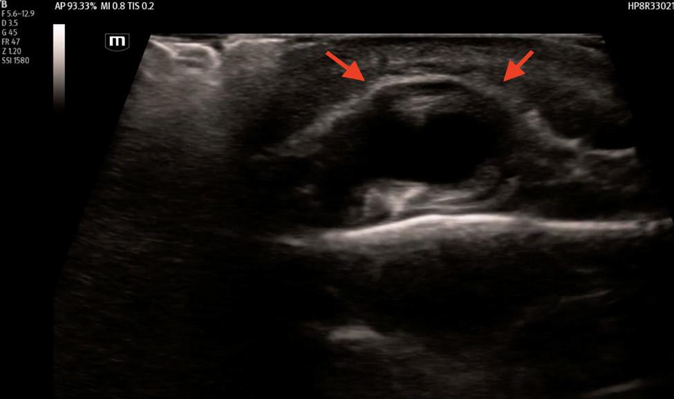
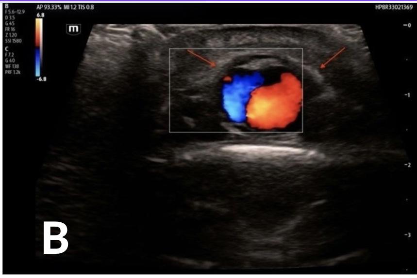
2
Image 3A. Point-of-care ultrasound (POCUS) gray-scale image using a linear probe demonstrates an anechoic, cystic structure (red arrowheads) adjacent to the occipital artery representing a pseudoaneurysm. Image 3B. POCUS image using a linear probe with color Doppler demonstrates turbulent flow with the characteristic yin-yang appearance within an anechoic, cystic structure (red arrowheads) adjacent to the occipital artery confirming the presence of a pseudoaneurysm.
Point-of-care Ultrasound Clarified the Diagnosis of an Occipital Artery Pseudoaneurysm After Blunt Trauma Nix et al. procedures.1,2 An injury to the arterial wall leads to a hematoma formation and eventually turbulent blood flow between the artery and the adjacent, communicating pseudoaneurysm.1-3 During the initial workup, duplex ultrasound, angiography, or both are useful in ensuring the prompt diagnosis of a pseudoaneurysm.3,4
Address for Correspondence: Kahra Nix, MD, Department of Emergency Medicine, University of Louisville, School of Medicine, 530 S. Jackson St, #C1H17 Louisville, KY 40202, email: kahra. nix@louisville.edu.
Conflicts of Interest: By the CPC-EM article submission agreement, all authors are required to disclose all affiliations, funding sources and financial or management relationships that could be perceived as potential sources of bias. The authors disclosed none.
Copyright: © 2025 Nix et al. This is an open access article distributed in accordance with the terms of the Creative Commons Attribution (CC BY 4.0) License. See: http://creativecommons.org/ licenses/by/4.0/
The authors attest that their institution requires neither Institutional Review Board approval, nor patient consent for publication of this case report. Documentation on file.
1. Guo Y, Chen H, Chen X, et al. Clinical importance of the occipital artery in vascular lesions: a review of the literature. Neuroradiol J 2019;32(5):366-75.
2. Changa AR, Meleis AM, Bassani L. Delayed occipital artery pseudoaneurysm following blunt force trauma. World Neurosurg 2016;89:732.e1-732.e7326.
3. Rumack C and Levine D. Chapter 25, Peripheral Arteries. In: Diagnostic Ultrasound, 6th ed. Amsterdam, Netherlands: Elsevier Academic Press; c2024:994-1017.
4. Keeling AN, McGrath FP, Lee MJ. Interventional radiology in the diagnosis, management, and follow-up of pseudoaneurysms. Cardiovasc Intervent Radiol. 2009;32(1):2-18.
Joseph Brutico, MD
Daniel Kreider, MD
Editor: R. Gentry Wilkerson, MD
Submission history: Submitted April 8, 2025; Revision received May 19, 2025; Accepted May 20, 2025
Electronically published July 24, 2025
Full text available through open access at http://escholarship.org/uc/uciem_cpcem DOI: 10.5811/cpcem.47142
Introduction: Cardiac myxomas are rare benign tumors of the heart that can become clinically relevant due to cardiovascular effects. Diagnosis can be challenging due to non-specific presenting symptoms. Point-of-care ultrasound (POCUS) provides a convenient first-line screening modality
Case Presentation: A 65-year-old male with a history of tobacco use presented to the emergency department (ED) with a month of progressive dyspnea with exertion and hematemesis. Cardiac POCUS and pulmonary computed tomography with angiography revealed a left atrial mass consistent with a cardiac atrial myxoma. The patient underwent coronary artery bypass grafting with excision of the left atrial myxoma via right atriotomy and atrial septal defect repair.
Discussion: Presented is a case of a patient presenting with progressive dyspnea diagnosed with a cardiac myxoma using POCUS in the ED. Cardiac myxomas have a wide variety of clinical presentations, and emergency physicians must maintain a high index of suspicion. Point-of-careultrasound is well suited for early diagnosis of this unique pathology. Surgical resection and tumor histopathology remain the mainstay of treatment. [Clin Pract Cases Emerg Med. 2025;9(3):355-357]
Keywords: POCUS; cardiac myxoma; echocardiography; case report.
A 65-year-old male with a history of tobacco use presented to the ED with a month of progressive dyspnea with exertion and hematemesis. On review of systems, the patient reported unintentional weight loss and night sweats. Examination revealed a cachectic male appearing older than stated age with slight tachypnea. Cardiac point-of-care ultrasound (POCUS) (Video 1) and pulmonary computed tomography (CT) with angiography (Images 1 and 2) revealed a 3 x 3 x 5 centimeter (cm) left atrial mass consistent with a cardiac atrial myxoma, which was later confirmed by pathology.
After left heart catheterization, the patient underwent coronary artery bypass grafting with excision of the left atrial myxoma via right atriotomy and atrial septal defect repair.
Cardiac myxomas are the most common primary cardiac
tumor. A vast majority of myxomas (80-90%) arise from the left atrium with fewer involving the right atrium (7-20%). Rarely, myxomas may be biatrial or arise from the ventricles.1 Symptoms vary and typically arise due to obstruction (heart failure), invasion of myocardial tissue (arrhythmias), or embolization (ischemia).2 Embolization occurs in up to 40% and is associated with villous tumors, size less than 4.5 cm, and valvular site of origin.3 Management begins with confirmation of diagnosis as common mimics include mural thrombi and valvular vegetations.2 While echocardiography is the diagnostic modality of choice, CT or cardiac magnetic resonance imaging may also be considered. Surgical resection and tumor histopathology remain the mainstay of treatment, with postoperative 30-day mortality < 5%. 4 The most common complication is cardiac arrhythmia with reported rates of up to 20%.5 This clinical pathology requires a high index of suspicion and use of multiple diagnostic modalities including cardiac POCUS.
CPC-EM Capsule
What do we already know about this clinical entity?
Cardiac myxomas are rare benign tumors of the heart that can present with a wide range of clinical symptoms. Management includes confirmation of diagnosis and surgical resection.
What is the major impact of the image(s)?
This case highlights cardiac myxoma as an important diagnosis to consider in the evaluation of a patient with dyspnea and has unique and recognizable echocardiographic findings.
How might this improve emergency medicine practice?
With a high level of suspicion, emergency physicians can accurately recognize this disease state using point-of-care ultrasound, leading to appropriate management and good long-term survival.
Patient consent has been obtained and filed for the publication of this case report.
Address for Correspondence: Daniel Kreider, MD, Wellspan York Hospital, Department of Emergency Medicine, 1001 S George St., York, PA 17403. Email: Dkreider3@wellspan.org.
Conflicts of Interest: By the CPC-EM article submission agreement, all authors are required to disclose all affiliations, funding sources and financial or management relationships that could be perceived as potential sources of bias. The authors disclosed none.
Copyright: © 2025 Brutico et al. This is an open access article distributed in accordance with the terms of the Creative Commons Attribution (CC BY 4.0) License. See: http://creativecommons.org/ licenses/by/4.0/
1. Lone R, Ahanger A, Singh S, et al. Atrial myxoma: trends in management. Int J Health Sci. 2008;2(2):141-51.
2. Elbardissi A, Dearani J, Daly R, et al. Embolic potential of cardiac tumors and outcome after resection. Stroke. 2009;40(1):156-62.
3. Wang Z, Chen S, Zhu M, et al. Risk prediction for emboli and recurrence of primary cardiac myxomas after resection. J Cardiothorac Surg. 2016;11(22):1-8.
4. Islam A. Cardiac myxomas: a narrative review. World J Cardiol 2022;14(4):206–19.
5. Okongwu C and Olaofe O. Cardiac myxoma: a comprehensive review. J Cardiothorac Surg. 2025;20(1):151.
Colton Conrad, MD*
Reginald Alouidor, MD‡ Christopher Allison, MD*†
* † ‡
University of Massachusetts Medical School – TH Chan School of Medicine, Baystate Medical Center Department of Emergency Medicine, Springfield, Massachusetts University of Massachusetts Medical School – TH Chan School of Medicine, Baystate Medical Center Department of Critical Care Medicine, Springfield, Massachusetts University of Massachusetts Medical School – TH Chan School of Medicine, Baystate Medical Center Division of Trauma/ Critical Care and Acute Care Surgery, Springfield, Massachusetts
Section Editor: Jacqueline Le, MD
Submission history: Submitted February 20, 2025; Revision received April 10, 2025; Accepted April 18, 2025
Electronically published June 8, 2025
Full text available through open access at http://escholarship.org/uc/uciem_cpcem DOI: 10.5811/cpcem.43521
Case Presentation: A 28-year-old woman with a history of cocaine and opioid use disorder presented to the emergency department with abdominal pain, nausea, and vomiting for two days. She’d had irregular bowel movements with constipation for quite some time. Physical exam was notable for diffuse peritonitis and melena on digital rectal exam. Patient had a witnessed episode of hematochezia. Computed tomography of the abdomen and pelvis with intravenous contrast demonstrated sigmoid colon intussusception, and the patient underwent emergent surgery for definitive treatment. Specimen was sent to surgical pathology and revealed no lead point.
Discussion: While sigmoid intussusception is not a rare finding, it is exceedingly rare in young adult patients who do not have a pathologic lead point. Lead points are areas of inflammation, lesions, or masses that snag the bowel and initiate the process of telescoping that ultimately results in an intussusception. This patient was not found to have such a lead point on gross examination during surgery or on extensive specimen examination in the pathology lab. Instead, her sigmoid intussusception is hypothesized to be secondary to decreased gut motility in the setting of chronic opioid use disorder. [Clin Pract Cases Emerg Med. 2025;9(3):358-360]
Keywords: intussusception; colectomy; gastroenterology; polysubstance use.
A 28-year-old woman with a history of cocaine and heroin use presented with abdominal pain, nausea, and vomiting worsening for two days. On physical exam, the patient was noted to have peritonitis in all four quadrants of the abdomen with absent bowel sounds and melanotic stool on digital rectal exam, as well as a witnessed episode of hematochezia. Vital signs were significant for tachycardia. Laboratory studies were significant only for a leukocytosis of 13.1 x 10³/liter (L) (reference range: 4.5-11.0 x 10³/L). The patient received antibiotics and antiemetic therapy, as well as pain control. Computed tomography (CT) of the abdomen and pelvis with intravenous (IV) contrast revealed the diagnosis of intussusception involving the sigmoid colon, and the
transverse and descending colon, as well as part of the omentum (Images 1 and 2). She underwent emergency surgery for definitive treatment (Image 3).
Computed tomography with IV contrast demonstrated sigmoid colon intussusception involving the transverse and descending colon, as well as parts of the omentum with evidence of ischemic necrosis. The patient underwent emergent sigmoidectomy, sub-total colectomy, partial omentectomy, and creation of end-ileostomy. The resected specimen was evaluated by pathology but demonstrated no lead point. Our patient had an extensive history of constipation, likely secondary to her opioid dependence.
Opioids act on the mu opioid receptors within the small and large bowel, leading to decreased smooth muscle contractions, diminished colonic peristalsis, and prolonged transit times.1 We hypothesize chronic opioid use contributed to her intussusception. Only one other case report in recent literature describes a patient with intussusception without pathologic lead point in the setting of chronic cocaine and opioid dependence.2 The prevalence of fentanyl contamination in cocaine and methamphetamine is estimated to be greater than 10%.3 Thus, patients who primarily use stimulants may unknowingly consume opioids on a regular basis, putting them at risk for decreased gut motility.
CPC-EM Capsule
What do we already know about this clinical entity?
Intussusception is relatively rare in adults and is generally associated with older age, adhesions from prior surgeries, inflammatory bowel disease, or malignancy.
What is the major impact of the image(s)?
The combination of computed tomography images and gross specimen photograph help the reader correlate typical radiographic findings with the physiologic process.
How might this improve emergency medicine practice?
This case serves as a reminder to consider atypical presentations of intussusception especially in young patients with risk factors such as chronic opioid use.
Image 3. Excised sigmoid, descending, and transverse colon, showing stacked layers of bowel within the sigmoid colon with ischemic necrosis and hemorrhagic walls (white arrow).
an existing lead point. Definitive treatment is resection via laparotomy, which exposes the entire colon for evaluation.5
The authors attest that their institution requires neither Institutional Review Board approval, nor patient consent for publication of this case report. Documentation on file.
Address for Correspondence: Colton Conrad, MD, University of Massachusetts Medical School – TH Chan School of Medicine, Baystate Medical Center, Department of Emergency Medicine, 759 Chestnut Street, Springfield, MA 01199. Email: coltonmichaelconrad@gmail.com
Conflicts of Interest: By the CPC-EM article submission agreement, all authors are required to disclose all affiliations, funding sources and financial or management relationships that could be perceived as potential sources of bias. The authors disclosed none.
Copyright: © 2025 Conrad et al. This is an open access article distributed in accordance with the terms of the Creative Commons Attribution (CC BY 4.0) License. See: http://creativecommons.org/ licenses/by/4.0/
1. Galligan JJ and Sternini C. Insights into the role of opioid receptors in the GI tract: experimental evidence and therapeutic relevance. Handb Exp Pharmacol. 2017;239:363-78.
2. Satoi S, Wilson S, Park E, et al. Intussusception resulting from uncontrolled severe opioid-induced constipation and/or chronic cocaine use. Am J Gastroenterol. 2021;116:S1448.
Intussusception in adults is rare, with an incidence of 2-3 cases per million annually, and comprising only 5% of all cases. Approximately 80-90% of adult intussusception cases extend from pathologic lead points that may represent inflammatory bowel disease, diverticular disease, polyps, or malignancy.4 The remaining 10-20% of intussusception cases without a pathologic lead point are termed “idiopathic.” The diagnostic modality of choice is CT with IV contrast, which may elucidate
3. Wagner KD, Fiuty P, Page K, et al. Prevalence of fentanyl in methamphetamine and cocaine samples collected by communitybased drug checking services. Drug Alcohol Depend 2023;1;252:110985.
4. Marinis A, Yiallourou A, Samanides L, et al. Intussusception of the bowel in adults: a review. World J Gastroenterol. 2009;15(4):407-11.
5. Huang BY and Warshauer DM. Adult intussusception: diagnosis and clinical relevance. Radiol Clin North Am. 2003;41(6):1137-51.

- Western Journal of Emergency Medicine Subscription
- CAL/AAEM News Service email updates
- Discounted AAEM pre-conference fees
- And more!
CAL/AAEM NEWS SERVICE
- Healthcare industry news
- Public policy - Government issues - Legal cases and court decisions