Visual Impairment in Children Due to Damage to the Brain
Edited by
gordon n. dutton
Department of Vision Sciences, Glasgow Caledonian University; Tennent Institute of Ophthalmology, Gartnavel General Hospital; and Royal Hospital for Sick Children, Yorkhill, Glasgow, UK and martin bax
Department of Medicine and Therapeutics, Imperial College, Chelsea and Westminster Hospital Campus, London, UK
Mac Keith Press
© 2010 Mac Keith Press
6 Market Road, London N7 9PW
Editor: Hilary Hart
Managing Editor: Caroline Black
Production Manager: Udoka Ohuonu
Project Manager: Catriona Vernal
The views and opinions expressed herein are those of the authors and do not necessarily represent those of the publisher
All rights reserved. No part of this publication may be reproduced, stored in a retrieval system, or transmitted in any form or by any means, electronic, mechanical, photocopying, recording or otherwise, without the prior permission of the publisher
First published in this edition 2010
British Library Cataloguing-in-Publication data
A catalogue record for this book is available from the British Library
Cover image used with permission from Sight-SimTM, courtesy of Drs Michael Bradnam, Aled Evans, and Ruth Hamilton, Department of Clinical Physics and Bioengineering, NHS Greater Glasgow and Clyde
isbn: 978-1-898683-86-5
Typeset by Prepress Projects Ltd, Perth, UK
Printed by TJ International Ltd, Padstow, Cornwall, UK
Mac Keith Press is supported by Scope
1.
2.2
4. imPairmenTs oF cenTral Visual FuncTion and iTs measuremenT 77
William V. Good and Anne B. Fulton
5. imPairmenT oF PeriPheral Vision and iTs measuremenT 85 Giorgio Porro and Dienke Wittebol-Post
6. abnormaliTies oF reFracTion and accommodaTion and Their managemenT
J. Margaret Woodhouse
7. clinical FeaTures oF PercePTual and cogniTiVe Visual imPairmenT in children wiTh brain damage oF early onseT
Gordon N. Dutton, with Elisabeth Macdonald, Suzannah R. Drummond, Shohista Saidkasimova, and Katherine Mitchell
8. imPairmenT oF cogniTiVe Vision: iTs deTecTion and measuremenT
8.1 struCtured CliniCal history-taking for Cognitive and perCeptual visual dysfunCtion and for profound visual disabilities due to damage to the brain in Children
Gordon N. Dutton, with Julie Calvert, Hussein Ibrahim, Elisabeth Macdonald, Daphne L. McCulloch, Catriona Macintyre-Beon, and Katherine M. Spowart
8.2 objeCtive behavioural and eleCtrophysiologiCal measures for assessing visual brain funCtion in infants and young Children 129
Janette Atkinson and Oliver Braddick
8.3 psyChometriC evaluation of higher visual disorders: strategies for CliniCal settings 149
Peter Stiers and Elisa Fazzi
9. The eFFecT oF imPaired Vision on deVeloPmenT
Elisa Fazzi, Sabrina G. Signorini, and Josée Lanners
10. PsychiaTric consideraTions in corTical Visual imPairmenT 174 Roger D. Freeman
11. children wiTh seVere brain damage: FuncTional assessmenT For diagnosis and inTerVenTion
Jenefer Sargent, Alison Salt and Naomi Dale
12. Visual imPairmenT in cerebral Palsy 194
Elisa Fazzi, Sabrina G. Signorini, and Paolo E. Bianchi
13. children wiTh inTellecTual disabiliTies and cerebral Visual imPairmenT: Problems wiTh deTecTion and diagnosis 205
Heleen M. Evenhuis
14. PracTical aPProaches For The managemenT oF Visual Problems due To cerebral Visual imPairmenT 217
Gordon N. Dutton, with Debbie Cockburn, Gillian McDaid, and Elisabeth Macdonald
15. sTraTegies To suPPorT The deVeloPmenT and learning oF children wiTh cerebral Visual imPairmenT aT home and aT school: communicaTion, orienTaTion, and mobiliTy 227
Marianna Buultjens, Lea Hyvärinen, Renate Walthes, and Gordon N. Dutton
16. aPProaches To The managemenT in schools oF Visual Problems due To cerebral Visual imPairmenT 236
Marianna Buultjens, Lea Hyvärinen, and Renate Walthes
17. sTraTegies To helP children who haVe boTh Visual and hearing imPairmenTs 245 Stuart Aitken
18. seTTing uP inTegraTed serVices For children wiTh cerebral Visual imPairmenT 257
Margot Campbell and Marianna Buultjens
19. classiFicaTion oF Visual FuncTioning and disabiliTy in children wiTh Visual Processing disorders 265 Lea Hyvärinen
20. Towards The deVeloPmenT oF a classiFicaTion oF Vision-relaTed FuncTioning – a PoTenTial Framework 282 August Colenbrander
Authors’ Appointments
Stuart Aitken sense scotland, glasgow; Call scotland, school of education, university of edinburgh, edinburgh, uk
Susann Andersson department of ophthalmology, the Queen sylvia Children’s hospital, gothenburg, sweden
Janette Atkinson department of developmental science, university College london, london, uk
Martin Bax imperial College (Chelsea and westminister hospital Campus), london, uk
Paolo E. Bianchi department of ophthalmology, irCCs san matteo hospital, university of pavia, pavia, italy
Oliver Braddick department of experimental psychology, university of oxford, oxford, uk
Marianna Buultjens monifieth, dundee, uk
Julie Calvert royal hospital for sick Children, yorkhill, glasgow, uk
Margot Campbell armistead Child development Centre, dundee, uk
Giovanni Cioni division of Child neurology, university of pisa, stella maris scientific institute, pisa, italy
Debbie Cockburn southbank Child Centre, glasgow, uk
August Colenbrander the smith kettlewell eye research institute, san francisco, Ca, usa
Naomi Dale department of neurodisability, great ormond street hospital for Children, london, uk
Suzannah R. Drummond tennent institute of ophthalmology, gartnavel general hospital, glasgow, uk
Gordon N. Dutton
tennent institute of ophthalmology, gartnavel general hospital; department of vision sciences, glasgow Caledonian university; the royal hospital for sick Children, yorkhill, glasgow, uk
Heleen M. Evenhuis erasmus university medical Center, intellectual disability medicine, department of general practice, rotterdam, the netherlands
Elisa Fazzi mother and Child department, medical faculty, university of brescia, brescia, italy
Olof Flodmark department of Clinical neuroscience, karolinska institute, stockholm, sweden
Roger D. Freeman department of psychiatry and department of pediatrics, university of british Columbia; neuropsychiatry Clinic, bC Children’s hospital, vancouver, bC, Canada
Anne B. Fulton department of ophthalmology, Children’s hospital boston, boston, ma, usa
William V. Good smith-kettlewell eye research institute, san francisco, Ca, usa
Melvyn A. Goodale Centre for brain and mind, department of psychology, university of western ontario, london, on, Canada
Andrea Guzzetta division of Child neurology, university of pisa, stella maris scientific institute, pisa, italy
Lea Hyvärinen faculty of rehabilitation sciences, technical university of dortmund, dortmund, germany; faculty of behavioural sciences, university of helsinki, helsinki, finland
Hussein Ibrahim royal hospital for sick Children, yorkhill; glasgow Caledonian university, glasgow, uk
Lena Jacobson department of Clinical neuroscience, karolinska institute, stockholm, sweden
Josée Lanners robert hollman foundation, Cannero riviera (vb), padova, italy
Daphne L. McCulloch
Gillian McDaid
Elisabeth Macdonald
Catriona Macintyre-Beon
Carey Matsuba
Eugenio Mercuri
Katherine Mitchell
royal hospital for sick Children, yorkhill; glasgow Caledonian university, glasgow, uk
royal hospital for sick Children, yorkhill, glasgow, uk
tennent institute of ophthalmology, gartnavel general hospital; royal hospital for sick Children, yorkhill, glasgow, uk
royal hospital for sick Children, yorkhill, glasgow, uk
british Columbia Children’s hospital, vancouver, bC, Canada
paediatric neurology unit, Catholic university, policlinico gemelli, rome, italy
the royal hospital for sick Children, yorkhill, glasgow, uk
Giorgio Porro department of ophthalmology, utrecht university hospital, utrecht, the netherlands
Daniela Ricci
Shohista Saidkasimova
paediatric neurology unit, Catholic university, rome, italy
tennent institute of ophthalmology, gartnavel general hospital, glasgow, uk
Alison Salt department of neurodisability, great ormond street hospital for Children, london, uk
Jenefer Sargent
department of neurodisability, great ormond street hospital for Children, london, uk
Sabrina G. Signorini Center of Child neuro-ophthalmology, Child neuropsychiatry unit, irCCs C. mondino institute, pavia, italy
Janet Soul
Katherine M. Spowart
Peter Stiers
harvard medical school, boston, ma, usa
royal hospital for sick Children, yorkhill, glasgow, uk
faculty of psychology and neuroscience, maastricht university, maastricht, the netherlands
Renate Walthes faculty of rehabilitation sciences, technical university of dortmund, dortmund, germany
Dienke Wittebol-Post utrecht university hospital, utrecht, the netherlands
J. Margaret Woodhouse school of optometry and vision sciences, Cardiff university, Cardiff, uk
Foreword
This book is a worthy sequel to the Castang Foundation meeting in November 2005 on visual impairment, bringing together experts in cognitive and perceptual dysfunction in children and how they can be helped. It is an ‘eclection’ of ideas and data on caring for children with visual brain damage, aimed at the medical and caring professions. The book starts with an authoritative statement by Mel Goodale on functional organization of the central visual pathways, and the important distinction between ventral and dorsal processing, which recurs throughout the book. Gordon Dutton discusses, with Elisabeth Macdonald, impairment of cognitive vision with issues of detection and measurement and, with others, cognitive dysfunction associated with brain damage in children.
Janette Atkinson and Oliver Braddick (Oxford, UK) discuss behaviour and electrophysiological measures for assessing visual brain function in infants and young children. The effects of impaired vision on development and visual impairment in the context of cerebral palsy are the themes of Elisa Fazzi’s work. Recent experimental techniques have their place here, including correlations between imaging and early visual development by Eugenio Mercuri and colleagues in Italy. Central visual function and how it is assessed are examined by William Good and Anne Fulton.
Clinical features of cognitive and visual impairments are analysed by Gordon Dutton and his colleagues at the Tennent Institute of Ophthalmology in Gartnavel General Hospital in Glasgow, UK.
Aside from the clinical and philosophical issues, there are political implications, especially as financial and medical, as well as educational, resources are made available or are withheld according to complicated social factors. By increasing understanding and awareness of these issues, this book will be of direct help to children with visual impairments. Many of the issues that are raised are technical, but there is an underlying appreciation of the importance of understanding these problems at many levels, from clinical to social, with sensitivity to emotional issues.
There is a concern throughout of how to tap into normal or damaged systems in infants without the use of language. So, experiments with eye movements and accommodation changes are particularly significant. For normal adults, eye movements are controlled from all levels of brain activity, including subtle cognitive estimates of probabilities and dangers and rewards. Presumably, as knowledge grows, and as techniques become more powerful,
it will be possible to gain richer insights into cognitive processing in infancy and childhood. Not only should this be of great clinical importance, but it should also tell us a great deal about human development and the effect of environment in home life and school. This clearly needs an interdisciplinary approach, which is just what emerges from the insights and future promises of this ambitious book.
Richard Gregory CBE, FRS† Emeritus Professor and Senior Research Fellow Department of Experimental Psychology University of Bristol
†Sadly, Richard Gregory passed away on 17 May 2010.
introduCtion
Gordon N. Dutton and Martin Bax
Increased awareness of cerebral visual impairment in children, combined with improved recognition of its wide-ranging manifestations, has led to it becoming the most common cause of visual impairment in children in the developed world. Yet the subject is in its infancy.
The development of a child’s visual acuity from early childhood, and its measurement, are well described (Teller 1997), but despite longstanding evidence that the newborn infant has an inbuilt capability to identify a face compared with other patterns (Goren et al 1975) and an early ability to imitate facial movements (Meltzoff and Moore 1983), surprisingly little information about the functional use of vision by the infant has been collected. It is known that the fetus learns to discriminate sounds such as a female voice while in utero (Lecanuet 1989), and the newborn infant can use smell to discriminate between a mother’s breast milk and that of a stranger (Macfarlane 1975). Given such very early facilitation of these senses to identify specific elements in the environment, it would be surprising if vision, with its larger allocation of dedicated brain function, does not play a major part in the child’s social (and psychosocial) development at a very early stage.
Much theorizing about the early psychosocial development of the child remains fixated on the oral Freudian model, despite the fact that, from research, we know that the oral mechanism reaches its level of sophistication very early on, because it is essential to the infant’s survival. The centres that control it are in the region of the midbrain and probably do not involve the cortex, in so far as the infant sucks and swallows satisfactorily from 34 weeks. The anencephalic child successfully swallows and survives for days, if not weeks. It appears much more likely that the early psychosocial developmental period for humans should be regarded as the visual rather than the oral period. Indeed, during breast feeding, once satiation is partially achieved, the child will break off from sucking and swallowing to gaze intently into the eyes of the mother (Blass et al 2001). The pacifier induces somnolence whereas the visual display excites interest. Visual identification of the face has been investigated in the classic studies of infants, and the importance of identification of the face leading to the development of the awareness of strangers (Piaget 1955) is self-evident. Here is not the place for a full review of the role of vision in early psychosocial development, but its importance cannot be overemphasized. Vision certainly plays a role in disturbed social interactions, which are seen in many neurodevelopmental disorders, such as autism (Pellicano et al 2007), but its role has yet to be fully elucidated.
This book originates from a Castang Foundation workshop with the same title, held in London in late 2005. It links the work of a range of authors who have made significant contributions to the literature on the subject of cerebral visual impairment and provides a structured amalgam of the viewpoints of different specialists. The authors each have different perspectives, some of which, to the reader, may appear to be contradictory, or at least not complementary. This applies particularly to the last two chapters, which present different viewpoints concerning the classification of cerebral visual impairment. At this stage in the evolution of knowledge, we believe this is an appropriate approach, as it provides the substrate for the development of a consistent model, which will gradually be built up through exchanging ideas and concept frameworks about this complex and wide-ranging subject.
A large proportion of the brain serves vision, but, in contrast to movement of the body, vision is an internalized function. Early-onset damage to the brain can interfere with the development of movement and manifests as cerebral palsy, but when vision is affected the result is less evident in all but the most severely affected cases. Yet the adverse effects on development can be profound. The classic model of thinking about vision, in which a picture is somehow formed by the eyes and processed in the striate cortex, has long been recognized to be limited. Yet in medical practice this conceptual framework has continued to hold sway, with impairment of visual acuity considered a prerequisite for both diagnosis and the provision of assistance. Damage to the visual pathways and occipital cortex impairs visual fields and visual acuities, whereas damage to the higher centres serving vision interferes with visual processing. These visual manifestations may occur either in isolation or in combination. But a wider concept of how damage to the brain can give rise to multiple visual difficulties has yet to be fully recognized. Children with such visual problems may, in some cases, be seen as having one of the syndromic classifications such as autism, cerebral palsy, or simply intellectual disability, but the visual element in these children is rarely recognized and may even be judged irrelevant to the child’s condition, despite potentially being pivotal.
When considering terminology, the term ‘cortical visual impairment’ has been defined as ‘loss of or highly inefficient visual acuity, essentially due to occipital lobe disturbance’ (J. Jan, British Columbia’s Children’s Hospital, BC, Canada; personal communication, 2009), whereas the term ‘cerebral visual impairment’ has been taken to encompass a wider range of disorders, including ‘visual disturbance on account of oculomotor incoordination, and visual, cognitive, and perceptual impairment owing to pathology affecting the visual association cortices and their interconnecting pathways’ (Fazzi et al 2004). Both terms are abbreviated in the literature to CVI, which can be a source of confusion. In this text, the term ‘cerebral visual impairment’ is predominantly used in view of its wider meaning. It also serves as a reminder that visual fields and visual acuities are rarely impaired in isolation and a search for evidence of visual–perceptual impairment in such children will usually be rewarded and will, in turn, lead to the implementation of a wider range of habilitative strategies. (The term ‘habilitative’ is chosen in preference to ‘rehabilitative’ because in most cases of cerebral visual impairment no function has been lost to rehabilitate. Similarly, the term ‘loss of vision’ can apply only to those who have lost vision. Thus, the term ‘visual impairment’ is preferable for those who have had low vision from birth.)
A wide spectrum of visual problems has now been described in children with cerebral visual impairment, which differs from that seen in adults, because the loss of a visual function is very different to impaired development of that same function. Moreover, early-onset damage to the brain is followed by brain growth and development, and the adult model – of damage to the visual system being immutable – does not apply. The fact that training is now known to lead to brain growth accompanied by greater cell size and greater numbers of synaptic connections in the occipital area indicates that strategies which optimize habilitation may affect ultimate visual outcome, and there is limited evidence that this is the case (Sonksen et al 1991). Simply the act of identifying, characterizing, and communicating the diagnosis of cerebral visual impairment to parents and carers can set the child on a new pathway of development. For example, lower visual field impairment is no longer attributed to clumsiness, but is managed appropriately; inability to copy from the ‘blackboard’ is understood to be a result of visual difficulties, and alternative measures are implemented; and impaired social interaction because of an inability to find someone in a group is recognized not as being caused by being socially aloof, but as a specific disability which can be managed appropriately. The resultant change in attitude of both parents and teachers can revolutionize a child’s life. Unlike impairment of vision owing to eye pathology, cerebral visual impairment in children can vary from hour to hour and day to day, and recognition that this is a typical feature and is not a result of bad behaviour is essential. The adverse emotional consequences of failure of diagnosis, and the child having to tolerate unfounded criticism of fully explicable behaviour, can be profound.
Focal damage to the visual brain leads to specific visual difficulties, which can be specifically characterized. By contrast, diffuse damage which affects all aspects of brain function can adversely impact upon visual function in a manner which is much more difficult to characterize, yet still needs to be recognized if appropriate measures are to be taken. It may be impossible to fully characterize the visual dysfunction in those who are profoundly impaired, yet the knowledge gained from those with focal damage can be used to afford a greater philosophical understanding of the visual problems in such children, which underpins the practical basis for how best they can be managed.
It is remarkable that knowledge concerning the specific cortical areas serving visual function goes back to the studies of soldiers with brain injuries during the First World War. Not only was the anatomy of the afferent visual pathways determined with accuracy (Holmes 1918a), but the profound visual consequences of damage to the posterior parietal cortex were graphically described in a second, but less well-recognized, paper (Holmes 1918b). This second paper described six soldiers who sustained bilateral posterior parietal shrapnel injuries. These soldiers all had lower visual field loss, impaired visual guidance of movement (optic ataxia) of the limbs (despite intact stereopsis in four cases), and profound simultanagnosia (being unable to identify more than one or two objects at a time). (This symptom, complex in its severe form, came to be known as Balint syndrome, and in its milder form is beginning to be referred to as dorsal stream dysfunction.) It is no coincidence that the selfsame features of impaired lower visual fields – inability to handle complex visual scenes and impaired visual guidance of movement – are evident in children with posterior superior periventricular white matter pathology (Jacobson and Dutton 2000). Affected children have coordination difficulties
and cannot see a friend in a group. They can appear antisocial and may be ascribed a range of alternative diagnoses if the visual origin of the behaviour is not recognized. While the ventral stream disorders are less clearcut, it is clear that they play a major role in visual function, and we can be sure that they have a large, as yet unrecognized, part to play in many of the syndromic neurodevelopmental disorders, such as cerebral palsy and autism.
Inevitably, the nature of service provision in different countries influences the thinking, with clinical experience constraining the patterns of visual disturbance seen by different practitioners. For example, those working in facilities for the visually disabled do not see how children with cerebal visual impairment manage in mainstream schooling or other special educational provision.
We were particularly aware that, while ‘simple’ problems affecting visual acuity, contrast sensitivity, and visual fields are identified, the role of perceptual and cognitive visual dysfunction in many disabilities may not be recognized or understood. We think particularly, for example, of the many children on the autistic spectrum who have problems with vision and visual interpretation, as these problems tend to be enveloped in the general description of the condition, such as autism, and not focused on diagnostic issues in their own right. The same applies to the field of cerebral palsy and is also probably of considerable importance in the field of intellectual disability. The possibility that such complex visual problems exist and may provide remediable explanations for some of the behavioural manifestations potentially renders this book important reading for those who look after such children, whether they be teachers, therapists, psychologists, doctors, or other health professionals. Simply thinking of children on the autistic spectrum as having fundamental visual problems, rather than purely behavioural issues, changes the cast of one’s mind as one approaches such a child. We hope that this book will lead people to look at children with disabilities in a different way and constantly keep in mind the role of the visual system in their disorder (without neglecting all the other areas of function which will affect the child). This book is not aimed at the paediatric ophthalmologist; rather, it is aimed at a whole community of clinicians, from paediatric neurologists through to psychologists, therapists, and teachers. To this end, the chapters with a more technical content therefore include summaries written with less technical language. We hope that all these groups will actively consider vision (in its broadest sense) and its role in the wide range of disorders which may present to them.
1 the FunCtionAl orgAnizAtion oF the CentrAl VisuAl pAthwAys
Melvyn A. Goodale
Introduction: What is vision for? A brief discussion of the origins of vision
Visual systems first evolved not to enable animals to see, but to provide distal sensory control of their movements. Vision as ‘sight’ is a relative newcomer on the evolutionary landscape, but its emergence has enabled animals to carry out complex cognitive operations on perceptual representations of the world. Thus, vision in humans and other primates (and other animals as well) has two distinct but interacting functions: (1) the perception of objects and their relations, which provides a foundation for the organism’s cognitive life and its conscious experience of the world and (2) the control of actions directed at (or with respect to) those objects, in which separate motor outputs are programmed and controlled online. These different demands on vision have shaped the organization of the visual pathways in the primate brain. Moreover, as we shall see, this distinction between ‘vision for perception’ and ‘vision for action’ provides a useful framework for understanding the functional organization of the human visual system, including the visual pathways within the cerebral cortex (for reviews see Goodale and Milner 2004, Milner and Goodale 2006). But before these different functions of vision are discussed in any detail, what is known about the organization of the projections from retina to different subcortical structures will be briefly reviewed.
From retina to brain
The retina not only transduces the electromagnetic radiation striking the photoreceptors into physiological signals that can be understood by the brain, but it also performs several computations on those signals, which involve combining information from a number of different photoreceptors (Masland 2001, Field and Chichilnisky 2007). Thus, by the time the sensory signals leave the eye on their way to the brain, a good deal of processing has already occurred. This processing, furthermore, is not uniform within the retinal system. The ganglion cells, whose axons leave the eye and constitute the optic nerve, are heterogeneous in the kinds of information they convey (Wässle 2004). Some ganglion cells, for example, carry information that is particularly useful for an analysis of the spatial distribution of light energy striking the retina; others carry information that is more related to the temporal dynamics of the retinal array, arising, for example, from the motion of a distal stimulus. Still others appear to be primarily concerned with the distribution of the different wavelengths of light entering the
eye, leading ultimately to the perception of colour (Dacey and Packer 2003, Solomon and Lennie 2007). Moreover, across different species, the organization of the eye and the signal transformations that occur in the retina vary enormously, no doubt reflecting the range of ecological niches in which animals live (Lamb et al 2007).
It is often not appreciated that the neuronal projections from the retina travel to a number of distinct target areas in the vertebrate brain. In other words, they do not form a set of parallel lines of information projecting together from one complex processing station to another, but instead diverge to very different processing targets right from the outset (Fig. 1.1). These different projections reflect both the evolutionary origins of different visual pathways as well as the different behavioural functions to which vision contributes.
The earliest set of projections to leave the optic tract and terminate in the brain are those terminating in a structure in the hypothalamus called the suprachiasmatic nucleus (SCN), which, as the name implies, sits right above the optic chiasm. The SCN is a critical structure in the brain circuit that controls circadian rhythms, such as the sleep–wake cycle and the production and release of circulating hormones (Herzog 2007). The retinal projections to the SCN provide input about ambient light levels that locks the intrinsic circadian rhythm of SCN neurons to the local light–dark cycle. When the local light–dark cycle changes dramatically, as it does when one flies across the Atlantic, it takes some time for the SCN neurons to be retrained to the new light–dark cycle (hence, one experiences ‘jet lag’).
Another set of ancient retinal projections, the accessory optic tract, terminates in three separate nuclei in the brainstem, known collectively as the accessory optic system (AOS). Neurons in the AOS are driven by large moving patterns of optic flow on the retina. They project via premotor relay nuclei to the cerebellum and spinal cord, and play a critical role (together with the vestibular system and proprioception) in visual stabilization, the control of posture, and the regulation of locomotion and heading (Simpson 1984).
A prominent set of pathways from the retina also projects to the midbrain, to a laminated structure known in simpler vertebrates and birds as the optic tectum, and in mammals as the superior colliculus (Figs 1.1 and 1.2). The superior colliculus plays a central role in the initiation and control of orientating movements of the eyes, head, and body towards salient visual stimuli in the peripheral visual fields and the maintenance of fixation on those stimuli (Sparks 2002). Other collicular circuits mediate visually guided escape responses by generating rapid movements away from looming visual stimuli that could be potential predators (Dean et al 1989). The deeper layers of the superior colliculus project to premotor nuclei in the brainstem involved in the control of the eye muscles and to parts of the spinal cord involved in the control of the neck musculature and the trunk and forelimbs (Crawford et al 2003, May 2005). The superficial layers of the superior colliculus send projections to visual areas in the cerebral cortex, via thalamic nuclei such as the pulvinar and medial dorsal nucleus (May 2005). The exact role of these projections is not well understood, although it is thought that some of them might send an efferent copy signal (corollary discharge) about shifts in gaze that could modulate attention and the processing of visual motion in the cerebral cortex (Sommer and Wurtz 2006).
Other retinal projections include those to various pretectal nuclei, which, as the name suggests, are located just in front of the optic tectum (or superior colliculus). One of these
Primary visual cortex
Interlaminar
Higher visual areas in cortex
Fig. 1.1 Schematic drawing of the main retinal projection sites in the brain. LGNd, lateral geniculate nucleus, pars dorsalis; parvo, parvocellular layers of LGNd; magno, magnocellular layers of LGNd; interlaminar, interlaminar regions of LGNd; SC, superior colliculus; pulvinar, pulvinar nucleus of the thalamus; LGNv, lateral geniculate nucleus, pars ventralis; AOTd, dorsal terminal nucleus of the accessory optic tract; AOTl, lateral terminal nucleus of the accessory optic tract; AOTm, medial terminal nucleus of the accessory optic tract; SCN, suprachiasmatic nucleus. For more details see Milner and Goodale (2006).
pretectal nuclei, the nucleus of the optic tract, has strong links with the AOS; another, the olivary pretectal nucleus, is part of the circuit mediating the pupillary light reflex and other reflexive responses such as light-evoked blinking (Gamlin 2005). There is also evidence that retinal projections to (unspecified) pretectal nuclei might play a role in obstacle avoidance during locomotion in both amphibians and mammals (Ingle 1982, Milner and Goodale 2006). Nevertheless, much remains to be discovered about the functions of the different pretectal nuclei, as well as neighbouring thalamic nuclei such as the ventral part of the lateral geniculate
LGNd
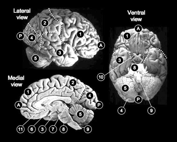
Fig. 1.2 The human brain, showing the lateral, medial, and ventral surfaces. A, anterior; P, posterior; 1, frontal lobe; 2, parietal lobe; 3, temporal lobe; 4, occipital lobe; 5, cerebellum; 6, thalamus; 7, superior colliculus; 8, pons; 9, medulla; 10, optic nerve; 11, corpus callosum.
nucleus (LGNv) and the retino-recipient region of the pulvinar (for a review see Kaas and Lyon 2007).
By far and away the most prominent visual pathway from the retina to the brain in humans and their primate cousins is the retinogeniculate projection, which terminates in the dorsal part of the lateral geniculate nucleus of the thalamus (LGNd). In other vertebrate classes, such as amphibians and reptiles, this pathway is barely evident. Even in birds, which are highly visual creatures, the homologue of the retinogeniculate projection is much smaller than the projection to their optic tectum. Only in mammals has this projection system become prominent. Neurons in the LGNd project in turn to the cerebral cortex, with almost all of the fibres, in primates at least, terminating in the primary visual area, or striate cortex (often nowadays called ‘area V1’) in the occipital lobe (Fig. 1.2). This geniculostriate projection and its cortical elaborations probably constitute the best studied neural ‘system’ in the whole of neuroscience. This fact is perhaps not unrelated to the general belief that subjective visual experience in humans depends on the integrity of this projection system.
Finally, it should be emphasized that almost all the subcortical structures discussed above receive not only direct input from the retina, but also inputs from other visual structures, including visual areas in the cerebral cortex. Thus, the superior colliculus, the pretectal nuclei, the pulvinar, and the lateral geniculate nucleus all receive inputs from area V1. In addition, many of the subcortical nuclei are highly interconnected. For example, the superior colliculus projects to pretectal nuclei, the pulvinar, and the lateral geniculate nucleus. Moreover, many
of these structures receive input from other modalities, such as audition and somatosensation. Nevertheless, each of these different visual structures plays a critical role in the control of a particular set of visually guided and/or visually modulated patterns of behaviour.
Two cortical visual pathways
Beyond the primary visual cortex in the primate cerebral cortex, visual information is conveyed to a bewildering number of extrastriate areas (Van Essen 2001). Despite the complexity of the interconnections between these different areas, two broad ‘streams’ of projections from primary visual cortex have been identified in the macaque monkey brain: a ventral stream projecting eventually to the inferotemporal cortex and a dorsal stream projecting to the posterior parietal cortex (Ungerleider and Mishkin 1982; see Figs 1.2 and 1.3). Although some caution must be exercised in generalizing from monkey to human (Sereno and Tootell 2005), recent neuroimaging evidence suggests that the visual projections from early visual areas to the temporal and parietal lobes in the human brain also involve a separation into ventral and dorsal streams (Grill-Spector and Malach 2004, Culham and Valyear 2006).

Fig. 1.3 Schematic representation of the two streams of visual processing in human cerebral cortex. The retina sends projections to the dorsal part of the lateral geniculate nucleus in the thalamus (LGNd), which projects in turn to primary visual cortex (V1). Within the cerebral cortex, the ventral stream arises from early visual areas (V1+) and projects to regions in the occipito-temporal cortex. The dorsal stream also arises from early visual areas but projects instead to the posterior parietal cortex. The posterior parietal cortex also receives visual input from the superior colliculus via the pulvinar. On the left, the approximate locations of the pathways are shown on an image of the brain. The routes indicated by the arrows involve a series of complex interconnections.
Inferotemporal cortex
Posterior parietal cortex
Pulvinar
Retina
Area V1+ LGNd
The PercePTion–AcTion hyPoThesis
Traditional accounts of the division of labour between the two streams (e.g. Ungerleider and Mishkin 1982) focused on the distinction between object vision and spatial vision. This distinction between what and where resonated remarkably with not only psychological accounts of perception, but also nearly a century of neurological thought about the functions of the temporal and parietal lobes in vision (Ferrier and Yeo 1884, Brown and Schäfer 1888, Schäfer 1888, Holmes 1918a).
In the early 1990s, however, the ‘what versus where’ story began to unravel as new evidence emerged from work with both monkeys and patients with neurological disorders. It became apparent that a purely sensory/perceptual account of ventral–dorsal function could not explain these findings. The only way to make sense of them was to consider the nature of the outputs served by the two streams – and to work out how visual information is eventually transformed into motor acts. In 1992, Goodale and Milner proposed a re-interpretation of the Ungerleider and Mishkin account of the two visual streams. According to their proposal, the dorsal stream plays a critical role in the real-time control of action, transforming moment-tomoment information about the location and disposition of objects into the coordinate frames of the effectors being used to perform the action (Goodale and Milner 1992, Milner and Goodale 2006). The ventral stream (together with associated cognitive networks) helps to construct the rich and detailed representations of the world that allow us to identify objects and events, attach meaning and significance to them, and establish their causal relations. Such operations are essential for accumulating and accessing a visual knowledge-base about the world. Thus, it is the ventral stream that provides the perceptual foundation for the offline control of action, projecting action into the future and incorporating stored information from the past into the control of current actions. In contrast, processing in the dorsal stream does not generate visual percepts; it generates skilled actions (in part by modulating more ancient visuomotor modules described in the previous section). The division of labour proposed by Goodale and Milner not only accounts for the neurological dissociations observed in patients with damage to different regions of the cerebral cortex, but it is also supported by a wealth of anatomical, electrophysiological, and behavioural studies in the monkey.
neuroPsychologicAl evidence
It has been known for a long time that patients with lesions in the superior regions of the posterior parietal cortex, particularly lesions that invade the territory of the intraparietal sulcus (or IPS) and the parieto-occipital sulcus, can have problems using vision to direct a grasp or aiming movement towards the correct location of a visual target placed in different positions in the visual field, particularly the peripheral visual field. This particular deficit is often described as optic ataxia (following Bálint 1909). But the failure to locate an object with the hand should not be construed as a problem in spatial vision; many of these patients, for example, can describe the relative position of the object in space quite accurately, even though they cannot direct their hand towards it (Perenin and Vighetto 1988). Moreover, sometimes the deficit will be seen in one hand but not the other. (It should be pointed out, of course, that these patients typically have no difficulty using input from other sensory systems, such as proprioception or audition, to guide their movements.) Some of these patients are unable to
use visual information to rotate their hand, scale their grip, or configure their fingers properly when reaching out to pick up an object, even though they have no difficulty describing the orientation, size, or shape of objects in that part of the visual field (see Fig. 1.4). Clearly, a ‘disorder of spatial vision’ (Holmes 1918a) fails to capture this range of visuomotor impairments. Instead, this pattern of deficits suggests that the posterior parietal cortex plays a critical role in the visual control of skilled actions (for more details see Milner and Goodale 2006).
The opposite pattern of deficits and spared abilities can be seen in patients with visual agnosia. Take the case of patient DF, who developed a profound visual-form agnosia following carbon monoxide poisoning (Goodale et al 1991). Although magnetic resonance imaging (MRI) showed evidence of diffuse damage consistent with hypoxia, most of the damage was evident in ventrolateral regions of the occipital cortex with V1 remaining largely spared. Even though DF’s ‘low-level’ visual abilities are reasonably intact, she can no longer recognize everyday objects or the faces of her friends and relatives, nor can she identify even the simplest of geometric shapes. (If an object is placed in her hand, of course, she has no trouble identifying it by touch.) Remarkably, however, DF shows strikingly accurate guidance of her hand movements when she attempts to pick up the very objects she cannot identify. Thus, when she reaches out to grasp objects of different sizes, her hand opens wider midflight for larger objects than it does for smaller ones, just like it does in people with normal vision (see Fig. 1.4). Similarly, she rotates her hand and wrist quite normally when she reaches out to
Patient RV: Optic ataxia
Patient DF: Visual-form agnosia
Fig. 1.4 Graphs showing the size of the aperture between the index finger and thumb during objectdirected grasping and manual estimates of object width for RV, a patient with optic ataxia, and DF, a patient with visual-form agnosia. RV (left) was able to indicate the size of the objects reasonably well (individual trials marked as open diamonds), but her maximum grip aperture in flight was not well tuned. She simply opened her hand as wide as possible on every trial. In contrast, DF (right) showed excellent grip scaling, opening her hand wider for the 50mm-wide object than for the 25mm-wide object. DF’s manual estimates of the width of the two objects, however, were grossly inaccurate and showed enormous variability from trial to trial. The data from DF were adapted from Goodale et al (1991).
grasp objects in different orientations, and she places her fingers correctly on the surface of objects with different shapes. At the same time, she is quite unable to distinguish between any of these objects when they are presented to her in simple discrimination tests. She even fails in manual ‘matching’ tasks in which she is asked to show how wide an object is by opening her index finger and thumb a corresponding amount (Fig. 1.4). DF’s spared visuomotor skills are not limited to grasping. She can step over obstacles during locomotion as well as control subjects can, even though her perceptual judgements about the height of these obstacles are far from normal. Contrary to what would be predicted from the ‘what versus where’ hypothesis, then, a profound loss of form perception coexists in DF with a preserved ability to use form in guiding a broad range of actions. Such a dissociation, of course, is consistent with the idea that there are separate neural pathways for transforming incoming visual information for the perceptual representation of the world and for the control of action. Presumably, it is the former and not the latter that is compromised in DF (for more details see Goodale and Milner 2004, Milner and Goodale 2006).
evidence from single-cell recording of The monkey
A broad range of studies on the dorsal and ventral streams in the monkey lends considerable support to the distinction between perception and action. For example, monkeys with lesions of inferotemporal (IT) cortex can orientate their fingers in a precision grip to grasp morsels of food embedded in small slots placed at different orientations – even though their ability to discriminate between different orientations is profoundly impaired (Glickstein et al 1998). Moreover, there is a long history of single-unit work showing that cells in IT and neighbouring regions of the superior temporal sulcus are tuned to specific objects and object features – with some cells maintaining their selectivity irrespective of viewpoint, retinal image size, and even colour (e.g. Gross et al 1972, Logothetis and Sheinberg 1996, Tanaka 2003). Moreover, the responses of these cells are not affected by the animal’s motor behaviour but are instead sensitive to the reinforcement history and significance of the visual stimuli that drive them. It has been suggested that cells in this region might play a role in comparing current visual inputs with internal representations of recalled images (e.g. Eskandar 1992), which are themselves presumably stored in other regions, such as neighbouring regions of the medial temporal lobe and related limbic areas (Squire et al 2007). In fact, sensitivity to particular objects can be created in ensembles of IT cells simply by training the animals to discriminate between different objects. There is also evidence for a specialization within separate regions of the ventral stream for the coding of certain categories of objects, such as faces and hands, which are of particular social significance to the monkey (Logothetis et al 1995). Finally, experiments that have used a binocular rivalry paradigm, in which competing images are presented at the same time to the two eyes, have shown that cells in IT are tuned to what the monkey reports seeing on a particular trial, not simply to what is present to the two eyes (Logothetis 1998). But neurons earlier in the visual system, such as area V1, do not show these correlations. They respond in the same way, no matter what the monkey indicates that it sees. Even in intermediate areas of the ventral stream, such as area V4, the correlations are relatively weak. These results provide indirect support for the claim that the ventral stream
plays a critical role in delivering the contents of our conscious percepts (in so far as activity in the monkey’s inferotemporal cortex reflects what the monkey indicates that it sees). These, and other studies too numerous to cite here, lend considerable support to the suggestion that the object-based descriptions provided by the ventral stream form the basic raw material for visual perception, recognition memory, and other long-term representations of the visual world (for a detailed discussion see Milner and Goodale 2006).
In sharp contrast to the activity of cells in the ventral stream, the responses of cells in the dorsal stream are greatly dependent on the concurrent motor behaviour of the animal. Thus, separate subsets of visual cells in the posterior parietal cortex, the major terminal zone for the dorsal stream, have been shown to be implicated in visual fixation, pursuit and saccadic eye movements, visually guided reaching, and the manipulation of objects (Hyvärinen and Poranen 1974, Mountcastle et al 1975). Moreover, the motor modulation is quite specific. For example, visual cells in the posterior parietal cortex that code the location of a target for a saccadic eye movement are quite separate from cells in this region that code the location for a manual aiming movement to the same target (Snyder et al 1997, Cohen and Andersen 2002). In other experiments, cells in the anterior intraparietal region of the parietal cortex (area AIP), which fire when the monkey manipulates an object, have also been shown to be sensitive to the intrinsic object features, such as size and orientation, that determine the posture of the hand and fingers during a grasping movement (for a review see Sakata and Taira 1994). Lesions in this region of the posterior parietal cortex produce deficits in the visual control of reaching and grasping similar in many respects to those seen in humans following damage to a homologous region in the posterior parietal cortex (Glickstein et al 1998). In one study, small reversible pharmacological lesions were made in area AIP in the monkey (Gallese et al 1994). When this region was inactivated, there was a selective interference with preshaping of the hand as the monkey reached out to grasp an object. The posterior parietal cortex is also intimately linked with the premotor cortex, the superior colliculus, and pontine nuclei – brain areas that have been implicated in various aspects of the visual control of eye, limb, and body movements. In short, the networks in the dorsal stream have the functional properties and interconnections that one might expect to see in a system concerned with the moment-to-moment control of visually guided actions (for a more detailed discussion see Milner and Goodale 2006).
neuroimAging sTudies of PercePTion And AcTion
Converging evidence from a host of recent neuroimaging studies in humans has revealed that visual information from V1 is conveyed to a series of retinotopically organized visual areas (for reviews see Tootell et al 1996, Van Essen et al 2001, Milner and Goodale 2006). These areas, which include V2, V3, V3A, V4, and V5 (sometimes called area MT, for middle temporal area), typically have visual receptive fields that are much larger than those of V1 and carry out more complex analyses on the visual input, extracting higher-order information about luminance, colour, texture, simple shape, and motion. As information moves further along into the ventral stream, however, a host of functional areas emerge that appear to be ‘tuned’ to particular stimulus categories (for a review see Grill-Spector and Malach 2004). Functional magnetic resonance imaging (fMRI) studies in humans have revealed the existence of separate
areas dedicated to the perception of faces and places. Thus, in the fusiform gyrus, a ‘face area’ has been identified (Kanwisher et al 1997), which is activated much more by pictures of faces than by other pictures such as everyday objects, buildings, or even scrambled pictures of faces (see Fig. 1.5). This fusiform face area (or FFA) is quite separate from another area in the parahippocampal gyrus (the parahippocampal place area or PPA), which is activated by pictures of buildings and scenes but much less by faces (Epstein and Kanwisher 1998). Yet another area has been identified which is selectively activated by pictures of everyday objects (such as fruit and vegetables, as well as manufactured objects, such as cameras and coffee cups). This region, which has been dubbed the lateral occipital area (or area LO) because of its location on the lateral surface of the occipital lobe, can be readily seen in fMRI scans by contrasting the activation that is present when people look at pictures of intact objects and contrasting that with the activation to scrambled pictures of the same objects (Malach et al 1995). This subtraction removes the brain activation caused just by the constituent lines and edges of the objects, revealing only the activity that is related specifically to the geometric structure of the objects. As more and more fMRI studies are carried out, more and more specialized areas are being identified. For example, in addition to the FFA, other face-selective areas have been identified, such as the occipital face area (OFA), which is located in close proximity to area LO. In addition, other visual areas have been shown to be selective for pictures (or videos) of tools and still others for body parts, such as arms and legs. Although both the degree of overlap among these different areas and their functional significance remain controversial, there is no doubting their separate existence. At the same time, there is considerable debate about whether these regions really represent category-specific areas or whether they are simply nodes of higher activation in what is really a highly distributed system.

Fig. 1.5 Functional areas of the ventral and dorsal streams that have been identified with functional magnetic resonance imaging. The approximate locations of these different areas are shown on the ventral and lateral surface of the cerebral hemispheres. There is some variation in location across individuals. LO, lateral occipital area, which helps to mediate object recognition; FFA, fusiform face area, which participates in face recognition; PPA, parahippocampal place area, which plays a role in scene recognition; hAIP, human anterior intraparietal cortex, which is critical for the visual control of grasping; LIP+, the human homologue of the lateral intraparietal area in the monkey, which plays a role in the control of voluntary saccades and covert shifts of attention; PRR, the parietal reach region, which participates in the visual control of reaching.
Another random document with no related content on Scribd:
played with great success. The principal support of our table we lose to-day, Mr. Brudenell, member for Rutlandshire, an extreem goodnatured, pretty kind of man; the company is going off so fast and the place is so thin, that I fear we shall miss him very much. My Aunt sends her love to you. She says she made you a promise of giving you a pair of lace ruffles or a guinea, which ever you chose, and desires you will consult with your mother which you will have, and if the lace, let her know it; for it’s sold here as well as at Bath. I should advise the money; for you have two pair of lace ruffles which I am sure is as much as you can possibly have occasion for; those you have must be taken off the footings, for the fine men weer them extreemly shallow; they should not be near a nail of a yard deep.
‘Pray make my compliments to everybody that enquires after me, and let me have a very long letter from you very soon. I have nothing to add to your entertainment, heartily wish I was at Bradfield, and beg you to tell me all you can that is doing there; and
‘Believe me with great sincerity,
‘Your most affectionate
‘E
LISA
MARIA.
‘Be sure don’t speak before my father of my playing at lottery.’
Extractsfromfurtherletters
‘My Uncle Ingoldsby I think looks very well. He asked after you, and so did my Aunt. He goes out of town for a fortnight next Monday, and Mr. and Miss go then. Dr. In. has made a new coach. Yesterday was the second day of using it, cost him 82l.; it’s very handsome, all but being painted in a mosaic, which all the smart equipages are. Miss Joy’s mother has made one this spring, cost 147l. There is hardly such a thing seen as a two-wheeled postchaise; nobody uses anything but four-wheeled ones, and numbers of them with boxes put on and run for chariots, and vastly pretty they are.
‘Now I must give you some account of the masquerade at Mrs. Onslow’s; Lady Onslow was there, Mr. and Mrs. Onslow, the Mr. Shelley we met at the Speaker’s, and Miss Freeman. Lady Onslow
was in a Venetian domino white lustring trimmed with scarlet and silver blonde. Mrs. Onslow’s dress we thought not at all pretty nor becoming; she had no jewels on, but was ornamented with mock pearl. Mr. Onslow was in a domino, as was Mr. Shelley; the first was very genteel and handsome, white lustring trimmed with an open shining gold lace and little roses of purple with gold in the middle of them. I never saw anything prettier. Miss Freeman was the sweetest figure I ever saw. Her dress, a dancer, blue satin trimmed with silver in the richest genteelest taste and very fine jewels. They say Fenton Harvey was the best figure there amongst the gentlemen, with his masque; on his dress was a domino which was reckoned the genteelest dresses.
‘Lady Coventry, amongst the ladies, was the best figure; Lady Peterson another much admired. The Town said beforehand that she was to be Eve and wear a fig leaf of diamonds; however, this was not true.’
Mrs.Tomlinson(néeElisaMariaYoung)toherFather
‘Honoured Sir,—Mr. Tomlinson and myself are your urgent petitioners for a favour which, if granted, will give us very great pleasure. It is that you will give my brother Arthur leave to make us a short visit; my mother (who we found safe and well at Chelmsford and have conducted hither) rejoins in the request; she desires you to determine in what manner is best for him to come hither on horseback, the joiner with him or in the stage coach, but either way, we beg to see him Tuesday at farthest, but on Monday if he comes on horseback. Be pleased to direct him to have his linnen washed, stocking (sic) mended, &c., and in case he comes on horseback, it may not be amiss to hint to him that he is not to reach London on one gallop, for his impatience may outrun his prudence. It was a great pleasure to hear a pretty good account of your health, and hope we shall hear often from Bradfield during my mother’s stay here.
‘Beg my love with Mr. T.’s to my brother. He desires to present his duty to you.
‘And I am, honoured Sir, ‘Your most affectionate and most dutifull daughter, ‘E. M. TOMLINSON.
‘Bucklersbury: Tuesday night, 10 o’clock (1757).’
Whilst at school I made in the playground a famous fortification, and then besieged it with mines of gun-powder, nearly blowing up two boys and an old woman selling pies. A better example was my habit of reading, which became a sort of fashion. I was thought to be of an uncommon stamp, and when the pupils returned home their parents became desirous of seeing the lad to whom they thought themselves indebted. My own acquisitions received a mortal shock on the marriage of my sister with Mr. Tomlinson, of the firm of Tomlinson & Co. The opportunity of introducing me into their counting-house was thought advantageous by my father, and in consequence orders came that I should receive immediate instruction in mercantile accounts; as a further preparation the sum of 400l.was paid to Messrs. Robertson, of Lynn, Norfolk, for a three years’ apprenticeship.
In February 1758 I took my last farewell of Lavenham, and paid a visit to my married sister in London. I remember nothing more of this visit than several performances of Mr. Garrick. When I took leave of my sister, who was far advanced in her pregnancy, she wept and said she might never see me more. This proved to be the case, as she died during her lying-in. She was a remarkably clever woman, with much beauty and vivacity of conversation, combined with much solidity of judgment. My mother grieved so much for her loss that she could never be persuaded to go out of mourning, but mourned till her own death, nor did she ever recover her cheerfulness. This had one good effect, and that a very important one for me; she never afterwards looked into any book but on the subject of religion, and her only constant companion was her Bible, herein copying the example of her father.
Every circumstance attending this new situation at Lynn was most detestable to me till I effected an improvement. This was done by hiring a lodging, surrounding myself with books, and making the
acquaintance of Miss Robertson, daughter of my employer’s partner. She was of a pleasing figure, with fine black expressive eyes, danced well, and also sang and performed well on the harpsichord; no wonder, as she received instructions from Mr. Burney.[12] He was a person held in the highest estimation for his powers of conversation and agreeable manners, which made his company much sought after by all the principal nobility and gentry of the neighbourhood. Here I must reflect, as I have done many times before, on the unfortunate idea of making me a merchant. The immediate expense absolutely thrown away differently invested would have kept me four years at the University, enabling my father to make me a clergyman and Rector of Bradfield. This living he actually gave to my Lavenham schoolmaster. The whole course of my life would in such a case have been changed. I should have known nothing of Lynn, and have taken a wife from a different quarter. I should probably have been free from all attraction to agriculture, and that circumstance alone would have changed the whole colour of my existence. I might never have been of any use to the public, but my years would have passed in a far more tranquil current, escaping so many storms and vicissitudes which blew me into a tempest of activity and involved me in great errors, great vice, and perpetual anxiety. This was not to be the case, and what I thought an evil star sent me to Lynn. In this place monthly assemblies were held, a mayor’s feast and ball in the evening, a dancing master’s ball and assemblies at the Mart. It was not common, I was told, for merchants’ clerks to frequent these, a suggestion I spurned, and attended them, dancing with the principal belles. I was complimented by the dancing master, who assured me that he pointed out my minuet as an example to his scholars. But pleasure alone would not satisfy me; I was by nature studious, and from my earliest years discovered a thirst for learning and books. These, the smallness of my allowance (I think not more than 30l. per annum), with my great foppery in dress for the balls, would not permit me to purchase and supply me with what I so much needed. Accordingly in 1758 I compiled a political pamphlet named ‘The Theatre of the Present War in North America,’ for which a bookseller allowed me ten pounds’ worth of books; as he urged me to another
undertaking I wrote three or four more political tracts, each of which procured me an addition to my little library. My first year’s apprenticeship had not expired before the death of my sister overthrew the whole plan which had sent me to Lynn. As 400l.had been paid for the agreed period of three years, I was kept there from no other motive. Under such circumstances it may be supposed that the counting-house and the business received not an atom more of attention than could be dispensed with. I was twenty years old on leaving Lynn, which I did without education, profession or employment. In June of this year (1759) my father died, and as he left debts, my mother thought it necessary to take an exact account of his effects. The following is the result:—
1
£1,106 18
I am sorry to add that money or money due to him made no part of the estimate. The fact was that my father died much in debt, and it was two years before my mother found herself tolerably free.
CHAPTER II
FARMING AND MARRIAGE, 1759-1766
The gay world A call on Dr. Johnson A venture Offer of a career Farming decided upon Garrick Marriage Mr. Harte Lord Chesterfield on farming Literary work Correspondence Birth of a daughter.
In 1761 I was at the Coronation, had a seat in the gallery of Westminster Hall, and being in the front row above the Duke’s table, I remember letting down a basket during dessert, which was filled by the present Duke of Marlborough. On this visit to London I had a mind to see everything, and ordered a full dress suit for going to Court. This was in September. In December I was again in London figuring in the gay world.[13] In January 1762 I set on foot a periodical publication entitled ‘The Universal Museum,’ which came out monthly, printed with glorious imprudence on my own account. I waited on Dr. Johnson, who was sitting by the fire so half-dressed and slovenly a figure as to make me stare at him. I stated my plan and begged that he would favour me with a paper once a month, offering at the same time any remuneration that he might name. ‘No, sir,’ he replied, ‘such a work would be sure to fail if the booksellers have not the property, and you will lose a great deal of money by it.’ ‘Certainly, sir,’ I said; ‘if I am not fortunate enough to induce authors of real talent to contribute.’ ‘No, sir, you are mistaken, such authors will not support such a work, nor will you persuade them to write in it; you will purchase disappointment by the loss of your money, and I advise you by all means to give up the plan.’ Somebody was introduced, and I took my leave. Dr. Kenrick,[14] the translator of Rousseau, was a writer of a very different stamp;
he readily engaged to write for me; so did Collier[15] and his wife, who between them translated the ‘Death of Abel.’[16] I printed five numbers of this work, and being convinced that Dr. Johnson’s advice was wise and that I should lose money by the business, I determined to give it up. With that view I procured a meeting of ten or a dozen booksellers, and had the luck and address to persuade them to take the whole scheme upon themselves. I fairly slipped my neck out of the yoke—a most fortunate occurrence, for, though they continued it under far more favourable circumstances, I believe no success ever attended it.
In September of the following year I broke a blood-vessel and was attended by a Lynn physician, who ordered me to Bristol Hotwells, as I was in a very consumptive state. I accordingly went, boarding and lodging in a house where I met very intelligent and agreeable society; amongst the number was one gentleman with whom I had many arguments concerning Rousseau and his writings, I, like a fool, much admiring both, my new acquaintance abusing them with equal heat. But the principal acquaintance I made at the Hotwells was Sir Charles Howard, K.B., then an old man. Being informed that I was a chess-player, he introduced himself to me in the pump-room and invited me to coffee and a game of chess. After some time and various conversations he made enquiries relating to my family and destination. I took it into my head that he seemed more affable when he was informed (for his enquiries were numerous) that Mr. Speaker Onslow and the Bishop of Rochester were my godfathers. On understanding that I was not bred to any profession and was without hope of any settlement in life, he asked me if I should like to enter the Army. I answered in the affirmative, but added that it could only be matter of theory, as I had not lived with any officers. He often recurred to the idea, and at last told me that he would give me a pair of colours in his own cavalry regiment, and bade me write to my mother for her approval.
This I did, and was not at all surprised by her reply. She begged and beseeched me not to think of any such employment, as my health and strength were quite inadequate to the life. I loved my dear mother too much to accept an offer against her consent. I also
became acquainted with an officer in the Army, Captain Lambert, who visited the Countess of B. at B. Castle. She was esteemed a demirep, handsome and fascinating. A little before I left Bristol I was introduced to her, and had my stay been longer should have made one in the number of her many slaves. On returning from Bristol to my mother at Bradfield, I found myself in a situation as truly helpless and forlorn as could well be imagined, without profession, business, or pursuit, I may add without one well-grounded hope of any advantageous establishment in life. My whole fortune during the life of my mother was a copyhold farm of twenty acres, producing as many pounds, and what possibility there was of turning my time to any advantage did not and could not occur to me; in truth, it was a situation without resource, and nothing but the inconsiderateness of youth could have kept me from sinking into melancholy and despair. My mother, desirous of fixing me with her, proposed that I should take a farm, and especially as the home one of eighty acres was under a lease expiring at Michaelmas. I had no more idea of farming than of physic or divinity, but as it promised, at least, to find me some employment, I agreed to the proposition, and accordingly commenced my rural operations, which entirely decided the complexion of all my remaining years. My connections at Lynn carried me often to that place, and my love of reading proved my chief resource. I farmed during the years 1763-4-5-6, having taken also a second farm that was in the hands of a tenant. I gained knowledge, but not much, and the principal effect was to convince me that in order to understand the business in any perfection it was necessary for me to continue my exertions for many years. And the circumstance which perhaps of all others in my life I most deeply regretted and considered as a sin of the blackest dye, was the publishing the result of my experience during these four years, which, speaking as a farmer, was nothing but ignorance, folly, presumption, and rascality. The only real use which resulted from those four years was to enable me to view the farms of other men with an eye of more discrimination than I could possibly have done without that practice. It was the occasion of my going on the southern tour in 1767, the northern tour in 1768, and the eastern in
1770, extending through much the greater part of the kingdom, and the exertion in these tours was admitted by all who read them (and they were very generally read) to be of most singular utility to the general agriculture of the kingdom. In these works I particularly attended to the course of the farmer’s crops, the point perhaps of all others the most important, and the more so at that period, because all preceding writers had neglected it in the most unaccountable manner. They relate good and bad rotations with the same apathy as if it was of little consequence in what order the crops of a farm were put in provided the operations of tillage and manuring were properly performed.
It has been very justly said that I first excited the agricultural spirit which has since rendered Britain so famous; and I should observe that this is not so great a compliment as at first sight it may seem, since it was nothing more than publishing to the world the exertions of many capital cultivators and in various parts of the kingdom, and especially the local practice of common farmers who, with all their merit, were unknown beyond the limits of their immediate district, and whose operation wanted only to be known to be admired.
In December 1762 I was again in London, and, as usual, constantly at the theatre. The parts in which Mr. Garrick acted to my great entertainment were, Macbeth, Benedict, Lear, Posthumus, Oakely,[17] Abel Drugger,[18] Sir J. Brute,[19] Sir J. Dominant,[20] Bayes, [21] Carlos,[22] Felix,[23] Ranger,[24] Scrub,[25] Hastings.[26] I must once for all remark that this astonishing actor so much exceeded every idea of representing character that the delusion was complete, Nature, not acting, seemed to be before the spectator, and this to a degree a thousand times beyond anything that has been seen since. The tones of his voice, the clear discrimination of feeling and passion in the vast variety of characters he represented, surpassed anything one could imagine, and raised him beyond competition. I have often reflected on the principal personages who figured in England during this age, and I am disposed to think that Garrick was by far the greatest, that is to say, he excelled all his contemporaries in the art he professed. Few men have been able to laugh at their own foibles with as much wit as Garrick. A striking instance was his little
publication called ‘An Ode to Garrick on the Talk of the Town,’ in which we find this stanza:
Two parts they readily allow Are yours, but not one more they vow, And they close their spite. You will be Sir John Brute[27] all day, And Fribble[28] all the night.
In 1765 the colour of my life was decided. I married. My wife[29] was a daughter of Alderman Allen, of Lynn, and great-granddaughter of John Allen, Esq., of Lyng House, Norfolk, who, according to the Comte de Boulainvilliers,[30] first introduced the custom of marling in the above-named county. We boarded with my mother at Lynn.
This year (1765) I was in correspondence with the Rev. Walter Harte,[31] Canon of Windsor, and author of the ‘Essays on Husbandry’ and the ‘Life of Gustavus Adolphus.’ He advised me to collect my scattered papers in the ‘Museum Rusticum,’ and, with additions, to publish them in a volume. This I did under the title of ‘A Farmer’s Letters.’ I visited Mr. Harte at Bath; his conversation was extremely interesting and instructive. I have rarely received more pleasure than in my intercourse with this amiable and deeply learned man. It is well known that he was tutor to Mr. Stanhope, natural son of Lord Chesterfield, to whom so many of that nobleman’s letters were addressed.[32]
ToMr.Harte
‘Blackheath: August 16, 1764.
‘Sir,—I give you a thousand thanks for your book, of which I’ve read every word with great pleasure and full as great astonishment. When in the name of God could you have found time to read the ten or twenty thousand authors whom you quote, of all countries and all times, from Hesiod to du Hamel?[33] Where have you ploughed, sowed, harrowed, drilled, and dug the earth for at least these forty years? for less time could not have made you such a complete