scıentific Carolina



Check out all of our previous issues at issuu.com/uncsci.




As the organization continues to grow, we would like to thank our Faculty Advisor, Dr. Lillian Zwemer, for her continued support and mentorship.





Mission Statement:
Founded in Spring 2008, Carolina Scientific serves to educate undergraduates by focusing on the exciting innovations in science and current research that are taking place at UNC-Chapel Hill. Carolina Scientific strives to provide a way for students to discover and express their knowledge of new scientific advances, to encourage students to explore and report on the latest scientific research at UNC-Chapel Hill, and to educate and inform readers while promoting interest in science and research.

Letter from the Editors:
Another fall semester at Carolina is coming to an end but research here on-campus goes on. At the core of any research is curiosity and you, as the reader, may also be curious as to what exciting research many scientists, faculty, and students at UNC Chapel Hill engage in. Carolina Scientific works to make such projects and scientific information more accessible to the campus-wide community. In the process, we also aim to foster scientific writing, creative designing, and critical thinking skills among students. Here, we hope to quench your curious minds through this Fall 2022 issue, from looking at the post-socialist consequences on the Yakutia population through a biological-anthropological perspective (p. 6) to investigating the potential brain cancer treatment (p. 18) to incorporating an innovative framework in biology classroom settings (p. 54). So grab your cup of hot coffee or tea, sit back, and enjoy our beautifully crafted issue of Carolina Scientific.
- Megan Bishop and Sarah (Yeajin) Kim
Dr. Mark Sorenson
Editors-in-Chiefs
Megan Bishop Sarah (Yeajin) Kim
Design Editor Cassie Wan Copy Editor Gargi Dixit Managing Editor Isaac Hwang Treasurer Ambika Bhatt Publicity Chair Sarah Giang Fundraising Chair Heidi Cao Associate Editors Meitra Kazemi Neil Sud Jasmeet Singh Maddy Stratton Online Content Manager Sreya Upputuri Faculty Advisor Lillian Zwemer, Ph.D.
Amil Agarwal Esha Agarwal Simran Bhatia Julia Boltz JR Cobb Grayson Coleman Ciara Daly Daniela Danilova Anooshka Deshpande Emily DeVille Sarah Giang Raquel Hernandez Venkata Mantri Rahul Mehta Ria Patel Renée Reeves Ellie Rogers Skye Scoggins Vina Senthil Natalie Travis Rebecca Turner Rachitha Vijayakumar Ashley Villanueva Bailey White Izamara Zamora
Tanisha Choudhury Emma Craver Nastia Hnatov Rachel Kneubuehl Abby Lehr Clara Lord Katrina Murch Skyler Peterson Amelia Varner Cassie Wan Kelly Yun
Whitney Abed Sneha Adayapalam Razmin Bari Ambika Bhatt Nicholas Boyer JR Cobb
Samanyu Dixit Corinne Drabenstott Reagan Gulledge Nastia Hnatov Lily Hohn Neha Jonnalagedda Sabrina Kolls Alacia McClary Jordan Moseley Manav Patel Nihith Ravikanti Madison Reavis Arora Rohrbach Prithika Roy Neha Saggi Julia Sallean Zaid Syed Stephen Thomas Rebecca Turner Samyuktha Vipin Sophia Vona Kelly Yun
Bhavika Chirumamilla Tanisha Choudhury Nastia Hnatov
Siberian Reindeer Farmers and PostSocialist Consequences Sarah Giang
The Significance of Mitochondrial Gene Expression in Cardiovascular Health Anooshka Deshpande
Protein vs Gene—A Match Worth Understanding Daniela Danilova
The Transforming Power of Transcription Factors: Examining the Role of TCF4 in PittHopkins Syndrome Renée Reeves
Methadone or Buprenorphine: The Combat of Neonatal Abstinence Syndrome Izamara Zamora
Future of the Flu Rachitha Vijayakumar
Potential Brain Cancer Treatment from Common Chemical Julia Boltz
Breaking “Barriers”: Understanding the Tumor Microenvironment and Tumor Subtypes in Pancreatic Cancer Vina Senthil
Photoredox Chemistry Lighting the Way to Simple Molecular Syntheses
Ashley Villanueva
Using Fluoride Ions to Create the Battery of the Future Amil Agarwal
Catalyzing a Brighter Energy Future JR Cobb
Computer Science Outside of Computer Science, The Reach of Machine Learning Venkata Mantri
Unlocked at Last—The Ivory Towers of Autism Research Rahul Mehta
Postpartum OCD Skye Scoggins
How to Hold Your Teacup: Delving into Essential Tremor-Induced Self-Isolationism Esha Agarwal
The Process of Becoming Raquel Hernandez
Treating Individual Disorders through Couple Therapy Natalie Travis
Emotional Guts Grayson Coleman Predicting Phytoplankton Diversity in the Anthropocene Bailey White Divide and Conquer Ellie Rogers
Tubular Tentacles: Uncovering the Mesmerizing Muscular Mechanisms of Squids Ciara Daly
Predicting Persistent Pain Simran Bhatia
When it Comes to Pollution, the Call is Coming from Inside the House Rebecca Turner
Re-evisioning the World: What Feminist Friendship Can Teach Us About Social Change Emily DeVille
TikTok, Toys, and Criminal Investigations: The Future to Biological Education Ria Patel

Although political shifts tend to affect populations in the short term, in the isolated landscape of Siberia the collapse of the Union of Soviet Socialist Republics triggered generations of physiological change. Sakha land, informally referred to as Yakutia, encompasses 1.2 million square miles of the Siberian landscape and 20% of the total land mass of Russia4 (Figure 1). Within that vast region, the Yakuts, a small population of Sakha reindeer farmers, live and work. With the fall of such a large international power in the early 1990s, the parliament of the Russian Republic of Sakha and many other nations, saw opportunities to declare sovereignty. Today, this series of declarations is dubbed matryoshka nationalism, named after the famous Russian nesting dolls.3 Similar to how smaller dolls rest inside of the larger dolls, many small nations, both supportive and non-supportive of the Socialist ideology, rested inside of the Soviet Union. Finally in 1991, leaders of the Yakutia parliament officially declared independence from the Soviet Union. However, this declaration led to major shifts in the economic and social structure of Yakutia, including a large drop in the western European social hierarchy.2 This large drop in social status began as a result of no longer associating with the previous political powerhouse of the Soviet
Union. In addition to physiological changes, the status drop also spurred periods of resource and agricultural instability.
Dr. Mark Sorensen, an associate professor within the University of North Carolina at Chapel Hill’s Anthropology Department and Co-Director of the Human Biology Laboratory, has centered his research around investigating the specific transitions that occurred in the Yakutia population after declaring independence.1 Since receiving a Ph.D. from Northwestern University in Biological Anthropology in 2003, Dr. Sorensen’s passion for investigating social and cultural adaptations has continued to thrive. His contemporary work into Yakutia looks deeper into the linkages between socio-cultural processes, population health, and human biology. Today, Dr. Sorensen continues to teach a variety of undergraduate and graduate biological anthropology classes to students from all academic backgrounds.
Physiological changes are defined as transitions within the body’s organ systems that occur with normal aging. These transitions can also be described as adaptations, as they can be influenced by environmental, societal, and cultural factors. For example, bones can become brittle due to a lack of calcium in the body’s environment, and muscles can lose mass due to insufficient physical activity. At the same time,

Figure 1. Boundaries of the Sakha Republic also known as Yakutia. Image courtesy of Sorensen et al.

the populations in Yakutia with the largest drop in social hierarchy experienced a major lack of resources, leading to drastic changes in immune function and overall health.2 Resources like agricultural stock and food supplies declined in number after the fall of the Soviet Union, since no outside organization was actively exporting them to Yakutia anymore. Due to this lack of resources, the Yakuts were forced to change their diets and began to cultivate more of their own foods. Unfortunately, many of the crops grown from their dirt and poor weather were low in nutritional content and health benefits. To supplement the newfound food scarcity, food consumption turned towards high carbohydrate and fatty foods -- or in other words -- junk food. A less consistent and healthy diet correlated with an overall decrease in physical activity within the population because a lower caloric intake corresponds with less overall available energy. Consequently, obesity rates climbed, which led to an exponential increase in the risk of cardiovascular disease among the Siberian population, specifically in more rural areas.2
While the increased risk for cardiovascular disease can be mainly attributed to unhealthy diets and lessened physical activity, there may be other causes. Dr. Sorensen found that in rural populations, psycho-social biomarkers of stress, or biological indicators of cognitive change, occur at increased rates.2 Causes of these stress biomarkers could include conditions of poverty from a decline in social status, or a lack of access to adequate medical care due to their remote location. Furthermore, the Siberian people had already developed adaptations to living at high altitudes, which conflicted with their new reality. The Yakutia people’s basal metabolic
rate (BMR), or the number of calories a body metabolizes as it performs basic functions, of the Yakutia people became elevated over generations. Generally speaking, the population’s metabolism now functions at a higher rate. People native to Yakutia also retain brown fat into adulthood. Brown fat is a specialized tissue that is activated whenever an individual is cold.2 The average person loses their brown fat by adulthood, as its main purpose is for homeostasis in newborns. The combination of high BMR and retention of brown fat help sustain the Yakutia population during cold stresses and changes in their photoperiod, or the time of day when organisms experience sunlight. However, Dr. Sorensen’s research tells us that though the previous adaptations may be positive, in conjunction with the nutrition deficient food and lack of modern health awareness, it is likely that these adaptations helped lead to a rise in heart disease and obesity rates within the rural Siberian populations.
In summation, Dr. Sorensen’s research with the Yakutia population shows us that the human body can allow for major changes and adaptations in the face of extreme social and international strife (Figure 2). In the future, the results obtained from Dr. Sorensen’s work could be applied to many different populations around the world and could help further our understanding of anthropological perspectives on human health. His research continues to intrigue and inspire the next generation of anthropologists, as he mentors Ph.D. student Jacob Griffin, who is currently engaging in research into the variety of cardiovascular and inflammatory risks in the Siberian population.2
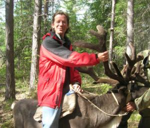
1. Caption: Mark Sorensen, Ph.D., on a reindeer. https:// humanbiologylab.web.unc.edu/mark-sorensen/

2.Interview with Dr. Mark Sorensen, 09/09/22
3.Kempton, D. R. (1996). The Republic of Sakha (Yakutia): The Evolution of Centre-Periphery Relations in the Russian Federation. Europe-Asia Studies, 48(4), 587–613. http:// www.jstor.org/stable/153137
4.The Northern Forum. Sakha Republic (Yakutia), Russia. https://www.northernforum.org/en/members/342-sakharepublic-yakutia-russia
5. Sorensen MV, Snodgrass JJ, Leonard WR, et al. Lifestyle incongruity, stress and immune function in indigenous Siberians: The health impacts of rapid social and economic change. Am J Phys Anthropol. 2009; 138(1):62-9. doi: 10.1002/ajpa.20899
6. Anthropology 318–Human Growth and Development, Introductory Powerpoint
In the United States, nearly 6.2 million adults suffer from heart failure.1 Despite how prevalent cardiovascular disease has become today, little remains known about the genetic mechanisms that contribute to it. Dr. Rau, an assistant professor in computational medicine and genetics at UNC Chapel Hill, is investigating the genetics underlying cardiovascular disease. He applies a method called systems genetics to his research, which involves linking gene expression with biological processes occurring in cells and organs to examine the pathophysiology of certain heart conditions.
Heart failure occurs when the heart is unable to pump enough blood to the body’s organs to meet their oxygen demands. It may begin when an individual is in their twenties or thirties due to poor lifestyle choices, which are detrimental to cardiac tissue. However, many people show symptoms of heart failure after the age of 65 because the heart has a high capacity to compensate for its damaged cells.2 Dr. Rau’s objective is to identify molecules, pathways, and genes that could serve as biomarkers for people to adjust their lifestyle and reduce their chance of developing heart failure later in life.
The heart contains four chambers: the right atrium, the right ventricle, the left atrium, and the left ventricle. As the heart goes through cycles, its chambers alternately contract and relax to enable blood to enter and exit. The cycles, known as cardiac cycles, consist of a diastolic phase and systolic phase. During the diastolic phase, the chambers relax as blood is filling them; during the systolic phase, the chambers contract as they are pumping blood. With age, however, the ventricles become stiff, impeding their ability to relax properly as blood fills them. As a result, the volume of blood filling the left ventricle, the chamber that pumps blood out of the heart to bodily organs, is reduced. This complication is called heart

failure with preserved ejection fraction (HFpEF), a form of diastolic dysfunction. The ejection fraction is the ratio of the volume of blood the left ventricle ejects out of the heart to the volume of blood that fills the left ventricle. A normal ejection fraction falls between 50% and 70%.3 Individuals with HFpEF may have an ejection fraction within this range; however, their left ventricle is not able to pump enough blood due to the decreased volume of blood filling it.
Figure 1. Ventricular diastole – ventricles fill with blood (left); ventricular systole – ventricles pump blood out of the heart (right). Image courtesy of LadypfHats via Wikimedia Commons.
Mitochondria supply ATP to fulfill a cell’s energy requirements. The organelle’s gene expression in heart tissue is crucial to proper cardiac function. Mitochondria can vary in number and gene expression among people based on many characteristics, some of which include gender
and lifestyle. More females tend to suffer from HFpEF and diastolic dysfunction than males. In order to examine the disparity, Dr. Rau and his research lab explored differences in mitochondrial gene expression between males and females. The lab used mice from the Hybrid Mouse Diversity Panel, a panel of about 150 distinct lines of mice, as their resource population. A technique known as adenoviral overexpression was implemented to examine the role of the mitochondrial gene ACSL6 in preserved ejection fraction. During adenoviral overexpression, ACSL6 was inserted into a nonreplicating virus. Then, the virus was injected into a cardiomyocyte and replicated using the cell’s machinery. Multiple copies of ACSL6 and its encoded protein were eventually produced, which allowed Dr. Rau and his lab to examine how overexpression of the gene impacted the action of cardiomyocytes. The lab discovered that ACSL6 enhanced cardiac function and that its expression in cardiomyocytes is lower in females than it is in males, which could explain why females tend to be at a higher
beats per minute. As a result, conclusions from mouse experiments may not always be applicable to humans.
Dr. Rau states that heart failure has a high mortality rate after it is diagnosed because it represents the end stage of a disease that has been progressing silently for decades, similar to cancer. While his goal remains to help people determine their risk of heart failure and encourage them to act early on through lifestyle changes, he mentions several treatment options too: statins, which decrease cholesterol levels, and betaadrenergic blockers, which reduce the stress on the heart by slowing it down and regularizing its rhythm. In the long run, however, medications may not be as effective in improving cardiovascular health as lifestyle changes. It is important that we lead an active, healthy lifestyle from a young age in order to lower the risk of heart failure later in life. Wouldn’t it be incredible if future generations have lower mortality rates from heart failure?
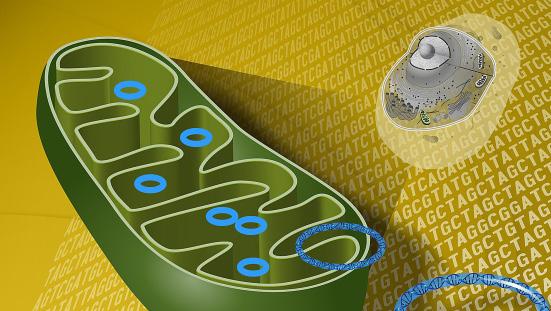
Figure 2. Mitochondria are elevated in cardiomyocytes. Image courtesy of BruceBlaus, CC BY-SA 4.0 via Wikimedia Commons Wikimedia Commons.
risk of diastolic dysfunction due to HFpEF.4
Dr. Rau has encountered numerous successes and obstacles in his research. One of his many successes includes being able to test and validate the role of fifteen novel genes in heart failure progression. This has spearheaded the development of diagnostic and screening tests for heart failure, which may be promising for people who are more prone to cardiovascular disease. An obstacle that stands in his way is recruiting more human subjects. Unlike humans, mice can be easily manipulated and researchers can readily access their heart tissue, which is why they are convenient to use. There are, however, physiological differences between mouse and human hearts. For instance, while a mouse heart beats at 600 beats per minute, a human heart beats at only 60
1. “Heart Failure.” Centers for Disease Control and Prevention, https://www.cdc.gov/heartdisease/ heart_failure.htm#:~:text=About%206.2%20million%20 adults%20in%20the%20United%20States%20have%20 heart%20failure.
2. Interview with Christoph Rau, PhD, 09/01/2022. 3. “Ejection Fraction.” Cleveland Clinic, https:// my.clevelandclinic.org/health/articles/16950-ejectionfraction.

4. Cao Y, Vergnes L, Wang YC, et al. Sex differences in heart mitochondria regulate diastolic dysfunction. Nature Communications. 2022 Jul;13(1):3850. DOI: 10.1038/s41467022-31544-5. PMID: 35787630; PMCID: PMC9253085.

Lthan expected. From the very beginning of an organism’s life, cells follow their genetic code to transform into the building blocks of everything from the skin to the heart. Along with the instructions embedded in the sequence of our DNA, certain external factors influence the way genes are expressed.1 Dr. Williams, Director of Hematopathology and associate professor at UNC’s Department of Pathology and Laboratory Medicine, focuses on their study in the field of epigenetics. His lab investigates interactions between proteins and DNA, and the resulting formation of macromolecule complexes. Specifically, he aims to understand protein structures and how they bind to the genetic code to influence its interpretation, ultimately using the findings to develop inhibitors of these protein-DNA interactions as novel therapeutics for cancer and inherited hemoglobin disorders.1
The pursuit begins with the methylation of DNA, or the addition of a methyl group (CH3) to one of its nucleic bases. The DNA molecule consists of two strands traversed by long sequences of four bases: adenine, guanine, thymine, and cytosine. To form the iconic helix, the strands link in a complementary manner, meaning adenine attaches to thymine, and guanine to cytosine. In humans, methylation occurs on the cytosine base, and only when it is directly followed by a guanine, a pattern known as a CpG. Due to DNA’s complementary nature, the CpG of one strand is mirrored by a CpG on the other strand. In a process known as symmetric methylation, both of the involved cytosines can be methylated to make a methylcyclosine molecule. Although this is a small alteration, it has a profound effect on gene expression. The reason lies in the discovery of a family of proteins known as the methylcyclosine binding domain, or MBD. One of its members, MBD2, has the ability to recognize the aforementioned molecule, bind to it, and recruit another set of proteins to form a structure known as the nucleosome remodeling and deacetylase complex (NuRD) (Fig. 1) to aid it in
When Dr. Williams collaborated with Dr. Hong Wang’s lab at NCSU, he was able to “see” this process through their “DNA tightrope” test, which involved stringing individual DNA molecules between microscopic beads to visualize movement along them. This probe showed MBD2 slide about freely on unmethylated DNA, but remain constricted when methylation was introduced2 (Fig. 2), supporting the idea that MBD2 interacts with the methylated regions to block the protein-making process that would have otherwise ensued.
Dr. David Williams, Jr.
The silencing of genes may sound alarming, but isn’t always unwelcome. As the human body develops, stem cells must be informed to develop into various cell types to later become parts of organs and carry out a plethora of functions that sustain the body. As part of the regulation process, there are methylated points in DNA that sit in front of genes coding for these functions to serve as signals for stifling some of them, allowing the sequence to take on a different interpretation. The problem arises with cancerous cells: “Cancer is when those normal signals go awry and you start to methylate areas you weren’t supposed to.”1 Looking at a cell’s genetic data, Dr. Williams is able to map the methylated points and observe that, in normal cells, CpG’s are methylated with the clear exception of CpG islands, or areas where there are several CpG’s in a row. These regions are associated with promoters, which are sections of DNA located before genes that serve as the starting point of the gene expression process. The difference with cancer is that CpG islands are also methylated, inviting MBD2 to bind3 to hundreds of previously unavailable sites and silence largely

tumor suppressor genes, whose expression is important in slowing the cancer’s proliferation. To tackle this issue, Dr. Williams and his collaborators have proposed targeting MBD2 to prevent it from recruiting the rest of NuRD and stop it from acting on the genome. A therapeutic that goes after MBD2 directly, as opposed to trying to obstruct methylation as a whole, can prove to be more precise and less toxic in its effect; and there is evidence of this approach’s possible success. Previous work in Dr. Ginder’s lab, a colleague of Dr. Williams, showed that inhibiting MBD2 reduced proliferation in the cell lines of the aggressive triple negative breast cancer.4
elevated gamma-globin expression in human cells.5 In a major leap, Dr. Williams and his collaborators were able to solve the interaction between MBD2 and the protein p66α, a structure so critical for the formation of NuRD that targeting it could inhibit MBD2-regulated gene silencing and increase gamma-globin production.1
The Williams Lab is engaged in groundbreaking work, as there are currently no drugs that act on portions of NuRD in an effort to restore the making of fetal hemoglobin. Unsurprisingly, this is a formidable task: finding a small molecule that can break apart the surfaces of two interacting proteins is difficult because of the many possible binding locations. Instead, Dr. Williams’ strategy has been to figure out smaller structures in NuRD between portions of proteins that could potentially be broken up by a drug, and he thinks his lab is getting closer. If the effort ends up succeeding, the end treatment could be significantly less toxic than other proposed remedies: although there are other factors that are known to turn on fetal hemoglobin, knocking them out comes with significant side effects1, a large setback when it comes to treating chronic conditions.
Biologically-targeted therapy is an area teeming with new discovery, and has the potential to triumph with its goal of treating disease while preserving quality of life. Today, computational tools like AlphaFold have the ability to predict protein structures from their amino acid sequences. Such technologies are invaluable hypothesis generators whose outputs have the potential to spur future investigations, or confirm the results of previous research, as they did for Dr. Williams and the MBD2/p66α structure1: “I would not have believed you if you told me I would still be studying the protein ten or fifteen years later.”1 Science never stops.
Figure 2. A kymograph shows MBD2 diffusing along methylated DNA with less range that it does along unmethylated DNA. Image courtesy of Leighton et al.

Dr. Williams is also invested in the treatment of hemoglobinopathies, or genetic disorders of hemoglobin, the protein in blood that carries oxygen. This facet of his research is closely related to his other role as a hemopathologist, a specialist focusing on diseases involving the bone marrow, such as white blood cell disorders and leukemias. For a while, professionals in the field have been trying to understand the hemoglobin “switch”. The hemoglobin molecule is made up of four protein sub-units known as globins. As a fetus, the body produces fetal hemoglobin (HbF), a form containing two alpha- and two gamma-globins. Around six months after birth, the body begins to make adult hemoglobin (HbA), containing two alphaand two beta-globins.1 The reason why the switch is key to understanding disease may be cleared up by examining sicklecell anemia, a genetic and chronic condition that is caused by a single mutation in the gene that codes for the beta-globin present in HbA. Clinical observation has shown that elevating levels of gamma-globincontaining HbF in sicklecell adults rids them of their symptoms5, so a possible treatment lies in understanding the mechanics behind the hemoglobin switch so that HbF production can be resumed. Once again, MBD2 revealed itself as a potential aim, as depleting it caused
1. Interview with David Williams, Jr., MD, PhD. 09/13/2022.
2. Leighton, G. O.; Irwin, E. M.; Kaur, P.; Liu, M.; You, C.; Bhattaram, D.; Piehler, J.; Wang, H.; Pan, H.; Williams, D. C. Densely Methylated DNA Traps Methyl-CpG Binding Domain Protein 2 but Permits Free Diffusion by Methyl-CpG Binding Domain Protein 3. 2022. https://doi.org/10.1016/j. jbc.2022.102428

3. Leighton, G.; Williams, D. C. JMB. 2020, 432, 1624-1639.
4. Ginder, G. D.; Williams, D. C. Pharmacology & Therapeutics. 2018, 184, 98-111.
5. Yu, X.; Azzo, A.; Bilinovich, S. M.; Li, X.; Dozmorov, M.; Kurita, R.; Nakamura, Y.; Williams, D. C.; Ginder, G. D. Haematologica. 2019, 104. 2361-2371.
6. David C. Williams Jr. Williams Lab. Williams Lab (unc. edu) (Accessed September 13th, 2022).
 By Renée Reeves
Each non-reproductive cell contains the entirety of an organism’s genetic code. This genetic instruction book has all the information needed to produce every protein made in your body, from motor proteins in muscle tissue to keratin that helps form hair, nails, and skin. If all cells have the DNA needed to form any structure in the body, how do different cells specialize in different things, only expressing the proteins they need to do their job? The answer to this cellular conundrum is found in transcription factors. Transcription factors are proteins that scan along the DNA looking for a specific sequence of base pairs—the smallest building blocks of the genetic code— to which they can bind. Once this sequence is found, these proteins can bind to the DNA, changing shape so that they can interact with other proteins. Through interactions with other proteins, transcription factors can increase, decrease, or turn on and off RNA production of a specific gene. RNA contains a sequence of base pairs that reflects the DNA sequence from which it came and is translated into protein in the cell. Since the amount of RNA produced in a cell is usually proportional to the amount of the protein it produces, transcription factors help control which proteins are present in which cells, creating the diversity of cellular form and function in our bodies. For transcription factors to affect cells differently, it is crucial that they be localized to the correct cells and expressed in the right amounts. If not, the wrong “pages” of the genetic code are read to a cell, and things start to get messy.

TCF4 is a gene that encodes a transcription factor normally localized to the brain, lungs, muscles, and heart. Because of TCF4’s role in regulating gene expression of hundreds of other genes starting early in development, deletions or mutations of TCF4 can lead to a host of deleterious effects and associated medical issues. Problems with TCF4 have been shown to contribute to Fuchs corneal dystrophy, autism, schizophrenia,
and Pitt-Hopkins syndrome.3 Consequently, this gene has been the subject of much research in recent years. PittHopkins Syndrome, or PTHS, is a rare neurodevelopmental disorder currently without a cure that has affects between 1:34,000 and 1:41,000 births worldwide.3 PTHS is characterized by developmental delays, cognitive and motor impairment, recurrent seizures, gastrointestinal issues, problems expressing language, and distinctive facial features.3 Research has shown that PTHS is caused by a deletion or mutation of one of an individual’s two copies of TCF4, which resides on chromosome 18, resulting in abnormally low levels of the protein.3 A promising therapeutic option for this disorder is gene therapy, which involves inserting a functional copy of the gene into select cells. If successful, the procedure would restore normal levels of the protein in cells and lessen or reverse the symptoms of PTHS in the treated individual.
One of the labs that studies PTHS is the Philpot Lab, a part of the Cell Biology and Physiology Department within the UNC School of Medicine. Dr. Vihma has been working with members of the Philpot lab as well as other researchers across the country to understand the role of TCF4 in Pitt-Hopkins in hopes of finding a cure for the disorder. Unlike many of her peers, Dr. Vihma’s path to neurodevelopmental research was anything but straightforward. As a child growing up in Estonia, she attended a rigorous music school, but developed a curiosity for science after following her friend’s mother into her lab to help refill pipette tips for some extra spending money. Early on, Dr. Vihma resolved to trade orchestras, ensembles, and a future in professional music for what truly fascinated her—chemistry, physics, and biology. Thriving in this new environment, Vihma eventually went on to university and then graduate school, earning her PhD in Gene Technology from Tallinn University of Technology. At Tallinn, Dr. Vihma studied how the activity of TCF4 is regulated in neurons. “At
Figure 3: Timeline of intracerebroventricular (ICV) injection into PTHS model mice: mice were injected at age P1, and subject to various behavioral tasks at age P60. Image courtesy of Dr. Vihma.

that time, there were already ideas in the air that Pitt-Hopkins could be cured using gene therapy,” she explained.1 Seeing that her training in TCF4 regulation perfectly positioned her to be a part of this emerging research, she accepted a postdoctoral position at the Philpot lab in 2019, almost 5,000 miles from home.
Much of Dr. Vihma’s research with the Philpot lab culminated in a study published earlier this year, which demonstrated early postnatal reinstatement of the TCF4 gene was able to partially “rescue” the PTHS disease phenotype in a mouse model—in other words, they partially restored the healthy phenotype to these mice. Prior to this study, however, Dr. Vihma and her colleagues had tried to identify the correct isoform of TCF4 to deliver to neurons. TCF4 is known to produce at least eighteen different isoforms—isoforms are proteins made from the same gene that have slightly different genetic codes due to differential cellular processing that occurs after the gene is being transcribed into RNA and before translation into protein.2 Their goal was to find an isoform that, upon delivery to neurons, would rescue the disease phenotype in PTHS mice. When they introduced the full-length isoform of TCF4 to neurons, which contained all the genetic information available in the TCF4 gene, there was no change in phenotype seen for PTHS model mice. This result led the researchers to wonder which part of their approach was incorrect: Was the wrong TCF4 isoform used? Was the level of protein introduced to neurons insufficient? Was the timing of protein introduction into neurons incorrect? This direct method of protein delivery to neurons left too many variables unknown, and this “painful failure” – as Dr. Vihma described it – prompted her and others in the Philpot lab to look for an alternative solution to reinstating TCF4 in neurons.1
The study catalyzed from these results, first authored by Dr. Hyojin (Sally) Kim, proved that gene therapy could be a viable treatment strategy for PTHS.2 This time, Dr. Vihma and her colleagues genetically engineered a mouse model of PTHS in which the levels of functional TCF4 were halved by introducing a transcriptional stop cassette (a gene construct) into the TCF4 gene. This stop cassette can be removed by an enzyme named Cre recombinase. Such a clever design allows to restore normal TCF4 levels in this PTHS model mouse without having to know which TCF4 isoform would be necessary. To model gene therapy approach in this mouse, they injected a viral vector that expresses Cre transgene neuron—specifically into the brain immediately after birth. Analyzing the brain of this mouse over the next few weeks of development showed that the normal levels of TCF4 had been
increased. To determine if insufficient TCF4 expression had been corrected early enough in development for the PTHS phenotype to be rescued in mice, the researchers then took measures of different behaviors known to present in PTHS model mice, such as anxiety-like and locomotor behaviors. They also took electroencephalograph (EEG) recordings to screen for altered patterns of neuronal activity characteristic of the PTHS phenotype and found that the phenotypic effects had been partially normalized.2
These findings helped push gene therapy further into the therapeutic spotlight as a feasible option for humans with PTHS. In a way, then, the researchers’ original experiment wasn’t a failure at all, but rather a springboard to their clever circumvention of the problem: developing a mouse model for a gene therapy study for PTHS. When asked what advice she would give to undergraduate students pursuing medical research, Dr. Vihma encouraged them “not to be afraid of rejections and failures, because that is part of science and part of life for everyone. . . If you stick with what you are studying, it may seem hard and confusing at first, but the longer you stay in the field, you start to see connections between everything you are learning, and the more you enjoy the learning process.”1
Despite the low incidence of PTHS, many researchers believe it is underdiagnosed, and the rising popularity of genetic testing will likely cause the number of worldwide cases to increase. Therapeutic options for PTHS have broad implications in neuroscience: certain aspects of PTHS such as trouble integrating sensory input and repetitive behaviors put this disorder on the autism spectrum.3 Thus, successful therapies found for PTHS could also lead to better treatments for autism-related disorders. Learning to harness the power of this transcription factor will improve the quality of life for individuals affected by PTHS and open doors to therapeutic research for many others.
1. Interview with Hanna Vihma, PhD. 9/18/22.
2. Kim H, Gao EB, Draper A, et al. Rescue of behavioral and electrophysiological phenotypes in a Pitt-Hopkins syndrome mouse model by genetic restoration of Tcf4 expression. eLife. 22;11:e72290. doi: 10.7554/eLife.72290
3. Pitt Hopkins Research Foundation. (2020, June 8). About Pitt Hopkins. Pitt Hopkins Research Foundation. Retrieved October 2, 2022, from https://pitthopkins.org/about-pitthopkins/
“When I was nine, my mom was a teacher for children with severe learning deficiencies; there was one student in particular who had full-blown fetal alcohol syndrome. We had the exact same birthday. I wondered how my life trajectory would have changed if my mom had consumed while pregnant.”1 Neonatal Abstinence Syndrome (NAS) is defined as the spectrum of clinical manifestations seen in newborns after drug exposure in the uterus before birth; it is diagnosed every 25 minutes in the United States. Primarily associated with opioid misuse and overdose, incidences of NAS have increased as one of many consequences of the opioid epidemic.

Hendrée Jones, Ph.D., is a professor in the Department of Obstetrics and Gynecology at UNC-Chapel Hill School of Medicine. She is recognized internationally in her field as an expert in the development and examination of behavioral and pharmacologic treatments for pregnant women and children in risky life situations. Dr. Jones is also the executive director of UNC Horizons, a holistic drug treatment program for pregnant parenting women and their drug-exposed children. After noticing the huge gap in the literature surrounding buprenorphine as an alternative treatment for opioid dependency during pregnancy Dr. Jones and her colleagues decided to investigate. They founded their research on the idea that buprenorphine, at the time, was known to have milder effects than methadone. Dr. Jones is a pioneer in her field, being part of the first team to look at buprenorphine as a method to treat pregnant individuals with opioid use disorder. Studies show that individuals who suffer from opioid
Image courtesy of Pixabay, CC0, via Wikimedia Commons
addiction are likely to relapse. To maintain abstinence, medications have been put on trial and researched heavily to aid in the battle against drugs. The Food and Drug Administration has approved a handful of medicines, two of which Dr. Jones has led groundbreaking research on: methadone and buprenorphine. Taking a drug-based approach to treatment may seem contradictory when considering that treatments are designed to address addiction to another drug. However, someone in recovery would benefit significantly from this approach because these medications minimize withdrawal and cravings while omitting the euphoria that the original harsher drug of choice induced.
Figure 1. Buprenorphine Molecule.

Image courtesy of Jones et al.
Methadone and buprenorphine work by eliminating withdrawal symptoms and relieving drug cravings by acting on opioid receptors in the brain, the same receptors that drugs like heroin, morphine, and opioid pain medications activate. Taking into consideration that buprenorphine was thought to have milder effects. If a patient were to abruptly stop taking buprenorphine as opposed to methadone or any harsher drug, they would have milder side effects. The team observed that when a baby is born, cutting the umbilical cord abruptly discontinues their exposure to opioid medication. They hypothesized that infants would experience milder withdrawal if exposed to buprenorphine instead of methadone. Dr. Jones was part of the first team to ever prescribe buprenorphine to three pregnant women with opioid use disorder. The study found that newborns

Figure 2. Mean Neonatal Morphine Dose, Length of Neonatal Hospital Stay, and Duration of Treatment for Neonatal Abstinence Syndrome. Image courtesy of Jones et al.
suffered no withdrawals. With this promising information, they felt ready to continue researching NAS and its propensity to arise after a mother is treated with methadone versus buprenorphine on a grander scale.
Dr. Jones and her team replicated their initial study and modified it to include a larger sample size. They screened opioid-dependent pregnant women between the ages of 18 and 41 at about 6 to 30 weeks of gestation at eight sites: six in the United States, one in Austria, and another in Canada. To limit bias surrounding the new “miracle” drug, they used a double-blinded, individualized dosing schedule for the study. Dosing for participants required numerous steps, including adjustments based on an individual’s willingness to take medication correctly, participant requests, urine toxicology results, and self-reported withdrawal symptoms or cravings. Participants were required to receive daily medications under observation in the study clinic. They consistently received seven tablets: three in the size of an 8-mg tablet and four in the size of a 2-mg tablet. Each tablet contained buprenorphine or a placebo.
Additionally, after receiving these tablets, participants received liquid containing methadone or a placebo. Trained staff evaluated NAS 10 days after the infant’s birth every 4 hours at all testing locations. An expert rater obtained NAS scores twice daily, at least 8 hours apart. An expert later provided a video of an infant undergoing NAS assessment every six months to maintain consistency in the reliability of the ratings at each site.
Results showed no significant differences between groups concerning peak NAS scores or head circumference; however, the total amount of morphine needed for treating NAS and the length of hospital stay for newborns did change.
On average, neonates exposed to buprenorphine required 89% less morphine than those exposed to methadone (mean total doses of 1.1 mg and 10.4mg) and spent 43% less time in the hospital (10.0 days vs. 17.5 days). Furthermore, neonates exposed to buprenorphine spent, on average, 58% less time in the hospital receiving medication for NAS than those exposed to methadone (4.1 days vs. 9.9 days). The study found that buprenorphine had better outcomes among the women who completed treatment. However, patients with
more severe opioid dependence were more likely to leave the buprenorphine group than the methadone group (see Figure 2). The greater satisfaction rate with methadone affirms its critical role in treating pregnant women dependent on opioids. Although there were no significant differences in the overall rate of NAS among infants exposed to buprenorphine or methadone, the benefits that buprenorphine displayed suggest that it should be a first-line treatment option in pregnancy.

Dr. Jones continues to advocate the use of buprenorphine by physicians to treat pregnant women with opioid use disorder because of its wider availability in lowincome and minority-driven communities. She strives to bring awareness to the discrimination and stigmatization of women suffering from opioid addiction who want to move towards healthier lifestyles. She urges individuals to look at the social, cultural, fiscal, and political hurdles these women must overcome to get treatment and be more open-minded to create a safer, healthier, and more inclusive society.
1. Interview with Hendrée Jones (09/17/21).
2. Jones, H. E.; Kaltenbach, K.; Heil, S. H., Stine; S. M., Coyle; M. G., Arria, A. M.; O’Grady, K. E.; Selby, P., Martin; P. R.; Fischer, G. Obstetrical & Gynecological Survey 2010, 66, 191–193. https://doi.org/10.1097/ogx.0b013e318225c419
3. NIDA. How do medications to treat opioid use disorder work? https://nida.nih.gov/publications/research-reports/ medications-to-treat-opioid-addiction/how-do-medica tions-to-treat-opioid-addiction-work (accessed September 20, 2022)
The dread of receiving a needle poke has come around again because of this year’s flu season. The flu takes a toll on us during the winter season as we see by the increase in hospitalizations and deaths associated with the infection, which is caused by the influenza virus. The influenza virus mutates all the time, so the vaccine that worked last year might not work against the new strains this year which is why we must get a flu shot every year. There is a protein in the influenza virus that docks on your cells and infects them. This protein is called hemagglutinin.2 When you get vaccinated, that is the main protein your body makes antibodies against. A typical flu shot consists of influenza virus that has been killed. When this inactivated virus is injected into the body, the immune system creates antibodies against the various proteins in the virus, like hemagglutinin, to prevent the virus from entering your cells. But what happens over the flu season is that the same hemagglutinin that was injected with the vaccines mutates in the virus and the antibodies against it no longer protect. Sometimes strains in a flu vaccines have a success rate as low as 10% because of 3 factors: inadequate predictions of viral strains in the vaccine, virus mutations over the course of the flu season, and virus mismatch due to growth in eggs.2
Dr. Kristy Ainslie, who is a distinguished professor in the Eshelman School of Pharmacy and division of Pharmacoengineering and Molecular Pharmaceutics was interviewed about the future of the flu shot. She received her bachelor of science, masters of science and PhD in chemical engineering.6 As a researcher at UNC, she is working on formulating a vaccine that offers a broader protection against many influenza viruses. When asked what her goals are for this project, she states “the ultimate goal would be to shift the influenza vaccine schedule - instead of getting the vaccine yearly, we could create a vaccine that would be needed only a handful of times, like the vaccines we receive during childhood.”5 The future of flu vaccines is to require only a handful of vaccinations, instead of yearly vaccines.
To help make a vaccine that protects for a longer time, researchers have taken data from past decades of influenza viruses and come up with a hemagglutinin protein that better protects against mutating influenza viruses.1 This process was developed by Dr. Ainslie’s collaborator, Dr. Ted

Ross at University of Georgia, and is termed COBRA: computationally optimized broadly reactive antigen. He has collected data on the influenza strains from the past 100 years and made a mutant Frankenstein’s monster like protein antigen through little changes to the protein’s peptide sequence that provides protection against those 100 years but also future years.1 When used as a vaccine, Ainslie and Ross have shown COBRA protein can provide protection against multiple influenza strains in a mouse model of infection.1-3
Ainslie, PhDWhen asked about what difficulties Dr. Ainslie thinks she will face during this research, she summarized it into three main questions: “Can you make them inexpensively? Can you apply them without a needle or administer the vaccine via the nose? Can you store them in a reasonable location?”5 For example, some of the COVID vaccines initially had to be stored in a freezer that is set at -80 degrees Celsius, which is a type of freezer is not readily accessible outside a university or hospital. Most pharmacies, like our homes, only have freezers that are set at -20 degrees Celsius or refrigerators that are set at 4 degrees Celsius. Dr. Ainslie gave the scenario of countries where the climate is hot and humid, and not as many resources for storage are not available. Ideally it would be good to create a vaccine that can withstand that climate for a duration of time, or even just being able to switch from a freezer storage to a refrigerator storage would shift the cost associated with the storage and administration of the vaccines.1 Using a polymer called acetalated dextran Dr. Ainslie has made nanoparticles that are shown in Figure 1. She has shown that using this polymer, she can protect proteins, like the COBRA antigen, when they are stored outside the freezer.4 Having storage outside a freezer can help with giving the vaccine in resource limited settings.
Vaccines can also be made less expensive by manufacturing them in a different manner. Often when

Figure 1. Electrosprayed acetalated dextran microparticles. Image courtesy of Kanthamneni et al.


vaccines are developed in a university lab like Dr. Ainslie’s, they are made in small beakers that might not scale up to the large amount needed to be given to lots of people, like what is given for the influenza vaccine. To study this, Dr. Ainslie has used a method where acetalated dextran particles are made through spraying them onto a surface. This can generate lots of particles very quickly as seen in Figure 2. Working with Dr. Ross, she has shown that they can make the particles in a larger amount very easily by adding more sprayers and that particles generated like this protect against multiple different vaccine strains.1
Most flu vaccines now are being administered intramuscularly as that gives us an immune response throughout most of our body, but does not provide much protection in the mucous membranes which is where the virus attacks first. Flumist is a live virus vaccine administered in the nose to protect against influenza. Dr. Ainslie and her colleagues are trying to make their vaccine administered in the nose, like Flumist so that it can be easily given in resource limited areas. By delivering it in the nose, a different type of immune response is generated than when vaccines are given in the muscle, allowing protective antibodies to form in the mucus. These protective antibodies can then help better clear the virus before it infects the body. To accomplish this, Dr. Ainslie has developed a mucoadhesive gel using polyethyleneimine and oxidized dextran. This hydrogel can be used to keep the COBRA protein and other vaccine components in the nose, while a protective vaccine response is generated. Dr. Ainslie and Dr. Ross has shown that this gel is protective against influenza in a mouse model of infection.3
Dr. Ainslie’s research along with her colleagues could push vaccinations to the future by allowing us to not have to worry about getting a vaccination year after year and de veloping one that can be given better in resource limited settings. She states: “Our research is focused currently on de veloping new formulation methods to reduce the number of pokes needed, make vaccines less inexpensive, have storage in more robust conditions and to be delivered intranasally - all to make it more accessible.”5
Figure 2. Homogenized acetalated dextran microparticles. Image courtesy of Kanthamneni et al.
1. Batty, C. J.; Gallovic, M. D.; Williams, J.; Ross, T. M.; Bachelder, E. M.; Ainslie, K. M., Multiplexed Electrospray Enables High Throughput Production of Cgamp Microparticles to Serve as an Adjuvant for a Broadly Acting Influenza Vaccine. Int J Pharm 2022, 622, 121839.
2. Eckshtain-Levi, M.; Batty, C. J.; Lifshits, L. M.; McCammitt, B.; Moore, K. M.; Amouzougan, E. A.; Stiepel, R. T.; Duggan, E.; Ross, T. M.; Bachelder, E. M.; Ainslie, K. M., Metal-Organic Coordination Polymer for Delivery of a Subunit Broadly Acting Influenza Vaccine. ACS Appl Mater Interfaces 2022, 14 (25), 28548-28558.
3. Varma, D. M.; Batty, C. J.; Stiepel, R. T.; GrahamGurysh, E. G.; Roque, J. A., 3rd; Pena, E. S.; Hasan Zahid, M. S.; Qiu, K.; Anselmo, A.; Hill, D. B.; Ross, T. M.; Bachelder, E. M.; Ainslie, K. M., Development of an Intranasal Gel for the Delivery of a Broadly Acting Subunit Influenza Vaccine. ACS Biomater Sci Eng 2022, 8 (4), 15731582.
4. Kanthamneni, N.; Sharma, S.; Meenach, S. A.; Billet, B.; Zhao, J. C.; Bachelder, E. M.; Ainslie, K. M., Enhanced Stability of Horseradish Peroxidase Encapsulated in Acetalated Dextran Microparticles Stored Outside Cold Chain Conditions. International Journal of Pharmaceutics 2012, 431 (1-2), 101-110.
5. Interview with Dr. Kristy Ainslie 9 September 2022
6. Kristy Ainslie, Ph.D.. (2022, July 11). Retrieved September 23, 2022, from https://pharmacy.unc.edu/directory/ ainsliek/
 By Julia Boltz
With the wide variety of chemicals and the great complexity of cancer, there are many avenues of research dedicated to testing compounds to treat cancerous cells. Occasionally, researchers discover that an already-commonplace chemical has the attributes for targeting cancer that scientists have long searched for. This is what happened in the lab of University of North Carolina at Chapel Hill’s Nobel laureate, Dr. Aziz Sancar. The Sancar lab recently discovered that the commonly used molecule called EdU, short for 5-ethynyl-2’-deoxyuridine, has previously-undiscovered properties that may make it uniquely suited to specifically target and destroy cancerous cells in the brain.
The molecule EdU has been commonly used in research since 2008 due to its properties of imitating a thymidine nucleotide analog that can be tagged. Thymidine is a part of DNA, more specifically one deoxyribose sugar and thymine (T) nucleotide. Thymine is one of the four bases that make up DNA,
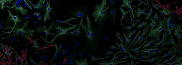
together with adenine (A), cytosine (C), and guanine (G). EdU is able to mimic thymidine in DNA and has a chemical makeup similar enough such that it binds with fluorescent molecules easily. Fluorescent molecules are those that emit light in a way that allows for the molecule to be visualized by a researcher. Due to both of these properties, scientists commonly use EdU in cells to visualize DNA replication by adding EdU to cell culture media. Subsequently this allows EdU to be incorporated in the place of the thymidine in DNA when cells are replicating, but it remains visible due to the fluorescent molecule (Figure 1). EdU has been used in various fields of research as one of the leading ways to visualize DNA in the cell, yet it has had unique properties all along that have just recently become better understood.
Dr. Aziz Sancar
Dr. Aziz Sancar has long studied
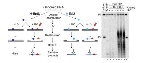
DNA damage and repair, for which he was awarded the Nobel Prize in Chemistry in 2015 for his work elucidating the mechanism of nucleotide excision repair. Recently, his lab is using the EdU molecule to continue their study of DNA repair and how the excised damaged DNA is eliminated in the cell. During this experiment, the lab noticed something was awry in their control group. The team exposed cells treated with EdU, for visualization purposes as previously mentioned, to UV light, which damages DNA, in order to study specific aspects of the DNA repair mechanism.1 However, when they compared the cells treated with EdU and were not exposed to UV light to the UV-irradiated cells, they found that both cells were undergoing nucleotide excision repair.1 Nucleotide excision repair is the process by which cells remove portions of the genome that have been damaged. The lab had expected the cells not exposed to UV light to show no DNA damage, which would allow the lab to compare these cells to those exposed to UV light and study the repair process.1 Thus, it was a complete surprise that the cells treated with EdU but not exposed to UV light still showed excision repair activity, despite having not been seemingly damaged (Figure 2).1 After this initial observation, and many additional control experiments, the lab determined

that cells recognize the molecule EdU as a form of DNA damage, thus triggering the excision repair pathway to remove it from the genome.1
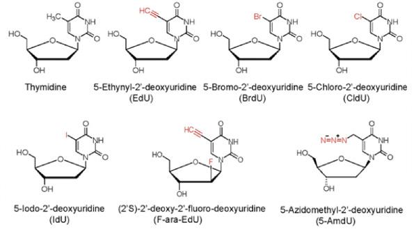
This new property of EdU is shocking to current researchers. so Dr. Sancar’s lab decided to explore other thymidine analogs besides EdU to see if they also were recognized as DNA damage by the cell.1 To their continued surprise, only EdU seemed to exhibit this property and other thymidine analogs were not processed by nucleotide excision repair.1 EdU only has minor chemical differences from other thymidine analogs, so the reason why it is uniquely recognized as damage to the genome, is not currently understood (Figure 3).1
A small stretch of DNA is removed from the genome during the process of nucleotide excision repair. The resulting gap is filled in with the same enzymes used for DNA replication. Thus when EdU is available, it is incorporated into these ‘repair patches’ generated during excision repair.1 A vicious cycle is initiated where EdU is incorporated into the genome, excised from the genome, and then re-incorporated from the genome.1 This repetitive process could ultimately lead to cell death and would explain previous observations that EdU is very toxic to replicating cells, unlike other thymidine analogs.1
Another unique property of EdU, is that it is able to pass through the blood brain barrier in the human body, a valuable property that has been
Figure 2.The chemical structure of Thymidine and Thymidine analogs that were studied, including EdU. Image courtesy of Li et al.
difficult to achieve for brain cancer drugs. Cancerous brain cells replicate rapidly, leading to the growth of tumors in the brain and potentially death. Since these cancer cells replicate so rapidly, EdU could be incorporated into the cancerous DNA, but not the DNA of nonreplicating normal brain cells, since a property of EdU is that is is incorporated into the DNA of only cells that replicate. Since cells, including cancer cells, view EdU as DNA damage, the cancer cells would excise the EdU, and thus initiate the vicious cycle that ultimately leads to cell death.1 Due to this unique ability of EdU to target replicating cells and ultimately cause death in the cell due to repeated DNA excision, Dr. Sancar states that he “believes there is great potential in EdU being used for brain cancer treatment”.1,2
Although significant research on
EdU including animal studies and clinical trials are still needed in determining the efficacy of EdU on treating cancer, Dr. Sancar is hopeful about the positive impact it could have for people. Research in EdU is moving into the next stages, with a collaboration between the Sancar Lab and a laboratory at Duke University that specializes in brain cancer, to begin studies on the effects on EdU on animals with brain cancer. EdU research in relation to treating brain cancer is still in a very early stage, but the potential of the molecule remains very promising. In conclusion, EdU serves as a reminder to not only the Sancar lab, but also the world that the potential for remarkable discovery could be closer than we ever imagined.
Figure 3. This figure depicts the pathway by which DNA treated with EdU excised a portion of their DNA containing the EdU, both when exposed to UV light and when not exposed. This is compared with DNA treated with another chemical BrdU on the left that was not interpreted as damage by the cell, meaning DNA was only excised in the BrdU cells exposed to UV light and not in those that were not. Image courtesy of Li et al.
1. Li Wang; Xuemei Cao; Yanyan Yang; Cansu Kose; Hiroaki Kawara; Laura A. Lindsey-Boltz; Christopher P. Selby; Aziz Sancar. PNAS. 2022, 119.
2. Interview with Aziz Sancar, MD, Ph.D. 09/09/2022.
 By Vina Senthil
For many patients with solid tumors, whether benign or malignant, surgical removal is often the best treatment option for cure. However, this is rarely the case for patients diagnosed with pancreatic cancer, which is both difficult to detect early and incredibly complex in nature. At a 5-year survival rate of only 11%, pancreatic cancer is among the deadliest cancers in the world.1 Of the various forms of pancreatic cancer, pancreatic ductal adenocarcinoma (PDAC) is the most common.
Dr. Jen Jen Yeh is a physician-scientist, surgical oncologist, and professor of surgery and pharmacology at the UNC-Chapel Hill School of Medicine. As a practicing surgeon, Dr. Yeh found it heartbreaking that surgery often wasn’t successful in treating PDAC patients. In fact, even if surgeons are confident after tumor removal, approximately 50% of patients will experience recurrence of the disease within just two years.1 Convinced that “surgery was never going to be enough,” Dr. Yeh began her research on PDAC. Dr. Yeh and her lab, located in the UNC Lineberger Comprehensive Cancer Center, have focused their research on analyzing various PDAC subtypes and identifying viable subtype-specific treatments.1 They aspire to eventually translate findings in the lab to clinical trials.

PDAC is exceptionally challenging to study, and Dr. Yeh attributes this to the scattered distribution of tumor cells within the tumor and the presence of the tumor microenvironment. The tumor microenvironment encompasses the “normal cells, molecules, and blood vessels that surround and feed a tumor cell.”1 Dr. Yeh visualizes the tumor microenvironment as “dense concrete encasing
the tumor cells.”1 Initially, scientists believed that the tumor microenvironment served as a barrier to protect tumor cells from eradication. Preclinical studies in the 1990s discovered that this “barrier” could be relaxed, and tumor cells could be successfully targeted this way. However, the clinical trials showed no improvement in patients’ condition, and in some cases, patients did worse than standard of care.

In the 2000s, studies found that if the “barrier” did not
Figure 1. PDAC tumor cells in a PDAC tumor are enclosed within the tumor microenvironment. Image courtesy of Dr. Yeh.
exist, then tumor cells grew rapidly. These findings resulted in uncertainty regarding the relationship between the tumor microenvironment and the tumor cells that it surrounds. It was unclear whether the tumor microenvironment was enabling tumor cells to persist, despite treatment, or whether it was actively preventing tumor cells from spreading throughout the body. Dr. Yeh explains that this “dichotomy” between using drugs to weaken the tumor microenvironment and maintaining its security in order to keep tumor cells from metastasizing is one that her research team, along with other researchers studying PDAC, must consider when investigating possible treatments.1
Accordingly, Dr. Yeh and her team’s research focused on assessing the role of the tumor microenvironment. In 2015, she published a research article that detailed a step forward in this effort. Dr. Richard Moffitt, a postdoctoral fellow of Dr. Yeh at the time, had figured out a computational method to observe RNA expression of the tumors and understand which signals come from the tumor as opposed to the tumor microenvironment.3 Dr. Moffitt then discovered two subtypes of tumor that behave differently--the basal and classical subtypes of tumors. PDAC patients found to have the basal subtype exhibited more aggressive tumors, while patients found to have the classical subtype experienced less aggressive tumors.
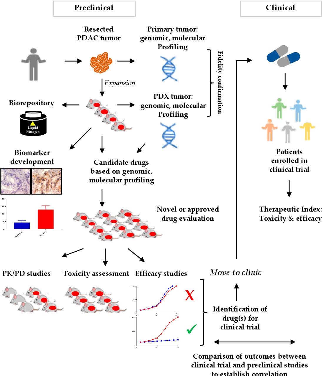
Figure 2. Patient-derived xenograft models are useful for studying PDAC and translating research to clinical trials. They involve the implantation of cancerous human tissue into lab mice. Because mice and humans are biologically similar, this allows researchers to better understand effects of treatments on humans.
fluorouracil, leucovorin, irinotecan, and oxaliplatin.2 Another treatment provided is a combination of two other drugs: nab-paclitaxel and gemcitabine. Clinical studies have shown that patients with the basal subtype of tumors may respond better to nab-paclitaxel and gemcitabine, though the response is not significant.
Exploring treatments for the basal subtype of PDAC is the next step for Dr. Yeh and her research team. Dr. Yeh hopes to investigate what the “Achilles heel of the tumor microenvironment” could be, and how the tumor and the tumor microenvironment interact with each other.1 There is evidence in the scientific literature that points to a scenario where while tumor cells are responsive to treatments or therapies, the tumor microenvironment may not be, and Dr. Yeh predicts that understanding the connection between the tumors and the tumor microenvironment might “change the whole story”.1
Currently, Dr. Yeh and her team are examining whether treatments can become more personalized to patients, depending on the subtype of tumors they have. However, in order to “put patients on a better path to treatment,” they must be able to translate their research findings into a clinical setting, meaning that a physician should be able to order these subtypespecific treatments, and lab testing must be CLIA-approved (approved by the Clinical Laboratory Improvement Amendments of 1988).1 Currently, the Medical College of Wisconsin is running a clinical trial, PurIST Classification-Guided Adaptive Neoadjuvant Chemotherapy by RNA Expression Profiling of EUS Samples Study (PANCREAS), to test this treatment approach.4 During the PANCREAS trial, patients will undergo a biopsy called Endoscopic Ultrasound-Guided Fine Needle Aspiration (EUS/ FNA), the results of which would be sent to the Medical College of Wisconsin.4 From these results, a specific treatment will be chosen for the patient. This process allows patients to avoid the negative side effects that come with some of the first line therapies that would not have been effective.
As a part of the PANCREAS trial, a PDAC patient may be tested at the UNC Hospitals, and if it is discovered that they have tumors characteristic of the classical subtype, they will receive one of the first-line treatments for PDAC, FOLFIRINOX. FOLFIRINOX is a combination of four old chemotherapy drugs:
But numerous obstacles lie ahead. In order to obtain accurate results about how a possible treatment will perform during clinical trials, it is important to use an accurate patient-derived model. Some engineers are attempting to create models that “mimic tissue” in order to address this need.1 An additional need is being able to detect recurrences of PDAC cases earlier. As the field progresses, imaging technologies that will allow for earlier detection and intervention are promising.
Despite PDAC’s poor prognosis for patients, Dr. Yeh remains optimistic. Due to better perioperative care and surgery techniques, more refined chemotherapy regimens, and more advanced technologies, outcomes for patients have greatly improved over the past decades. “Once a core number of discoveries have been made, progress in finding treatments accelerates,” says Dr. Yeh.1
1.Interview with Jen Jen Yeh, M.D. 09/12/22.
2.“FOLFIRINOX - NCI,” pdqDrugInfoSummary, 28 Nov. 2011, online, Internet, 22 Sep. 2022. , Available: https://www. cancer.gov/about-cancer/treatment/drugs/folfirinox.
3. Moffitt, Richard, et al. “Virtual microdissection identifies distinct tumor- and stroma-specific subtypes of pancreatic ductal adenocarcinoma.” Nat Genet. 2015 Oct;47(10):116878. doi: 10.1038/ng.3398. Epub 2015 Sep 7. PMID: 26343385; PMCID: PMC4912058.
4. Tsai S. “PurIST Classification-Guided Adaptive Neoadjuvant Chemotherapy by RNA Expression Profiling of EUS Aspiration Samples.” clinicaltrials.gov; 2022 [accessed 2022 Oct 12]. Report No.: NCT04683315. https://clinicaltrials. gov/ct2/show/NCT04683315
Photosynthesis, or the creation of carbohydrates from carbon dioxide and water, is one of the most wellknown chemical reactions. Behind this conversion from light energy to chemical energy is a fascinating chemical process called photoredox catalysis, where light rays excite organic molecules, facilitating the transfer of electrons.1 This fundamental, life-sustaining reaction is not only the fuel of plant life, but is also the fuel to incredible scientific discoveries at UNC Chapel Hill.
Inspired by photosynthesis, Dr. David Nicewicz and his colleagues began to wonder how photoredox catalysis could be used to make new reactions with simple chemistry, a task that would help the in the creation of medicines. “Let’s say you’re in the pharmaceutical industry and you have a promising molecular structure… oftentimes you have to start your synthesis from scratch, synthesizing a series of molecules to test your hypothesis”1 says Dr. Nicewicz, while contemplating one of the principle struggles of chemistry: the limitations of molecule manipulation due to each molecule’s unique, intrinsic properties that govern their capabilities to react under varying conditions. This problem has tasked organic chemists with a mission: to take simple molecules and design new, streamlined reactions that allow for functionalization that would have naturally been unfavorable.

An example of an unfavorable reaction is the addition of a fluoride to a molecular structure called an aromatic ring. Fluorine is an element that is challenging to use because it does not tend
to donate electrons, is slightly basic, and often makes hydrogen bonds. Nicewicz and his team handle a difficult, unstable isotope of fluorine, 18F, and they grapple with the speed of the isotope’s decay, as it takes a lengthy 120 minutes for it to decay to half of its original concentration.2 On the other hand, aromatic rings are systems of remarkably stable carbon bonds, making them resistant to additions of new atoms. Simple chemical processes inducing the combination of these two groups is necessary, especially so that less specialized technicians could perform the reaction and the dangerous, toxic intermediate stages of a longer synthesis process could be minimized or eliminated.
In collaboration with Professor Zibo Li of UNC’s Radiology department, Dr. Nicewicz experimentally determined the

Figure 1. Catalytic C-H to C-18F reaction. Image courtesy of Dr. Nicewicz.

appropriate reagents and processes for the addition of the fluorine isotope to an aromatic ring. The team, with the help of CHEM 262 Lab undergraduate students at UNC, created a multitude of various dyes with the potential to act as catalysts in these new reactions. When these special dyes absorb a photon of blue light, they achieve an excited state and will either accept or give up an electron to drive a reaction. The assistance of these catalysts and the appropriate laboratory conditions have shortened the reaction kinetics to only a 30-minute reaction time and the fluorine isotopes were successfully added to the molecules.2
The creation of a simplified reaction to allow for 18F to bind to an aromatic ring has allowed for significant pharmaceutical benefits because of a functional imaging technique called Positron Emission Tomography (PET). When 18F emits a positively charged electron, called a positron, and encounters an electron, the two annihilate and emit a gamma ray. These gamma rays are detected using magnetic resonance imaging (MRI) in real time, allowing scientists to determine the exact location of a drug in a body’s system.
Dr. Nicewicz and his team found that the scope of their research was much wider than anticipated and had additional implications in human health. Hyperparathyroidism is a condition that causes the enlargement of parathyroid glands in the neck. The treatment for this condition is to remove the glands to avoid thyroid cancer. Unfortunately, in a third of cases, it is hard to identify the location of the cancer with a CT scan, causing the need for multiple surgeries. However, with the addition of 18F to an aromatic-ring containing medication, the enlarged parathyroid glands light up on the imaging scans, allowing for specialists to make precise measurements for the incisions they make in surgery to remove the gland.1 In another practical example, a derivative of the amino acid tyrosine with a fluorine
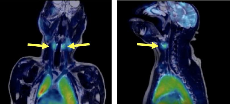
addition can act selectively as a probe for the identification of brain/spinal cord cancer and breast cancer. Dr. Nicewicz and Professor Zibo Li have also been able to create a company, LED Radiofluidics, which makes their research available to hospitals and research facilities, who can now link it their own equipment to create specially designed molecules themselves.1
Many scientific pursuits are limited by the constraints of chemical processes. Oftentimes the organic molecules desired to further research are very difficult or impossible to create. In the future, Dr. Nicewicz hopes for the development of new reactions that will simplify the synthesis of these molecules, making the goals of other fields of science easier to attain. “These molecules allow for chemical reactions to happen that couldn’t happen in any other way unless you start with different materials. They allow for us to streamline these processes so that we can really have an impact on patient health,” says Dr. Nicewicz. “It’s taken our science to something I never would’ve envisioned happening, [a] direct impact on biology, and it’s pretty cool.”1
Figure 2.
References:
1. Interview with David Nicewicz, Ph.D. 9/19/22
2. Direct arene C–H fluorination with 18F− via organic photoredox catalysis https://www.science.org/doi/ abs/10.1126/science.aav7019?cookieSet=1 (accessed September 19th, 2022)
3. New Positron-annihilation Techniques for Materials Research https://www.sciencedirect.com/topics/chemistry/ positron (accessed October 3, 2022)
“In the future, Dr. Nicewicz hopes for the development of new reactions that will simplify the synthesis of these molecules, making the goals of other fields of science easier to attain.”
The most powerful type of battery may be coming to your phone in just a few years. For the past 30-35 years lithium-ion batteries have dominated the markets and can be found in nearly everything from your car to your phone.3 Being surrounded by these batteries begs the question if these are indeed the best type. Lithium-ion batteries work through the movement of lithium ions from the cathode (the positive end) of the battery to the anode (the negative end), and is conducted in a liquid phase that allows the transfer of lithium ions between the electrodes (cathode and anode).1
A better alternative may be the fluoride-ion battery. Though they work in a similar fashion, fluoride may yield a larger energy output due to fluoride having a higher electronegativity than lithium. Electronegativity is the tendency of an atom to take an electron, which would allow for the atom to have a full octet of 8 electrons, making it stable. Due to fluoride’s higher electronegativity, the ion would move from the anode to the cathode and discharge more energy as it moves from the highly unstable form in the anode to the highly stable form in the cathode.1

Dr. Scott C. Warren, Ph.D., and his lab group have been working on making Fluoride-ion batteries a reality. The main goal behind this project is to develop a less expensive and more sustainable battery that would have higher performance than lithium-ion batteries. Currently, the group is working on developing potential electrodes that would allow for the fluoride ion to move to the cathode, creating a large discharge in energy.1
 By Amil Agarwal
The criteria for these new electrodes haven’t yet been thoroughly researched, so Dr. Warren and his lab searched for compounds containing fluoride along with having other broad characteristics until they acquired a list of about 10,000 compounds.3 In order to find potential electrodes, two strategies were devised. The more optimal of the two, the Dynamic, Decoupled, and Iterative strategy (DDI strategy), resulted in calculations being conducted approximately 100-times faster than previous calculations without a significant loss in accuracy. These calculations revealed how each material would perform in a fluoride-Ion battery.3
As part of their study, a group of 300 materials were chosen at random to calculate their performance using accurate but slow methods. Then, for comparison, Dr. Warren and his lab repeated these calculations using the DDI method. They were able to compute all the calculations for each material candidate within hours, compared to 2-3 months for the conventional method.1
By the end of the DDI process, a handful of potential electrides (ionic compounds in which an electron acts as the anion) were determined.
Figure 2. Diagram representing the difference between the Hierarchal and DDI strategy used to determine potential electrides for Fluoride-ion Batteries. Figure courtesy of Sundberg et al.
These electrides held characteristics that allowed them to theoretically produce the high energy output sought after. Some of the characteristics included that the metal atoms that form the coordination sphere around the fluorine ion should be highly electropositive.3 Some of the metals determined to be the best fit for the anode electrode were the lanthanide series along with yttrium and scandium.2 This is due to the large positive charge on these ions allowing the negatively charged fluoride ion to be highly stable when in the cathode. Additionally, through the DDI system, metallic materials of the form M1M2F6 were discovered that could potentially become electrolytes for the fluoride-ion battery.3
One of the setbacks Dr. Warren and his group faced was the large oxygen content in the scandium carbide (Sc2C), in which a layered state is the most optimal structure.2 The reason why oxygen is a large factor is that even 0.5 atomic percent can affect the structure largely by changing it into a cubic shape, which would result in a lower energy output than intended.2 To combat this, Dr. Warren and his lab used both an inert gas environment along with a titanium sponge in order to remove any oxygen present in the scandium carbide. While this did work to some extent, a pure layered state would have been the most effective.2
Based on these materials and the high energy output the battery could produce, a techno-economic analysis was conducted and showed that if the right materials were chosen and an optimal battery was created, this would increase the energy output by 4-10 times that of a lithium-ion battery.1 This would allow for the fluoride-ion battery to be applied anywhere and everywhere due to it having a lower cost with a higher output. In addition to this, when Dr. Warren researched the capability of Sc2CF2 as an electrode with scandium carbide (Sc2C) being the electride, he found that the scandium carbide acted as a semiconductor, an unforeseen beneficial characteristic of the material.2 This prompted a large interest in the material and is being heavily researched along with its uses in photodetectors and as part of light-absorption technology.1
Dr. Warren has largely contributed to the growing field of fluoride-ion batteries which held its first international conference on fluoride-ion batteries in the summer of 2022 in Germany.1 Dr. Warren says that the next few steps will be key to deciding the future of fluoride-ion batteries and how fast they can be implemented in the main markets. The biggest issue is to find a liquid or solid electrolyte which would allow for the fluorine ions to move across between the cathode and the anode.1 A liquid electrolyte is currently used in lithiumion batteries and allows for the electrodes to grow and shrink as they are charged and discharged. However, the solid electrolytes would allow for better mechanical robustness and would lead to fewer chemical reactions between the battery and its surroundings.1 This past summer, Dr. Warren worked in conjunction with the University of Stuttgart to research a potential solid electrolyte and had promising results which would only further advance the field as a whole.1 As Dr. Warren and his lab research more potential electrides and possible electrolytes, it is only a matter of time before the fluoride-ion battery is widely incorporated into devices and appliances around you and becomes as ubiquitous as the lithium-ion batteries of today.
1. Sundberg, J.D., Druffel, D.L., McRae, L.M. et al. Highthroughput discovery of fluoride-ion conductors via a decoupled, dynamic, and iterative (DDI) framework. NPJ Comput. Mater. 8, 106 (2022). https://doi.org/10.1038/ s41524-022-00786-8.
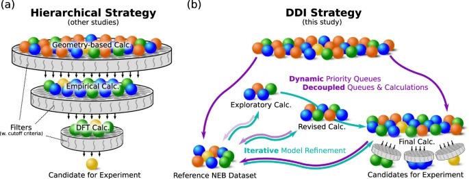
2. Interview with Dr. Scott C. Warren, PhD., 09/21/2022.
3. Lauren M. McRae, Rebecca C. Radomsky, Jacob T. Pawlik, Daniel L. Druffel, Jack D. Sundberg, Matthew G. Lanetti, Carrie L. Donley, Kelly L. White, and Scott C. Warren Journal of the American Chemical Society 2022, 144 (24), 10862-10869, DOI: 10.1021/ jacs.2c03024.

 By J.R. Cobb
The energy crisis unifies many of the world’s most pressing issues, from food to sustainable energy to global warming and more. They have always been intertwined: a lack of sustainable energy sources means creating more fossil fuels, which releases more greenhouse gases, which hinders crop yields. This interdependency clouds a solution. In the Department of Chemistry at the University of North Carolina at Chapel Hill, though, Dr. Alexander Miller tackles this monstrous set of problems through catalysis. He leads a research group in the Center for Hybrid Approaches in Solar Energy to Liquid Fuels, known as CHASE. Supported by a $40 million grant from the U.S. Department of Energy, CHASE spans six research institutions and is headquartered in Chapel Hill.1
Dr. Miller has long seen the connection between catalysts, chemistry, and energy. As an undergraduate at the University of Chicago, he worked in a research group studying “unusual bonding motifs in chemistry.”1 Essentially, they attempted to produce molecules that were thought to be impossible. Transitioning to graduate school at the California Institute of Technology, he sought to investigate linkages between chemistry and energy, and their applications to the global environment.
Catalysts are special types of molecules which speed up chemical reactions without being consumed, making non-naturally occurring reactions possible.
Figure 1. Chemical structures of ethanol and methanol, two fuels whose solar-driven production is being investigated at CHASE. Figure courtesy of J.R. Cobb.
They can be metals, proteins, or carbon-containing compounds, to name a few. Catalysts already play a major role in protecting the environment. The catalytic converter, for example, uses metals including platinum and palladium to convert carbon monoxide and nitrogen oxides found in automobile exhaust into carbon dioxide and nitrogen.1 Simply put, it converts very potent greenhouse gases into less potent ones. The catalytic converter is just one of myriad applications of catalysis to the real world.
At CHASE, Dr. Miller is curious to know how one would use sunlight to make gasoline alternatives. Dr. Miller himself said that answering this question “can be overwhelming at times,” since “the scale of fuels is daunting.”1 However, his group breaks the enormity of the energy crisis into more manageable pieces. First, he says, they select a problem to investigate— currently, that is the DOE’s sunlight-to-fuel challenge. Then, they design catalysts that may help solve that problem. They do this “at an exquisite level of detail,” using visual tools such as x-ray diffraction to understand their new reactions at the most elementary level.1 Their process is iterative, calling for the creation of a new catalyst, intense analysis of what made that catalyst work (or, as he notes, often fail), and then adjusting. Building off the last cycle allows the catalysts to improve year after year, building both a deep intrinsic understanding of
Figure 2. Visualization of Dr. Miller’s research catalyst, at the intersection of photoelectrocatalysis and Nickel-based catalysts. Image courtesy of Stratakes et al.
the chemistry, as well as a stairway towards a final solution. Dr. Miller’s work is therefore fundamental, as he and his group are creating new chemistry rather than engineering existing technologies up to a commercial scale.
Dr. Miller explains that the “solar” in CHASE’s name alludes to the hub’s new approach to solar energy.1 Today’s solar power is almost entirely photovoltaic, running energy captured from sunlight through an electronic circuit in, say, a solar panel. An inherent flaw with this system is that when the sun goes out, so does the power.1 This is perhaps the greatest obstacle to solar power’s integration into energy infrastructure. CHASE attempts to solve this by using sunlightdriven chemical reactions to produce synthetic fuels. An example of this is the conversion of CO2—an abundant greenhouse gas—into useful liquid fuels such as the racecar fuel methanol and the gasoline additive ethanol.1
This new approach to solar power grants engineers an energy storage solution, as large-scale batteries to store energy from photovoltaics have yet to prove economical or efficient. Liquid fuels store their own energy in their chemical bonds and are easily integrated into existing energy infrastructure. Importantly, chemical energy stored in those fuels doesn’t disappear when the sun sets, meaning CHASE’s research can help solve the photovoltaic cell’s fatal flaw.1
Focusing on the conversion of CO2 is important: if this process can be used to generate synthetic fuels, then those fuels will be re-burned and will re-release the CO2, yielding a closed cycle.1 When compared to the continuous release of CO2 into the atmosphere seen now, the closed cycle is much more sustainable. Currently, there is a continuous cycle of CO2 emission, beginning with fossil fuel extraction, and ending with combustion of liquid fuels, producing a net positive output of carbon dioxide; that is, more is released than consumed.1 Dr. Miller notes that a closed loop is more sustainable since the same CO2 is released by liquid fuel combustion that is then consumed again by making more liquid fuels. The result is a system that can theoretically be carbon-neutral, aiding in the fight against climate change.1
The group’s research has led them to substantial pieces of novel chemistry. More recently, they synthesized a catalyst which holds promise for solar energy production. Bis(diphosphine)nickel hydrides serve as photoelectrocatalysts
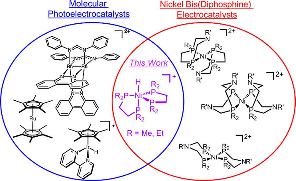
Figure 3. Visualization of sunlight-driven mechanism of catalyst action. Image courtesy of Stratakes et al.
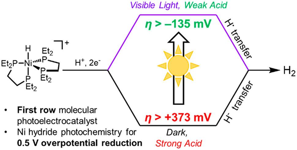
This special type of catalyst uses sunlight to perform its catalytic work of speeding up chemical reactions.2 Dr. Miller’s catalyst of interest is the first instance of a first-row transition metal—abundant and affordable metals in the middle of the periodic table—being used as a photoelectrocatalyst .
These types of catalysts usually rely on expensive, rare elements such as ruthenium and iridium, so the development of an economical alternative is significant.2 Moreover, this catalyst does not require the use of expensive semiconductor materials or “sacrificial chemical reductants.”2 Yet, it produces the desired products at rates rivaling its more expensive counterparts, giving it an edge.2 As more researchers investigate catalysts like this one, more pathways towards economical and sustainable solar fuels will open.
Despite his focus on the cyclic process of catalysis, Dr. Miller notes that in his work at CHASE, there is a sentiment of urgency, saying “we gotta get this done!”1 With climate change and the consequences of fossil fuels making headlines constantly, this does not come as a surprise. However, Dr. Miller’s iterative process with a focus on granular detail may help scientists open new doors to sustainable energy production, all through the power of the sun. The energy crisis unifies many of the world’s pressing issues, so helping to unravel it may help untangle a web of other issues as well.
1. Interview with Dr. Alexander J.M. Miller, 12 September 2022.
2. Stratakes, B.M.; Wells, K.A.; Kurtz, D.A.; Castellano, F.N.; and Miller, A.J.M. J Am Chem Soc. 2021 143 (50), 2138821401
 By Venkata Mantri
Image by TheumasNL, CC BY-SA 4.0 via Wikimedia Commons
Machine learning (ML) and artificial intelligence (AI) have received tremendous attention in the last few years. Nowadays, complex topics like AI are known to most people, and this is for good reason. Even outside the movies, progress in AI and ML research is growing at an explosive rate, with significant advancements every year. In the last 5 years, there have been many advancements being made in almost all major sub-areas of AI.1 However, while many assume that these advancements stay within the realm of computer science or software, in reality this progress extends to multiple other disciplines.2
AI is a broad term that generally refers to machines displaying features of human intelligence and behavior. While ML is a subsection of AI that involves making a machine that can learn from a set of data and draw its own connections, rather than having to be hard-coded by a person. Hard-coding a software to recognize a dog from a cat would require numerous instructions, from fur to the overall structure of the face, but using the proper machine learning system (MLS), one could create a program that would simply need to be trained by a large set of pictures (the training set) indicating whether they are a dog or a cat and the MLS would learn from this. This makes it so that a machine can understand a complex task without countless lines of code and numerous revisions. Within ML, multiple areas of research are trying to make it more efficient and applicable. One major area is uncertainty, which is where UNC researcher Dr. Junier Oliva primarily operates.
In MLSs, uncertainty and incomplete data can lead MLSs to make incorrect predictions. Dr. Oliva and his lab deal with “the general development of better machine learning methods”3 by
incorporating and using uncertainty present in the MLSs. They do this by using distribution and recognition within an MLS, instead of the typical averaging or regression. An example Dr. Oliva gave was for trying to figure out the heart rate of a specific patient. Certain patients may have only a high or low heart rate without a middle heart rate, but if a MLS was to be trained on this data without incorporating uncertainty the MLS would likely average the varied data and give very inaccurate predictions, since many patients won’t be around the calculated average. However, if the MLS could understand the uncertainty of the data, it could give a distribution as an output and report which heart rates would be most likely for patients. This offers a richer and superior depiction of what the heart rate would be.3 Additionally, uncertainty could mean lacking data. If an MLS was able to indicate that it lacks the data/expertise for a certain
Figure 1. Illustration of an ML algorithm called a neural network. The inputs are called the parameters and are provided from a training set. The learning for this model happens in the hidden layer between the input and the output. Image courtesy of Dr. Oliva.
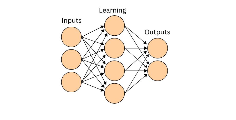
input, instead of simply outputting an inaccurate prediction, it would prevent time waste and inaccuracies. With the integration of uncertainty, ML will only become more powerful and effective for solving the problems present in A.I and computer science as a whole, but the scope isn’t so limited.4
ML can stretch far beyond computer science to places like biology and chemistry, since ML has a great advantage in handling/interpreting large amounts of data.5 When talking about his recent research involving both ML and biology, Dr. Oliva talked about how it would be hard for people to make sense of 200,000 cells and all their characteristics. However, a MLS would have a much easier time parsing through all the data, and showing a sample of 50 or 500 cells which are representative of the entire sample, and this doesn’t even consider the option of visualizing what connections the MLS made to try and find novel relationships within cell-biology.6
Dr. Oliva has an aim of creating what he calls the “machine detective” which is “a system that is autonomously able to gather new information to allow it to make better predictions on the fly” with the goal being to make ML “systems that know what they don’t know”.3 Simply put, it would be a MLS that can tell if a MLS is fit to make a prediction and do a cost-benefit analysis on upgrades that could be made. If the MLS doesn’t seem to have the necessary information for a specific case, the machine detective can recognize this and say the program might not predict the best for that instance, while also assessing the costs and benefits of upgrading the system whether it is with more data, better functionality, or other upgrades. If this ML system was to be made, it would be extremely beneficial to computer science and multiple other disciplines.
Figure 2. Figure illustrating how a large sample of cells can be represented in smaller sets, then interpreted for useful data. Image courtesy of Baskaran et al.

When they did a similar project, Dr. Oliva and his team were determined to create the MLS in a way that took uncertainty into account, and incorporated the distribution within the entire sample into the cells that it provided instead of simply averaging them. This way, the entire sample is accurately represented. This ability to take in immense amounts of data, sort it, learn from it, and then represent it to humans is something that is desperately needed within molecular-biology and chemistry. Using MLSs to test drug/compound feasibility in molecules would be a huge boon since large amounts of time and resources can be dedicated to having a drug be developed, properly tested, move onto clinical trials, etc. Therefore, it would be very efficient to have an MLS that could simulate or model how a molecule behaves and filter out which molecules are worth investigating further. This way, resources can be allocated much more efficiently, allowing us to find strong molecular candidates sooner.
The potential benefits of ML to every field will only increase as it develops, which happens because of people like Dr. Oliva and his lab. Currently,

1. Michael L. Littman, Ifeoma Ajunwa, Guy Berger, Craig Boutilier, Morgan Currie, Finale Doshi-Velez, Gillian Hadfield, Michael C. Horowitz, Charles Isbell, Hiroaki Kitano, Karen Levy, Terah Lyons, Melanie Mitchell, Julie Shah, Steven Sloman, Shannon Vallor, and Toby Walsh. “Gathering Strength, Gathering Storms: The One Hundred Year Study on Artificial Intelligence (AI100) 2021 Study Panel Report.” Stanford University, Stanford, CA, September 2021. Doc: http://ai100.stanford.edu/2021-report. Accessed: September 16, 2021.
2. Pugliese, R., Regondi, S., & Marini, R. (2021). Machine learning-based approach: Global Trends, Research Directions, and regulatory standpoints. Data Science and Management, 4, 19–29. https://doi.org/10.1016/j. dsm.2021.12.002
3. Interview with Dr. Junier Oliva
4. Webb, S. (2018). Deep Learning for Biology. Nature, 554(7693), 555–557. https://doi.org/10.1038/d41586-01802174-z
5. Ching, T., Himmelstein, D. S., Beaulieu-Jones, B. K., Kalinin, A. A., Do, B. T., Way, G. P., Ferrero, E., Agapow, P.-M., Zietz, M., Hoffman, M. M., Xie, W., Rosen, G. L., Lengerich, B. J., Israeli, J., Lanchantin, J., Woloszynek, S., Carpenter, A. E., Shrikumar, A., Xu, J., … Greene, C. S. (2018). Opportunities and obstacles for deep learning in biology and medicine. Journal of The Royal Society Interface, 15(141), 20170387. https://doi.org/10.1098/rsif.2017.0387
6. Communications, B. (2021, March). Connecting Biology with machine learning. Drug Discovery News. Retrieved September 30, 2022, from https://www.drugdiscoverynews. com/connecting-biology-with-machine-learning-15114
 By Rahul Mehta
Autism Spectrum Disorders (ASDs) are among the most prevalent developmental disorders present in humans, affecting about 5.5 million adults and one in forty-four children in the United States.¹ Though it has been established that autism occurs on a spectrum, with symptoms present from a young age, a single treatment remains elusive given the diverse phenotypic presentation of disease. Supplementing this issue is the disease’s pervasive nature and its efficacy in obstruction of student learning, which can potentially lend merit to pseudoscientific techniques that may be implemented within families attempting to provide educational remedies for their affected students.
Pseudoscientific practices are formed as programs that appear to give credibility to unproven hypotheses by means of presenting them in a manner that appears scientific. Unverified reports and anecdotes supplementing unfounded data analyses are usually the foundation of such methods. We can examine the Rapid Prompting Method (RPM) as an example of popular pseudoscientific intervention in the case of Autism therapy techniques. Created by the mother of ASD afflicted student, Soma Mukhopadhya, much of the intervention’s success can be attributed to anecdotal evidence. The technique follows a “teach ask” protocol, prompting a facilitator to present students with a concept (for
example, “the motor needs to rotate one time for the robot to move”), then immediately asking a question (“how many times does the motor have to rotate for the robot to move?”). Since its genesis, RPM has been proven to rely too greatly on the facilitator— the mediator of discussion subconsciously prompts ideas from the person from the person with communication difficulty, restricting freedom of thought.² Despite this, mother of thirteen-year-old Mike Keller claims that the treatment was a breakthrough for her nonverbal son, allowing communication by means of keyboard when following the teach-ask protocol.² Such subjective experiences have progressed pseudoscientific techniques in a field where the promise of universal treatment therapy dwindles.
Figure 1. Breakdown of mean costs of a typical CTM during intervention, and postintervention periods. Figure courtesy of Cidav et al.
Through cases such as that of Mike Keller, we can better understand the confusion for families when choosing among thousands of intervention techniques for ASD learning therapy. It may be asserted that the rise of pseudoscience in treatment is reasonable. Distinguished Professor at the UNC School of Education, Dr. Brian Boyd, argues that the issue lies in communication between researchers and families in “ensuring disseminated research is made accessible.”³ Among various evidence-based treatments -- empirically proven procedures that improve single fundamental behaviors -- Boyd asserted a high percentage of backing among a group known as Comprehensive Treatment Models (CTM). A CTM can be defined as a program focused on progressing multiple developmental areas such as social skills, adaptation, and cognitive

Figure 2. Breakdown of experimental prodecure in UNITED testing: Experimental group (UNITED) vs Control (IAU). Figure courtesy of Locke et al.
functioning through multiple evidence-based interventions from a young age. The foundation for such intervention lies in applied behavior analysis (ABA) treatment which costs upwards of $30,000 in the United States.⁴ Thus, in conjunction with colleagues, Boyd is attempting to improve the accessibility of CTMs among other effective ASD learning interventions across the field of education.
Currently, Dr. Boyd is a William C. Friday Distinguished Professor in Education at UNC Chapel Hill and Vice President of the International Society for Autism Research. Dr. Boyd began his journey into special-needs education at The College of William and Mary where he worked as a counselor at Camp Royal, a division of North Carolina’s Autism society that provides an accepting atmosphere for individuals affected by an ASD. During his time working as a camp counselor, he recalls a personally influential experience with the mother of a young ASD student who recounted the child’s first words — “some approximation of ‘pentagon’,” Boyd recalled.² Touched by such experiences, Dr. Boyd became involved in the development of novel evidence-based interventions at the preschool level. After earning a Ph.D. in special education at the University of Florida, he developed a focus on marginalized populations and students of color. In promoting accessibility among evidence-based CTMs and other interventions with empirical basis to these targeted demographics, Boyd and other colleagues have been testing a project called Using Novel Implementation Tools for Evidence-based Intervention Delay, or UNITED.
UNITED involves the simultaneous implementation of three evidence-based learning interventions for autism students, with the goal of “increasing rapid uptake of effective practices to reach vulnerable demographics.”⁵ These treatments include marginalized family navigation intervention, Mind the Gap (MTG); social engagement intervention, Remaking Recess (RR); and adolescent selfdetermination intervention, Self-Determined Learning Model of Instruction (SDMLI). The theoretical framework of UNITED is based upon social and organizational theories, which means ensuring that those involved grasp the systematic process of identifying team members proficient at particular tasks and setting clearly defined roles among these team

members. Such foundations will assist the development of this multifaceted implementation strategy in low-resource settings. Given the demand and complexity of resources required in the implementation of singular evidence-based interventions, UNITED’s “light touch” strategy, delivering multiple interventions at a lower cost, could bring widespread use of more science-backed interventions and eventually, CTMs to the Autism community.
1. CDC. 2020. “CDC Releases First Estimates of the Number of Adults Living with ASD.” Centers for Disease Control and Prevention. April 27, 2020. https://www.cdc.gov/ ncbddd/autism/features/adults-living-with-autismspectrum-disorder.html.
2. Chandler, Michael Alison. 2017. “Parents Want to Give Their Autistic Children a Voice in Schools, but Scientists Call Their Technique ‘False Hope.’” Washington Post, February 28, 2017, sec. Social Issues. https://www.washingtonpost.com/local/ social-issues/parents-of-autistic-children-are-pushingschools-to-allow-controversial-communicationtechniques/2017/02/28/1bd33da2-ed6a-11e6-9973c5efb7ccfb0d_story.html?utm_term=.1b4347276015.
3. Interview with Brian Boyd, Ph.D, M.Ed. 09/16/2022
4. Hodgson, Robert, Mousumi Biswas, Stephen Palmer, David Marshall, Mark Rodgers, Lesley Stewart, Mark Simmonds, Dheeraj Rai, and Ann Le Couteur. 2022. “Intensive Behavioural Interventions Based on Applied Behaviour Analysis (ABA) for Young Children with Autism: A Cost-Effectiveness Analysis.” Edited by Shinya Tsuzuki. PLOS ONE 17 (8): e0270833. https://doi.org/10.1371/journal. pone.0270833.
5. Locke, Jill, Elizabeth McGhee Hassrick, Aubyn C. Stahmer, Suzannah Iadarola, Brian Boyd, David S. Mandell, Wendy Shih, Lisa Hund, and Connie Kasari. 2022. “Using Novel Implementation Tools for Evidence-Based Intervention Delivery (UNITED) across Public Service Systems for Three Evidence-Based Autism Interventions in Under-Resourced Communities: Study Protocol.” BMC Psychiatry 22 (1). https://doi.org/10.1186/s12888-022-041059.



 by Nastia Hnatov
Have you ever frantically rushed to double-check if you left the oven on, even when you are confident that you turned it off? While it’s not uncommon to experience these anxious, second-guessing thoughts, Dr. Jonathan Abramowitz studies similar, but more extreme, thoughts and behaviors on a clinical level.
Dr. Abramowitz is a professor and researcher in UNC-Chapel Hill’s Department of Psychology and Neuroscience. His Anxiety and Stress lab focuses primarily on anxiety disorders, such as Obsessive Compulsive Disorder, which

Dr. Abramowitz’s groundbreaking research is critical to families’ wellbeing as obsessive thoughts and compulsive behaviors can negatively impact both the parents and the newborn child.
is a psychological disorder that causes anxiety-inducing unwanted obsessional thoughts to manifest frequently, and as a result, causes individuals to behave compulsively in hopes of resolving their concerns. OCD can be a crippling life disorder, but psychologists have found treatment options that help individuals live functionally. Dr. Abramowitz stated, “We’re learning that many women and their partners experience obsessions and compulsions during pregnancy and into postpartum. This has been overlooked because of a focus on depression, but we know that OCD can also be a problem.”1
His lab is conducting research on the development and maintenance of postpartum OCD in parents to mitigate this issue.
Specifically, postpartum OCD stems from birth parents having obsessive thoughts about their newborns wellbeing and displaying severe responsive behaviors. For instance, a mother with postpartum OCD may worry about their child suffering from sudden infant death syndrome (SIDS), the unexplained
By Skye Scogginsdeath of a baby, without reason. These anxious and intrusive thought compels the mother to excessively check on her newborn baby’s wellbeing, causing her significant fatigue and distress. Moreover, Dr. Abramowitz explained that postpartum depression can cooccur with OCD, which can increase the severity of their depression.1

Dr. Abramowitz’s lab has recently studied the efficacy of a prevention program for postpartum OCD in expecting parents. The control trial was a traditional childbirth education (CBE) program, while the prevention program included the CBE program along with “CBT skills and techniques designed to prevent postpartum OCD”.2 Cognitive Behavior Therapy (CBT) is a treatment that utilizes rational thought processes and behavioral changes. Dr. Abramowitz’s lab measured data of 64 expecting parents periodically before and after birth using a baseline assessment, Postpartum Thoughts and Behaviors Checklist (PTBC), the Yale-Brown ObsessiveCompulsive Scale (YBOCS), Dimensional Obsessive-Compulsive Scale (DOCS), and more.2 DOCS is an assessment tool Dr. Abramowitz’s team created to measure OCD symptoms. Their data found that incorporating CBT prevention education into CBE could decrease the likelihood of developing postpartum OCD.1
Dr. Abramowitz’s lab is furthering its research beyond this initial finding, and recently
utilizing a clinical questionnaire (DOCS) and a clinical evaluation via Zoom.1
A potential obstacle of this longitudinal study is that participants tend to drop out due to its long duration. However, Dr. Abramowitz’s team is combating this potential issue. Each patient is incentivized monetarily to motivate the moms to participate in the entire study, and they are not required to do anything beyond the periodical assessments. Furthermore, Dr. Abramowitz explained that having psychological check-ups are extremely beneficial to families participating in the study.1
received a grant from the National Institute of Mental Health to work collaboratively with Johns Hopkins University. This research aims to reveal predictors of postpartum OCD through a three-year longitudinal study involving 300 mothers. Dr. Abramowitz’s team will study 150 mothers over the course of their pregnancy, and every mother will be tested at each trimester and several times after birth. Dr. Abramowitz explained this study investigates “how psychological and biological factors interact to lead to postpartum OCD.”1 Moreover, he noted that this study is unique in its amalgamation of biological and psychological assessment technique. Biologically, researchers are taking assessments using blood, saliva, and urine samples; psychologically, th ey are

Dr. Abramowitz’s groundbreaking research is critical to families’ well-being, as obsessive thoughts and compulsive behaviors can negatively impact both the parents and the newborn child. In the years to come, Dr. Abramowitz’s team hopes to discover how to prevent the development of postpartum OCD and coping techniques.
1. Interview with Jonathan Abramowitz, PhD. 09/08/2022
2. Abramowitz, Jonathan S. “Experiential Avoidance and the Misinterpretation of Intrusions as Prospective Predictors of Postpartum Obsessive-Compulsive Symptoms in First-Time Parents.” Journal of Contextual Behavioral Science 20 (April 2021): 137–43.

 By Esha Agarwal
The royals have been increasingly present in the news recently, from Queen Elizabeth II’s death to Prince Harry and Meghan Markle’s controversies, and one of the most common associations people make with the royal family is the laundry list of etiquette rules they must follow. Contrary to popular belief, sticking the pinky finger up when drinking from a teacup is not the most sophisticated method of sipping one’s tea. However, for the millions of patients struggling with neurodegenerative diseases and the effects of aging, simple tasks, and joys such as drinking from a cup or writing with a pen become unimaginably difficult.
Essential tremor is the most prevalent movement disorder in the world and is characterized by an involuntary, rhythmic oscillation of a body part such as the hands, head, feet, and more, according to Dr. Theresa Zesiewicz, a professor and neurologist at the University of South Florida.1 Because it most commonly manifests in the hands, this article will use the phrase “essential tremor” to refer to a constant shaking of the hands. Essential tremor has been medically classified as a “benign”, or unthreatening health/life condition due to its minimal effect on life expectancy. However, this condition makes it difficult to perform everyday tasks such as holding cutlery to eat or drink from a cup, and as a result, can lead to functional disability, social embarrassment, and isolationism. Therefore, essential tremor is psychologically burdensome and thus can have a severe effect on the behavior and mental health of patients.1
“As soon as I entered the room, the patient said, ‘Hey Doctor, I just want to hug you.’” With a soft smile on his face, Dr.
Krishna reflected on a particularly moving moment: a grateful patient who had not been able to walk in five years had shown him a video from the previous afternoon of the patient walking around their backyard for half an hour after a life-changing spinal procedure.7 Dr. Vibhor Krishna, a UNC-Chapel Hill Associate Professor and board-certified neurosurgeon with expertise in functional neurosurgery and neuromodulation, considers helping patients struggling with neurodegenerative disease symptoms, such as essential tremor, improve their quality of life as his biggest motivation. After getting his medical degree from All India Institute of Medical Sciences College in India, Dr. Krishna came to the US, where he cumulatively spent over a decade getting an MS in Epidemiology from Harvard University and completing internships, residency, and fellowship at a variety of institutions to specialize in functional neurosurgery.8
Dr. Vibhor Krishna was drawn to this field in his residency, during a clinical trial when a previously depressed patient underwent a Deep Brain Stimulation treatment and found that afterward, they could “see colors for the first time in several years”.7 Before Dr. Krishna expanded on the nitty-gritty of his


research, he defined what the discipline he works in, functional neurosurgery and clinical and translational research, means to him. “[It is] the opportunity to serve patients with different movement disorders, invest time in doing research and…leverage recent advances in neuroscience to improve patient outcomes... [and it]… is derived from the desire to serve patients and help in challenges they face in their lives…This is a way to help fellow humans, help society improve health, wellbeing, and do things they enjoy, doing rather than focusing on sickness”.7
Figure 2. The difference between ultrasound ablation therapy and ultrasound imaging as used for patients such as pregnant mothers. Photo by Wikimedia Commons.
Dr. Krishna and his research team have received an R01 grant from the National Institute of Health this past July.2,7 An R01 grant is the oldest grant mechanism funded by the NIH and is awarded to those who perform health-related research that supports a discrete and specified project in the researcher’s area of specialty.3 As a functional neurosurgeon, Dr. Krishna will be utilizing this grant to study and optimize the technique of focused ultrasound ablation. Focused ultrasound ablation utilizes mechanical waves, usually those with a frequency above 20 kHz, that transmit energy through molecular movements. Because these waves can travel through mediums such as tissue and bone, focused ultrasound (FUS) ablation is an outpatient, noninvasive, nonsurgical procedure. The FUS ablation procedure when performed requires no more than four hours and involves about 10-15 sonications, the application of sound energy for particle agitation.4,5 The several initial sonications are made so the target area for ablation can be properly assessed and evaluated. The final therapeutic sonications of the procedure create a lesion in the brain that can help with the symptoms of the neurological condition the patient is being treated for. In this study conducted by Dr. Krishna, the efficacy of an FUS ablation procedure that serves to decrease essential tremor will be evaluated.
While Dr. Krishna has utilized focused ultrasound ablation as a research method for almost nine years, he feels that the treatment can be improved. Thus, optimizing this treatment is the main goal of his NIH-funded study. The three key aspects Dr. Krishna hopes his study will be able to aid in the optimization of are as follows: accurately designing and delineating the ablation site with neuroimaging prior to treatment, enhancing treatment delivery — which is currently not standardized — by ablating a specific volume of the target, and improving the monitoring post-treatment with neuroimaging to improve safety, outcome, and accuracy.7
The FUS ablation procedure has a variety of potential implications. Because it has the ability to treat essential tremor, FUS ablation may have additional applications for patients experiencing neurodegenerative disorders, such as Parkinson’s Disease. In one of Dr. Krishna’s studies, a Phase 2 trial for Parkinson’s Disease research, focused ultrasound ablation demonstrated both safety and effectiveness in improving motor symptoms and other fluctuations patients experience.7 FUS ablation can also help patients with epilepsy as well as possibly open the blood-brain barrier to deliver therapeutics that can treat other brain disorders or even battle against brain tumors.7 As the entire field of neuromodulation, or the targeted delivery of electrical stimulus or impulse to alter nerve activity in the body, advances, it will have applications in cognition and memory disorders and consciousness.6
While holding one’s pinky up while sipping one’s drink still may not be the way to show etiquette at the table, thanks to FUS ablation, neuromodulation, and the research being performed by Dr. Krishna and his research team, researchers are hopeful that will be able to continue holding our teacups the way we want far into the future.
Figure 3. Cross-Section of the brain. Photo by Wikimedia Commons.
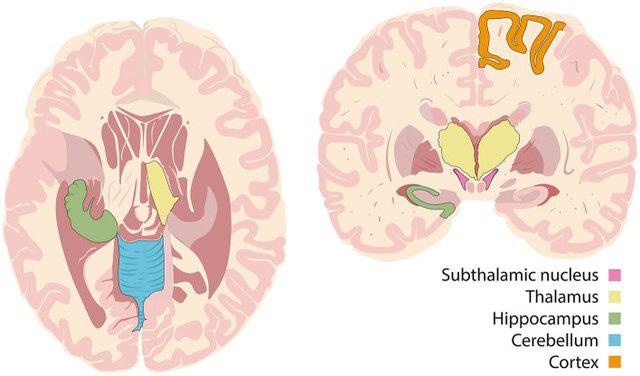
1. Zesiewicz, T. Overview of Essential Tremor. Neuropsychiatric Disease and Treatment 2010, 401. https:// doi.org/10.2147/ndt.s4795.
2. Hardy, M. Dr. Vibhor Krishna Receives Grant to Fund Essential Tremor Treatment Research https://www.med.unc. edu/neurosurgery/dr-vibhor-krishna-receives-grant-to-fundessential-tremor-treatment-research/ (accessed 2022 -09 -21).
3. NIH Research Project Grant Program (R01) | grants.nih.gov https://grants.nih.gov/grants/funding/r01.htm.
4. Sonication - an overview | ScienceDirect Topics https:// www.sciencedirect.com/topics/engineering/sonication.
5. Krishna, V.; Sammartino, F.; Rezai, A. A Review of the Current Therapies, Challenges, and Future Directions of Transcranial Focused Ultrasound Technology. JAMA Neurology 2018, 75 2, 246. https://doi.org/10.1001/ jamaneurol.2017.3129.
6. Garcia, N. Neuromodulation Defined https://www. neuromodulation.com/neuromodulation-defined.

7. Interview with Vibhor Krishna M.B.B.S. MS, 09/08/22.
8. Vibhor Krishna, MD https://www.med.unc.edu/ neurosurgery/physicians/physicians-faculty/vibhor-krishnamd/ (accessed 2022 -09 -22).
It is human nature to wonder why one is the way they are. Recent research has shown that human development is influenced by early adversity. Early adversity is the exposure to environmental circumstances at a young age which then influences the growth of a young individual.1 Examples of early adversity include abuse, neglect, and economic hardship. Dr. Margaret Sheridan has dedicated her personal research to finding the significance of early adversity in adolescent growth.
Dr. Sheridan earned her Ph.D. in clinical psychology from the University of California, Berkeley. She then completed a clinical internship at New York University Child Study Center at Bellevue Hospital. She enjoys spending time with children and reading novels where


she can analyze a character's psychological development as the story progresses. With this background, she has developed a strong passion for research with an emphasis on children and their influences on their early life. Dr. Margaret Sheridan is an associate professor in the Department of Psychology and Neuroscience at the University of North Carolina at Chapel Hill and is also the director of the Child Imaging Research on Cognition and Life Experiences (CIRCLE) Lab.2 She studies the impact of different life experiences on behavior and the developing brain. At the CIRCLE Lab, Dr. Sheridan enjoys mentoring others in research and hopes her work can help change the world for the better.
One goal for Dr. Sheridan and her team is to discover how early adversity affects a child's risk of psychopathology.3 Psychopathology refers to the study of abnormal cognition, experiences, and behavior. The CIRCLE Lab created the Wellness Health and Life Experiences (WHALE) pilot study to help find the answer to the team’s goal. While introducing the study,
1.
Dr. Sheridan states: “We know that little kids' brains are ready to learn information and learn from their environment… therefore if you experience a form of adversity it changes your brain's development in some fashion.”4 With this idea in mind, Dr. Sheridan developed a theory questioning how lack of deprivation and threat affects a child's cognitive development. Deprivation is the lack of having something and threat is the condition of feeling in danger to oneself or others. For the WHALE pilot study, the deprivation is a lack of emotional and/or psychical attachment from their parent(s) and the threat of poverty.
Dr. Sheridan’s WHALE study included 63 participants ages 4-7 years old. Participant screening used aspects by race and socioeconomic status. 50.8% of the participants were white, 34.9% were African American, and 12% of the participants identified as Multicultural or Hispanic.5 All participants belong to a low socioeconomic status (SES) and are current residents of North Carolina. Exclusions to participate in the study included: a major medical condition, a neurological illness, pervasive developmental disorders, and any limitations in understanding English.
The body of the study consisted of questionnaires sent to parents of the child’s parents and in-person interviews with the children themselves. IQ tests, as
violence or who were around threats earlier in their development can predict whether a loud noise is a threat or not. It was also noted that children who can not accurately predict whether the loud noise was a threat or not, their heart rates increased significantly.
well as visual observations were conducted to see if there was any significant exposure to deprivation or threat. Results show that there is a correlation between deprivation to executive function. However, the presence of threat did not affect cognitive development. Although there was a lack of correlation between threat and cognition, it does have significance to participants' psychological reactions. Children who experienced
Dr. Sheridan and researchers from the CIRCLE Lab will now conduct a full WHALE study with around 300 participants. The procedure is similar to the pilot study, but it will now include neuroimaging. Neuroimaging, also known as “brain scanning,” is the process of producing live images of the brain. Neuroimaging techniques can show the brain’s structure and function. The results of the new and improved WHALE study will provide more context to human development.

Figure 2. A participant. Image courtesy of Dr. Sheridan
As the improved WHALE study beings, Dr. Sheridan would like us to know the goal of the study is to promote awareness of early adversity and its risks. With this goal in mind, she hopes her work will eventually lead to a change in governmental policy and intervention. A study this extensive could not be done alone, therefore Dr. Sheridan would like to give special thanks to her graduate students, Lucy Lurie and Micaela Rodriguez, and undergraduate research assistant, Kayla Simone Brown.
1. Machlin, Laura, et al. “Differential Associations of Deprivation and Threat with Cognitive Control and Fear Conditioning in Early Childhood.” Frontiers, Frontiers, 1 Jan. 1AD, https://www.frontiersin.org/articles/10.3389/ fnbeh.2019.00080/full.
2. Interview with Margaret Sheridan, Ph.D. 09/20/22
3. Circle Lab, https://circlelab.unc.edu/.
4. Duffy, Korrina A, et al. “Early Life Adversity and Health-Risk Behaviors: Proposed Psychological and Neural Mechanisms.” Annals of the New York Academy of Sciences, U.S. National Library of Medicine, Sept. 2018, https://www.ncbi.nlm.nih.gov/pmc/articles/PMC6158062/.
“We know that little kids' brains are ready to learn information and learn from their environment… therefore if you experience a form of adversity, it changes your brain's development in some fashion”
When it comes to treating psychological disorders, most imagine sitting in a room alone with a therapist, but Dr. Donald Baucom, Ph.D., and his team are researching a different approach—bringing your significant other into the scene. Dr. Baucom has always been interested in understanding intimate interpersonal relationships and how they impact social interactions. When Dr. Baucom was in graduate school, cognitive behavioral therapy, one of today’s most effective approaches for helping people with psychological disorders, was just developing. Cognitive behavioral therapy suggests that psychological problems are based on “faulty or unhelpful thinking” and “learned patterns of unhelpful behavior”2 and that people with psychological problems can learn ways to cope with these thinking patterns to curtail their symptoms. In their early work, Dr. Baucom and his colleagues applied cognitive behavioral theory to help couples that were dissatisfied or unhappy with their relationships. Despite success in treating couples as a whole, Dr. Baucom observed that often, one or more partners were experiencing their own distress independently of the other person in the relationship. As a result, he concluded that though relationships do exist as a single unit in and of themselves, they are made up of two distinct individuals. This led to the research question that drives his current work, which is whether a significant other can be used to help an individual who’s struggling.1

This proposition gave rise to a couple-based approach
to cognitive behavioral therapy. This approach is all about helping couples manage their individual disorders by teaching them strategies for coping and how to implement those into their daily life. Dr. Baucom explains that “a partner is an important part of a person’s environment. So, in one way you’re bringing their environment into the room for the treatment, but now when treatment ends... you’ve educated the environment.”1 This couple-therapy approach applies to both objectively healthy and unhealthy relationships because cognitive behavioral therapy focuses on teaching the couple to work together as a team to overcome individual challenges rather than placing the blame on the partner.
Cognitive behavioral couple therapy can be used to treat many psychological disorders, including depression and obsessive-compulsive disorder (OCD). Dr. Baucom collaborates with peer specialists in different psychological disorders to understand the individual condition of a given patient. For example, he may team up with a depression specialist to treat a relationship in which one partner has a depression disorder. Depression may cause people to feel unmotivated so the other partner may offer to take care of their responsibilities. While this may provide temporary relief, this does not resolve the symptoms of depression. Using cognitive behavioral couple therapy, the therapist might suggest to the partner to encourage and reward the other when they do things such as exercising or going out together. As another example, perhaps a partner has a form of OCD that causes them to fear germs and is afraid to help their partner cook chicken because it is raw. A therapist would recommend that the partner encourages the other to help because exposure can improve symptoms of OCD. In the words of Dr. Baucom, “You’ve got to touch the chicken.”1
Cognitive behavioral couple therapy is incredibly

versatile in its applications and has been shown to help couples who are struggling with major life events. “Life sometimes changes, and we have got to adapt,” says Baucom.1 For example, when one partner has a heart attack, there are a lot of health behavior changes that need to be made by both partners, such as in regards to managing diet, exercise, and watching children. The same reasoning applies when one partner is diagnosed with cancer because a therapist can help couples cope with such a big life change. Often, partners avoid talking about the complex decisions that are necessary, such as choosing a treatment plan or addressing financial aspects of treatment, as it can be frightening or overwhelming. Cognitive behavioral couple therapists often advise couples to discuss these decisions, instead of withholding their feelings. They also aid in the implementation of treatment methods and forming support systems for such challenging situations.
After 40 years of research, results show that cognitive behavioral couple therapy is highly effective. Typically, one to two years after active treatment of a psychological disorder ends, patients relapse into previous thoughts and behaviors. Experiencing and coping with mental illness alone can be challenging, but with this couple-approach, patients have someone they care about and spend a lot of time with supporting them and helping them apply what they learned in therapy. Evidence demonstrates that this strategy promotes long-term maintenance of goals likely because patients have changed their “environment” (Figure 2).
Dr. Baucom and his team are working to destigmatize cognitive behavioral couple therapy in the United States and are expanding their research to make LGBTQA+ couples. For the last 10 to 15 years, Dr. Baucom has worked with the National Health Service in England (NHS), training therapists to use this approach. Due to the lack of knowledge on this topic, many patients reject the idea of bringing their partner in because they claim that they have a good relationship and fear their partner may think the therapist is blaming them for the disorder affecting the significant other. The coupletherapy approach is an expanding endeavor across different demographics. In particular, Baucom and his team are diligently working to help LGBTQA+ people feel comfortable with therapists using this approach. Research on same-sex relationships is lacking. Because of this, the same strategies for different-sex couples are applied to same-sex couples, despite same-sex couples experiencing vastly different challenges such as discrimination and atypical gender roles.
Figure 1. Differences in reduction of OCD symptoms after various treatments. Graph courtesy of Dr. Baucom.

Baucom and his team hope to make patients feel welcome by specifically addressing their unique understanding of same-sex relationships on their website and advertisements. Dr. Baucom and his team hope to “broaden the paradigm” and make cognitive behavioral couple therapy accessible to everyone.1 References
1. Interview with Donald H. Baucom, Ph.D. 09/21/22
2. American Psychological Association. (2017, July). What is cognitive behavioral therapy? American Psychological Association. Retrieved September 23, 2022, from https://www.apa.org/ptsd-guideline/patients-andfamilies/cognitive-behavioral
3. Berry, A. C. (2009, January 15). Extinction retention predicts improvement in social anxiety symptoms onlinelibrary.wiley.com. Retrieved September 23, 2022, from https://onlinelibrary.wiley.com/doi/10.1002/da.20511
4. Everything you need to know about exposure and response prevention therapy. A Guide to Exposure and Response Prevention Therapy | McLean Hospital. (2021, September 24). Retrieved September 23, 2022, from https://www.mcleanhospital.org/essential/ erp#:~:text=Exposure%20and%20response%20 prevention%20(ERP,the%20problematic%20cycle%20 of%20OCD.
Figure 2. Comparison of result maintenance 6 months and 12 months after treatment from couple based and individual exposure treatment. Graph courtesy of Dr. Baucom.
5. Griffin, R. M. (n.d.). 11 natural depression treatments. WebMD. Retrieved September 23, 2022, from https://www. webmd.com/depression/features/natural-treatments
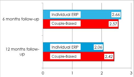
“Dr. Baucom and his team hope to ‘broaden the paradigm’ and make cognitive behavioral couple therapy accessible to everyone.”
Could your gut be affecting your mood? That is the question currently being tackled by Dr. Matthew Redinbo and his team over in Carolina’s Genome Science Building. Dr. Redinbo is determining whether the unpredictable effectiveness of psychiatric medications could be due to interferences caused by the complex chemistry of our gut bacteria. These medications should be effective as they accurately target their intended cascades and proteins produced by the human genome. This has made him wonder if they could be interrupting normal microbial processes involving serotonin, 95% of which is in our gastrointestinal (GI) tract. Motivated by previous research where gut microbial intervention occurred in a
cancer pharmaceutical, Dr. Redinbo believes those same mechanics are at the heart of this study. “How do microbes impact neurotransmitters and how do psychiatric medications impact microbes?” This is the question he intends to address in his next publication.

A cancer study 10 years ago was the start to his voyage into the microbiome. In 2010, Dr. Redinbo and a group of researchers found that a drug widely used for pancreatic and colon cancer had a side effect of eliminating the epithelial cells of the intestines. The drug, camptothecin (generically known as irinotecan), was designed as an inhibitor to topoisomerase I—an enzyme which has shown to be hyperactive in tumor development. When finished inhibiting topoisomerase in the tumor, the drug
R. Redinbo, PhDis excreted from humans through the GI tract. What was found, however, was that camptothecin would kill cells along patients’ intestinal walls to such a degree that the drug could not be used to its full potential. Through further testing and brilliant ingenuity, his team discovered that certain bacteria were reactivating camptothecin once it entered the colon. They supplemented patients with inhibitors that block

drug
“Due to the high levels of serotonin in the gut, it is not far-fetched to hypothesize that the bacteria present could use serotonin in some way.”
reactivation by gut microbes, making camptothecin significantly more effective within tumor models. These same bacteria and proteins of interest are now being investigated in their relation to serotonin levels and SSRI’s.


Selective Serotonin Reuptake Inhibitors—referred to as SSRI’s—are a class of anti-depressive medications that are typically used as the first line of defense against depression. Serotonin can be thought of as the “happy hormone” in relation to depression. These drugs work by preventing serotonin from being reabsorbed by neurons and, thus, increasing a positive mood. Serotonin does not just make people happy, though. It is also used in peristalsis – the rhythmic contractions of the GI tract that move food along the digestive system. Due to the high levels of serotonin in the gut, it is not far-fetched to hypothesize that the bacteria present could use serotonin in some way. In fact, that is precisely what Dr. Redinbo has discovered. The same enzymes that were isolated in the study of camptothecin have been found to interreact with serotonin. Using the same mechanism from the camptothecin study, serotonin was found to reach the gut in an inactive form and become reactivated by microbial intervention. However, SSRI’s possess a potent inhibitor to a family of gut microbial enzymes that also process
serotonin. Thus, when these drugs are administered, they poison some of these microbial processes. This supports Dr. Redinbo’s hypothesis and could be part of the reason why SSRI’s have such a variable effect on patients.
This project has been in the works for nearly two years. With the intent to publish by the end of this year, the Redinbo lab is looking forward at what this preliminary research would entail for the psychiatric community. Redinbo has already partnered with a psychiatrist within the School of Medicine with the interest in collecting patient samples and experimenting with fecal matter in ex vivo testing. Nasty as it may be, as Dr. Redinbo says, once you get past the “ick factor,” collecting these samples becomes crucial to understanding the gastro-mechanics of a patient. By doing deep sequencing on the genomes of these samples, the proteins of these bacterial colonies can be isolated and studied to identify what psychiatric medications could work best for each patient. “Let’s tailor the use of the medications toward what we believe you would respond best to,” as Dr. Redinbo put it. There may be a possible identifier that could be determined from collecting samples in clinical trials which could shift this research from scientific inquiry into groundbreaking medical practice.
“How does disease develop? How
do you treat disease? How could we treat disease better?” These are all questions that drives the research done by Dr. Redinbo and his team. By serendipity, he found himself in the microbiome and the research done could one day shape the way medicine views gut bacteria. This is just the start for Dr. Redinbo and the results to come may alter the way we think of disease and its treatment in the years to come.
1. “Selective Serotonin Uptake Inhibitors (SSRIs).” Mayo Clinic, Mayo Foundation for Medical Education and Research, 17 Sept. 2019, https:// www.mayoclinic.org/diseasesconditions/depression/in-depth/ssris/ art-20044825.
2. Wallace BD, Wang H, Lane KT, Scott JE, Orans J, Koo JS, Venkatesh M, Jobin C, Yeh LA, Mani S, Redinbo MR. Alleviating cancer drug toxicity by inhibiting a bacterial enzyme. Science. 2010 Nov 5;330(6005):831-5. doi: 10.1126/science.1191175. PMID: 21051639; PMCID: PMC3110694.
3. Interview with Matthew R. Redinbo, Ph.D.
They may be small, but they sure are mighty. Phytoplankton are an incredibly diverse, singlecelled micro-organism that perform photosynthesis—the process of capturing the sun’s light energy and converting it into chemical energy. Like plants, phytoplankton intake carbon dioxide and release oxygen during photosynthesis. They form the base of aquatic food chains and produce about half of the oxygen we breathe.

Dr. Adrian Marchetti is the principal investigator of the Marchetti Lab, a biological oceanography lab in the Department of Earth, Marine and Environmental Sciences at UNC-Chapel Hill. Dr. Marchetti joined the department in 2011 and has led several research, cruise expeditions across the globe since.
The Marchetti Lab uses a combination of laboratory and field techniques to research how phytoplankton are affected by and adapt
to their environment. The lab has studied phytoplankton communities in Antarctica, in the North Pacific Ocean, off the coast of North Carolina, and in the Galápagos Archipelago.
nutrient-dense water.
Dr. Adrian Marchetti
The Galápagos Islands are special—after moving offshore, the ocean floor drops to reveal deep ocean surrounding the Archipelago. The Galápagos formed because of volcanic activity, volcanic hot spots erupt and lift tectonic plates, layering them on top of one another. The islands have a distinct, conical shape because of repeated volcanic eruptions. The Galápagos lies at the convergent point of three main ocean currents, which drives variation in water temperature all around the islands. Figure 1 depicts the geography of the Galápagos and shows theses currents, the Humboldt Current, the Panama Flow, the Cromwell Current. Deep ocean upwelling is strong around the islands, which replaces surface water with cold,
The diverse ocean conditions in the Galápagos are home to many different species of phytoplankton.1 Phytoplankton growth is limited by three key factors: light, temperature, and nutrients. Phytoplankton thrive because they float near the surface of the water and capture sunlight that does not penetrate through deep ocean water. Unlike other marine primary producers, such as seagrass and macroalgae, phytoplankton cannot survive in deep ocean waters. A tremendous amount of food and nutrients that fuel the other food webs on the islands start with phytoplankton.1
“We’re interested in the Galápagos Islands and where they are situated because it is on the front lines of El Niño,” Dr. Marchetti said.1 El Niño is a recurring climate pattern associated with rising sea temperatures and declines in nutrient upwelling—this is particularly true around the Galápagos.2 The drop in nutrient availability causes a decline in phytoplankton primary production, the process by which phytoplankton capture sunlight and
Figure 1. Map detailing sampling sites in the Galápagos during the Marchetti Lab’s 2018 research expedition. The three major currents that drive aquatic diversity on the islands are labeled as well. Figure courtesy of Dr. Marchetti
convert it into chemical energy. Dr. Marchetti and his team are using El Niño as a natural laboratory to understand how phytoplankton respond to these changing conditions in the Galápagos. Dr. Marchetti stated they can use El Niño as “a window into what they can predict for the entire ocean accounting climate change.”1
To date, the Marchetti Lab has led four cruises to study the impacts of El Niño in the Galápagos. During their most recent expedition in 2018, they sampled water from 23 sites across the Archipelago between September 30 and October 25. The scientists used DNA metabarcoding – large-scale taxonomic identification of DNA samples through DNA sequence analysis – and microscopic examination to measure phytoplankton communities.2 Dr. Marchetti said they wanted to know how environmental conditions brought on by El Niño impacted phytoplankton,


and what this meant for primary production.1 The climate patterns of El Niño weaken upwelling around the islands, which means there are less nutrients available to phytoplankton. They found that the lack of nutrients and rise in water temperature causes phytoplankton productivity to decline.1

The effects from El Niño can be catastrophic – the decline in phytoplankton productivity can be seen all the way at the top of the food web. All the ecosystems in the Galapagos, marine and terrestrial, suffer during El Niño. Less energy for phytoplankton means less food for fish,which means less food for birds and mammals, and less food for the inhabitants of the islands. The geography of the Galapagos limits the amount of land-based protein that farmers can raise, so they must source most of their protein from the ocean. When fish populations decline, fishermen are forced to ship food from the mainland, which can be extremely expensive and inaccessible.1
is really important for management of the Galápagos ecosystem,” Dr. Marchetti said.1 Although the effects of El Niño and La Niña are temporary, climate change threatens to warm oceans and alter aquatic ecosystems. Climate change can cause nutrient-poor areas of the ocean to expand, which can drive shifts in phytoplankton species size, composition, and density.1 Through his research, Dr. Marchetti hopes to be able to predict how phytoplankton communities will change across the globe as ocean temperatures continue to rise.
Figure 2. Graphs depicting distribution of seas surface temperatures (SST) and variations in nutrient availability across the Galápagos during 2018 expedition. Figure courtesy of Dr. Marchetti.
Figure 3. Dr. Marchetti researching water conditions in the Galápagos in 2018 with a sunset in the background. Image courtesy of Dr. Marchetti
Dr. Marchetti hopes to return to the Galápagos this November to investigate how La Niña, the periodic cooling of sea-surface temperatures, is changing the ecosystem. During La Niña, upwelling is stronger, which increases the amount of nutrients available to phytoplankton. Thus, phytoplankton thrive, causing increases in biomass all the way up the food chain. Larger animals thrive as they can grow faster, grow larger, and reproduce more often. The conditions of La Niña could be one of the reasons why animals in the Galápagos are able to survive in the conditions El Niño. “Understanding all sides of the productivity spectrum
Figure 4. Dr. Marchetti analyzing a water sample in their make-shift lab on the ship, courtesy of Dr. Marchetti.
1. Interview with Christopher Willett, Ph.D. 9/20/13.
2.Willett, C.S. 2012. “Hybrid breakdown weakens under thermal stress in population crosses of the copepod Tigriopus californicus.”
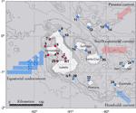
Journal of Heredity. (103) 103-114.
3.Dictionary.com: copepod. http:// dictionary.reference.com/browse/ copepod?s=t&ld=1172 (accessed October 17, 2013).
Process of Cytokinesis
Image by Wikimedia Commons
From a fertilized egg cell growing into a fetus, to repairing a paper cut on your pinky finger, most bodily procedures require cell division for growth, development, and the maintenance of health and homeostasis. However, the stepwise process that occurs is much more intricate than the simple circular diagrams seen in middle school science classes.

Structure defining function is a well-known biological principle. Just as a building cannot maintain its integrity and function without a specific framework, a cell’s ability to function similarly relies upon its structure, which undergoes several changes during division. Cells must rearrange and build their own “cellular machinery” from a variety of physical and chemical components to ensure that genetic material is accurately distributed to daughter cells. Though this rearrangement causes some structural changes, the structure and function of the cell will be dramatically affected if cytokinesis—the physical cell shape change that occurs at the end of mitosis and meiosis—is not properly executed. Cytokinesis is not only vital in ensuring that organisms can grow and injuries can be repaired, but it and its precise execution will ultimately dictate the structure and therefore function of cells.¹
Dr. Amy Shaub Maddox, a professor and cell biologist at UNC Chapel Hill, studies the cytoskeleton and the changes in cellular architecture that occur during division. Her lab makes movies of model animal cells dividing and then measures a variety of parameters, including the shape and size of the cell and the design principles that are the foundation for

cellular architecture and assembly. The research has several applications in the understanding of fertility, growth, early development, and disease.2

“There has to be a continued effort to understand natural processes important for human and global health. We have to have better medicines that don’t make people sick while they’re trying to make them better,” said Dr. Maddox when asked about the direction that her work should go in the future.2
One major focus and direction in which Dr. Maddox’s research is oriented is understanding the initiation and progression of cancer, which often results from uncontrolled or unregulated cell division. Understanding the molecular and physical mechanisms that dictate cytokinesis in particular could be invaluable in the development of cancer-targeting drugs and therapies. Incomplete cytokinesis causes a variety of issues that may contribute to cancer: tetraploidy, for instance, is a condition in which an individual has four copies of a
Figure 1. Mitosis Appearances in Breast Cancer Cells.
“We have to have better medicines that don’t make people sick while they’re trying to make them better.”Dr. Amy Shaub Maddox
chromosome in each cell. Another is binucleation, which is the formation of cells that contain two nuclei, causing genomic instability. Furthermore, excessive cytokinesis can contribute to tumor development. Most of the drugs used to address cancers today are cytotoxic agents, which inhibit division and kill cancer cells. Though they can be effective at stopping the progression of cancer, the harmful chemicals in these drugs also attack healthy cells that frequently divide, such as intestinal tract and hair follicle cells. These complications can cause nausea and hair loss in cancer patients undergoing treatment, as well as a myriad of other unpleasant symptoms.2
Cytokinesis is largely driven by proteins, which indicates the possibility of drug development that directly targets the function of these proteins. The first physical change in cell structure occurs with the formation of the cleavage furrow from the constriction of a complex of actin microfilaments, myosin proteins, and a variety of structural and regulatory proteins known as the contractile ring. The plane on which cellular division will occur is further regulated by another protein complex known as the mitotic spindle, which organizes the genetic material within the cell before division. It then determines the location of the contractile ring, causing the cell to divide symmetrically or asymmetrically, depending on whether cells of different sizes must be made. The contractile ring slowly pinches the cell and creates an indentation on its surface, gradually deepening and spreading around the cell. Before the contractile ring completely splits a cell in two, new membrane must be inserted into the plasma membrane of the cell that is being created from its parent. Cytokinesis is completed when the contractile ring has completely split the cell in two.1
Dr. Maddox’s lab has researched and published several findings about proteins that drive the process of cytokinesis. One protein in particular, anillin, is being researched in relation to its numerous roles in cytokinesis. The protein’s name comes from the Spanish word, “anillo,” which means ring, as it is a part of the contractile ring that constricts and pinches the cell to promote separation during cytokinesis. As a key regulator of cytokinesis, it has been linked to cancer and observed in studies that suggest that its contributions to changes in cellular shape and structure may alter cell motility and facilitate metastasis, the spread of cancer from its primary site to other areas.3
As her research continues, Dr. Maddox cites collaboration as a necessary aspect of research that culminates in discovery and greater understanding. “There’s the subject of your science, which, for me, is cells, but people are at least as big a part of it as the research subject, the personalities, and the team...Camaraderie and sympathy and people pushing each other to their best standards, that’s really what research is,” said Dr. Maddox.2 Through collaboration, perseverance, and persistent curiosity, Dr. Maddox and her lab will continue to make remarkable advances in understanding one of the most important life processes, ultimately paving the way to a better future.
“The actual proteins we study are important and essential for cell division, and if we are able to generate or discover a compound that screwed up the function of that protein, then it would screw up that essential process,” explained Dr. Maddox. However, targeting cytokinesis for cancer treatment does not come without risk. Though it may effectively inhibit continued cancer cell division, it can lead to some of the harmful, unwanted effects of incomplete cytokinesis.2
Figure 3. Expression patterns of the ANLN gene, which codes for the anillin protein, in different cells. Graph by Wikimedia Commons

1. Rafelski, Susanne M., and Wallace F. Marshall. “Building the Cell: Design Principles of Cellular Architecture.” Nature News, Nature Publishing Group, https://www.nature.com/ articles/nrm2460#:~:text=Cell%20architecture%20is%20 dictated%20by,or%20 dynamic%20balance%2Dpoint%20 mechanisms.
2. Interview with Amy Shaub Maddox, PhD 09/06/22
3. AS;, Piekny AJ;Maddox. “The Myriad Roles of Anillin during Cytokinesis.” Seminars in Cell & Developmental Biology, U.S. National Library of Medicine, https:// pubmed.ncbi.nlm.nih.gov/20732437/.

 By Ciara Daly
by Bhavika Chirumamilla
Under the surface, the marine environment is peaceful; a shoal of fish dart by, glimmering scales reflecting the light. In a millisecond, the peace is disturbed by the rapid extension of a squid’s tentacles. A thrashing fish is snatched, unable to evade evolutionarily perfected limbs made deadly by extraordinary muscles tailored to precisely and rapidly capture prey. While this scene may be a common occurrence in the ocean every time squid hunt, it represents a muscular phenomenon quite unlike any other. Due to the incredibly quick response time, impeccable accuracy of movement, and span of the near 200% elongation of each tentacle from its resting state, squid tentacles are so unique they may someday be mimicked by engineers to create soft robotics that can achieve things as large as site excavation to as precise as neurosurgery.1
Professor William Kier, a researcher who focuses on squid tentacles at UNC-Chapel Hill, has devoted most of his extensive career seeking to understand the mechanisms that permit squid tentacles to accomplish their amazing feats. Early marine scientists and biologists were puzzled by the fact that hydrostats can even move in the first place - without a skeleton to act as a lever, how can muscles move? The answer involves a combination of biological architecture, engineering, and marine biology, a series of subjects Kier was seeking to combine as a young scientist intrigued by ocean organisms.

As you turn the pages of this Carolina Scientific issue, the movement of your muscles are dependent upon your skeleton. Our muscles contract and, as a result, pull on our bones to trigger movement like a lever. Invertebrates without skeletons are called hydrostats: they use pressure put on a fluid by muscle instead of a skeleton to move. In these animals, such as worms, a muscle is structured
Dr. William KierFigure 1. A variety of animals that have muscles that are mus cular hydrostats. Image courtesy of Smith and Kier.

into bands around a fluid filled membrane. When the rings of muscle contract, the result is like a water balloon being squeezed, and the limb elongates. Conversely, when that pressure is released as the bands return to their relaxed state, the limb retracts. However, squids and other cephalopods belong to an even more specialized group of animals called muscular hydrostats. Not only do their muscles’ function and structure differ from ours because they cannot rely on skeletons to facilitate movement and support muscles, they also do not use fluid compression to achieve elongation and contraction. Instead, tentacles consist solely of muscle fibers that tug to put pressure upon other muscle tissue and induce the same elongation and contraction as regular hydrostats.
The Kier lab has produced a plethora of works contributing to the scientific community’s understanding of muscular hydrostats, particularly squid tentacles. His latest cutting-edge research on the miracles of tentacles focuses on muscular mechanisms such as the rapid activation rate and extension. “They [squids] are approaching the prey and then in fact aiming and triggering this extremely rapid extension of the tentacles and there is no correction during this time...optimizing the rate of activation and making sure it occurs simultaneously across the tentacles is very important”, Kier explains.
When muscles receive instructions to move, it comes packaged in the form of neuroelectric signals and changes in electric voltage that induce biochemical pathways that finally
spur movement. When considering activation rate, ion channels such as sodium channels play a major role. These channels are located on the cellular membrane, which encapsulates the cell and its contents, and it is connected to nearby neurons. When an ion channel on a membrane receives a neuroelectric impulse sent from a connected neuron, the action potential, an imbalance in charges and concentrations across the membrane that induce ionic movement, is in the form of positive sodium ions that increase in concentration surrounding the exterior of the membrane. This highly positive charge outside of the cell that is more positively charged than the fluid inside of the cell leads the channels to open and for the ions to move inside the cell to even out the concentrations again. This flow of ions triggers a series of reactions and signaling that eventually lead to the attraction between muscle fibers that draw the muscles closer together before once again relaxing.2

To put squid tentacles to the test in the laboratory instead of in the sea, the sample species tested for this study included both the California market squid and longfin inshore squid, which were captured in the wild and placed in tanks of seawater before being decapitated. Squids possess two tentacles and eight arms. The arms are covered in suckers, unlike the tentacles, and are less capable of rapid extension and drastic elongation, leading them to play less of a role in striking at prey.3 They can thus serve as a control group for experimental procedures on squid tentacles. The limbs were thinly sliced into cross-sections and then placed in seawater petri dishes to maintain the balance of water concentration in muscle cells, as would be found in the oceanic natural habitat of each squid. Kier’s team tested each sample’s muscular power and ability using and running pulses of electric current through the muscle fibers, mimicking neural impulses, to measure the voltage changes and action potential of the tentacle muscles versus the arm muscles.1
The experiments yielded some insightful results, pinpointing key biological components that distinguished squid tentacles from other muscles. All tentacle fibers tested with an electric current were found to have voltage-dependent sodium currents, while the control group of arm muscle fibers did not: this means that sodium channels play a major role in how squid muscles in the tentacles are capable of rapidly responding to stimulus and neuroelectric impulses. In addition, another fascinating discovery was that the density of sodium conductance was 10 times higher in the squid tentacles, indicating a stronger sensitivity to stimulus and an inclination to more rapid response at lower levels of input. Some similarities were also noted between the control and experimental groups. For instance, both the fibers from arms and tentacles had similar kinetic
activation and inactivation of sodium channels, meaning that they are not completely different in action potential functioning and that it is unlikely to be a reason for the unique properties of tentacular muscle.1
This information advances the foundational knowledge on muscular hydrostats, particularly squids, that Kier has built throughout his extensive career. This research continues to enlighten our understanding of tentacles, but also of the broader group of muscles as a whole and the biological properties that determine muscular specialization and niches. These findings also have potentially major technological applications - engineers have sought to collaborate with Kier, learning from his expertise in the unique properties of tentacles. In particular, members of a field called soft robotics seek to replicate the flexibility, extension, and lack of rigidity of muscular hydrostats. One day, these robotic mimicries of tentacles could serve to complete far more precise and detailed work than current robotic arms are capable of, perhaps going as far as miniaturized tentacle-like appendages serving as probes for neurosurgery or larger soft robotic projects that can excavate and navigate fields such as demolition and construction sites.
Figure 3. OCTOR, a prototype mimicking a tentacle and trunk, the product of biologists and engineers collaborating to design soft robotics. Image courtesy of Dr. Kier.

A seasoned scientist and Tarheel of 37 years, Professor Kier retired this June, and at the tail end of his career, his passion for muscular hydrostats and science remains stronger than ever. Still surprised by where his path in marine science and biology has led him as he followed his fundamental interest of how animals work and by the potential technological applications of the tentacle research he has performed, Kier remarks, “The greatest delight of doing science... is that you’ll work through something and realize ‘I just figured out something that no one else knew’”.
1. Gilly, William F., Corbin Renken, Joshua J. C. Rosenthal, and William M. Kier. “Specialization for Rapid Excitation in Fast Squid Tentacle Muscle Involves Action Potentials Absent in Slow Arm Muscle.” Journal of Experimental Biology 2020, 223. https://doi.org/10.1242/jeb.218081.
2. Ruiz, Lera, Manuel de, and Kraus, Richard, L. “VoltageGated Sodium Channels: Structure, Function, Pharmacology, and Clinical Indications.” Journal of Medicinal Chemistry 58, no. 18 (September 24, 2015): 7093–7118. https://doi. org/10.1021/jm501981g.
3. Scully, Caitlin. “Get to Know The Four Types of Cephalopods.” Birch Aquarium Blog, October 11, 2018. https://aquarium.ucsd.edu/blog/get-to-know-the-fourtypes-of-cephalopods/ (accessed September, 15, 2022).
4. Smith, Kathleen and Kier, William M. “Trunks, Tongues, and Tentacles: Moving with Skeletons of Muscle.” American Scientist (January-February 1989) http://labs.bio.unc.edu/ Kier/pdf/Smith_Kier_1989.pdf (accessed August, 30, 2022)
Despite being one of the oldest systems of the body, there is surprisingly very little known about pain. Even though pain treatment, management, and prediction have been at the forefront of research in neurology for years, there are few explanations for why similar circumstances may lead to varied pain outcomes in different individuals. Dr. Sarah Linnstaedt, an associate professor of anesthesiology at UNCChapel Hill, became interested in this phenomenon as a postdoc at Duke University. While working with microRNA (miRNA), she began to realize its potential to be used as a biomarker—a measurable characteristic used to detect normal and abnormal function, such as concentration of a certain protein or molecule in the blood—to predict chronic pain development (Figure 3). Dr. Linnstaedt decided to further investigate this possibility and, focused her research on pain development following a traumatic incident because “a lot of people experience trauma in their life, but not everybody will go on to develop chronic pain”.1
following a common traumatic incident. First, she separated the study participants into different groups: those experiencing persistent pain, or pain that lasts 3+ months despite treatment, and those not experiencing persistent pain. She then collected and analyzed blood RNA samples from all individuals in both groups to determine differences in miRNA expression. In order to identify recurring patterns among the separate groups, the results from the miRNA expression analysis were compared to the level and duration of pain experienced by each individual. Dr. Linnstaedt was able to conclude that high concentrations of two kinds of miRNA (miR-135a-5p and miR-3613-3p) in the bloodstream shortly after stress exposure predicted persistent pain development in car crash victims. Additionally, the study results suggested that women were more vulnerable to persistent pain than men and that X chromosome miRNA was identified as a major contributor to persistent pain development. Through the study, Dr. Linnstaedt was able to prove that blood samples taken in the hours following a traumatic incident could provide some clarity into the mechanisms surrounding pain development.2
Dr. Sarah Linnstaedt
To begin her research on this topic, Dr. Linnstaedt identified a sample of 53 car crash survivors, all coping with varying levels of pain
The miRNA project has continued to inspire current research in Dr. Linnstaedt’s lab. Presently, Dr. Linnstaedt is focused on a new biomarker project in victims of traumatic events. For this project, she is collaborating on a study called AURORA to look at over 4000 individuals who have experienced any traumatic event in their lifetime. The AURORA study not only includes car crash survivors, but also survivors of sexual assault, combat, and major traumatic brain injury. The blood
Figure 2. Graphic representation of Dr. Linnstaedt’s AURORA study design. Image courtesy of Dr. Sarah Linnstaedt.
RNA samples from the AURORA participants are taken at three different time points: at the time of trauma, two weeks posttrauma, and six months post-trauma. In blood samples, the concentration of various cytokines, which are small proteins with important roles in immune response, will be analyzed and compared to the level and duration of pain experienced by the individual in a manner similar to the miRNA study (Figure 1).The results from this study can then be used to determine if a single or combination of cytokine levels immediately after sustaining trauma are predictive of chronic pain. Additionally, because of the different timepoints in the study, Dr. Linnstaedt will also be able to determine how cytokine blood levels change in an individual following a traumatic incident and how these changes correlate with pain development or persistence (Figure 2). The study team faced a number of challenges during the development and participant recruitment phase, largely due to the COVID-19 pandemic. “COVID has certainly impacted our human resource studies because we have not been able to enroll in the same capacity as we used to before,” explained Dr. Linnstaedt, describing how AURORA enrollment sites were shut down for many months at the start of the pandemic, which led to a lower-than-desired number of total study participants.1
In the near future, Dr. Linnstaedt hopes to increase the accuracy of biomarker-based pain prediction by increasing the sample size of her experimental group to represent an even larger group of people. By doing so, she will be able to establish more accurate, general trends regarding persistent pain
prediction that will target a larger segment of the population. Upon achieving this, Dr. Linnstaedt plans on expanding her research to include development of a therapeutic intervention that could change or modify these vulnerabilities shortly after a traumatic event has occurred.
Dr. Linnstaedt believes that, at the clinical level, “Trying to understand vulnerabilities at the biological level and identifying these predictive markers will help enable identification of individuals at risk”.1 She hopes that her research will help clinicians, emergency department doctors, and other healthcare professionals improve treatment of patients who have suffered a major traumatic event. By being able to predict an individual’s pain development at the time of trauma, a healthcare team can act early to prevent—or even reverse—changes at the biological level that make an individual more prone to worse pain outcomes.
Figure 3. Using blood samples to identify diagnostic biomark ers. Image courtesy of Csaba Deli/Shutterstock.


1. Interview with Sarah Linnstaedt, Ph.D. 09/20/22. 2. Linnstaedt, S. D.; Walker, M. G.; Parker, J. S.; Yeh, E.; Sons, R. L.; Zimny, E.; Lewandowski, C.; Hendry, P. L.; Damiron, K.; Pearson, C.; Velilla, M.-A.; O’Neil, B. J.; Jones, J.; Swor, R.; Domeier, R.; Hammond, S.; McLean, S. A. MicroRNA Circulating in the Early Aftermath of Motor Vehicle Collision Predict Persistent Pain Development and Suggest a Role for MicroRNA in Sex-Specific Pain Differences. Mol Pain 2015, 11, s12990-015-0069–3. https://doi.org/10.1186/s12990-015-00693.
 By Rebecca Turner
When people think about air pollution, what typically comes to mind is the outside environment, rather than the indoor air that surrounds them 90% of the time. While researchers often study outdoor air quality, many tend to ignore indoor air quality. Chemical reactions in the atmosphere do not stop in an outdoor environment, and pollutants from outside inevitably come inside, too.
Dr. Glenn Morrison, a professor in the Department of Environmental Sciences and Engineering at UNC-Chapel Hill, has dedicated his career to studying indoor chemistry and pollutants. Because indoor environments are so different from their outdoor counterparts, molecules indoors can go through significant chemical changes. The key to understanding indoor chemistry lies in understanding surface chemistry, because indoor surfaces are where the built environment and outdoor air meet.1 Indoor surfaces adsorb and accumulate compounds from the air, changing them in the process.
Indoor environments have a higher surface-area-tovolume ratio from walls and furniture, meaning gas particles collide with surfaces at a greater rate than in outdoor environments. All indoor ozone molecules will collide with a surface and potentially react before leaving a building, compared to approximately a 1% chance of doing so outdoors. Morrison also researches oxidants in an indoor environment: while the hydroxyl radical is the most important oxidative reactant outdoors, ozone is the most important indoors. Another aspect to be studied are the actual surfaces themselves, including glass, wood, drywall, fabric, carpet, paint, and all other varied surfaces. Finally,
Figure 1. Structure of ammonium perfluorooctanoate (C8) Molecule. Image courtesy of Rhodeodendeonbusch via Wikimedia Commons.

researchers want to know what is driving this chemistry, and the main suspects are light, temperature, and acidity. Clearly, there are many unknowns.
Broadly Dr. Morrison studies the field of indoor chemistry, but he also researches particular indoor air pollutants. In focus, he researches a nasty group of chemicals called perfluoroalkyl and polyfluoroalkyl substances, commonly known as PFAS. PFAS are legacy pollutants, which are highly stable substances that continue to exist in the environment long after their production is stopped and are unlikely to break down in a person’s lifetime. PFAS can be found in many everyday products such as waterproof clothing, non-stick pans, food packaging and takeout containers.
PFAS pollutants have garnered more attention in recent years due to their detrimental health effects. Teflon, the substance used in non-stick pans, is known to contain a PFAS known as ammonium perfluorooctanoate (C-8). The production of C-8 has been linked to leaking significant amounts of the chemical into the environment and drinking water, to the point that 99% of Americans have C-8 in their blood.2 C-8 concentrations in the bloodstream have been linked to testicular and kidney cancers, ulcerative colitis, thyroid disease, preeclampsia, and high cholesterol, along with neurological disorders. C-8 is one of the most well-known and well-tested PFAS chemicals, but due to industrial manufacturing, “there’s thousands of versions of these perfluorinated chemicals out there that are made

Figure 2. Possible Drivers of Indoor Chemiestry. Image courtesy of Ault et al.

either intentionally as parts of products, or they are breakdown products.”3 Since PFAS chemicals do not break down efficiently into reactive forms, they have nowhere to go except to travel through the environment, and in particular, “there [is] enough of their precursor left over that it [has been] released over time into indoor environments, or [been] released in washing, and gets back into our water systems.”3
While PFAS exposure through waterways is now understood, Dr. Morrison and his colleagues are currently researching other relevant methods of exposure. PFAS is a broad classification, and chemicals within this category can vary significantly in size and structure. Consequently, scientists are left asking what sources of PFAS humans are being exposed to.
One recent study Dr. Morrison participated in looked at PFAS emitted from floor waxes, which often contain PFAS to maintain their properties, like low surface tension.4 As a result, the concentrations of PFAS can be incredibly high, and since floor waxing is an intensive process, these chemicals are easily suspended in the air due to aggressive stripping and waxing techniques. The researchers found that during floor waxing, the concentrations of 5 types of PFAS were higher in the air than before waxing, showing an occupational hazard to floor technicians who perform these activities regularly.
Another field with high exposure to PFAS is firefighting, which is the next field Morrison and colleagues plan to study for PFAS exposure. Most firefighting foams contain PFAS, which can then harm firefighters. However, firefighting foams are a substance deemed essential enough such that the potential harm from PFAS exposure may not outweigh the benefits of fighting fires.
Ultimately, further research will continue to be important regarding PFAS-containing substances. While most people would likely recognize that PFAS does not need to be in everyday items like takeout containers, it could be a necessary evil for certain industries. Furthermore, when PFAS like C-8 are banned
from production, the new chemicals created in their place may be just as harmful, if not worse. According to Morrison, “When we identify chemicals that are problematic, we’ll look at not just those chemicals, but the class.” The subsequent phasing out of groups of chemicals, rather than individual chemicals, is less expensive and more widely effective.
Regarding indoor air chemistry, the field has received more attention since COVID transmission became a significant public health focus. The main hurdle indoor chemistry encounters are the lack of national attention. Whereas the Clean Air Act is essential for regulating outdoor air quality, no such legislation exists to protect indoor air and research. “It’s not until we have policies in place that crystallize regulation like the Clean Air Act,” Morrison says, that indoor air quality will be given the attention it deserves.3 One of the many lessons learned from COVID is that indoor air is important and deserves more attention to protect human health.
1. Ault, A. P.; Grassian, V. H.; Carslaw, N.; Collins, D. B.; Destaillats, H.; Donaldson, D. J.; Farmer, D. K.; Jimenez, J. L.; McNeill, V. F.; Morrison, G. C.; O’Brien, R. E.; Shiraiwa, M.; Vance, M. E.; Wells, J. R.; Xiong, W. Indoor surface chemistry: Developing a molecular picture of reactions on indoor interfaces. Chem 2020, 6(12), 3203–3218. https://doi. org/10.1016/j.chempr.2020.08.023.
2. Soechtig, S. (Director). (2018). The Devil We Know [Film]. Cinetic Media.
3. Interview with Glenn Morrison, Ph.D. September 12, 2022. 4. Zhou, J.; Baumann, K.; Chang, N.; Morrison, G.; Bodnar, W.; Zhang, Z.; Atkin, J. M.; Surratt, J. D.; Turpin, B. J. Per- and polyfluoroalkyl substances (PFASs) in airborne particulate matter (PM2.0) emitted during floor waxing: A pilot study. Atmos. Environ. 2022, 268, 118845. https://doi.org/10.1016/j. atmosenv.2021.118845.
 By Emily DeVillle
“Please wait, the meeting host will let you in soon.” Although it has been merely two years, this sentence would not be so familiar in 2019, or even early 2020, as it is now. Since the COVID-19 pandemic, people from across the globe have become intimately familiar with Zoom, the software in which many classes, family reunions, and work meetings were held throughout the pandemic. Among the many losses of the pandemic, face-to-face contact may have been one of the most detrimental. Some of the greatest joys in life are derived from friendships made and solidarity formed with people met. Virtual interactions pale in comparison to those in-person. Not only do face-to-face interactions bring joy to people’s lives but they can also help them to persist when life gets challenging. When friends show up, people are inspired to show up for themselves and keep moving forward. It is these types of friendships that Anusha Hariharan researches here at UNC-Chapel Hill. Anusha Hariharan is an eighthyear doctoral candidate and socio-cultural anthropologist. Specifically, Hariharan’s research focuses on the friendships formed between feminist activists in South India and how they sustain their friendships by building solidarity and engaging in care practices.
In Tamil Nadu, a state in South India, there is a group of feminist activists. These women, (ages ranging from mid-50s to mid-70s), have been friends since nearly 1989, although their activism predates their friendship. It

is the everyday lives, as well as the ethical commitments of these feminist activists that interest Hariharan. In fact, much of the importance in Hariharan’s research lies in what most would consider mundane actions. However, it is through these actions that these women can build strong friendships and coalitions to support each other in their individual and collective pursuits to bring about social change. Initially, Hariharan’s fieldwork was concerned with a different inquiry: everyday tasks among Dalit feminist movements. Dalit feminists examine feminism and social justice through the lens of the Dalit people, or the “Untouchables.”
The Dalits occupy essentially the bottom rung of the caste in India. And although “Untouchability” has been outlawed in India, Dalit feminists and Dalit activists challenged the norms of atrocities committed against “Untouchables” in India that were before seen as routine behavior. However, during the time Hariharan spent with these Dalit feminists, they faced several changes and challenges. Because of these challenges, Hariharan decided that it was not the right time to tell their story, and order to uphold her ethics of research, and respect for the women she worked with, chose to walk away.
Figure 2. Map of Tamil Nadu, the South Indian state in which the feminist women featured in Hariharan’s research reside. Photo courtesy of TUBS, Wikimedia Commons

However, the experience left Hariharan wondering what remains when funding runs out, or when the group disagrees on what issues to focus on, etc. This inquiry is what led Hariharan to the group of women who live across Tamil Nadu. Although the group has grown and shrunk in numbers throughout the time she spent with them, Hariharan interviewed twenty-two of these women. The women, who had each been involved in various forms of activism with other women and male comrades, felt an absence in a feminist collective. So, they came together, across the state and formed one. Every three months, this feminist coalition would meet for an extended weekend “retreat,” alternating locations so the labor of hosting was divided equally amongst the women. As a primarily grass-roots movement, the women had to find a space that would let them stay for free whether it was through donations or an organization they were attached to, often these retreats were held in the multipurpose rooms of various churches. During the retreats, the women would come together from across the state, discuss various topics of social change and transformation, and bring their concerns as feminists. These retreats created a space where feminist activists took large scale issues in politics such as political equality or more localized issues such as domestic violence and forged solidarity in what Hariharan refers to as, “an intimate sphere.” These women would talk into the late hours of the night, addressing different issues across the various cities they inhabit, and cooking together. Activities such as these are often undervalued because they are associated with the feminine and are not seen as useful in the realm of activism. However, Hariharan argues that it is these actions, or what she refers to as, “ethical labor” that can better shape the way people re-envision the world they want to live in, and how they inhabit that world. When people care for the world
they envision, they also care for the people in that world as they are a part of it.
Although people tend to focus on solidarity and coalition building solely in times of crisis, it is important to also take notice of the maintenance of friendships and support groups during times of stagnation as Hariharan says, “because coalition building, and solidarity building can not only happen when things go wrong” (Hariharan 2022).
Figure 3. Some of the feminist women, and closest interlocutors in Hariharan’s research in Tamil Nadu. Image courtesy of Anusha Hariharan

Feminist activists are faced with not only horrifying atrocities, but many disappointments at the hands of systems in power. Because of the emotionally charged circumstances under which they work, these women lean on each other, or find other sources of spiritual grounding through meditation, reflection, or even religion. In her research, Hariharan has found that the feminist that has some form of spiritual grounding, is the feminist that thrives throughout their lifespan of activism. There seems to be an idea about activism that anger is the most dominant and productive emotion however, this leaves unresolved emotions. It is through these strong feminist friendships and moments of spiritual grounding and reflection, that the unresolved feelings of grief and pain may finally be healed.
There is so much people can learn from Hariharan’s research not just about feminist friendship and solidarity but also how they may re-envision the world they wish to live in and how they might inhabit that world. Change, or rather, “real” change cannot come from one person. As she prepares to complete and defend her dissertation, Hariharan hopes to broaden her research and continue to tell the history of feminist activism through the lens of friendship. In addition, Hariharan hopes to make her research accessible to not just those in academia but also to reach other activists and individuals interested in feminist history.
Figure 4. This table represents the Indian Caste System. Photo courtesy of Equality Labs from the Publication Scroll, 2018
1. Interview with Anusha Hariharan. 09/15/2022
2. Hariharan, A., “us. together for 30 Years”: Care, intimacy, feminist friendship.
3. Jati: The caste system in India.

Figure 3. TikTok – one of the most famous social media apps today that is based on creative video content. Dr. Laura Ott’s students made videos explaining biological processes under this platform.
 By Ria Patel
Imagine this: you are sitting in a college level biology course with nearly 150 other students getting ready to learn how transmembrane proteins in the plasma membrane aid in the generation of intercellular signals. Your professor enters the lecture hall and says, “What did you think what the TikTok video I assigned for class today?” or “Students, take out some playdough and pick up a 3-D printed plasma membrane –these will be the materials we use in class today.”
For many people, this scenario remains as a matter of an imaginative mind – truly, how can such a complex biological process be taught through a crime science investigation, a few toys, and an informal social media platform? However, for an ambitious professor at UNC-Chapel Hill, Dr. Laura Ott, this socalled “imaginative scenario” may actually become an integral part of a collegiate level biology education. Dr. Laura Ott is a teaching assistant professor in the Department of Biology at UNC, where she teaches a variety of biology-related courses such as BIOL 101: Principles of Biology, BIOL 103: How Cells Work, BIOL 252: Human Anatomy & Physiology, BIOL 448: Advanced Cell Biology, and others. With a passion for teaching
biology, helping undergraduates reach their educational and professional goals, and conducting disciplinary-based education research, Dr. Ott strives to find scholarly, novel, and scientific approaches to improving educational experiences and outcomes for students interested in furthering their education in biology. To do this, Dr. Ott draws heavily from the 2011 Report - Vision and Change in Undergraduate Biology Education: A Call to Action, which helped set the modern framework for biology education1. With the framework in mind, Dr. Ott uses innovate activities to expand her students’ knowledge on the five major themes of life and key competencies that biology majors should demonstrate.1
During her postdoc at NC State University, Dr. Ott taught molecular biology courses, as well as designed and assessed novel courses and curricula to improve student learning. Focusing on the core competencies identified by the Vision and Change report, Dr. Ott and colleagues created introductory lab exercises or modules that helped students advance their technical laboratory and data analysis skills and apply biological knowledge to real-world scenarios.2
One example of these introductory lab exercises includes “Who Scared the Cat?” which gives students the “opportunity to gain knowledge and experience in basic bioinformatics and molecular biology laboratory techniques and analysis in the context of a mock crime scene investigation.”2 Dr. Ott used a scientific approach to determine how these novel laboratory

“Who knew crime scene investigations would promote student learning?!”
curricula impacted student learning by assessing her students through lab reports.2 Her assessments revealed positive gains associated with the corresponding learning outcomes.2 Who knew crime scene investigations would promote student learning?!

Dr. Ott later became a professor at UNC in 2020, where she continued her research on improving educational experiences and learning outcomes for biology students. Last semester, Dr. Ott implemented an activity that uses tactile models for her students to manipulate so that they could visualize complex biological processes in 3D.1 Students in her BIOL 252 course manipulated 3D printed “toys”, or tactile teaching tools, that she and some UNC Biology Undergraduates, along with faculty and undergraduate colleagues at NCSU, developed as part of an active learning activity in the class. The activity allowed students to explore how microbes in the gut digest and ferment carbohydrates into short chain fatty acids.1 In fact, Dr. Ott and her faculty and undergraduate collaborators have written a paper for this activity, which is currently under review.
More recently though, Dr. Ott has been emphasizing one specific key competency identified by the Vision and Change Report: the ability to communicate and collaborate with other disciplines.3 In other words, she emphasizes the value in communicating science both formally and informally to the public.1
Good science communication is, undoubtedly, a fundamental aspect of being a scientist. If there is
one takeaway from the COVID-19 pandemic, it is that communicating science to a general audience needs major improvement. Knowing this, Dr. Ott has collaborated with another biology professor at UNC, Dr. Eric Hastie, to promote science communication and understand how students’ views on science communication changes through the creation of social media content. After obtaining a grant from UNC, Dr. Ott was able to have her students make TikTok videos that help inform a general audience on various biological processes through a creative, entertaining, and informal lens.1 After this initial collaboration, Dr. Ott and Dr. Hastie received 2 other grants from the Center of Faculty Excellence and the UNC System to establish the Carolina Biology Education Research Lab, which includes a science communication component with associated technology (e.g., microphones and greenscreen) and provides undergraduate students an opportunity to engage in disciplinary-based education research1 Through these grants, Dr. Ott, in collaboration with Dr. Hastie, hopes to continue engaging in biology education research and find novel ways to engage undergraduates in this process to develop their skills, such as science communication!
1. Interview with Laura E. Ott, Ph.D. 09/13/2022
2. Who Scared the Cat? A Molecular Crime Scene Investigation Laboratory Exercise †. Journal of Microbiology & Biology Education 2016, 17 (3), 451–457. https://doi.org/10.1128/jmbe.v17i3.1122.
3. Brewer CA, Smith DS, editors. 2011. Vision and Change in Undergraduate Biology Education: A Call to Action. American Association for the Advancement of Science. https://visionandchange.org/wp-content/uploads/2011/03/ Revised-Vision-and-Change-Final-Report.pdf.




is a scientist after all? It is a curious man looking through a keyhole, the keyhole of nature, trying to know what’s going on.”
- Jacques Yves CousteauFall 2022 | Volume 16 | Issue 1


publication
at least in
“What