Welcome to the Ginny L. Clements Breast Cancer Research Institute Annual Symposium. Exploring precision medicine and addressing health disparities are two important themes for this year’s symposium. Breast cancer precision medicine—tailoring a treatment specifically to each patient—is the aspiration for patients and goal of cancer clinicians and scientists. Bringing innovative care to those who are most underrepresented in our community is foremost in our quest for quality cancer care for all.
Through the newest trials, research and discoveries, the ambitious aim of targeted care is now materializing. We hope what you learn today will give you hope for the future of cancer treatment, prevention and care.
The symposium begins with internationally renowned breast surgeon and researcher Dr. Lisa A. Newman speaking about breast cancer disparities. Having extensively investigated ethnicity-related breast cancer disparities for more than 20 years, Dr. Newman's research is keenly relevant to our own catchment area focus.
We are also honored to have Komal Jhaveri, MD, FACP, a breast oncologist and early drug development specialist, presenting on the trial results from INAVO 120 and EMBER-3 in HR+/HER2- metastatic breast cancer.
Agenda
Registration & Breakfast
8:30 a.m.
Introduction & Welcome
9:00 a.m.
Rachna Shroff, MD, MS, FASCO
Associate Director of Clinical Investigations, University of Arizona Cancer Center
Suresh Garimella, PhD
President and University Distinguished Professor
Ginny L. Clements
Breast Cancer Survivor, Philanthropist, Founder, The Ginny L. Clements
Breast Cancer Research Institute
Keynote Speaker
9:20 a.m.
Lisa Newman MD, MPH, FACS, FASCO, FSSO
Professor and Chief, Section of Breast Surgery
Chief Breast Surgical Oncology Programs, NYP-WCM Network Department of Surgery
Medical Director and Founder, International Center for the Study of Breast Cancer Subtypes
Oncologic Anthropology: Understanding the correlation between African ancestry and triple negative breast cancer
UACC Speaker
10:10 a.m.
Rachel Adler, PhD, APRN, PMHNP-BC, AGNP-C
Professor and Chair
Nursing and Health Sciences Division University of Arizona College of Nursing
El Corrido as Legacy through Musical Autobiography (CALMA): Feasibility of a Culturally Relevant Meaning-Centered Therapeutic Intervention for Mexican Americans with Advanced Cancer
Break
10:40 a.m.
UACC Speaker:
1:15 p.m.
Janet Funk, MD, MS, FACP
Vice Chair of Research, Department of Medicine, COM-T Professor, Department of Medicine, Division of Endocrinology Professor, Nutritional Sciences and Wellness University of Arizona
Exploration of Natural Compounds and Their Impact on Breast Cancer
UACC Speaker:
1:50 p.m.
Stephen Adamo, PhD
Assistant Professor, Cognition & Neural Systems
Director, Attention Detection And Medical Observation (ADAMO) Lab
From Cognitive Psychology to Medical Image
Perception: How do Radiologists Perform when Searching for Simulated Signs of Breast Cancer in 2D and 3D environments
Ajibola Adelakun
The Interplay of Helicases, G-quadruplexes, and R-loops in Transcription-Replication Conflicts
and Genome Stability
Dr Jacob C. Schwartz Pharmaceutical Sciences
The Interplay of Helicases, G-quadruplexes, and R-loops in Transcription-Replication Conflicts and Genome Stability
Introduction and Background
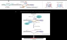
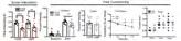
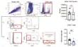
Maintaining genomic stability is essential for preserving cellular health and preventing diseases, especially cancer Helicases are crucial enzymes that unwind DNA and RNA structures during transcription and replication processes Two significant obstacles to replication have been identified: Gquadruplexes, which are four-stranded structures formed by guanine-rich DNA/RNA sequences, and R-loops, which are RNA-DNA hybrids These structures can lead to genomic instability by causing conflicts between transcription and replication Recent research has emphasized the critical role of helicases in resolving these structures, suggesting potential therapeutic avenues To explore these concepts in drug discovery, researchers utilize A673 AID/TIR1 cells, a modified Ewing sarcoma cell line featuring an auxin-inducible degron system This system enables swift and reversible breakdown of the EWSR1-FLI1 fusion protein, a key factor in Ewing sarcoma, when treated with auxin analogs such as 5-Ph-IAA The drug screening experiments aim to evaluate how various compounds affect these cells, both with and without EWSR1-FLI1 depletion This approach may uncover novel therapeutic strategies for Ewing sarcoma and other diseases related to genomic instability
Figure 2: Schematic depicting AID based degron approach for depletion of endogenous EWSR1-FLI1.
Figure 3: Drug Screening and
Methods
Discussion and Conclusion
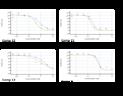
Our research illuminates the complex interactions between Gquadruplexes (G4s), R-loops, and specific helicases in maintaining genomic stability within cancer cells We identified four compounds out of 30 that demonstrated significant effects on A673 cells, with enhanced efficacy in cells lacking the EWS-FLI1 fusion protein The involvement of BACH1, DHX9, and DHX36 helicases, known for their roles in resolving G4 structures and R-loops, points to their significance in this process The differential response based on EWS-FLI1 presence indicates a possible avenue for targeted Ewing sarcoma therapies Our findings emphasize the importance of further investigating the specificity and effectiveness of G4targeting compounds Additionally, they highlight the potential for exploring combination therapies and applications in various genetic disorders This study lays the groundwork for future research aimed at leveraging these molecular interactions to develop novel therapeutic strategies in cancer and beyond
Future Research
Investigate the specific mechanisms by which BACH1, DHX9, and DHX36 resolve G4s and R-loops, particularly in the context of EWS-FLI1 presence or absence
Employ techniques such as HepG4-seq and HBD-seq to map native co-localized G4s and R-loops in Ewing sarcoma cells before and after helicase overexpression
• Investigate potential synergistic effects between G4-targeting compounds and drugs targeting other cellular processes affected by G4s and R-loops
Develop and test inhibitors specific to BACH1 DHX9 or DHX36 to further elucidate their individual roles in G4 and R-loop resolution
Funding
This research is funded by the Bi-National Science Foundation (BSF)2021273 to RT and JCS
Figure 1: Formation and resolution of G4s and
Figure 5: Dose response
Jesse Altemus
Identification and validation of peroxisomal protein interactor hits identified via a novel proteomics method
Jacob Schwartz Medical Pharmacology
Identification and validation of peroxisomal protein interactor hits identified via a novel proteomics method Jesse Altemus1, Rachel Victor, Jacob Schwartz1,2 1Department of Pharmacology, University of Arizona College of Medicine, Tucson, AZ 2University of Arizona Cancer Center, Tucson, AZ
INTRODUCTION
METHODS
REFERENCES
Jennifer Bea
Behavioral Measurement and Intervention Shared Resource (BMISR)
Health Promotion Sciences
Behavioral Measurement and Interventions Shared Resource (BMISR)
Angela Yung, Jennifer Bea, Rina Fox The University of Arizona Cancer Center, Tucson AZ
INTRODUC TION
The University of Arizona Cancer Center (UACC) BMISR provides support to local and national researchers studying lifestyle behavior quality of life and patient reported outcomes related to cancer prevention and control within diverse populations.
BMISR is a unique “one stop” resource that offers investigators one access point to several research services ranging from study design consultation to measurement and instrument selection to intervention delivery. BMISR provides innovative approaches to investigator data collection needs utilizing a large team of trained student research assistants. Through this unique model, BMISR creates cost-effective partnerships at the University of Arizona (UA) and beyond.
•24-hour dietary recalls
•Body
MEASUREMENT
•Physical activity assessment methods
administered surveys
and functional tests including
DEVELOPMENT
OUR REACH
HOW TO ENGAGE BMISR FOR YOUR PROJECTS
• CONTACT US. When writing grant proposals, email BMISR with a project inquiry and the services of interest.
GRANT TEXT AND LETTERS OF SUPPORT BMISR can provide grant text and letters of support to strengthen your application.
CONSULTATION. Providing BMISR with your study specific aims will allow us to best support you. BMISR directors, as well as experts on the advisory board, can provide consultation for study design.
COST ESTIMATE. BMISR will develop and provide a cost estimate and project scope for your approval. Upon agreement, include BMISR services in grant budget.
AWARD NOTIFICATION Upon notice of award, you and BMISR can review cost estimate and project scope and revise as needed. BMISR will create a comprehensive timeline for services. BEHAVIORAL
BMISR TRAINING. BMISR coordinators will train their team and/or your team for project specific protocols/ instruments and create a workflow to ensure all dead-lines are met.
CONTINUAL COMMUNICATION. Work with BMISR to develop a schedule for reports and meetings. BMISR will maintain contact with the coordinator throughout the study via emails and conference calls as needed.
•Researchers requested support for database development (n=15), dietary measurement (n=14), bilingual interviews (n=5), physical activity measurement (N=4), health coaching (n=2), and mHealth studies (n=2).
•Researchers conducted studies in populations of adolescents, food bank patrons, American Indians, Hispanics, healthcare providers, cancer survivors and their informal caregivers.
•During this time period, 57 publications included results and services generated by the BMISR or were supported by BMISR personnel.
Uloma Beauty Elvis-Offiah
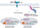
EGFR activation reverses gastrin suppression by MENIN by inducing its nuclear export
Dr Juanita L Merchant Cancer Biology program
EGFR activation reverses gastrin suppression by MENIN by inducing its nuclear export
Uloma B. Elvis-Offiah1,2, Suzann Duan1, Sulaiman Sheriff1, Juanita L Merchant1,2
1University of Arizona College of Medicine, Department of Medicine, Division of Gastroenterology; 2Programs in Cancer Biology, University of Arizona, Tucson, AZ 85724
ABSTRACT
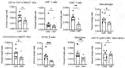
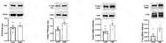
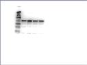
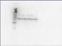
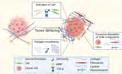
Background: Gastroenteropancreatic neuroendocrine tumors (GEP-NETs), particularly gastrinomas, secrete excess gastrin due to MEN1 gene mutations or post-translational inactivation of MENIN, a tumor suppressor that represses gastrin transcription EGFR ligands, abundant in the duodenal microenvironment, may drive gastrinoma development by interfering with MENIN function This study investigates how EGFR activation alters MENIN’s localization, stability, and ability to suppress gastrin Methods Human gastric carcinoma cells expressing WT and NLS-mutant MENIN constructs were treated with epiregulin (EREG) Gastrin expression was assessed via qPCR, MENIN localization via immunofluorescence, and MENIN stability via proteasome inhibition assays Results: MENIN suppresses gastrin expression in a nuclear localization-dependent manner WT MENIN and constructs with at least two NLSs reduced gastrin by 50% while the deletion of all three NLSs abolished suppression EREG treatment induced gastrin in cells expressing WT or partially NLS-deficient MENIN but had no effect when all NLSs were deleted NLS-deficient MENIN was unstable and degraded via the proteasome, as confirmed by MG132 rescue Immunofluorescence revealed that EREG promoted MENIN’s cytoplasmic translocation, demonstrating that EGFR activation disrupts MENIN’s nuclear retention, stability, and suppression of gastrin This study highlights that EGFR activation induces gastrin expression by driving MENIN translocation to the cytoplasm and promoting its degradation, highlighting nuclear retention of MENIN or EGFR inhibition as potential therapeutic strategies for MEN1-associated gastrinomas
BACKGROUND
MEN1 Inactivation Promotes Gastrinomas
1. GEP-NETs, particularly gastrinomas lead to excessive gastrin secretion due to MEN1 mutations a gene that encodes tumor suppressor protein, MENIN
2. In normal cells, MENIN localizes to the nucleus via three nuclear localization signals (NLSs), suppressing gastrin by interaction with transcription factors and nuclear proteins
3. Duodenal gastrinomas retain the wild-type (WT) MEN1 allele suggesting that loss of function may occur through translocation or degradation of MENIN rather than loss of heterozygosity (LOH)
4. The duodenal microenvironment, rich in EGFR ligands (e g amphiregulin), may drive MENIN translocation to the cytoplasm, impairing its function
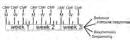
MEN1 Gene Exons and Encoded MENIN NLSs
METHODS
PCR cloning used to generate Menin C-terminal domain deletion constructs
Used gastrin-expressing human gastric carcinoma cell line model with
Proteasome
I WT MENIN and NLS-retaining constructs suppressed gastrin transcriptional activity and expression
III EGFR activation promoted WT MENIN nuclear export and degradation in the cytoplasm
Preclinical Studies of the Neuropathological Effects of Breast Cancer Chemotherapy
James Bibb Department of Translational Neurosciences
methotrexate, and 5-fluorouracil (CMF) in preclinical studies using mice.
• CMF treatment in mice mimicking chemotherapy for breast cancer caused anxiodepressivelike behaviors, impaired motor function, reduced contextual memory, and deficits in fear memory extinction, similar to cognitive and emotional impairments observed in breast cancer patients.
Chemotherapy disrupts neuroimmune homeostasis, as shown by depletion of brain-resident and infiltrating immune cells including microglia, macrophages, dendritic cells, CD4+ T-cells.
Chemotherapy treatment increased Cdk5 activity in the striatum, leading to elevated Cdk5 dependent phosphorylation of substrates (DARPP-32, synapsin 1, and Tau) suggesting a role for Cdk5 in the neuronal changes associated with chemobrain symptoms.
CMF treatment enhances excitability in the NAc, increasing I/O response, paired pulse ratio (PPR), and long-term potentiation (LTP) in cortico-ventral striatal neurons.
Single-nucleus RNA sequencing revealed 15 cell types in the NAc including dopamine receptor-linked MSNs and support cells, with no changes in cell types after CMF treatment, setting the stage for studying gene expression and chromatin alterations.
Breast cancer chemotherapy causes behavioral and cognitive impairments (increased anxietylike behaviors and impaired learning and memory), accompanied by immune cell depletion, corticostriatal circuit disruption, and aberrant Cdk5 signaling. Alterations in brain circuitry and neuroimmune system function may drive the behavioral and cognitive dysfunction associated with chemotherapy.
• Current studies are in progress to identify cell-type-specific molecular and epigenetic mechanisms underlying these effects using multiomic approaches
Further comprehensive studies are required to investigate the impact on neural circuitry function and the cell-type-specific effects. Additionally, it would be beneficial to conduct these investigations within the framework of cancer modeling, utilizing murine models that either harbor allografts or are transgenically engineered to replicate cancer pathologies with clinical relevance.
REFERENCES
Zahra Fatahivanani1, Sujoud Al kawasmy1, Julian Ramos1, Jessica Contreras1, Alan Umfress2 , Ayanabha Chakraborti1 & James A. Bibb1
Schematic of CMF treatment and post-treatment assessments
Fig 1. CMF chemotherapy affects body weight and induces behavioral alterations in mice.
Fig
Graphical
Janet Funk
Interrogating steroid hormone signaling in a murine model of ER+ human breast cancer bone metastases
Department of Medicine
Interrogating steroid hormone signaling in a murine model of ER+ human breast cancer bone metastases
RESULTS
INTRODUCTION
line here is an important limitation.
v At the same time, the current findings can inform interpretation of pre-clinical breast cancer investigations where, for example, ER+ orthotopic tumor studies are primarily performed in E2-treated OVX mice, while ER+ BMET models are most often conducted using
METHODS
Janet Funk
Natural Product Dietary Supplement Use by Women Diagnosed with Breast Cancer Department of Medicine
Natural Product Dietary Supplement Use by Breast Cancer Survivors
Anthony M. Rossi1, Meg Hauer2, Betsy C. Wertheim3, Hilary Kleppel1, Jennifer W. Bea3,4,5, Janet L. Funk3,5,6
1Honors College, 2College of Medicine, 3University of Arizona Cancer Center, 4Department of Health Promotion Sciences, Mel & Enid Zuckerman College of Public Health, 5Department of Medicine, College of Medicine, and 6School of Nutritional Sciences & Wellness, College of Agriculture and Life Science, The University of Arizona.
RESULTS
INTRODUCTION
§ Women with breast cancer are frequent users of natural product-derived dietary supplements (NP).
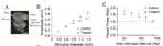
§ Many NP bioactive constituents, including those with musculoskeletal protective effects (e.g., quercetin, curcumin), circulate as inactive glucuronides requiring in vivo activation (deconjugation) by ß-glucuronidase (GUSB) (Fig 1).
§ While risks and benefits of most NP in breast cancer are not well characterized, we have recently documented an association of GUSB with preservation of lean mass in breast cancer survivors, a group that suffers accelerated loss of muscle and bone (Fig 2).
OBJECTIVE
§ NP use in breast cancer is often underreported to health care providers and has not been assessed in observational studies for over a decade.
§ A study was undertaken to document current NP use in those diagnosed with breast cancer, including:
o associations with disease characteristics and breast cancer treatments, and o primary sources of information when deciding to use a particular NP
METHODS
§ Study population: breast cancer patients and survivors
§ Recruitment: primarily via social media (Facebook advertisements, Twitter posts, breast cancer support and interest group posts, various UA platform posts)
§ Survey: deidentified online questionnaire (English; Spanish) about NP use and breast cancer via REDCap link
§ Statistical analyses: multivariate logistic regression was performed to assess relation between NP use and participant demographics or disease characteristics, adjusted for key demographic and clinical variables
§ This protocol was approved by UA IRB (#1710898912).
2. NP Use in Breast Cancer
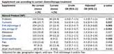
Most participants (68%) reported current NP use, with 27% concurrently using at least three products.
Top-reported NP products (>15% prevalence) were probiotics, turmeric, fish oil/omega-3, melatonin, and cannabis.
Fifty percent of participants reported use of NP that circulate as inactive glucuronides (blue bars) requiring ßglucuronidase activation.
Source of funding: The University of Arizona Honors College Thesis Award (to AMR) and NCI (CA023074 to UACC).
3. NP Use According to Breast Cancer Characteristics
§ Overall NP use was:
o more common with estrogen-receptor positive (HR+) tumors (ORadj 1.53, p = 0.005)
o lower in the South (ORadj 0.55, p < 0.001) and Northeast (ORadj 0.50, p < 0.001) vs West
o less common in African American participants (ORadj 0.56, p = 0.025)
o but did not differ by age, ethnicity, time since diagnosis, advanced disease, HER2 status, ever/current radiation or chemotherapy or current endocrine therapy use
§ Cannabis use was more frequently reported by those with advanced disease (ORadj 2.1, p < 0.001), as was ginger (ORadj 1.8, p = 0.041), and cannabis use trended higher in those undergoing current radiation treatment (ORadj 1.8, p = 0.050).
§ In those currently undergoing chemotherapy, NP use trended lower (p< 0.05 for NP in blue), excepting cannabis or ginger, but use of NP with possible adverse effects when combined with chemotherapy (turmeric, any omega-3) was still > 15%.
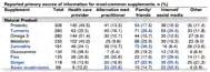
§ For five of the top nine NP (see blue font), family/friends or internet/social media were the most common information source when choosing to use these supplements.
§ Breast cancer survivors commonly reported using multiple NP supplements, including those requiring GUSB for activation.
§ Given the overall paucity of research on the risks and benefits of NPs in breast cancer and frequent NP use without health care provider (HCP) recommendation, it is important for HCP to inquire about supplement use in this population.
§ Additional research focused on NPs commonly used by breast cancer survivors may also be merited.
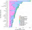
1. Consort Diagram and Participant Characteristics
Janet Funk
A possible tumorigenic role for extranuclear estrogen receptor-alpha (ER) signaling in the bonedisseminated ER+ breast cancer microenvironment Department of Medicine
A possible tumorigenic role for extranuclear estrogen receptor-alpha (ER) signaling in the bone-disseminated ER+ breast cancer microenvironment
RESULTS
INTRODUCTION
treatments, experiments were undertaken to query the centrality of this pathway to ER+ breast cancer progression specific to bone
v As previously described,1-3 intact female nude mice, which require estrogen supplementation to support human ER+ breast cancer xenografts,4 were inoculated (intracardiac, IC) with human ER+ MCF-7 BMET-derived breast cancer cells (43 4M cells) that form estrogen and TGFß-dependent BMET 2 v Supplementation with 17-ß estradiol (E2) pellets (60-day release, 0 72mg) or a nuclear ER-only agonist (estetrol, E4; 3 or 4 mg/day, a 100-fold higher dose than E2 consistent with its 100-fold lower ER affinity)5 was started immediately prior to tumor cell inoculation Bone dissemination was not altered by either treatment 3 v Osteolytic ER+ BMET formation was monitored radiographically, with histologic confirmation of ER+ BMET (cytokeratin- and/or ER-positive tumor cells by IHC)1-3 v As indicated, in some experiments, nude mice (ovary-intact or ovariectomized [OVX]) were inoculated with 43 4M cells (in Matrigel) via the mammary fat pad Bone effects of E2 or E4 on bone mineral density in intact or OVX mice were determined by DXA
v Statistical analyses were also conducted as previously described 1-3
Spencer Holmes
Radiomics in Oncology: Advancing Precision Medicine Through Quantitative Imaging
Dr Hina Arif Medical Imaging
Radiomics in Oncology: Advancing Precision Medicine Through Quantitative Imaging
However, this can be time consuming and may result in large inter-reader variability Automated algorithms that can provide reliable and reproducible ROIs are being developed and are markedly improving efficiency in segmenting suspicious lesions
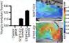
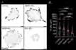
3 Mask generation An automated procedure was developed using Python scripting to generate the mask for each ROI from the annotated images (Figure 2) The mask allows analyzing radiomics features within each ROI
4 Feature extraction An open-source Python package3 was used for extracting radiomics features In addition to first order statistics, features based on shape, gray level co-occurrence matrix, gray level run length matrix, gray level size zone matrix, neighboring gray tone difference matrix, and gray level dependence matrix, for a total of 93 features were extracted
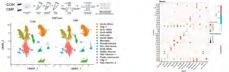
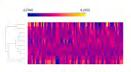
5. Analysis and Model Development A data mining toolkit (Orange data mining, version 3 38 1) was used for analysis and model development For each radiomics feature, the distribution was summarized using violin plot (Figure 3A) A heatmap was generated to visualize the variation in radiomics features among the nodules studied (Figure 3B) To improve visualization, K-means based clustering was used to generate 10 clusters
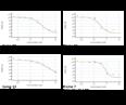
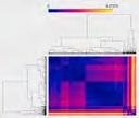
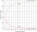
Correlation analysis: The pair-wise correlations among the radiomics features were determined To visualize the correlation, the normalized Euclidean distances were obtained and were used to generate a distance map (Figure 4) To reduce dimensionality, principal component analysis (PCA) was performed and seven components explained more than 90% of the variance (Figure 5)
Summary Upon successful completion, the developed radiomics pipeline can potentially enable non-invasive assessment of tumor heterogeneity, disease progression, and treatment response
References:
1. Mayerhoefer ME, Materka A, Langs G, Häggström I, Szczypiński P, Gibbs P, Cook G. Introduction to Radiomics. J Nucl Med. 2020 Apr;61(4):488-495.
2. Shur, Joshua D et al. “Radiomics in Oncology: A Practical Guide.” Radiographics a review publication of the Radiological Society of North America, Inc vol. 41,6 (2021): 1717-1732.
3.
Somto Ike
Image Analysis of Mechanically Conditioned Breast Cancer Tissue in Mouse Models
Dr.
Ghassan Mouniemne
Department of cellular and molecular biology
Image Analysis of Mechanically Conditioned Breast Cancer Tissue in Mouse Models
[1]Somto Ike, [2]Marco Padilla Rodriguez, [3] Faith Rice, [3] Jennifer Barton [4]Ghassan (Gus) Mouneimne University of Arizona Cancer Center, 1515 N Campbell AVE, Tucson, AZ, 85724
BACKGROUND
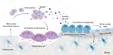
Breast cancer metastasis is significantly influenced by the tumor microenvironment (TME). Mechanical forces within the TME, including tissue stiffness, contribute to cancer progression and metastasis. Collagen, a key extracellular matrix (ECM) protein, undergoes structural changes that promote aggressive cancer growth, with increased collagen fiber crisscrossing observed in tumors derived from mechanically conditioned tissues. These alterations indicate heightened fibrosis, which correlates with the aggressiveness of the cancer. Understanding how these mechanical forces interact with the ECM provides insights into cancer metastasis. Fibronectin holds TGFbeta (Transforming growth factor-beta) in an inactive state, and mechanical activation from cancer cells releases TGF-beta, which in turn increases tissue stiffness via CAF (Cancer Associated Fibroblasts) activation. This feedback loop between ECM stiffness and tumor growth is critical in understanding metastatic progression.
METHODOLOGY
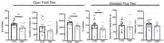
We employed second-harmonic imaging microscopy (SHIM) to visualize collagen fibers in both soft and stiff breast cancer tissues.
SHIM takes advantage of second-harmonic generation (SHG), a nonlinear optical effect, to provide detailed contrast of collagen structures.
The images were preprocessed using a customized GA3 (General Analysis3) recipe in NIS-Elements software. This included cropping the images, denoising, and enhancing contrast with LUT modifications. We applied skeletonization to the collagen fibers, quantifying branches and endings. The data were then analyzed and compared between soft and stiff tissues to identify patterns related to tissue stiffness and tumor progression.
RESULTS AND ANALYSIS
CONCLUSIONS
Our analysis demonstrated that tissues that are mechanically conditioned by stiff matrix ex-vivo exhibit generate tumors with increased collagen fiber crisscrossing in-vivo, reflecting heightened fibrosis and aggressiveness of the cancer. Softer tissues, in contrast, had less collagen branches and endings, suggesting greater tissue flexibility and reduced fibrotic behavior. These findings support the hypothesis that ECM stiffness correlates with cancer progression. Understanding these mechanical interactions may provide potential targets for therapeutic intervention aimed at modifying tissue stiffness to inhibit cancer metastasis.
REFRENCES
[1] Pijanka, Jacek K et al. “Quantification of collagen fiber structure using second harmonic generation imaging and two-dimensional discrete Fourier transform analysis: Application to the human optic nerve head.” Journal of biophotonics vol. 12,5 (2019): e201800376. doi:10.1002/jbio.201800376
[2] Mai, Z., Lin, Y., Lin, P. et al. Modulating extracellular matrix stiffness: a strategic approach to boost cancer immunotherapy. Cell Death Dis 15, 307 (2024). https://doi.org/10.1038/s41419024-06697-4
[3] Wershof, Esther et al. “A FIJI macro for quantifying pattern in extracellular matrix.” Life science alliance vol. 4,3 e202000880. 27 Jan. 2021, doi:10.26508/lsa.202000880
ACKNOWLEDGEMENTS
Kaneez Khakwani
Magnetic Resonance Imaging based non-invasive accurate diagnosis and characterization of intrahepatic cholangiocarcinoma
Hina
Arif Tiwari
Medical Imaging
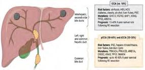
Cholangiocarcinoma is a rare, highly aggressive malignancy of the biliary epithelium, often diagnosed at an advanced stage due to its subtle and non-specific symptoms. Early and accurate imaging is critical for diagnosis, staging, and guiding management, with MRI particularly MRCP emerging as the preferred non-invasive modality for comprehensive biliary assessment.
Cholangiocarcinoma is anatomically classified into three distinct subtypes based on the location of tumor origin within the biliary tract: intrahepatic, perihilar, and distal. Intrahepatic cholangiocarcinoma arises from the small bile ducts within the liver parenchyma and is often associated with mass-forming lesions. Perihilar cholangiocarcinoma, also referred to as a Klatskin tumor, originates at the confluence of the right and left hepatic ducts and represents the most common subtype. Distal cholangiocarcinoma develops in the common bile duct near the head of the pancreas and may present similarly to pancreatic head carcinoma. Among these, intrahepatic cholangiocarcinoma has been gaining increased clinical attention due to its rising incidence, distinct molecular profile, and challenges in early detection and management.
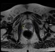
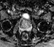
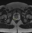
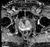
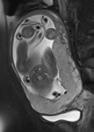
Magnetic Resonance Imaging (MRI), combined with Magnetic Resonance Cholangiopancreatography (MRCP), is the preferred modality for evaluating intrahepatic cholangiocarcinoma. The protocol includes T1weighted sequences for anatomical assessment, T2-weighted imaging to evaluate biliary dilation and tumor extent, and diffusion-weighted imaging (DWI) for lesion detection. Dynamic contrast-enhanced imaging aids in assessing vascular involvement and enhancement patterns. MRCP offers high-resolution, non-invasive visualization of the biliary tree, enabling accurate delineation of strictures and obstruction. Intrahepatic cholangiocarcinoma typically appears as a large, solitary liver mass with irregular or lobulated margins, often accompanied by satellite lesions. MRI shows hypointensity on T1 and hyperintensity on T2-weighted images, with signal variation based on tumor subtype. The tumor has a hypovascular nature, exhibiting delayed, heterogeneous or homogeneous gadolinium enhancement with varying patterns, including peripheral enhancement and progressive filling.
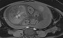
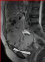
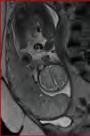
A: Axial T2 image shows T2 intermediate liver lesion at segment 4, 5 and 8. B-E: Are the dynamic T1GE pre and post contrast imaging showing heterogeneously enhancement of the lesion with few satellite lesions. Pathology confirmed intrahepatic cholangiocarcinoma.
A: Axial T2WI shows intermediate signal hepatic mass (5 cm). B-E: Are the axial dynamic pre and post contrast T1GE phases showing peripherally enhancing lesion at segment 8 with minimal central filling. Background features of chronic liver disease. Pathology confirmed intrahepatic cholangiocarcinoma.
A-D: Are the dynamic T1GE pre and post contrast imaging showing infiltrative mass at hepatic segment 5 and 6. E: Axial venous phase T1GE showing thrombosis of the right portal vein without direct tumoral extension or restriction on DWI image. Pathology confirmed intrahepatic cholangiocarcinoma.
Intrahepatic Cholangiocarcinoma is a challenging malignancy with late presentation and complex anatomy. MRI, especially with MRCP, is crucial for diagnosis, staging, and surgical planning, guiding treatment and improving outcomes
authors have nothing to disclose.
Pavicevic,
Erik Larsen
Accessing the IMPACT (Individual Molecular Registry of Patients for Accelerated Clinical and Translational medicine)
Dr. Ritu Pandey UAHS
IMPACT (Individual Molecular Registry of Patients for Accelerated Clinical and Translational medicine), a UAHS strategic initiative for genome data analytics of UACC patients to foster precision research
Larsen3, Chenbo Sun1, Rudy Salcido4, Michele
Ritu Pandey1,2,3
Gabriella Mamo
MR imaging has high soft-tissue contrast resolution.
• MR is reserved as a complementary exam for equivocal findings at US.
• No fetal or maternal radiation risks.
MRI is superior to other modalities in diagnosis of placenta accreta disorders according to FIGO survey of 2017-2018.
Succenturiate
Makena Manes
Dissecting the Microenvironment: Cytoskeletal Interfaces in Cancer Invasion
Dr. Ghassan Mouneimne Cancer Biology
Dissecting the Microenvironment: Cytoskeletal Interfaces in Cancer Invasion
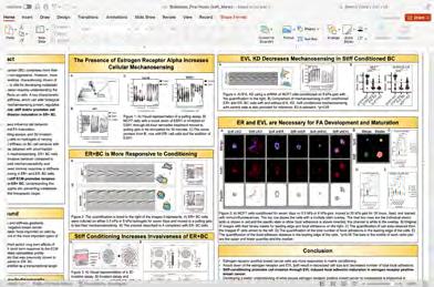
OVERVIEW - Research in our laboratory is focused on understanding how aberrant structural organization of the cytoskeleton influences cellular behavior, such as cancer cell invasion and metastasis Our goal is to identify distinct actin cytoskeletal architectures that impact the response of cancer cells to well-known genetic and microenvironmental factors during cancer progression We are focused on understanding how cancer cells integrate biochemical and mechanical cues from their surroundings and translates them into cellular responses that contribute to their invasiveness We hope that such knowledge will allow us to target key factors regulating these responses in the attempt to curb metastatic progression Our experimental approaches include modeling cancer in in vitro organotypic systems, in vivo mouse models, and clinical samples donated by cancer patients In addition, we have a strong foundational infrastructure to use advanced omics techniques in our approaches
MECHANICAL MEMORY
The mechanical properties of the primary tumor microenvironment influence cancer progression and prognosis However, whether these properties continue to inform cell behavior at distal metastatic sites had not been explored Our lab discovered that stiff primary tumors induce persistent epigenetic changes in breast cancer cells, driving aggressive and dangerous osteolytic metastases These changes to the transcriptional landscape are maintained by the osteogenic transcription factor and gene bookmarker RUNX2 in a process called “mechanical conditioning” (Watson et al (2021 Cell Rep) We designed a gene panel, MeCo, comprised of genes that are differentially regulated by stiffness, which serves as a prognostic tool to predict bone metastases Top R ght Mechan
TUMOR MICROENVIRONMENT & MIGRATION
The tumor microenvironment influences many aspects of cellular behavior, including cell migration Cells sample their microenvironment through focal adhesions (FAs), which allows them to mature via EVL-mediated actin polymerization Our lab has shown EVL as a crucial regulator of mechanosensing (Puleo et al (2019) JCB), which is the initial step cell migration toward increasing stiffness Utilizing acute and long-range migration assays in combination with 3D invasion assays, our lab focuses on the changes in migration & cytoskeletal dynamics at FAs
Right Assays to assess the effect of the physical microenvironment on migration and invasion (A) Illustration depicting mechanosensing assay using a micropipette (B) Sensing index and turning angle analyses, where the crosshairs denote micropipette position (C) Illustration of 3D invasion assay to assess the invasive potential of tumor cells (D)
Illustration of durotaxis assay using a stiffness gradient of 1mm (E)
Illustration of the data collected and used from the durotaxis assay
Left EVL is required for FA maturation (A) Representative inverted TIRF images of paxillin staining in control (LKO), Mena KD, VASP KD, and EVL KD MCF7 cells Scale bars are 10μm (B) Dot plot shows quantification of FA area Data are collected from three independent experiments; all data points are shown
ACTIN DYNAMICS IN CELL MOTILITY
The dynamic regulation of actin cytoskeletal architecture is how cells convert environmental cues into effective behaviors such as migration Our lab identified two antagonistic actin pathways that promote or suppress directed cell migration – lamellipodial actin networks (LANs) and cortical actin bundles (CABs), respectively These structures are differentially regulated by the physical and biochemical micro-environment, and in breast cancer cells, by Estrogen Receptor activity (Padilla-Rodriguez et al (2018) Nat Comm)
Utilizing live-cell imaging in 2D and 3D, and acute cellular manipulations via optogenetic tools, our lab strives to discover the spatiotemporal network between sensing, protein dynamics, actin reorganization and cell motility
Right Antagonism contributing to lamellipodial (CABs; myosin- and MCF7 cells immunolabeled ROIs (C) MCF7 cells Left: 3D rendering of Time series and kymograph treated with 50µm ROCK min Scale 10µm
Celia Sahli
Machine Learning for Breast Tumor Classifica-
tion: Insights from Nuclear Morphology
Kenry
Pharmacology and Toxicology
Orli Sanyal
A Conceptual Model to Assess Barriers to Cancer Care for Native Americans in Arizona
Dr. Julie Armin College of Medicine
Manlin Shao
Interaction Between Tumor Cells and Tumor-Associated Macrophages in p53-Mutated Oral Squamous Cell Carcinoma
Carlos Caulin Department of Otolaryngology
Ganga Velma
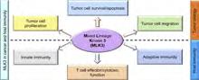
Discovery of novel NAMPT/MLK3 dual inhibitor for Triple-negative Breast Cancer (TNBC)
Gregory Thatcher
Pharmacology and Toxicology
Discovery of novel NAMPT/MLK3 dual inhibitor for Triple-negative
Breast Cancer (TNBC)
Ganga Reddy Velma, Soumya Reddy Musku, Sandeep Kumar, Sweta Misra, Piush Srivastava, Rakesh Sathish Nair, Martha Ackerman-Berrier Rui Xiong, Laura Bloem, Ajay Rana*, Gregory R J Thatcher*
Location: Skaggs building, College of Pharmacy, 4th Floor Rm. 474
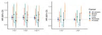
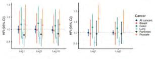
Associations between air pollution and cancer survivorship in the U.S: the NIH-AARP Diet and Health study
Chris Lim Department of Community, Environment, and Policy
BACKGROUND
Jeonggyo Yoon1, Rena R. Jones2, George D. Thurston3, Chris C. Lim1 1Department of Community, Environment, and Policy, Zuckerman
RESULT
Ambient particulate matter (PM2.5, <2.5 µm) is a known human carcinogen, but research on associations with cancer survival remains limited. We sought to investigate the long-term effects of air pollution exposure on post-diagnosis cancer survival, utilizing data from the NIH-AARP Diet and Health Study.
METHODS
Study populat on & Morta ity Ascertainment :
ü The NIH-AARP Diet and Health Study, aged 50–71 years, from 6 U.S. states (CA, FL, LA, NJ, NC, PA) and 2 metropolitan areas (Atlanta, GA; Detroit, MI), with follow-up from 1995 to 2011.
ü Vital status was determined through regular linkage of the cohort with multiple data sources: the Social Security Administration Death Master File, National Death Index Plus, cancer registry records, questionnaire responses, and other mailings.
ü Analyzed associations with survival of all cancers combined (N=92,818), and lung (N=11,213), colon (N=6,389), prostate (N=25,686), breast (N=10,821), and pancreas cancer (N=1,901).
• A r Pol ution Exposure Assessment:
ü Annual average PM2.5 concentrations (ug/m3) (1980–2010) were estimated at the census tract level using a spatio-temporal model based on geographic predictors, with extrapolation for pre-1999 levels before nationwide monitoring began.
ü Annual average NO₂ concentrations (ppb) (1990–2012) were estimated at the census tract level using a national land use regression model incorporating regulatory monitoring data, satellite-based measurements, and geographic predictors.
• Statistica Methods:
ü Cox proportional hazards models with time-varying exposures adjusted for demographic, socioeconomic, and behavioral factors (age, sex, race and ethnicity, education level, body mass index, diet, alcohol, and smoking history).
ü Different lag structures were examined to assess cumulative and delayed effects on survival outcomes.
DISCUSSION
Associations between air pollution and cancer survivorship varied by cancer type. Our findings highlight the need for survivorship research that addresses cancer type-specific effects and identifies strategies to improve survivor outcomes.
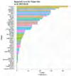
Table 1 Descriptive characteristics of the participants at baseline (n=92,818)
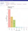
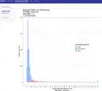
Figure 2. Hazard ratios (95% CI) for cause-specific cancer death associated with PM₂
Your donations to the Ginny L. Clements Breast Cancer Research Institute support advances in innovative clinical and supportive care, education, and early detection, and can help us recruit the best researchers and clinicians.