The laboratory inside the microscope Darmstadt researchers led by Leopoldo Molina-Luna observe the tiniest of electronic components in action. Not only can their electron microscope image individual atoms; it can also produce, heat and electronically control the sample.
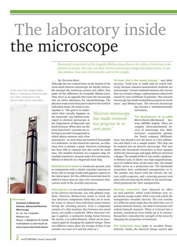
At the top of the sample holder, there is a laboratory of microscopic size where temperature and electric field can be controlled.
_ By Christian Meier Although the two control boxes at the bottom of the room-sized electron microscope are hardly noticeable amongst the numerous screens and cables, they make all the difference for Leopoldo Molina-Luna. They serve as an upgrade that turns the microscope into an entire laboratory for nanotechnology. The physicist wants more than just to observe the world of individual atoms. He wants to manipulate it. ‘The goal is to understand what actually happens on the nanoscale,’ says Molina-Luna, expert in electron microscopy at the Department of Materials and Earth Sciences. When they use the term ‘nano-level’, scientists are referring to an order of magnitude in which objects measure only a few nanometres - no more than one hundred thousandth of a millimetre. In this minuscule universe, an influenza virus is already a giant. However, technology has been able to contract into this world for some time. The smallest elements of a computer chip, for example transistors, measure only a few nanometres. Billions of them fit on a fingernail-sized chip.
‘At least, that is the model concept,’ says Molina-Luna. ‘Until now, it could only be tested indirectly, because classical measurement methods are macroscopic.’ Current methods measure the current flow at a certain voltage, a phenomenon collectively caused by tens of billions of particles. ‘But electron microscopy has made tremendous progress in recent years,’ says Molina-Luna. The electron microscope has become a ‘multidimensional’ instrument.
‘Electron microscopy has made tremendous progress in recent years.’
Photo: Katrin Binner
Nanoelectronics wants to break new ground, such as components with a similar reminder function as brain cells or storage media with gigantic capacity in the tiniest space. For this, different materials must be added to layers that are only a few nanometres thin: custom work in the invisible microcosm.
Information Institute of Materials Science, Advanced Electron Microscopy Dr. rer. nat. Leopoldo Molina-Luna Phone: ++49 (0)6151/16-22309 molina@aem.tu-darmstadt.de www.mawi.tu-darmstadt.de/aem
2
Although you can see such diminutive components in the electron microscope, you only glimpse snapshots. However, Molina-Luna wants to watch the tiny electronic components while they are at work. He wants to observe how individual atoms behave during the switching process. Such a component consists of a thin insulator slice between two metal layers, not unlike a sandwich. When electrical voltage is applied, a conductive bridge forms between the metals, a so-called filament, which can be interrupted again by changing the applied voltage. These two different states allow the storage of data if one encodes one state as 0 and the other as 1.
hoch3 FORSCHEN / Issue 8 / Spring 2019
The development of so-called Micro-Electro-Mechanical Systems (MEMS) helped. These are complex electromechanical devices of microscopic size. Molina-Luna‘s cooperation partner, the Dutch company DENSsolutions, has shrunk a test lab down to a few millimetres and fixed it on a sample holder. This chip can be inserted into an electron microscope. This now allows the Darmstadt researchers to heat samples within the microscope and apply different electrical voltages. The sample holder is very stable, emphasizes Molina-Luna. It allows very high magnifications, up to 25-million times. At the same time, the sample holder serves as a production site: The Darmstadt researchers have synthesized nanoparticles on it. The powder was mixed with the solvent, the solvent could evaporate, and a sintering process took place after introducing the holder in the microscope, which produced the ‘new’ nanoparticles. Recently, researchers have observed an effect on such particles, which could facilitate new, super-dense storage media. With a core and a shell, the nanoparticles resemble cherries. The core consists of a different metal oxide than the shell (the core of sodium bismuth titanate, the shell of strontium-rich sodium bismuth titanate). When it is forced into a particle, mechanical stress builds up in its interior. Researchers controlled the strength of the stress by changing the proportion of strontium. The mechanical stress leads to so-called flexoelectricity: Inside, the electrical charge carriers split