Dental News, Volume XXIII, Number III, 2016


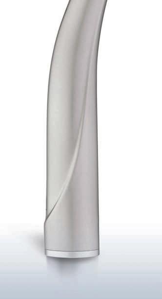
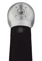
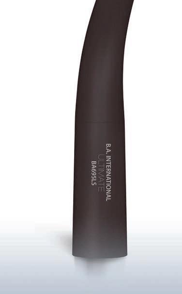
Surgical Micromotor System Ultrasonic Bone Surgery System
Surgical Micromotor System Ultrasonic Bone Surgery System


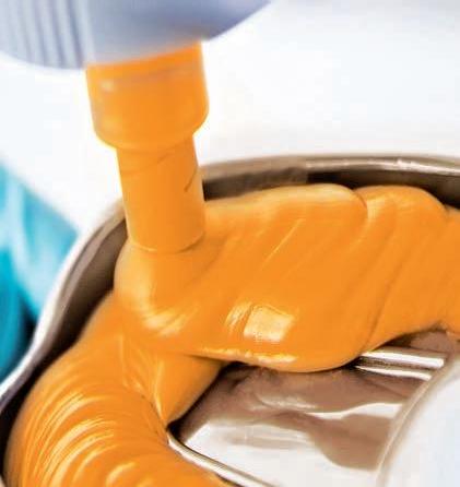
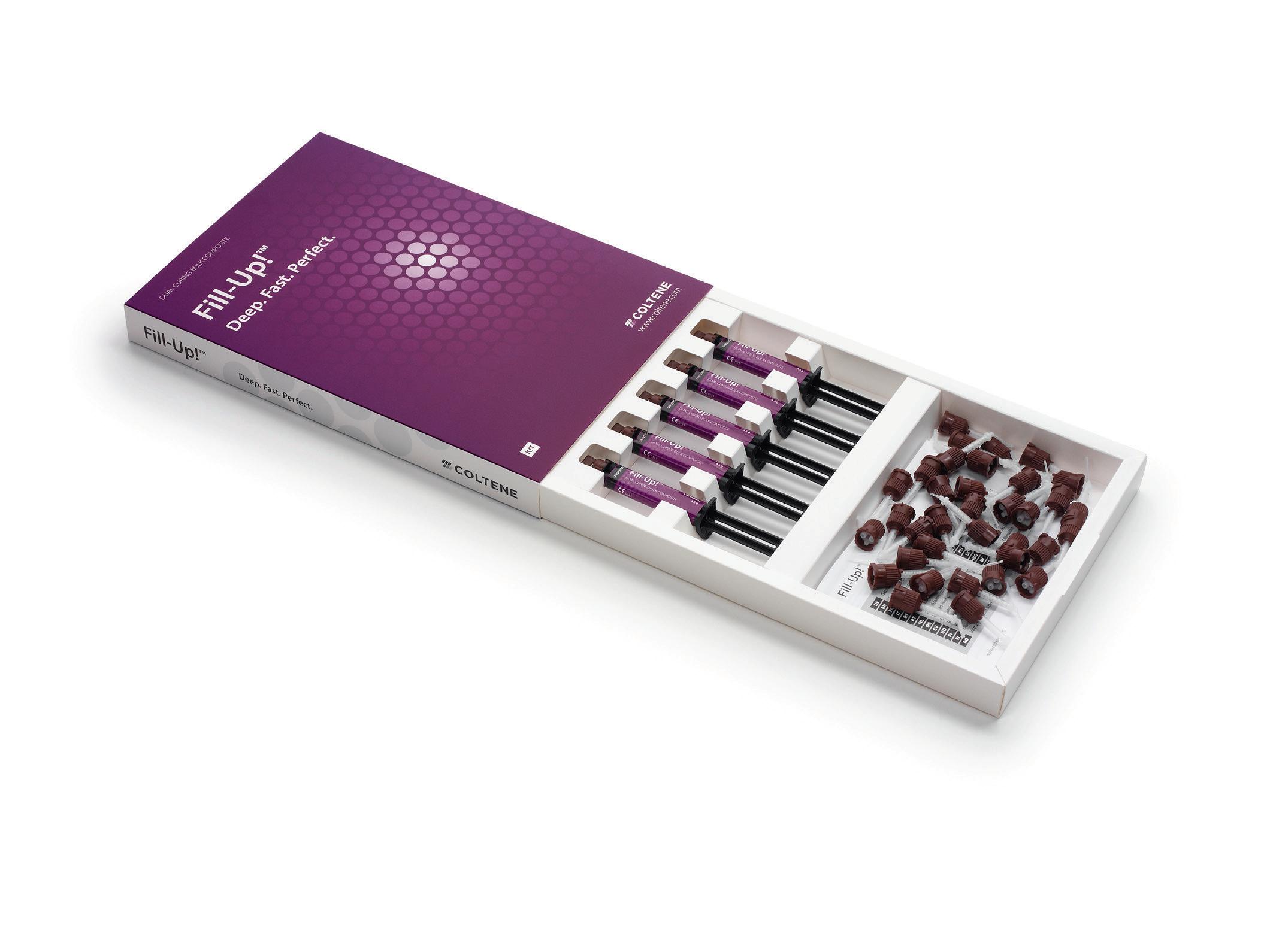
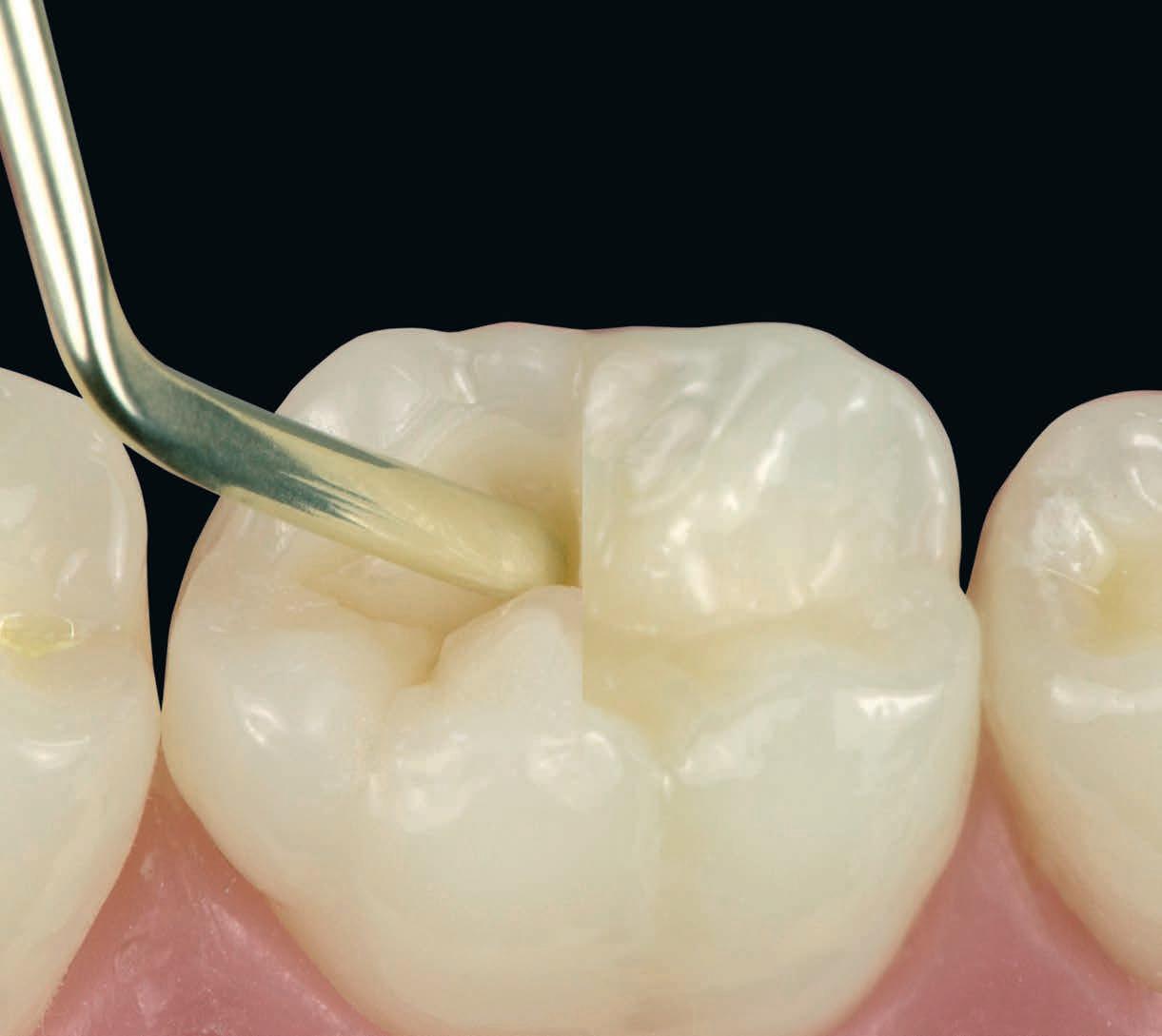
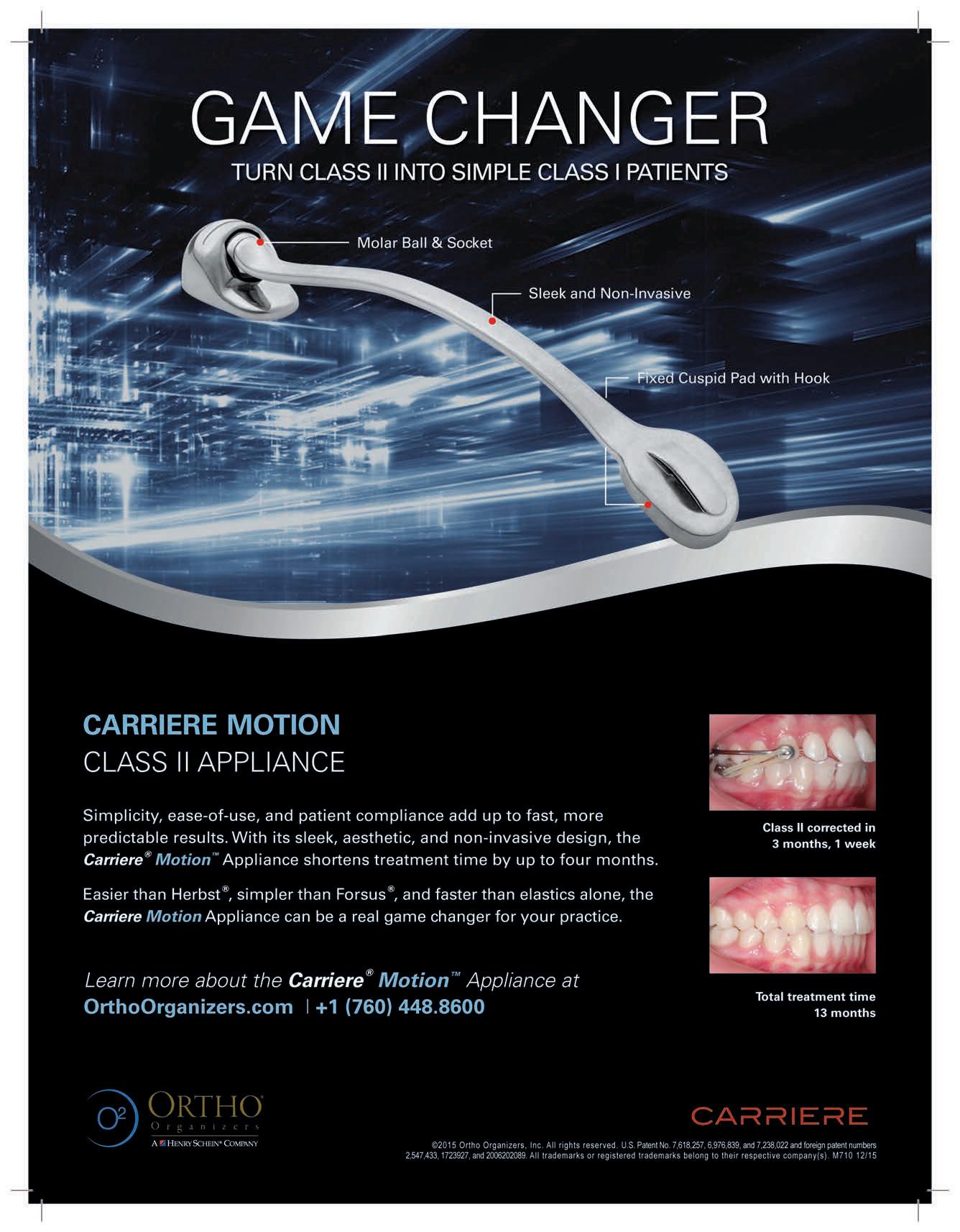
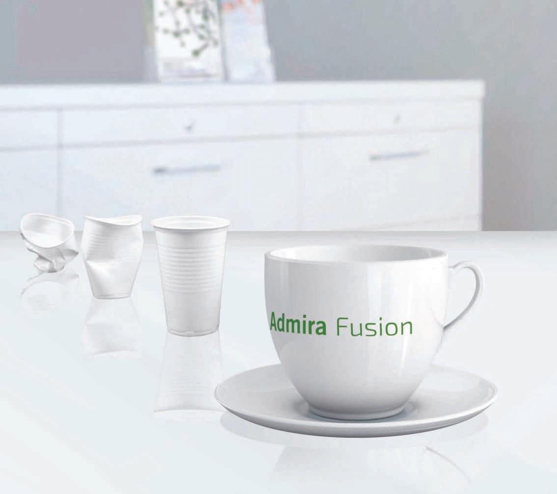
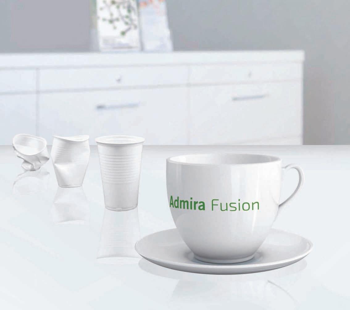
The nano-optimized 4-mm composite
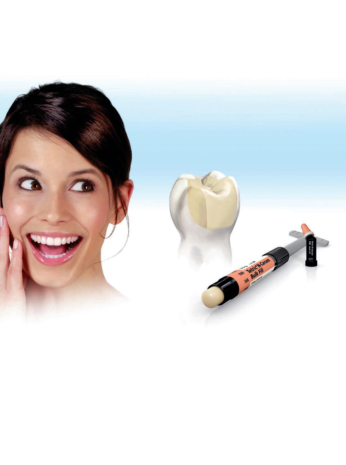
• Bulk filling is possible due to Ivocerin®, the patented light initiator
• Special filler technology ensures low shrinkage stress
• Esthetic results are achieved quickly and efficiently in the posterior region 4 mm to success

National Oral Health Survey of Oral Health Status of Bahraini School
Children Aged 6, 12 & 15 Year Old
The effect of root preparation with Protaper® rotary files on the development of apical root cracks in mild and moderate fluorotic teeth
One Year Program in Dental Implants: Interview with Pr. Nabil Barakat
Ultrasonic Implant Site Preparation technique using PIEZOSURGERY®

LDLS 2016
Lebanese Dental Laboratories Show
May 12 - 14, 2016
Le Royal Hotel Dbayeh, LEBANON
May 25 - 28, 2016
St. Joseph University - Faculty of Dentistry Beirut, Lebanon
EXCIDA 2016
Exhibition and Congress of Iranian Dental Association
May 17 - 20, 2016 International Exhibition Center Tehran, Iran
EDSIC 2016
Egyptian Dental Syndicate International Congress August 31 - September 2, 2016 Intercontinental City Stars Cairo, Egypt
3SHAPE 53
ACTEON 45, 49
A-DEC 17
BA INTERNATIONAL 29
BIEN AIR 15
BISCO 19
CAMLOG 35
CARESTREAM 24
COLGATE 9, 47
COLTENE 25
DENTAURUM 13
DENTSPLY 41
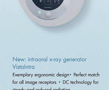
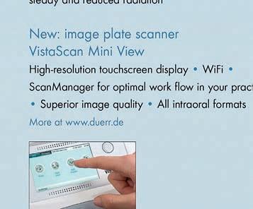
DURR 69
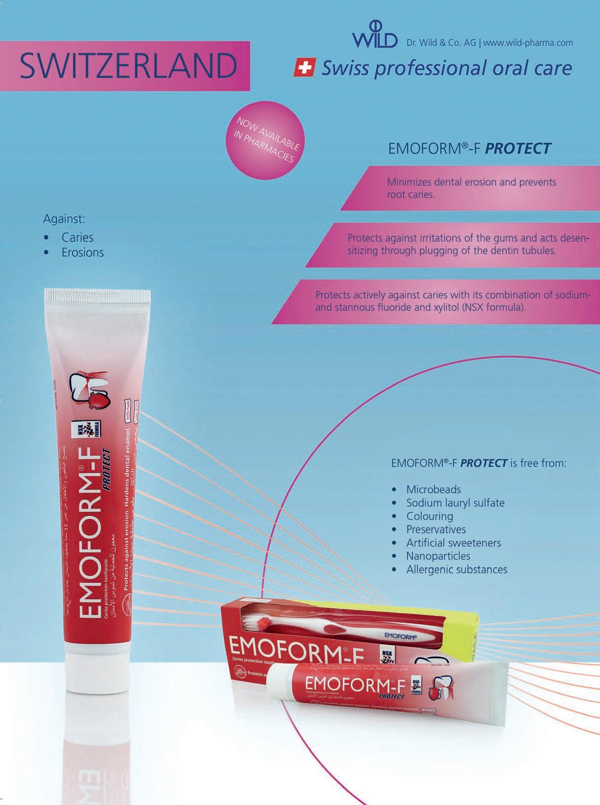
EMOFORM 4, 5
FKG 70
GC 67
GSK C3, 33, 39, 51
HENRY SCHEIN 6
ITENA 37
IVOCLAR 1, C4
KERR C2
MECTRON 21
MEDESY 73
MICRO MEGA 23 MORITA 55
NSK C1
63
31
40
10 SCHEU 76 SDI 57
SIRONA 65 SWIDENT 8 ULTRADENT USA 36 VOCO 65
W&H 7 ZHERMACK 2

Henry Schein is a name you can trust
Henry Schein is the largest global dental distributor of health care products and Services to general practitioners, specialists and laboratories throughout the world. Our network of exclusive Henry Schein Middle East Distributors offer dentists a complete portfolio from the following high-quality and value-priced consumable and equipment product manufacturers that you can rely on to ful ll your practice needs.


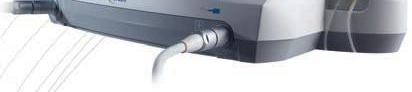
The new Implantmed assists you in performing implant procedures with maximum precision. The unit is easy to operate and guarantees longer working without fatigue, thanks to the lightweight, yet powerful motor and ergonomically designed contra-angle handpiece. The integrated thread cutter function is a must when treating especially dense bone.
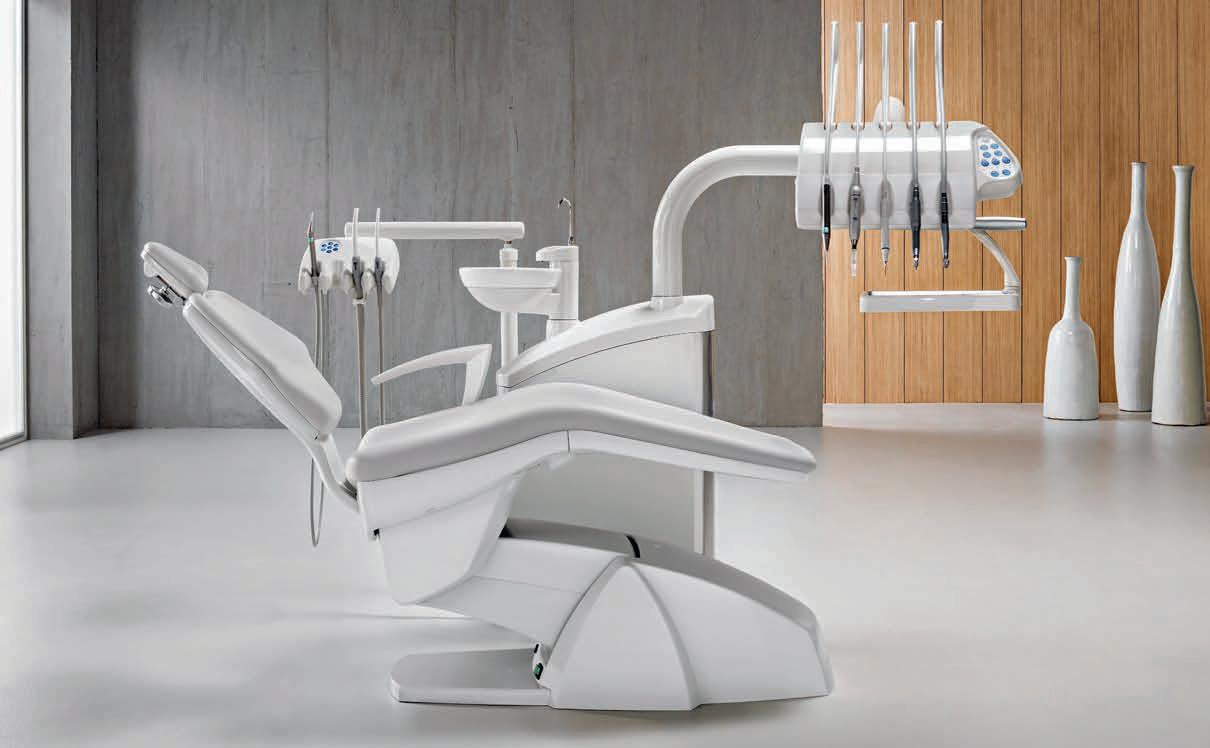
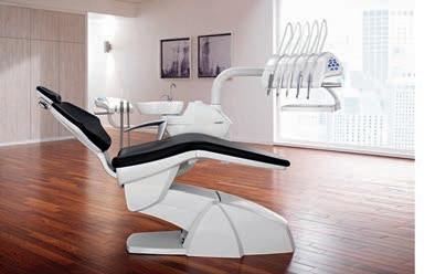
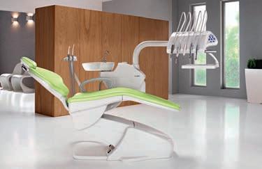
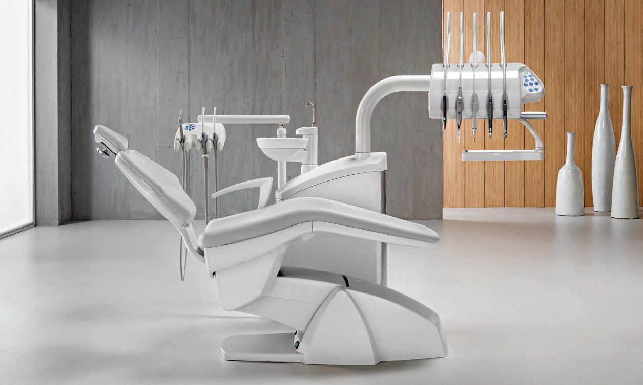
Choose Swident and you’re in good hands. You’ll discover a Company with a precise, rigorous approach and a product range as reliable as a Swiss watch. You’ll discover all equipments designed, made and tested in Italy with maximum attention to the smallest detail.
units guarantee
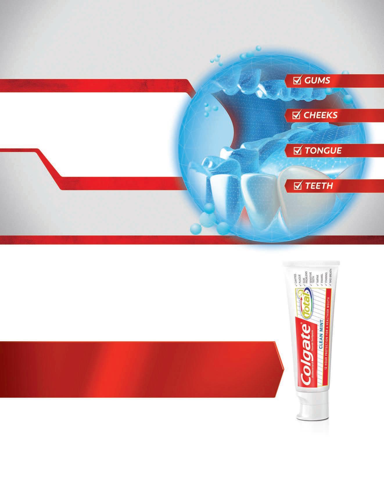
O Regular toothpastes† only protect the hard tissue, which is 20% of the mouth2
O The remaining 80% of the mouth is the tongue, cheeks, and gums, which can provide a bacteria reservoir for plaque biofilm recolonization
WHY SETTLE FOR 20% WHEN YOU CAN OFFER PATIENTS PROTECTION TO 100% OF THE MOUTH’S SURFACES?
*In addition to fluoride for cavity protection, Colgate Total® provides 12-hour antibacterial protection for teeth, tongue, cheeks, and gums.
†Defined as non-antibacterial fluoride toothpaste.
References: 1. Fine DH, Sreenivasan PK, McKiernan M, et al. J Clin Periodontol. 2012;39:1056-1064. 2. Collins LMC, Dawes C. J Dent Res. 1987;66:1300-1302.
made in germany
Henry Schein Dental, Dr. Ghassan Nasser Hussein , Sales and Marketing Director (Henry Schein) Middle East and North Africa, Mobile: +971 50 4813292, Tel: +971 6 5252842, Fax: +971 6 5531291 E-mail: ghassan.nasser@henryschein.com


Ritter Concept GmbH, Germany, Christian Findeisen Sales and Key Account Manager , Middle East/ Africa Ritter Concept GmbH, Mobile: +971 56 9578689
E-mail: christian.findeisen@ritterconcept.com www.ritterconcept.com
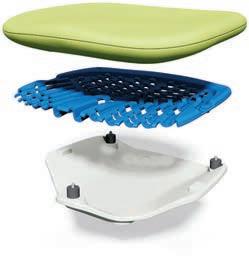
The treatment unit ARIA SR and the furniture line Cameo combine efficiency and userfriendly handling with pure aesthetics and individuality and improve the productivity in the dental office with simple and practical solutions. The careful selection of high-quality materials, a very attractive design and outstanding functionality guarantee fantastic results. In terms of design the products amaze with elegant glass elements in fresh colours that can be customized according to the all preferences, with personal images, motifs or clinic logos. Create your distinctive brand recognition! ARIA SR & Cameo: Aesthetics and individuality for the individual style of your clinic


Individual design of glass surfaces: Choose your preferred colours, patterns or photos!
Alfred Naaman, Nada Naaman, Jihad Fakhoury, Dona Raad, Antoine Saadé, Lina Chamseddine, Tarek Kotob, Mohammed Rifai, Bilal Koleilat, Mohammad H. Al-Jammaz
Suha Nader
Marc Salloum
Micheline Assaf, Nariman Nehmeh
Josiane Younes
Albert Saykali
Gisèle Wakim, Marielle Khoury
Tony Dib 1026-261X
DENTAL NEWS IS A QUARTERLY MAGAZINE DISTRIBUTED MAINLY IN THE MIDDLE EAST & NORTH AFRICA IN COLLABORATION WITH THE COUNCIL OF DENTAL SOCIETIES FOR THE GCC.
Statements and opinions expressed in the articles and communications herein are those of the author(s) and not necessarily those of the Editor(s) or publisher. No part of this magazine may be reproduced in any form, either electronic or mechanical, without the express written permission of the publisher.
DENTAL NEWS – Sami Solh Ave., G. Younis Bldg.
POB: 116-5515 Beirut, Lebanon.
Tel: 961-3-30 30 48
Fax: 961-1-38 46 57
Email: info@dentalnews.com
Website: www.dentalnews.com
www.facebook.com/dentalnews1
www.instagram.com/dentalnews
twitter.com/dentalnews
Dental News App on both Appstore & Google play


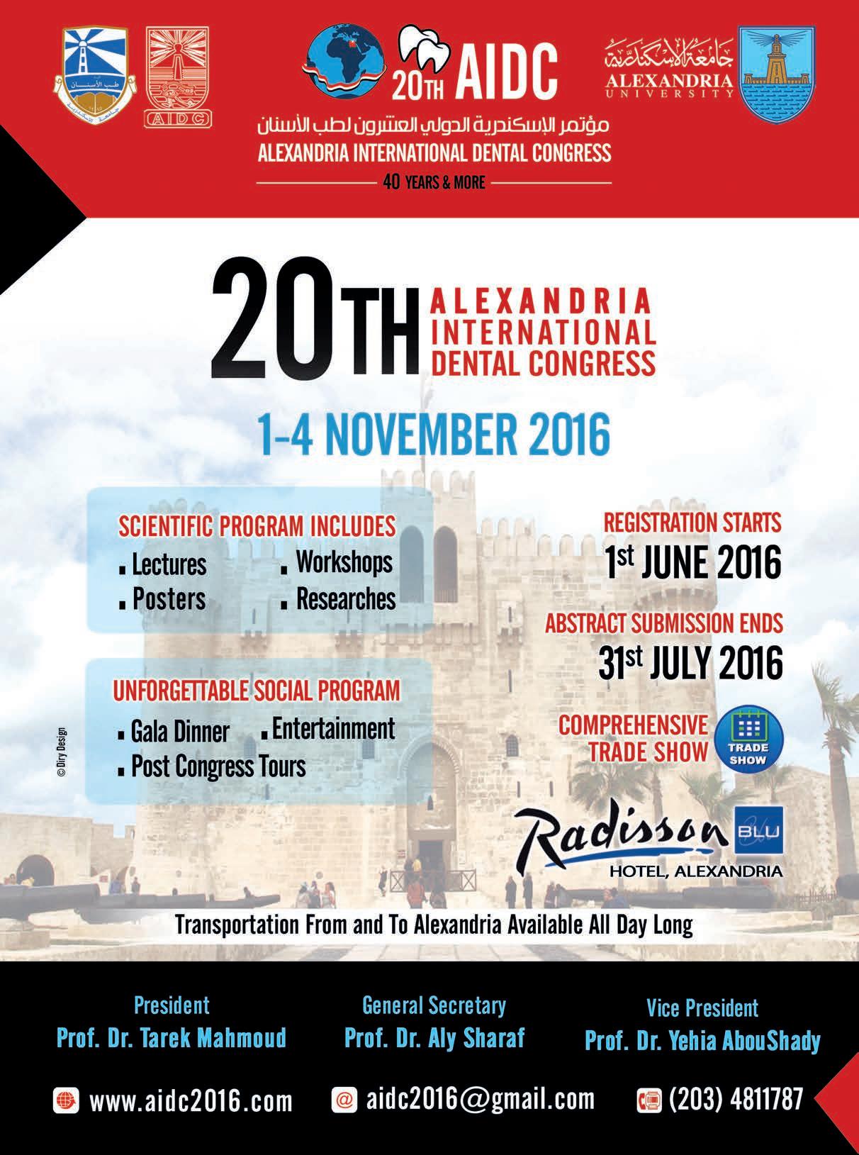
AIDC 2016 - The 20th Alexandria International Dental Congress


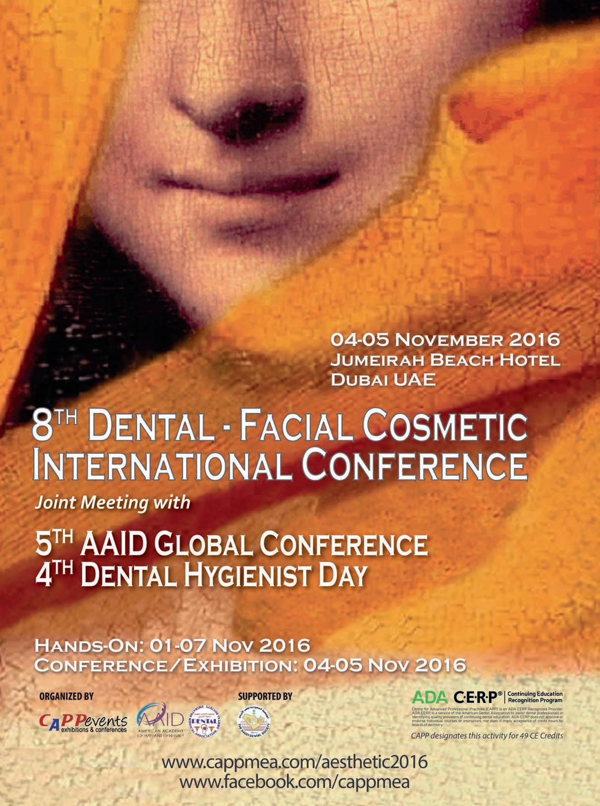
DFCIC 2016 - The 8th Dental-Facial Cosmetic International Conference


KDA 2016 - The 19th Kuwait Dental Association Dental Conference


SDS – KSU 2017 - The 28th Saudi Dental Society Meeting


2017 - The 21st UAE International Dental Conference & Arab Dental Exhibition
November 1-4, 2016 Alexandria, EGYPT Website: www.aidc2016.com
November 4 - 5, 2016 at the Jumeirah Beach Hotel, Dubai, UAE Website: www.cappmea.com
November 17-19, 2016 at the Jumeirah Beach Hotel, Kuwait, KUWAIT Website: www.kda.org.kw
January 10-12, 2017 at the International Convention & Exhibition Center, Riyadh, KSA Website: www.sds.org.sa
February 4-5 , 2017 at the Royal Thalassa Monastir, TUNISIA Email: atorecd@yahoo.fr
February 7 - 9, 2017 at the Dubai International Convention & Exhibition Center, Dubai, UAE Website: www.aeedc.com


March 21-25, 2017 Cologne, Germany Website: www.english.ids-cologne.de
May 18 - 20, 2017 Rimini, Italy. Website: www.expodental.it


August 29 - September 1, 2017 Madrid, Spain. Website: www.fdiworldental.org


Dr. Azhar Ali Ahmad Naseeb Consultant Dental Public Health Member, GCC Oral & Dental Health Committee anaseeb@health.gov.bh
Key words
Children, Dental caries, periodontal diseases, Fluorosis, Epidemiology, Bahrain
Part II continued
Result was revealed also that the uppermost DMFT was recorded in southern governorate among all other governorates and equal to 12.64 (dmft at 6 years old =5.33) + (DMFT at 12Years old =2.93) + (DMFT at a 15 Years old=4.38) and that because of new dental caries incidence found in age 6 and 15 years old except age 12 years old found to be the highest score recorded in Muharraq governorate (Fig.16).
Fig 16
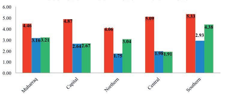
16: Trends of Dental Caries dmft/DMFT at Age 6, 12 & 15 Years Old/Governorate.
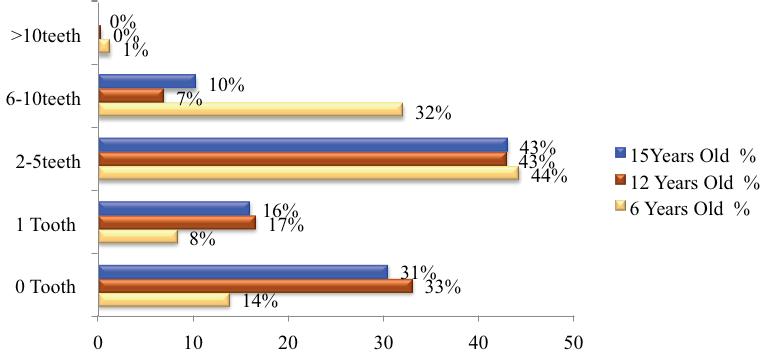
The severity of dental caries was recorded in all age groups in term of how many tooth considered per child has exposed to dental caries; score zero was consider to be child free of dental caries, one tooth, 2-5 teeth, 6-10 teeth and more than 10 teeth with dental caries (Fig.17).
Fig 17


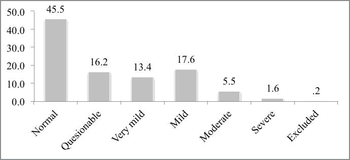
Result of dental fluorosis was showed that there were no sign of dental fluorosis to be recorded in 45.5% of children, while 54.5% were recorded to have variety of enamel fluorosis on the labial surfaces of upper anterior incisors and canine’s teeth with symmetrical diffusing format. (Fig.18)
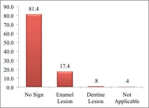
Also the result of enamel and dental erosion was recorded in 81.4% of children, while 17.4% were recorded to have enamel lesion and 0.8% in dentin lesion. (Fig.19)
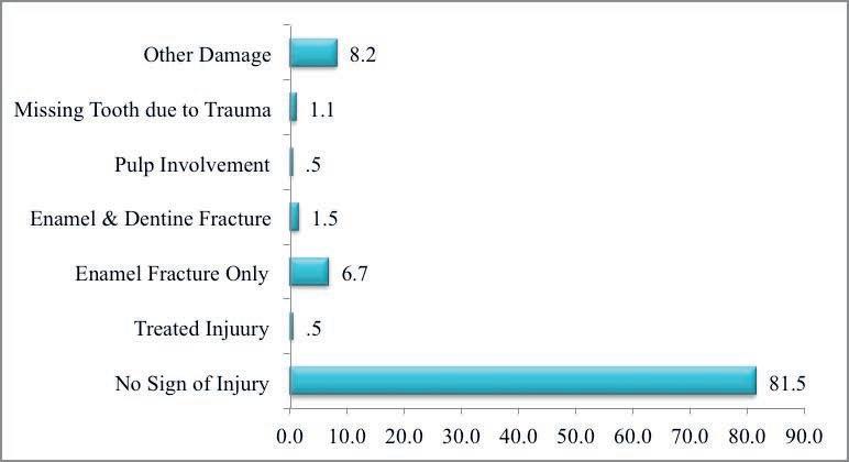
Children with no sign of traumatic injury were 81.5% of the total examined and the remaining of children had a range of trauma that included enamel (6.7%), enamel & dentin (1.5%), pulp involvement (0.5%) fracture and missing tooth due to trauma (1.1%), only 0.5% found to be treated from trauma. (Fig. 20)


Producing the best instruments to simplify the work of practitioners and constantly improve patient comfort. This has been Bien-Air’s mission since its creation in 1959.

Ergonomics, precision and reliability are at the core of the development of every new product. Paying careful attention to professionals every day, Bien-Air has made numerous innovations, always setting the bar higher. A true culture of excellence sitting perfectly with the tradition of Swiss-Made products from the renowned Watch Valley.

The results were disclosed that all the children have no sign of mucosal lesions, upper or lower denture and or an urgent intervention were required.
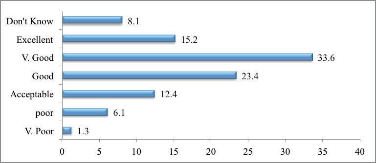
12 years school children were subjected to a face to face interview and results revealed that 8.1% of the children at age 12 years old did not know how to describe the health of their teeth and gum while 91.9% knew how to describe the health of their teeth and gum that varies in knowledge from very poor to excellent that was scored equal to 15.2% only. (Fig. 21)
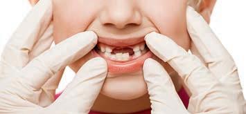
A 30.6 % of children were unsatisfied with the appearance of their teeth while 51.8 % of them were satisfied and 17.6% only answered normal i.e. neither satisfied nor unsatisfied (Fig.22), 22.2% of the children avoided smiling/laughing because of their teeth appearance (Fig.23), and 8% of the children complained that other schoolchildren make fun of them because of their teeth appearance (Fig.24)
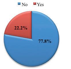
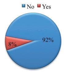
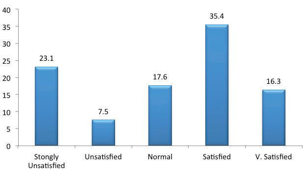
Figure 22: % of children/12 years old were satisfied about appearance of their teeth.
Figure 23: % of children/12 years old were avoided smiling/laughing because of their teeth.
Figure 24: % of children/12 years old were other schoolchildren make fun of them due to appearance of their teeth.
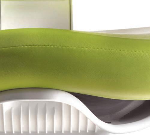
Not to mention everything else.
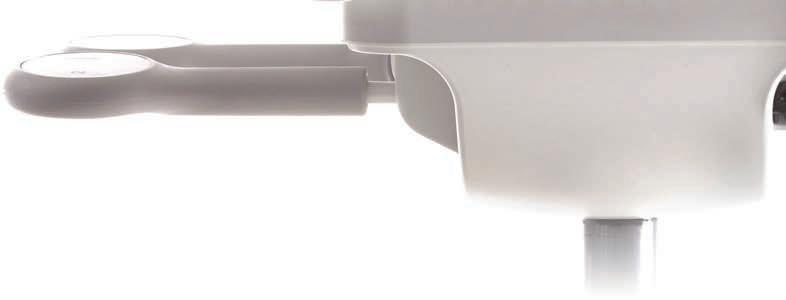
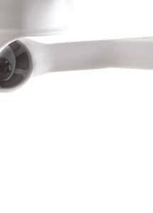
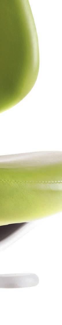
The ability to perform your job comfortably is essential. And the new feature-rich A-dec 500 stools have you covered. Based on a unique dynamic seating system design, individual performance zones work in unison to conform and move with your body. With an ultimate ergonomic solution like this, you can focus on what’s really important—your patients.
Discover your ultimate comfort at a-dec.com/500stools

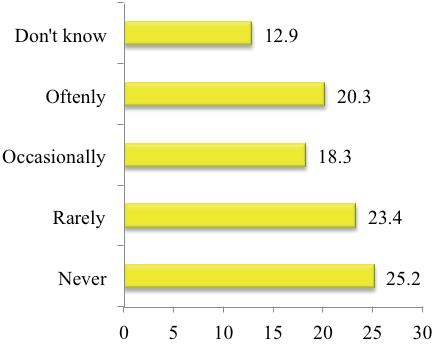
% of children/12 years old and child got toothache/felt discomfort during the past 12 months.
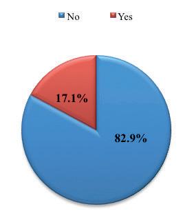
Figure 26:
% of children/12 years old and dental pain was the main cause to miss their school classes.
25.2% of children responded that they never had toothache or felt discomfort during the past 12 months, while 74.8% of them had (Fig.25). 17.1% of the children missed their school classes due to dental pain (Fig.26).
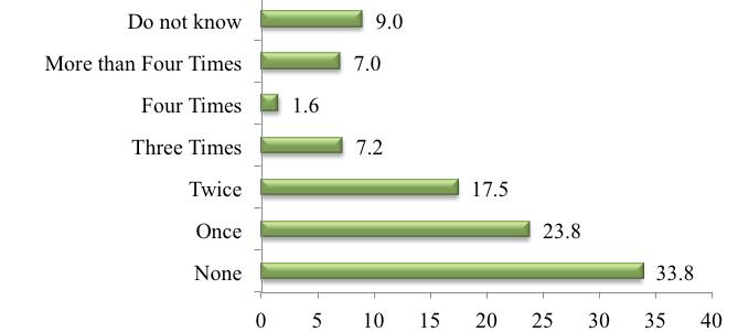
The results showed also, that 33.8% of the children never went to the dentist during the last 12 months while 66.2% of them visited the dentist. On how frequent per year they visited dentist they responded as follows: 7% of children had visited the dentist more than four times, 1.6% had visited four times, and 7.2% three times, 17.5% two times and 23.8% visited the dentist once a year (Fig. 27).
% of children/12 years old and how often did they go to dentist during the last 12 months.
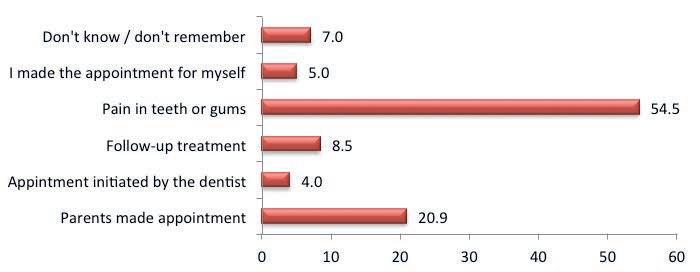
Pain in teeth and gum scored 54% as uppermost reason that let children visit their dentist during the last 12 months (Fig.28).
%


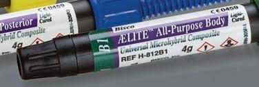
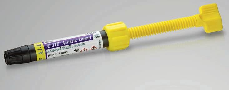
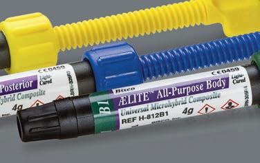
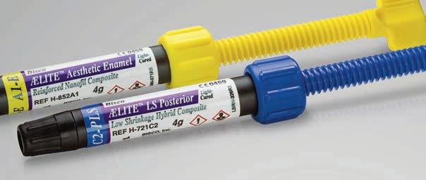
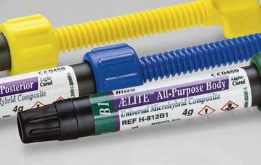


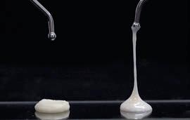
High Strength, Low Shrink, Simply Beautiful
• Excellent polishability and strength
• Radiopaque for easy identi cation on radiographs
• A variety of shades mimic the natural dentition

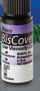
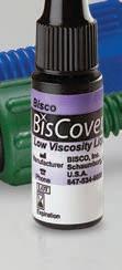
Low Viscosity Liquid Polish
• Seal all your composite restorations while leaving a smooth polished/glazed surface


• Superior handling properties allow for easy placement, contouring and sculpting nal anatomy


Figure 29:
% of children/12 years old were brushed their teeth by using a brush and toothpaste & frequency of brushing.
A 96.7% of the students brushed their teeth by using a brush and toothpaste; 58.2% of them reported to brush twice or more a day, 22.7% once a day, 6.8% several time a week or a month and 2.3% brushed their teeth once a week (Fig.29)
However, 3.3% only of the students reported they never brushed their teeth at all (Fig.29).

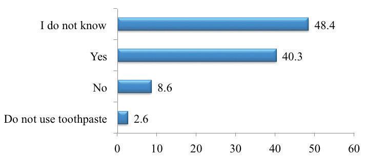
Children were asked about using fluoridated tooth paste while brushing their teeth; 40.3% answered yes, while 8.6% answered no and another 48.4 responded that they do not know about fluoridated tooth paste and 2.6% only of children have never used tooth paste while brushing their teeth (Fig.30).
Figure 30:
% of children/12 years old were brushed their teeth by using fluoridated toothpaste
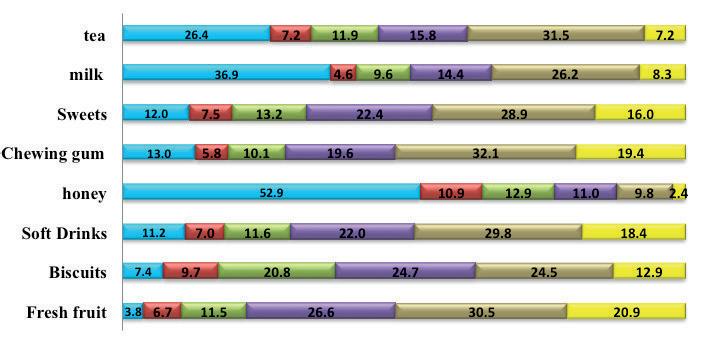
% of children/12 years old and frequency in eat/drink sugary food and drinks


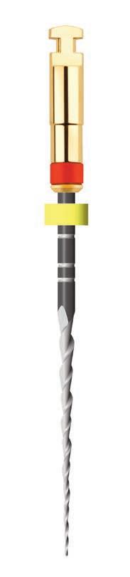

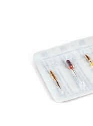
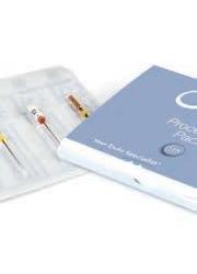


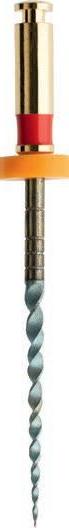
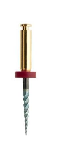
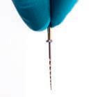
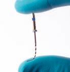
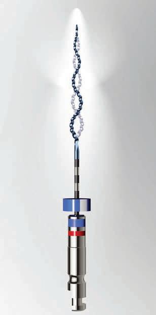
THE NEW NITI FILE GENERATION

Extremely break resistant le for quick preparation


> Up to 700% higher fracture resistance
> Specially hardened surface


> Less lling required for treatment success

ORIFICE OPENER (optional) 25 / .12


Glidepath File 10 / .05
HyFlex™ OneFile 25 / ~


FINISHING FILES (optional)


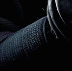


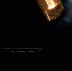


CONTROLLED MEMORY


40 / .04
50 / .03


60 / .02


termine appropriate preventive and therapeutic tactic; the dental hygienist can be the key dental team member responsible for the creation, implementation and evaluation of CAMBRA instead of waste of resources. This approach was proposed by a group of experts based on scientific literature as a means of caries risk assessment through disease indicators, risk factors and protective factors to determine the associated clinical protocols or interventions Featherstone, J.D.B., DomejeanOrliaguet, S., Jenson, L., Wolff, M., & Young, D.A. (2007) and (Featherstone, J.D., Adair, S.M., Anderson, M.H., Berkowitz, R.J., Bird, W.F., Crall, J.J., Den Besten, P.K., Donly, K.J., Glassman, P., Milgrom, P., Roth, J.R., Snow, R., & Stewart, R.E. (2003).
The World Health Organization (WHO) defined health as the “complete state of physical, mental, and social well-being and not merely the absence of infirmity” (WHO 1948). In this definition, WHO indicates three dimensions of well-being. Physical well-being that relates to function normally in activities such as bathing, dressing, eating, and moving around. Mental well-being implies that intellectual faculties are intact and that there is no burden of fear, anxiety, stress, depression, or other negative emotions. Social well-being relates to one’s ability to participate in society, fulfilling roles as family member, friend, worker, or citizen or in other ways engaging in interactions with others. Bergner M, Bobbitt RA, Carter WB, Gilson BS. (1981). As Gift and Atchison (1995) stated, measuring health-related quality of life allows assessment of “the trade-off between how long and how well people live.” Gift and Atchison (1995). Diseases and disorders that result in dental and craniofacial defects can frustrate that goal, disturbing self-image, self-esteem, and well-being. Oral complications can compromise the quality of life such as problems the children could face with appearance, speaking, chewing, taste, and bad smell from oral cavity due to dental caries; and periodontal disease. In this study the psychological impact of teeth appearance has been found that 30.6 % of children were unsatisfied with the appearance of their teeth and 22.2% of the children were found that they avoided smiling/laughing because of their teeth appearance, and 8% of the children were found that other schoolchildren make fun of them because of their teeth appearance. A 17.1% of children had experienced dental pain and missed school, and perform poorly in
school. A 74.8% of children responded that they had toothache or felt discomfort during the past 12 months.
From above the presented result it can be concluded that oral health has direct impact on people’s life; Davied Locker stated that “Oral health affects people physically and psychologically and influences how they grow, enjoy life, look, speak, chew, taste food and socialize, as well as their feelings of social wellbeing (Locker D. 1997). Also, Sheiham, A. (2016) supported the same idea “Severe caries detracts from children’s quality of life: they experience pain, discomfort, disfigurement, acute and chronic infections, and eating and sleep disruption as well as higher risk of hospitalization, high treatment costs and loss of school days with the consequently diminished ability to learn” (Sheiham, A, 2016). These findings suggest that improving children’s oral health status may be a vehicle to enhancing their educational experience.
Furthermore, 33.8% of the children never went to the dentist during the last 12 months, and 23.8% of them once a year had visited the dentist. However, pain in teeth and gum was the uppermost reasons that drove children to visit their dentist in the last twelve months as this is reported by (54.5 %) students. Oral diseases are the fourth most expensive diseases to treat. Treating caries, estimated at US$ 3513 per 1000 children, would exceed the total health budget for children of most low-income countries (Yee R, Sheiham A. 2002). The situation for adults in developing countries is worse, as they suffer from the accumulation of untreated oral diseases. Millions with untreated caries have cavities and suppuration, yet planners continue to overlook oral diseases, despite their significant impact on cost and quality of life. This oversight will lead to more decay and expensive, ineffective clinical interventions. (Yee R, Sheiham, 2002).
Brushing teeth helps to remove food and plaque the sticky film that forms on your teeth and contains bacteria. American Dental Association (ADA) recommended that in addition to brushing teeth at least twice a day, person should floss daily, eat a healthy diet and limit between-meal snacks, replace toothbrush every three to four months or sooner if the bristles are frayed, and schedule regular dental checkups (American Dental Association, 2013). Fortunately, 96% of interviewed children were brushing their teeth, but only 33.8% never brushed their teeth. Though 33.8% never visited a dentist in


the last 12 months but still there is vast majority 66.2% of children did either once or more than four times and the main reason about 54.5% of children were visiting the dentist is because of pain in teeth or gum. Children found also that they consumed sugary food and drinks several time a day and moreover they are not regularly visiting the dentist; this explains the high component of dmft/ DMFT. Though enamel fluorosis free was 45.5% among all age groups but still different rankings of fluorosis incidences was discovered during this study about 54.5% and most of it was in the form of symmetrical diffuse. This can be explained by the fact that most areas in Bahrain are fluoridated and more over people consumption drinks and food from other sources that contain fluoride. Thus, an artificial adding of fluoride should be scientifically studied very well.
Despite there is an overall minute improvement in oral health status of Bahraini 6, 12 and 15 year old children and mean of national DMFT =2.26 for 12 years-old is considered higher than the global standard DMFT for the year 2010 which should not be higher than (1) one (Hobdell et al, 2000 ), furthermore still there is a dental caries incidence among all age groups and an occurrence of gingival bleeding among all age groups also periodontal pocket 3-4mm and periodontal pocket more than 6mm was found in 15 year old children.
Hence, it can be understood that childhood dental caries and periodontal diseases is a serious dental public health problem among Bahraini children, particularly the very young children. Prevalence of dental caries and periodontal disease are high across the Kingdom of Bahrain.
Therefore, a serious action by all dental health services and authorities to prevent dental caries and periodontal diseases should become a priority, dental disease and periodontal diseases surveillance should be encouraged and as well as more studies should be carried out to tackle risk factors associated with same diseases, and implementation of strategies directing to prevent and control oral diseases in the Kingdom of Bahrain. Nevertheless, a mutual cooperation between all sectors governmental and private towards caries free generation should be addressed, and always one should not forget prevention is always better than curative services.
1. AMERICAN DENTAL ASSOCIATION (2013): BRUSHING YOUR TEETH. AMERICAN DENTAL ASSOCIATION HTTP://WWW MOUTHHEALTHY ORG/EN/AZ-TOPICS/B/BRUSHING-YOUR-TEETH ASPX. ACCESSED JAN. 31, 2013.
2. ASMCHP,(1999): ISSUE BRIEF; PUTTING TEETH IN CHILDREN’S ORAL HEALTH POLICY AND PROGRAMS: THE STATE OF CHILDREN’S ORAL HEALTH AND THE ROLE OF STATE TITLE V PROGRAMS, DECEMBER, 1999. ASSOCIATION OF MATERNAL AND CHILD HEALTH PROGRAMS, 1999.
3. BERGNER M, BOBBITT RA, CARTER WB, GILSON BS. (1981): THE SICKNESS IMPACT PROFILE: DEVELOPMENT AND FINAL REVISION OF A HEALTH STATUS MEASURE. MED CARE 1981 AUG; 19(8):787-805.
4. EKLUND S, MOLLER IJ, LECLERCQ MH. (1993)-WHO (2013): CALIBRATION OF EXAMINERS FOR ORAL EPIDEMIOLOGICAL SURVEYS. WORLD HEALTH ORGANIZATION, 1993(ORH/EIS/EPID.93.1 IN 5TH EDITION ORAL HEALTH SURVEYS BASIC METHOD, (2013).
5. FEATHERSTONE, J.D.B., DOMEJEAN-ORLIAGUET, S., JENSON, L., WOLFF, M., & YOUNG, D.A. (2007): CARIES RISK ASSESSMENT IN PRACTICE FOR AGE 6 THROUGH ADULT JOURNAL OF THE CALIFORNIA DENTAL ASSOCIATION, 35(10), 703-713.
6. FEATHERSTONE, J.D., ADAIR, S.M., ANDERSON, M.H., BERKOWITZ, R.J., BIRD, W.F., CRALL, J.J., DEN BESTEN, P.K., DONLY, K.J., GLASSMAN, P., MILGROM, P., ROTH, J.R., SNOW, R., & STEWART, R.E. (2003): CARIES MANAGEMENT BY RISK ASSESSMENT: CONSENSUS STATEMENT, APRIL 2002. JOURNAL OF THE CALIFORNIA DENTAL ASSOCIATION, 31(3), 257-269.
7. GIFT HC, REISINE ST, LARACH DC (1992): THE SOCIAL IMPACT OF DENTAL PROBLEMS AND VISITS. AMERICAN JOURNAL OF PUBLIC HEALTH 1992; 82:1663-8.
8. GIFT HC, ATCHISON KA. (1995). ORAL HEALTH, HEALTH, AND HEALTH-RELATED QUALITY OF LIFE; MED CARE. 1995 NOV; 33(11 SUPPL):NS57-77. REVIEW
9. HOBDELL ET AL, 2000: PAULA MOYNIHAN AND POUL ERIK PETERSEN (2004). DIET, NUTRITION AND THE PREVENTION OF DENTAL DISEASES. PUBLIC HEALTH NUTRITION: 7(1A), 201-226. 2004.
10. LOCKER D., (1997): CONCEPTS OF ORAL HEALTH, DISEASE AND THE QUALITY OF LIFE IN: SLADE GD, EDITOR. MEASURING ORAL HEALTH AND QUALITY OF LIFE. CHAPEL HILL: UNIVERSITY OF NORTH CAROLINA, DENTAL ECOLOGY; 1997, PP. 11-23.
11. NASEEB, A., (2005): NATIONAL ORAL HEALTH SURVEY 2005-2008. KINGDOM OF BAHRAIN. MINISTRY OF HEALTH. MOH-BAHRAIN UNPUBLISHED SURVEY DOCUMENTS
12. PETERSEN PE. THE WORLD ORAL HEALTH REPORT (2003): CONTINUOUS IMPROVEMENT OF ORAL HEALTH IN THE 21ST CENTURY – THE APPROACH OF THE WHO GLOBAL ORAL HEALTH PROGRAM. COMMUNITY DENTISTRY AND ORAL EPIDEMIOLOGY 2003; 32 SUPPLE 1:3-24.
13. RICHARD WATT. (2005): STRATEGIES AND APPROACHES IN ORAL DISEASE PREVENTION AND ORAL HEALTH PROMOTION. BULLETIN OF THE WORLD HEALTH ORGANIZATION 83 (9). PP 711-718.
14. RUGG-GUNN, A. J. (1992): BRITISH SOCIETY OF PEDIATRIC DENTISTRY POLICY DOCUMENT SUGARS AND THE DENTAL HEALTH OF CHILDREN. INTERNATIONAL JOURNAL OF PEDIATRIC DENTISTRY 2: 177-180.
15. SHEIHAM, A, 2016: ORAL HEALTH, GENERAL HEALTH AND QUALITY OF LIFE IN BULLETIN OF THE WORLD HEALTH ORGANIZATION: (HTTP://WWW WHO INT/BULLETIN/VOLUMES/83/9/EDITORIAL30905HTML/EN/)
16. U.S. DEPARTMENT OF HEALTH AND HUMAN SERVICES, 2000: ORAL HEALTH IN AMERICA: A REPORT OF THE SURGEON GENERAL, CHAPTER 10: FACTORS AFFECTING ORAL HEALTH OVER THE LIFE SPAN. ROCKVILLE, MD: U.S. DEPARTMENT OF HEALTH AND HUMAN SERVICES, OFFICE OF THE SURGEON GENERAL. AVAILABLE AT: <WWW2.NIDCR NIH GOV/SGR/SGROHWEB/CHAP10.HTM#CHILDREN>. ACCESSED JULY 30, 2012.
17. VARENNE, B., PETERSEN, P. E. AND OUATTARA, S. (2006): ORAL HEALTH BEHAVIOR OF CHILDREN AND ADULTS IN URBAN AND RURAL AREAS OF BURKINA FASO, AFRICA. INTERNATIONAL DENTAL JOURNAL, 56, 61-70.
18. ORAL HEALTH SURVEYS BASIC METHOD (2013): 5TH EDITION, (2013).
19. WWW CIO GOV BH/CIO ENG, 2016.


Endodontics
The effect of root preparation with Protaper® rotary files on the development of apical root cracks in mild and moderate fluorotic teeth
 Dr. Chems Belkhir: Professor of Endodontics
University of Monastir
Dr. Chems Belkhir: Professor of Endodontics
University of Monastir
Key words
Apical root crack, Protaper, working length, dental fluorosis.
Introduction
Dental fluorosis is a common pathology in Tunisia. The majority of studies addressing this topic have focused on the histological characteristics, esthetic aspect and bonding problems of fluorotic teeth. The literature reviews involving structural and histological alterations of different tissues of the tooth crown are relatively few, even more rare those describing the root of fluorotic teeth. Many studies have described the adverse effects of different rotary Ni-Ti instruments on the apical third of sound dental roots but no study has reported their effects on the root of fluorotic teeth1,2,13
The aims of our study, involving the third apical of maxillary incisors and premolars presenting mild to moderate fluorosis, are:
• To study the modifications observed in the apices of these teeth after endodontic treatment using Protaper® system.
• To determine the risk of occurrence of apical cracks according to the degree of fluorosis (mild and moderate).
Extracted human mandibular premolars and maxillary incisors with straight roots were selected for this study. The reasons for extraction and the age of the patients at the time of extraction were unknown. The roots were inspected for evidence of open apices, fracture lines, or anatomic irregularities and discarded if any of these characteristics were found. Seventy five teeth were finally selected and stored in distilled water throughout the study.
These teeth were divided into 4 groups according to Thylstrup and Fejerskov index:
Group 1: fifteen maxillary incisors with mild fluorosis (TFI 1-3).
Group 2: fifteen maxillary incisors with moderate fluorosis (TFI 4).
Group 3: ten mandibular premolars with mild fluorosis (TFI 1-3).
Group 4: ten mandibular premolars with moderate fluorosis (TFI 4).
Group 5: fifteen maxillary incisors without fluorosis (control group).
Group 6: ten mandibular premolars without fluorosis (control group).
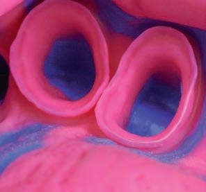
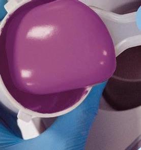
The root was wrapped with a single layer of aluminum foil and embedded in autopolymerizing resin (Major Ortho®) set in an acrylic tube (12 mm high and 20 mm in diameter). The root was then removed from the tube, and the aluminum foil peeled off. The crowns were removed at 2 mm above the proximal cement enamel junction to ensure a straight-line access and to provide a reference plane.
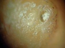
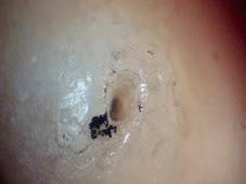
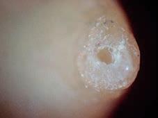
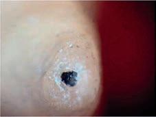
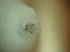
The apical surface surrounding the major apical foramen of each specimen was photographed with a stereomicroscope (Zeiss) under magnification of 4 and with a digital camera.
The patency of the canal was checked, and the canal length was measured by inserting a #8 K-file into the canal until the tip of the file became visible through the major apical foramen. The distance between the file tip and the reference plane was defined as the canal working length (WL).
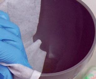
A simulated periodontal ligament was represented by a hydrophilic vinyl polysiloxane impression material. The root surface and the ‘‘socket’’ were


coated with a hydrophilic vinyl polysiloxane impression material (Examixfine, GC Corporation), and the root was immediately repositioned. Thus, the polysiloxane replaced the space created by the foil.
The following sequence of Protaper® rotary files (Dentsply Maillefer) was used at 300 rpm to prepare the canals: an SX file was used to enlarge the coronal portion of the canal, and then the following files were used to working length – 0,5mm (WL-0,5mm): S1, S2, F1 and F2. Irrigation was performed with 33cc sodium hypochlorite at 2.5% before and after the passage of each instrument. China Ink (Le Coq®) was used in final rinsing to stain the root apex in order to reveal any cracks, and the teeth were then rinsed with water.
A single operator performed all the procedures. After root preparation, the apical surface surrounding the major apical foramen of each specimen was photographed with a stereomicroscope (Zeiss) under magnification of 4 and with a digital camera.
Fisher Exact Test was used to compare the number of cracks between fluorosed teeth and non fluorosed teeth. Statistical analysis were performed at a 5% significance level.
Fisher Exact Test revealed no significant differences between the study groups:
Comparison between fluorosed teeth and non-fluorosed teeth: p=0,28.
Comparison between mild fluorosed teeth and non-fluorosed teeth: p=0,5.
Comparison between moderate fluorosed teeth and non-fluorosed teeth: p=0,2. (Table 1)
Comparison between fluorosed incisors and non-fluorosed incisors: p=0,35.
Comparison between mild fluorosed incisors and non-fluorosed incisors: p=0,5.
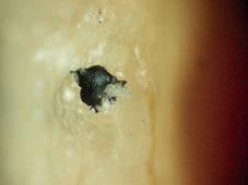
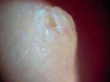
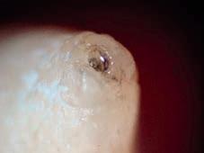
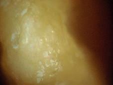
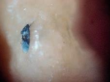
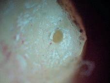
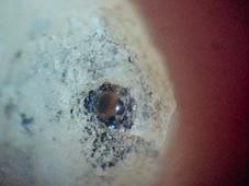
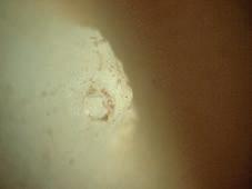
Comparison between moderate fluorosed incisors and non-fluorosed incisors: p=0,32. (Table 2). (Figures 1, 2, 3, 4, 5 and 6).


1: Mild fluorosed incisors: (A) before preparation. (B) After preparation: apical crack is produced.
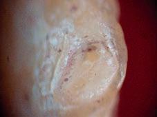
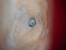
2: Mild fluorosed incisors: (A) before preparation. (B) After preparation: no apical crack is produced.

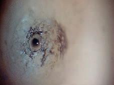
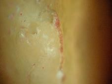


Comparison between fluorosed premolars and non-fluorosed premolars: p=0,66. Comparison between mild fluorosed premolars and non-fluorosed premolars: variables=0. Comparison between moderate fluorosed premolars and non-fluorosed premolars: p=0,05. (Table 3). (Figures 7, 8, 9 and 10).
Discussion
Through this experimental study, we have studied the modifications observed after root shaping using Protaper® system at the apices of mild-tomoderate incisors and premolars and comparing them to those of sound teeth. Many studies have described the adverse effects of different rotary Ni-Ti instruments on the apical third 1,2 of sound dental roots but no study has reported their effects on the root of fluorotic teeth. In order to simulate the anatomic conditions in vitro, we have tried to reproduce the anatomical structures surrounding the tooth. The periodontal ligament plays the role of a damper of occlusal forces applied on the tooth due to hydraulic con-


tent of periodontal space (blood vessels and ground substance) acting as a room full of fluids that absorbs shocks. Low viscosity silicones are characterized by thermal stability, compression and tensile strengths. They act as shock absorbers. These characteristics make silicone the material of choice to simulate the periodontal ligament even though the complexity of its anatomic and biological aspects seems to be difficult to imitate 13
Thus, the use of silicone can cause changes in the distribution and dissipation of the forces applied on the tooth 1,2,16
Polymerization reaction of acrylic resin is exothermic; a relative dehydration of the tooth is noted. This dehydration decreases the elastic modulus of radicular dentin that becomes harder, thus more brittle2
Our in vitro study has shown that dental fluorosis mild and moderate doesn’t affect the capacity of radicular dentin to withstand, same as sound dentin, the forces caused by rotary endodontic files. Dentin tubules traverse the entire thickness of dentin. They contain relatively less inorganic material, but a


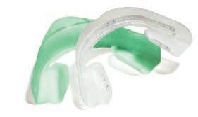


• Simple, fast and great-tasting
• No impressions or custom trays necessary
• Ready to use
• UltraFit tray adaptable to any smile


Opalescence GO, the professional alternative for home whitening:

• 10% & 15%, 15 to 60 minutes wear for a 5 to 10 days treatment
• 20% of water with Potassium Nitrate and Fluoride

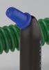
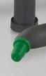
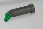
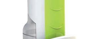
EFFICIENT ROOT CANAL CLEANING
PATENTED CONCEPT
Tip oscillation to allow perfect disinfection.
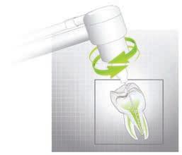
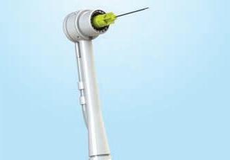
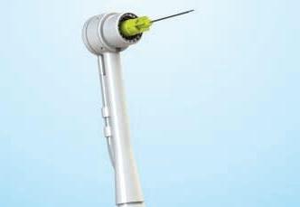
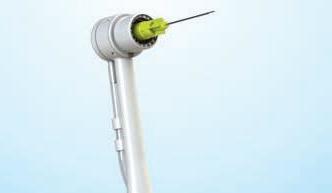
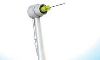
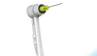
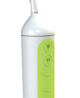
IRRIGATYS : the new two-in-one handpiece with dual functions
Two-in-one system provides and activates liquid for the perfect clean.
A removable tank allows the irrigation of the root canal with Hypochlorite and EDTA. The irrigation line leads the solution through the Irriga-Tip This patented technology, developed after 6 years of research, optimizes the result of the complex procedure of root canal irrigation.


higher collagen concentration (about 90%) in the intertubular dentin, which consists of 97% of Type I collagen. Peritubular dentin grows from the inner surface of the dentinal tubule cavity and forms a clear and highly mineralized layer that is structurally denser than the intertubular dentin, and it contains essentially no collagen 8-21
On the other hand, in response to the effects of severe enamel fluorosis, dentin shows hypermineralization, as found in other enamel disorders. Levels of Ca and P are also significantly higher than in healthy dentin 11-21
Dentin hypermineralization and hardness correspond to a structural pattern of sclerotic dentin, characterized by a narrowing of the lumen, and compaction of peritubular and intertubular dentin. This change in hardness has been also associated with the role of dentin as a possible biomarker for fluoride exposure 18-21
Vieira’s study has shown that for a fluorosis ITF0 to 4 the structural and mechanical modifications (elastic modulus) were limited 17
Another factor also intervening in the reduction of elasticity and flexural strength of dentin is sodium hypochlorite irrigant NaClO 3, 5, 6, 9. Besides the mechanical effect of flushing debris from the canal and lubricating instruments, sodium hypochlorite has an antibacterial wide spectrum and an ability to dissolve organic tissues14, 15
Some studies have proven that using NaClO as irrigant at high concentrations decreases the elastic modulus and flexural strength of dentin by altering its mechanical properties due to the degradation of organic components 4,7,10
For our study, a concentration of 2.5% was used. These alterations have not been proven with low concentrations of NaClO (0.5% to 2.25%) and shorter exposure time (1 to 10min). 7
Root canal shaping was performed using Protaper® system according to crown down concept. The progressive shaping of coronal and middle portions of the canal to the foramen makes easier the access of instruments to the apical portion. Some studies4 have proven that NiTi rotary systems can increase dentinal defects. Our study has shown that Protaper® induces little cracks in both sound and fluorotic teeth which can be explained by the technical features of the instrument. Protaper® has a convex triangular cross section that decreases the rotational friction between the blade of the file and dentin, an active blade
without radial land and a negative cutting angle that enhances the cutting action. Besides, Protaper® files each have a noncutting, modified guiding tip that decreases canal transportations. The taper and the diameter of the instrument may cause dentinal defects 12,19
Bier et al. study 4 has shown that the rate of occurrence of cracks and apical fractures depends on the diameter of the rotary instrument utilized for the preparation of the apical third. This study has demonstrated that dental roots presenting the highest number of cracks are those prepared with Protaper® system following the operative sequence to the file F3 (30/100 diameter and apical taper of 09). In our work, we have finished the preparation of the apical third to the file F2 (25/100 diameter and apical taper of 08) which can explain the low number of cracks observed.
Sathorn et al. 12 confirmed our findings by demonstrating that keeping the apical foramen as small as possible decreases the risk of occurrence of radicular cracks and fractures.
The excessive and extended contact between endodontic instruments and canal walls induces the appearance of high stress and pressure zones. It’s from this weakness zone that cracks arise. The excessive contact is particularly observed when big diameter files are utilized for the preparation of the apical third which is the most tapering portion thus the most fragile of the dental root 2
The position of the apical terminus also takes part in the occurrence of radicular cracks. Wu et al. 20 suggest locating working terminus at 2 to 3 mm short of the apical foramen in vital teeth and at 1 to 2 mm short of this foramen in necrotic teeth. This suggestion enables to keep the foramen as small as possible and gives the apical portion of the canal sufficient resistance and stiffness.
Adorno et al. study 2 has shown that excessive removal of radicular dentin may predispose the root and particularly the apical third to cracks and even radicular fractures. The apical cracks were more numerous in teeth where working length is equal to the length of the radicular canal. This study concluded that leaving 1 mm of non-instrumented radicular dentin at the apical zone reduces the risk of occurrence of radicular cracks and fractures.
For our study, we have chosen to stop our preparation at 0.5 mm short of the apex, thus allowing treating almost the entire apical portion with little damage of the apical radicular dentin.

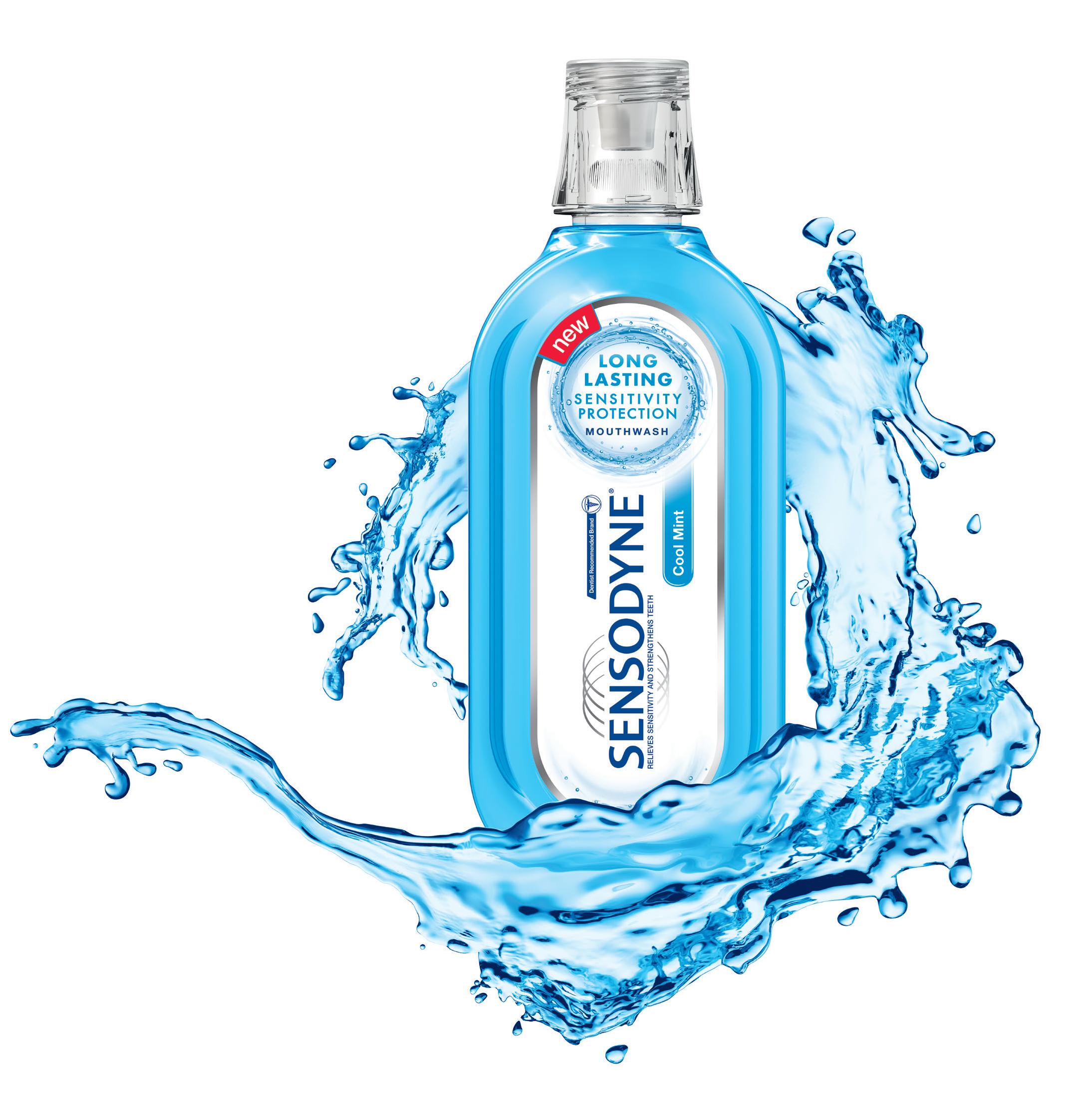
Ultra-low abrasion for your patients who need sensitivity relief and seek gentle whitening

Clinically proven relief from the pain of sensitivity*1-4
Gently lifts stains and help prevent new stains from forming5-7
Ultra-low abrasive formulation appropriate for your patients with exposed dentine8

Recommend Sensodyne – specialist expertise for patients with dentine hypersensitivity

*With twice-daily brushing

References. 1. Jeandot J et al. Clinc (French) 2007; 28: 379–384. 2. Nagata T et al. J Clin Periodontol 1994; 21(3): 217–221. 3. GSK data on file. DOF Z2860473. 4. Leight RS et al. J Clin Dent 2008 19(4) 147-153. 5. Schemehorn BR et al. J Clin Dent 2011 22(1) 11-18. 6. Shellis RP et al. J D ent 2005 33(4) 313-324. 7. GSK data on file. DOF Z2860415. 8. GSK data on file. DOF Z2860435.
Arenco Tower, Media City, Dubai, U.A.E. Tel: +971 4 3769555, Fax: +971 3928549 P.O.Box 23816. For full information about the product, please refer to the product pack. For reporting any adverse event/side effect related to GSK product, please contact us on contactus-me@gsk.com
Prepared: December 2014, CHSAU/CHSENO/0034/14f.
value
At the end of this study, we can conclude that:
• Mechanical preparation of the canal using Protaper® rotary files to WL-0.5mm induces fewer apical cracks in mild and moderate fluorotic incisors and premolars.
• These fluorotic teeth are not more fragile than non fluorotic teeth at their apical portions.
• There is no association between the severity degrees of fluorosis (mild, moderate) and the occurrence of apical defects.
1. ADORNO CG , YOSHIOKA T, SUDA H. THE EFFECT OF ROOT PREPARATION TECHNIQUE AND INSTRUMENTATION LENGTH ON THE DEVELOPMENT OF APICAL ROOT CRACKS. JENDOD 2009,35: 389-92
2. ADORNO CG , YOSHIOKA T ,SUDA H. CRACK INITIATION ON THE APICAL ROOT SURFACE CAUSED BY THREE DIFFERENT NICKEL-TITANIUM ROTARY FILES AT DIFFERENT WORKING LENGTHS. JENDOD 2011,37: 522-5
3. ARI H, ERDEMIR A. EFFECTS OF ENDODONTIC IRRIGATION SOLUTIONS ON MINERAL CONTENT OF ROOT CANAL DENTIN USING ICP-AES TECHNIQUE. J ENDOD 2005; 31: 187-9
4. BIER SC, SHEMESH H, TANOMARU-FILHO M, WESSELINK PR, WU MK THE ABILITY OF DIFFERENT NICKEL-TITANIUM ROTARY INSTRUMENTS TO INDUCE DENTINAL DAMAGE DURING CANAL PREPARATION. J ENDOD 2009,35: 236-8
5. CORRER GM, BRUSCHI ALONSO RC, GRANDO MF, BORGES AF, PUPPIN-RONTANI RM. EFFECT OF SODIUM HYPOCHLORITE ON PRIMARY DENTIN. A SCANNING ELECTRON MICROSCOPY (SEM) EVALUATION. J DENT 2006, 34: 454-9.
6. HU X, LING J, GAO Y. EFFECTS OF IRRIGATION SOLUTIONS ON DENTIN WETTABILITY AND ROUGHNESS. J ENDOD 2010; 36:1064-7
7. HU X, PENG Y, SUM CP, LING J. EFFECTS OF CONCENTRATION AND EXPOSURE TIMES OF SODIUM HYPOCHLORITE ON DENTIN DEPROTEINATION: ATTENUATED TOTAL REFLECTION FOURIER TRANSFORM INFRARED SPECTROSCOPY STUDY. J ENDOD 2010,36: 2008-11.
www.promedica.de to see our upcoming exhibitions
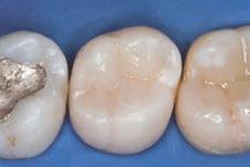
Light-curing nano-ceram composite
• nano-reinforced ceramic particles
• special resin matrix

• significantly less free monomers
• highly esthetic
• universal for all cavity classes
• comfortable handling, easy modellation
• also available as a flowable version
8. MA S, CAI J, ZHAN X, WU Y. EFFECTS OF ETCHANT ON THE NANOSTRUCTURE OF DENTIN: AN ATOMIC FORCE MICROSCOPE STUDY. SCANNING 2009; 31:28-34.
9. MARENDING M, PAQUE F, FISHER J, ZEHNDER M. IMPACT OF IRRIGANT SEQUENCE ON MECHANICAL PROPERTIES OF HUMAN ROOT DENTIN. J ENDOD 2007,33: 1325-8.
10. PASCON FM, KANTOVITZ KR, SACRAMENTO PA, NOBRE-DOS-SANTOS, PUPPINRONTANI RM. M,. EFFECT OF SODIUM HYPOCHLORITE ON DENTIN MECHANICAL PROPERTIES. A REVIEW. J DENT 2009; 12: 903-8.
11. ROJAS-SÁNCHEZ F, ALAMINOS M, CAMPOS A, RIVERA H, SÁNCHEZ-QUEVEDO MC. DENTIN IN SEVERE FLUOROSIS: A QUANTITATIVE HISTOCHEMICAL STUDY. J DENT RES 2007; 86: 857-861.
12. SATHORN C, PALAMARA JE, MESSER HH. A COMPARISON OF THE EFFECTS OF TWO CANAL PREPARATION TECHNIQUE ON ROOT FRACTURE SUSCEPTIBILITY AND FRACTURE PATTERN J ENDOD 2005; 31:283-7.
13. SHEMESH H, BIER CA, WU M, TANOMARU-FILHO M,WESSELINK PR. THE EFFECTS OF CANAL PREPARATION AND FILLING ON THE INCIDENCE OF DENTINAL DEFECTS. INT ENDOD J 2009;42: 208–13.
14. SIQUEIRA JF JR, ROCAS IN FAVIERI A, LIMA KC. CHEMOMECHANICAL REDUCTION OF THE BACTERIAL POPULATION IN THE ROOT CANAL AFTER INSTRUMENTATION AND IRRIGATION WITH 1%, 2,5% AND 5,25% SODIUM HYPOCHLORITE. J ENDOD 2000; 26:331-4.
15. SIM TP, KNOWLES JC, NG YL, SHELTON J,GULABIVALA K. EFFECT OF SODIUM HYPOCHLORITE ON MECHANICAL PROPERTIES OF DENTIN AND TOOTH SURFACE STRAIN. INT ENDOD J 2001;34:120-32.
16. SOROS C, ZINELIS S, LAMBRIANIDIS T, PALAGHIAS G. SPREADER LOAD REQUIRED FOR VERTICAL ROOT FRACTURE DURING LATERAL COMPACTION EX VIVO: EVALUATION OF PERIODONTAL SIMULATION AND FRACTURE LOAD INFORMATION ORAL SURG ORAL MED ORAL PATH ORAL RADIOL ENDOD. 2008. 106:E64-70.
17. VIEIRA A, HANCOCK R, DUMITRIU M, LIMEBACK H, GRYNPAS MD. FLUORIDE’S EFFECT ON HUMAN DENTIN ULTRASOUND VELOCITY (ELASTIC MODULUS) AND TUBULE SIZE CI 2006;114: 83–88 A, HANCOCK R, LIMEBACK H, MAIA R, GRYNPAS M. IS FLUORIDE CONCENDENTIN AND ENAMEL A GOOD INDICATOR OF DENTAL FLUOROSIS? J DENT RES 2004; 83: 76-80.
LR, ROSKELLEY C, SUTTON T. THE RELATIONSHIP OF ROOT CANAL ENLARGEFINGER-SPREADER INDUCED VERTICAL ROOT FRACTURE. J ENDOD 1997;23:533-


AIHAM
DEMONSTRATING THE LATEST CERAMICS FROM IVOCLAR VIVADENT

MR. RASHED MATTIT FROM RENFERT EXPLAINING THE NEW PRODUCTS


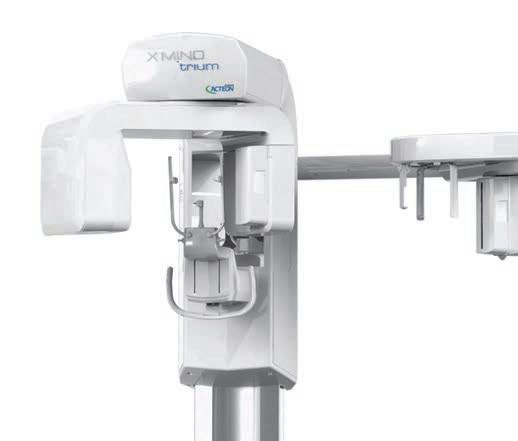
• u erior ima e ualit
• i le el and intuiti e D oft are
• olution of m
• electa le i e of ield of ie from mm to mm
• Dedicated metal artifact reduction filter
• com lete and e clu i e er ice ro ided cteon ot line on ite trainin remote oft are a i tance


 LEFT TO RIGHT: WALID ELKHOURY, CHRISTIAN CONCI FROM YETI, AYAD DAWALIBI
LEFT TO RIGHT; MICHELE TEMPERANI, ELIAS SABBAGH, ELIE MINA
RITA MOUSSA, JOYCE MINA, RABIH HAMOUCHE, ELIE MINA, SAMI SAMAHA
IMAD LAHOUD, FOUAD AWADA, JAMAL ELHAJJ,TAJ AND RASHED MATTIT FROM RENFERT
LEFT TO RIGHT; OSAMA SHEIK-ELBALAD, ZAHI JANHO, CHAWKI RICHA
FADI BALHAWAN, MICHEL KHAYAT, MARINA KELAGHBIAN, CHRISTOPHER ABOU SABHA
PHOTO FROM THE TAMER BOOTH
PHOTO FROM THE PHARMACOL BOOTH
LEFT TO RIGHT: WALID ELKHOURY, CHRISTIAN CONCI FROM YETI, AYAD DAWALIBI
LEFT TO RIGHT; MICHELE TEMPERANI, ELIAS SABBAGH, ELIE MINA
RITA MOUSSA, JOYCE MINA, RABIH HAMOUCHE, ELIE MINA, SAMI SAMAHA
IMAD LAHOUD, FOUAD AWADA, JAMAL ELHAJJ,TAJ AND RASHED MATTIT FROM RENFERT
LEFT TO RIGHT; OSAMA SHEIK-ELBALAD, ZAHI JANHO, CHAWKI RICHA
FADI BALHAWAN, MICHEL KHAYAT, MARINA KELAGHBIAN, CHRISTOPHER ABOU SABHA
PHOTO FROM THE TAMER BOOTH
PHOTO FROM THE PHARMACOL BOOTH
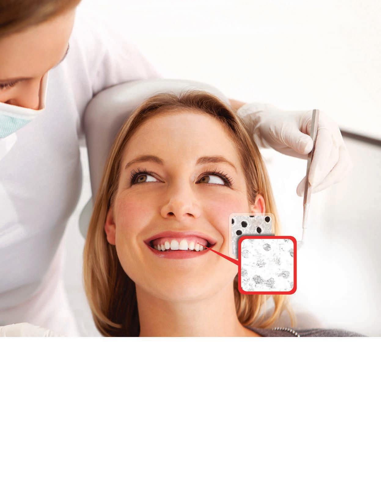
BEFORE
Open tubules AFTER
Closed tubules in 60 SECONDS with Colgate® Sensitive Pro-Relief™ Toothpaste*
Extensive scientific research has shown that Colgate® Sensitive Pro-Relief™ protects against the triggers and causes of sensitivity, and is proven to occlude dentin tubules in 60 seconds.*
Finally, a way to quickly improve your patients’ satisfaction and comfort. *When

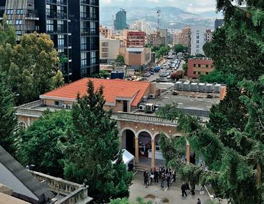
May 25 - 28, 2016
St. Joseph University - Faculty of Dentistry Beirut, Lebanon


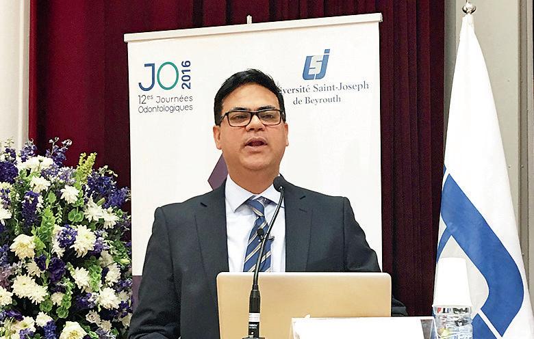
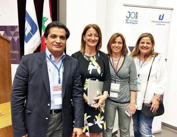
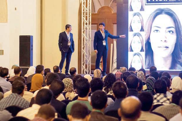
The 12th Dental days organized by the Faculty of Dentistry USJ are being held at the Campus of Medical Sciences in Beirut. The opening ceremony held on May 25 was preceded by a live webcast.
Over 20 international speakers and 40 local speakers presented their work under the conference theme: “Innovations and Controversies.” About 1,000 delegates enjoyed 100 master lectures, live broadcasts, and practical workshops. More than 40 exhibitors presented their products in the gardens of the Campus.
At the opening ceremony, Prof. Salim Daccache S.J. emphasized two points that mark the progress of the congress: “the first is this pertinent idea to gather all the students of the Faculty of Medicine USJ, those of the Lebanese University and the Beirut Arab University to attend to their peers and teachers at a meeting of young researcher named young podium to confirm that younger generations are an inherent part of any dynamic and practical
scientific research and the fact that they come together to help prepare future cooperation and mutual enrichment, especially in the field of dentistry.
The aim is to identify innovations at the level of institutions or individuals, to assert them by discussion and debate and then put them into use in projects and partnerships. In this context, I can only emphasize the dynamic certification conducted by the Faculty of Dentistry with the goal of maintaining its position of excellence in Lebanon and the Arab world.
It is true that technology has undoubtedly an important place in this area but any innovation can not put aside the human and moral element that characterizes any approach that seeks the well-being.
It is in this sense that I see the importance of controversy in your approach, which seeks the best for the human individual and at community level. “


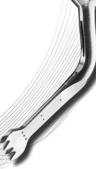


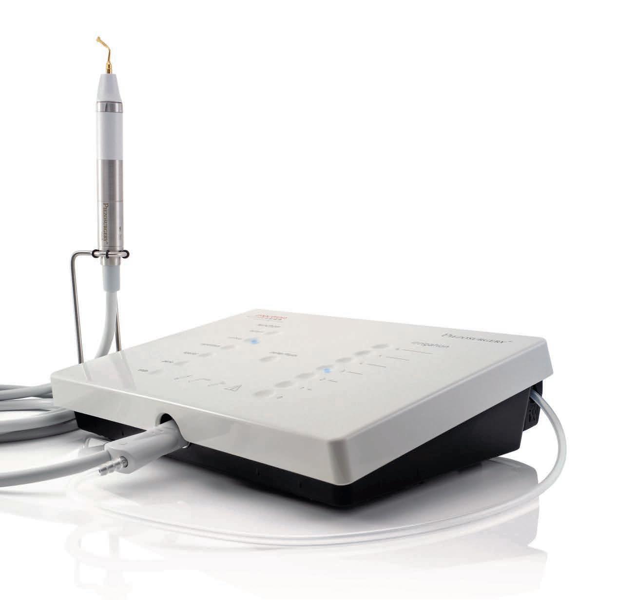
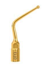
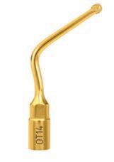
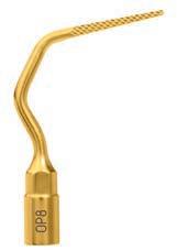
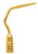
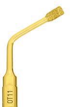
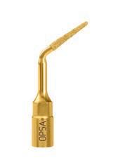
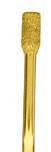
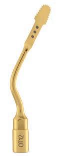
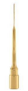
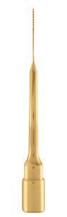
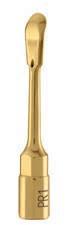
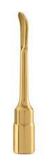
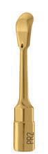
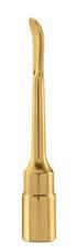
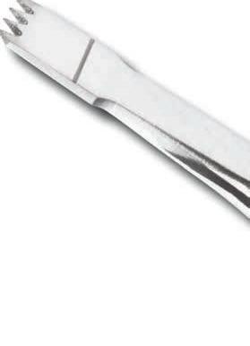
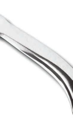
• Numerous robust surgical ultrasonic tips for: Bone surgery, Sinus Lift (lateral & crestal), Crest Splitting, Piezocision™, Extraction, Crown lengthening
• Compatible with Piezotome® Solo, Piezotome® 2 and Implant Center™ 2 units
• For safe and precise surgeries with great healing and reduced post-operative pain*


SIMONE GRANDINI BETWEEN THE DENTAL STUDENTS
12e Journées Odontologiques


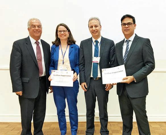
Pioneers in Dentistry Annual Symposium Expanding the Envelope of Dental Specialty
May 28, 2016 - Saint Joseph University, Faculty of Dental Medicine
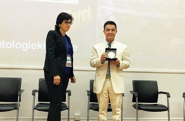
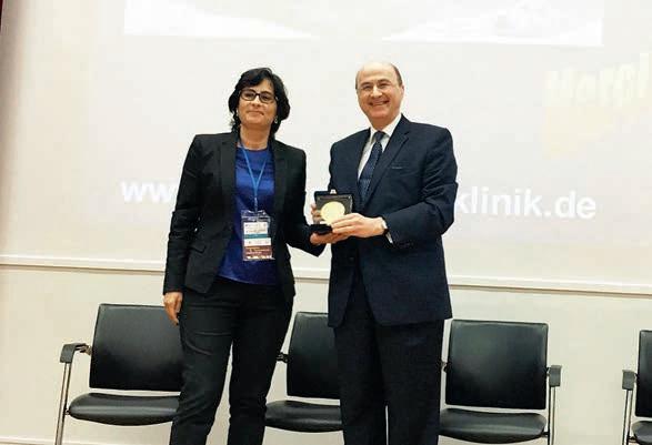
PIONEERS IN DENTISTRY RECOGNITION TO PR.

DR. NADA
DEAN FACULTY OF DENTISTRY IN
UNIVERSITY PRESENTING A TROPHY TO DR. JOSEPH GHAFARY, FOUNDER OF PIONEERS IN DENTISTRY


 DRS. ISSAM KHALIL, JOSETTE CAMILLERI, RANDA HARIK , CARLA ZOUGHEIB MOUBARAK
FOUAD KHOURY
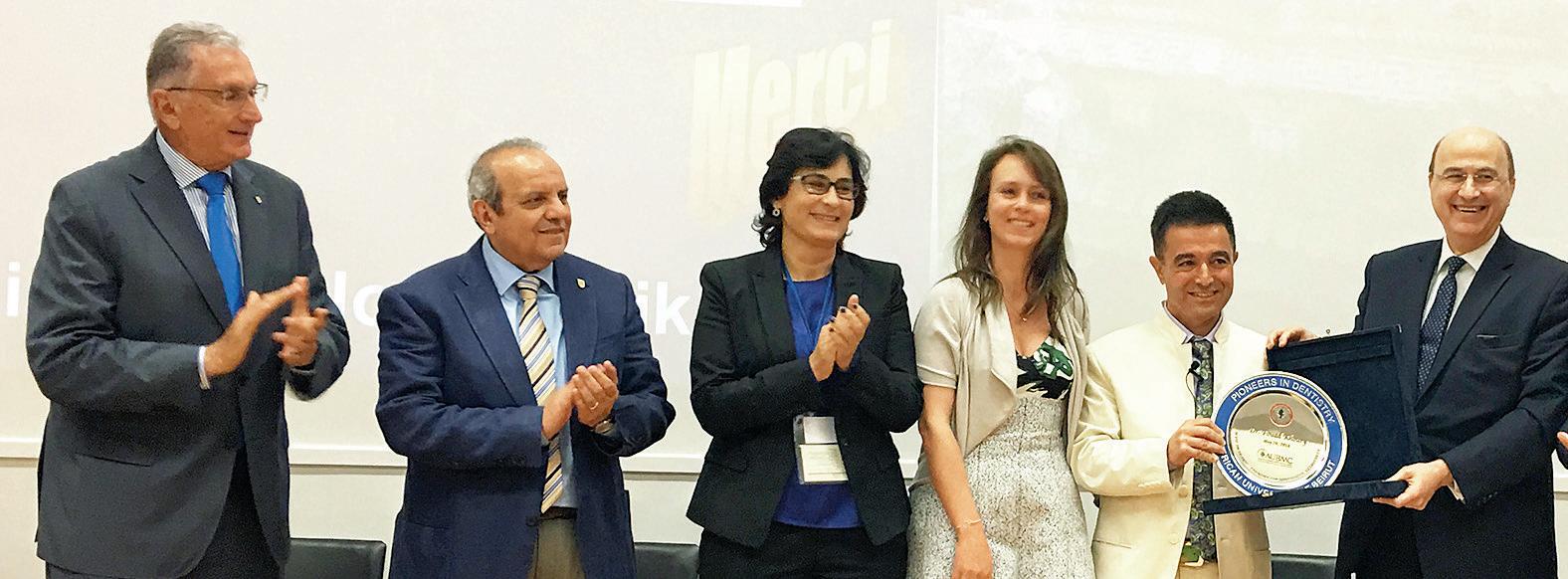
LEFT TO RIGHT; DRS. NABIL BARAKAT, ESSAM OSMAN, NADA NAAMAN, MRS AND DR FOUAD KHOURY, JOSEPH GHAFARI
DRS. MOUNIR DOUMIT, SYBILLE VITAL, JEAN CLAUDE ABOU CHEDID, AJAY JUNEJA
NAAMAN,
SAINT JOSEPH
DRS. ISSAM KHALIL, JOSETTE CAMILLERI, RANDA HARIK , CARLA ZOUGHEIB MOUBARAK
FOUAD KHOURY
LEFT TO RIGHT; DRS. NABIL BARAKAT, ESSAM OSMAN, NADA NAAMAN, MRS AND DR FOUAD KHOURY, JOSEPH GHAFARI
DRS. MOUNIR DOUMIT, SYBILLE VITAL, JEAN CLAUDE ABOU CHEDID, AJAY JUNEJA
NAAMAN,
SAINT JOSEPH

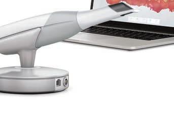
The award-winning TRIOS ® intraoral scanner gets your patients back up on the horse quickly. TRIOS ® 3 makes your work more efficient, more precise and your patients, more comfortable.
Three solutions in one:
• Intraoral scanner for fast, easy-to-do 3D real color digital impressions
• Digital shade measurement while you scan – for more accurate and predictable results
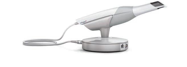
• Integrated intraoral camera included in the scanner


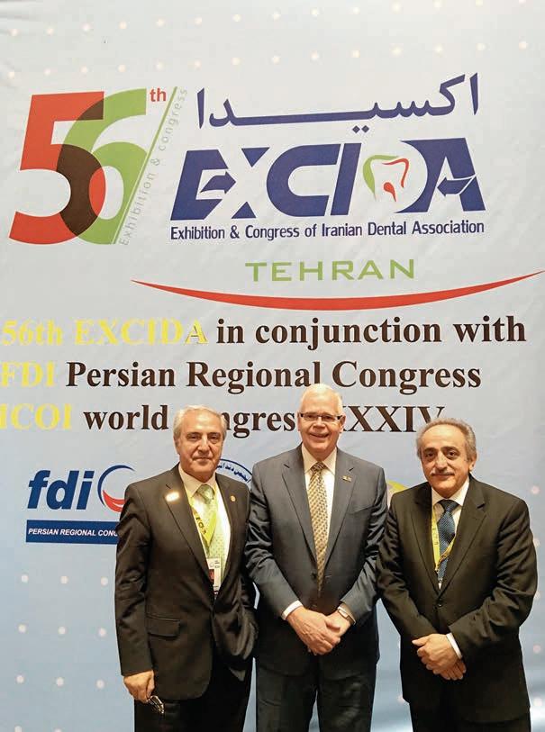
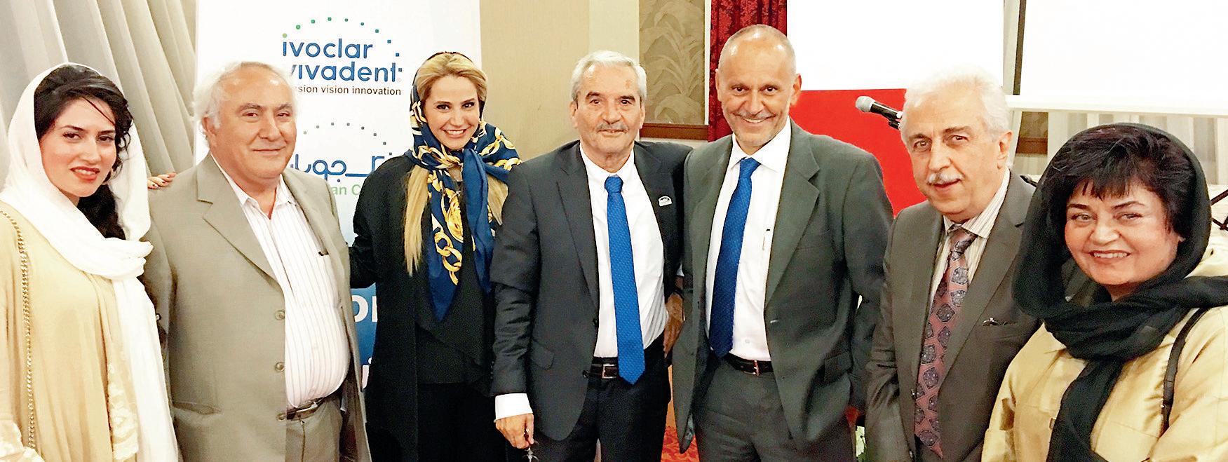
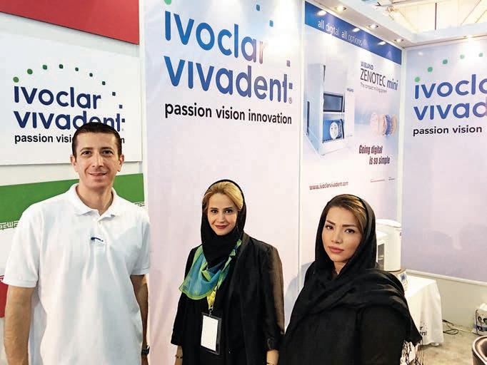
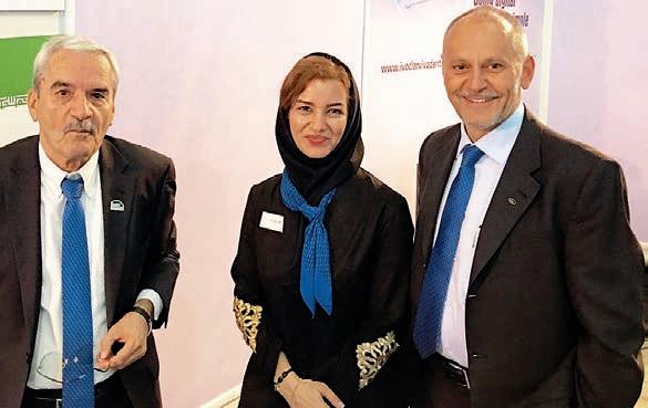
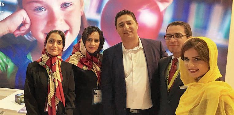
May 17 - 20, 2016 International Exhibition Center Tehran, Iran
Dear colleagues,
Dear participants of 56th EXCIDA and 1st FDI Persian Regional Congress,
As the president of Iranian Dental Association (IDA), this is my great pleasure to deliver a welcome message on the auspicious occasion of 56th EXCIDA and the first FDI Persian Regional Congress that will be convened in Tehran on 17-20 May 2016.
Founded in 1963, IDA is dentists’ organization aimed at improving public dental health and promoting art and science of dentistry.
IDA strives for excellence in dental profession through sup-


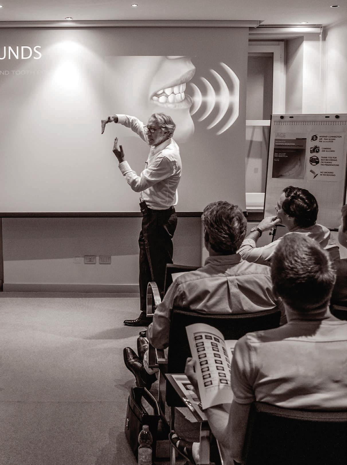
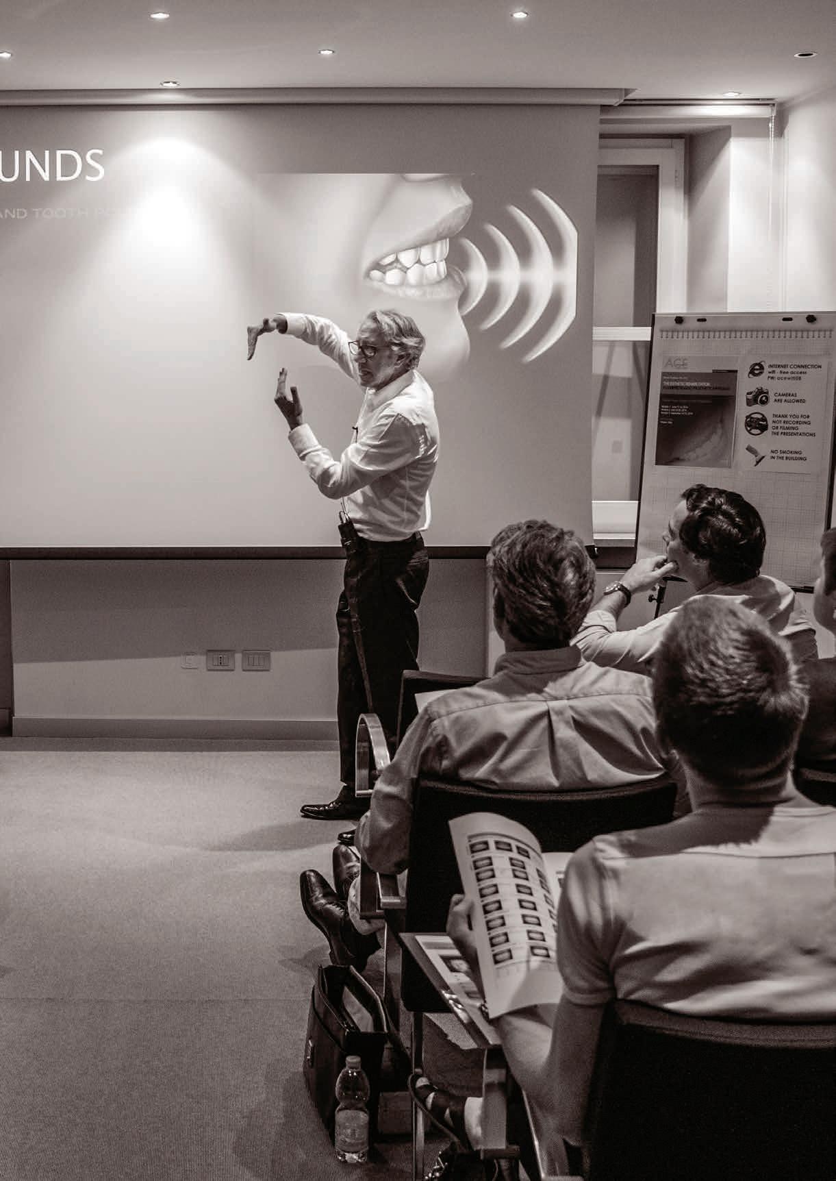
CHARLES GOODACRE TALKING ABOUT THE RESTORATION OF ENDODONTICALLY TREATED TEETH
porting all national dental societies as well as holding annual scientific congresses.
The 2016 congress which is co partnered by FDI features scientific panels of national as well as international renowned dental scholars and provides the opportunity for a comprehensive overview of the latest research developments in dentistry.
I look forward to seeing our colleagues in the coming congress.
With warm regards,
Dr. Gholamreza Ghaznavi President of Iranian Dental Association


Iô°TÉÑŸG äGƒ°ûë∏d ⁄É©dG ‘ ∫hC’G ±õÿG
• √óMh ±õÿG ¤EG óæà°ùJ ⁄É©dG ‘ º«eôJ IOÉe ∫hCG
• ¢ü∏≤àdG ó¡÷ ¢†Øîæe iƒà°ùeh (ºé◊G øe %1^25) ¢†Øîæe Iôª∏H ¢ü∏≤J @
• á«fƒ∏dG äGÒ¨à∏d kGóL ΩhÉ≤eh ‹ÉY …ƒ«M πÑ≤J hP ƒ¡a Gòd ,kÉjhɪ«c πeÉN
• á«Ø∏ÿGh á«eÉeC’G ≥WÉæŸG ‘ äÉÑ∏£àŸG ≈∏YCG »Ñ∏j
• èFÉàf É¡∏c


August 31 - September 2, 2016
Intercontinental City Stars Cairo, Egypt


Distinguished guests, Dear friends and colleagues,
Many dentists have a negative perception of the syndicate, claiming that its role is confined to attesting their documents or private practice licenses, arranging some trips, and the little amount of pension they receive when they retire.
For some, The role of the syndicate ends by receiving their membership cards. Our challenge is to prove the opposite. Our goal is to position the syndicate where it belongs; the umbrella that protects all dentists, and its social responsibility towards the community in general, and dental healthcare in particular.
The role we are looking for is a coordinating role, not a dominating one. Thus we are extending our hands to all

relevant parties and stakeholders, Universities, ministry of health, ministry of higher education, the military, and even private sector, all aiming to achieve one main goal, proper dental healthcare service in Egypt.
Tools are too much to be listed, but starts from proper licensing structure and credit hours, limiting the number of undergraduates, launching dental assistant schools, effecting the ruling of infection control allowance, organizing or rather harmonizing dental scientific events, and much more in our minds.
Our power is in our unity.
Welcome to EDSIC 2016, and I wish you a pleasant evening.
Dr. Ehab Heikal
Secretary, The Egyptian Dental Syndicate International Congress
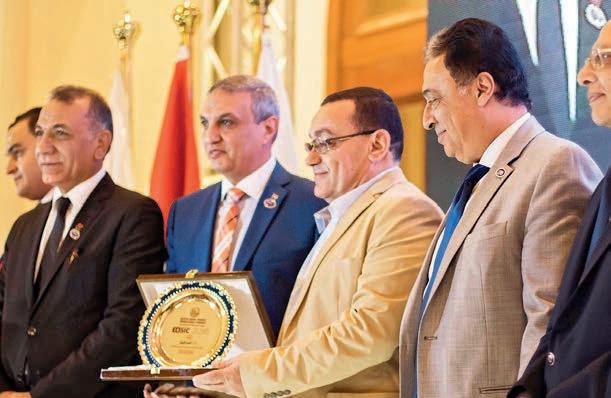
TROPHY DISTRIBUTION TO DR. AHMAD FAWZI, PRESIDENT OF THE ESTHETIC ASSOCIATION
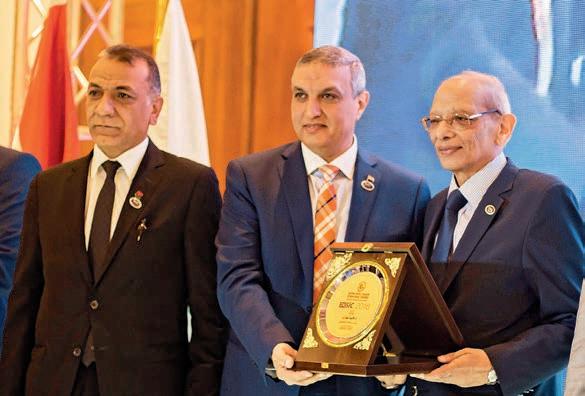
TROPHY DISTRIBUTION TO PR. REDA ABDELRAHMAN, DEAN FACULTY OF DENTAL MEDICINE, FUTURE UNIVERSITY
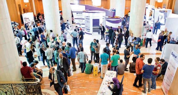
TROPHY TO PR. MAGID AMINE EXHIBITION FLOOR

MOUNIR DOUMIT, FDI, DIRECTOR CONTINUING EDUCATION FOR THE MIDDLE EAST


 PR.
DR. SHAFIC EL HAKIM
PR.
DR. SHAFIC EL HAKIM
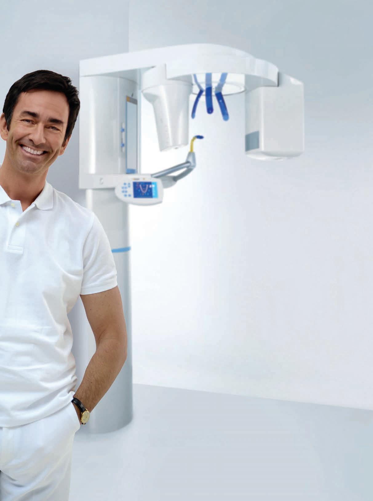
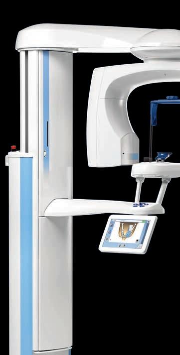
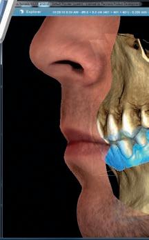
By choosing an OR THOPHOS XG you are investing in a secure future. This is because the units in the OR THOPHOS XG family o er you high quality, durability and the best image quality with the lowest dose and a perfect workflow. S o it‘s no surprise that more than 100,000 dentists all over the world have decided on an OR THOPHOS XG. Enjoy every day. With Sirona.


 DR. MOHAMMED HAMMO
DR. ESLAM KASSEM
DR. ABDEL RAHMAN TAWFIK
DR. MOHAMMED HAMMO
DR. ESLAM KASSEM
DR. ABDEL RAHMAN TAWFIK
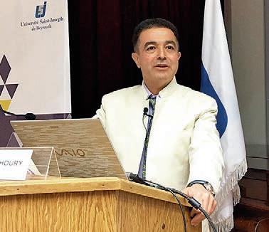
Pr. Nabil Barakat is an Oral and Maxillo-Facial Surgeon from Loyola Dental School Chicago. He was the Founder and Chairman of the Department of Oral and Maxillo Facial Surgery at the Lebanese University Dental School from 1982 until 2006. He is the President of the Lebanese Association of Osseointegration.
He occupied many positions, among them, President of the International College of Dentists Middle East Section, President of the East Mediterranean Association of Osseointegration, and Coordinator of the Post Graduate programs of the Lebanese University. He is the pioneer of Implant Dentistry in the Middle East and an International Lecturer. The editorial team of Dental News met Professor Nabil Barakat and asked him about the One Year Program.
Prof. Nabil Barakat, you are begining a one year program in Bahrain. Can you tell us about your history in implant dentistry?
I am an Oral and Maxillo-Facial Surgeon trained in Chicago and back to lebanon since 1970. in 1986 I met prof. Per-Ingvar Brånemark the discoverer of the osseointegration Phenomenon and we became friends and travelled the whole middle east into using dental implants during
the nineties. We started our first course in 1990 in Beirut, Lebanon. And the course will be given in Bahrain is to fulfill the requirement of the National Health Authority in Bahrain and all the GCC countries.
Once you take that course that includes 250 hours of lectures, Hands-on and live transmissions, you will be able to place implants in your own office and do the prosthetic part.
What is special with this program?
The program is in association with a well know university in the area, the saint joseph university in beirut, and all the teachers in this program will be professors from this university.
How many implants will the dentist put?
Each participant will place 10 implants. if he is placing the implant within the fourth session he will be able to uncover these implants and do the prosthetic part in the 7th or 8th session of 10 and therefore finish the case.
Course Description
The course should give the participants theoretical, hands-on and clinical information about Dental implants. This course will cover all aspects of implant dentistry and is addressed to dental practitioners interested in implant dentistry for their practice.
Course Objectives
• Assimilation of the fundamental principles, which are the basis for any intervention in this field.
• Proper selection of the therapeutic armamentarium in order to find appropriate solution for all types of edentation.
• To teach the participant the principle of applying the technique to the patient and not the patient to the technique.
Course Certificate
At the end of the One Year program, each participant will receive a certificate of attendance, from the Saint Joseph University Faculty of Dentistry.

GC EUROPE N.V.
Head Office
Researchpark
Haasrode-Leuven 1240
Interleuvenlaan 33
B-3001 Leuven
Tel. +32.16.74.10.00
Fax. +32.16.40.48.32
info@gceurope.com
http://www.gceurope.com


Course Date
Course Fee
BD 8,000 this also include four Nobel Biocare Dental Implants and healing abutments. Prosthetic components are not included.
Payment plan in four installments. First 25% upon registration and the remaining should be completed three months before graduation. (Payments are nonrefundable)
For registration and more information about the one year program, please contact Seef Medical Training Center:
Phone: +973 16011771
Email: info@seefmedicaltraining.com
Address:
Bldg 103, First floor, Road 59 Amwaj Island, 257 Next to Choueifat School
Website: www.seefmedicaltraining.com


MECTRON S.P.A.
Via Loreto, 15/A
16042 Carasco (Ge) – Italy
Tel. +39 0185 35361
www.mectron.com - mectron@mectron.com
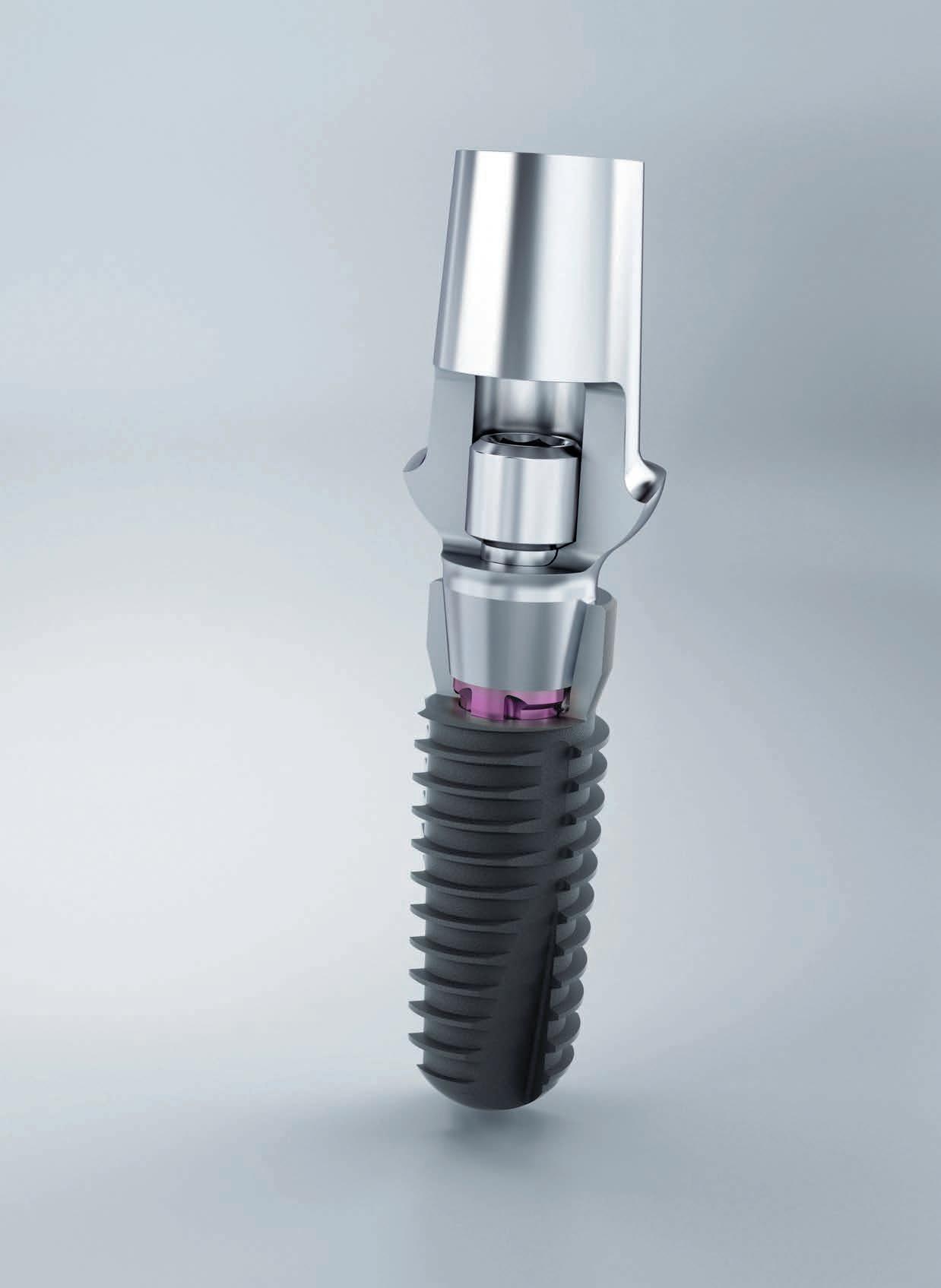
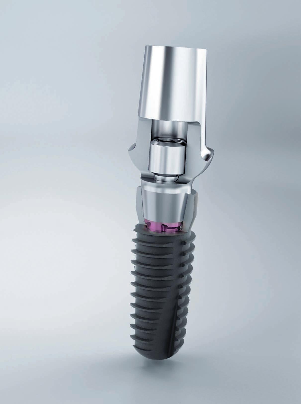
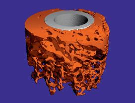
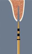
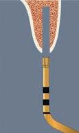
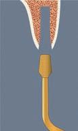
PIEZOSURGERY® Implant Site Preparation, more commonly known as UISP Technique (Ultrasonic Implant Site Preparation Technique) was born from an intuition of Prof. Tomaso Vercellotti, followed and developed within the “International Piezosurgery Academy” and in collaboration with different research groups at the University.
This method offers significant advantages in the healing process, preserving trabecular bone architecture and allowing both to reduce cortical and cancellous bone trauma with respect to the rotating instruments and to leave the bone surface thoroughly cleansed by milling residues. If we analyze site preparation techniques from the mechanical point of view, it is clear that the cutting action is promoted by a variable number of bumps of a cutting disk on the bone structure; the main difference between ultrasonic preparation and preparation with the drill consists precisely in the frequency of the impacts of the working part of the instrument on the bone mineralized structure.
In the ultrasonic technique, there are about 30,000 micro-shocks per second, whereas with drills these are a few dozens or hundreds. This is confirmed by the high micronization of the chips with ultrasonic preparation, and allows better control of the surgical instrument during osteotomy.
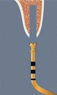
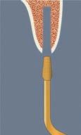
As in the traditional preparation with drills, the piezoelectric implant site preparation involves the use of a sequence of inserts with an increasing diameter with internal and external irrigation, to allow the placement of each type of implant; however, it is also possible to carry out one or more steps with drills in the case of particular geometries or implants with particular anatomical or clinical needs.
The development of the UISP technique has led in a few years to the realization of many biomolecular, biomechanical, histological and radiographic studies, of which there is extensive bibliography available at www.piezosurgeryacademy.com
The first clinical results on the UISP technique applications have led to the publication of favourable case studies, including one published in 2014 on the International Journal of Periodontics and Restorative Dentistry: a multicenter work carried out on 3579 implants with 3 years of follow-up which was conducted on mature edentulous ridges, as well as on regenerated, post-extraction sites and on advanced reconstructive surgery with simultaneous placement of implants.
Another milestone in the history of the UISP technique was in 2011 in Lugano during the 1st International Congress of the International Piezosurgery Academy, where the definition of “Ultra-osseointegration” was introduced and discussed to explain the biomolecular effects that ultrasonic preparation has made possible, as proven by a major study of the University of Turin, published by Prof. Preti et al. in the Journal of Periodontology in 2007.
These results have also been confirmed in several in-vitro and in-vivo studies, which analysed the anatomical features of the bone after site
preparation and the consequent healing physiology, showing a reduction of bone damage in sites prepared with the UISP technique compared to traditional drills.
These studies have demonstrated the positive effects of the microsurgical precision cutting of ultrasounds and those of intensive cleaning by piezoelectric cavitation which frees the site from preparation drilling residues.
In addition, other studies:
• Stacchi et al. (Clinical Implant Dentistry and Related Research 2013) have shown, after the UISP preparation, a minor loss of stability of the implants in the early stages of healing.
• Di Alberti et al. (Quintessence International 2010) have shown an higher peri-implant radiographic bone density in the UISP sites compared to those prepared with drills, suggesting an increased bone formation near the implant.
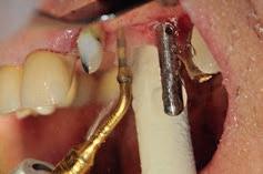
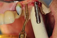
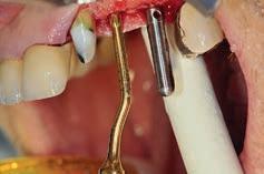
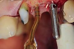
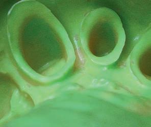
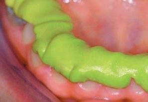
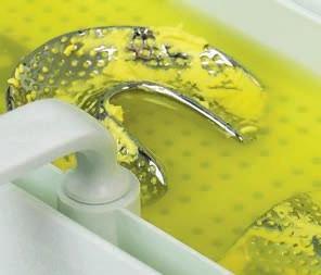
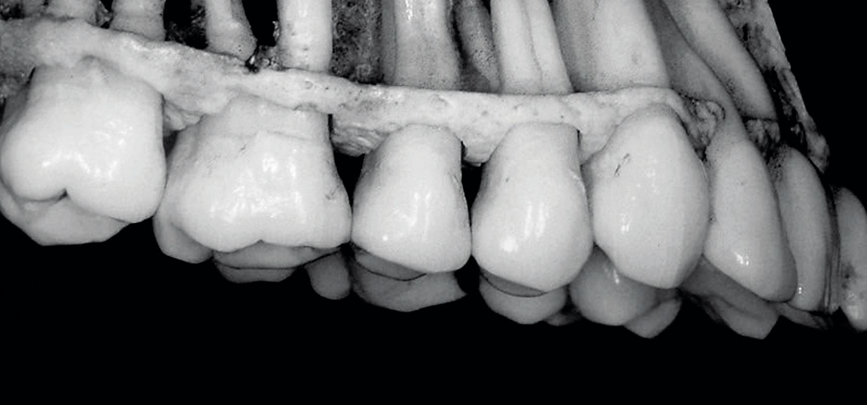
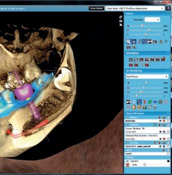
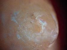
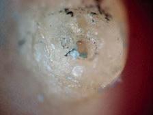
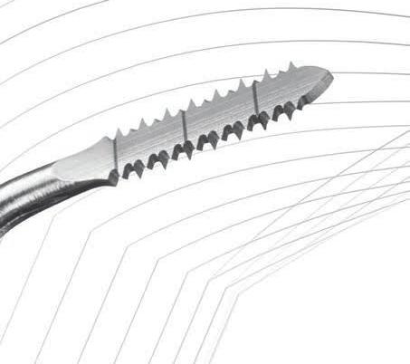
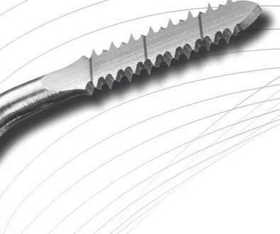
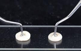
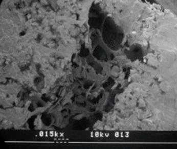
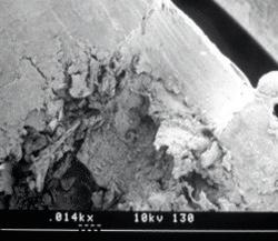
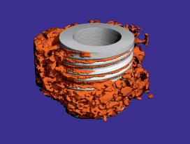
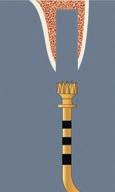
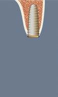
Figures 5-8: Ultrasonic site preparation
Therefore, with ultrasonic preparation, we have:
1. Greater number of vascular channels open in the bone cortex,
2. Less bone damage due to trauma or overheating.
3. Better cleaning of the site from preparation debris.
4. Lifting with endosteum cavitation from trabecular bone.
5. Faster or equal healing of the implants depending on bone quality.
Ultrasonic implant site preparation was particularly advantageous in different clinical situations:
Where immediate loading is chosen and it is imperative to try to obtain and maintain the highest possible stability. As is known, in these cases it is necessary to search for the cortical bone on which to engage the implants; the ultrasound action allows perceiving the different vibration between cortical and cancellous bone.
• In post-extraction sites, in order to easily address the preparation axis as correctly as possible.
• To correct a site initially prepared on an incorrect axis.
• In the presence of poor bone quality or of a very thin cortical as is often the case the upper maxillar bone.
• When it is necessary to preserve delicate tissues, such as the sinus membrane or the alveolar nerve that the ultrasonic vibration does not damage, unlike a rotary drill.
In summary, ultrasounds determine less trauma to the bone structure, a more favourable healing response and a very low inflammatory phase with earlier bone reparative neo-apposition.
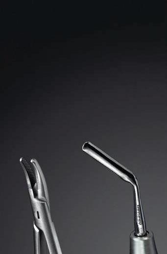


 Fig 5
Fig 6
Fig 8
Fig 5
Fig 6
Fig 8
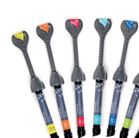
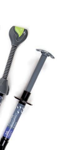
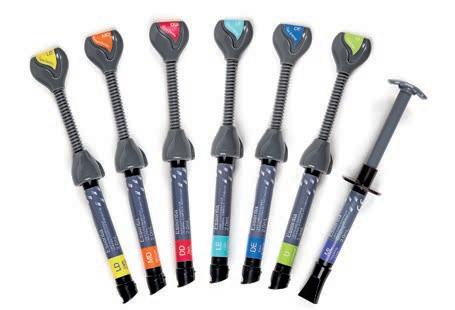
Essentia is a new concept from GC featuring:
Essentia breaks free from the conventions with a unique shade system
• New shade concept, bold & straightforward: seven shades to solve all clinical cases!
• Intuitive system which will simplify the daily work of clinicians.
• Reduced inventory: only seven syringes; all shades will be of use
Essentia simplifies shade selection with six main restorative options
• Anterior: 4 options based on the patient’s age: Bleach/Junior, Young, Adult & Senior
• Posterior: easy duo-layering technique, or a simple monoshade using the Universal
Essentia features optimal handling & optical properties in all situations
• Dentins are made soft for an easy sculpting, and display an excellent shade adaptation
• Enamels are slightly more compact, and guarantee an excellent gloss
• The Universal shade is packable for an easy application in the posterior area
• The Masking Liner is injectable and very opaque: perfect for deep discoloured cavities
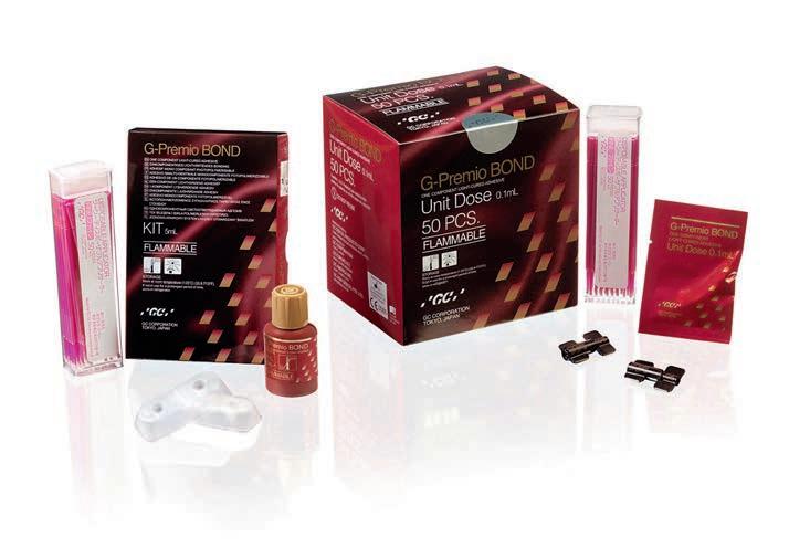
As the bond is equally important, G-Premio Bond offers the ease of use & versatility without any impact on the quality of the bond. G-Premio BOND offers the advantages of a universal but with a top performance in all situations.

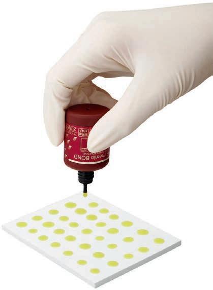
G-Premio BOND: a one-bottle universal bonding compatible with all etching modes and which can be used not only for direct bonding, but also for repair cases & hypersensitivity treatment.
G-Premio BOND offers unique features which help the dentists to strive for…
With an excellent bond strength to both enamel and dentin regardless of the etching mode and an excellent stability, G-Premio BOND ensures a durable bond in time thanks to a combination of three functional monomers.
ZERO discolorations
With a perfect penetration and wettability and an efficient drying procedure, G-Premio BOND displays an extremely thin film thickness of 3µm to guarantee durable aesthetics.
ZERO post-op sensitivity
With a mild effect on dentin tubules in self-etch and a full infiltration by the bonding agent in total-etch, thanks to an optimal wettability.
Website: www.gceurope.com
The new IPS e.max Ceram Selection range comprises special Enamel and Effect powders that are characterized by their vibrant colours and exceptional light-optical properties. They are used in combination with the existing IPS e.max Ceram materials. Individual characteristics are created quickly and easily with these powders and restorations are imparted with highly esthetic, lifelike features.


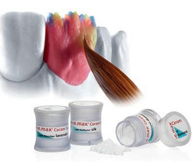


The new Enamel and Effect materials are available in twelve shades, which are divided into three groups. The six “Special Enamel” powders have an enamel-like translucency. They are used to adjust the saturation and chroma of the restoration. As their names suggest, the three “Light Reflector” Effect powders reflect light, while the three “Light Absorber” powders absorb light.
Special Enamel: enamel-like translucency
The following shades are available: aqua, citrine, honey, apricot, quartz and diamond. Citrine, honey, apricot and quartz are used to adjust the saturation and chroma of the restoration and to accentuate the enamel area. Aqua is an intensively coloured enamel powder that highlights the bluish translucent appearance of incisal edges.
Light Reflector: light reflecting
They are supplied in the following shades: silk, salmon and cream. Silk increases the brightness of the incisal area. Salmon and cream are suitable for imitating the colour of reflecting areas in the cervical and incisal third.
Light Absorber: light absorbing
BIOSTAR® and MINISTAR S® – top performance in pressure moulding for all applications in practice and laboratory
• heater reaches working temperature in 1 second
• scan technology for immediate code programming
• great variety of pressure moulding material for all applications
• leading technology for almost 50 years
They are available in the following shades: fog, lavender and taupe. Fog reduces the brightness of the incisal area. Lavender and taupe are used to create light absorbing areas in the incisal and cervical third and in proximal edges.
Experts collaborated in the development
A team of highly esteemed dental technicians composed of Oliver Brix (Germany), August Bruguera (Spain) and Gérald Ubassy (France) played a key role in the development of IPS e.max Ceram Selection. Website: www.ivoclarvivadent.com


1 - 2 DECEMBER, 2016 AT THE SEEF MEDICAL TRAINING CENTER, BAHRAIN


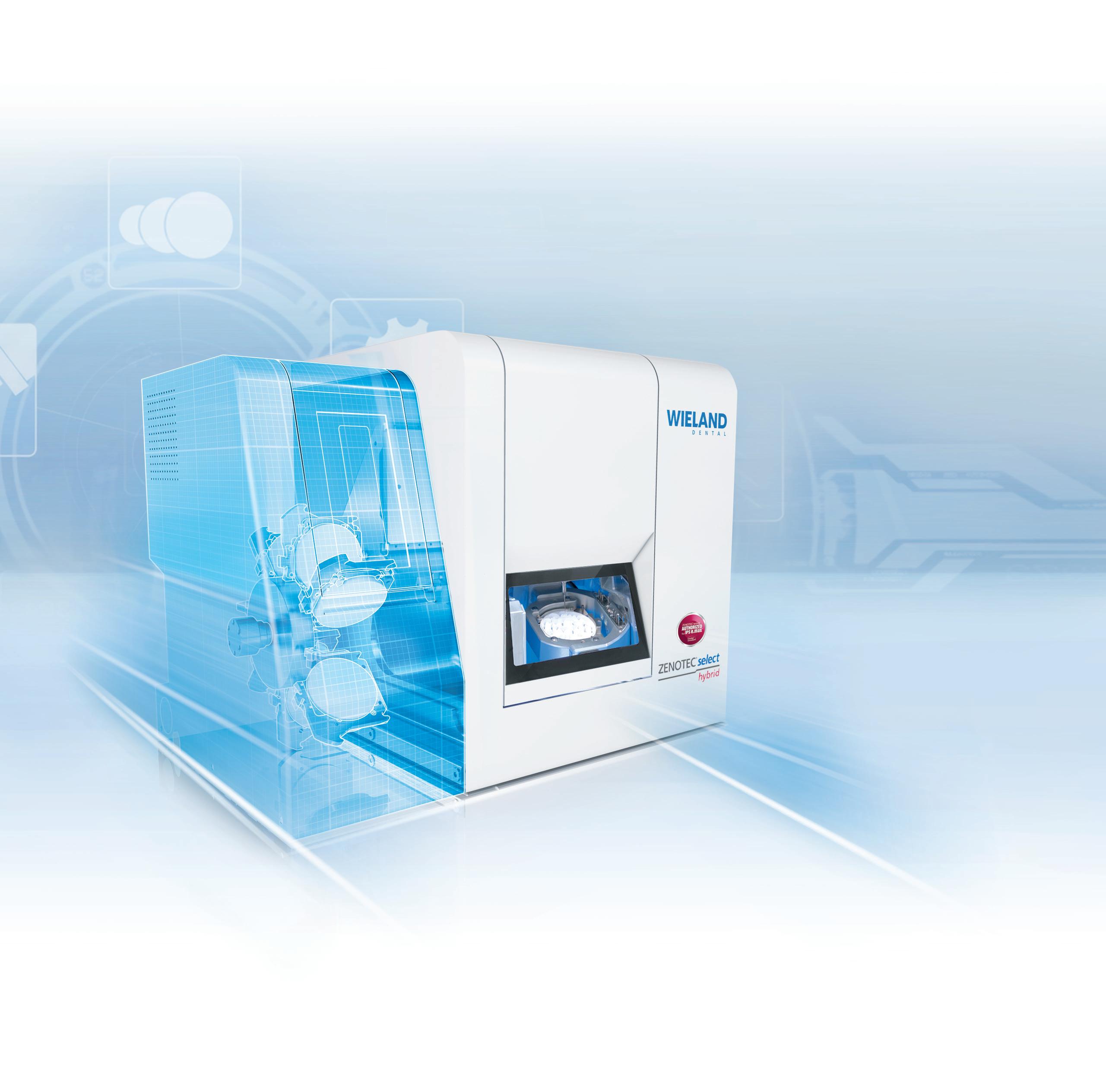
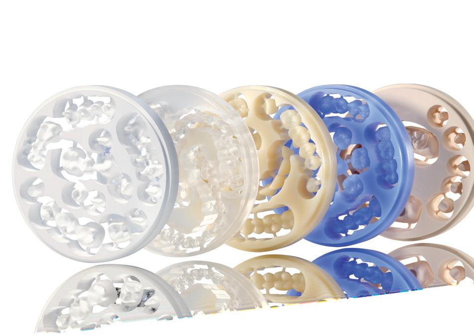
• Automated material changer for enhanced efficiency • Dry milling of zirconium oxide, acrylic resin, wax
• Wet-grinding capabilities for IPS e.max® CAD for Zenotec
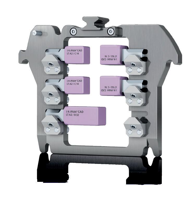
• The IPS e.matrix multiholder maximizes productivity and flexibility