


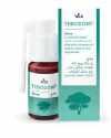
TEBODONT ®
TEBODONT ®
TEBODONT ®
TEBODONT ®
Gel
Gel
Gel
Gel
In case of irritated gums and oral mucosa, contains tea tree oil
In case of irritated gums and oral mucosa, contains tea tree oil
In case of irritated gums and oral mucosa, contains tea tree oil
In case of irritated gums and oral mucosa, contains tea tree oil
TEBODONT
TEBODONT ®
TEBODONT ®
TEBODONT ®
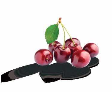
Spray
Spray
Spray
Spray
In case of irritated gums and oral mucosa, inhibits the formation of plaque, cares for and regenerates, contains tea tree oil
In case of irritated gums and oral mucosa, inhibits the formation of plaque, cares for and regenerates, contains tea tree oil
In case of irritated gums and oral mucosa, inhibits the formation of plaque, cares for and regenerates, contains tea tree oil
In case of irritated gums and oral mucosa, inhibits the formation of plaque, cares for and regenerates, contains tea tree oil
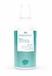
TEBODONT ®
TEBODONT ®
Mouthrinse
Mouthrinse
Mouthrinse
Mouthrinse
Inhibits the formation of plaque, cares for and strengthens the gums, contains tea tree oil, without fluoride
Inhibits the formation of plaque, cares for and strengthens the gums, contains tea tree oil, without fluoride
Inhibits the formation of plaque, cares for and strengthens the gums, contains tea tree oil, without fluoride
Inhibits the formation of plaque, cares for and strengthens the gums, contains tea tree oil, without fluoride
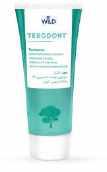
TEBODONT ®
TEBODONT ®
TEBODONT ®
TEBODONT ®
Toothpaste
Toothpaste
Toothpaste
Toothpaste
Inhibits the formation of plaque, strengthens the gums, contains tea tree oil, without fluoride
Inhibits the formation of plaque, strengthens the gums, contains tea tree oil, without fluoride
Inhibits the formation of plaque, strengthens the gums, contains tea tree oil, without fluoride
Inhibits the formation of plaque, strengthens the gums, contains tea tree oil, without fluoride
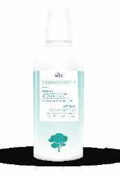
TEBODONT ®-F
TEBODONT ®-F
Mouthrinse
Mouthrinse
Mouthrinse Inhibits the formation of plaque, for the prophylaxis of caries, cares for and strengthens the gums, contains tea tree oil and sodium fluoride
Inhibits the formation of plaque, for the prophylaxis of caries, cares for and strengthens the gums, contains tea tree oil and sodium fluoride
Mouthrinse Inhibits the formation of plaque, for the prophylaxis of caries, cares for and strengthens the gums, contains tea tree oil and sodium fluoride
Inhibits the formation of plaque, for the prophylaxis of caries, cares for and strengthens the gums, contains tea tree oil and sodium fluoride
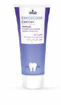
Gum Care
Gum Care
EMOFORM® Gum Care
EMOFORM® Gum Care
Toothpaste
Toothpaste
Toothpaste
Toothpaste
In case of gum problems, contains mineral salts
In case of gum problems, contains mineral salts
In case of gum problems, contains mineral salts
In case of gum problems, contains mineral salts
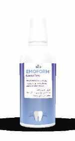
EMOFORM Gum Care
EMOFORM® Gum Care
EMOFORM® Gum Care
Mouthbath concentrate In case of gum problems, contains mineral salts
Mouthbath concentrate In case of gum problems, contains mineral salts
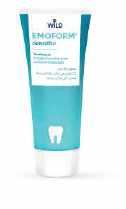
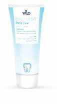
Sensitive
Sensitive
EMOFORM® Sensitive
EMOFORM® Sensitive
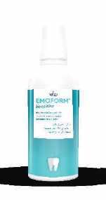
Sensitive
Sensitive
EMOFORM® Sensitive
EMOFORM® Sensitive
Whitening toothpaste For white and shiny teeth, contains finest diamond particles EMOFORM Gum Care
Mouthbath concentrate In case of gum problems, contains mineral salts
Mouthbath concentrate In case of gum problems, contains mineral salts
Toothpaste
Toothpaste
Toothpaste In case of sensitive gums, contains mineral salts
In case of sensitive gums, contains mineral salts
Toothpaste In case of sensitive gums, contains mineral salts
In case of sensitive gums, contains mineral salts
Mouthbath concentrate
Mouthbath concentrate
In case of sensitive gums, contains mineral salts
Mouthbath concentrate In case of sensitive gums, contains mineral salts
Mouthbath concentrate In case of sensitive gums, contains mineral salts
In case of sensitive gums, contains mineral salts
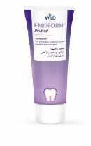
Protect
Protect
EMOFORM
Protect
EMOFORM Protect
Toothpaste
Toothpaste For prevention of caries, hardens dental enamel
For prevention of caries, hardens dental enamel
Toothpaste For prevention of caries, hardens dental enamel
Toothpaste For prevention of caries, hardens dental enamel
Diamond
Diamond
EMOFORM®
Diamond
EMOFORM® Diamond
Whitening toothpaste
Whitening toothpaste
For white and shiny teeth, contains finest diamond particles
Whitening toothpaste For white and shiny teeth, contains finest diamond particles
For white and shiny teeth, contains finest diamond particles
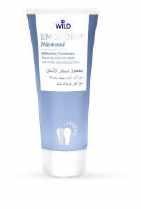
EMOFLUOR® Daily Care
EMOFLUOR® Daily Care
EMOFLUOR® Daily Care
® Daily Care
Toothpaste
Toothpaste
EMOFLUOR® Daily Care
EMOFLUOR® Daily Care
EMOFLUOR® Daily Care
EMOFLUOR® Daily Care
Mouthrinse
Mouthrinse
Toothpaste For daily care of sensitive teeth, contains stabilised stannous fluoride
For daily care of sensitive teeth, contains stabilised stannous fluoride
For daily care of sensitive teeth, contains stabilised stannous fluoride
Toothpaste For daily care of sensitive teeth, contains stabilised stannous fluoride
Mouthrinse
For the daily care of sensitive teeth and for the prophylaxis of caries
For the daily care of sensitive teeth and for the prophylaxis of caries
For the daily care of sensitive teeth and for the prophylaxis of caries
Mouthrinse For the daily care of sensitive teeth and for the prophylaxis of caries
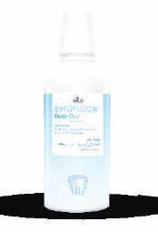
EMOFLUOR® Intensive Care
EMOFLUOR® Intensive Care
EMOFLUOR® Intensive Care
Intensive Care
Gel
Gel
Gel
Gel
For targeted protection against sensitive teeth and erosions, contains stabilised stannous fluoride
For targeted protection against sensitive teeth and erosions, contains stabilised stannous fluoride
For targeted protection against sensitive teeth and erosions, contains stabilised stannous fluoride
For targeted protection against sensitive teeth and erosions, contains stabilised stannous fluoride
EMOFLUOR® Twin Care
EMOFLUOR® Twin Care
EMOFLUOR® Twin Care
Toothpaste
EMOFLUOR EMOFLUOR ® EMOFORM
® Twin Care
Toothpaste
Toothpaste
Toothpaste
Dual protection against erosions and sensitive teeth with stannous fluoride and vVARDIS technology
Dual protection against erosions and sensitive teeth with stannous fluoride and vVARDIS technology
Dual protection against erosions and sensitive teeth with stannous fluoride and vVARDIS technology
Dual protection against erosions and sensitive teeth with stannous fluoride and vVARDIS technology
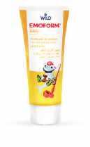
EMOFORM® Kids
EMOFORM® Kids
EMOFORM Kids
EMOFORM® Kids
Toothpaste for children
Toothpaste for children
EMOFORM® Youngstars
EMOFORM® Youngstars
EMOFORM Youngstars
EMOFORM® Youngstars
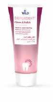
DEPURDENT® Clean & Polish
DEPURDENT® Clean & Polish
DEPURDENT® Clean & Polish
DEPURDENT® Clean & Polish
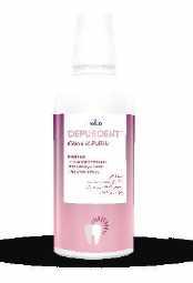
DEPURDENT® Clean & Polish
DEPURDENT® Clean & Polish
DEPURDENT® Clean & Polish
DEPURDENT® Clean & Polish
Toothpaste for children
From the first milk tooth, up to and including 5 years
From the first milk tooth, up to and including 5 years
From the first milk tooth, up to and including 5 years
Toothpaste for children From the first milk tooth, up to and including 5 years
Toothpaste for children Comprehensive protection for the teeth, as of 6 years
Toothpaste for children
Toothpaste for children Comprehensive protection for the teeth, as of 6 years
Paste for teeth cleaning
Paste for teeth cleaning
Mouthrinse
Mouthrinse
Toothpaste for children Comprehensive protection for the teeth, as of 6 years
Comprehensive protection for the teeth, as of 6 years
Removes dental plaque and teeth discoloration, with polishing eect
Paste for teeth cleaning
Removes dental plaque and teeth discoloration, with polishing eect
Removes dental plaque and teeth discoloration, with polishing eect
Paste for teeth cleaning
Removes dental plaque and teeth discoloration, with polishing eect
Mouthrinse
Mouthrinse
Helps preserve the natural whiteness of your teeth, helps prevent caries
Helps preserve the natural whiteness of your teeth, helps prevent caries
Helps preserve the natural whiteness of your teeth, helps prevent caries
Helps preserve the natural whiteness of your teeth, helps prevent caries
Al Rabee
Volume XXXII, Number 2, 2025
EDITORIAL TEAM COORDINATOR
Alfred Naaman, Nada Naaman, Khalil Aleisa, Jihad Fakhoury, Dona Raad, Antoine Saadé, Lina Chamseddine, Tarek Kotob, Mohammed Rifai, Bilal Koleilat, Mohammad H. Al-Jammaz
Suha Nader
Marc Salloum
Micheline Assaf, Nariman Nehmeh
Josiane Younes
Albert Saykali
Gisèle Wakim
Tony Dib 1026-261X
DENTAL NEWS IS A QUARTERLY MAGAZINE DISTRIBUTED MAINLY IN THE MIDDLE EAST & NORTH AFRICA IN COLLABORATION WITH THE COUNCIL OF DENTAL SOCIETIES FOR THE GCC.
Statements and opinions expressed in the articles and communications herein are those of the author(s) and not necessarily those of the Editor(s) or publisher. No part of this magazine may be reproduced in any form, either electronic or mechanical, without the express written permission of the publisher.
DENTAL NEWS – Sami Solh Ave., G. Younis Bldg. POB: 116-5515 Beirut, Lebanon.
Tel: 961-3-30 30 48
Email: info@dentalnews.com Website: www.dentalnews.com

FDI
World Dental Congress 2025

Saudi
Prosthodontic Society 2025

BIDM 2025

CADEX Central Asia 2025

www.instagram.com/dentalnews
39th CAPP Int’l Dental ConfEx
September 9 – 12, 2025 Shanghai | CHINA 2025.world-dental-congress.org/En
September 26 – 27, 2025 Crown Plaza | Riyadh, KSA www.prosthodontics.org.sa
October 3 – 5, 2025 Seaside Arena | Beirut, LEBANON www.lda.org.lb
SIDC Jeddah 2025
AEEDC 2026
SIDC Riyadh 2026
October 9 – 11, 2025 ALMATY | KAZAKHSTAN www.cadex.kz
November 14 – 15, 2025 Madinat Jumeirah | Dubai, U.A.E www.cappmea.com
November 27 – 29, 2025 Jeddah Hilton | Jeddah, K.S.A. www.sidc.org.sa
January 19 – 21, 2026 World Trade Center | Dubai, U.A.E. www.aeedc.com
February 5 – 7, 2026
Riyadh Front Exhibition & Conference Center | Riyadh, K.S.A. www.sidc.org.sa


Abdelrahman K. Eldabe, BDS, MSc, PhD,a Doaa Adel-Khattab, BDS, MSc, PhD,b and Kirollos H. Botros, BDSc
When registering intraoral features and implant position, an intraoral scanner (IOS) is a popular substitute for traditional impression materials, increasing patient comfort, accuracy, and predictability1 The traditional splinted impression technique has been reported to provide an accurate definitive cast,2–5 but, for the precise and distortion free capture of the implant positions, a stiff and dimensionally stable splinting material is essential. 6,7
Qasem Obiedat, BDS
Maxillo-Facial
Surgeon; Jordanian Ministry of Health
Amman, Jordan asemobeidatsurg@gmail.com
The mesiodens is an extra tooth that is situated in the middle of the two central incisors. It is the most prevalent supernumerary teeth. Whereas genetic factors and dental lamina proliferation have been linked to mesiodens, its exact etiology is still unknown. Often, it causes cyst development, misaligned teeth, food entrapment, poor aesthetics, and other oral health problems. Therefore, to avoid these pathologic and orthodontic issues, early diagnosis and treatment are advised. This essay examines recent research on the causes, prevalence, diagnoses, and treatments of this concern.
A high degree of trueness, generally within a range of ≤50 µm, has been reported with IOSs when used to capture single implant and short span implant-supported prostheses.8,9 Scanning for complete arch implant-supported prostheses is more challenging because of the lack of fixed landmarks, and unacceptable passivity has been reported, particularly in mandibular prostheses.8–12 A discrepancy of <150 µm has been recognized as the clinically acceptable level of misfit for complete arch implant-supported prostheses.13–15 Photogrammetry has been used, without stitching, to scan implant coordinates while omitting the dental and gingival anatomy.16 The exclusion of unstable mucosa during scanning and the avoidance of stitching make the passive seating of implant prostheses a more predictable procedure.17–19 Although a promising technique, clinical studies of photogrammetry are scarce.10,20
As well as, understanding the various removal techniques and their effects on the patient’s dentition.
1. Mesiodentes compose of 3 or more mesiodens (supernumerary teeth) and are uncommon compared to mesiodens (single supernumerary tooth) found in the midline of the maxilla. Typically, mesiodentes are asymptomatic, impacted and have a cone-like crown and one root.1
Statement of problem. Intraoral scanning of complete arch implant-supported prosthesis is still not predictable in all scenarios. Photogrammetry was introduced to overcome physical and anatomic limitations. The use of a new intraoral scanner combined with photogrammetry technology in a simplified workflow may improve the ease of fabrication and accuracy of complete arch implant-supported prostheses.
They often have their roots facing the occlusion and their crowns towards the nasal cavity in an inverted configuration.1
Purpose. The purpose of this clinical study was to evaluate the degree of trueness of a conventional intraoral scanner (IOS) and an intraoral photogrammetry scanner (IPS) for complete arch implant-supported prostheses.
Mesiodens diagnosis, mesiodens removal, esthetic concerns of mesiodens, orthodontic treatment after mesiodens removal, supernumerary teeth in adults, surgical extraction.
Mesiodens comes from a Latin origin “mesio” meaning “medio” or middle and “dens” meaning tooth. It is a supernumerary tooth that usually resides between the upper central incisors as the occurrence is rare in the lower region. mesiodens usually results in oral problems such as malocclusion, food impaction and poor aesthetics. It’s the most prevalent type of supernumerary teeth.
Current commercially available photogrammetry systems have been considered extraoral systems that require an additional recording of the soft tissue whether with a conventional impression or a digital scan. Recently an intraoral photogrammetry scanner (IPS) has been introduced that can scan
2. The prevalence of supernumerary teeth in permanent teeth is 1–14%, in accordance with the literature. When compared to females, males are impacted around twice as often. Most extra teeth (90–98%) are found in the maxilla, and 90% of them are only found in the pre-maxilla.2
Listed from the most to the least frequent locations of supernumerary teeth:
• The mesiodens
• Maxillary fourth molars
• Maxillary premolars
• Mandibular premolars
• Maxillary lateral incisors
• Mandibular fourth molars
Material and methods. Participants who had received 4 implants in an edentulous arch for an implant-supported complete arch fixed dental prosthesis were recruited. Three recordings were obtained for each participant: IOS, IPS (tests), and a conventional splinted open-tray impression (reference). Three-dimensional (ΔEUC), and angular deviations (ΔANGLE) for both groups (IOS and IPS) were evaluated and compared with the reference scan. Potential effects (correlation) of the impression device (IOS and SPG) and type of arch (maxilla and mandible) were evaluated. A paired t test was used to evaluate both the angular and 3-dimensional deviation for each implant position. A point-biserial correlation was conducted to assess the relationship between jaw type and angular and ΔEUC deviations in the 2 groups.
• Maxillary premolars
In some syndromes, mesiodens may present as a part of the symptoms; however, this condition might be seen in normal individuals. It seems that positive family history is one of the predisposing factors.
This research did not receive any specific grant from the funding agencies in the public, commercial, or non-profit sectors. The authors declare no conflict of interest and this research did not receive any specific grant from any funding agencies.
• aLecturer, Oral Medicine, Periodontology and Diagnosis, Faculty of Dentistry, Assiut University, Assiut, Egypt.
• bAssociate Professor, Oral Medicine, Periodontology and Diagnosis, Faculty of Dentistry, Ain Shams University, Cairo, Egypt.
• cDemonstrator, Oral Medicine, Periodontology and Diagnosis, Faculty of Dentistry, Assiut University, Assiut, Egypt.
This study aims to cover most of the major topics related to mesiodens diagnosis, surgical removal considerations postsurgical extraction recommendations relevant influencing factors associated with the supernumerary tooth eruption.
Results. Thirteen edentulous arches (9 maxillae, 4 mandibles) in 11 individuals were rehabilitated with a monolithic zirconia screw-retained prosthesis supported by 4 implants, totaling 52 implants. Conventional intraoral scanners (IOS) and intraoral photogrammetry scanners (IPS) were used to scan 104 implant positions, which were then compared with the corresponding reference scans. (mean ΔEUC IOS 59.8 µm, IPS 30.7 µm; mean ΔANGLE IOS 1.4 degrees, IPS 0.78 degrees). A paired t test revealed statistical significance in favor of IPS in terms of both Euclidian and angular deviation (P<.001). In the IOS group, angular deviation had a positive statistically significant correlation with the type of arch (rpb=0.34, n=52, P=.013).
The etiology behind supernumerary teeth formation is unclear. Many ideas, including the dualism of the tooth bud, the hyperactivity of the dental lamina, and a confluence of hereditary and environmental variables have been suggested. However, only two widely accepted hypotheses have been supported.
1. The dichotomy hypothesis of dental germs, a tooth bud divides into two pieces, producing two teeth that may be of equal or different sizes.
Conclusions. The IPS significantly improved the ease and accuracy of recording complete arch implant-supported prostheses.
2. The dental lamina’s local, autonomous, or conditioned hyperactivity causes the development of extra teeth.

Embodies our belief of bringing highly reliable Japan-quality products to dentists in the
Osaka, Japan https://dental.takarabelmont.co.jp/

primary dentition. Moreover, the presence of extra teeth may be a single, independent defect or may be linked to certain diseases, such as cleft lip and palate, Down syndrome, cleidocranial dysplasia, etc.
incision are used to remove a maxillary mesiodens. The scientists discovered that this method effectively removed the mesiodens without harming the nearby teeth or tissues.
The reported frequency in the general population ranges from 0.15 to 1.9%, and it’s believed to affect more men than women. According to reports, 82% of the time it affects the maxilla, more precisely the premaxillary area compared to the anterior region of the mandible.
Regardless of the type of arch, intraoral photogrammetry appears promising for scanning implants for complete arch implant-supported prostheses.
Radiographic diagnosis
One-fifth of all supernumerary teeth are found in the permanent dentition, making them less prevalent in the primary dentition. Moreover, the presence of extra teeth may be a single, independent defect or may be linked to certain diseases, such as cleft lip and palate, Down syndrome, cleidocranial dysplasia, etc.
3D printing can be effectively utilized in the fabrication of the following appliances: diagnostic models, removable orthodontic appliances, presurgical nasoalveolar molding, occlusal splints, space maintainers, expanders, aligners and retainers.
Radiography most accurately Cone-beam computed tomography (CBCT), is used for the diagnosis of supernumerary teeth including mesiodens. Due to the fact that CBCT has the ability to get around most of the technical challenges of plain radiographs projection and the capacity to provide a high-resolution three-dimensional (3D) interpretation of the maxillofacial tissues.3
EPIDEMIOLOGY
tissue and that has built-in photogrammetry technology so that it can capture implant locations through special intraoral scan flags. The IPS can accomplish both soft tissue and scan flag scanning and can also merge them in the included software program without the need for a thirdparty software program, as required with extraoral photogrammetry scanners.
RADIOGRAPHIC DIAGNOSIS
Diagnostic models measurements performed with 3D digital models represent high validity, reliability, and reproducibility. They are a viable alternative to traditional plaster models and identical copies of a digital model can be reproduced without distortion or deformation which can negatively affect the appliance that is built upon it5. Warping and distortion are two common issues traditionally encountered using gypsums and other polymer materials when used for dental impressions.
The reported frequency in the general population ranges from 0.15 to 1.9%, and it’s believed to affect more men than women. According to reports, 82% of the time it affects the maxilla, more precisely the premaxillary area compared to the anterior region of the mandible
When a mesiodens is present, it is critical that it be treated quickly. This is due to the possibility that leaving it could result in dental issues later in life, such as a diastema (space between your two front teeth), displacement of surrounding teeth, increased crowding in the area of the mesiodens, problems with bite creation, and root resorption of nearby teeth, as well as, delayed eruption of surrounding teeth.
Therefore, the mesiodens in a timely related manner is mandatory.
MANAGEMENT
The authors are unaware of a previous prospective clinical trial aimed primarily at investigating and comparing the accuracy (degree of trueness) of IOS and IPS for complete arch implant recording. Trueness refers to how close the scanned fixture positions are to reality. The secondary outcome was to examine the potential association between the type of arch (maxilla versus mandible) and the degree of trueness. The chair-side scanning duration of the approaches was also evaluated. The null hypotheses were that no variations would exist between intraoral scanning and intraoral photogrammetry capturing systems for complete arch implant-supported prostheses and that neither capturing system would be affected by the type of arch (maxilla versus mandible).
Radiography most accurately Cone-beam computed tomography (CBCT), is used for the diagnosis of supernumerary teeth including mesiodens. Due to the fact that CBCT has the ability to get around most of the technical challenges of plain radiographs projection and the capacity to provide a high-resolution three-dimensional (3D) interpretation of the maxillofacial tissues.3
Removable Orthodontic appliances that can be fabricated by 3D printing include the Hawley appliance and functional appliances like Twinblock or activators. Traditional pouring of plaster casts have effectively been eliminated by utilizing scans such as TRIOSTM or 3ShapeTM6. Other manufacturers worldwide of 3D orthodontic appliances include DentalWingsTM, Shining 3DTM
The way supernumerary teeth are handled depends on their type, where they are in the mouth, and where they are in the dentition cycle. It is advised to remove the mesiodens sooner in order to improve the prognosis. Since mesiodens frequently erupt into the oral cavity, it is generally not recommended to extract them during primary dentition since doing so increases the risk of injuring the permanent incisor. However, following the removal of mesiodens, the permanent central incisors spontaneously emerge in the early mixed dentition stage. Additionally, it encourages optimized tooth alignment and reduces the need for orthodontic therapy. Following the removal of a mesiodentes, the dentition must be closely monitored.
Reevaluation is advised six months following mesiodens extraction and if the permanent incisor does not erupt normally after 12 months of extraction, closed eruption accompanied with orthodontic mechanotherapy is recommended.
1.
In this study,
incisor does not erupt normally after 12 months of extraction, closed eruption accompanied with orthodontic mechanotherapy is recommended.
technology, where acrylic layers are added on to each other, while bonding occurs between levels to fabricate an appliance8. This method has proven to be faster, more adaptable and better fitting for patients.
preventing incision releases and filling the surgical site with platelet-rich fibrin (PRF). A 13-year-old patient was referred to our clinic to remove a supernumerary tooth positioned between the maxillary central incisors. A mucoperiosteal flap was raised bilaterally through tunneling following anesthesia and the excision of the labial frenulum. The tooth was carefully extracted using an “apexo” elevator after a delicate osteotomy was completed. This method offers a predictable, conservative approach, lower operation complications, no scarring in the front maxilla, and no adverse cosmetic effects.
In this study, a lateral tunneling technique and frenulum incision are used to remove a maxillary mesiodens. The scientists discovered that this method effectively removed the mesiodens without harming the nearby teeth or tissues. preventing incision releases and filling the surgical site with platelet-rich fibrin (PRF). A 13-yearold patient was referred to our clinic to remove a supernumerary tooth positioned between the maxillary central incisors. A mucoperiosteal flap was raised bilaterally through tunneling following anesthesia and the excision of the labial frenulum. The tooth was carefully extracted using an “apexo” elevator after a delicate osteotomy was completed. This method offers a predictable, conservative approach, lower operation complications, no scarring in the front maxilla, and no adverse cosmetic effects.
The labial frenulum was anesthetized laterally with 4% lidocaine+ 100,000 epinephrine. The frenulum was excised and the incision was made wider using a 15C blade and hemostatic forceps.
A mucoperiosteal flap was raised bilaterally by tunneling to improve the area’s visibility. As a result, the flap had considerable movement and could retract. The flap’s margins were sutured with 5.0 nylon suture thread in order to separate and manipulate it without injury.4
The labial frenulum was anesthetized laterally with 4% lidocaine+ 100,000 epinephrine. The frenulum was excised and the incision was made wider using a 15C blade and hemostatic forceps.
The labial frenulum was anesthetized laterally with 4% lidocaine+ 100,000 epinephrine. The frenulum was excised and the incision was made wider using a 15C blade and hemostatic forceps.
Space maintainers are used to intercept malocclusion before it is established within the dentition of a child receiving orthodontic care. The following appliances are also used for anchorage reinforcement, where a tooth or teeth must be held stationary during treatment until movement can be elicited. These appliances can now be routinely fabricated with 3D printing: a) Trans palatal arch is 3D printed through metal printing and the bonding site is designed on the molars not completely circular, but only confined to palatal surface. b) Hybrid Nance appliance: Nance appliance again serving both purposes, can be 3D printed through metal printing. c) Lingual arch: Lingual arch fabrication with bands designed to be printed through 3D metal printing (fully or partial) around the molars with a connector along the lingual surface of the teeth 9
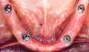
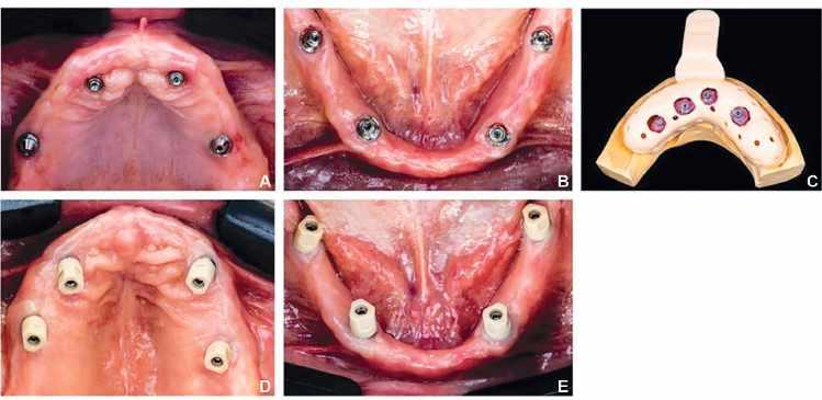
All participants were recruited from the periodontology clinic of the Faculty of Dentistry Ain Shams University and Assiut University. Approval for the study had been provided by the ethical committee (FDASU-Rec IR032422). The inclusion criteria consisted of participants who had received 4 implants in a single edentulous arch (Fig. 1), were to receive a 1-piece complete arch implant-supported prosthesis, were available for follow-up visits, and were medically healthy (American Society of Anesthesiology [ASA] classification I or II). Implants were placed fully guided with an approximate location in the canine and first molar areas, respecting the anteriorposterior implant spread,21,22 with an inter- implant angulation not exceeding 15 degrees. Otherwise, angled multiunit abutments (MUA) would be used instead of straight MUA. MUAs with suitable cuffs (1 to 3 mm) (IS- II Active Fixture; Neobiotech) were fixed during the implant insertion procedure so that the finish line would be slightly subgingival. All participants had healthy peri-implant soft tissue, except 1 participant with redness around 2 implants associated with slight mobility of the healing abutments.
A mucoperiosteal flap was raised bilaterally by tunneling to improve the area’s visibility. As a result, the flap had considerable movement and could retract. The flap’s
A mucoperiosteal flap was raised bilaterally by tunneling to improve the area’s visibility. As a result, the flap had considerable movement and could retract. The flap’s margins were sutured with 5.0 nylon suture thread in order to separate
and orthognathic asymmetries. Subtractive technology, where a block of acrylic is reduced to a desired shape and size was originally used to fabricate occlusal splints. But 3D printing has offered a different method, called Additive
When a mesiodens is present, it is critical that it be treated quickly. This is due to the possibility that leaving it could result in dental issues later in life, such as a diastema (space between the two front teeth), displacement of surrounding teeth, increased crowding in the area of the mesiodens, problems with bite creation, and root resorption of nearby teeth, as well as, delayed eruption of surrounding teeth. Therefore, the mesiodens in a timely related manner is mandatory. The way supernumerary teeth are handled depends on their type, where they are in the mouth, and where they are in the dentition cycle. It is advised to remove the mesiodens sooner in order to improve the prognosis. Since mesiodens frequently erupt into the oral cavity, it is generally not recommended to extract them during primary dentition since doing so increases the risk of injuring the permanent incisor. However, following the removal of mesiodens, the permanent central incisors spontaneously emerge in the early mixed dentition stage. Additionally, it encourages optimized tooth alignment and reduces the need for orthodontic therapy. Following the removal of a mesiodentes, the dentition must be closely monitored. Reevaluation is advised six months following mesiodens extraction and if the permanent
The following are some images taken during the procedure by Dr. Qasim, as well as radiographic
3D printing has seen much success with expansion appliances that shorten treatment times and adapt better. Rapid Palatal Expanders (RPEs) have been designed in various ways, with the most common forms being a single or connected band(s), bands with arms or a faces mask where the arms are equipped with hooks that attach . There are also modifications to expansion appliances and distalizers, appliances used to gently push maxillary molars backwards as seen with 3D printed Hyrax-Hayrake-Blue-grass combination appliance, an appliance that is a combination of three appliances: the hyrax, a split Hayrake for habit-breaking and a movable bluegrass bead for
When 3D printing is utilized to fabricate customized orthodontic brackets, they are usually made from polycrystallines alumina ceramic or metal and 3D printed into twin brackets with idealized geometries, which creates tooth movement that is highly efficient. Customization
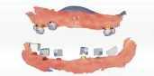
scanner group. A, B, Representative participant in second stage ready for prosthetic phase. C, Custom 3-dimensionally printed open tray with to-be-splinted open-tray impression copings for reference cast fabrication. D, E, Multiunit abutment scan bodies for digital scan capturing.
The healing abutments were retightened, and the tissues appeared healthy within a week. The implant stability quotient was assessed by a damping capacity device (Any check; Neobiotech), yielding a minimum ISQ mean value of 70 on the day of recording. All participants signed an informed consent.
The sample size had been calculated based on the primary outcome measure, which was the Euclidean deviation (ΔEUC) for each implant location. According to a previous study10 reporting a standard deviation of 115.5 µm and a mean difference of 49.6 µm, calculations were based on a dependent t test with a power=95%, and α=.05 yielded a total sample size of 57. The number was increased to 60 to account for dropouts. The sample size adequacy was assessed using a software program (PS Power and Sample Size version 3.1.2; PS). In this study, 11 participants with 52 implants were recruited, resulting in 104 recorded virtual implant locations, constituting the entire sample size for comparison with the relative reference scan. For a verified conventional splinted impression, an open-tray impression technique was made by attaching the open-tray impression copings (Multiunit transfer impression copings; Neobiotech) unsplinted to the multiunit abutments on the implant fixtures with the copings being picked up directly using a polyvinyl siloxane (PVS) impression material (Affinis Precious; Coltène).5 Type IV dental stone (Marmoplast N; Siladent) was poured into the impression after being mixed with a vacuum mixer (Twister Vacuum mixing unit; Renfert).
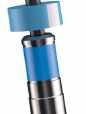
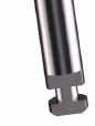
A light-polymerizing tray material (Megatray; Megsdenta Dentalprodukte GmbH) was used for splinting the impression copings extraorally on the primary cast. The resin devices were then sectioned and numbered. The 2-step approach saved chairside time, with minimal mess.23 A custom impression tray was designed digitally using a software program (Zirkon zahn.tray; Zirkohnzahn GmbH) and 3-dimensionally (3D) printed with a digital light processing (DLP) 3D printer (AccuafabD1s; Shining3D) (Fig. 1C).
In the second visit, the sectioned (to be splinted) open-tray impression copings were seated in the patient´s mouth. A radiograph was made to ensure that they were completely seated. They were then splinted with a low-shrinkage flowable composite resin (Gradia Direct LoFlo; GC Corp). A single operator (A.K.E.) made all impressions. The definitive cast was poured using Type IV dental stone (Marmoplast N; Siladent) in accordance with the manufacturer�s guidelines. Titanium frameworks were milled from conventional definitive casts to verify the conventional impressions. The mucosal surfaces of the frameworks were designed to minimize contact with the gingiva, thereby reducing resistance from soft tissues during clinical evaluation. The accuracy of the conventional casts as a control was evaluated by assessing the fit of the frameworks intraorally during the subsequent visit. The Sheffield 1 screw test was used to assess any inaccuracy.10,18 Following the tightening of
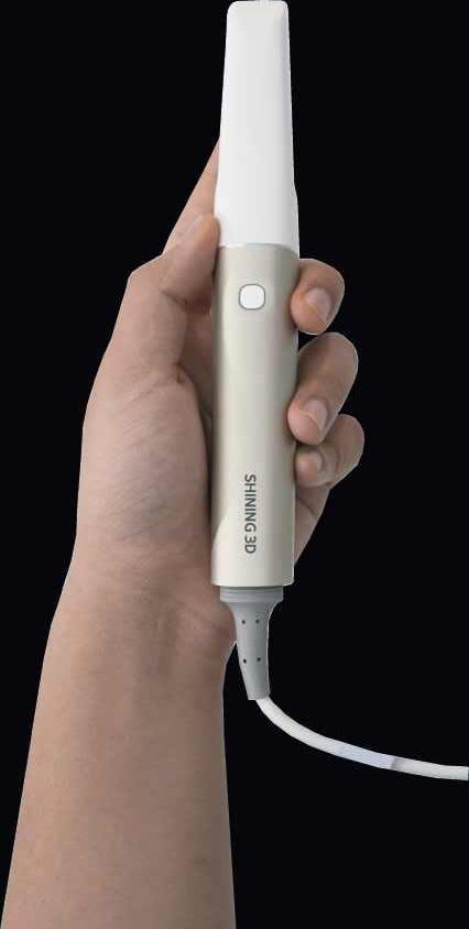
screws, the fit was evaluated radiographically using a paralleling technique. Conventional impressions and the following steps were repeated when an improper fit was identified to obtain verified casts as the control. Then, 2 digital scans were recorded, one with a conventional intraoral scanner (AoralScan 3; Shining3D) (Fig. 1D, E) and another with an intraoral photogrammetry scanner (Elite; Shining3D) by a single operator (A.K.E.) (Fig. 2). Both scanners had been calibrated immediately before the recordings. The scanning strategy adhered to the manufacturer’s recommendations throughout all procedures.
For each technique, the chairside time to accomplish the recording (conventional impression or digital scan) was calculated. For the conventional impression, the total time of the 2 visits was calculated. Regarding the digital scan either by IOS or IPS, the whole time of the scanning visit was calculated, including adjustments of the scan flags or scan bodies, tightening, and capturing. A monolithic screw-retained zirconia dental prosthesis was digitally constructed using definitive cast STL files (reference files) and subsequently delivered to the patient. (Figs. 3, 4). The authenticated definitive cast of the traditional impression was digitized using a high-resolution la- boratory scanner (DS MIX; Shining3D), achieving an accuracy of <7 µm according to the International Organization for Standardization (ISO) 12836 standard24 to produce a digital definitive cast (STL) file to be used as a reference. Three digital files were acquired for each participant: 1 reference scan (derived from the validated definitive cast), 1 IOS scan, and 1 IPS scan. All STL files were imported into the dental CAD software program (exocad Dental CAD; exocad GmbH), and scan bodies were used to generate multiunit Ti bases from a corresponding digital library. Subsequently, the generated STL files were imported into a software program (Mimics version 2.4; Materialise Mimics) for accuracy evaluations. The 2 STL files were superimposed with the STL file of the reference cast using the “N-point registration” and “global registration” algorithms. All the alignment processes were executed by a single investigator (K.H.B.) and verified by a second investigator (A.K.E.) to exclude errors. The Mimics software program performed multiple iterations automatically using an iterative closest point (ICP) algorithm to refine alignment. The number of iterations was set to 20 so that the probability of error was negligible.
The 3D variation between the 2 STL files was assessed regarding linear and angular deviation. The Euclidean distance is used to measure the differences between 2 points in 3D space. A central point and central axis of the virtual MUA Ti base were used for deviation measurements. The outcome measurements were defined by numbering the implants in the arch sequentially from implant 1 to implant n (n=number of implants), moving from the patient’s right to left (Fig. 5). All statistical analyses were conducted using a statistical software program (SAS version 9.4; SAS Institute). The linear and angular deviations were
Transcend universal composite provides unprecedented shade matching with just one Universal Body shade due to its patented Resin Particle Match™ technology that eliminates the need for a blocker.
If you prefer a layering technique Transcend composite also includes four dentin shades and two enamel shades.
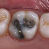
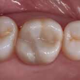
Deep amalgam staining presents one of the most difficult restoration situations to clinicians. In this case only the Transcend composite Universal Body shade was used to replace the amalgam no blocker needed. Note the excellent color blending of the preserved oblique ridge.
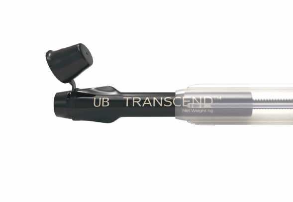
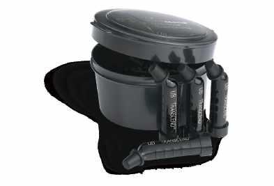
Scan the QR code to learn more about Transcend Universal Composite or go to ultradent.eu/transcend
subjected to descriptive analysis. The 3D deviation for each implant position was calculated using the Euclidean distance, and significance was evaluated through a paired t test. The distance deviations and the clinically acceptable misfit threshold (100 µm),15,25–27 regarded as the hypothetical mean,
were analyzed using a 1-sample t test. A pointbiserial correlation was conducted to assess the relationship between jaw type and both angular and ΔEUC deviations in the IOS and IPS groups. One-way ANOVA was used to compare the mean scanning time for conventional and scanning techniques (α=.05).
Figure 2.
Intraoral photogrammetry group. A, B, C, D; Scan codes (scan flags) with special dot orientation tightened according to manufacturer instructions (8 to 15 Ncm). Orientation of scan flags toward center, thus facilitating capture of fixture locations in single imaging.
Figure 3. A, B, Designing maxillary and mandibular implant screw-retained monolithic prosthesis after aligning intraoral data with facial scans. C, D, E, F; 3-dimensionally printed polymethyl methacrylate (PMMA) prosthesis acts as rigid prototype to evaluate passivity, intaglio adaptation, smile line, canting, occlusion, and patient satisfaction (esthetic and functional parameters).


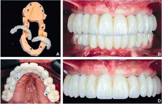
Figure 4.
A, Evaluating prosthesis on 3-dimensionally printed cast with multiunit abutment (MUA) digital laboratory analogs in place. B, C, D, Screw- retained monolithic zirconia prostheses restoring esthetics and function.
Thirteen edentulous arches, comprising 9 maxillae and 4 mandibles, were rehabilitated in 11 patients using monolithic zirconia screw-retained implant prostheses supported by 4 implants, resulting in a total of 52 implants. Implant positions were recorded using both a conventional intraoral scanner (IOS) and an intraoral photogrammetry scanner (IPS), resulting in a total of 104 implant positions for comparison with the relative reference scans. Normality was checked with QQ plots, histograms, and the Kolmogorov–Smirnov and Shapiro–Wilk test, indicating that the data did not significantly deviate from normality (Fig. 6). The ΔEUC deviation of the IPS group had lower values (M=30.7, SD=18) than those of the IOS group (M=59.8, SD=30). A t test for paired samples indicated that the difference was statistically significant (P<.001), with a 95% confidence interval (−40.33, −17.88). The angular deviation of the IPS group had lower values (M=0.78, SD=0.27 degree) than those of the IOS group (M=1.4, SD=0.44 degree) (P<.001), 95% Confidence interval (−0.8, −0.53) (Fig. 7). Tables 1 and 2 shows Euclidian and angular deviation for each arch in both the IPS and IOS groups, respectively. Table 3 shows the differences from the reference scans of both the IOS and IPS groups. A 1- sample t test revealed that the overall mean of the 3D linear deviations of
the IPS group (M=30.7, SD=18 µm) was significantly lower than 100 µm (P<.001). Also, the conventional IOS group had an overall mean (M=59.8, SD=30 µm) which was also significantly lower than 100 µm (P<.001). The angular deviation of IPS (M=0.78, SD=0.27) was lower than the hypothetical mean (P<.001). However, the angular deviation of the conventional IOS (M=1.4, SD=0.44 degree) was significantly higher than the accepted degree of angular deviation (P<.001). A point-biserial test assessed the correlation between jaw type and angular and ΔEUC deviations in both groups (IPS and IOS). In the IPS group, the relationships between angular deviation and arch type (rpb=0.15, n=52, P=.281) and between EUC and the type of arch (rpb=−0.11, n=52, P=.428) were statistically similar, indicating that intraoral photogrammetry trueness was not affected by the jaw type. In the IOS group, a statistically significant positive relationship existed between angular deviation and arch type. (rpb=0.34, n=52, P=.013). The correlation between EUC and the arch type (rpb=0.1, n=52, P=.463) was statistically similar. The chair side time for obtaining the conventional and the digital scans was statistically sig- nificantly different (P<.001) (Tables 4, 5).
Thirteen edentulous arches, comprising 9 maxillae and 4 mandibles, were rehabilitated in 11 patients using monolithic zirconia screw-retained implant prostheses supported by 4 implants, resulting in a total of 52 implants. Implant positions were recorded using both a conventional intraoral scanner (IOS) and an intraoral photogrammetry scanner (IPS), resulting in a total of
was statistically significant (P<.001), with a 95% confidence interval (−40.33, −17.88). The angular deviation of the IPS group had lower values (M=0.78, SD=0.27 degree) than those of the IOS group (M=1.4, SD=0.44 degree) (P<.001), 95% Confidence interval (−0.8, −0.53) (Fig. 7). Tables 1 and 2 shows Euclidian and angular deviation for each arch in both the IPS and IOS groups, respectively. Table 3 shows the differences from the
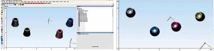
Figure 5. A, B, Alignment of STL files of both groups (IOS and IPS) to STL file of reference scan to determine both Euclidian and angular deviations for trueness assessment in Mimics software program. IOS, intraoral scanner; IPS, intraoral photogrammetry; STL, standard tessellation language.
A, B, Alignment of STL files of both groups (IOS and IPS) to STL file of reference scan to determine both Euclidian and angular deviations for trueness assessment in Mimics software program. IOS, intraoral scanner; IPS, intraoral photogrammetry; STL, standard tessellation language.
Figure 6.
A, B, C, D, QQ plots and histograms show that data did not significantly deviate from normality.
Figure 7.
Table 1.
Euclidian and angular deviation for each arch in intraoral photogrammetry (IPS) group
Table 2. Euclidian and angular deviation for each arch in intraoral scanner (IOS) group
Table 3.
Mean, and standard deviation of linear, Euclidian, and angular deviations of both intraoral scanner (IOS) and intraoral photogrammetry (IPS) groups

Table 4.
Mean and standard deviation of chairside time for three recording techniques (conventional splinted open tray, IPS, IOS).
IOS, intraoral scanner; IPS, intraoral photogrammetry; SD, standard deviation.
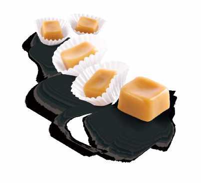


• Double protection – crystalline protectiv layer and comprehensive desensitisation (5 % NaF ≙ 22.600 ppm fluoride)
• Aesthetic – tooth-shaded varnish
• Universal – available in tube version and SingleDose
• Variety of flavours – mint, caramel, cherry and bubble gum
For more information please contact VOCO‘s Business Development in Middle East/Northern Africa, Mohamad El Fil (+961-3-805758 / m.elfil@voco.com).
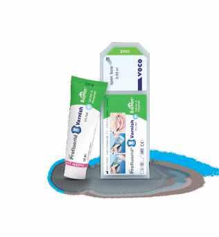
Table 5.
The primary outcome of this single cohort clinical trial was to compare the accuracy of intraoral scanning (IOS) and an intraoral photogrammetry capturing system (IPS) in patients with complete arch prostheses supported by
4 implants. Based on the results of this study, the null hypothesis that no variations would be found between intraoral scanning and intraoral photogrammetry capturing systems for complete arch implant-supported prostheses was rejected, as both the Euclidian (ΔEUC) and angular deviations were statistically higher in the IOS group than in the IPS group. The null hypothesis that both capturing systems would not be affected by the arch type was partially rejected, as the means of deviations were higher in the mandible than in the maxilla in the IOS group; however, no difference between the maxillary and mandibular arches was found when using the IPS capturing system.
The splinted open-tray impression technique is still the standard for capturing complete arch implantsupported prostheses. Papaspyridakos et al6 reported the superiority of the splinted technique for delivering a passive complete arch, 1-piece fixed prosthesis. Recently, Revilla-León et al16 compared the splinted conventional impression technique with photogrammetry, revealing the higher trueness (3 µm difference) and precision (18 µm difference) of the splinted technique.16 The present study used the splinted technique to produce a reference cast, which was digitalized using a high-resolution laboratory scanner (DS MIX; Shining3D) with an accuracy of <7 µm (ISO 12836) to generate a digital definitive cast (STL) file.
Several approaches have been used to quantify the deviations of implant positions: some report data in 3 dimensions (X, Y, Z), others evaluate lengths and angles with a single point that is considered a fixed landmark, and a completely different approach has been used to compare the scanned scan bodies using the best-fit technique.19 Passive fit for screw-retained definitive prostheses requires precise abutment platform data. To make the results more clinically relevant, only information from the abutment platforms was extracted and
used to compute the 3D deviation. The present study design focused on calculating 3D deviations for each implant position through a best-fit alignment algorithm applied to the reference and test scans. The variations of each implant can be analyzed from linear (ΔY, ΔX, ΔZ), 3D (ΔEUC) and angular (ΔANGLE) perspectives.10,28
The authors are unaware of a previous clinical trial that assessed the accuracy of complete arch digital scans executed with intraoral photogrammetry and intraoral scanning. Previous studies have assessed IOSs in complete arch implant scanning, but its use in daily practice for this purpose remains controversial.17,29,30 This debate arises from a lack of agreement regarding the permissible misfit range of a single implant in a complete arch supported by 4 and 6 implants, as well as the suitable clinical measurement method.13,14 To prevent long-term complications, a threshold value of 150 µm has been recommended.25 Moreover, manufacturing and its tolerance, which may create deviations ranging from 20 to 100 µm, should be considered,31,32 as should the implant number per arch, as the increase in the number of implants inversely affects the tolerable deviations.10,33 Recently, interimplant distance, interimplant angulation, scan body type, intraoral scanner type, and operator experience have been reported to significantly affect the accuracy of scanning.34,35
Yan et al 28 compared the accuracy of an extraoral photogrammetry system and intraoral scanning in patients with complete arch implant-supported prostheses. In all participants, photogrammetry yielded less deviation, and the results were statistically significant compared with those of intraoral scanning (range 2.70–92.80 µm, median 17.00 versus 21.30 to 815.60 µm, and median of 48.95 µm). Also, the accuracy of photogrammetry was not affected by the position or number of implants, unlike IOS. The authors did not recommend IOS for complete arch implant scanning, as the results did not pass the accepted threshold level.28 In the present study, although the IOS 3D deviation was statistically significantly different (P<.05) compared with IPS (IOS M 59.8 µm, SD=30 versus IPS M=30.7 µm, SD=18),the results were still below the accepted threshold of the misfit. This might be attributed to only 4
implants per arch for all participants. Similarly, a statistically significant correlation was found between IOS angular deviation and the mandibular arch (P<.05). However, no correlation was found between the arch type and IPS deviation, whether Euclidian or angular (P>.05).
In the present study, both IOS and IPS showed a lower Euclidian deviation mean (M 59.8 µm, 30.7 µm respectively) when compared with the hypothetical mean when considering 100 µm as the upper limit of acceptable fit. However, the angular deviation means of the IOS (1.4 degree) exceeded the acceptable theoretical limit of 1-degree angular deviation, while IPS had a 0.78-degree deviation, lower than the accepted deviation. Manzella et al15 investigated the degree of tolerable misfit by utilizing a gypsum indicating device as a brittle material that would fracture with any degree of intolerable misfit. The indicating device did fracture with a 150-µm horizontal misfit, 50-µm vertical misfit, and 1 degree of angular deviation when considering these values as the threshold of acceptable misfit.15 The clinical meaning of this deviation difference between IPS and IOS has to be further interpreted considering the overall number of implants for each arch. For complete arch digital implant scans, these variations might suggest that IPS is a more dependable option than IOS, although it is still advised to attempt a rigid prototype before making a definitive screw-retained complete arch implant-supported prosthesis. Similar outcomes have been reported in a recent in vivo study17 comparing IOS and extraoral stereophotogrammetry systems (SPG), whose clinical performance was analyzed and compared in the same participant. SPG was reported to be more accurate than IOS (mean EUC SPG 87.6 µm; mean SPG angle 0.38 degree versus mean EUC IOS 137.2 µm; mean IOS angle 0.79 degree) and was not affected by the position or number of implants.17
In the present study, the use of IPS affected not only the accuracy of capturing but also the ease of the process itself, demonstrated by the statistically significant difference in scanning times when IPS (mean 14.1 min SD 1.5) and IOS (mean 18.2 min SD 3.6) were used (P<.001). Unlike conventional intraoral scanners, photogrammetry was not affected by soft tissue or by blood or saliva making it better at scanning complete arches immediately after surgery. Limitations of the present study included that, because of the clinical design, no repetitions were conducted; therefore, precision was not evaluated, and only trueness could be
calculated. The study was only on prostheses supported by 4 implants; other implant numbers per arch should be further investigated. No standardization of the type of arch (maxilla or mandible) added to the study limitations. Also, as the results of this study were based on only 1 intraoral scanning system, IPS should be compared with different intraoral scanning technologies and with extraoral photogrammetry systems.
Based on the findings of this clinical study, the following conclusions were drawn:
1. Intraoral scanning can be used for complete arch implants in some patients.
2. Intraoral photogrammetry system (IPS) significantly improved the ease of fabrication and accuracy of complete arch prostheses supported by 4 implants.
3. Unlike IOS, the trueness of intraoral photogrammetry (IPS) was not affected by the type of jaw.
All the performed procedures were conducted according to the ethical principles and standards of the institutional and national research committee and with the Declaration of Helsinki and its later amendments (revised in October 2018). The study protocol was independently reviewed and approved by the Institutional Ethical Committee, Faculty of Dentistry, Ain Shams University (FDASU-Rec IR032422). Informed written consent was obtained from all individual participants included in the study.
The data sets used and analyzed during the current study are available from the corresponding author on reasonable request.
Whiter teeth can give your patients the confidence to smile more.

Opalescence whitening is on a mission to help give your patients brighter, whiter smiles so they can look their best and feel their best, turning good days into better ones. As the global leader in professional whitening,1 Opalescence has brightened over 100 million smiles.1 That’s a lot of better days.
and the largest dental school in the U.S.
The peri-implant bone level was found to be stable at the level of the first thread after 9 years of loading (Fig 3).
• 300,000 patient visits annually ensure superb clinical training for students
• Nearly 10% of dentists in the U.S have been educated at NYU Dentistry
1- Arcuri L, Pozzi A, Lio F, Rompen E, Zechner W, Nardi A. Influence of implant scanbody material, position and operator on the accuracy of digital impression for complete-arch: A randomized in vitro trial. J Prosthodont Res. 2020;64:128–136.
• NYU Dentistry is ranked 3 in the U.S. in National Institutes of Health research funding
• 21,000+ alumni network practicing worldwide
2- Hoods-Moonsammy V, Owen CP, Howes D. A comparison of the accuracy of polyether, polyvinyl siloxane, and plaster impressions for long-span implant-supported prostheses. Int J Prosthodont. 2014;27:433–438.
• 1,959 students across all academic programs
3- Leggeri A, Carosi P, Mazzetti V, Arcuri C, Lorenzi C. Techniques to improve the accuracy of intraoral digital impression in complete edentulous arches: A narrative review. Appl Sci. 2023;13:7068.
• 93 NIH-funded and other funded researchers advance science every day
• 14 Academic Societies, each lead by a Senior Mentor, promote a strong sense of community and afford DDS students small-group learning and mentoring experiences while still having access to the vast resources of a large university
4- Kachhara S, Nallaswamy D, Ganapathy DM, Sivaswamy V, Rajaraman V. Assessment of intraoral scanning technology for multiple implant impressions:A systematic review and metaanalysis. J Indian Prosthodont Soc 2020:41–52.
Fig 3 - Bone level 9 years postloading.Bone positioned at the level of the first thread. Minimal bone loss occurred over 9 years of functional loading.
• An average of 27 students per class year in each Academic Society, supported by its own Student Success Network, which connects every DDS student with a network of academic advisors, peer tutors, and peer and faculty mentors who provide one-on-one guidance and support to promote success from the moment students enter at Orientation
THE NEEDLE: NYU Dentistry’s Center for Oral Health Policy and Management Addresses Advocacy and Leadership Issues
11- Paratelli A, Vania S, Gómez-Polo C, Ortega R, Revilla-León M, Gómez- Polo M. Techniques to improve the accuracy of complete arch implant intraoral digital scans: A systematic review. J Prosthet Dent. 2023;129:844–854.
The NYU Dentistry Center for Oral Health Policy and Management, an interdepartmental, interdisciplinary action laboratory, was founded in 2021 on the premise that the current oral health policy and management environment in the U.S. requires a holistic approach to the situation — one that has been lacking.
12- Pérez-Giugovaz MG, Mosier M, Revilla-León M. An additively manufactured intraoral scan body for aiding complete-arch intraoral implant digital scans with guided integration of 3D virtual representation. J Prosthet Dent. 2022;127:38–43.
13- Rutkunas V, Larsson C, Vult von Steyern P, Mangano F, Gedrimiene A. Clinical and laboratory passive fit assessment of implant-supported zirconia restorations fabricated using conventional and digital workflow. Clin Implant Dent Relat Res. 2020;22:237–245.
Fig 4 - Six months after placement of the unstable removable overdenture. Note the severe bone loss on the implant in the maxillary right first premolar site (down to the sixth thread) and the maxillary right first molar site (down to the third thread). However, no bone loss was observed on the intermediate implant.
“While tremendous strides have been made in improving the oral health status of Americans through scientific breakthroughs, many are left without access to basic dental care,” notes Dean Charles Bertolami. “Dental benefits remain separated from other health care coverage and out of reach for many individuals and families,” he added.
14- Katsoulis J, Takeichi T, Gaviria AS, Peter L, Katsoulis K. Misfit of implant prostheses and its impact on clinical outcomes. Definition, assessment and a systematic review of the literature. Eur J Oral Implantol. 2017;10:121–138.
5- Papaspyridakos P, Chen CJ, Gallucci G, Doukoudakis A, Weber HP, Chronopoulos V. Accuracy of implant impressions for partially and completely edentulous patients: A systematic review. Int J Oral Maxillofac Implants. 2014;29:836–845.
6- Papaspyridakos P, Lal MSK, George MS, White S. Effect of splinted and nonsplinted impression techniques on the accuracy of fit of fixed implant prostheses in edentulous patients: A comparative study. J Prosthet Dent. 2012;108:83.
7- Hariharan R, Shankar C, Rajan M, Baig MR, Azhagarasan NS. Evaluation of accuracy of multiple dental implant impressions using various splinting materials. Int J Oral Maxillofac Implants. 2010;25:38–44.
8- Moon YG, Lee KM. Comparison of the accuracy of intraoral scans between complete-arch scan and quadrant scan. Prog Orthod. 2020;21:4–9.
Prosthetic failure of the maxillary left premolars and first molar occurred in October 1999. The fixed partial prosthesis became loose due to recurrent decay and poor crown-to-root ratio. It was decided to extract the remaining teeth and convert to an implant-supported fixed restoration. Three Brånemark implants (Nobel Biocare, Göteborg, Sweden) were placed in the maxillary left quadrant, and the patient was referred to her dentist for the placement of a temporary removable prosthesis to restore esthetics and function while implant osseointegration was achieved. The dentist removed the maxillary right implant-supported partial prosthesis and placed an overdenture. The patient was seen in May 2000 for abutment connection on the maxillary left implants. Periapical radiographs were obtained to assess the osseointegration. Severe bone loss was observed on the implants in the maxillary right first premolar site and the maxillary right first molar site (Figs 4 to 6).
9- Yilmaz B, Gouveia D, Marques VR, Diker E, Schimmel M, Abou-Ayash S. The accuracy of single implant scans with a healing abutment-scanpeg system compared with the scans of a scanbody and conventional impressions: An in vitro study. J Dent. 2021;110:103684.
10- Pozzi A, Arcuri L, Lio F, Papa A, Nardi A, Londono J. Accuracy of complete- arch digital implant impression with or without scanbody splinting: An in vitro study. J Dent. 2022;119:104072.
Charles Bertolami
In addition, the rigorous curriculum for dental students focuses predominantly on basic sciences and clinical care, but most learn little about the complexity of the dental and general health care systems of which they will soon be a part. The center aims to change this through new programming and academic offerings on oral health policy and leadership.
15- Manzella C, Bignardi C, Burello V, Carossa S, Schierano G. Method to improve passive fit of frameworks on implant-supported prostheses: An in vitro study. J Prosthet Dent. 2016;116:52–58.
16- Revilla-León M, Rubenstein J, Methani MM, Piedra-Cascón W, Özcan M, Att W. Trueness and precision of complete-arch photogrammetry implant scanning assessed with a coordinatemeasuring machine. J Prosthet Dent. 2023;129:160–165.
“NYU Dentistry is uniquely well positioned to undertake these challenges,” said Richard Valachovic, DMD, MPH, a clinical professor at NYU Dentistry and president emeritus of the American Dental Education Association, who serves as founding director of the center.
Fig 5-6 - Six months after placement of the unstable removable overdenture. The implants were connected with a rigid bar, and the unstable overdenture was adjusted.
17- Pozzi A, Carosi P, Gallucci GO, Nagy K, Nardi A, Arcuri L. Accuracy of complete-arch digital implant impression with intraoral optical scanning and stereophotogrammetry: An in vivo prospective comparative study. Clin Oral Implants Res. 2023;34:1106–1117.
Several of the College’s access to care and advocacy initiatives align with critical issues related to oral health policy and management.
18- Zhang YJ, Qian SJ, Lai HC, Shi JY. Accuracy of photogrammetric imaging versus conventional impressions for complete arch implant-supported fixed dental prostheses: A comparative clinical study. J Prosthet Dent. 2023;130:212–218.
19- Orejas-Perez J, Gimenez-Gonzalez B, OrtizCollado I, Thuissard IJ, Santamaria-Laorden A. In Vivo complete-arch implant digital impressions: Comparison of the precision of three optical impression systems. Int J Environ Res Public Health. 2022;19:4300.
Another priority for the NYU Dentistry for Oral Health Management and Policy is to develop the next generation of policy-oriented leaders for the dental and related health care professions through creating new leadership programming and courses.
The removable prosthesis was found to be very unstable; it was rocking around the maxillary right implants and had been doing so for 6 months, according to the patient. In collaboration with the dentist, all 6 implants were splinted, and a properly fitted removable prosthesis was fabricated. Oral hygiene was reinforced to improve the patient’s home care. The peri-implant condition was re-evaluated radiographically every 3 months. The bone lesions started to heal within 3 months after elimination of the traumatic condition. At 6
20- Sánchez-Monescillo A, Sánchez-Turrión A, Vellon-Domarco E, Salinas- Goodier C, PradosFrutos J. Photogrammetry impression technique: A case history report. Int J Prosthodont. 2016;29:71–73.
21- Sun X, Cheng K, Liu Y, et al. Biomechanical comparison of all-on-4 and all- on-5 implantsupported prostheses with alteration of anteriorposterior spread: A three-dimensional finite element analysis. Front Bioeng Biotechnol. 2023;11:1–12.
22- Malo P, De Araújo Nobre M, Lopes A, Moss SM, Molina GJ. A longitudinal study of the survival of all-on-4 implants in the mandible with up to 10 years of follow-up. J Am Dent Assoc. 2011;142:310–320.
23- Patil P, Madhav VNV, Alshadidi AAF, et al. Comparative evaluation of open tray impression technique: Investigating the precision of four splinting materials in multiple implants. BMC Oral Health. 2023;23:1–13.
24- ISO International Organization for Standardization. Dentistry – Digitizing devices for CAD/CAM systems for indirect dental restorations –. Test methods for assessing accuracy. 12836. 2015:19.
25- Jemt T. In vivo measurements of precision of fit involving implant- supported prostheses in the edentulous jaw. Int J Oral Maxillofac Implants. 1996;11:151–158.
26- Jemt T, Book K. Prosthesis misfit and marginal bone loss in edentulous implant patients. Int J Oral Maxillofac Implants. 1996;11:620–625.
27- Rutkunas V, Gedrimiene A, Akulauskas M, Fehmer V, Sailer I, JegeleviciusC D. In vitro and in vivo accuracy of full-arch digital implant impressions. Clin Oral Implants Res. 2021;32:1444–1454.
28- Yan Y, Lin X, Yue X, Geng W. Accuracy of 2 direct digital scanning techniques—Intraoral scanning and stereophotogrammetry—for complete arch implant-supported fixed prostheses: A prospective study. J Prosthet Dent. 2023;130:564–572.
29- Kanjanasavitree P, Thammajaruk P, Guazzato M. Comparison of different artificial landmarks and scanning patterns on the complete-arch implant intraoral digital scans. J Dent. 2022;125:104266.
30- Chochlidakis K, Papaspyridakos P, Tsigarida A, et al. Digital versus conventional full-arch implant impressions: A prospective study on 16 edentulous maxillae. J Prosthodont 2020:281–286.
31- Ortorp A, Jemt T, Bäck T, Jälevik T. Comparisons of precision of fit between cast and CNC-milled titanium implant frameworks for the edentulous mandible. Int J Prosthodont. 2003;16:194–200.
32- Abduo J. Fit of CAD/CAM implant frameworks: A comprehensive review. J Oral Implantol. 2014;40:758–766.
33- de França D, Morais M, das Neves F, Carreiro A, Barbosa G. Precision fit of screw-retained implantsupported fixed dental prostheses fabricated by CAD/CAM, copy-milling, and conventional methods. Int J Oral Maxillofac Implants. 2017;32:507–513.
34- Zhang YJ, Shi JY, Qian SJ, Qiao SC, Lai HC. Accuracy of full-arch digital implant impressions taken using intraoral scanners and related variables: A systematic review. Int J oral Implantol. 2021;14:157–179.
35- Gómez-Polo M, Ortega R, Sallorenzo A, et al. Influence of the surface humidity, implant angulation, and interimplant distance on the accuracy and scanning time of complete-arch implant scans. J Dent. 2022;127.
Corresponding author:
Assoc Prof Doaa Adel-Khattab Oral Medicine, Periodontology and Oral Diagnosis Faculty of Dentistry Ain Shams University Cairo EGYPT
Email: dr.doaa.adel-khattab@dent.asu.edu.eg
CRediT authorship contribution statement
All authors have made substantial contributions to the conception and design of the study A.K.E., and K.H.B., conceptualized and designed the study, D.A., and A.K.E., have been involved in data collection and data analysis. A.K.E., K.H.B., and D.A., have been involved in data interpretation, and drafting of the manuscript, A.K.E., and D.A., revised it critically and have given final approval of the version to be published.
Copyright © 2025 by the Editorial Council of The Journal of Prosthetic Dentistry. All rights are reserved, including those for text and data mining, AI training, and similar technologies.
https://doi.org/10.1016/j.prosdent.2025.03.041
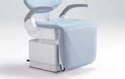
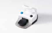
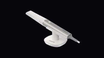
IPG - Intraoral Photogrammetry
Two-in-One System
ScanDesignDelivery


If you’ve ever experienced hearing troubles, you might not have considered that your oral hygiene could play a role. Without proper care, harmful bacteria can develop around your teeth and gums, potentially leading to issues such as infected tooth roots, periodontal disease, and TMJ disorders.
Neglecting to brush, floss, or attend regular dental check-ups can result in problems like bacteria buildup in the roots of your teeth, gum infections, or jaw issues. Since your jaw and several nerves are closely connected to your ears, maintaining good oral health can help prevent ear pain and hearing problems. Let’s explore some key ways oral hygiene can impact your ears and hearing.
An infected tooth can cause severe pain and throbbing. When bacteria penetrate the tooth’s protective enamel and reach the root’s nerve, they can cause significant discomfort and possibly lead to an abscessed jaw.
But the impact doesn’t stop there. Oral bacteria can spread to blood vessels in your ears and other areas, while pain from the nerves in your teeth can radiate through your jaw. This is especially true for the nerves in the upper jaw, which are located near the ear canals. Consequently, a toothache might also trigger ear pain, such as an earache.
To avoid these complications, schedule a dental appointment if you notice a toothache. Your dentist can perform root canal therapy to remove infected nerve tissue from the tooth root, relieving pain and preventing the spread of bacteria into your bloodstream.
Bacteria from an infected tooth can spread to the gums, prompting your immune system to fight back through inflammation. Unfortunately, this response can damage gum tissue, potentially leading to tooth loss—a condition known as periodontal disease.
Periodontal disease can also affect your hearing. As your body combats bacteria, blood vessels that supply the nerve cells in your ears may narrow. If insufficient blood reaches these nerves, they can begin to deteriorate, potentially causing hearing loss.
To prevent this, it’s essential to maintain good
dental hygiene through regular brushing, flossing, and dental check-ups. If periodontal disease develops, visit your dentist promptly for treatment to protect both your oral health and your hearing.
The temporomandibular joint (TMJ), located near your ears, enables your jaw to move up, down, and side to side, allowing you to eat and speak. When this joint is injured or affected by a condition that limits jaw function and causes pain, it’s referred to as a TMJ disorder.
Because the TMJ is situated close to the ears, it can lead to symptoms that impact ear health, including:
A full or clogged sensation in the ear
Muffled or slight hearing loss
Tinnitus (ringing in the ear)
Regular dental visits can help detect early symptoms and prevent more severe complications. Dentists can also provide corrective care for tooth-grinding issues or alignment problems that contribute to TMJ disorders.
These issues are largely preventable with consistent oral hygiene practices, such as brushing, flossing, and attending routine dental check-ups. As healthcare professionals, we’re here to address your concerns and answer any questions about your hearing health.
Katie Koebel, M.Cl.Sc., is the Senior Manager of Audiology at HearingLife, Canada’s largest group of hearing centres with over 350 locations across the country. HearingLife clinics use the most advanced hearing aid technology, clinical support, and diagnostic equipment. Katie is an Audiologist registered with CASLPO and has been providing her clients with the best possible hearing health care with HearingLife for over 17 years.
If you’re at high risk of hearing damage, it is advisable to have your hearing checked regularly and advocate for proper hearing protection in your workplace. For more information on different types of hearing loss and education resources, you can visit HearingLife online or at one of our locations across Canada.
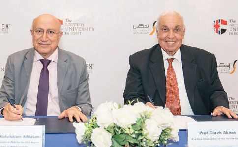
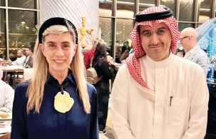
Cairo, June 24, 2025: In a landmark move signaling a new era for strategic partnerships, INDEX Conferences and Exhibitions, a member of the leading UAE, based INDEX Holding, has signed a Memorandum of Understanding (MoU) with the British University in Egypt. This collaboration aims to establish an integrated model that combines academic depth with professional expertise in medical education, scientific research, and the organization of specialized global events.
This agreement goes beyond a typical academic partnership; it embodies INDEX’s vision as a regional powerhouse driving impact and exporting knowledge and expertise across the Arab world. The MoU was signed during an official visit by the INDEX Holding delegation to Cairo, reflecting strategic efforts to reshape the relationship between education and industry — particularly the conference and events sector — transforming events into platforms for future, building rather than mere seasonal occasions.
The initiative reinforces INDEX’s commitment to transferring the UAE’s pioneering model of integration between education, innovation, and development to the broader region through effective partnerships. These efforts
support the establishment of sustainable educational and healthcare ecosystems on both regional and international levels.
The signing ceremony took place at the British University in Egypt’s headquarters and was attended by His Excellency Amb. Dr. Abdulsalam AlMadani Roving Ambassador of the Parliamentary Assembly of the Mediterranean (PAM) for the GCC Region and Chairman of INDEX Holding, alongside Dr. Tarek Abbas, Vice President of the British University in Egypt. In the presence of Mr. Tareq AlMadani, CEO of INDEX Conferences and Exhibitions and INDEX Design, and Ms. Sara AlMadani, CEO of INDEX Media, along with a high dignitary of professors from the university’s medical and pharmaceutical faculties.
The MoU aims to enhance cooperation in organizing conferences and exhibitions, as well as in health, medical, and scientific programs, continuing education, and the development of educational and awareness initiatives that contribute to improving healthcare quality across the Arab world and Africa.
His Excellency Dr. Abdulsalam AlMadani highlighted the significance of this collaboration, emphasizing that the MoU opens new horizons for fruitful partnerships reflecting the UAE’s leading role and its national institutions in supporting health and education efforts regionally and globally. He stated: “We hold the conviction that the foundation of healthy and sustainable communities lies in the enhancement of regional and international cooperation, and that scientific advancement constitutes the cornerstone of the progress of nations and societies. Accordingly, this
partnership represents a transformative milestone in connecting knowledge with practical application, and a significant step forward in our pursuit of establishing integrated educational and professional systems—where the conference functions as an instrument of change, the exhibition as a gateway to innovation, and education as an ongoing journey that transcends the confines of the university. We firmly believe that Egypt, endowed with its abundant human resources, creative intellects, and established institutions, stands as a strategic partner in shaping a more radiant Arab future founded upon integration, innovation, and the investment in intellectual capital.”
For his part, Dr. Tarek Abbas, Vice President for the Medical Sector at the British University in Egypt, emphasized the strategic importance of this partnership with INDEX Conferences & Exhibitions. He stated that signing the MoU represents a pivotal step towards expanding regional and international cooperation in medical education, training, and continuous professional development. Dr. Abbas also underscored the strong scientific and research partnership between the British University in Egypt and the Egyptian Dental Association with the Global Scientific Dental Alliance, chaired by His Excellency Dr. Abdulsalam AlMadani. This longstanding collaboration has contributed to cementing the university’s role in supporting academic and research excellence on an international level. He described the current visit as a crowning achievement in this ongoing journey, aimed at building a bright future for coming generations.
Dr. Tarek Abbas added that the British University in Egypt is the first university in Egypt and North Africa to receive the UK Quality Assurance accreditation (QAA) in 2024, coinciding with the university’s celebration of its 20th anniversary.
Following the signing ceremony, Dr. Tarek Abbas, Vice President for the Medical Sector at the British University in Egypt, accompanied Dr. Abdulsalam AlMadani and his delegation on a tour of the university’s Faculty and Hospital of Dentistry, under the supervision of Professor Dr. Ferdous Rizk, Acting Dean of the Faculty. The delegation reviewed the latest medical and educational facilities, showcasing the university’s commitment to providing an advanced academic and research environment that supports excellence in healthcare and education.
Furthermore, both parties expressed their anticipation for launching joint initiatives, aimed at translating the goals of this collaboration into actionable programs and innovative projects that contribute to enhancing the quality of life and achieving a sustainable impact on Arab and African communities. They emphasized that this partnership represents an advanced model that strengthens the region’s position as a leading platform for innovation and development in the health and education sectors.
Through this cooperation, INDEX—whose global platforms include the Dubai International Pharmaceuticals and Technologies Conference and Exhibition (DUPHAT), the UAE International Dental Conference and Arab Dental Exhibition (AEEDC Dubai), Dubai World Dermatology and Laser Conference and Exhibition (Dubai Derma), Healthcare Future Summit, the International Family Medicine Conference & Exhibition, and the Annual Radiology Meeting among other specialized events with a global footprint—reinforces its pioneering role. INDEX’s efforts go beyond event organization to become an integral part of a comprehensive ecosystem for knowledge creation, enabling institutions to reach their true audiences, and forging sustainable strategic partnerships that transcend geographical boundaries and build bridges between generations, in pursuit of a more advanced future.
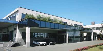

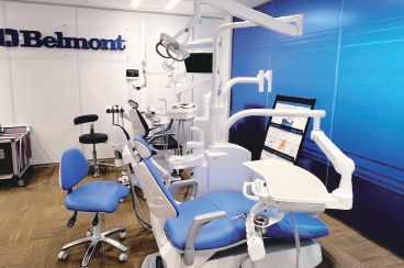
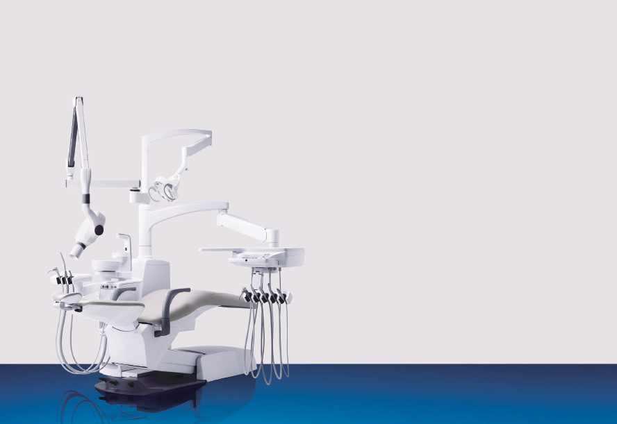

Company name: VOCO GmbH
Country of origin: Germany
Website: www.voco.com
The family-run dental company VOCO located in Cuxhaven (Germany) is one of the leading manufacturers in the industry, both nationally and internationally.
The product portfolio comprises more than 100 preparations, with a focus on preventive, restorative, prosthetic and digital dentistry.
The products are manufactured at the headquarters and are therefore 100 percent “Made in Germany”. 440 people are employed in the departments of research, production and administration in Germany.
www.voco.com
IonoStar Plus is a glass ionomer restorative material with numerous special features. Very easy to extract from the capsule, the material at first has excellent wetting characteristics resulting in optimal marginal adaptation. Its viscosity then changes within a few seconds, making the material malleable for at least one minute without sticking. It thus provides ideal viscosity at every stage of application.
Moreover, IonoStar Plus has a curing time of just two minutes, after which work can continue immediately. This is a valuable advantage, particularly in the treatment of patients with low compliance, such as children.
• Perfect marginal adaptation and packability in one product, thanks to a change in viscosity during application
• Fast setting time of only 2 minutes from placement of the filling
Another 400 employees are responsible for sales worldwide and take care of dentists and depots on site.
In addition to user-friendliness, the highest safety standards and product quality have top priority at VOCO: Since September 2021, VOCO has been one of the first German dental companies to be fully certified under the European Medical Device Regulation (MDR) in accordance with (EU) 2017/745.
Meaning that medical products of class I and Ila manufactured at the Cuxhaven site are always produced and placed on the market in accordance with valid European guidelines.
• The first glass ionomer material with tooth-like fluorescence
• High level of fluoride release
• The new capsule design reaches smaller cavities and difficult-to-access areas of the mouth
• High compressive strength and abrasion resistance
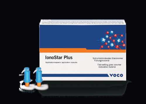
Simplified shade system through Cluster-Shades
Admira Fusion 5 uses five different Cluster-Shades, each bundling several VITA® classical shades. This provides adaptation to the natural colour gradient of the tooth as well as more efficient and easy shade selection.
Thanks to the simplified shade system, all 16 VITA® classical shades can be covered with only five ClusterShades. Multi-shade layering is not necessary.
This means that you are optimally positioned for your everyday practice with only five shades and can thus considerably streamline your inventory. This enables a better overview, and incorrect stocks are a thing of the past.
Simplified shade system
• all VITA classical shades can be covered with just 5 Cluster-Shades – there is no need for multiple layering
• significantly reducing your inventory
• new resin matrix enables enhanced chameleon effect through optimised light scattering
Universal
• completely universal – for the highest demands in anterior and posterior regions
• Easy and fast handling
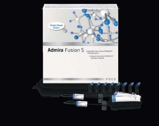
Company name: Ultradent Products Inc.
Country of origin: Salt Lake City, UT (USA) Website: www.ultradent.com
Ultradent Products Inc. was founded in 1978 in Salt Lake City by Dr. Fischer dentist, researcher and university professor (now Founder & CEO Emeritus of Ultradent Products, Inc.) with the aim of improving the techniques and products used by dentists in the entire world, in order to solve patient problems faster and without invasive methods.
Now, marking its 44th year as a family-owned, international dental supply and manufacturing company, Ultradent has continued its vision to improve oral health globally by creating better dental products that continue to set new industry standards.
MUHAMMAD AHMAD SABAWOON LTD Khushal khan, mina block 32, muqabil sello, Kabul Phone: (00) 788775555 chughtai1@hotmail.com
BAHRAIN
DENTAL WORLD & MEDICAL SUPPLIES
Office 11, bldg 932 Road 632 , block 706, salmabad Phone: (00)973 (178) 96322 dwms.bh@gmail.com
CYPRUS
PM DENTAL
11A KORITSAS, Nicosia Phone: (00)35722594050 p.m.dental@cytanet.com.cy
EGYPT
ELWAN DENTAL SUPPLIES
70 Merghani street 10th floor Managger, Cairo Phone: (00)202.33873883 elwanyegypt@yahoo.com
IRAN
GOLNAR NIKAN DANDAN
Unit 9, #64 Building, Opp Bahar St, Enghelab Ave P.O. Box: 1148836873, Tehran Phone: (00)9821 77533716 kavehgolnar@yahoo.com
IRAQ
SMART DENT
Almansoor street, beside zain
Ultradent currently researches, designs, manufactures, and distributes more than 500 materials, devices, and instruments used by dentists around the world, has subsidiaries in 9 countries and sells products in over 125 countries. Ultradent also works to improve the quality of life and health of individuals through financial and charitable programs.
communication bldg
Bldg no. 3 Floor 2, Baghdad Phone: (00)7810775922 smartdent79@yahoo.com
MALEK DENTAL & MEDICAL SUPPLY
jo 75 Al Buhturi Street, 2nd Circle, Jabal Amman, P.O. Box 7067, Amman Phone: (00)7810775922 mhdentamed@go.com.jo
KUWAIT
ADVANCED TECHNOLOGIES
Hawali 32060, P.O.Box 44558
Kuwait City Phone: (00)962795533513 ali@atc.com.kw
LIBYA
ALBYAN ALMOTAGADED FOR IMPORTING OF EQUIP & MEDICAL SUPPLIES CO LTD
Mohd Fkini St., Bldg No. 20, PO Box 82525, Tripoli Phone: (21) 333-3021 albyan_centre@hotmail.com
MOROCCO
A.M.E.D.
47, Rue de bruxelles, Casablanca Phone: (00)212.522823134 ihssan.b@amed.ma
OMAN
PIONEER TRADE & MEDICAL SUPPLY
Khalid Al Said Investment Co. PO Box 77 PC 103, Muscat Phone: (00)68 9780 4272 raed@pioneersoman.com
PAKISTAN
PAK-MED DENTAL SUPPLIES
pmdsupplies@gmail.com
Phone: (00)92 52 325 722
BILAL ENTERPRISES
Chughtai1@hotmail.com
QATAR
CEDARS DENTAL CENTER
P.O. Box 47684
Al Hilal, Doha Phone: (00)974.44864088 chaanine@cedars-dental.com
SAUDI ARABIA
DENTAL ERA
Al-Abdel Latif Plaza, King Fahd Rd, 3RD Floor, Office # 306 P.O. Box 126122, Jeddah Phone: +966 12 2752 382 ashraf.mubarak@dentalera.com
SYRIA
BADRIG AYDENIAN
Shouhada street, Damascus Phone: (00)963 (11) 444 6429 aydbaco@gmail.com
TUNISIA
DISTRI MED
Av. De Madrid, Tunis Phone: (+216) 71 334 812 distri-med@planet.tn
UNITED ARAB EMIRATES
ELWAN TECHNICAL SUPPLIES
P.O. Box 43305, Abu Dhabi Phone: (00)97126393292 etselwan@emirates.net.ae
When you want to give your patients the best results, your only choice is Opalescence™ teeth whitening!
Opalescence™ PF whitening | The most customizable of our whitening options. Use Opalescence™ PF whitening with custom trays made by your dentist for a personalized whitening solution day or night.
Opalescence Go™ whitening | It’s a portable, quick application. You pop in the UltraFit™ tray for the directed time and then brush afterwards. The tray gets thrown away and you pop in a new one next time! You can keep it anywhere there’s a fridge and a toothbrush.
Opalescence™ Boost™ in-office whitening | This miraculous product is so potent, it still needs to be applied by your dentist. It’s worth mentioning here because it’s an excellent option for fast, powerful teeth whitening. Think of it as a boost to start your whitening experience. Complete your whitening treatment with an Opalescence™ whitening take-home option.
Opalescence™ Whitening Toothpaste | Did you know that foods and other factors can cause surface stains on your teeth? A safe, everyday whitening toothpaste gently scrubs away surface stains and helps maintain a healthy smile. Achieve the best results by using toothpaste in tandem with our other whitening products which work to whiten below the surface.

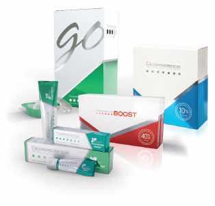

“Everything that VALO was, VALO X is just a better version. I’m really proud of that.” Neil Jessop, Senior Vice President of R&D Ultradent Products
When the original VALO™ curing light was launched in 2009, it changed the way people thought of a curing light. As we have continued to add to the VALO™ curing light family, we have worked tirelessly to ensure that every light has improved upon the past while retaining the durability, accessibility, and power that set the original VALO™ light apart from the rest.
Today, we wanted to ensure the VALO™ X light was the most innovative curing light we’ve created. We redesigned the VALO™ curing light from the bottom up to ensure that the VALO™ X light has the most cutting-edge components and state-of-theart features.
Find your perfect VALO™ light!
I-View Gold Intraoral sensor
I-View Gold Intraoral sensor
I-View Gold Intraoral sensor
Thanks to the advanced calibration tool, the I-View Gold captures high-definition images without any filters. Calibration files are used to remove any imperfections during acquisition, reducing noise and enhancing detail. As a result, the images captured by the I-View Gold require no further processing, ensuring reliable images, accurate diagnosis and time efficiency.
Thanks to the advanced calibration tool, the I-View Gold captures high-definition images without any filters. Calibration files are used to remove any imperfections during acquisition, reducing noise and enhancing detail. As a result, the images captured by the I-View Gold require no further processing, ensuring reliable images, accurate diagnosis and time efficiency.
DMP enhances its digital product range by presenting the first ever mobile shade guide application. - Chromatcher
DMP enhances its digital product range by presenting the first ever mobile shade guide application. - Chromatcher
Thanks to the advanced calibration tool, the I-View Gold captures high-definition images without any filters. Calibration files are used to remove any imperfections during acquisition, reducing noise and enhancing detail. As a result, the images captured by the I-View Gold require no further processing, ensuring reliable images, accurate diagnosis and time efficiency.
DMP enhances its digital product range by presenting the first ever mobile shade guide application. - Chromatcher
- DICOM compatibility
- DICOM compatibility
- DICOM compatibility
- Bridge feature for connecting to other dental management software.
- TWAIN option
- TWAIN option
- TWAIN option
- Multiuser connection
- Multiuser connection
- Multiuser connection
This unique app offers the user (dentist or technician) the ability to accomplish the restoration effortlessly. The Chromatcher is used to determine the tooth shade of the patient by taking a photo of the desired tooth area and matching it with DMP’s shade system for restorative materials. Therefore, the user knows the required shade immediately through the Chromatcher avoiding the use of conventional shade guides. This process applies for all aesthetic restorations using DMP’s restorative materials.
This unique app offers the user (dentist or technician) the ability to accomplish the restoration effortlessly. The Chromatcher is used to determine the tooth shade of the patient by taking a photo of the desired tooth area and matching it with DMP’s shade system for restorative materials. Therefore, the user knows the required shade immediately through the Chromatcher avoiding the use of conventional shade guides. This process applies for all aesthetic restorations using DMP’s restorative materials.
This unique app offers the user (dentist or technician) the ability to accomplish the restoration effortlessly. The Chromatcher is used to determine the tooth shade of the patient by taking a photo of the desired tooth area and matching it with DMP’s shade system for restorative materials. Therefore, the user knows the required shade immediately through the Chromatcher avoiding the use of conventional shade guides. This process applies for all aesthetic restorations using DMP’s restorative materials.
- Bridge feature for connecting to other dental management software.
- Bridge feature for connecting to other dental management software.
Download Chromatcher now, free at Apple Store! (available for iPhone, iPad & iPod touch)
Download Chromatcher now, free at Apple Store! (available for iPhone, iPad & iPod touch)
www.dmpdental.com
www.dmpdental.com
www.voco.dental

Download Chromatcher now, free at Apple Store! (available for iPhone, iPad & iPod touch)


The trend in the development of adhesive systems is to improve adhesion while simplifying clinical manipulation steps. Along with that, the use of universal adhesives has been increasing and its performance has been reported to be clinically sufficient.
multi-talent where safe and reliable
The trend in the development of adhesive systems is to improve adhesion while simplifying clinical manipulation steps. Along with that, the use of universal adhesives has been increasing and its performance has been reported to be clinically sufficient.
The trend in the development of adhesive systems is to improve adhesion while simplifying clinical manipulation steps. Along with that, the use of universal adhesives has been increasing and its performance has been reported to be clinically sufficient.
Nanoceram-Bright Flow is a light curing flowable version of the Nanoceram-Bright composite using the same nanohybrid technology to achieve optimum results. It is characterized by its superior aesthetics through its chameleon effect and high gloss polishability. This highly thixotropic product guarantees easy handling and precise application.
Nanoceram-Bright Flow is a light curing flowable version of the Nanoceram-Bright composite using the same nanohybrid technology to achieve optimum results. It is characterized by its superior aesthetics through its chameleon effect and high gloss polishability. This highly thixotropic product guarantees easy handling and precise application.
Nanoceram-Bright Flow is a light curing flowable version of the Nanoceram-Bright composite using the same nanohybrid technology to achieve optimum results. It is characterized by its superior aesthetics through its chameleon effect and high gloss polishability. This highly thixotropic product guarantees easy handling and precise application.
Indications
Indications
Anterior and limited posterior restorations
Anterior and limited posterior restorations
Indications
Liner for class I and II restorations
Liner for class I and II restorations
The multi-talent where safe and reliable bonding is required
The multi-talent where safe and reliable bonding is required
Thanks to the new silane bonding agent ARS, BeautiBond Xtreme bonds to enamel, dentine and various indirect restorative materials (composite, precious and non-precious alloys, glass-ceramics, alumina and zirconia). An additional primer is not
Thanks to the new silane bonding agent ARS, BeautiBond Xtreme bonds to enamel, dentine and various indirect restorative materials (composite, precious and non-precious alloys, glass-ceramics, alumina and zirconia). An additional primer is not required.
Thanks to the new silane bonding agent ARS, BeautiBond Xtreme bonds to enamel, dentine and various indirect restorative materials (composite, precious and non-precious alloys, glass-ceramics, alumina and zirconia). An additional primer is not required.
Sealing larger pits and fissures
Sealing larger pits and fissures
Anterior and limited posterior restorations
Liner for class I and II restorations
Sealing larger pits and fissures
Repair of composite restorations and ceramic veneers
Splinting of mobile teeth
Splinting of mobile teeth
Blocking out of undercuts
Splinting of mobile teeth
Blocking out of undercuts
Blocking out of undercuts
Characteristics - Advantages
Characteristics - Advantages
Excellent handling characteristics
Excellent handling characteristics
Excellent sculptability
Characteristics - Advantages
Excellent sculptability
Repair of composite restorations and ceramic veneers
Repair of composite restorations and ceramic veneers
Low shrinkage-Very good marginal adaptation
Excellent handling characteristics
Extremely high polishability
Excellent sculptability
Extremely high polishability
High resistance to wear
High resistance to wear
Superior aesthetics
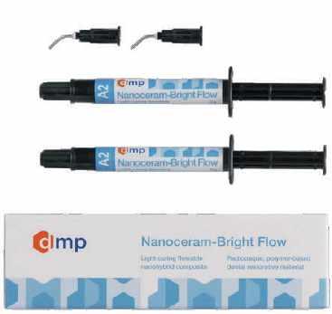
Low shrinkage-Very good marginal adaptation
Low shrinkage-Very good marginal adaptation
Extremely high polishability
With BeautiBond Xtreme, SHOFU continues to evolve the latest generation of a true universal adhesive. By introducing a new type of Acid Resistant Silane coupling agent (ARS), a chemical composition with excellent stability has been designed to allow the surface treatment of a wide range of adherends with an easy-to-use system. BeautiBond Xtreme is a light-curing, self-etching all-in-one universal adhesive for bonding direct and indirect restorations. It has “inherited” the excellent properties of the former BeautiBond Universal bonding system, such as bond strength, low technique sensitivity, convenient application and simplified one-step application procedure.
Superior aesthetics
Strongly radiopaque
High resistance to wear
Strongly radiopaque
Superior aesthetics
With BeautiBond Xtreme, SHOFU continues to evolve the latest generation of a true universal adhesive. By introducing a new type of Acid Resistant Silane coupling agent (ARS), a chemical composition with excellent stability has been designed to allow the surface treatment of a wide range of adherends with an easy-to-use system. BeautiBond Xtreme is a light-curing, self-etching all-in-one universal adhesive for bonding direct and indirect restorations. It has “inherited” the excellent properties of the former BeautiBond Universal bonding system, such as bond strength, low technique sensitivity, convenient application and simplified one-step application procedure.
www.dmpdental.com
Strongly radiopaque
www.voco.dental
www.shofu.com/global
Advanced Motor Technology
Delivers high-torque, ultra-smooth performance with a precision control system for precise endodontic procedures with minimal noise.
Precision Control System
Real-time feedback delivers accurate angle control and customizable torque, adapting to complex clinical needs.
Versatile Operating Modes
Intelligent modes enable seamless switching, ensuring the precision system controls efficiency across diverse endodontic protocols.
Wireless Ergonomic Design
Cordless handpiece reduces fatigue, enhancing comfort and maneuverability, which is critical for precision control during intricate treatments.
Contra-angle handpiece with 360° rotation and multi-angle display ensures precise control from any position, supported by the precision control system.
Built-in apex locator provides real-time, accurate length detection, streamlining shaping and measuring with reliable precision control.
The precision control system simplifies procedures by removing the need for external file clips or sleeves, ultimately lowering consumable costs.
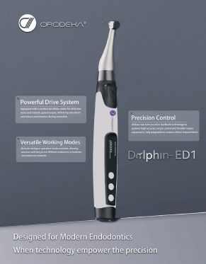
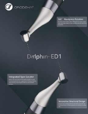

Cologne, Germany-March 25, 2025
Shining 3D, a pioneer in high-precision 3D digital technology, marks its 20th anniversary with the launch of four innovative digital dental solutions: the revolutionary all-in-one digital dental solution - e-Motion Workstation, Aoralscan Elite Wireless intraoral scanner, AccuFab-F1 3D printer, and FabCure N2 nitrogen curing box. With a relentless commitment to innovation,user-centricdevelopment, and comprehensive end-to-end workflows, these new solutions further solidify Shining 3D’s position at the forefront of digital dentistry.
Since its founding in 2004, Shining 3D has continuously pushed the boundaries of digital technology, investing over 20% of its annual revenue into Research and Development. This commitment has led to the independent development of core 3D technologies, resulting in 182 authorized patents, 416 pending, and 182 software copyrights. With its Global presence, Shining 3D remains a trusted leader in the digital dental industry.
Shining 3D’s success is driven by a deep understanding of user needs, ensuring its technology aligns with the evolving demands of dental professionals. The company offers a fully integrated digital dental workflow, covering scanning, designing, and printing with highprecision intraoral and desktop scanners, advanced 3D printers and printing materials, post-processing solutions, and cloud-based services. This holistic approach enhances efficiency, precision, and reliability for dental professionals worldwide.
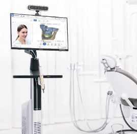
e-Motion Workstation: A Complete Digital Dentistry Solution
The e-Motion Workstation is the world’s first all-inone digital dental solution, seamlessly integrating an intraoral scanner, face scanner, intraoral photogrammetry, and advanced mandibular movement tracking system. This innovative system captures multiple types of data within a single device, creating a dynamic virtual patient for more precise diagnosis and treatment planning.
Key features of the e-Motion Workstation include:
• Mandibular Movement Tracking: Captures realtime jaw movements to improve occlusion analysis and prosthetic design.
• Facial Scanner: Enhances aesthetic treatment planning with highly detailed facial data.
• Intraoral Scanner: Captures the digital impression of teeth and the supporting structures.
• Intraoral Photogrammetry (IPG): Provides exceptional accuracy, particularly for edentulous and implant cases.
• Multi-Modal Data Integration: Aligns intraoral, facial, mandibular movement, and CBCT data in one coordinate system for a comprehensive digital workflow.
By optimizing complex protocols, the e-Motion Workstation significantly improves efficiency and treatment predictability in complicated cases such as edentulous implant restorations and occlusal rehabilitation. With precise jaw relationship determination and implant positioning, dental professionals can achieve superior prosthetic accuracy, improved patient comfort, and longterm stability of restorations.
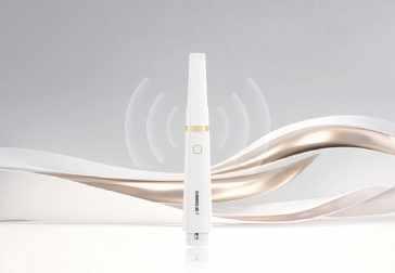
Aoralscan Elite Wireless: Next-Level Intraoral Scanning
Weighing just 194 grams, the Aoralscan Elite Wireless is the world’s lightest wireless intraoral scanner that provides outstanding flexibility and movement freedom. Powered by Wi-Fi 6 technology, it delivers stable and high-speed data transfer, while its wireless charging cradle ensures effortless operation. Its innovative design allows seamless use across multiple dental chairs, optimizing workflow efficiency. With Intraoral Photogrammetry (IPG) technology, the Aoralscan Elite Wireless delivers exceptional scanning accuracy, making it ideal for All-on-X procedures and full-arch restorations.
Watch official product video: https://youtu.be/06X4KS1mOnc
Read more:
www.shining3ddental.com/solution/e-motion/
Read more:
www.shining3ddental.com/solution/aoralscanelite-wireless/
be housed in the newly constructed, stateof-the-art Apa Aesthetic Suite. He has stepped up to the plate and showed his commitment to the continuance of a program that has started him and many others on their journey to provide responsible aesthetic restoration as the focus in their careers,” said Dr. Calamia.
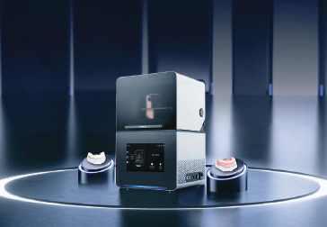
in one of these five thematic areas, evidence of excellent scholarship, and research potential.
While the program is still in the planning stages, the College anticipates that the DScDent program would appeal to international dental school faculty and those who aspire to become faculty. International students may be attracted to our DScDent program because of its relatively short duration and because it could be efficiently combined with specialty or other clinical programs via separate degree tracks.
The FabCure N2 nitrogen curing box is an opensystem device that optimizes post-processing for dental restorations, enhancing physical properties and aesthetics. Its nitrogen curing technology ensures consistent, high-quality results, improving both durability and color stability of printed dental parts. As a crucial addition to the digital dental workflow, the FabCure N2 enables stronger, more predictable restorations.
3D Printer: High-Speed Precision Printing
In the Apa Honors Program in Aesthetic Dentistry, students will continue to receive didactic and clinical experience, participating in lectures, seminars, literature reviews, and case presentations as well as dedicated weekly clinical sessions to treat advanced aesthetic cases. The clinical sessions enable students to develop treatment plans and complete aesthetic restorations using a range of cutting-edge materials and methods for inlays, onlays, crowns, porcelain veneers, and ceramic bridges. Students learn to use high-tech equipment for planning and restoration, including CAD/CAM and lasers, as adjuncts to traditional restorative treatment.
Designed for stability and speed, the AccuFab-F1 3D printer features dual heating modules for both the print platform and resin tank, ensuring optimal material performance. With a maximum light intensity of 18 mW/cm², it excels in printing highceramic filler resins and high-viscosity materials with outstanding accuracy. Its modular design simplifies maintenance, while the quick-release resin tank film streamlines workflow efficiency for both clinics and labs.
Students enter the honors program through a competitive application and interview process; eight to 12 DDS students are accepted to this D4 program.
Dental faculty with education and credentials in teaching and learning, clinical research, and leadership are relatively rare and yet are a critical aspect of improving oral health globally. We invite you to contact us and learn more about our plans for this program and other exciting new initiatives on the horizon at NYU Dentistry. Our tradition of innovative thinking – all in service of improving public health – provides the headwind needed to continue to advance programs, partnerships, and people who are energized to join NYU Dentistry and imagine a world where everyone smiles.®
Read more: www.shining3ddental.com/solution/ fabcure-n2/
“At NYU Dentistry, we truly value Dr. Apa’s ongoing dedication, vision, and commitment to advancing aesthetic dentistry, making a lasting impact. His pursuit of excellence serves as a source of inspiration to our students,” said Anabella Oquendo, DDS, assistant dean for international programs and director of the Apa Advanced
Watch official product video: https://youtu. be/9Id-5AMVhG8
Read more: www.shining3ddental.com/solution/
Health; Leadership; Educational Technology or Sub-specialization in Clinical Fields such as implantology, care for people with disabilities, and aesthetic dentistry. Successful applicants to the DScDent program would exhibit a career interest
Founded in 2004, Shining 3D has spent two decades developing high-accuracy 3D digital solutions for industries such as healthcare, engineering, and manufacturing. Leveraging this expertise, its dedicated sub-brand, Shining 3D Dental, applies these advanced technologies to transform digital dentistry. With a strong foundation in precision technology, continuous R&D investment, and user-driven innovation, Shining 3D Dental provides comprehensive, end-to-end workflows that empower dental professionals worldwide to achieve greater accuracy, efficiency, and clinical success.
Contact: Shining 3D Dental
Email: marketing@shining3d.com
For more information on Shining 3D’s digital dental solutions, visit shining3ddental.com.

Fiber posts are frequently used in endodontics for extensive coronal substance defects when the crown is no longer sufficiently supported, particularly prior to restoration with an indirect restoration. The influence of these posts on clinical success and their survival rate was investigated by Alenezi et.al. in their recently published meta-analysis [1]. In this research, 17 different clinical studies with 7,330 endodontically treated teeth in 7,278 patients were analyzed. The results showed a significantly higher survival rate for teeth restored with posts. The authors concluded that post placement can improve the clinical performance and survival probability of endodontically treated teeth.
Fiber posts are frequently used in endodontics for extensive coronal substance defects when the crown is no longer sufficiently supported, particularly prior to restoration with an indirect restoration. The influence of these posts on clinical success and their survival rate was investigated by Alenezi et.al. in their recently published meta-analysis [1]. In this research, 17 different clinical studies with 7,330 endodontically treated teeth in 7,278 patients were analyzed. The results showed a significantly higher survival rate for teeth restored with posts. The authors concluded that post placement can improve the clinical performance and survival probability of endodontically treated teeth.
RTD DT Light Post
RTD (Recherches Techniques Dentaires, a Dentsply Sirona company based in France) is an innovator in fiber posts field. Founded in 1968, RTD was one of the very first companies worldwide to invent and patent a fiber post. RTD has continued to innovate fiber posts ever since. Other fiber posts in the market can have an uneven distribution of fibers or even air bubbles trapped in the resin; RTD has optimized their products to eliminate those potential weak spots for the post and thus for the coronal restoration (Fig. 1).
RTD (Recherches Techniques Dentaires, a Dentsply Sirona company based in France) is an innovator in fiber posts field. Founded in 1968, RTD was one of the very first companies worldwide to invent and patent a fiber post. RTD has continued to innovate fiber posts ever since. Other fiber posts in the market can have an uneven distribution of fibers or even air bubbles trapped in the resin; RTD has optimized their products to eliminate those potential weak spots for the post and thus for the coronal restoration (Fig. 1).
The DT Light Post with its typical double taper is RTD’s most well-known product. Its shape contributes to a better stress distribution compared to single taper or parallel posts and follows the natural geometry of the root canal, requiring less dentin removal, especially in the apical third where unnecessary dentin removal can increase the risk of fracture.
The DT Light Post with its typical double taper is RTD’s most well-known product. Its shape contributes to a better stress distribution compared to single taper or parallel posts and follows the natural geometry of the root canal, requiring less dentin removal, especially in the apical third where unnecessary dentin removal can increase the risk of fracture.
[1] ALENEZI, Ali A., et al. Clinical behavior and survival of endodontically treated teeth with or without post placement: a systematic review and meta-analysis. Journal of Oral Science, 2024, S. 24-0098.
[1] ALENEZI, Ali A., et al. Clinical behavior and survival of endodontically treated teeth with or without post placement: a systematic review and meta-analysis. Journal of Oral Science, 2024, S. 24-0098.
Competitor product with uneven fiber distribution and air bubbles.
Competitor product with uneven fiber distribution and air bubbles.
RTD post with homogeneous distribution of fibers and no air bubbles.
RTD post with homogeneous distribution of fibers and no air bubbles.
After two decades, the FDI World Dental Congress (WDC) returns to mainland China, with the global dental community gathering in Shanghai. From September 9 to 12, 2025, the highly anticipated event will take place at the National Exhibition and Convention Center (Shanghai), hosting one of the largest exhibitions in its history and coinciding with China’s booming dental market. As 191 national and regional dental associations converge in the city, Shanghai will become the center of global dentistry. This prestigious event, a collaboration between FDI World Dental Federation (FDI), the Chinese Stomatological Association, and Reed Sinopharm Exhibitions, will bring together over 700 dental companies and attract more than 35,000 visitors. Known as the Olympics of dentistry, FDI WDC 2025 will provide a unique platform for sharing insights, innovations, and best practices, shaping the future of oral health.
huFrieDy booth
Zone, the process of applying Chinese labeling can be completed within the zone, streamlining compliance procedures. Additionally, local subsidies in regions such as Beijing and Sichuan further create a supportive environment, enabling international brands to establish and grow in China’s expanding oral healthcare market.
l. to r. Drs ziAD AllAhem, nAhAwAnD thAbet norAh AlAjAji
l. to r. DeAn Ali rAzooKi, DeAn nADA nAAmAn, Dr tony Dib
China’s dental market is experiencing rapid growth, driven by rising living standards and increased awareness of oral health. This has fueled demand for high-quality dental products and services. Its size grew from 92.3 billion yuan in 2015 to 261.07 billion yuan in 2023. Currently accounting for 6% of the global market, China’s dental industry still holds ample room for expansion. With over 120,000 dental institutions and growing consumer demand, especially among younger generations, the market offers vast opportunities for both domestic and international players.
While market potential provides the “pull,” China’s policy reforms actively create the “push” for international engagement. The government has rolled out a suite of incentives to attract foreign dental technology providers. In the Hainan Free Trade Port, medical institutions can import registered or specially approved devices with zero tariffs and VAT. In the Shanghai Free Trade
Complementing policy support, China’s dental sector is undergoing a tech-driven metamorphosis—a transformation poised to dominate center stage at FDI WDC 2025. The exhibition’s “IntelliDent Stage” will spotlight how innovations like 3D-printed implants and AIassisted diagnostics are improving treatment outcomes and accessibility. For example, AIpowered teledentistry platforms enable remote diagnosis between cities like Shanghai and Xinjiang, bridging urban-rural care divides. This fusion of policy empowerment and technological leapfrogging cements China’s dual role as a massive consumer and cutting-edge innovator inglobal dental tech.
l. to r. Drs. ziAD sAlAmeh, hAtem el DAmAnhoury, DeAn heshAm mArei, husein Al lAwAti
FDI WDC 2025 is expected to be the largest exhibition and congress in FDI’s history. It’s a four-day exhibition and scientific programme, covering a 60,000m2 area. It will host over 191 dental associations, attract more than 35,000 visitors, and welcome over 700 companies— including industry leaders like Haleon, Straumann, Solventum, Ivoclar, Bondent, and Zumax. With 300+ speakers, the congress will be a knowledgesharing and networking bonanza. We also offer exclusive social activities, including the Gala Dinner and Shanghai Night, providing a perfect platform for global dental professionals to connect.
In our exhibition hall, we offer matchmaking and guided tour services to help you quickly find what you need among a wide range of products. We also prioritize innovation through the IntelliDent
Stage and exclusive new product launches in the exhibition hall. These activities and services are designed to enhance your experience and ensure you get the most out of your visit. Language should never be a barrier. We offer English translation and consultation services to ensure smooth communication for all our international guests.
It’s the perfect time to visit Shanghai for the dental industry professionals. Shanghai, the host city of the FDI WDC 2025, is a metropolis that blends tradition with innovation. As FDI returns to mainland China after nearly two decades, Shanghai has become the centre of global dentistry. This dynamic city offers a unique cultural atmosphere, delicious local cuisine, and stunning landscapes. While attending the congress, you can take
advantage of China’s favourable visa policies, such as the 240-Hour Transit Visa Exemption, to fully experience the charm of this city. Stroll through the tranquil Yu Garden, marvel at the futuristic skyscrapers along the Bund, and savour the beloved Shanghai cuisine, including the famous soup dumplings. This international business hub also offers a glimpse into traditional Chinese culture, creating a captivating contrast. Make your journey to Shanghai for the FDI WDC 2025 both professionally rewarding and personally enriching, as you explore all that this multifaceted city has to offer
For more details, visit www.world-dental-congress.org.
See you in Shanghai!


Join us for an unforgettable learning and networking experience in the vibrant city of Shanghai!

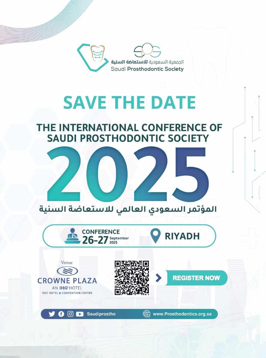
The 2025 FDI World Dental Congress (FDIWDC25) will be held in China and is expected to be a groundbreaking event in the field of dentistry. Set against the vibrant backdrop of Shanghai, the congress promises to be one of the largest in its history, featuring over 700 companies and more than 35,000 dental professionals. In this interview, Prof. Guo Chuanbin, president of the Chinese Stomatological Association, which will be co-hosting the World Dental Congress in 2025, shares some insights into the exciting opportunities that FDIWDC25 will offer for dental professionals to engage with cutting-edge technologies and discusses the significant impact of artificial intelligence (AI) and 3D printing on dental practices worldwide.
The 2025 FDI World Dental Congress (FDIWDC25) will be held in China and is expected to be a groundbreaking event in the field of dentistry. Set against the vibrant backdrop of Shanghai, the congress promises to be one of the largest in its history, featuring over 700 companies and more than 35,000 dental professionals. In this interview, Prof. Guo Chuanbin, president of the Chinese Stomatological Association, which will be co-hosting the World Dental Congress in 2025, shares some insights into the exciting opportunities that FDIWDC25 will offer for dental professionals to engage with cutting-edge technologies and discusses the significant impact of artificial intelligence (AI) and 3D printing on dental practices worldwide.
Prof. Chuanbin, what can participants expect from FDIWDC2025?
Prof. Chuanbin, what can participants expect from FDIWDC2025?
FDIWDC25 is set to be one of the largest exhibitions in the history of the World Dental Congress. Spanning an impressive 60,000 m2 of hall space, the exhibition will host over 700 companies from around the world, including leading global brands and hundreds of Chinese manufacturers and technology providers specialising in dental equipment and solutions.
FDIWDC25 is set to be one of the largest exhibitions in the history of the World Dental Congress. Spanning an impressive 60,000 m2 of hall space, the exhibition will host over 700 companies from around the world, including leading global brands and hundreds of Chinese manufacturers and technology providers specialising in dental equipment and solutions.
Prof. Guo Chuanbin, president of the Chinese Stomatological Association
Prof. Guo Chuanbin, president of the Chinese Stomatological Association
FDIWDC25 will offer an unparalleled opportunity for dental professionals to engage with the Chinese market, which is fuelled by a booming economy and an expanding middle class. This event will also be invaluable for international attendees to explore a wealth of technologies and digital innovations from competitive local suppliers. More than 35,000 professionals are expected to attend, and the exhibition will be a vibrant hub for networking, learning and discovering the latest advancements in dental care.
FDIWDC25 will offer an unparalleled opportunity for dental professionals to engage with the Chinese market, which is fuelled by a booming economy and an expanding middle class. This event will also be invaluable for international attendees to explore a wealth of technologies and digital innovations from competitive local suppliers. More than 35,000 professionals are expected to attend, and the exhibition will be a vibrant hub for networking, learning and discovering the latest advancements in dental care.
How will FDIWDC25 address the impact of new technologies in dentistry?
How will FDIWDC25 address the impact of new technologies in dentistry?
With the rapid development of science and technology, a growing number of AI applications are transforming dental diagnosis and treatment. Innovations such as implant robots, intelligent navigation systems, virtual simulation teaching and digital mandibular movement simulation are becoming increasingly integral to modern dental practices.
With the rapid development of science and technology, a growing number of AI applications are transforming dental diagnosis and treatment. Innovations such as implant robots, intelligent navigation systems, virtual simulation teaching and digital mandibular movement simulation are becoming increasingly integral to modern dental practices.
Alongside these advancements, the rise of CAD/CAM and 3D-printing technologies has led to the widespread use of new materials, reshaping the future of oral health. This is why we have chosen “Shaping the Future of Oral Health” as the theme for FDIWDC25.
Alongside these advancements, the rise of CAD/CAM and 3D-printing technologies has led to the widespread use of new materials, reshaping the future of oral health. This is why we have chosen “Shaping the Future of Oral Health” as the theme for FDIWDC25.
The congress will feature a series of lectures focused on these cutting-edge technologies and materials. Additionally, an AI-enabled dentistry event area will be set up in the exhibition hall, providing a platform for showcasing and exchanging ideas on these innovations. Over four days, attendees will have the opportunity to hear from national and international experts in a total of 100 hours of engaging lectures.
The congress will feature a series of lectures focused on these cutting-edge technologies and materials. Additionally, an AI-enabled dentistry event area will be set up in the exhibition hall, providing a platform for showcasing and exchanging ideas on these innovations. Over four days, attendees will have the opportunity to hear from national and international experts in a total of 100 hours of engaging lectures.
How will the event’s location help create a unique experience for visitors?
How will the event’s location help create a unique experience for visitors?
Shanghai is one of the most vibrant and cosmopolitan cities in the world today. When exploring Shanghai in 2025, visitors will discover that it is much more than just an international business hub. The city is a showcase of the latest in style and fashion, offering the most exquisite tastes in lifestyle, unique dining experiences and stunning entertainment options. We are confident that the participants’
time in Shanghai will be unforgettable and full of surprises and will leave them longing to return for more.
time in Shanghai will be unforgettable and full of surprises and will leave them longing to return for more.
Known as Hu, Shanghai is a multicultural metropolis that seamlessly blends modernity with traditional Chinese elements. You can explore ancient temples and gardens alongside bustling markets and futuristic shopping malls. You can feel the pulse of history in Yu Garden and the City God Temple, enjoy the breathtaking night views along the Bund and marvel at the skyscraper skyline in Lujiazui. Shanghai is a popular travel destination that captures the essence of China’s rapid development, offering visitors a unique opportunity to experience the country’s dynamic growth.
Known as Hu, Shanghai is a multicultural metropolis that seamlessly blends modernity with traditional Chinese elements. You can explore ancient temples and gardens alongside bustling markets and futuristic shopping malls. You can feel the pulse of history in Yu Garden and the City God Temple, enjoy the breathtaking night views along the Bund and marvel at the skyscraper skyline in Lujiazui. Shanghai is a popular travel destination that captures the essence of China’s rapid development, offering visitors a unique opportunity to experience the country’s dynamic growth.
Shanghai is one of the most vibrant and cosmopolitan cities in the world today. When exploring Shanghai in 2025, visitors will discover that it is much more than just an international business hub. The city is a showcase of the latest in style and fashion, offering the most exquisite tastes in lifestyle, unique dining experiences and stunning entertainment options. We are confident that the participants’
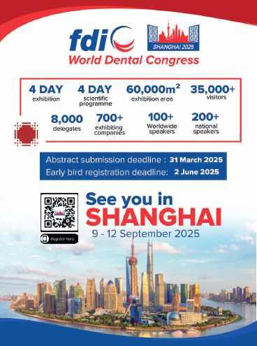

While aesthetic universal composites have to be layered and polymerised in 2 mm increments, which is complicated, bulk-fill composites offer the advantage of application into the cavity in increments of up to 4 mm. However, due to their insufficient aesthetics, bulk-fill composites are normally only used for the posterior region. VOCO has now resolved this issue with the new universal composite GrandioSO Unlimited.
The aesthetic material can be applied into the cavity in 4 mm increments and is indicated both for the anterior and posterior regions. As a result, dental practices now only need one packable universal composite for their day-today work! With this extension to the GrandioSO product family, tried and tested for more than a decade, VOCO is setting a new standard for universal composites.
The universal composite without layers thanks to visual transformation technology (VTT)
In an uncured state, the material is so translucent that it enables a reliable depth of
cure of 4 mm. During light polymerisation, a visual transformation (visual transformation technology) takes place, whereby the translucency of the material visibly decreases and the desired shade is achieved at a level of opacity that matches the natural tooth. The result is an aesthetic material for the anterior and posterior regions, which can be applied into the cavity in layers of 4 mm thanks to this special technology.
In addition to universal use and the usual simple handling, GrandioSO Unlimited offers further attractive advantages: thanks to the simplified shade system, the 5 cluster shades are able to cover all 16 classic VITA shades. The optimised light scattering leads to a targeted and intensified chameleon effect, ensuring perfect adaptation of the restorative material to the natural tooth shade. As a result, dental practices are ideally equipped for their day-today work with just five shades and can reduce their inventories. The cluster shades A1-A3.5 that are frequently used require a polymerisation time of just 10 seconds. With an impressive filler content of 91%, GrandioSO Unlimited is setting new standards in the high-performance composites class when it comes to stability and strength. The low volume shrinkage of 1.44% and the resulting low shrinkage stress also reduce the risk of poor marginal integrity and marginal discolouration, and contribute to topquality restorations.
GrandioSO Unlimited is compatible with all conventional bonding agents and is available in both syringes and caps.
www.voco.dental
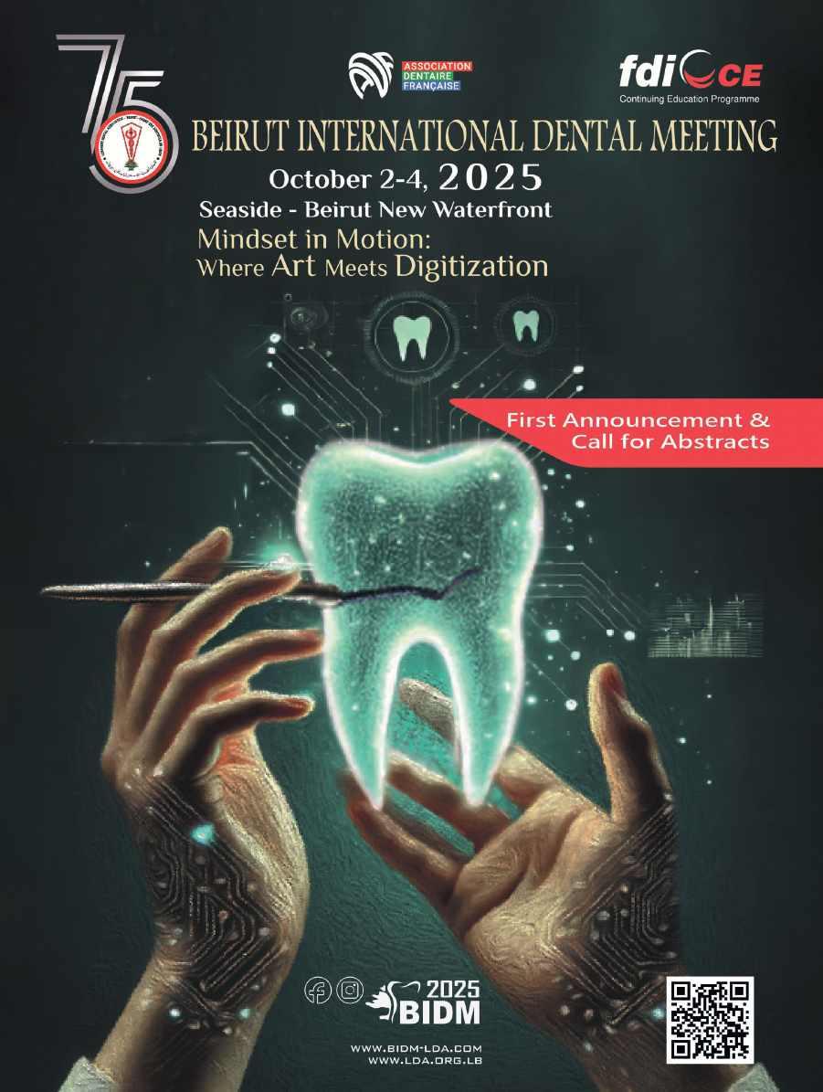
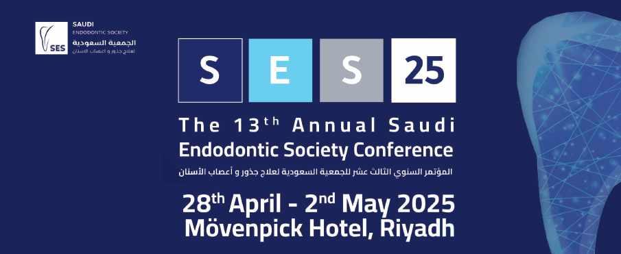
It’s with great pleasure to welcome you to the 13th Saudi Endodontic Society Annual Meeting which will be held in Riyadh, Saudi Arabia between the period of 28th of April and 2nd of May 2025. This version of the conference will cover the more advanced and debatable topics in Endodontics by expert local and international speakers.
There will be 14 lectures and 9 workshops covering all the topics needed by general practitioners, Endodontic residents and specialists. There will be a special session for Young Graduates to present their unique cases to the expert audience and to show their capabilities and skills, beside that there will be a scientific poster competition for the best 3 posters.
Alongside this event is an exhibition with selected exhibitors showing their updated and advanced products with special offers to our members. We look forward to seeing and welcoming you at the conference.
Prof. Abdulaziz
Abu-Melha President of the Saudi Endodontic Society

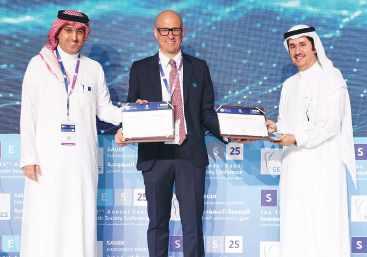
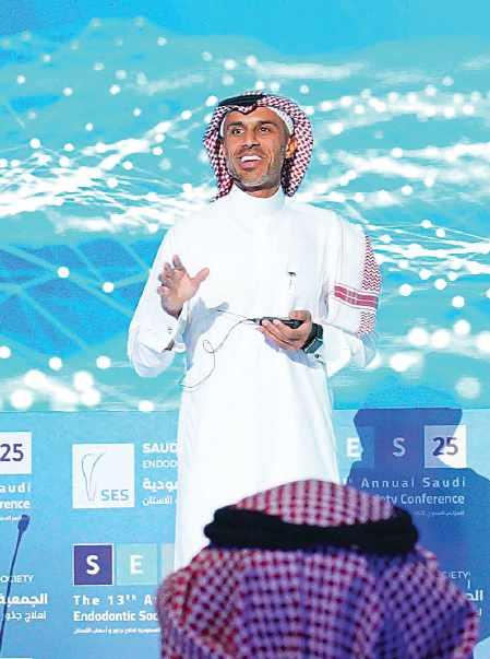
Dr. obaDa aUstah leCtUring aboUt ConteMPorary aPProaChes to vital PUlP theraPy
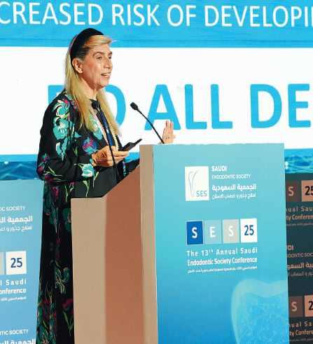
Dean Prof. elisabetta Cotti, at the University of Cagliari, leCtUre entitleD aPiCal PerioDontitis anD the host
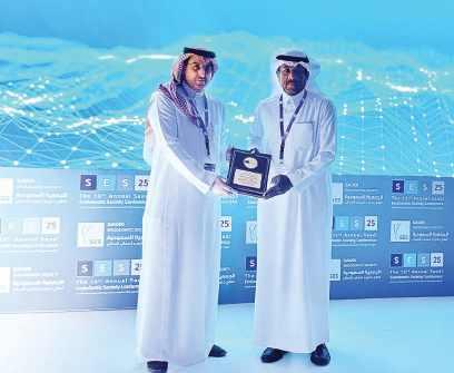
Prof. saaD al-nazhan granteD the honorary MeMbershiP by Prof. abDUlaziz abU Melha, PresiDent of the saUDi enDoDontiC soCiety, in reCognition of his oUtstanDing lifelong aChieveMents
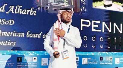
Dr. MohaMMeD al-harbi leCtUre aboUt non-sUrgiCal retreatMent, giving teeth a seConD ChanCe
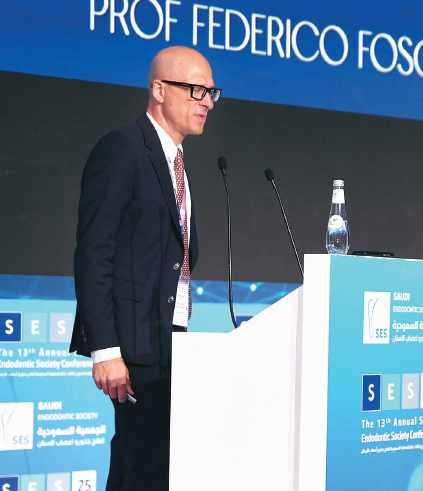
Prof. feDeriCo fosChi, heaD of enDoDontiCs at UCl eastMan Dental institUte
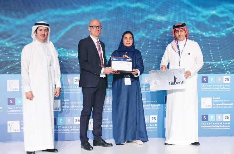


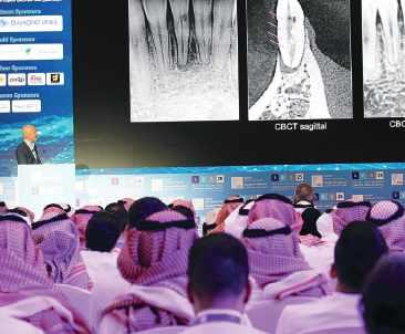
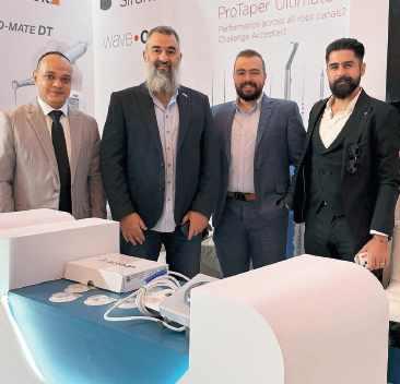
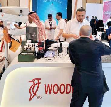
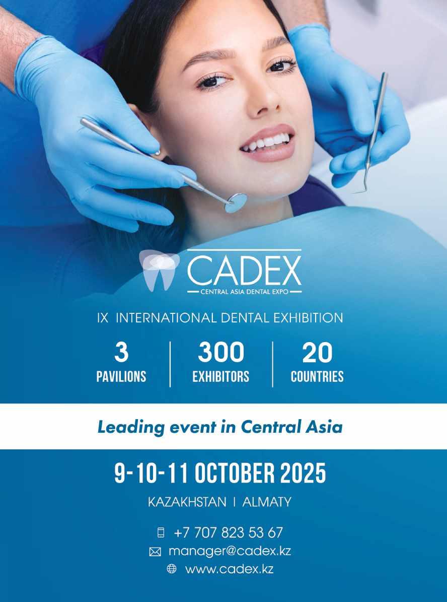

Over 12,000 Visitors Joined Us at the 5th Jordanian Dental Show
The 5th edition of the Jordanian Dental Show (JDS 2025), held from May 13 to 15, 2025, at the Jordan Center for International Exhibitions – Macca Mall in Amman, exceeded all expectations. With more than 12,000 attendees, the atmosphere was vibrant, and the energy throughout the exhibition was truly exceptional.
Organized by JOMESRA – the Jordanian Association for Medical and Scientific Materials Traders – JDS and under the Leadership of Mr. Ayman AbulHawa and Eng. Ziad Qwaider, it has become one of the region’s most anticipated dental trade events. This year’s show gathered key players from across the dental industry, including clinicians, manufacturers, suppliers, distributors, and academics.
JDS 2025 featured an impressive lineup of exhibitors showcasing the latest advancements in dental technology and products, including cutting-edge equipment, materials, CAD/CAM systems, oral hygiene solutions, and innovations in digital dentistry. Attendees had the chance to experience live product demonstrations, attend seminars, and explore valuable business and networking opportunities.
As the official media partner, Dental News played a key role in covering the event, providing regional visibility and supporting exhibitors and participants through comprehensive media outreach.
Thanks to the enthusiastic participation of the dental community, this edition of JDS delivered a unique and unforgettable experience. The exceptional turnout and engagement reflected the growing importance of this event in the MENA region.
With the success of this year’s show, the promise for the next edition is clear: even bigger, even better.
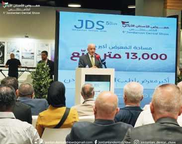
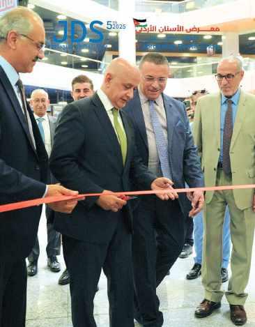


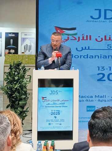
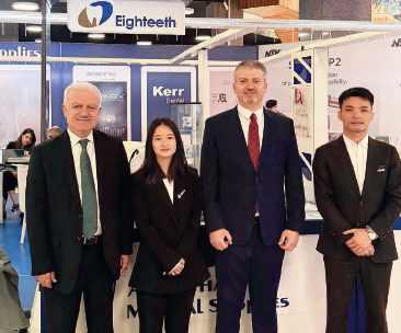
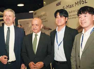
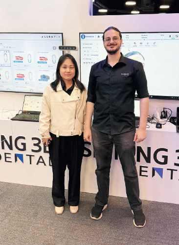
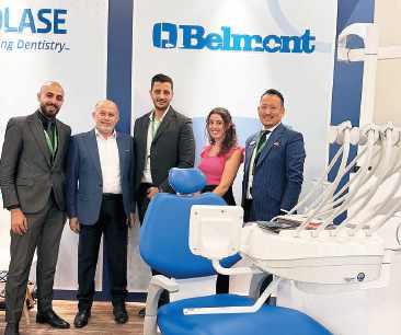
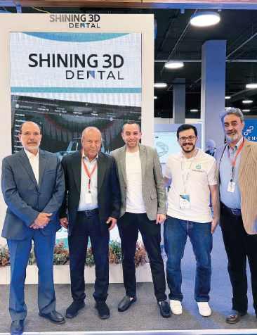
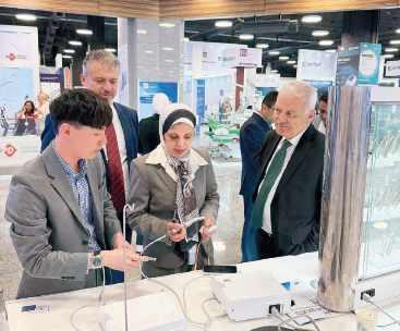
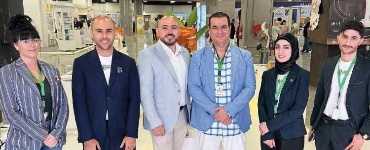
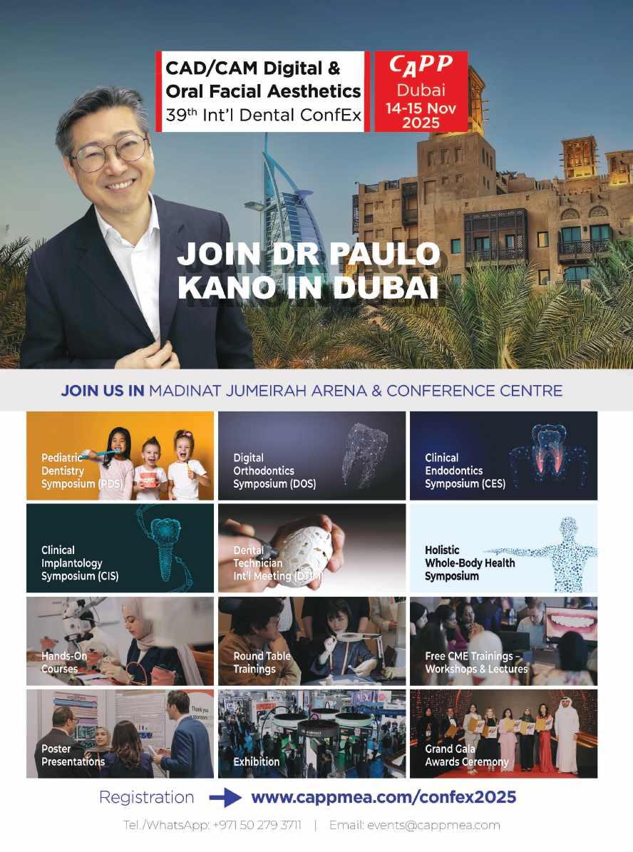


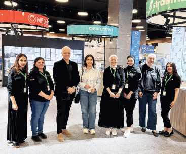
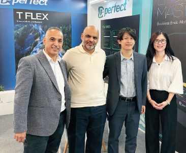
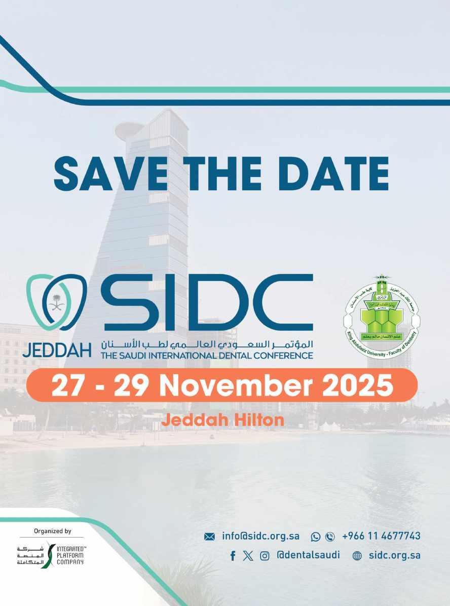
15-16-17 May 2025

Milan, May 20, 2025 – The 2025 edition of Expodental Meeting concluded with great success, reinforcing its undisputed leadership role in the Italian and international dental sector. The event saw a significant turnout of professionals, companies, and stakeholders, showcasing a complete and cuttingedge overview of the latest innovations in technologies, materials, and services.
The exhibition hosted 400 exhibitors, marking a notable increase compared to the previous year. A strong international presence was confirmed, with significant participation from Germany and China, and a total of 19 countries represented. A sign of dynamism is the increase in new exhibitors, from 82 to 90, and the 10% increase in net exhibition space. The number of visitors also recorded a 5% increase, with 21,000 attendees over the three days.
On Friday, May 16, the event had the honor
of hosting the Undersecretary of State for Health, Hon. Dr. Marcello Gemmato, who visited the exhibition area with considerable interest, where he was able to view the innovations and high technological content of Italian dental products. He also participated in the presentation of the economic analysis of the dental sector, prepared by ANDI. His presence testified to the great attention of the institutions towards the dental sector and the crucial importance of a constant dialogue between politics and professionals to face the challenges and seize future opportunities.
Also of great resonance was the round table on Saturday morning: “Dentistry and Orthodontics, between digitalization and multidisciplinarity: a present/future between Clinic, Ethics, Economics and dynamic interactive Technology. Is the team ready for the challenges of Change?”. A lively moment of discussion on the evolution of the sector.
The round table represented a key event, presented by Fabio Velotti (President of UNIDI) and Pietro di Michele (representative of SUSO), with an introduction by Antonio Pelliccia and the expert moderation of journalists Margherita De Bac and Giuliano Giubilei. The event, strongly
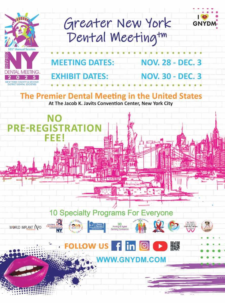


desired by UNIDI and SUSO (Italian Union of Orthodontic Specialties), brought together the top representatives of the Italian dental scene. Present were the President of CAO (Commission of the Register of Dentists) Andrea Senna, the Councilor of CNEL (National Council of Economy and Labor) Francesco Riva, the President of ANDI (National Association of Italian Dentists) Carlo Ghirlanda, the President of AIO (Italian Dental Association) Gerhard Seeberger, the President of CDUO (College of University Professors of Stomatological Disciplines) Lorenzo Lo Muzio, the President of SUSO Fabrizio Sanna, and representatives of the main scientific societies and sector associations SIDO, SIOF, AIOP, SIDP, SIPMO, U.N.I.D., ATASIO, AIDI, SISIO, IDEA, AIASO, FNO -TSRM – PSTRP, ANTLO, CNA-SNO, ORTEC, the representatives of dental producers CEFLA, ADVAN, LEONE, ALIGN TECHNOLOGY, CARESTREAM DENTAL and Federconsumatori.
The various thematic areas within the exhibition were a great success, offering visitors a complete and up-to-date exhibition itinerary on the latest trends and innovations. The high level of interest and participation in the scientific sessions, practical workshops,
and “live on stage” events (an absolute novelty of 2025) confirmed the vitality and value that distinguish the dental community.
Of particular note was the presence of the foreign delegation, a large group of 60 dealers from Albania, Angola, Saudi Arabia, Azerbaijan, Bosnia Herzegovina, Brazil, Chile, Colombia, Croatia, Ecuador, United Arab Emirates, Ethiopia, Ghana, Kosovo, Malaysia, Pakistan, Peru, Slovenia, Thailand, Uganda, Uruguay, who came to Rimini thanks to the joint efforts of ICE Agency and UNIDI.
“Expodental Meeting 2025 concludes its time at the Rimini exhibition center with the solid awareness of having offered a platform for networking and professional development of the highest level,” declared Fabio Velotti, President of UNIDI. “I would like to express my sincere thanks to all the participants, exhibitors, speakers, partners, scientific societies, and institutions that contributed to the success of this edition, and I invite everyone to meet again from May 14 to 16, 2026, to continue building together the bright future of dentistry.”
For information: Susi Marotti | s.marotti@unidi.it |+39 340 7496695
May 30 & 31, 2025
Mövenpick Hotel & Resorts, Beirut, Lebanon
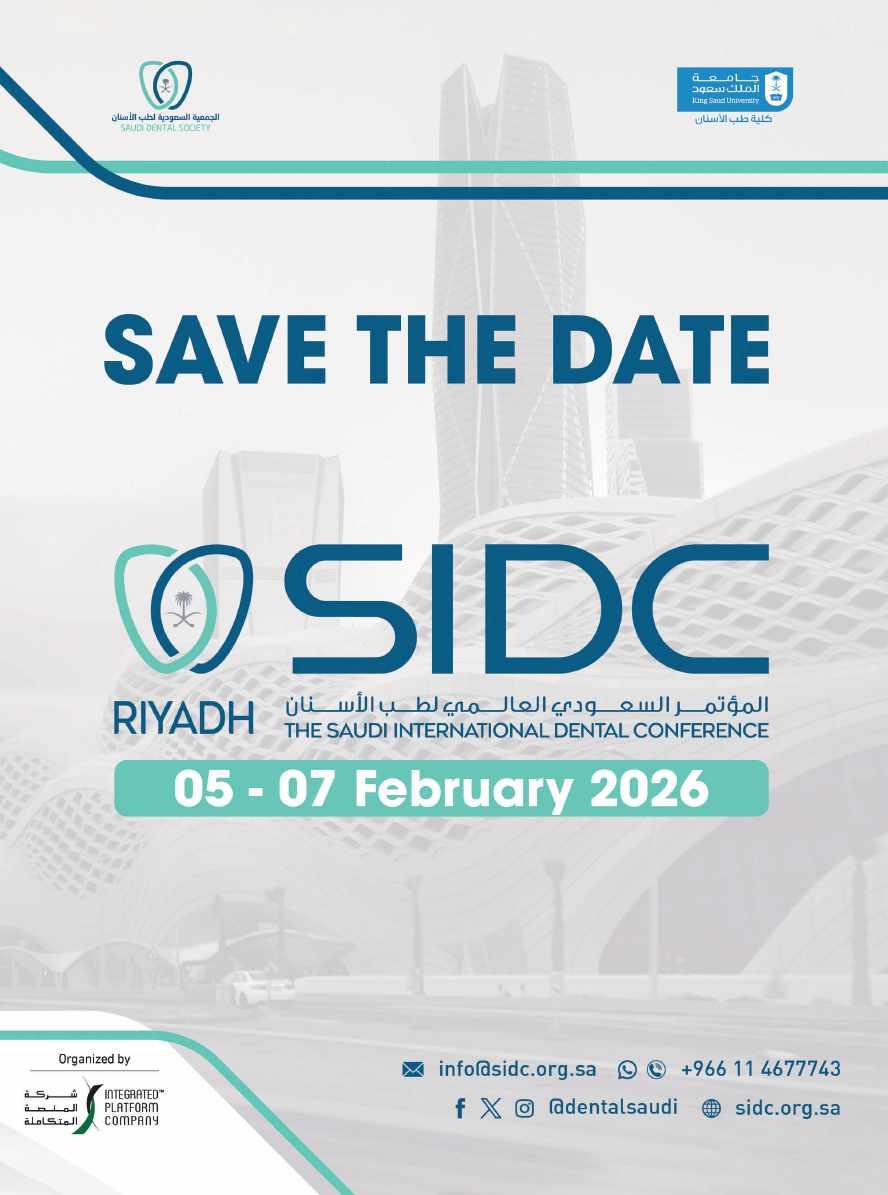

the
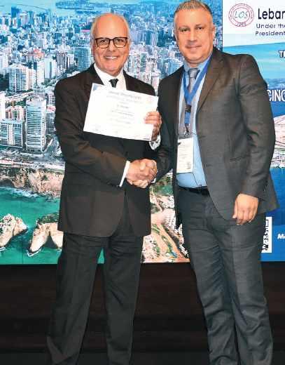

Driven by quality, our goal is to make the professional feel comfortable in its daily endo workflow.
KENDO delivers a wide and reliable range of endodontic products made in Germany as: K-REAMERS, K-FILES, H-FILES, ROOT FILLERS
A VDW brand - over 150 years of endo experience.
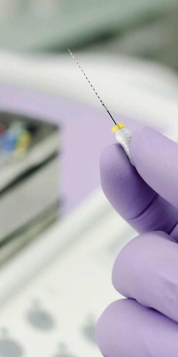
Viscosity
• Good visibility – contrasty to the gingiva
• A clean product – quick and easy to spray off
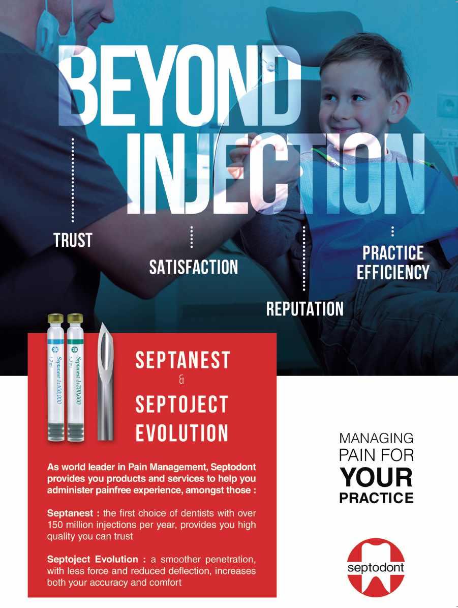

For nearly six decades, endodontists and patients have trusted RTD for the most high-stakes restorations. In fact, we were one of the firsts to develop and bring fiber posts to market, leveraging on deep knowledge and expertise. Today, we’re one of the largest producers all over the world, with a wide range of posts designed for optimal outcomes and unbeatable peace of mind.
Learn more about RTD at rtddental.com