www.kavo.com/e30
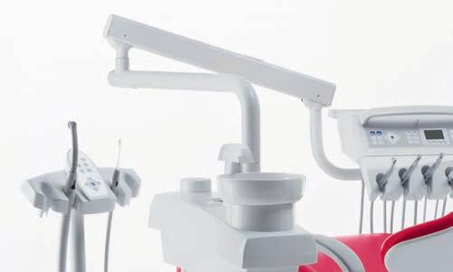
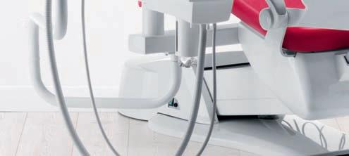
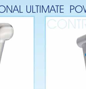
KaVo ESTETICA® E30 – everything your heart desires, within your reach.
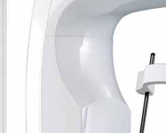
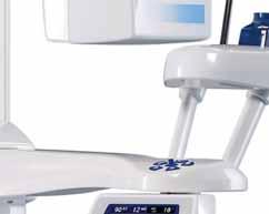
KaVo ESTETICA E30 - looks good, feels excellent.

The KaVo ESTETICA E30 opens a new dimension of Dental Excellence: the essence of high KaVo quality, reliability and efficiency at affordable entry level pricing.
Clever technology and a love for detail.

Easy and intuitive to use, safe and economical to operate. This treatment unit combines convenience and efficiency as part of your daily workload.
Each detail of the KaVo ESTETICA E30 is aimed at efficiency, flexibility and ease of use. The perfectly matched components ensure cost-efficient operation with high reliability. The integrated service functions lead to low costs and operational safety.


Eruption Of Oral Lichen Planus After Interferon Therapy For Hepatitis C Infection: Case Report
Dr. Wafa Ali Al-Shamali,Dr. Mohamed Ahmed, Dr. Rasha Matter, Dr. Saqer Abdulrahman
Using Microabrasion And In-Office Bleaching To Treat Fluorosis In Permanent Anterior Teeth
Dr. Mayada Jemâa, Pr. Sonia Zouiten, Pr. Neila Zokkar, Ms. Belkhir, Pr. Abdellatif Boughzala
The Importance Of The Contact Point In Class II Restorations
Dr. Marco Calabrese
Maintaining Missing Central Space Using Tad
Dr. Faraj A. Sedeqi
Steam As The Preferred Tabletop Sterilizing Agent, And The Raging Controversy Between The “B” And “S” Approaches
Oman International Dental Congress
Al Bustan Hotel, Muscat February 27-28, 2013
35th International Dental Show
Koelnmesse, Cologne, Germany
March 12-16, 2013
Kuwait Dental Association Conference
Radisson Blu Hotel, Kuwait
April 13-15, 2013

74. April 24-26, 2013
Quintessence Dental Arab Congress
Al Faisaliah, Riyadh, KSA

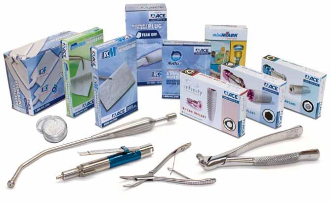
ACE Surgical 15
ACTEON 23
A-DEC 79
AL TURKI 40
BA Intl 19
BIEN AIR 39
BISCO 62
CARESTREAM 55
CAVEX 29
KAVO C2
KERR 72
LISTERINE 77
MEDESY 34
MICRO MEGA 53
MORITA 41
NSK C1
ORTHO ORGANIZERS 80
PLANMECA 45
COLTENE 27
E4D 17
DENTSPLY 47
Vita Enamic The First Hybrid Dental Ceramic In The World With A Dual-Network Structure
Scican VITA
DISCUS PHILIPS 67
DURR 57
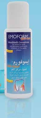
EMOFORM 5
GC 52
GSK C3, 31, 51, 61
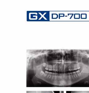
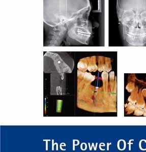
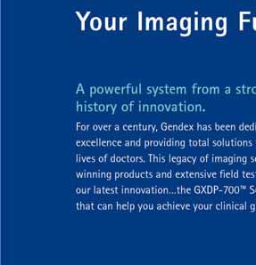
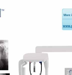
GENDEX 6
HENRY SCHEIN 63
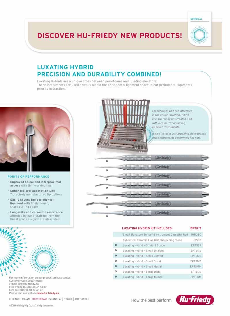
HU FRIEDY 37
IVOCLAR 1, C4
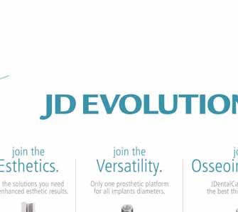
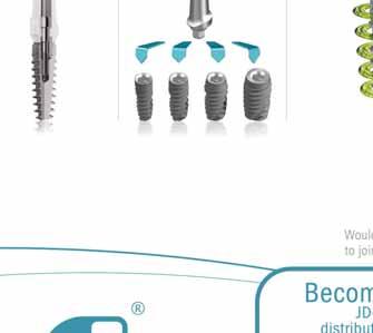
JDENTAL CARE 21
RITTER 13
SCI CAN 43
SIRONA 25
SULTAN 59
SOREDEX 9
THOMMEN 10
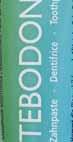
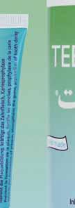
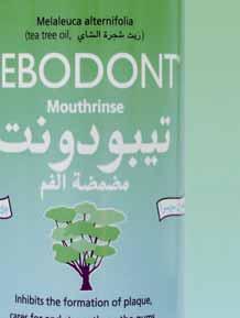
TEBODONT 4
ULTRADENT 35
VITA 49
VOCO 7
W&H 8
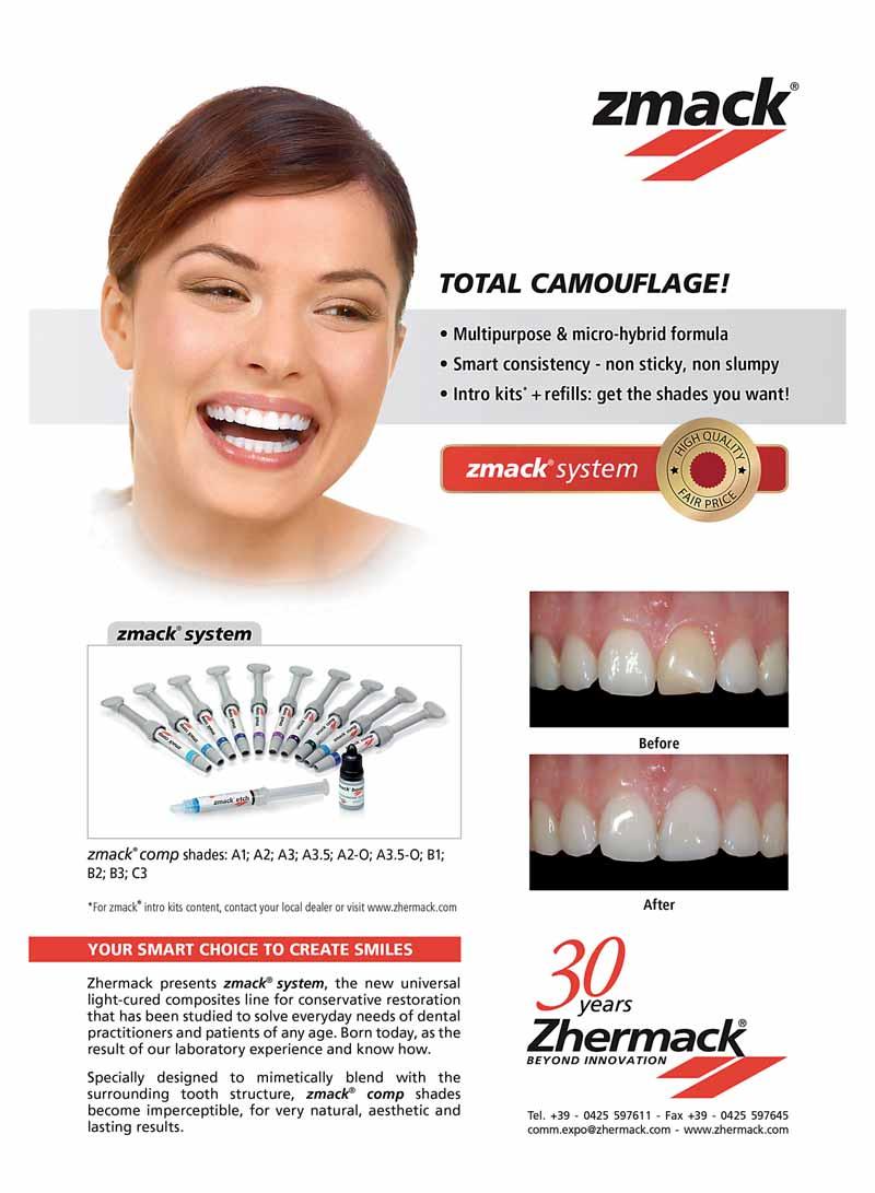
ZHERMACK 2


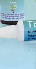
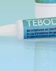


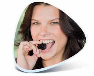
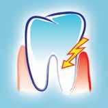
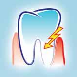
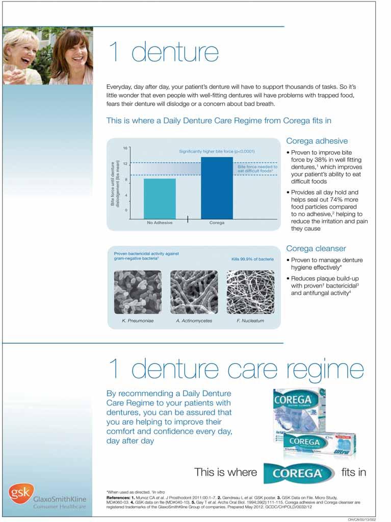
Special toothpaste and mouthbath with Ems salts for sensitive teeth and denuded toothnecks, irritations of the gums, plaque


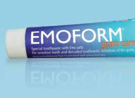
Swiss made
desensitizes teeth and denuded toothnecks
firms up the gums and combats dental plaque neutralizes acids harmful to the teeth

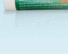
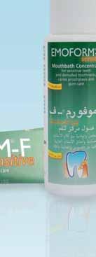
Special toothpaste and mouthbath for sensitive teeth and denuded toothnecks, caries prophylaxis and gum care


Swiss made
desensitizes teeth and denuded toothnecks
caries prophylaxis stimulates salivation


.πjóÑdG äRƒÑªμdG
¿Éæ°SÓd Ö∏°üàdG »Fƒ°V äRƒÑªμdG
IÒÑμdG º¨∏e’G äGƒ°û◊ πãe’G πjóÑdG øeõHh º∏e 4 ácɪ°S äGP äÉ≤Ñ£H ≥«Ñ£àdG á«fÉμeG :º«eÎdG RÉ‚G áYô°S ¿GƒK 10 Ö«∏°üJ
π°†ØH á«©«Ñ£dG ¿Éæ°S’G ™e RÉટG ʃ∏dG ≥HÉ£àdG øeƒj :πeÉ°ûdG ¿ƒ∏dG
AÉHô◊G á«°UÉN
πμ°ûdÉH É«dÉM ôaƒàe …OÉ°üàb’G …QÉéàdG


Tune in to Swiss precision and innovation with Thommen! Based on more than 25 years of clinical experience, in-house research and development as well as high-quality Sw iss manufacturing, you will fi nd that the Thommen Implant System excels through its proverbial simplicity!

EExclusive distributor in the Middle East: Star Science International GmbH Jupiterstrasse 57 3015 Bern | Switzerland Tel. +41 31 941 07 31 star.science@bluewin.ch

www d e n ta ln e ws c o m
Volume XX, Number II, 2013
EDITORIAL TEAM
Alfred Naaman, Nada Naaman, Jihad Fakhoury, Dona Raad, Antoine Saadé, Lina Chamseddine, Tarek Kotob, Mohammed Rifai, Bilal Koleilat, Mohammad H. Al-Jammaz
COORDINATOR
ART DEPARTMENT
SUBSCRIPTION
ADVERTISING
Suha Nader
Ibrahim Mantoufeh
Micheline Assaf, Nariman Nehmeh
Josiane Younes
PHOTOGRAPHY
TRANSLATION DIRECTOR ISSN
Albert Saykali
Gisèle Wakim, Marielle Khoury
Tony Dib
1026-261X
DENTAL NEWS IS A QUARTERLY MAGAZINE DISTRIBUTED MAINLY IN THE MIDDLE EAST & NORTH AFRICA IN COLLABORATION WITH THE COUNCIL OF DENTAL SOCIETIES FOR THE GCC.
Statements and opinions expressed in the articles and communications herein are those of the author(s) and not necessarily those of the Editor(s) or publisher. No part of this magazine may be reproduced in any form, either electronic or mechanical, without the express written permission of the publisher.
DENTAL NEWS – Sami Solh Ave., G. Younis Bldg.
POB: 116-5515 Beirut, Lebanon.
Tel: 961-3-30 30 48
Fax: 961-1-38 46 57
Email: info@dentalnews.com
Website: www.dentalnews.com www.facebook.com/dentalnews1
www.facebook.com/dentalnews1
twitter.com/dentalnews
Dental News App on both Appstore & Google play


August 7 – 10, 2013 at the Ritz-Carlton, Washington DC, USA info@estheticacademy.org
August 28 – 30, 2013 Istanbul, Turkey Email:congress@fdi2013istanbul.org Website: www.fdi2013istanbul.org
September 6 – 7, 2013 at K3, Josef-Herold-Strasse 12, A-6370 Kitzbühel, Austria www.icdd-2013.com
September 26 – 28, 2013 Campus President RAFIC HARIRI, Hadath, Lebanon Email: bidm@lda.org.lb Website: www.bidm-lda.com
September 29 – October 1, 2013 at the King Saud University, College of Medicine, Riyadh Website: accaff1_symposia@ngha.med.sa
October 3 – 5, 2013 at Cinema Lux Turin/Italy Email: www.escdonline.eu
October 8 – 9, 2013 at Jumeirah Beach Hotel, Dubai, UAE Website: www.cappmea.com
October 31 – November 3, 2013 at Movenpick Resort©Raouch Beirut, Lebanon Email: info@leborthosoc.org Website: www.leborthosoc.com
November 5 – 8, 2013 at InterContinental Citystars Hotel, Cairo, Egypt Website: www.eda-egypt.org
November 28 – 30, 2013 at the Hilton Habtoor, Beirut, Lebanon Email: info@paec2013.org Website: www.paec2013.org
November 29 – December 4, 2013 New York, NY, USA Email: victoria@gnydm.com Website: www.gnydm.com
February 4 – 6, 2014 at Dubai International Convention & Exhibition Centre (DICEC), UAE Website: www.aeedc.com
Oral Pathology
REPORT
PLANUS AFTER INTERF
N THERAPY FOR HEPATITIS C INFECTION: CASE REPORT
Dr. Wafa Ali Al-Shamali wshamali2@yahoo.com
Dr. Mohamed Ahmed El-Khalawany
Dr. Rasha Matter Al-Shemmari
Dr. Saqer Abdulrahman Al-Surayei
Abstract
Background: The association between oral lichen planus (LP) and hepatitis C virus infection (HCV) has been discussed in several papers worldwide. The exact pathogenesis of oral LP in HCV-positive patients is still uncertain There are several studies, which highlight the role of alpha-interferon (INF) being used for treatment of HCV- positive patients, resulting in eruption
or exacerbation of oral LP. Case description: We present a case of erosive LP limited to oral cavity
in a 44-year- old Egyptian man with chronic HCV infection who was treated with INF and ribavirin
Despite an extended period of treatment, there was no significant effect on the viral activity (viral
load). Interestingly, following five months of
termination of anti-hepatitis therapy, there was recurrence of oral LP lesions which was confirmed
histopathologically. His condition improved
dramatically by Protopic cream 0.1%.
Conclusion: Altered immunogenicity of HCV
appears to be the likely explanation, hence understanding the importance of follow-up of the patient post anti-hepatitis C therapy.
Introduction
Oral lichen planus (LP) is a relatively common chronic inflammator y condition that affects the oral mucous membrane with variable clinical traits.
Since the first description of oral LP associated with hepatitis C infection was reported in 1991,1 there have been several reports suggesting the association between HCV infection and oral LP.2
Many studies have showed higher prevalence (1.6 -20%) of oral LP in HCV-positive patients (27). In contrast, some researchers found weak or no correlation between chronic HCV infection and LP. 8 -11
A region-based correlation between HCV infection and LP has been described by some
researchers worldwide.12 However, the possible etiopathogenic mechanism that links the two diseases remains unclear. Immunogenic dysregulation of host infected with HCV, reaction to anti-hepatitis medications particularly alphainterferon or viral infection are considered to be the current acceptable etiopathogenic factors causing oral LP. 12-13
The clinical and histological features of oral LP associated with hepatitis C infection subjects are no different from the control patients. Although, erosive form of oral LP is common clinical
phenotype noted in seropositive hepatitis C individuals, the management of oral LP in patients with or without hepatitis remains the same.
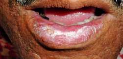
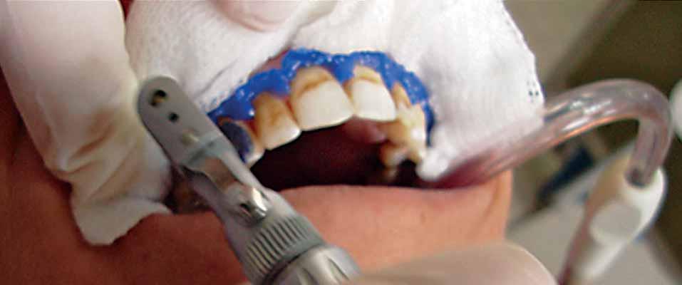
A 44-year- old Egyptian male was referred from dermatology department at Far waniya hospital to oral medicine clinic, who presented with painful
and swollen ulcerated lower lip on March 2010.
On examination, there was apparent swollen
lower lip with central erosive areas oozing fresh
blood from the eroded surfaces on light palpation,
and there were white fine and coarse lace-like
mucosal changes abutting the eroded lesions
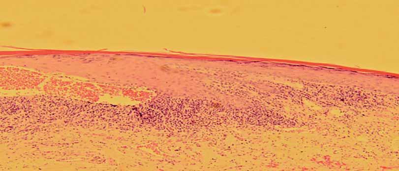
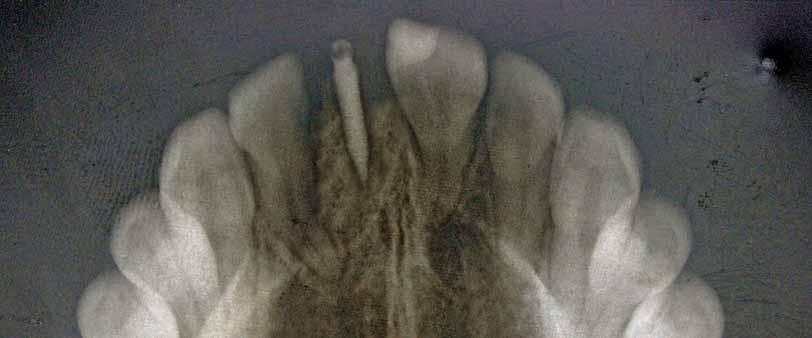
(fig 1). Also, there was bilateral lymphadenopathy
with mobile, tender lymph nodes palpable in
the submandibular triangular region. Intra-oral
examination revealed
fig 1
A swollen lower lip with central erosive and hemorrhagic areas.
Ritter Implants Ivory Line - the German Implant System: Two-Piece Implants QSI/TFI and One-Piece Implants MCI with full range of all prosthetic components and abutments. Clever, easy and beneficial !

Please contact directly:
Oral Pathology
LICHEN PLANUSbilateral white and red lesions on posterior part of the buccal mucosa. These lesions had striking
reticular pattern (reminiscent of LP) centered on
erythematous mucosal areas. The lesion on right
buccal mucosa was found rubbing against heavily
restored molar tooth with amalgam (figs 2 and 3).
No other lesions were seen on the rest of oral
cavity mucosa. The clinical presentation of the lip fig 2
g 3
and oral cavity lesions were consistent with LP.
On reviewing his medical histor y, he had been
diagnosed with hepatitis due to HCV infection
(genotype 4) in 2008 for which he received
combined therapy of pegylated interferon-alpha (180 mcg SC, weekly for 48 weeks) and ribavirin (1000 mg PO daily for 48 weeks). The patient reported an oral soreness and burning sensation after one month of the anti-hepatitis therapy inception for the first time. The exact diagnosis of oral lesion and subsequent therapy provided by dermatology department had not been known to us. Nevertheless, oral condition was quiescent through the period of the therapy. The oral symptoms reappeared five months following discontinuation of anti-hepatitis therapy with
increased severity resulting in severe pain, difficulty in eating, swallowing and speaking.
In addition, he noticed progressive swelling of the lower lip with bleeding ulcers over the next 6 weeks. Besides his known medical condition,
he is on insulin to manage his diabetes (type II).
Furthermore, he is not a cigarette smoker and
he does not drink alcohol. An incision biopsy
of lower lip lesion revealed interface dermatitis
confirming our clinical provisional diagnosis.
Microscopically, the specimen exhibited ortho -
keratosis with prominent granular layer, intense band-like lymphohistiocytic infiltrate with plasma
cell predominance and hydropic degener
of basal c
ll
fig 4

Exhibit lichenoid changes “reticular pattern” on right (A) and left (B) buccal mucosae.
Reveals hydropic degeneration with intense lympho-histoicytic infiltrates. (Hematoxylin and eosin stain at higher magnification 20X)
fig 5 fig 6
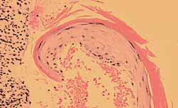
Prominent Civatte body (*).
(Figs 4- 6). The patient was treated with protopic cream 0.1% three-four times daily for 2 weeks. The lower lip status improved dramatically.
fig 7
Dramatic improvement of lower lip lesion after two weeks of topical tacrolimus use.


For over 45 years, ACE Surgical Supply has been committed to providing our customers with the best possible products available at unbeatable prices. We are the only multi-disciplinary surgical supply company. ACE continues to develop and
manufacture the highest quality, state-of-the-art products, while keeping a focus on customer service. Our highly qualified team is always available to answer any questions you may have about our extensive product line.
Oral Pathology
LICHEN PLANUS
Discussion
Among viruses, human herpes viruses, human papilloma virus and hepatitis viruses have been
linked with oral LP, albeit on the basis of equivocal data.12 There have been several studies, which suggest an association between LP and HCV infection 3,7 In a recent review the pooled data from all studies revealed a statically significant difference in the population of HCV seropositive subjects among LP patients when compared
with the controls. Interestingly, geographic heterogeneity seems to play an important role
in this LP-HCV association. As indicated by studies from the Mediterranean basin showing a significant association whereas studies from
Northern Europe did not present any such association Furthermore, in studies from countries
with high prevalence such as Egypt, negative or
insignificant association between HCV infection and oral LP has been reported12,14 The discrepancy may be explained by genetic differences among
the population studies and this may possibly be
the reason for development of LP in our patient.
The exact etiopathogenesis of oral LP in HCV-
positive individual is still uncertain. Nonetheless, eruption of oral LP in our case could have resulted from a lichenoid reaction to the medication used
in the treatment of hepatitis C, particularly alpha-
interferon This hypothesis (i.e. drug reaction)
was plausible in some studies.15,20 Most of these case reports demonstrate the aggravating effect of the interferon rather than causative effect for
the development of oral LP in patient with HCV
infection. Besides, in our case, reappearance of severe erosive oral LP while not receiving INF
therapy, suggests that it may not have played a
significant role in its pathogenesis. Nonetheless, this may be viewed as it having more aggravating
rather than causal effect. Therefore, it would be a good practice to screen the oral cavity of HCV-positive patients prior to initiating antiviral
therapy. So, the possible eruption of oral LP
can be anticipated and managed appropriately
especially in those with quiescent LP. Besides INF
therapy, other confounding factors appeared to have contributed to the possible initiation or
aggravating already present of oral LP in our case, such as presence of amalgam on left mandibular
molar and chronic rubbing of buccal mucosa.
Unfortunately, we are not aware of the intra-
oral examination findings of the patient prior to
anti-hepatitis treatment. Also, there is plethora of literature suggesting role of immune dysregulation
in the pathogenesis of oral LP involving the cellmediated immunity. However, viral factors, such as genotypes of HCV and HCV-RNA levels, are less
important pathogenic cause.12 Why oral mucosa is
most frequently affected is still unknown. Several
experimental studies conducted proposing a theor y of “compartmentalization of mucosa”
that still does not give a clear explanation to this phenomenon 21,22 In HCV seropositive subjects,
erosive oral LP is commonly prevalent lesion 23,24
Mega et al 25 noted three types of OLP. He found
lymphocytic inflammation deeply infiltrating
lamina propria in OLP associated with a HCV
infection and that could be associated with the
erosive trait, as noted in our case. Management of HCV associated oral LP lesion is no different from
oral LP in HCV-negative subjects. Since there is
no cure different therapies are aimed primarily to ameliorate the signs and symptoms of oral LP.
Although corticosteroids have been the mainstay of management, other immunosuppressant and immunomodulator y agents have also contributed significantly towards treatment of the disease.12,26,27,28 A comparative systemic review of 28 randomised controlled clinical trials of therapy for symptomatic oral LP has concluded that there is insufficient evidence to support the effectiveness of any specific treatment as being superior 26, 29 A plausible therapeutic approach should be guided by severity of the patient’s condition. In our case, tacrolimus cream (0.1%) was prescribed and used three to four times daily for 2 weeks. Some studies recommend use of tacrolimus as second line of treatment especially in reluctant lesion. We preferred to use it due to severity of the lesion, which is found to be effective in other studies. 12,26 In order to prevent a flare up of the condition, we avoided use of systemic immunosuppressant therapy. Many researchers have emphasised on maintenance of good oral hygiene, relief of mechanical trauma by dental procedure or dentition itself, and discrete use of dental allergen materials (e.g. amalgam) for preventing development or irritating already existing oral LP . Up to the time of writing this case
report, his oral condition is fairly controlled with topical steroids in addition to tacrolimus. Due to chronicity of LP, relapses of his oral condition did

SCAN
Powder-free Scanner
E4D is the original powder-free scanner that captures the true anatomy

SUPPORT Support-On-Sight
E4D is backed by a dedicated support team of clinical and technical experts to optimize your result


DESIGN
Intuitive User Interface
E4D’s design tools and easy-to-follow navigation guide you through the entire process

At E4D University, you and your staff receive comprehensive, hands-on training to maximize your skills

MILL
Precision Mill
In-office restoration milling means same day dentistry –a great fit for both you and your patient

With its advanced scanning, design and milling capability, the E4D creates a high quality, exceptionally well-fitting restoration. Visit E4D.com/perfectfit to learn how E4D is the perfect fit for your practice.
For quality and accuracy, the E4D chairside CAD CAM system stands alone. Independent studies and clinicians confirm the accurate fit and clinical efficacy of the E4D restoration. What’s more, the E4D gives you the flexibility to practice on your own terms and your own schedule. And our hands-on training and support assures you of flawless integration. Which means it perfectly fits your success.

Stay Connected with E4D
occur but with lesser frequency and severity. The potential for malignant transformation of OLP is still controversial. The frequency ranges from 0.4% to 6.25% with the highest rates in the erythematous and erosive lesions.30,31,32 Follow up is mandatory not only to control his oral LP but also to detect early malignant transformation.
1. MOKNI M, RYBOJAD M, PUPPIN D. LICHENPLANUSANDHEPATITIS C VIRUS. J AM ACAD DERMATOL. 1991; 24 (5 PT 1): 792.
2. NITA CHAINANI-WU, FRANCINA LOZADA-NUR, NORAH TERRAULT. HEPATITIS C VIRUS AND LICHENPLANUS: AREVIEW. ORAL SURG ORAL MED ORAL PATHOL ORAL RADIOL ENDOD. 2004; 98 (2):171-183.
3. LC FIGUEIREDO, F CARRILHO, HF DE ANDRADE, DA MIGLIAN. ORAL DISEASES 2002 JAN; 8 (1): 42- 6.
4. GHADERI R, MAKHMALBAF Z. SHIRAZ E-MEDICAL JOURNAL. 2007; 8(2): 72-9.
5. SANCHEZ-PEREZ J, DE CASTRO M, BUEZO GF, FERNANDEZ-HERRERA J, BORQUE MJ, GARCIA-DIEZ A. LICHENPLANUSANDHEPATITIS C VIRUS: PREVALENCEANDCLINICAL PRESENTATIONOFPATIENTSWITHLICHENPLANUSANDHEPATITIS C VIRUSINFECTION. BR J DERMATOL. 1996 APR; 134(4):715-9.
6. THAIS DIAS TAVARES GUERREIRO, MARILIA MOURA MACHADO, THAIS HELENA PROENCADE FREITES. ASSOCIATIONBETWEENLICHENPLANUSANDHEPATITIS C VIRUSINFECTION: APROSPECTIVESTUDYWITH 66 PATIENTSOFTHEDERMATOLOGYDEPARTMENTOFTHE HOSPITAL SANTA CASADE MISERICORDIADE SAO PAULO. AN BRAS DERMATOL. 2005; 80(5):475-80.
7. NIMA MAHBOOBI, FARZANEH AGA-HOSSEINI, KAMRAN BAGHERI LANKARANI HEPATITIS C VIRUSANDLICHENPLANUS: THEREALASSOCIATION. HEPAT MON. 2010; 10(3):161-4.
8. SIMON C, TUCKERAND IAN H. COULSON. LICHENPLANUSISNOTASSOCIATEDWITH HEPATITIS C VIRUSINFECTIONINPATIENTSFROM NORTH WEST ENGLAND. ACTA DERM VENEREOL. 1999; 79:378-9.
9. KARIN SOARES GONCALVES CUNHA, ANGELA CORREA MANSO, ABEL SILVEIRA CARDOSO, JACQUELINE BITTENCOURT ALTHOFF PAIXAO, HENRIQUE SERGIO M. COELHO, SANDRA REGINA TORRES, AND RIODE JANEIRO. ORAL SURG ORAL MED ORAL PATHOL ORAL RADIOL ENDOD. 2005; 100:330-3.
10. YU ZHOU, LU JIANG, JIE LIU, XIN ZENG, QIAN-MING CHEN. THEPREVALENCEOF HEPATITIS C VIRUSINFECTIONINORALLICHENPLANUSINANETHNIC CHINESECOHORTOF 232 PATIENTS. INT J ORAL SCI. 2010; 2(2): 90-97.
11. DEL OLMO JA, PASCUAL I, BAGAN V, SERRA MA, ESCUDERO A, RODRIGUEZ F, RODRIGO JM. PREVALENCEOFHEPATITIS C VIRUSINPATIENTSWITHLICHENPLANUSOFTHEORAL CAVITYANDCHRONICLIVERDISEASE. EUR J ORAL SCI. 2000 OCT; 108(5):378-2.
12. GIOVANNI LODI, CRISPIAN SCULLY, MARCO CARROZZOO, MARK GRIFFITHS, PHILIP B. SUGERMAN AND KOBKAN THONGPRASOM. CURRENTCONTROVERSIESINORALLICHEN PLANUS: REPORTOFANINTERNATIONALCONSENSUSMEETING. PART1. VIRALINFECTIONS ANDETIOPATHOGENESIS. ORAL SURG ORAL MED ORAL PATHOL ORAL RADIOL ENDOD 2005; 100:40-51.
13. A. A. AL ROBAEEAND A. A. AL ZOLIBANI, ETAL. ORALLICHENPLANUSAND HEPATITIS C VIRUS: ISTHEREREALASSOCIATION? ACTA DERMATOVEN APA. 2005; 15(NO1):14-9.
14. G. LODI, M. GIULIANI, A. MAJORANA, A. SARDELLA, C. BEZ, F. DEMAROSI, A. CARRASSI, ETAL. LICHENPLANUSANDHEPATITIS C VIRUS: AMULTICENTRESTUDYOFPATIENTSWITHORALLESIONSANDASYSTEMATICREVIEW. BRITISHJOURNALOFDREMATOLOGY 2004 DEC; 151 (6): 1172-81.
15. NAGAO Y, SATA M, IDE T, SUZUKI H, TANIKAWA K, ITOH K, KAMEYAMA T. DEVELOPMENTANDEXACERBATIONOFORALLICHENPLANUSDURINGANDAFTERINTERFERON THERAPYFORHEPATITIS C. EUR J CLIN INVEST. 1996 DEC; 26 (12): 1171-4.
16. NAGAO Y, KAWAGUCHI T, IDE T, KUMASHIRO R, SATA M. EXACERBATIONOFORAL EROSIVELICHENPLANUSBYCOMBINATIONOFINTERFERONANDRIBAVIRINTHERAPYFOR CHRONICHEPATITIS C. INT J MOL MED. 2005 FEB; 15(2):237-41.
17. GROSSMANN SDE M, TEIXEIRA R, DE AGUIAR MC, DO CARMO MA. EXACERBATION OFLICHENPLANUSLESIONSDURINGTREATMENTOFCHRONICHEPATITIS C WITHPEGYLATED INTERFERONANDRIBAVIRIN. EUR J GASTROENTEROL HEPATOL. 2008 JUL; 20(7):702-6.
18. BARRECA T, CORSINI G, FRANCESCHINI R, GAMBINI C, GARIBALDI A, ROLANDI E. LICHENPLANUSINDUCEDBYINTERFERON-ALPHA-2ATHERAPYFORCHRONICACTIVEHEPATITIS C. EUR J GASTROENTEROL HEPATOL. 1995 APR; 7(4): 367-8.
19. PROTZER U, OCHSENDORF FR, LEOPOLDER-OCHSENDORFA, HOLTERMULLER KH. EXACERBATIONOFLICHENPLANUSDURINGINTERFERONALFA-2ATHERAPYFORCHRONICACTIVE HEPATITIS C. GASTROENTEROLOGY. 1993 MAR; 104(3): 903-5.
20. AREIAS J, VELHO GC, CERQUEIRA R, BARBEDO C, AMARAL B, SANCHES M,
MASSA A, SARAIVA AM. LICHENPLANUSANDCHRONICHEPATITIS C: EXACERBATION OFTHELICHENPLANUSUNDERINTERFERON-ALPHA-2ATHERAPY. EUR J GASTROENTEROL HEPATOL. 1996 AUG; 8(8); 825-8.
21. CARROZZO M, QUADRI R, LATORRE P, PENTENERO M, PAGANIN S, BERTOLSSO G. MOLECULAREVIDENCETHATTHEHEPATITIS C VIRUSREPLICATESINTHEORALMUCOSA. J HEPATOL. 2002;37: 364-9.
22. PILLI M, PENNA A, ZERBINI A, VESCOVI P, MANFREDI M, NEGRO F. ORALLICHEN PLANUSPATHOGENESIS: AROLEFORTHE HCV-SPECIFICCELLULARIMMUNERESPONSE. HEPATOLOGY. 2002; 36:1446-52.
23. CARROZZO M, GRANDOLFO S, CARBONE N, COLOMBATTO P, BROCCOLETTI R, GARZINO-DEMO P, GHISETTI V. J ORAL PATHOL MED. 1996 NOV; 25(10):527-33.
24. MICHELE D, MIGNOGNA MD, LUCIO LO RUSSO, ETAL. ORALLICHENPLANUS: DIFFERENTCLINICALFEATURESIN HCV-POSITIVEAND HCV-NEGATIVEPATIENTS. INT J DERMATOL. 2000 FEB. 39(2):134-9.
25. MEGA H, JIANG W, TAKAGI M. IMMUNOHISTOCHEMICALSTUDYOFORALLICHEN PLANUSASSOCIATEDWITHHEPATITIS C VIRUSINFECTION ORALLICHENOIDCONTACTSENSITIVITYREACTIONANDIDIOPATHICORALLICHENPLANUS. ORALDISEASES. 2006;7(5): 296-305.
26. THONGPRASOM K, CARROZZO M, FURNESSS, LODI G. INTERVENSIONFORTREATING ORALLICHENPLANUS. COCHRANE DATABASEOF SYSTEMIC REVIEWS 2011, ISSUE 7.
27. N LAVANYA, P JAYANTHI, UMADEVI K RAO, K RANGANATHAN. ORALLICHEN PLANUS: ANUPDATEONPATHOGENESISANDTREATMENT. J ORAL MAXILLOFAC PATHOL 2011; 15: 127-132.
28. MAHNAZ SAHEBJAMEE, FATEMEH ARBABI-KALATI. MANAGEMENTOFORALLICHEN PLANUS. ARCHIVESOF IRANIAN MEDICINE. 2005; 8(4): 52-6.
29. ANALIA VEITZ KEENANAND DEBRA FERRAIOLO. INSUFFICIENTEVIDENCEFOREFFECTIVENESSOFANYTREATMENTFORORALLICHENPLANUS. EVIDENCE-BASEDDENTISTRY. 2011; 12: 85-86.
30. GIOVANNI LODI, CRISPIAN SCULLY, MARCO CARROZZOO, MARK GRIFFITHS, PHILIP B. SUGERMAN AND KOBKAN THONGPRASOM. CURRENTCONTROVERSIESINORALLICHEN PLANUS: REPORTOFANINTERNATIONALCONSENSUSMEETING. PART1. VIRALINFECTIONS ANDETIOPATHOGENESIS. ORAL SURG ORAL MED ORAL PATHOL ORAL RADIOL ENDOD 2005; 100:164-78.
31. ATESSA PAKFETRAT, ABBAS JAVADZADEH-BOLOURI, SAMIRA BASIR-SHABESTARI, FARNAZ FALAKI. ORALLICHENPLANUS: A RETROSPECTIVESTUDYOF 420 IRANIANPATIENTS MED ORAL PATOL ORAL CIR BUCAL. 2009 JUL; 14(7):E315-8.
32. MARIJA BOKOR-BRATIC, IVANA PICURIC. THEPREVALENCEOFPRECANCEROUSORAL LESIONS. ORALLICHENPLANUS. ARCHIVEOFONCOLOGY. 2001; 9(2):107-9.


28 to 31 August 2013 - Istanbul, Turkey
Bridging Continents for Global Oral Health


Aesthetic Dentistry
Dr. Mayada Jemâa dr.jemaamyada@gmail.com
Pr. Sonia Zouiten
Pr. Neila Zokkar
Ms. Belkhir
Pr. Abdellatif Boughzala
Abstract
In today’s world, there is a strong focus on perfect physical appearance. (Didier Dietsch, 2008).
Different esthetic dental procedures have been developed since many patients are ver y
dissatisfied with their appearance.3
Discolored teeth affected by fluorosis, due to the fluoridation of drinking water, are frequently seen in our population This kind of pathology leads to the whitish, opaque, unpleasant appearance of enamel which is often visible at speaking distance. Proposed treatments, depending on fluorosis severity, range from expensive ceramic veneers to free hand bonding restorations and abrasive chemical treatments. (S.Ardu et al, 2009) Bleaching is considered before porcelain veneer placement to either eliminate the need for veneers, reduce the amount of opacifiers needed to mask discoloration, or to give the patient the option of attempting a less expensive/ invasive treatment before committing to veneers. (Van B. Haywood, 2003). The aim of this article is to describe an easy technique for managing enamel discoloration via microabrasion followed by inoffice dental bleaching.
Key words
Dental fluorosis, Microabrasion, in-office bleaching
Introduction
The intrinsic discoloration is incorporated into the structure of enamel or dentine and can’t be eliminated by simple prophylaxis using pumice or tooth paste.4 Dental fluorosis which is an intrinsic discoloration is defined as hypomineralization of enamel resulting from excessive ingestion of fluoride during tooth development. It is characterized by diffuse opacities on the enamel surface. These are differentiated from
other conditions by the characteristic bilaterally symmetric distribution of the enamel defects. (Howard E. Strassler et al. 2011). Current research suggests that superfluous amounts of fluoride cause retention of amelogenin proteins in the developing tooth structure, there by inhibiting enamel maturation This interference results in porosities in the enamel at the time of tooth eruption. (Howard E. Strassler et al. 2011). The enamel affected by fluorosis contains higher levels of protein content than normal enamel. When normal enamel protein content ranges from 0.07 to 0.14 per cent, fluorosed enamel content ranges from 0.03 to 0.56 per cent.11 The safe level for daily fluoride intake is 0.05 to 0.07 mg F/Kg /day. Above this level, the risk of developing fluorosis due to chronic fluoride consumption will be evident.12 Dental fluorosis has been categorized under various grades as follows: (Naveen Chhabra et al. 2010). Grade 0: Normal, translucent, smooth, and glossy teeth;
Grade I: White opacities, faint yellow line;
Grade II: Changes as in Grade I and brown stains;
Grade III: Brown line, pitting, and chipped off edges;
Grade IV: Brown, black and/or loss of teeth
The severity of dental fluorosis depends on when and for how long the overexposure to fluoride occurs, the individual response, weight, degree of physical activity, nutritional factors and bone
growth, suggesting that similar dose of fluoride may lead to different levels of dental fluorosis. (Jenny Abanto Alvarez et al. 2009). It is ver y difficult to correct deeper enamel fluorosis via only Microabrasion For this reason, a combination of different techniques such as microabrasion/inoffice bleaching is recommended to mask deeper defects since they are conser vative, provide highly satisfactor y results, without excessive wear of sound dental tissue. 12


Aesthetic Dentistry
TREATING FLUOROSIS
We should undertake a standard extra- oral and
intra- oral examination 4 The initial examination and diagnosis are ver y important before starting
the treatment. And of course they cannot be performed without the patient consulting the dentist.8 Proper examination and diagnosis, including radiographs, are needed to rule out pathology that will require completely different
treatment from bleaching. (Van B. Haywood, 2003). The differential diagnosis between fluorosis
and non-fluoride-induced opacities needs to establish differences between symmetrical and asymmetrical and/or discrete patterns of opaque defects. These criteria imply that all symmetrically distributed and non-discrete opaque conditions of enamel are fluorosis. (Jenny Abanto Alvarez et al. 2009). A histor y of eventual tooth sensitivity should be investigated 9 No bleaching procedure should be initiated without appropriate dental examination
Microabrasion
Microabrasion is a procedure developed by Dr
Theodore Croll6 helping in removing superficial
stains related to discrete or moderate fluorosis.1
It is composed of a mixture of hydrochloric acid and pumice that is rubbed onto the surface of the tooth repetitively until the outer layers of
the enamel containing the stains are abraded
away. The stains in the outer layers of enamel can be removed, leaving a smooth, glassy enamel surface.6 It is recommended to use a rubber dam so as to protect the gums from the acid
After that, the pumice-acid slurr y is then applied to the teeth and rubbed with a ver y slow speed rubber cup. After a few layers of enamel are removed, the slurr y is rinsed with water and
the result is evaluated This process is repeated until the stain is gone or the process must be stopped for other reasons such as enamel getting too thin or tooth getting sensitive.6 At the end, it is recommended to bath teeth with a fluoride gel so as to reduce eventual postoperative sensitivity.6 This technique is simple to perform and the depth of enamel removed in 10 applications is approximately 100 μm. (0.1 mm.).4 Microabrasion is a time-proven technique that is safe and effective for the atraumatic removal of superficial enamel defects.(HO Heymann, 1997).
In- office vital tooth bleaching has been used for many years in dentistr y and is known to be a reliable technique for quickly lightening discolored teeth. (Ruta Zekonis et al. 2003). For this technique, we use higher concentration of hydrogen peroxide (35%) than we can use at-home bleaching and for this reason, the
bleaching agent will penetrate the tooth more
rapidly with in- office bleaching.14 In general, the in- office bleaching procedure for vital teeth
involves several necessar y and important steps:
(M. S. Gutmann and J. L. Gutmann, 2001)
dam along with a petroleum jelly to protect the
gingival tissues.
placement of the bleaching agent. A gel or liquid
bleaching agent, usually 35% hydrogen peroxide,
is then applied to the enamel surface. If the liquid
form is used, gauze squares saturated with the
bleaching agent are placed on the facial surfaces.
Allow the bleaching agent to remain on the teeth
for 20 -30 minutes. Apply a heat source, usually a
visible light curing lamp or a laser, to accelerate
the chemical reaction
and monitor patient discomfort to avoid tissue
burns or excess heat build-up in the pulp.
Analgesics, such as ibuprofen, acetaminophen, or aspirin, may be recommended for the first 24
hours if any tooth sensitivity is noted
bleaching agent should be removed with water before removing the rubber dam.
Different studies reveal that, 2- 6 visits with about 45 minutes application per time in- office
bleaching are necessar y to obtain acceptable results.14 We also should take into account the
possible appearance of tooth sensitivity that is why we must wait at least one week between
visits.14 The use of bleaching light may lead to the augmentation of pulpal temperature which
depends on the exposure time and the light source.9 In addition to that, tooth sensitivity and pulpal irritation may be higher with the use of bleaching light or even heat application 9,15 One of the most safety advantages of the in- office dental
bleaching is that it is under dentist’s control 16
The disadvantages are: the cost which can be
considered as expensive for some patients, the

Discover MeToo, a totally new range of professional whitening products, both comprehensive and wide-ranging to cover all your patient’s requirements!
Visit www.metoo-teeth-whitening.com and discover the great advantages of MeToo along with innovative tools to support you in your practice. Iwant to smile
Download our unique whitening simulator and show your patients how great they could look after a procedure.
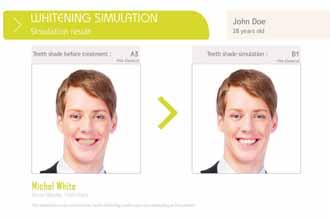

Download MeToo simulation software

Because everybody deserves to smile, ACTEON created MeToo!

Aesthetic Dentistry TREATING FLUOROSIS
Fig1: Initial view of a patient’s teeth affected by fluorosis
duration of the treatment and the unpredictable
results.16 Some dangerous features may be the post-treatment sensitivity, the increasing
temperature of the pulp and eventual discomfort concerning the gingival barrier 16
Case reports
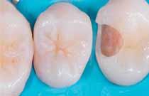
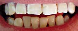
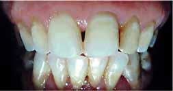
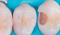
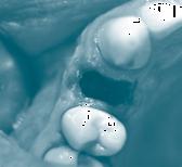
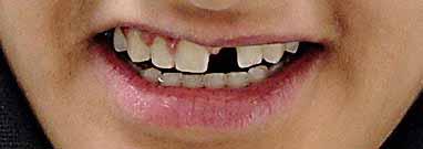
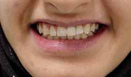
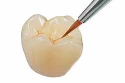
Case N°1
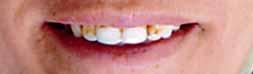
A 30 -year old male patient with complaints of dissatisfaction about the discolored teeth (Figure 1) came to the department of conser vative dentistr y. Patient gave histor y of discoloration from his childhood. No other relevant medical
histor y was reported by the patient. His oral
hygiene was good
Diagnosis: Dental fluorosis (Grade III: Brown line, pitting, and chipped off edges)
Treatment plan: Microabrasion followed by in-
office dental bleaching.
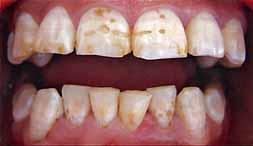
2: Light curing rubber dam application and Microabrasion with Opalustre ®
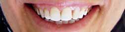
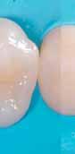
4: Final view of the clinical case after the end of the In-office bleaching (with Hydrogen Peroxide 35%) in 3 visits
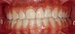
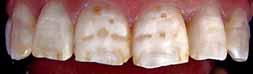
Case N°2: A 47-year old
with the discolored upper front teeth (Figure 5) came to the depar
childhood. Good oral hygiene.
Diagnosis: Dental fluorosis (Grade III: Brown line, pitting, and chipped off edges)
Treatment proposed was Microabrasion followed by in- office dental bleaching.
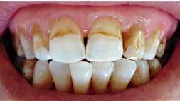
5: Labial view showing the severely discolored maxillary incisors.
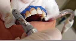
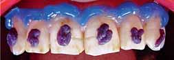
Fig 6: Microabrasion procedure ( Placement of the gingival barrier + application of the micro abrasion gel on the surface of the enamel of the upper anterior teeth for stain removal + the gel was rubbed with a slow speed rubber cup + the teeth was rinsed off and air dried Examination)
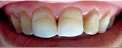
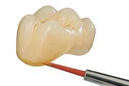
Fig 7: Intra-oral view of the maxillary incisors after two Treatments (4 visits): Microabrasion and in-office tooth whitening (Hydrogen Peroxide 35%)
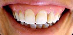
Fig 8: Post-treatment view illustrating the improved aesthetics following tooth whitening, microabrasion and restoration with resin composite of the two central incisors.
The new CEREC Omnicam combines powder-free ease of handling with natural color reproduction to provide an inspiring treatment experience for the patient. Discover the new simplicity of digital dentistry. Enjoy every day. With Sirona.
UOROSIS
Fig 9: sensitivity occurred and we stopped it with the use of Flor Opal ® gel
fig 9
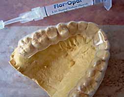
fig 10
Fig 10: After 14 months
and simple. It allows us to obtain excellent results
when treating superficial enamel stains.19
It does not require the use of anesthesia that is
why dentist can have a better relationship with
patients.20 The Opalustre™ microabrasion slurr y
(Ultradent Products Inc, Utah, USA) is composed
of 6.6% hydrochloric acid and silicon carbide
microparticles.11 According to (F Ng et al. 2007),
the Hydrogen peroxide (HP) is an oxidizing
agent which breaks down into free radicals that
will combine to create ox ygen and water. And
that the HP oxidizes, carbox ylates and lightens
chromophores, particularly within the dentine.
Most current in- office whitening systems are
based on HP solutions of 25 to 35 per cent.
Higher peroxide concentrations also have been
shown to be effective in tooth whitening;
however, these are professionally super vised to a
greater extent. (Kevin J. Donly et al. 2002) The
higher concentrations of peroxide lead to faster
rate of bleaching, but added to that a possible
Fig 11: Before
Fig 12: After
higher incidence of dental sensibility.8 While the
fig 11 fig 12
Discussion
Due to the recent increase in dental fluorosis, extensive research has been performed to understand the aetiology and pathogenesis of this systemic disease. (S. Ardu et al. 2009)
The aetiology is well established and based on the excessive consumption of fluoride during specific critical ages.7 Browne indicated that this critical period is 15 to 24 months of age for males and 21 to 30 months of age for females.18
Bleaching may represent a conser vative first approach for many cosmetic conditions. (Kevin J.
Donly et al. 2002) Conser vative treatment options
such as microabrasion and/or tooth whitening
can produce dramatic improvements in brown and yellow discoloration, providing a satisfactor y
interim result before more invasive procedures
are considered, if necessar y.(F Ng et al. 2007) The
microabrasion procedure is considered as safe
higher concentrations may reach the end point
sooner, they also “overshoot” the color and have
a greater relapse, and a longer time for the color to become stable. (Van B. Haywood, 2003)
One of the common adverses of vital bleaching
is dental sensitivity because the peroxide can penetrate the enamel, dentine and even the pulp
chamber 11 Other side effects such as irreversible pulpitis and pulp necrosis can occur with the uses of 35% of hydrogen peroxide.3
When the concentration of PH increase, the degree of penetration in dental structure will increase, also we can note a greater penetration in hypomineralized enamel 11 The patient in the second case reported sensitivity with the in- office whitening system containing 35% of HP that is why we opted for desensitizing gel Flor Opal ®.
This may have been because of the relatively higher concentration of HP used (35%).
According to (HO Heymann, 1997), we must wait a minimum of one week after bleaching procedure before placing any resin-bonded restoration on tooth structure because the strength of resin bonds to freshly bleached enamel and dentine
are reduced. A study of (Christian Hannig et al 2006) showed that bleaching procedure has an impact not only on surface micro -hardness of composites but also for deeper layers of adhesive filling materials. Bleaching
TREATING FLUOROSIS
versus Porcelain veneers? Dentists should make a choice for their patients between teeth whitening or veneers. If there is any regression in whitening after esthetic translucent veneers are placed, the teeth can be relightened from the lingual. (Van B. Haywood, 2003). In cases when teeth bleaching didn’t produce the expected result, the patient is confident since the most conservative options have been attempted first, and that porcelain veneers are the best option they have for an esthetic smile.8 We shouldn’t also ignore the minor cost of bleaching compared with the expensive cost of dental veneers that is why the bleaching procedure is usually the first choice for patients to retrieve better smile.8
This combined approach (Micro abrasion and in office bleaching) may be considered an interesting alternative to more invasive prosthetic techniques such as ceramic veneers. In addition to that, this minimal invasive technique allows acceptable aesthetic results and even a possible cost reduction for these patients.
1. OPTIMIZINGSMILECOMPOSITIONANDESTHETICSWITHRESINCOMPOSITESANDOTHERS
CONSERVATIVEESTHETICPROCEDURES DIDIER DIETSCH,DMD,PHD THE EUROPEANJOURNALOFESTHETICDENTISTRY VOLUME3, NUMBER1,SPRING 2008
2.VIABLEAPPROACHTOMANAGESUPERFICIALENAMELDISCOLORATION
NAVEEN CHHABRAAND KIRAN P.SINGBAL CLIN DENT. 2010 OCT-DEC; 1(4): 284–287.
3.SOME CURRENT PERSPECTIVESON TOOTH BLEACHINGAND MANAGEMENTOF TOOTH STAINS PR.M.S.GUTMANN - PR.J.L.GUTMANN
DENTALNEWS,VOLUME VIII,NUMBER III, 2001
4. TREATMENTOFINTRINSICDISCOLORATIONINPERMANENTANTERIORTEETHINCHILDREN
ANDADOLESCENTS ALYSON WRAYAND RICHARD WELBURY
INTERNATIONALJOURNAL OFPAEDIATRICDENTISTRY
VOLUME 11, ISSUE 4, JULY 2001, PAGES: 309–315
5.MANAGEMENTOF FLUOROSIS USING MACRO- AND MICROABRASION
HOWARD E.STRASSLER,DMD;AUTUMN GRIFFIN,DDS; AND MARGRIT MAGGIO DMD DENTALCETODAY COM
6. HTTP://WWW DENTALARTANDSCIENCE COM/MICROABRASION/MICROABRASION HTM
7.A COMBINEDCHEMO-MECHANICALAPPROACHFORAESTHETICMANAGEMENTOFSUPERFICIALENAMELDEFECTS
S.ARDU,N.BENBACHIR,M.STAVRIDAKIS,D.DIETSCHI,I. KREJCIAND A.FEILZER
BRITISHDENTALJOURNALVOLUME 206 NO. 4 FEB 28 2009 205
8.FREQUENTLY ASKED QUESTIONS ABOUT BLEACHING VAN B.HAYWOOD,DMDCOMPENDIUM / APRIL 2003 VOL. 24, NO. 4A
9.TOOTH WHITENING/BLEACHING:TREATMENT CONSIDERATIONSFOR DENTISTSAND THEIR PATIENTS ADACOUNCILON SCIENTIFIC AFFAIRS SEPTEMBER 2009 (REVISED NOVEMBER 2010) 2009 AMERICAN DENTAL ASSOCIATION
10.NONRESTORATIVETREATMENTOFDISCOLOREDTEETH:REPORTSFROMAN INTERNATIONAL SYMPOSIUM HOHEYMANN JADA 1997;128(6):710-711
11.AESTHETICMANAGEMENTOFSEVERELYFLUOROSEDINCISORSINANADOLESCENTFEMALE FNG,DJMANTON AUSTRALIAN DENTAL JOURNAL 2007;52:3.
12.DENTALFLUOROSIS:EXPOSURE, PREVENTIONANDMANAGEMENT JENNY ABANTO ALVAREZ , KARLA MAYRA P.C.REZENDE , SUSANA MARÍA SALAZAR
MAROCHO , FABIANA B.T.ALVES ,PAULA CELIBERTI , ANA LIDIA CIAMPONI
MED ORAL PATOL ORAL CIR BUCAL. 2009 FEB 1;14 (2):E103-7.
13.CLINICAL EVALUATIONOF IN-OFFICEAND AT-HOME BLEACHING TREATMENTS RUTA ZEKONIS,BRUCE AMATIS ,MICHAEL ACOCHRAN,SALAH EAL SHETRI,GEORGE JECKERT,TIMOTHY JCARLSON, OPERATIVE DENTISTRY, 2003, 28-2, 114-121
14.ACOMPARISONOF AT-HOMEAND IN-OFFICE BLEACHING VAN B.HAYWOOD DENTISTRY TODAY 2000:19(4):44-53
15.EXTERNALBLEACHINGTHERAPYWITHACTIVATIONBYHEAT LIGHTORLASER ASYSTEMATICREVIEW.BUCHALLA W,ATTIN T.DENT MATER 2007;23:586-96.
16.HISTORY SAFETY ANDEFFECTIVENESSOFCURRENTBLEACHINGTECHNIQUESANDA PLICATIONSOFTHENIGHTGUARDVITALBLEACHINGTECHNIQUE VAN B.HAYWOOD QUINTESSENCE INT 1992;23:471 488.
17.EFFECTOFBLEACHINGONSUBSURFACEMICRO-HARDNESSOFCOMPOSITEANDAPOLYACIDMODIFIEDCOMPOSITE CHRISTIAN HANNIG,SEBASTIAN DUONG, KLAUS BECKER,EDGAR BRUNNER,ELKE KAHLER,THOMAS ATTIN 2006 ACADEMYOF DENTAL MATERIALS PUBLISHEDBY ELSEVIER LTD
18.FLUORIDEMETABOLISMANDFLUOROSIS.BROWNE D,WHELTON H, O’MULLANE D. JDENT 2005; 33: 177-186
19.ASSOCIATIONOFMINIMALLYINVASIVEPROCEDURESFORTHEREHABILITATIONOFDISCOLOREDPERMANENTTEETH.CLINICALCASEREPORT MAIRA ATHAIDE,LEONARDO MUNIZ MAGAZINE FGMNEWS – EDITION 2- OCTOBER 2009
20.MICROABRASIONOFTOOTH4.4 WITHWHITENESS RM ENRICO COGO,PIETROSIBILLA,ROBERTO TURRINI MAGAZINE FGMNEWS – EDITION 2OCTOBER 2009
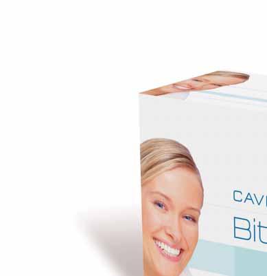
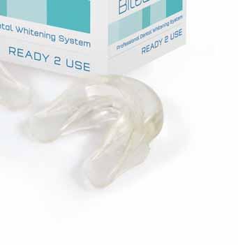
Cavex Bite&White Ready 2 Use Art. nr. BW030 (6 trays per box)
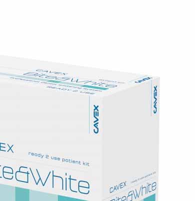
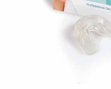
Cavex Bite&White Ready 2 Use – prefilled whitening trays
Home whitening has never been easier!
The Cavex Bite&White professional home whitening system is a safe, rapid and easy-to-use dental whitening system for use at home and is exclusively available through the dentist. Simply tear open the bag, place the tray in your mouth for an hour and enjoy the result. How easy is that? Cavex Bite&White contains 6% hydrogen peroxide (partly as carbamide peroxide), a material that has proven itself as a whitening agent on a global basis. Potassium nitrate has been added to sooth in case of sensitivity. www.biteandwhite.nl or dental@cavex.nl
READY 2 USE
READY 2 USE
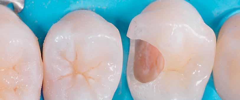
Case report
Dr. Marco Calabrese victoria.sowerby@dentsply.com
Clinical case
Class II restorations using composite resins present a number of technical problems, including the creation of a tight interproximal
contac t points.
A tight proximal contac t will balance the mesial
and distal forces and prevent food impac tion
For a while now, the use of preformed matrices
and separator rings, in combination with
wedges, has made it possible to obtain good restorations with satisfac tor y morphology.
An even more efficient system has recently been introduced, which combines preformed matrices, innovatively designed wedges, and nickel-titanium rings providing optimum separation that remains consistent over time.
This clinical case demonstrates the procedure
for conser vative restoration of teeth 15 and 14 using this innovative system.
The radiograph (Figure 1) shows that the
patient has a distal carious lesion of 14 and a
mesial lesion of 15 (Figure 2). After isolating the
operative field with a rubber dam (Figure 3), the
cavity on 14 is prepared (Figures 4 and 5)
fig 1
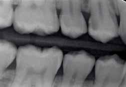
Fig 2
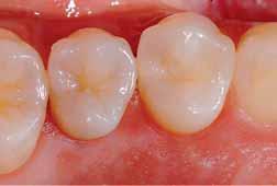
Fig 3
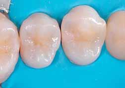
Fig 3: Isolating the operative field with a rubber dam .
Fig 4
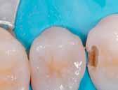
Fig 4: Accessing the carious lesion .
Restorative Dentistry
Case report
Fig 5: Finished cavity on 14.
Fig 6: Mesial lesion on 15.
Fig 7: Final restoration of 15.
Fig 8: Fitting the Palodent® Plus matrix to tooth 14, placement of wedge and ring .
Fig 9: Excellent adaptation of the matrix around 14 thanks to the ring design with Vshaped tines that accommodate the wedge perfectly .
5
through which the mesial lesion on 15 can be reached (Figure 6) and the tooth is restored (Figure 7). The Palodent® Plus matrix is
then placed on tooth 14, with simultaneous placement of a wedge and ring (Figure 8). The unique design of the nickel-titanium ring means
that the matrix fits per fec tly around the tooth
(Figure 9). Nex t comes the bonding phase; DE TREY® Conditioner 36 (36% phosphoric
acid) is applied first (Figure 10), followed by XP-
Bond® adhesive (wet bonding method). The
cavity is then par tially filled with SDR® – Smar t
Dentin Replacement (Figure 11); after waiting
a few seconds for the produc ts to self-level
per fec tly inside the cavity, it is polymerised
The distal wall is created with Ceram.X®mono+ composite, shade A2 (Figure 12). The matrix is
removed, leaving minimal amounts of excess material to be removed (Figure 13). The restoration is then completed using Ceram
X®mono+ composite, and finished (Figure 14).
10
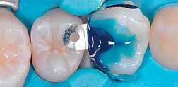
Fig 10: Bonding phase: applying conditioner (DETREY® Conditioner 36).
11
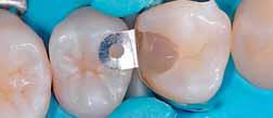
Fig 11: Partial filling of the cavity with SDR® - Smart Dentin Replacement.
Fig 12
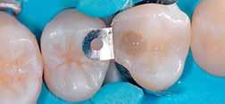
Fig 12: Creating the distal wall with CeramX®mono+ composite
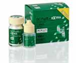
The power of advanced Glass Ionomer technology, with the simplicity of powder and liquid delivery.


Restorative Dentistry
Case report
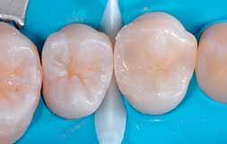
Fig 13: Applying a final layer of Ceram-X® mono+ shade A2 and removing the matrix. Note that there is minimal excess material to be removed during finishing.
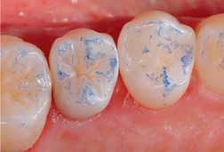
Fig 14: Final restoration of tooth 14, perfect interproximal contacts and bite check.
Conclusion
Composite restorations per formed using techniques designed for amalgams (round matrices) do not create the correc t anatomical
contours. However, with the use of Palodent®
Plus sec tional matrices it is now possible to create a proximal contac t that is elliptical in the
buccolingual direc tion about 1 mm apical to the
height of the marginal ridge. The interdental
papilla fills the space apical to this contac t and
prevents lateral food impac tion The Palodent®
Plus system makes it possible to create a good
tooth contour adjacent to the papilla, which is
necessar y to reproduce the original shape.
The use of the innovative DENTSPLY sec tional
matrix in Class II restorations allows the dentist to produce more predic table and morphologically
correc t restorations.
is glad to introduce

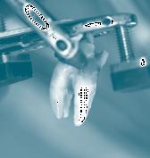
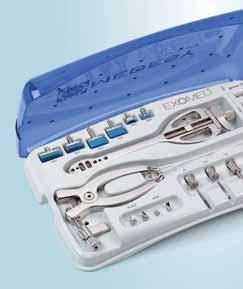


PERFECT ALVEOLUS after extraction with Minimal Trauma

EXOMED™ allows the extraction of teeth and roots with minimal trauma: it preserves the periodontal and alveolar tissues, which remain fully undamaged!
For additional information about EXOMED™ and to see all the clinical cases kindly visit www.exomed.it Or address your questions to exomed@medesy.it


The Ultradent Premium Class offers treatment units that you can configure as individually as your dream car. We are a modern dental company that flexibly manufactures our products based on your needs. In Germany. With outstanding quality. And absolute perfection. We are the experienced partner of completely satisfied dentists. Providing exceptional reliability and intuitive operation. With the newest technologies and multimedia. Ultradent Premium units will captivate you.

EXPERIENCE THE NEW STANDARD FOR OUR PREMIUM DENTAL UNITS: With vision U – the future tool for best practice.
EACH NEWULTRADENT PREMIUM CLASS UNIT NOW COMES WITH VISION U: The revolutionary, interactive, touchscreen-based multimedia system.
WITH VISION U, THE DOORS OF THE FUTURE OPEN TO YOUR PRACTICE:
> Large 21.5“ multi-touch screen – responds to „SmartTouch“ gestures
> Innovative patient entertainment – all informations are freely selectable
> Optical support – digital intraoral camera with autofocus and barcode reader, 2- and 3D x-ray viewer
> Simple quality assurance – automatic recording of all performance data before, during, and after treatment
> Integrated maintenance and service platform – reduces downtime and saves costs
Orthodontics
Case report
Dr. Faraj A. Sedeqi faraj_dds@yahoo.com
Abstract
Typically developmental anomalies are not uncommon in or thodontic cases. One of the most common contributor to malocclusion is hypodontia. Maxillar y lateral incisors are known
to be some of the most common congenital missing teeth This introduces an imbalance to the maxillar y and mandibular dental arch length 1 Treatment for the replacement of the
missing tooth depends on a number of fac tors, such as arch length, the number of missing teeth,
patient profile and smile line. Treatment options are either to close the space by positioning
the adjacent tooth into the missing tooth site, close it with a fixed bridge2 or an implant suppor ted crown Treatment plans for patients
with missing maxillar y incisors have traditionally
included either space closure or space opening for future restoration The most common objec tives to or thodontic space closure are that
the treatment outcome may not look “natural”, making retention questionable, also making the func tional occlusion compromised. Clinicians in general prefer to create space for the missing
lateral incisor with single -tooth implants or resinbonded bridges.3 -12 Implants are becoming the
treatment of choice for replacing missing teeth
One disadvantage with implants is that they
should not be placed until all residual growth has subsided That, however, means for most
or thodontics patients who are adolescent, have to wait 4- 6 years until the appropriate age of 18 for the implants. Maintaining the space for
a long time can be challenging especially with
teenagers if their cooperation and the retainer
wear is compromised. Gradually with time
the bone at the missing tooth site remodels thereby making it thin and may not suppor t
or be wide enough for the Implant.13 TAD or
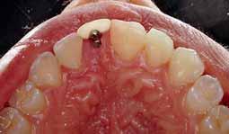
mini screws are commonly used in or thodontic cases for anchorage ; they are relatively cheap and have proved their success in suppor ting tooth movement. Or thodontic management of a congenital missing upper lateral incisor is the subjec t of this case repor t. The primar y or thodontic consideration was to maintain the space for the Implant and maintain bone integrity where a TAD with an acr ylic prosthetic tooth was placed as a space maintainer
Treatment Objectives
Ideally, the treatment objec tives for the final restoration of the missing tooth would be commenced only after there is a downward incline towards to any residual remaining growth 14 However, achievement of this objec tive would lead to fur ther bone loss, thereby making it unsuitable for either an implant or a fixed bridge prosthesis at the suitable age. Therefore to preser ve the bone height and thickness a temporar y anchorage implant was thought of.
The alternative method to reestablish normal
alveolar process is by tooth transplantation which
can inherent a potential for bone induc tion as indicated by B.U. Zachrisson et al 15
Treatment Procedure
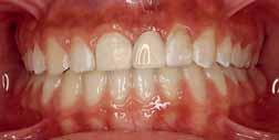
The clinical patient is a 14 year old female with congenitally missing left central incisor, generalized spacing on the upper anterior with
a midline shift to the left. On examination she
presented a straight profile and a Class I molar
relation. After the alignment and the midlines were coincided with the space opened up,
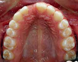
the braces were removed and spaces were maintained using removable retainers (fig. 1,2).
In order to avoid collapsing of the arch and
Orthodontics
2
Occlusal view (mirror image) space created for the TAD
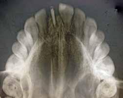
Occlusal Radiograph showing the TAD in place
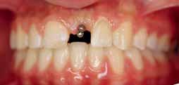
Fig: 3 TAD in place
further degradation of the bone height, a TAD
with an acr ylic tooth was shaped and trimmed accordingly to preser ve the present conditions (fig. 3,4,5). As the radiographs showed that there was enough bone thickness; the roots were diverged and it had sufficient bone shelf in the edentulous area. An 8 mm screw of IMTEC® was placed parallel to the adjacent roots in alignment with the adjacent teeth The head was placed and
checked for clearance from the lower incisors. The acr ylic tooth was trimmed and checked for occlusal interferences. Wax was placed in between to check for the approximate fit prior to placing it firmly with composite (fig. 4,5).
Treatment results
After the 3 months’ through the retention period, the TAD was placed The radiographs showed fairly good response without any occlusal interference from the lower anteriors. There
weren’t any significant loosening, infection or damage to the underlying structures (fig. 6,7). The shape and color of the prosthetic tooth had a significant matching to the adjacent teeth This helped in avoiding a collapse of the facial fullness and her profile. Thus improving her smile and gaining her confidence drastically (fig. 5,8,9).
Fig: 4
Occlusal view (mirror image) - TAD in place with the prosthetic tooth placed over it to check for interference
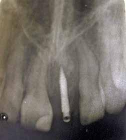
Radiograph showing the TAD in place
Micro-Series: welcome to a new dimension.
30% shorter and 23% lighter, Micro-Series offers perfect balance, exceptional power and versatility.
The new Bien-Air Micro-Series offers ultra-short contra-angles and straight handpieces combined with the new state-of-the-art MX2 LED micromotor. With its ultra-compact size, the MX2 offers the same performance as our world leading MX micromotor. This includes power, versatility, and perfect speed control, as well as auto-reverse and torque limitation capabilities ideal for endo.
Micro-Series: welcome to a new dimension.
photograph prior to TAD placement
photograph after firmly fixing the prosthetic tooth to the
Conclusion
Treatment of congenitally missing anterior teeth by the use of TAD is fairly a new idea which has little supporting literature. Our idea and our credit goes to Dr. John Graham for his concept on bone preser vation using TAD, which was presented in the AAO annual meet 2009. ® 3M IMTEC corporation
1.MCNEILL,R.W.;JOONDEPH,D.R.:CONGENITALLYABSENTMAXILLARYLATERALINCISORS:TREATMENTPLANNINGCONSIDERATION,ANGLE ORTHOD.43:24–29,1973.
2.TURPIN,D.L.:TREATMENTOFMISSINGLATERALINCISORS,AM.J. ORTHOD.DENTOFACIAL ORTHOP.125:129, 2004.
3.ROSA,M.;ZACHRISSON,B.U.:INTEGRATINGESTHETICDENTISTRYANDSPACECLOSURE INPATIENTSWITHMISSINGMAXILLARYLATERALINCISORS,J.CLIN. ORTHOD. 35:221–234,2001.
4.CARLSON,H.:SUGGESTEDTREATMENTFORMISSINGINCISORCASES,ANGLE ORTHOD 22:205-216,1952.
5.ASHER,C.;LEWIS,D.H.:THEINTEGRATIONOFORTHODONTICANDRESTORATIVEPROCEDURESINCASESWITHMISSINGMAXILLARYINCISORS.BR.DENT.J. 160(7):241-245, 1986.
6.TUVERSON,D.L.: ORTHODONTICTREATMENTUSINGCANINESINPLACEOFMISSINGMAXILLARYLATERALINCISORS.AM.J. ORTHOD.58:109-127, 1970.
7.MC NEIL,R.W.;JOONDEPH,D.R:CONGENITALLYABSENTMAXILLARYLATERALINCISORS: TREATMENTPLANNINGCONSIDERATIONS.ANGLE ORTHOD.43: 24-29, 1973.
8.ZACHRISSON,B.U.;MJOR,I.A.:REMODELLINGOFTEETHBYGRINDING.AM.J. ORTHOD. 68: 545-553, 1975.
9.SENTY,E.L.:THEMAXILLARYCUSPIDANDMISSINGLATERALINCISORS:ESTHETICSAND OCCLUSION.ANGLE ORTHOD.46:365-371, 1976.
10.ZACHRISSON,B.U.:IMPROVING ORTHODONTICRESULTSINCASESWITHMAXILLARY INCISORSMISSING.AM.J. ORTHOD.73:274-289, 1978.
11.BALSHI,T.J.: OSSEOINTEGRATIONANDORTHODONTICS: MODERNTREATMENTFOR CONGENITALLYMISSINGTEETH.INT.J.PERIODONT.RESTOR.DENT.13:499-505, 1993. 12.SABRI,R.:MANAGEMENTOFMISSINGMAXILLARYLATERALINCISORS.J.AMER.DENT ASSOC.130:80-84, 1999.
13.IKUYA,M.;YOICHI,T.;EISHIN,W.;HIROHIKO,S.;TADAHIKO,I.:INFLUENCEOF CORTICALBONETHICKNESSANDIMPLANTLENGTHONIMPLANTSTABILITYATTHETIMEOF SURGERY – CLINICAL, PROSPECTIVE, BIOMECHANICALANDIMAGINGSTUDY,BONE. 37: 776-780,DECEMBER 2005.
14.ZACHRISSON,B.U.:LETTERSTOTHEEDITOR;SINGLEIMPLANTS OPTIMALTHERAPYFOR MISSINGLATERALINCISORS?.AM.J. ORTHOD.126(6):A13-A15, 2004.
15.EWA,M.C.;ARILD,S.;BJØRN,A.;ZACHRISSON,B.U.:AUTOTRANSPLANTATIONOF PREMOLARSTOREPLACEMAXILLARYINCISORS:A COMPARISONWITHNATURALINCISORS.AM J. ORTHOD.118: 592-600,2000.

Table top sterilizers in a clinical setting have
been around since the time of Louis Pasteur
The beginnings of preser vation and sterilization techniques go back to ancient years. Aristotle
recommended to Alexander the Great, his
troops should boil water before they drank
it. The beneficial effect of passing surgical instruments through flame was well known to ancient civilizations. Heat as a preser vative method in medical industr y was introduced in 1809 by Nicholas Appert, with his method of sealing vegetables and fruits in glass jars and
then heating them. Louis Pasteur recommended to French vintners heating the new wine at 55°C in the absence of air in order to avoid serious problems. Later he noticed that moist heat was more effective than dr y heat. The proposition of sterilization (1879) was simple enough: an integrated chamber with burners located at the bottom, and a serpentine connection to
an adjacent water tank that would deliver a fixed amount of distilled water to the chamber at the outset of each cycle…the autoclave was developed. An autoclave is a device used to sterilize equipment and supplies by subjecting
them to high pressure saturated steam at set temperature for a set time depending on the size of the load and the contents. The name comes from Greek auto - ultimately meaning self, and
Latin clavis meaning key — a self-locking device.
Under this 19th centur y theor y, the burners would then bring the temperature up to 121°C, the internationally accepted minimum for sterility to take place, and the resulting steam would permeate ever y corner, nook and cranny within and annihilate any pathogenic or non-pathogenic organism in this enclosed environment. A brilliant idea indeed that has been with us since the 1880s and that allowed for the notion of sterile instruments at or in the vicinity of clinical pointof-use to be actively pursued by the pioneers of microbiology. In order to understand the implications of cross contamination, and because of the forgetful nature of the human brain, it would be prudent to review basic definitions.
Sterilization (when applied to the eradication of microorganisms): The total annihilation of pathogenic and non-pathogenic organisms in any given environment rendering a product free of viable microorganisms.* Disinfection: The reduction in numbers of pathogenic and nonpathogenic organisms in any given environment that may or may not be detrimental to human health.** As we can see, the first (sterilization) is an absolute as there are no degrees of sterility inherent to the definition, whereas the second (disinfection) is not an absolute, and we can thus speak of low, medium, and high levels of disinfection This indicates that in the proposition “total annihilation” the key word is “total.” And here we begin to understand why steam has made it as the predominant sterilizing agent. A word on steam: It is commonly accepted that the ideal sterilization process is one that can be used between patient procedures; one that does not damage or corrode heat-sensitive instruments; one that is inexpensive; and one that consistently


Scican
penetrates narrow orifices, channels, and the lumen of instruments of certain complexity. All of the above makes pressurized steam the
perfect candidate for the job as it is cost-effective, presents with no environmental concerns, and it proves to be highly per vasive. An important concept to remember regarding steam is that the increase in temperature from boiling point to 134°C is instant, and that the temperature of moist heat can be raised by increasing chamber pressure, with the accepted formula as follows: 121°C sustained for thirty minutes will bring about sterility, but if we are able to increase the temperature to 132°C, we may decrease the time of exposure down to four minutes with the same result, that is, sterility. The main objection to the traditional approach of steam in an integrated chamber was exposed in 1989 by Professor
Fodder, who demonstrated that leaving 5 percent or more of air in the chamber effectively prevents a true claim of sterility. The rationale behind this
is that steam tends to spread out in layers, and
because steam is lighter than air, all air must vacate
the chamber in order to avoid the formation of airpockets, into which steam could not penetrate.
Obviously, without such penetration sterilization could not happen and would be particularly
absent within the hollow of instruments. So, it is established then that air removal is a condition sine qua non for sterility to occur. Effective air removal was achieved in 1990 with the advent of STATIM®, a revolutionar y design that moved away from the integrated chamber approach to the removable cassette-based chamber, where sterile instruments can be aseptically transported right to the point of use. This is air removal by positive pressure pulse displacement (dynamic air removal), where a steam generator injects pressurized steam into the chamber. Saturated steam*** now forces the air out through a valve, with the remainder of the air being removed by opening this valve at inter vals. The effect is the creation of positive pressure pulses.
EN13060
EN13060 is the European Standard for small steam sterilizers, i.e. steam sterilizers whose chamber volume does not exceed 60 litres. As a longstanding, active member of the working group responsible for this standard, SciCan is intimately familiar with the requirements of EN13060+A2
2010
This European Standard specifies the performance
requirements and test methods for small steam sterilizers and sterilization cycles which are used for medical and dental purposes or for materials that are likely to come into contact with blood or body fluids. This Standard is intended for sterilizer manufacturers and is also used and referenced by many non-European health/regulator y authorities and sterilizer users. At the heart of this Standard is the definition of the types of approved cycles:
B, S, and N, as is defined in the beginning of EN13060**** :
B cycle – The sterilization of all wrapped or nonwrapped solid, hollow load products type A and porous products as represented by the test loads in the standard
S cycle – The sterilization of products as specified by the manufacturer of the sterilizer N cycle – The sterilization of non-wrapped solid products.
B-cycles use a vacuum pump to create a vacuum that ensures air removal before the chamber is pressurized with steam. The process is known as fractionated vacuum (negative pressure) and, relative to gravity displacement cycles, allows for better steam penetration through the entire load, but it must be tested daily for adequate air removal by using an approved process challenge device.
S-cycles can use a variety of technologies to ensure air removal before the chamber is
pressurized with steam. The STATIM Cassette Autoclave, as stated above, uses positive pressure pulse displacement


Scican General Dentistry
(dynamic air removal). S-cycles do not require
daily air removal testing. In accordance with
EN13060:2004+A2 2010 Section 7.1, STATIM
sterilizers are tested on a microbiological basis and have proven to consistently achieve a sterility
assurance level of 10 - 6 (or a 6 -log reduction in microorganisms) for a variety of loads (solid, hollow, hinged, etc.). Furthermore, as required by
Section 7.2 and 7.3, STATIMs 2000S and 5000S
have passed Type testing, and each unit shipped has successfully undergone Works testing (the results of the Works test are provided with each unit).
N -cycles do not use multiple steam purges
during the preparation phase of the cycle that
would ensure air was removed from cavities of
instruments such as found in dental handpieces.
Here the trapping of air is a real concern and the
formation of air pockets a possibility and therefore
N -cycles, similar to gravity displacement cycles are
only adequate for solid instruments.
Microbiological Testing
Because STATIMs are S-cycle machines, they are ver y likely the sterilizers that have the most
microbiological test data to prove their efficacy in destroying microorganisms, especially in medical
and dental environments, while other autoclave
manufactures (B-cycles) rely on EN867-5:2001 approved PCDs (process challenge device) to claim load processing. STATIM users can therefore have
the utmost assurance in the sterilization efficacy of the STATIMs. They have been microbiologically
tested and proven to successfully and effectively
sterilize a variety of instruments. A PCD is a
mechanical device which simulates the worst case of conditions for attainment of the specified sterilization conditions within the items to be sterilized as defined by EN867-5:2001, definition 3.2. The device is constructed so that
a biological or non-biological indicator system
can be placed within the device in the position which it is most difficult for the sterilizing agent to reach The design of the process challenge
device depends on the nature of the goods to be sterilized and the sterilization procedure.
The performance of the STATIM autoclaves
have been validated via microbiological testing,
conducted by a well respected researcher and
leader in the field of infection control To regularly
validate the STATIMs and ensure they continue to attain the specified sterilization conditions,
the STATIM Process Challenge Device (PCD) has been designed in order to demonstrate that the sterilization parameters required and validated
during microbiological testing have indeed been obtained This device tests the unit and ensures that the mechanical components and software controls are functioning correctly and match those of the units tested during microbiological testing. In sum, the removal of the air***** from the chamber prior to the commencement of “killing” time is the fundamental consideration
in selecting the best autoclave for any clinical
practice. The methodology through which this
removal is achieved is of secondar y importance.
The fact remains, in spite of the opinions of manufacturers, advocates, partisans and others, that both the S and the B cycles expel the air
from the sterilization chamber to such an extent where sterilization of hollow instruments (such as dental handpieces) can be attained. Where the removal of air through positive pressure (STATIM) establishes its dominance is in the reduced duration of the cycle, aspect this rightly coveted by most professionals, since the instrument turnaround time could be as short as 10 minutes. This is achieved as a result of having the technological capability of injecting steam at will, and of creating conditions of sterility (temperature and pressure) through a chamber whose walls are extremely thin. In mechanical terms, the effect of this approach is a gentler sterilization process for your expensive instruments.
* Definitions of sterility in the medical literature var y in composition depending on the source, but the definition given here encompasses general
consensus.
** Definitions of disinfection in the medical literature var y in composition depending on the source, but the definition given here encompasses general consensus.
*** EN 13060 2004+A2 2010, § 3.34
**** EN 13060 2004+A2 2010, Table 1
***** EN 13060 2004+A2 2010, § 3.3


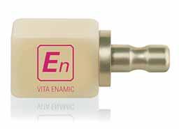
In January 2013, a new generation of materials for chairside treatment using CAD/CAM technology were made available. The future of chairside CAD/CAM restoration is VITA ENAMIC (Fig. 1). A
Fig. 1: With its dual-network structure, the VITA ENAMIC hybrid ceramic represents a new class of materials.
composite material that is unique worldwide in combining the benefits of conventional ceramic and composite materials. Both scientific studies and initial clinical experience confirm that with the VITA ENAMIC hybrid ceramic, a new dimension in stability, reliability, precision and cost-efficiency can be achieved.
Material concept
With the development of VITA ENAMIC, a whole new approach has been adopted, creating a hybrid ceramic comprised of a dominant ceramic network reinforced by a polymer network structure. With both networks fully integrated with one another, VITA ENAMIC offers considerable benefits for patients and for practice and laboratory use –from a lower tendency towards brittle fracture than pure ceramics, through to greater abrasion resistance than composite materials.
Findings of materials science
With flexural strength values of approximately 150-160 MPa, the results for VITA ENAMIC
are within the same range as those for silicate ceramics. And at 30 GPa, the material also offers a modulus of elasticity comparable to that of human dentin. Up until now, no dental restoration material has matched this “natural” elastic range. The result is unusually high stability as demonstrated by internal and external studies. Thanks to the elasticity provided by the integrated polymer network, VITA ENAMIC absorbs the applied load, achieving the best results (approx. 2890 Newtons, see Fig. 2) in a fracture load study
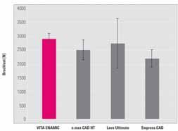
Fig. 2: In this test setup, VITA ENAMIC demonstrates the highest fracture load of approx. 2890 Newtons and the lowest standard deviation.
Source: Internal study, VITA R&D
of any of the materials tested. This result also correlates with the study results published by Dr. Petra Güß of the clinic for oral and maxillofacial surgery at the University of Freiburg. Here it was found that when a dynamic load is applied in a chewing simulator, VITA ENAMIC crowns demonstrate a 100 % survival rate, both for walls of normal and of reduced thickness (Fig. 3). As a measure of the reliability of a material, the Weibull modulus indicates the benefits of VITA ENAMIC particularly effectively. An internal study conducted by the VITA Research & Development
The first hybrid ceramic with dual network structure for unsurpassed absorption of masticatory forces
VITA ENAMIC sets new standards for resistance by combining strength and elasticity and providing unsurpassed absorption of masticatory forces. VITA ENAMIC ensures utmost dependability and efficient processing for dental practices and laboratories. And patients feel
that VITA ENAMIC restorations are identical to natural teeth. VITA ENAMIC is particularly suited for crown restorations in the posterior area and minimally invasive restorations. More information at www.vita-enamic.com facebook.com/vita.zahnfabrik
The En formula for success: strength + elasticity = reliability ²
Esthetic Dentistry
Fig. 3: The survival rate of VITA ENAMIC crowns with walls of normal and reduced thickness is 100%.
Source: University of Freiburg, Dr. Güß
department shows that at a value of 20, the
Weibull modulus for the VITA ENAMIC hybrid
ceramic is more than double that of comparable
materials for the fabrication of monolithic single-
tooth restorations (Fig. 4).
Fig. 4: Of the materials examined in this test, VITA ENAMIC offered the greatest reliability. The Weibull modulus is 20.
Source: Internal study, VITA R&D
In practical application, the superior properties
of the new hybrid ceramic make it ideally suited to crown restorations in areas with high occlusal
that in the same way as silicate ceramics, VITA
ENAMIC also facilitates superior-quality etching
using hydrofluoric acid gel This is an important
factor in ensuring permanent and tight contact
adhesion between the restoration and the tooth substance.
VITA ENAMIC will be available initially in block
size EM -14 (12 x 14 x 18 mm), as well as in the
shades 0M1, 1M1, 1M2, 2M2 and 3M2, and in
two degrees of translucency. The hybrid ceramic
can be processed in the usual manner using the
CEREC and inLab MC XL systems, for example.
Software version V4.0 or higher is prerequisite.
For easy, efficient pre-polishing and high-gloss polishing of VITA ENAMIC restorations that is
gentle on the material, a special polishing set is
provided (Fig. 6).
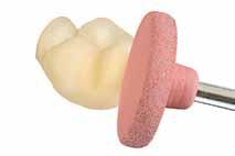
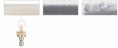
Fig. 5: VITA ENAMIC exhibits high edge stability even in areas with thin margins. Top view: 30° wedges: left: VITA ENAMIC, right: e.max
CAD
load, and facilitate reduced wall thickness for minimally-invasive restorations. They also enable greater precision, improved edge stability and, as a result, finer, more accurate milling results than
previously possible using conventional CAD/CAM ceramics. SEM images illustrate the difference compared to conventional ceramics (Fig. 5).
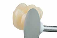
Fig. 6: These instruments, which have been developed especially with VITA ENAMIC in mind, allow superior, plaque-resistant surface results to be achieved.
At the same time, the innovative composite
material can also be milled more cost-effectively
than comparable CAD/CAM materials – the
milling times for VITA ENAMIC restorations are
the shortest both in normal and in fast milling mode, while also ensuring a longer ser vice life for diamond milling tools. It is also important to note
The VITA ENAMIC STAINS KIT, which contains six stains as well as accessories (Fig. 7), can be used for characterization The stains are bonded to the restoration as part of a polymerization process.
Surface sealing can be performed using the chemical glaze material VITA ENAMIC GL AZE, which increases the durability and brilliance of the shade in the oral environment. Processing could not be easier – simply condition the surface of the
7: Simply apply the VITA ENAMIC stains to the restoration and polymerize – that’s all that’s required to quickly characterize the shade of VITA ENAMIC restorations.
restoration, mix and apply the shades, perform intermediate polymerization, apply chemical glaze and complete final polymerization. Firing is not required.
Summary
Thanks to the dual ceramic-polymer network, the new VITA ENAMIC composite combines the benefits of ceramic and composite materials in one outstanding product, providing a quantum leap in the development of CAD/CAM materials. It is approved for single-tooth restorations such as inlays, onlays, veneers and crowns, and is also distinguished not least by the superior comfort it offers to patients, thanks to material properties similar to those of natural dentition. fi
With Fuji IX GP Extra in powder-liquid, GC, the glass ionomer world leader, is providing the most advanced GI technology to Powder Liquid users.
Actually, advances in glass technology have resulted in the development of GC Fuji IX GP EXTRA, a high strength glass ionomer cement with a new generation of glass filler which gives the restorations a very high translucency that changes little over time. The result is natural aesthetics never before achievable with conventional glass ionomer. GC special glass filler used in GC Fuji IX GP EXTRA releases three times more fluoride than conventional glass ionomers so it provides even better protection against caries. The comfortable working time combined with a fast setting time mean you can start finishing and polishing just two and a half minutes after mixing.
The indications of Fuji IX GP Extra are for permanent and temporary teeth. It can be used for Class I and II restorations in children’s teeth, non-load-bearing Class I and II restorations in adult teeth, Class V and root surface restorations, core build-up and for use with a composite or an inlay in the immediate or delayed sandwich technique.
Find out more about Fuji IX GP Extra powder-liquid at www.gceurope.com


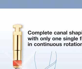
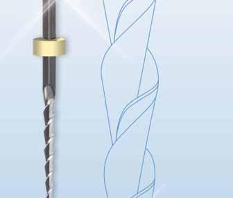
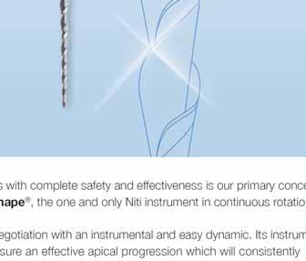


We go
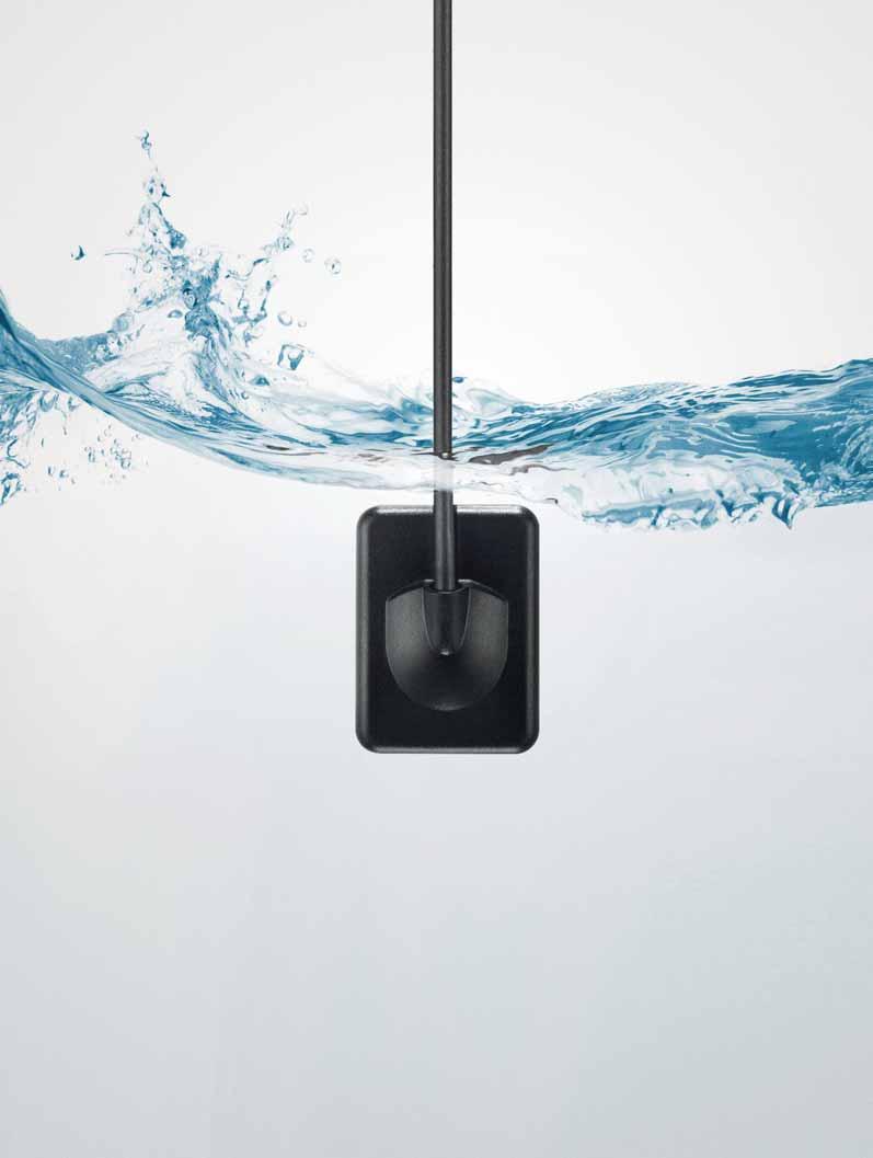
Over 30 years ago, we redefined dental intraoral radiography with the invention of the RVG sensor. Since then, every sensor we make is even stronger than the last. Our years of testing and continuous development assure you of a level of durability and image quality that is simply second to none.
Instant, high-quality image output with an unsurpassed true resolution of up to >20 lp/mm
Waterproof and fully submersible sensor head for optimal disinfection and increased durability
Flexible and robust cable, designed and tested to withstand strong bends and pulls
Shock-resistant casing and silicon padding offer extra protection from falls, bites and other damage
Available from participating Dealers. Call +44 1442 838908 or e-mail ernesto.jaconelli@carestream.com to arrange a


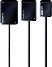


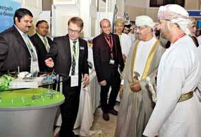
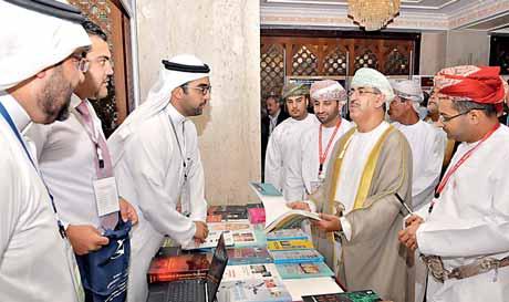
OMAN INTERNATIONAL DENTAL CONFERENCE
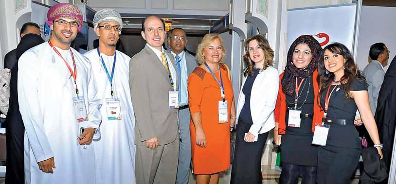


COMPRESSED AIR SUCTION IMAGING DENTAL CARE
HYGIENE


Tornado – the new generation of compressors from Dürr Dental
Dürr Dental, the inventor of oil-free dental compressors, presents an unbelievably quiet and powerful compressor for dentistry in the form of the new “Made in Germany“ Tornado.
▪ Oil-free, dry, and hygienic
▪ One of the quietest of its kind
▪ Dust- and Sandresistant
▪ Antibacterial inner tank coating
▪ Low-maintenance thanks to membrane-drying unit
Duerr Dental Middle East, P.O.Box: 87355, Al Ain - U.A.E., Mobile: +971 (0) 50 - 550 84 12, Fax: +971 (0) 3 767 - 5615, email: koll.m@duerr.de


MASTERDENTALTECHNICIANSAND DENTISTS, WILLHAVETOWORKTOGETHER MORECLOSELYINTHEFUTURE

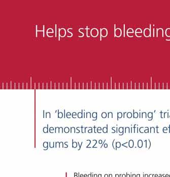
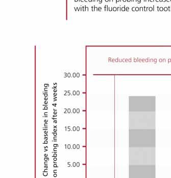
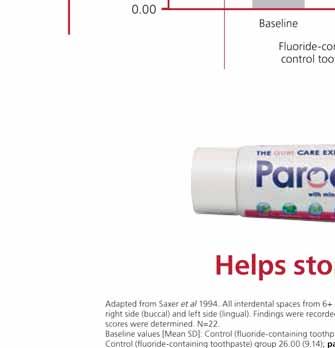


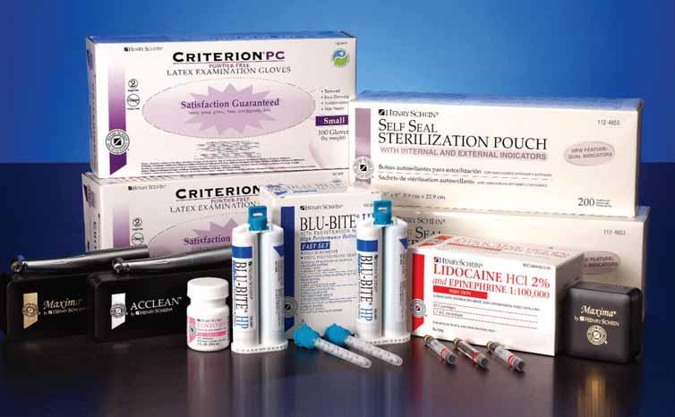
Henry Schein Brand products offer our clients maximum value without compromising on quality.
We offer quality products you can trust to fulfill your practice merchandise needs—each bearing the Henry Schein Seal of Excellence, your guarantee of satisfaction.
Henry Schein, Inc. is the world’s largest provider of health care products and services to office-based dental, medical, and animal health practitioners.


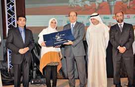


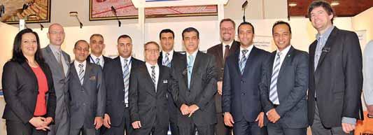


New Philips Zoom offers advanced light technology that gives you more control and your patients even greater results. And with a worldwide public awareness campaign to drive patients to you and new programs to help you easily integrate Philips Zoom WhiteSpeed light-activated whitening into your practice, you’ll have the answer to the confident, beautiful smile your patients are asking for.
Ask about the new Philips Zoom WhiteSpeed today. Visit philipsoralhealthcare.com. For more information, contact your local sales representative or call +971 50 643 1707.


April 24-26, 2013 Riyadh, KSA
PROF. ABDULLAH AL SHAMMERY THE RECTOROFTHE RIYADH COLLEGESOF DENTISTRYAND PHARMACY
www.facebook.com/dentalnews1


DR. ESAM TASHKANDIGIVINGTHE TROPHYTO DR. ANDREA RICCI


AHMAD KHOJARECEIVINGTHETROPHYFROM PROF. ABDULLAH AL SHAMMERY TROPHY TO DR. FEDERICO FERRARISFROM ITALY
The Second International Quintessence Dental Arab Congress jointly organized by the Riyadh Colleges of Dentistr y and Pharmacy (RCsDP) and the Quintessence International Publishing Group and the 8th Riyadh International Dentistr y Meeting (RIDM) closed on 26 April 2013. The meeting was held at the Al Faisaliah Hotel in Riyadh City and covered eleven scientific papers presented by internationally known dental experts and authors of dental books from Switzerland, Ital
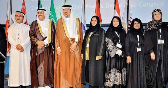


 PICTURES FROMTHE EXHIBITION FLOOR
LEFTTORIGHT DRS. MASSIRONI,RASPERINI,RICCI,FERRARIS
PICTURES FROMTHE EXHIBITION FLOOR
LEFTTORIGHT DRS. MASSIRONI,RASPERINI,RICCI,FERRARIS


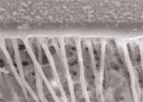

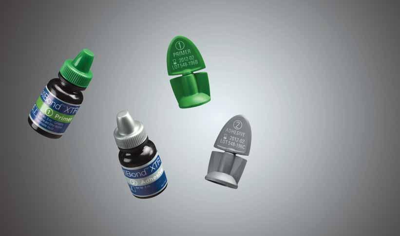
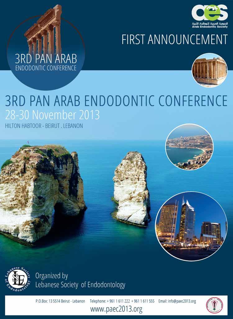


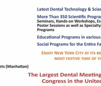

SlimCollection
Hu-Friedy’s SlimCollec tion, which consists in a full line of sleek new or thodondic instruments that feature smaller, thinner, lighter working ends for more maneuverability.
Slim collec tion consists in eight new instruments, which guarantee the or thodontist a more efficient and precise working method The pliers, tweezers and cutters of the new produc t line are considerably narrower, thinner and lighter than conventional produc ts to greatly improve intraoral adaptation, flexibility and view. The result: More maneuverability, more patient confor t and time savings for doc tor and patient.
For more information www.hufriedy.eu
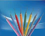
AngleFix Concept
Prosthetic Restorations on angled tioLogic© Implants
The AngleFix concept provides the tioLogic© implant system with yet another indication it enables edentulous patients to be treated with immediate restorations such as screw retained bridges or a bar construc tions in the maxilla or mandible. It consists of various fully compatible prosthetic components, which enable the tioLogic© implants to be inser ted at an angle, in order to avoid augmentation in the posterior region and enable anatomically critical areas to be treated with care. The prosthetic suppor ting sur face is displaced in a distal direc tion, the prosthesis is additionally suppor ted and the local bone substance is put to best possible use. The AngleFix abutments are available for the implant lines S, M and L in straight (0°) and angled at 18° and 32°. The angled abutments can be positioned exac tly using the integrated PentaStop®. The design of the abutments is such, that implant axis deviations of up to 44° to one another are possible. There are closure screws, impression copings, laborator y analogs and acr ylic and titanium copings available for the ex tended prosthetic restoration There are two angle aids available in 18° and 32° for the exac t alignment of the implants. Alternatively, the implant axis can also be determined using the navigation system tioLogic© pOsition and the drilling template made with this. If you should have any questions, the Dentaurum Implants Hotline is available with experienced implantologists and dental technicians to give assistance. For more information www.dentaurum-implants.de
The Next Generation of the Endo Gold Standard
PROTAPER NEX T is the newest innovation to the DENTSPLY Maillefer PROTAPER system, which has been the gold standard in endodontics for many years. It is still based on the
PROTAPER philosophy, including a single file sequence for all clinical cases, variable tapers for optimised crown-down technique and commonly approved apical finishing diameters.
PROTAPER NEX T has an off-centred rec tangular cross sec tion, providing a snake -like “swaggering” movement of the file in the root canal The DENTSPLY proprietar y M -WIRE
NiTi provides improved flexibility to the file as well as greater resistance to c yclic fatigue.
As a result, PROTAPER NEX T covers more difficult clinical cases, improves safety and also
shor tens the shaping time.
For more information www.dentsplymea.com
Exomed™
From our R&D depar tment a new company has been created, Medesy Engineering, whose unique mission is to develop new materials and new produc ts.
E xomed™ is the ex traordinar y result of Medesy Engineering, E xomed™is an instrument which allows the ex trac tion of teeth and roots with minimal trauma.
Among the most impor tant advantages:
- The periodontal and alveolar tissues remain fully undamaged after the ex trac tion,
the neighbouring tissues are prac tically intac t after the operation
- No need of forceps and root elevators, nor to per form a luxation and syndesmotomy before ex trac tion!
- The implant inser tions are possible even immediately after the ex trac tion:
the alveolar sites have no lacerations
- The patient will recover from the ex trac tion in a much shor ter time.
- The ex trac tion of roots is really easier and faster THE MORE YOU KNOW I T, THE
MORE YOU USE IT! SIMPLE & REVOLUTIONARY. EXOMED™, SIMPLY NO STRESS. For more information www.exomed it
NEW! Cavex Bite&White Ready 2 Use ‘pre-filled whitening trays’
The Cavex Bite&White Ready 2 Use professional home whitening system is a fast, safe and above all ver y simple whitening system for use at home. Just open the packaging, place the pre -filled tray in your mouth and enjoy the result after an hour. Mere child’s play! The highly viscous Cavex Bite&White Ready 2 Use whitening gel contains 6% hydrogen peroxide (par tly as carbamide peroxide), a sustainable and ac tive combination of effec tive whitening substances with a proven reputation worldwide as an effec tive whitening agent. Adding potassium nitrate avoids sensitivity. The unique ‹one -size -fits-all› Dr yflex® whitening tray is made of a special type of plastic which the mouth tolerates well Thanks to its 95% compatibility, the whitening tray is suited to almost ever yone. Cavex Bite&White Ready 2 Use complies totally with new European legislation for teeth whitening agents. For more information www.biteandwhite.eu
VITA ENAMIC the first hybrid dental ceramic in the world
VITA Zahnfabrik have created a new generation in ceramic materials: VITA ENAMIC is the first hybrid dental ceramic in the world with a dual-network struc ture that combines the best of what ceramic and composite materials have to offer The CAD/ CAM blocks are not only suitable for fabricating conventional inlays, onlays, veneers and crowns for anterior and posterior applications, but they can also be used for minimally-invasive restorations such as non-prep veneers and for restorations in areas where space is limited For more information www.vita-zahnfabrik.com

Self-Ligating Bracket


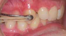
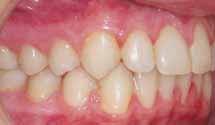
* Typical case: Patient 16 years Start of treatment, prior to placement of Carriere Distalizer Appliance 5.10.10
With its non-invasive design, the Carriere Distalizer Appliance corrects Class II malocclusion at the beginning of treatment, prior to bracket placement when patient motivation is highest. Call us today at 888.851.0533 or visit us online at OrthoOrganizers.com. Carriere Ortho 3D A FREE App. for iPads, iPhones, and Android tablets and phones