Volume 1 Number 2 December 2025/January 2026 ISSN 3088-6775
19
EDITORIAL
Dr David McReynolds 2 12 15 3 8 7 20
Planning ahead: considerations for our ageing population
RESEARCH
Management of lip paraesthesia post root canal treatment via unilateral sagittal split osteotomy: case report
Ahmed AlMajmaie, Laith Al Sabek, Ahmed ElMinshawi, Tom Barry
QUIZ
RESEARCH
Learning style preferences of undergraduate dental students in Northern Ireland
Ryan McConville, Marc Lewis Emrys Edwards, Amanda Willis
CLINICAL TIPS
Facially driven and digitally guided surgical crown lengthening in the contemporary management of amelogenesis imperfecta
Dr David McReynolds, Dr Ioannis Polyzois, Mr Alexander Lichtmannegger MDT, Mr Enrico Steger MDT
CLINICAL FEATURE
Procurement in dentistry – sustainable purchasing of equipment and consumables
Dr Brett Duane, Dr Eamon Croke, Dr Michaela Dalton, Dr Nick Armstrong
NEW DENTAL SCIENCE
Abstracts from the latest dental literature
RESEARCHER PROFILE
Looking at the bigger picture
Dr Cristiane da Mata
BDS MFD (RCSI) Dip TLHE MPH
Phd FFD RCSI
Honorary Editor
journaleditor@irishdentalassoc.ie
Deputy Editor
Dr David McReynolds BA BDentSC MFDS RCSEd DChDent (Pros) FFD RCSI
Editorial Board
Dr Meriem Abbas BDS (NUI) MFDS RCSEd PGDip TLHE
Una Farrell Dip Dental Hygiene
Dr Catherine Gallagher MB BCh BAO BDS NUI FDS RCSEng FFD RCSI
Dr Geraldine McDermott BA BDentSc MFDS (RCSI) PGradDip ConSed (TCD) MSc Healthcare Leadership (RCSI)
Dr Clair Nolan BDS (NUI) MSc (Endo) U. Lond
Dr Adedeji Daniel Obikoya BChD MFDS (RCSI) MSc
Dr Judith Phelan BDS (NUI) MDS (NUI) MSc (U Lond) MRD (RCS Eng and Glas)
Dr Patrick Quinn BCL BDS LLM MDPH
Dr Catherine Vaughan BDS (NUI)
International Editorial Board
Assoc. Prof. Lamyia Anweigi BDS MMedSc MFDS PhD (Qatar)
Dr Ana Cecilia Diniz Viana BDS MD PhD (Brazil)
Dr John Macken BDS PGCertMEd PhD MFDS RCSEng ACIEA FHEA (UK)
Prof. Leonardo Marchini DDS MSD PhD (USA)
Dr Ramiar Karim BDS MSc Paed Dent (Germany)
Dr Elaine Smyth BA BDentSc DChDent (USA)
Prof. Murali Srinivasan BDS MDS MBA MAS PD (Switzerland)
Prof. Sayaka Tada DDS PhD (Singapore)
jida.scholasticahq.com
Irish Dental Association
Unit 2 Leopardstown Office Park, Sandyford, Dublin 18.
T: +353 1 295 0072
F: +353 1 295 0092 www.dentist.ie
Planning ahead: considerations for our ageing population
We are delighted to feature a guest editorial from Prof. Gerry McKenna, Chair of Oral Health Services Research and Gerodontology in the Centre for Public Health at Queen’s University Belfast.
Dental students are taught the importance of patient assessment and long-term treatment planning. I recall an article suggesting that we think about where our patients will be in five and ten years’ time, and factor this into our decisions.1 Changing epidemiology has made this increasingly important, with the emergence of a rapidly ageing population. While older patients traditionally attended to have new complete dentures constructed, we are now seeing the emergence, and consequences, of a partially dentate older population. Older patients increasingly suffer from a range of systemic diseases, including cardiovascular disease, diabetes, osteoarthritis, and cancer. Alongside personal and safety concerns, the management of these diseases can be a tipping point for older patients becoming dependent on others for care or even entering a care home. Currently, more than 25,000 older people live in registered care homes in Ireland, with significant numbers also receiving care in their own homes. About half of all care home residents have at least some of their own natural teeth, but their oral health is much worse than their peers in the community. With increasing age, residents’ ability to care for their mouth deteriorates, polypharmacy can lead to xerostomia and diets are oen rich in sugars. These factors all increase the risk of oral diseases and directly impact on systemic comorbidities.2
While in some areas of the country our colleagues in the Health Service Executive (HSE) or the Community Dental Service (CDS), plus a diminishing number of dedicated general dental practitioners, provide excellent treatment for these patients,
References
1. Jacobs DJ, Steele JG, Wassell RW. Crowns and extra-coronal restorations: considerations when planning treatment. Br Dent J. 2002;192(5):257-267.
2. Pretty I. The life course, care pathways and elements of vulnerability. A picture of health needs in a vulnerable population. Gerodontology. 2014;31(Suppl. 1):1-8.
3. Tsakos G, Brocklehurst PR, Watson S, et al. Improving the oral health of older people in care homes: results from a randomised feasibility study. Community Dent Oral Epidemiol. 2025;53(4):413-423.
4. McKenna G, Allen PF, Hayes M, DaMata C, Moore C, Cronin
many do not receive even basic oral care. Efforts have been made to establish programmes to train care home staff in assisted tooth brushing programmes and preventive care, but significant challenges remain.3
These concerns should refocus dentists’ treatment planning decisions to consider conservative, evidence-based and patientcentred options. A strong body of evidence shows that simple treatment philosophies such as the shortened dental arch are very effective, and can provide adequate function without sacrificing maintenance and retrievability.4 Dental implants are also a successful treatment option, but prosthesis design must account for maintenance and access for those cleaning it.5 It is now a common issue in care homes that patients and carers are completely unable to clean, maintain or service complex and poorly designed full arch restorations. Pragmatic, implantretained solutions are available, based around removable prostheses, which do account for the need for forward planning, but also improve patients’ quality of life, aesthetics and function without compromising cleansability.6
The dental profession must take responsibility for appropriate treatment planning, particularly when replacing missing teeth. Decisions taken today will impact on patient care in the future and without appropriate consideration for maintenance and recall, complex restorations are doomed to failure.
M. Impact of oral rehabilitation on the quality of life of partially dentate elders in a randomised controlled clinical trial: 2 year follow-up. PLoS One. 2018;13(10):e0203349.
5. Schimmel M, Srinivasan M, McKenna G, Muller F. Effect of advanced age and/or systemic medical conditions on dental implant survival: a systematic review and meta-analysis. Clin Oral Implants Res. 2018;29(Suppl. 16):311-330.
6. Schimmel M, Araujo M, Abou-Ayash S, et al. Group 4 ITI Consensus Report: patient benefits following implant treatment in partially and fully edentulous patients. Clin Oral Implants Res. 2023;34(Suppl. 26):257-265.
JIDA Science is an official publication of the Irish Dental Association. The opinions expressed in JIDA Science are, however, those of the authors and cannot be construed as reflecting the Association’s views. The editor reserves the right to edit all copy submitted to JIDA Science
Ahmed AlMajmaie
Oral and Maxillofacial Surgery Department
University Hospital Galway
Laith Al Sabek
Oral and Maxillofacial Surgery Department
University Hospital Galway
Ahmed ElMinshawi
Oral and Maxillofacial Surgery Department
University Hospital Galway
Tom Barry
Oral and Maxillofacial Surgery Department
University Hospital Galway
Corresponding author: Ahmed AlMajmaie E: ahm.almuj@gmail.com
Management of lip paraesthesia post root canal treatment via unilateral sagittal split osteotomy: case report
Précis: Surgical exploration via sagittal osteotomy effectively resolved an inferior alveolar nerve injury caused by overfilled root canals, leading to significant symptom improvement.
Abstract
This case report discusses a rare and severe complication arising from endodontic treatment, specifically overfilling and extrusion of root canal sealer into the mandibular canal, leading to injury of the inferior alveolar nerve (IAN).
A 42-year-old female presented with severe pain and complete paraesthesia in the lower lip and chin area ten days after endodontic treatment. Radiographic examinations revealed left overfilled root canals extending approximately 5cm along the mandibular canal.
A unilateral sagittal split osteotomy was performed using piezosurgery to protect the nerve. Meticulous dissection was performed to release it from the canal. Notably, rigid paste debris was observed in proximity to and within the nerve canal. The nerve exhibited signs of swelling and was enveloped by granulation tissue. A thorough cleaning procedure ensued, followed by the repositioning of the two mandibular cortices, securing them in place with AO 5mm diameter bicortical screws. Orthodontic brackets were used to stabilise the occlusion for six weeks. Two weeks after the surgery, the patient reported that the pain had significantly improved, with only a tingling sensation remaining. The feeling of pressure was completely relieved. At one-year follow-up, the sensation was partially restored and no pain was reported. This case underscores the potential for serious complications arising from endodontic treatments and highlights the efficacy of surgical exploration, particularly through sagittal osteotomy, in addressing nerve injuries caused by overfilled root canals.
Journal of the Irish Dental Association Science December 2025/January 2026;1(2):3-7
Introduction
Endodontic treatment techniques have been evolving over the last few decades, including improvement in instrumentation and obturation techniques, which provide more accurate and faster outcomes. However, the complications of such techniques can be serious and lead to unfavourable results.1-3 In this case report, we present an extremely rare complication affecting the inferior alveolar nerve (IAN) post root canal overfilling and extrusion. During root canal obturation, where the apices of teeth are intimately related to the mandibular canal, the extrusion of sealer into the mandibular canal can cause nerve injury both mechanically and chemically.2,3 Mechanically, injury results from a traumatic compression of the nerve within the inferior alveolar canal. 4,5 Furthermore, chemical neurotoxic effects may arise from the constituents of the endodontic obturation materials.6-8
Although spontaneous nerve recovery can occur after root canal sealer extrusion with pharmacological agents, in some cases, surgical removal and debridement of the sealer from the mandibular canal can result in almost complete resolution of nerve symptoms.7
In this article, we report on the surgical treatment of a patient with paraesthesia following an IAN injury caused by the extrusion of a large amount of root canal filling material (AH Plus sealer) into the mandibular canal.
Case report
A 42-year-old female teacher was referred to the oral and maxillofacial surgery service at University Hospital Galway by her general dental practitioner (GDP) after an incident during which she underwent root canal treatment of the lower left first molar. Medically, the patient has a history of multiple sclerosis (MS) and she was a non-smoker. The patient presented ten days after obturation with severe pain and complete paraesthesia of the left side of her lower lip and chin area. The patient reported that she had immediately developed pain in the region of the distribution of the left IAN and paraesthesia after undergoing the endodontic treatment. According to her dentist’s explanation, the treatment consisted of canal instrumentation using a rotary preparation system, irrigation with
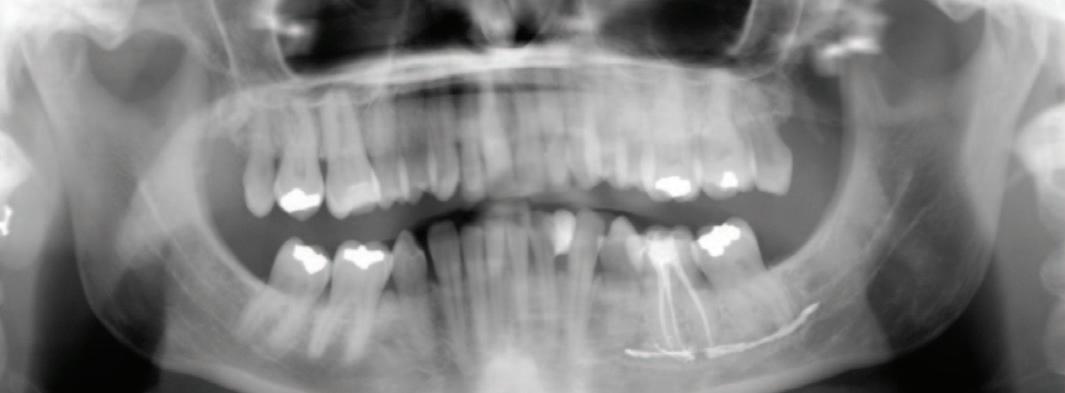
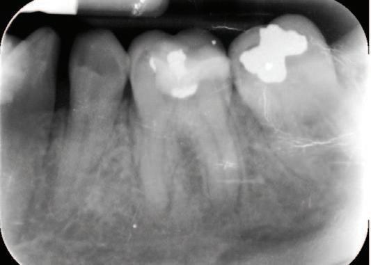
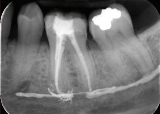
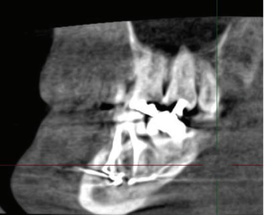
FIGURE 2: Left: Periapical radiograph pre endodontic treatment. Right: Post-endodontic treatment radiograph showing the presence of root canal filling material (AH Plus) inside the inferior alveolar canal.
FIGURE 3: CBCT scan demonstrating the presence of the material inside the canal.
FIGURE 1: Orthopantogram post endodontic treatment demonstrating the extension of the root canal material (AH Plus) along the inferior alveolar canal.
sodium hypochlorite, EDTA 17% solution and chlorhexidine, and obturation with gutta-percha using a warm vertical obturation technique with AH Plus root canal sealer.
The patient was initially treated with a steroid (prednisolone 40mg), paracetamol with codeine, and vitamin B12. Vitamin B12 is utilised to promote neurosensory recovery in peripheral neuropathy and to treat nerve injuries resulting from oral surgeries, such as third molar removal, dental implant placement, local anaesthesia, or trauma.9 However, this medication had no effect on the symptoms.
Radiographic examinations consisted of digital orthopantomogram (OPG), peri-apical (PA), and cone beam computed tomography (CBCT) scans. The scans revealed that the lower left first molar root canals were obturated with a radiopaque material, and showed root canal filling extending beyond the apices of the tooth and approximately 5cm along the mandibular canal ( Figures 1 , 2 and 3 ).
The causes of lip paraesthesia in this case were either chemical injury or pressure injury due to compression of the nerve inside the canal. The
decompression of the nerve and subsequent nerve lateralisation were discussed with the patient in detail, including the risks and benefits of the surgery.
Treatment
The patient commenced treatment with steroids and vitamin B12 before surgery. However, patient uncertainty about the surgical option delayed the intervention for approximately four weeks.
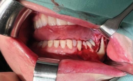
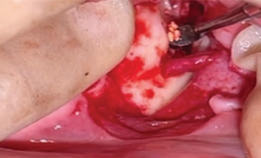
A unilateral sagittal split osteotomy was performed using piezosurgery to protect the nerve. The alveolar nerve, extending from the apical region of the left first and second molars to the mental foramen, was uncovered, and a meticulous dissection was performed to release it from the canal. Notably, rigid paste debris was observed in proximity to and within the nerve canal. The nerve exhibited signs of swelling and was surrounded by granulation tissue (Figures 4 and 5 ).
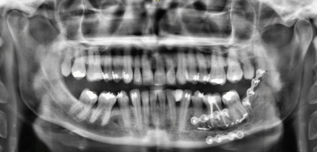
A thorough cleaning procedure ensued, followed by the repositioning of the two mandibular cortices, securing them in place with AO 5mm diameter
FIGURE 4: Unilateral sagittal split osteotomy.
FIGURE 5: The root canal filling material being removed from the canal.
FIGURE 6: Postoperative orthopantomogram showing almost complete retrieval of the material from the canal.
bicortical screws, and without insertion of intermaxillary fixation (IMF) screws. Orthodontic brackets were used to stabilise the occlusion for six weeks ( Figure 6 ).
Two weeks after the surgery, the patient reported that the pain was significantly improved, with only a tingling sensation remaining. The feeling of pressure was completely relieved. At one-year follow-up, sensation was restored and no pain was reported.
Discussion
This case report illustrates a treatment option for managing endodontic sealer extrusion following a serious endodontic complication. A 42-year-old patient underwent a unilateral sagittal split osteotomy to remove an extruded endodontic sealer from the inferior alveolar canal. Despite careful dissection and debridement of the nerve, complete removal of the sealer was not achieved, as the objective was to relieve pressure and toxicity by removing as much material as possible without causing nerve damage.
The anatomical proximity of the premolar and molar apices to the mandibular canal necessitates careful radiographic evaluation during the planning of endodontic treatment. In some cases, the apices of the molar teeth are in contact with the mandibular canal, which can cause inadvertent extrusion of the root canal sealer. An initial pretreatment radiograph of the mandibular teeth will reveal the proximity of the canal to the apices. Taking a radiograph during endodontic treatment is a necessary step in ensuring the correct working length, as well as preventing perforation and possible subsequent damage to the IAN.
Several factors could lead to sealer extrusion, including: overinstrumentation of a root canal with manual or rotary instruments; excessive pressure; and/or, poor adaptation of the gutta-percha apically, allowing sealer to be extruded through the apical foramen with warm condensation techniques.7,10,11 Additionally, the method of placing endodontic sealer has a significant impact. Studies suggest that using a Lentulo spiral exerts less intracanal pressure than the use of a syringe when applying filling pastes.12 Hence, it is imperative when injecting root canal sealer into canals that the tip does not bind in the canal and gentle force is exerted. Overextensions can be prevented by maintaining accurate working lengths and careful obturation techniques. Younger patients with wider root canal systems or cases of apical resorption may lack an adequate apical stop to prevent the extrusion of gutta-percha or endodontic sealers. In such situations, techniques that create apical barriers using calcium hydroxide or mineral trioxide aggregate (MTA) may be beneficial.
The literature highlights the potential for trauma to the IAN due to overextended root-filling material in the mandibular canal, particularly given the close anatomical proximity of the mandibular canal to the apices of
posterior mandibular teeth. It has been reported that IAN injuries after endodontic treatment account for 1.4-6% of total trigeminal nerve injuries.1315 Clinical reports attribute post-endodontic paraesthesia, dysaesthesia, and pain to two fundamental mechanistic factors. The first causative mechanism of peripheral nerve injury is compression. The confined anatomical space of the osseous mandibular canal renders the enclosed nerve susceptible to compartment syndrome. Compression of the nerve’s arterial blood supply induces an acute phase characterised by heightened vascular permeability, oedema, and ischaemia, resulting in diminished oxygen delivery to the nerve.16,17 Despite the peripheral nervous system’s inherent resilience to ischaemia, prolonged exposure to stretch and compressive forces can instigate irreversible changes, including fibroblast invasion, scarring, and fibre degeneration. Recovery from nerve damage is contingent upon the duration and severity of the trauma. Thus, immediate decompression of the compartment is imperative to avert irreversible sequelae such as reactive fibrosis or neuroma.
The second causative mechanism of peripheral nerve injury is chemical neurotoxicity. Eugenol, a derivative of phenol, can infiltrate the nerve and induce protein coagulation, leading to the chemical destruction of the axon.8 In the context of surgical interventions, decortication and apicectomy are commonly employed techniques for endodontic sealer removal. However, sagittal osteotomy emerges as a highly effective method, particularly when addressing nerve compression around the second or third molars. In this specific region, characterised by thicker bone between the nerve and the periosteal surface of the buccal cortex and limited visual control, decortication or apicectomy become more challenging and less secure, heightening the risk of nerve injury. Sagittal osteotomy, routinely utilised in orthognathic surgery, facilitates a clear and extensive exposure of the nerve along a significant portion.
Conclusion
In conclusion, as the severity of nerve damage escalates with the duration of the injury, prompt surgical exploration, involving the removal of material and decompression of the IAN, is imperative and effective upon the early manifestation of sensory disturbances. Thus, emphasis should be placed on exercising caution during root canal treatment, and obtaining radiographs both during and after the procedure. Ensuring a timely referral for surgical evaluation is also critical.
Ethical statement
Written informed consent describing the purpose and scope of this study has been obtained from the patient who was included in this study.
References
1. Gatot A, Tovi F. Prednisone treatment for injury and compression of inferior alveolar nerve: report of a case of anesthesia following endodontic overfilling. Oral Surg Oral Med Oral Pathol. 1986;62(6):704-706.
2. Kothari P, Hanson N, Cannell H. Bilateral mandibular nerve damage following root canal therapy. Br Dent J. 1996;180(5):189-190.
3. Dempf R, Hausamen JE. Lesions of the inferior alveolar nerve arising from endodontic treatment. Aust Endod J. 2000;26(2):67-71.
4. LaBanc JP, Epker BN. Serious inferior alveolar nerve dysesthesia after endodontic procedure: report of three cases. J Am Dent Assoc. 1984;108(4):605-607.
5. Morse DR. Endodontic-related inferior alveolar nerve and mental foramen
6. Brodin P, Røed A, Aars H, Orstavik D. Neurotoxic effects of root filling materials on rat phrenic nerve in vitro J Dent Res. 1982;61(8):1020-1023.
7. Pogrel MA. Damage to the inferior alveolar nerve as the result of root canal therapy. J Am Dent Assoc. 2007;138(1):65-69.
8. Escoda-Francoli J, Canalda-Sahli C, Soler A, Figueiredo R, Gay-Escoda C. Inferior alveolar nerve damage because of overextended endodontic material: a problem of sealer cement biocompatibility? J Endod. 2007;33(12):1484-1489.
9. Trising W. Comparative study between vitamin B 1-6-12 and vitamin B 12 for neurosensory recovery after bilateral sagittal split ramus osteotomy. Chulalongkorn University Theses and Dissertations (Chula ETD). Published online January 1, 2018.
10. Siqueira JF Jr. Microbial causes of endodontic flare-ups. Int Endod J. 2003;36(7):453-463.
11. Conrad SM. Neurosensory disturbances as a result of chemical injury to the
CPD questions
To claim CPD points, go to the MEMBERS’ SECTION of www.dentist.ie and answer the following questions:
l D. Gingival recession paresthesia. Compend Contin Educ Dent. 1997;18(10):963-968, 970-973, 976-8 passim; quiz 98.
1. What was the primary complication encountered in the reported case following endodontic treatment?
l A. Fractured tooth
l B. Root resorption
l C. Inferior alveolar nerve (IAN) injury
l D. Chronic infection
Quiz
Submitted by Dr Richard Lee Kin.
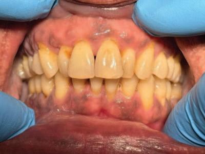
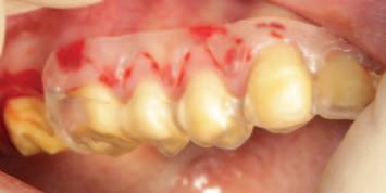
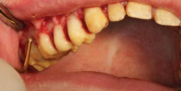
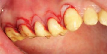
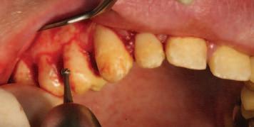
1. Describe the appearance and distribution of the pigmentation visible in the gingiva.
2. What is the most likely cause of this pigmentation?
3. How can this type of pigmentation be distinguished from pathologic causes of oral pigmentation?
4. Is treatment required for this condition?
5. How should you document it?
6. Is it a risk factor for any oral diseases?
inferior alveolar nerve. Oral and Maxillofacial Surgery Clinics of North America. 2001;13(2):255-263.
12. Ahlgren FKEK, Johannessen AC, Hellem S. Displaced calcium hydroxide paste causing inferior alveolar nerve paraesthesia: report of a case. Oral Surg Oral Med Oral Pathol Oral Radiol Endod. 2003;96(6):734-737.
13. Klazen Y, Van der Cruyssen F, Vranckx M, et al . Iatrogenic trigeminal posttraumatic neuropathy: a retrospective two-year cohort study. Int J Oral Maxillofac Surg. 2018;47(6):789-793.
14. Hillerup S. Iatrogenic injury to oral branches of the trigeminal nerve: records of 449 cases. Clin Oral Investig. 2007;11(2):133-142.
15. Tay ABG, Zuniga JR. Clinical characteristics of trigeminal nerve injury referrals to a university centre. Int J Oral Maxillofac Surg. 2007;36(10):922-927.
16. Grant GA, Goodkin R, Kliot M. Evaluation and surgical management of peripheral nerve problems. Neurosurgery. 1999;44(4):825-839.
17. Sunderland S. A classification of peripheral nerve injuries producing loss of function. Brain. 1951;74(4):491-516.
2. What surgical technique was employed to manage the injury in this case?
3. Which of the following symptoms significantly improved aer the surgical intervention?
l A. Coronectomy
l B. Apicectomy
l C. Unilateral sagittal split osteotomy
l D. Alveolar ridge augmentation
l A. Tooth mobility
l B. Facial swelling
l C. Tingling sensation and pressure in the lower lip
7. When should a referral be considered? Answers on page 18
CPD
Ryan McConville Centre for Dentistry
Queen’s University Belfast United Kingdom
Marc Lewis Emrys Edwards Centre for Dentistry
Queen’s University Belfast United Kingdom
Amanda Willis Centre for Dentistry
Queen’s University Belfast United Kingdom
Corresponding author: Marc Edwards, Centre for Dental Education, Grosvenor Road, Belfast BT12 6BP
E: Marc.edwards@qub.ac.uk
Learning style preferences of undergraduate dental students in Northern Ireland
Précis: This paper’s findings suggest that learning style preferences do not appear to explain the existing gender differences in academic attainment of dental undergraduate students.
Abstract
Understanding how students approach learning is of interest in health professional education, although there is limited evidence that identifying learning styles improves educational outcomes. This study investigated learning style preferences of clinical dental students at Queen’s University Belfast, using the VARK questionnaire, and explored whether preferences were associated with gender or academic achievement. Of 86 respondents (72% response rate), 69% preferred multimodal learning styles, with quadmodal (all four VARK modes) being the most common at 41%. Multimodal refers to a preference for more than one learning style. No relationship was found between learning style, gender, or academic distinction. Notably, a higher proportion of females achieved distinctions, a finding warranting further exploration. These results suggest that while multimodal learning is common, learning style preferences do not explain gender differences in academic performance. Given the lack of evidence for the efficacy of adapting teaching to learning styles, caution is warranted in using this information to inform curriculum design.
Journal of the Irish Dental Association Science December 2025/January 2026;1(2):8-11
Introduction
In Queen’s University Belfast, the primary degree in dentistry is the Bachelor of Dental Surgery (BDS) in the Faculty of Medicine, Health, and Life Sciences. Distinctions are awarded to the top 10% of students, providing they achieve at least 70% overall and pass at first attempt. Between 2014 and 2019, a higher percentage of females typically achieved honours (8.6%) or distinctions (37.8%) when compared with males (2.2% and 26.4%, respectively). Furthermore, during the same period, an average of 24% of females were awarded medals and/or prizes compared with 12% of males.
Previous studies have looked at the differences between male and female undergraduate students’ academic performance in health-related disciplines, including nursing 1 and medicine. 2,3 Student ability, family motivation, the quality of secondary education, and learning styles have been suggested as possible explanations for differences in academic achievements between genders.4-6 However, there is limited research into the learning styles and preferences of dental undergraduate students.
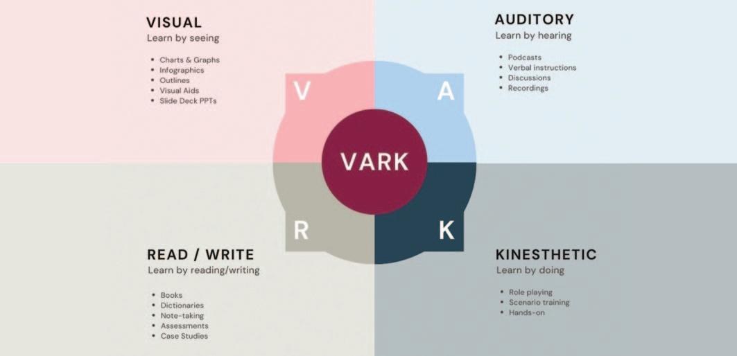
While learning styles have been widely discussed in education, systematic reviews have found little evidence that tailoring teaching to learning style preferences improves outcomes.4,5 In fact, focusing on learning styles may not be an effective use of limited educational resources and may even limit students’ beliefs in their own learning ability.1 Nonetheless, understanding students’ preferences may offer insights into their engagement with different teaching methods. Understanding students’ learning styles can be effective in organising and modifying the learning environment, and the teaching and learning process.6 Learning style refers to an individual’s preferences for learning, including how one absorbs, processes, and retains new information.7 There are several methods to measure learning styles, with the VARK questionnaire developed by Fleming and Mills (1992) being the most widely used. VARK is an acronym for the sensory modalities used to present information.8
Visual learners (V) process information by studying or drawing tables, diagrams or pictures. Aural learners (A) prefer to hear the information and process information best by listening to lectures, attending tutorials, and using tape recorders to play back learning sessions. They prefer to listen rather than take notes. Read-write learners (R) prefer to see the written words. They like to read text and take notes verbatim and re-read these. Kinaesthetic learners (K) prefer to acquire information through experience and practice, and prefer to learn information that has a connection to reality. They use live experience and practice to learn (Figure 1).
A learner’s style preference can be singular with one main preferred modality, bimodal with two preferences, trimodal with three, or quad-modal with the preference including all four types. Multimodal refers to a preference for more than one learning
FIGURE 1: Summary of the different VARK learning styles (https://whatfix.com/blog/multimodal-learning/).
style (e.g., bimodal, trimodal or quad-modal). As a tool to evaluate learning style preferences, the VARK questionnaire has achieved widespread use among different study populations in undergraduate and postgraduate settings. This is due to its validity, relative ease of implementation, simplicity and reliability.8 For example, Childs-Kean et al. (2020) explored students’ learning styles using the VARK model in different health programmes including medicine, dentistry, pharmacy, and nursing, finding that learning styles of students varied between programmes.9 At Queen’s University Belfast, the dental curriculum incorporates a range of teaching methods, including lectures, small group tutorials, clinical practice, and laboratory sessions, which align with various VARK modalities.
Methods
The primary aim of this study was to explore the learning style preferences of clinical dental students. Secondary aims were to investigate associations between learning style and: (a) gender; and, (b) academic achievement (honours or distinctions). Following a successful application for ethical approval within the Faculty of Medicine, Health and Life Sciences, the VARK questionnaire was completed voluntarily by fourth- and fih-year undergraduate dental students at Queen’s University Belfast. Only fourth- and fih-year students were included as they have completed the majority of the clinical curriculum, providing a more consistent basis for comparison. The anonymised survey was distributed to a total of 120 students via email, which included a description of the study, a consent page, and a link to the questionnaire in Microso Forms. The questionnaire consisted of 24 multiple-choice questions, each with four options. The students were requested to choose more than one option if applicable. Questions one to nine captured demographic information and questions 10-24 focused on VARK learning styles. The distribution of the VARK preferences was calculated according to the guidelines provided on the VARK website. Accordingly, learning preferences were categorised as unimodal (V, A, R, or K), bimodal (VA, VR, VK, AR, AK, and RK), trimodal (VAR, VAK, VRK, and ARK), or quad-modal (VARK).
Descriptive statistics were used to summarise demographic data and learning style preferences. Chi-square tests were used to compare categorical variables (e.g., learning style preference by gender, and by distinction or honours status), with significance set at p<0.05. Analyses were conducted using SPSS version 27.
Results
A total of 86 respondents completed the questionnaire, 48 from BDS IV (34 female, 14 male) and 38 from BDS V (24 female, 14 male). This was a response rate of 72%. Of the respondents, 67% were female and 33% male. The gender distribution within each year group was similar to the overall cohort, with females comprising approximately two-thirds of respondents in both years.
A total of 73 students were in the 21-24 year age category, 13 were older than 25, and 20% were postgraduate students.
Of the study group, 69% (N=59) preferred multimodal learning styles. The most common learning style was quad-modal (VARK) at 41%, the next most common being unimodal (31%), then bimodal (16%) and trimodal (12%). Table 1 summarises the distribution of learning style preferences by gender. There was no significant difference between males and females.
The most preferred unimodal learning style was K, with 20 of the 27 (74%) unimodal learners preferring this method. The most preferred bimodal style was visual and kinaesthetic (VK), and the most preferred trimodal style was visual, read/write and kinaesthetic (VRK).
Table 1: Learning style preference by gender.
n No distinction n Distinction/honours
A total of 23 students had received at least one distinction or honour throughout the course of their dentistry exams, accounting for 18% of male students and 31% of females. Students self-reported if they had received an honour or distinction on the anonymised questionnaire. There was no significant difference between those with or without distinctions. No significant differences were observed in learning style preferences or distinction rates between BDS IV and BDS V students (Figure 2).
Discussion
There is a relative abundance of studies analysing different learning styles in variable fields such as medicine, engineering, nursing, and allied health specialties. However, studies examining VARK learning preferences among dental students are few, especially within the UK and Europe. The objective of this study was to investigate the learning preferences of dental students in Queen’s University Belfast, while examining the effects of gender, and whether attainment of distinctions/honours had any relationship with learning style preferences. Most students in the present study preferred a multimodal learning style with no significant difference between bimodal, trimodal, and quad-modal styles. This is in agreement with the findings of previous studies from Saudi Arabia and the USA that multimodal style is the dominant learning preference among undergraduate dental students.10,11 In contrast, Nazir et al. (2018) found that 76% of dental students preferred a single learning style.12 However, this difference may be explained by research suggesting that learning preferences can change over time and between different academic environments.9
Of the students preferring a unimodal learning style, K was the most common followed by V. K has been a preferred style of dental undergraduates in several international studies, suggesting that students prefer more active learning strategies.12-14 It is likely that most undergraduate dental programmes already incorporate hands-on learning with visual components such as the use of models and explanatory videos.
The predominance of multimodal learning preferences among clinical dental students may reflect the complexity of dental education, which requires the integration of visual, auditory, reading/writing, and kinaesthetic skills. Clinical dentistry involves not only theoretical knowledge but also practical skills and
patient communication, necessitating a flexible approach to learning. This aligns with the demands of oral care, where students must synthesise information from multiple sources and modalities to provide comprehensive patient care. In relation to gender preferences, there were no significant differences in learning style between female and male dental students. This is in agreement with Murphy et al. (2004), Al-Saud (2013), and Nasiri et al. (2016), where no significant differences were observed between genders among dental students.11,13,14 Conversely, Aldosari et al. (2018) found that female dental students had a significantly higher preference for bimodal learning styles when compared with their male counterparts, indicating that variability in gender preferences may exist in different contexts.10 However, this study only focused on dental students at a single institution in Riyadh, specifically including students from the third and fourth years.5
Al-Saud (2013), Aldosari et al. (2018), and Nazir et al. (2018) have all reported an association between a dental student’s GPA (Grade Point Average) and their learning style preference, and found that students with low GPA demonstrated unimodal learning style, whereas students with higher academic performance had multimodal learning style preferences. However, in this study, no significant difference was noted in learning style between those with and without distinctions/honours, suggesting that differences may be a result of other factors. Nazir et al. (2018) investigated learning styles among dental students in Saudi Arabia, including students from all years of study, which may explain the higher proportion of unimodal preferences compared to our senior-only cohort.
From the present data, it is clear that a higher percentage of females achieved distinctions or honours when compared with males, in keeping with previous years. This is in agreement with a study of 770 fourth- and fih-year Jordanian dental students.15 Sawair et al.’s (2009) results showed that the cumulative GPAs of the female graduated students were significantly higher than those of the male students. Similarly, Nawa et al. (2020) found that males were significantly associated with lower GPA trajectories, and withdrawal or repeating years, among Japanese dental students.16 However, the present finding that there appears to be no significant difference between the learning styles of either female and male or those with or without distinctions, suggests that other factors, such as study habits, motivation, or external support, may contribute to the observed gender
FIGURE 2: Distinction/honours status by learning style.
disparity in academic achievement. Further research is warranted to explore these potential influences.
No association was found between learning style and gender or distinctions/honours. This may be because none exist, or because the study was under-powered. No power calculation was used as this was a convenience sample, which may limit the ability to detect subtle associations.
While understanding learning preferences may inform curriculum design, there is limited evidence that matching teaching methods to learning styles improves outcomes.1 Overemphasis on learning styles may even restrict students’ beliefs in their learning potential.
Academic performance is likely influenced by a complex interplay of individual, environmental, social, and cultural factors. Focusing solely on gender may
References
1. Ezzeddine N, Hughes J, Kaulback S, Houk S, Mikhael J, Vickery A. Implications of understanding the undergraduate nursing students’ learning styles: a discussion paper. J Prof Nurs. 2023;49:95-101.
2. Khanal L, Giri J, Shah S, Koirala S, Rimal J. Influence of learning-style preferences in academic performance in the subject of human anatomy: an institution-based study among preclinical medical students. Adv Med Educ Pract. 2019;10:343-355.
3. Rezigalla AA, Ahmed OY. Learning style preferences among medical students in the College of Medicine, University of Bisha, Saudi Arabia (2018). Adv Med Educ Pract. 2019;10:795-801.
4. Farooq MS, Shafiq M, Berhanu G. Factors affecting students’ quality of academic performance: a case of secondary school level. J Qual Technol Manag. 2011;7:1-14.
5. Mozaffari HR, Janatolmakan M, Sharifi R, Ghandinejad F, Andayeshgar B, Khatony A. The relationship between the VARK learning styles and academic achievement in dental students. Adv Med Educ Pract. 2020;11:15-19.
6. Kell C, Van Deursen R. Student learning preferences reflect curricular change. Med Teach. 2002;24(1):32-40.
7. Pashler H, McDaniel M, Rohrer D, Bjork R. Learning styles: concepts and evidence. Psychol Sci Public Interest. 2008;9(3):105-119.
8. Fleming ND, Mills C. Not another inventory, rather a catalyst for reflection. To Improve the Academy. 1992;11(1):137-55.
9. Childs-Kean L, Edwards M, Smith MD. Use of learning style frameworks in health
CPD questions
To claim CPD points, go to the MEMBERS’ SECTION of www.dentist.ie and answer the following questions:
1. The preferred learning style among fourth- and fih-year dental students at Queen’s University Belfast is:
l A. Bimodal
l B. Trimodal
l C. Quin-modal
l D. Quad-modal
l E. Unimodal
oversimplify the issue; future research, including qualitative studies, could provide deeper insights.
Conclusion
Differences in learning style do not appear to explain the differences in academic attainment experienced among female and male undergraduate dental students in Queen’s University Belfast. Further research will be required to explore other explanations for this, including individual, environmental, social, and cultural factors. Given the lack of evidence that adapting teaching to learning styles improves outcomes, caution is warranted in using learning style information to inform curriculum design.
science education. Am J Pharm Educ. 2020;84(7):ajpe7885.
10. Aldosari MA, Aljabaa AH, Al-Sehaibany FS, Albarakati SF. Learning style preferences of dental students at a single institution in Riyadh, Saudi Arabia, evaluated using the VARK questionnaire. Adv Med Educ Pract. 2018;9:179-186.
11. Al-Saud LM. Learning style preferences of first-year dental students at King Saud University in Riyadh, Saudi Arabia: influence of gender and GPA. J Dent Educ. 2013;77(10):1371-1378.
12. Nazir MA A-AA, Farooqi FA. Influence of gender, class year, academic performance and paternal socioeconomic status on learning style preferences among dental students. J Clin Diagn Res. 2018;12(6):ZC04-ZC8.
13. Murphy RJ, Gray SA, Straja SR, Bogert MC. Student learning preferences and teaching implications. J Dent Educ. 2004;68(8):859-866.
14. Nasiri Z, Gharekhani S, Ghasempour M. Relationship between learning style and academic status of Babol dental students. Electron Physician. 2016;8(5):2340-2345.
15. Sawair FA, Baqain ZH, AlOmari IK, Wahab FK, Rajab LD. Effect of gender on performance of undergraduate dental students at the University of Jordan, Amman. J Dent Educ. 2009;73(11):1313-1319.
16. Nawa N, Numasawa M, Nakagawa M, Sunaga M, Fujiwara T, Tanaka Y, et al. Differential effects of individual and school factors on the academic trajectories of Japanese dental students. J Dent Educ. 2020;84(7):792-798.
2. Which of these is an example of a visual learning style?
l A. Listening to lectures
l B. Taking notes and re-reading them
l C. Role-play
l D. Studying diagrams or pictures
3. Which of these is a recognised learning style according to the VARK questionnaire?
l A. Virtual
l B. Artificial
l C. Read-write
l D. Kinetic
Facially driven and digitally guided surgical crown lengthening in the contemporary management of amelogenesis imperfecta
From digital planning to clinical reality with precise and predictable results.
Introduction
Amelogenesis imperfecta (AI) is frequently associated with other developmental dentofacial defects, such as orthognathic malformation.1 This may give rise to clinically significant discrepancies in the smile, such as excessive or uneven gingival display. When considering significant changes to a dentition, it is critical that due consideration is given to all foundational discrepancies at an early stage of diagnosis and treatment, as this period is the principal window of opportunity to prevent their propagation into the definitive reconstruction. Although it is theoretically possible to address foundational discrepancies post reconstruction, such an approach is effectively ruled out, as doing so would necessitate costly re-treatment of a segment. Therefore, it is important that the nature of such discrepancies is communicated to the patient and the option to engage in their management is considered seriously at this stage.
This clinical tip guide aims to illustrate how contemporary digital tools may be used to plan and implement facially driven and digitally guided surgical crown lengthening with a view to addressing an uneven gingival display within a smile, with a high degree of clinical control.
Clinical tips
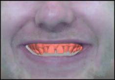
n A three-dimensional (3D) virtual patient is created through the merging of an intra-oral scan (3Shape TRIOS, Copenhagen, Denmark), a facial triangulation scan (Face Hunter 3D facial scanner, Zirkonzahn Srl, Gais,
Italy) and patient-specific occlusal data (PlaneSystem, Zirkonzahn Srl, Gais, Italy) on Zirkonzahn.Modifier software (Zirkonzahn Srl, Gais, Italy) ( Figure 1A ) as previously published in this Journal 2 The 3D facial scanner and the PlaneSystem are seamlessly integrated into a workflow that aligns perfectly with the design software in use. This integration ensures that all acquired data is transferred into the software with 1:1 accuracy, preserving every detail and enabling the creation of a precise 3D virtual reproduction of the patient’s physiognomy and oral condition. The design software, on the other hand, places special emphasis on aesthetics. The two diagnostic tools, in combination with the design software, support the entire restorative team with a comprehensive planning and communication solution, with patient-centred outcomes in mind.
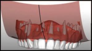
n A treatment rehearsal or treatment feasibility study may be simulated, to include a virtual diagnostic wax-up of the dentition, including repositioning of the gingival margin locations, where required. A digital sphere, based on the occlusal theory of the sphere of Monson, was applied in a novel approach to guide the harmonious positioning of a proposed gingival margin in this case ( Figures 1B and 1C ).3 The outcome of proposed changes may be assessed in the context of the virtual smile display ( Figure 1D ).
n Pending approval of the proposed gingival margin locations, a digital
FIGURE 1: A 3D virtual patient feasibility study may be rehearsed through the merging of a 3D facial tessellation scan, achieved with Face Hunter, and an intra-oral scan, using Zirkonzahn.Modifier software. From this, a dimensionally accurate 3D virtual wax-up may be created to include modelling of proposed crown lengthening. A digital sphere, based on occlusal compensation curves (such as the sphere of Monson) was applied in this case as a novel reference guide for the harmonious and aesthetically pleasing positioning of proposed gingival zeniths, which integrate well into facial balance as determined through the smile display. Readers should note that root anatomy depicted in the digital wax-up is imported from a library for illustrative purposes and is not representative of the patient’s true root anatomy in this particular case.
Dr David McReynolds BA BDentSc MFDS RCSEd DChDent (Pros) FFD RCSI (Pros) Academic Prosthodontist
Corresponding author: Dr David McReynolds, Department of Restorative Dentistry and Periodontology, Division II, Dublin Dental University Hospital, Lincoln Place, Dublin 2. E: david.mcreynolds@dental.tcd.ie
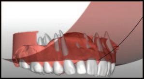
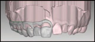
FIGURE 2: Based on the diagnostic findings of the 3D virtual feasibility study, a fully tooth-supported and fully digitally fabricated surgical guide may be developed via a process of computer-aided design and computer-aided manufacturing (CAD/CAM). The guide indicates the proposed gingival margin location as well as the proposed incisal edge and cusp height locations.
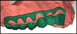
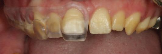
FIGURE 3: A pre-operative radiographic assessment and clinical try-in of the surgical guide gives the clinician an indication of the extent of crown lengthening surgery required. The clinician may confirm that sufficient crown–root ratio, distance from furcation anatomy, keratinised tissue, and attached gingivae will remain post surgery, and ensure the complete and comfortable seating of the guide at this stage. It is of note that the stainless steel crown on tooth UR6 was lost to retention failure between the radiographic assessment and the clinical assessment in this case.
FIGURE 4: During the surgical procedure, the upper, inner edge of the window within the guide provides the surgeon with the exact site of the proposed definitive gingival margin location and in doing so, indicates the precise location of the internal bevel incision. Following a sulcular incision and curettage of the excess gingival tissue, a fullthickness mucoperiosteal flap is raised. Osteotomy, followed by osteoplasty, is performed using a variety of round end cutting burs and/or Piezo tips with a view to re-establishing a tailored biological width from the alveolar osseous crest to the proposed free gingival margin crest.
crown lengthening guide may be designed, 3D-printed in Temp Premium Flexible transparent resin (Zirkonzahn Srl, Gais, Italy), post-processed ( Figure 2 ) and subsequently tried in the patient’s mouth.
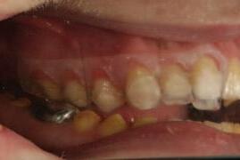
n Following clinical and radiographic assessment of the width of the keratinised mucosa, the positioning of the gingival margin in relation to the cemento-enamel junction (CEJ) and the distance of the CEJ from the bone crest, the surgical guide is tested for stability and the proposed gingival margins are approved ( Figure 3 ). To achieve an optimised outcome, the operator should envisage that at least 2mm of keratinised tissue, including at least 1mm of attached gingivae, remain intact post surgery.4 A favourable crown–root ratio and integrity of furcation anatomy must remain intact post surgery.
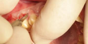
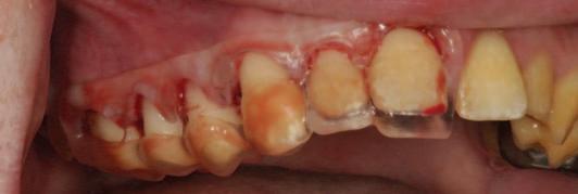
n During the surgery, the upper, inner edge of the window within the guide provides the surgeon with the exact site of the proposed definitive gingival margin location and in doing so, indicates the precise location of the initial internal bevel incision (Figures 4A-4C ).5
n The incisions are usually made with a 15C or SM67 surgical blade (SwannMorton, Sheffield, England). After removal of the excess tissue and revealing of the new crown length, a full-thickness mucoperiosteal flap is raised for direct visualisation of the surrounding bone and the CEJ anatomy. The area for supra-crestal tissue attachment is recreated by osteotomy followed by osteoplasty, using a variety of round end cutting burs and/or Piezo tips. The operator must tailor the distance between the new alveolar osseous crest and the free gingival margin to create a stable
following 12 weeks of healing, the clinician may confirm that there is alignment between the planned and the actual location of the new proposed gingival margin. In doing so, the clinician ensures they remain in control of the treatment plan.
biological width, with average appropriate distances being approximately 2-3mm.6,7 The flap is then repositioned and sutured using internal vertical mattress sutures (Figures 4D-4F ).8
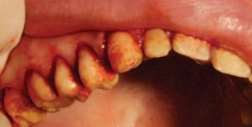
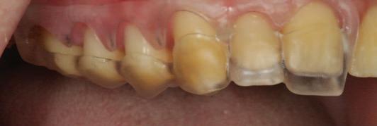
n Immediately post surgery and once again following healing and gingival remodelling at 12 weeks, the alignment between the planned outcome and the actual outcome of surgery may be assessed to ensure that the goals of treatment have been met, and that a high degree of control is maintained over the treatment plan ( Figure 5 ).
Conclusions
Innovations in the digital workflow are providing clinicians with indispensable tools for accurately manifesting planned surgical outcomes into clinical realities, with precise and predictable results beyond what freehand techniques can offer.9 Such outcomes are becoming increasingly
focused on patient-centred outcomes, such as the creation of a facially driven and aesthetically pleasing smile display.
CRediT author statement
Dr David McReynolds: conceptualisation, clinical procedures, visualisation, original draft preparation, writing – review and editing.
Dr Ioannis Polyzois: clinical procedures, original draft preparation, writing –review and editing.
Mr Alexander Lichtmannegger: conceptualisation, laboratory procedures, visualisation, original draft preparation, writing – review and editing.
Conflict of interest: funding was generously provided for the laboratory steps illustrated in this clinical tip guide by Zirkonzahn Srl, Gais, Bolzano, South Tyrol, Italy.
References
1. Crawford PJM, Aldred M, Bloch-Zupan A. Amelogenesis imperfecta. Orphanet J Rare Dis. 2007;2:17.
2. McReynolds D, Lichtmannegger A, Steger E. Digital planning in the contemporary management of amelogenesis imperfecta. J Ir Dent Assoc. 2025;71(2):81-84.
3. Nam SE, Park YS, Lee W, Ahn SJ, Lee SP. Making three-dimensional Monson’s sphere using virtual dental models. J Dent. 2013;41(4):336-344.
4. Scheyer ET, Sanz M, Dibart S, et al. Periodontal so tissue non-root coverage procedures: a consensus report from the AAP Regeneration Workshop. J Periodontol 2015;86(2 Suppl):S73-S76.
5. Coachman C, Valavanis K, Silveira FC, et al. The crown lengthening double guide and the digital perio analysis. J Esthet Restor Dent. 2023;35(1):215-221.
6. Gargiulo A, Krajewski J, Gargiulo M. Defining biologic width in crown lengthening. CDS Rev. 1995;88(5):20-23.
7. Lanning SK, Waldrop TC, Gunsolley JC, Maynard JG. Surgical crown lengthening: evaluation of the biological width. J Periodontol. 2003;74(4):468-474.
8. Lee EA. Aesthetic crown lengthening: classification, biologic rationale, and treatment planning considerations. Pract Proced Aesthet Dent. 2004;16(10):769-780; quiz 80.
9. Li Y, Liu M, Zhou T, Lyu J, Tan J, Liu X. Accuracy of three types of digital guides for crown lengthening surgery: an in vitro study. J Dent Sci. 2024;19(1):39-45.
FIGURE 5: Following closure of the mucoperiosteal flap and again
Procurement in dentistry – sustainable purchasing of equipment and consumables
Reviewing your practice’s procurement policies can play a significant role in making dentistry more environmentally sustainable.
Learning outcomes:
This article aims to assist the reader to:
n purchase environmentally safe products such as chemicals and materials; n manage ordering products to maximise efficiency and reduce the number of deliveries;
Introduction
Sustainable procurement in dentistry means choosing products and suppliers in such a way as to minimise environmental impacts without compromising patient safety. In the UK, NHS dental procurement has been shown to account for 18% of the carbon footprint of dentistry.1 Sustainable procurement can lead to lower operational costs because of reduced waste and increased efficiency. Other benefits to the practice include enhanced patient recognition, as patients are increasingly environmentally aware. Dental suppliers will benefit from selling safer products and can use this more sustainable approach to promote their business. Sustainable procurement in dentistry should be viewed as a practice-level initiative that links into a broader regional/national health system strategy to reduce greenhouse gas (GHG) emissions (50% reductions required for the Irish public sector by 2030).2 Embedding dental procurement within these frameworks ensures consistency with the wider EU Green Deal and national decarbonisation targets.3 Procurement decisions can be evaluated in different ways. The four pillars of sustainability are social, human (including healthcare), environmental, and economic. These factors must prosper in a balanced way for a sustainable future. To that end, the triple bottom line theory is a sustainability framework used in business that measures a business’s success across three parameters: its social impact (people); its environmental impact (planet); and, its financial performance (profit). This guides the procurer to pre-assess purchases not only for monetary value but to minimise carbon and waste, to ensure ethical labour practices in supply chains, protect staff and patient health by reducing chemical exposure, and deliver best value when life cycle costs (LCCs) are considered. The reader may have seen a recent paper by one of the authors demonstrating that manufacturing toothbrushes locally, or at least within Europe, has more positive social outcomes across several levels; this also applies to other procurement products.4
Actions for the practitioner
Avoid waste: order less and order less frequently
Ordering supplies less frequently will reduce the pollution associated with
Dr Brett Duane
Associate Professor in Dental Public Health, Dental Science, TCD
Dr Eamon Croke
General dental practitioner, Part-time lecturer, Dublin University Hospital, Member of the IDA Quality and Patient Safety Committee
n buy equipment that is the best value over time; n value equipment and consumables that are reusable or recyclable; and, n reduce consumption where possible.
delivery. Good stock control is very important to avoid waste, and reduce costs, for example, rotating consumables to ensure that older products are in front of newer ones.
The authors are aware of progress by the Irish dental supply companies to electrify their fleets and significantly reduce their transport emissions.5 In the absence of electrified transport, where practical, practices and suppliers should consolidate shipments to minimise transport-related emissions.
Dental team members should Rethink the products they buy and try to avoid disposable or single-use items. Reduce orders as much as is practical. Reuse products that can be reused. Recycle products as appropriate. Consider having a system or a designated person who oversees stock management. Doing an annual audit can identify the most used products and analyse their sustainability. By design, the ideal product would be:6
n safe to use on patients and fit for purpose; n manufactured without fossil fuels;
n manufactured with renewable energy;
n supplied with minimum, reusable packaging;
Dr Michaela Dalton
General dental practitioner HSE, Member of the IDA Quality and Patient Safety Committee
Dr Nick Armstrong
Retired Principal Dental Surgeon HSE, Member of the IDA Quality and Patient Safety Committee
Corresponding author: Dr Nick Armstrong E: armstrongnick5@gmail.com
n broken down into environmentally harmless elements; n simple in design, and easy to clean and reuse or recycle; n transported in the most environmentally acceptable way; and, n inexpensive.
Supplier and manufacturer decisions are influenced by a combination of market demand, regulatory requirements, cost structures, and reputational considerations, rather than simple market forces alone.7 Evidence from sustainable healthcare supply chains shows that clear procurement signals from clinicians and institutions, together with policy incentives, standards (e.g., ISO 14001, NHS Evergreen, EU Green Public Procurement), and transparency requirements, can act as major facilitators of change.8,9,10 Conversely, fragmented demand, lack of product labelling, and uncertainty about clinical acceptability oen remain key barriers to supplier innovation.1 Collaboration between dental professionals, procurement leads, and suppliers is therefore essential to shi the market toward lower-carbon, ethically sourced products.
Suppliers should be encouraged to reuse packaging by collecting it for reuse. It would be more efficient to only have one monthly delivery, with used packaging collected by the supplier for reuse; if it cannot be reused it can be recycled. Ideally, suppliers should be committed to ISO 14001 or other international standards reflecting sustainability. Ideal packaging8 would conserve as many natural resources as possible and be reusable, and production would be low energy. The dental team should discuss sustainability with their supplier and request more sustainable delivery and products where possible. Suppliers should be encouraged to advertise their environmentally acceptable products and highlight these products in their catalogues and advertising.
Plastics
Plastic pollution is a major crisis worldwide and plastic products should be avoided or reduced where possible.11,12 Ideally, materials for order should be grown rather than fossil fuel based. Grown products such as bioplastics should be grown on non-arable land, where possible, to preserve land for food production.12 Plastics are not inert materials. To achieve flexibility, colour, durability, and flame resistance, they contain a wide range of chemical additives, including plasticisers (e.g., phthalates), flame retardants (polybrominated diphenyl ethers [PBDEs], tetrabromobisphenol A [TBBPA]), stabilisers (lead, cadmium, bisphenol A), and antioxidants (butylated hydroxytoluene [BHT], butylated hydroxyanisole [BHA]).13 These substances are not chemically bound to the polymer and can migrate, leach, or volatilise throughout the product’s life cycle during manufacture, use, or disposal.14 Human exposure occurs through skin contact, inhalation of microplastic particles, and especially, ingestion via food packaging as additives transfer into food and beverages.15 Several of these compounds act as endocrine disruptors (e.g., BPA, phthalates), neurotoxicants (e.g., PBDEs, lead stabilisers), or carcinogens (e.g., vinyl chloride monomers).16 Microplastics can also absorb and carry persistent organic pollutants (POPs) like dioxins and polychlorinated biphenyls (PCBs), amplifying their bioavailability once ingested or inhaled.17 As a result, plastics contribute not only to environmental contamination, but also to chronic low-level chemical exposure in humans, particularly through consumer and healthcare products that come into contact with the body or food.18
Cleaning products
Cleaning contractors and cleaners should be required to only use safe chemical cleaning solutions that readily break down to environmentally safe components.
A study of domestic cleaners showed a reduction in lung function potentially linked to the inhalation of toxic substances.19
The dental team should avoid using chlorine and other polluting products.11 There are safer alternative cleaning agents on the market, and these should be the cleaning products of choice. If your supplier does not stock these products, it should be encouraged to stock safe products. Alternatively, purchase them from companies that do.
Further research and education are required to ensure that dental professionals can accurately identify which cleaning and disinfection products are safe and environmentally responsible to use. In Ireland, the use and management of all chemical agents in the workplace are governed by the Safety, Health and Welfare at Work (Chemical Agents) Regulations 2001-2021, which require employers to identify hazardous substances, assess exposure risks, and implement control measures to protect staff and patients. Compliance with these regulations should form the foundation of sustainable chemical procurement and use within dental settings.20
Equipment
Single-use equipment should be avoided unless there is a very strong reason for its use. Some items are single use for infection control reasons. Disposable kits have a higher environmental impact in many areas, including the minerals and metals used, resulting in greater damage to the environment including freshwater use and climate effects.21 Switching from single-use to reusable exam kits can reduce climate impacts by approximately one-third.21
Avoid plastic disposable single-use suction tips and air/water syringe tips. These should be replaced by steel products ideally, or reusable and autoclavable plastic suction tips. Recycling plastic consumables, such as suction tips and plastic barriers, should take place where appropriate.
Dental equipment can be energy intensive. Always check the efficiency of equipment as advised by the manufacturer. Buy energy-efficient autoclaves, washer-disinfectors, suction units and compressors.
Procurers should think beyond the purchase price of any equipment they buy. Looking at the whole LCC helps to compare products by looking at the total cost over their entire lifetime, not just the price on the invoice. This includes the energy needed to run the equipment, the cost and frequency of maintenance or consumables, and the final disposal or recycling cost. For example, a cheaper autoclave that uses more electricity and water may cost far more to operate over ten years than a higher-efficiency model. A simple way to apply LCC is to ask three questions before purchase:
1. What will it cost to run and service each year?
2. How long will it last before replacement?
3. Can parts be repaired or recycled at end of life?
Example:
Consider a practice deciding between two suction systems:
System A costs €3,000, uses 1.2kWh of electricity per hour, and typically lasts for seven years.
System B costs €4,000, uses only 0.6kWh per hour, and lasts for 10 years.
System A seems cheaper. However, with ten years of daily use, the energy savings from System B (at a price of €0.35/kWh) would amount to €1.36 a day and the longer lifespan means that its total annual purchase cost (€400 compared with €423 per year) is also lower.
Even without complex spreadsheets, thinking in terms of costs over time, not just the purchase price, enables dentists to choose equipment that is financially and environmentally sustainable, aligning with the principle of ‘best value over time’ rather than lowest upfront cost.¹
PPE
Use biodegradable personal protective equipment (PPE) materials, for example, polylactic acid (PLA) masks and gowns, as these have a lower environmental cost. PLA masks, manufactured from plant-derived polymers such as corn starch, are available as biodegradable alternatives to conventional polypropylene (PP) surgical masks.¹ Although visually similar, they can be distinguished through several physical and labelling characteristics. PLA masks typically exhibit a slightly stiffer and glossier texture, oen with a sweet or starchy odour, whereas PP masks feel soer and more elastic.
On the packaging, they are labelled as “bioplastic”, “plant-based”, or “compostable”, and commonly certified under EN 13432, ASTM D6400/D6868, or compost standards.22,23 Unlike PP masks marked with the recycling code PP-5, PLA masks usually carry compostability logos and instruct users not to recycle them in conventional plastic streams.6 However, it is important to note that most commercial ‘eco’ masks are hybrid designs in which only the external or filter layers are PLA, while ear loops or nose wires remain synthetic, limiting full biodegradability.
Under current HSE guidance, used masks from treatment areas should continue to be managed as healthcare risk waste, regardless of whether they are polylactic or polypropylene based. Only unsoiled PLA masks used in non-clinical areas (e.g., reception staff, waiting rooms) may be directed to industrial composting streams, but only where certified as EN 13432 and accepted by the waste provider.24
Preferably, reusable gowns and visors should be considered. Non-sterile gloves are adequate for most dental procedures. Reusable silicone gloves can be used for non-critical procedures. Gowns and aprons should be washable. Cotton or other plant- or animal-based fabrics are favoured compared to the fossil fuelbased fabrics such as polypropylene or polyester, as these shed particles of plastic during washing. Reusable gowns may potentially reduce GHG emissions, energy consumption and solid waste generation.
Office
supplies
Practices should aim to be paperless if possible. Social media and other online tools can be used to communicate with patients. Paper used in the practice should be good quality recycled paper. Purchase any necessary stationery in bulk.
Office and waiting room furniture should be environmentally sustainable and should not contain plastic components. Wooden furniture should be from a sustainable source and not made from tropical hardwood.
References
1. Duane B, Ramasubbu D, et al. Environmental sustainability and procurement: purchasing products in the dental setting. Br Dent J. 2019:226(6):453-458.
2. Department of Climate, Energy and the Environment. Public Sector Climate Action Mandate and the Public Sector Climate Action Strategy. https://www.gov.ie/en/department-of-climate-energy-and-theenvironment/publications/public-sector-climate-action/
3. ISO 20400 (Sustainable Procurement), EC Green Public Procurement Guidelines
Household items such as tea and coffee should come from a sustainable source and preferably be organic. Fairtrade, Rainforest Alliance and other certified brands are easily attained.
Dental laboratory
The use of digital intraoral scanning and local laboratory partnerships significantly reduces the environmental footprint associated with dental laboratory work. Digital workflows eliminate the need for physical impressions, plaster casts and disinfection chemicals, while also reducing transport requirements and emissions.¹ When work must be sent to external laboratories, selecting local or national providers instead of overseas facilities avoids the disproportionate carbon impact of air freight (0.9kg CO2e per tonne), which can be up to 10 times higher per kilogram of cargo than ground transport (0.080-0.120kg CO2e per tonne).25 Laboratories that operate under ISO 14001 (environmental management) or ISO 9001 (quality management) standards should be prioritised.6
Challenges and solutions
Despite repeated warnings, governments and industries are not sufficiently meeting the challenges of sustainable development. Barriers include concerns about profitability, vested interests within the fossil fuel industry, and widespread disinterest or disinformation.26
Yet, meaningful progress depends not only on systemic change but also on the actions of individuals and sectors willing to take responsibility. Within dentistry, Irish practices face specific obstacles to sustainable procurement, including potentially higher upfront costs, limited product options, and stringent infection control requirements. Over time, growing demand and regulatory pressures are likely to reduce these costs. Practices can help accelerate progress by sourcing greener alternatives, engaging with suppliers, and appointing sustainability leads to co-ordinate training and promote environmentally responsible practices. Reusable items that can be safely decontaminated should be prioritised, and disposable products should be made from sustainable materials. Continued collaboration between professional organisations, suppliers, and practitioners is essential to share knowledge, strengthen sustainability policies, and embed best practice across the dental supply chain.
Take home messages:
n procurement practices can reduce environmental damage in a sustainable way;
n working with suppliers and manufacturers on more environmentally friendly approaches will help us to move towards a more sustainable future; and, n practitioners should embed the concept of rethink, reduce, reuse and recycle in practice procurement policies.
3rd ed. Brussels, 2021, and the WHO Sustainable Health Procurement Guidance 2023.
4. Duane B, Steinbach I, Martinez-Cadenas R, Ashley P. Erratum to “What is the best toothbrush from a social impact perspective?” [Journal of Dentistry 105708]. J Dent. 2025;161:1060016.
5. Personal communications between authors Eamon Croke and Nick Armstrong and dental companies Henry Schein, DMI and Medray.
6. Duane B. Sustainable Dentistry, Making a Difference. Springer, 2023.
7. Walker H, Brammer S. Sustainable procurement in the United Kingdom public sector. Supply Chain Manag. 2009;14(2):128-137.
8. ISO. ISO 14001. https://www.iso.org/standard/60857.html
10. European Commission. Green public procurement. https://single-marketeconomy.ec.europa.eu/single-market/public-procurement/strategic-procurement/ green-public-procurement_en
11. Armstrong N, Dalton M, Croke E, Kahatab A. Sustainability in dentistry part II: sustainability and the use of chemicals in dentistry. J Ir Dent Assoc. 2024;70(2):9092.
12. Kahatab A, Armstrong N, Dalton M, Croke E, Duane B. Plastics: time for a rethink. J Ir Dent Assoc. 2025:71(2)85-87.
13. Hahladakis JN, Velis CA, Weber R, Iacovidou E, Purnell P. An overview of chemical additives present in plastics: migration, release, fate and environmental impact during their use, disposal and recycling. J Hazard Mater. 2018;344:179-199.
14. Lithner D, Larsson Å, Dave G. Environmental and health hazard ranking and assessment of plastic polymers based on chemical composition. Sci Total Environ. 2011;409(18):3309-3324.
15. Groh KJ, et al. Overview of known plastic packaging-associated chemicals and their hazards. Environ Sci Technol. 2019;651(Pt 2):3253-3268.
16. World Health Organization. State of the Science of Endocrine Disrupting Chemicals. Geneva; 2013.
17. Teuten EL, Saquing JL, Knappe DRU, et al. Transport and release of chemicals from plastics to the environment and to wildlife. Philos Trans R Soc B Biol Sci.
Quiz answers
1. Appearance and distribution:
n brown to dark brown, flat, diffuse areas mainly on the attached gingiva and interdental papillae;
n symmetrical pattern, more pronounced in the anterior and lower gingiva; and,
n no swelling, ulceration, or loss of surface texture.
2. Most likely cause:
n physiologic (ethnic) pigmentation due to increased melanin production by melanocytes in individuals with darker skin types; and,
n present from birth or early childhood, representing a normal genetic variation.
3. Distinguishing physiologic from pathologic pigmentation:
n physiologic pigmentation: bilateral, symmetrical, flat, present from birth, stable over time, no symptoms, no mucosal changes; and,
n pathologic pigmentation: irregular, focal, new in onset, may be associated with drugs, systemic disease, or neoplastic changes, may be symptomatic or evolving.
4. Treatment:
n no treatment required, as it is a normal, benign finding; and,
n cosmetic depigmentation (e.g., laser) may be considered for aesthetic reasons; results may vary.
2009;364(1526):2027-2045.
18. Andrady AL. Microplastics in the marine environment. Mar Pollut Bull. 2011;62(8):1596-1605.
19. Svanes O, Bertelson RJ, Lygre SHL, et al. Cleaning at home and work in relation to lung function decline and airway obstruction. Am J Respir Crit Care Med. 2018;197(9):1157-1163.
20. Irish Statute Book. Safety, Health and Welfare at Work (Chemical Agents) Regulations 2001-2021. https://www.irishstatutebook.ie/eli/2001/si/619/made/en/print
21. Byrne D, Saget S, Davidson A, et al. Comparing the environmental impact of reusable and disposable dental examination kits: a life cycle assessment approach. Br Dent J. 2022 Aug;233(4):317-325.
22. ASTM International. ASTM D6400-21 – Standard Specification for Labeling of Plastics Designed to be Aerobically Composted in Municipal or Industrial Facilities. West Conshohocken, PA; 2021.
23. European Committee for Standardization (CEN). EN 13432:2000 – Requirements for Packaging Recoverable through Composting and Biodegradation. Brussels; 2000.
24. EPA Ireland. National Waste Management Plan for a Circular Economy –Biodegradable Waste Guidance. Wexford: Environmental Protection Agency; 2022.
25 DEFRA (UK Government). Greenhouse Gas Reporting: Conversion Factors 2024, Freight Transport Tables.
26. Intergovernmental Panel on Climate Change (IPCC). Climate Change 2023: Synthesis Report. Contribution of Working Groups I, II and III to the Sixth Assessment Report of the Intergovernmental Panel on Climate Change. Geneva: IPCC; 2023.
5. Documentation:
n take intraoral photographs/scan for baseline record;
n record detailed clinical notes including distribution, symmetry, and any associated findings;
n review medical and medication history to rule out systemic causes; and,
n monitor for changes during routine dental visits.
6. Risk factor for periodontal disease:
n current evidence suggests that physiologic (ethnic) gingival pigmentation may be associated with an increased risk of periodontal disease, although further research is needed to determine whether this relationship is causal or influenced by confounding factors.
7. Referral
Referral to an oral medicine specialist should be considered if:
n pigmentation is new, rapidly changing, or asymmetrical;
n there is ulceration, induration, bleeding, or pain; or,
n there is suspicion of systemic disease (e.g., Addison’s disease) or drug-related pigmentation.
Questions on page 7
Survival of fixed prosthetic restorations on vital and nonvital teeth: A
systematic review
Hawthan M, Larsson C, Chrcanovic BR.
Purpose
To evaluate the survival rate of full-coverage tooth-supported fixed prosthetic restorations, single crowns (SCs), and fixed dental prostheses (FDPs), taking into consideration the potential influence of tooth vitality, presence and type of post, and type of prosthetic restoration material.
Materials and methods
In October 2022, two authors independently conducted a search in PubMed, Web of Science, and Scopus electronic databases, as well as a hand search to identify clinical human studies on full-coverage SCs and FDPs supported by vital and/or non-vital abutments and/or a combination of both, with a minimum observation period of 24 months.
Results
Out of 4,198 studies identified through the database search and 22 through hand searching, 26 studies fulfilled the inclusion criteria and were included in the analysis of the present systematic review. Included studies scored six points and more according to the Newcastle-Ottawa Scale (NOS). The highest estimated five-year survival rate was observed for (metal-ceramic and all-ceramic) SCs on vital teeth (98.3%; 95% CI [98.1, 98.6%]) and allceramic SCs on non-vital teeth with fibre post (95.0%; 95% CI [94.5-95.4%]). Metal-ceramic SCs on vital teeth (97.1%; 95% CI [95.6-98.7%]) showed a statistically significant higher estimated five-year survival rate compared to metal-ceramic SCs with cast metal post (90.7%; 95% CI [87.4-94.0%], P < 0.001), fibre post (91.3%; 95% CI [90.9-91.6%], P<0.001) and without post (85.7%; 95% CI [80.7, 90.6%], P<0.032). All-ceramic SCs with fibre post had a statistically significant higher estimated five-year survival rate (95.0%; 95% CI [94.5-95.4%]) compared to metal-ceramic SCs on non-vital teeth with fibre post (91.3%; 95% CI [90.9-91.6%], P<0.001). SCs (all-ceramic and metal-ceramic) with fibre post had a statistically significantly higher estimated five-year survival rate of (92.7%; 95% CI [92.4-92.9%]) than SCs made of metal-ceramic and retained by cast metal post (90.7%; 95% CI [87.4-94.0%], P<0.001). For FDPs, the five-year survival rate was significantly higher for FDPs on vital abutments (84.9%; 95% CI [75.9, 93.9%]) compared to FDPs retained by non-vital abutment/s (81.3%; 95% CI [80.3, 82.2%], P = 0.049) irrespective of presence, type of post, and FDP material. The results are limited by the limited number of studies and the presence of uncontrolled confounding clinical variables.
Conclusions
Within the limitations of the study, tooth vitality is suggested to contribute positively to the survival of SCs and FDPs.
J Prosthodont . 2024;33(2):110-122.
How many tooth colours are there?
Hein S, Morovič J, Morovič P, Saleh O, Lüchtenborg J, Westland S.
Objectives
This study aimed to estimate the number of distinct tooth colours using a large dataset of in-vivo CIELAB measurements. It further assessed the coverage error (CE) and coverage error percentage (CEP) of commonly used shade guides and determined the number of shades needed for an ideal guide, using the Euclidean distance ( Δ Eab) and thresholds for clinical perceptibility (PT) and acceptability (AT) as evaluation criteria.
Methods
A total of 8,153 untreated maxillary and mandibular anterior teeth were measured in vivo using calibrated dental photography. Cardinality was applied to determine the number of unique natural tooth colours. The CE and CEP were calculated for the Vita Classical and Vita 3D-Master shade guides, while the cardinality method was also used to estimate the number of shades required to adequately cover the estimated gamut of natural tooth colours.
Results
The cardinality analysis revealed 1,173 unique natural tooth colours. The CE for the Vita Classical shade guide was 4.1ΔEab, with a CEP of 75% beyond AT, while the 3D-Master shade guide had a CE of 3.3ΔEab and a CEP of 70% beyond AT. Based on cardinality computation, 92 discrete shades are required to adequately cover the estimated gamut of natural tooth colours with a CE of 1.2ΔEab and CEP of 0.3% beyond AT.
Conclusions
Cardinality computations estimated 1,173 unique tooth colours, while 92 discrete shades are estimated for full coverage. Such a number is impractical for physical shade guides, but new digital tools and 3D printing may offer future solutions. Both the Vita Classical and 3D-Master shade guides do not fully represent the range of natural tooth colours.
Clinical significance
This study highlights the limitations of existing shade guides and underscores the potential for new developments.
Dent Mater . 2025;41(1):51-57.
Looking at the bigger picture
Dr David McReynolds is a Visiting Research Fellow at The Irish Longitudinal Study on Ageing (TILDA), and Deputy Editor of the JIDA. He is an academic prosthodontist, currently undertaking a PhD.
Tell us about your background and what led you to dentistry, and to dental research?
I really wanted to become a musician, but my sister who is a professional musician said, if you can do anything else, I suggest you do it because you’ll struggle to make money as a musician. I thought dentistry was a nice balance between working every hour of the day and maybe having some time for music. I was a general dentist for two years, then went back to specialise in prosthodontics and did that for three years. I was headhunted to become an Assistant Professor at the DDUH – that’s what got me into my PhD.
Can you describe your work as part of TILDA?
TILDA is a World Health Organization (WHO) Collaborating Centre for Longitudinal Studies on Ageing in the Life Course. It’s a very prestigious place to be. The dental school was looking to recruit a dedicated PhD student to take part. My job was to look into the mouths of older adults living in Ireland and record details. On the basis of that, I’m doing research to show epidemiological findings of the dental health of that group. But the wonderful thing about TILDA is that it’s also a general health and social research lab. So you can start to connect someone’s dental health to other variables. You can ask, for example, does the number of teeth a person has predict if they’re socially withdrawn?
What challenges have you encountered during your research, and how have you overcome them?
The easiest thing for me as a clinical dentist was to assess the patients and build a rapport with them. As a clinician, you’re already quite meticulous with recording details. But the huge challenge was taking that data and working on statistical analytical soware to do massive computations. Nobody was there to teach me how to code a PhD analysis. I had to teach myself.
How do you see your research contributing to clinical practice or patient outcomes in dentistry?
I’ve been very lucky in TILDA to have had introductions to and an audience with the Department of Health and the National Oral Health Office. I think they take my research very seriously. So, I’m hoping that my findings will help to inform policy on how we treat older adults and what social welfare schemes will be available to them.
Are there any misconceptions in the field that your research helps to clarify or correct?
I think the current perception is that oral hypofunction happens in the nursing home population, whereas I’m showing that it’s very prevalent in the community.
Another thing is that there seems to be a tremendous focus on tooth decay being the main problem. My study is finding that tooth decay is a very minimal problem on a population level. I am finding that in that age group, almost everybody has some level of gum disease or periodontitis. That’s really interesting because the social welfare schemes have almost zero provision for periodontal care, but they’ve got a humongous provision for tooth decay. I’m hoping that we can update our understanding of what diseases are actually affecting people.
Which emerging technologies or methods do you believe will most significantly shape the future of dental research?
I’m very fortunate that I came into my PhD in the era of large language models. You have to know what you’re doing with it, but generative AI has been fantastic for helping people get past that initial mountain climb of learning how to code.
How important is interdisciplinary collaboration in your work?
It’s massive in TILDA. The study is led by Regius Prof. Rose Anne Kenny, who’s a medical gerontologist. Classically, dentistry has been segregated from medicine. But I think Regius Kenny is a very progressive individual who recognises that oral health is integral to overall health. There are other research studies like TILDA worldwide, but to my knowledge, none of them has a fulltime dentist on site. With TILDA, because we’ve got medical gerontologists, social workers, statisticians, IT expertise, you get the ability to ask very big questions about where oral health fits into a person’s general health, but also their social background.
What advice would you give to young dental researchers just starting out in the field?
Don’t wait for anybody else to tell you what to do. You have to be self driven, figure out what you need to do, and do it your own way. The second thing is learn lessons from people that have come before you. There are two books that I would highly recommend: How to Read a Paper by Trisha Greenhalgh and The Doctor’s Guide to Critical Appraisal by Narinder Gosall. If any aspiring researcher reads those books, it will completely demystify medical research.
Outside of your research, how do you maintain balance and well-being in such a demanding field?
With all the health research, I’ve got into exercise. So, I’m improving my physical fitness throughout the PhD, which, I think, complements the work that you do cognitively. Separately, I’m an avid video gamer. I play The Legend of Zelda with my girlfriend every Sunday.