The role of minimally invasive non-surgical therapy (MINST) in regenerating intrabony defects
Dr Varkha Rattu, Prof. Luigi Nibali
QUIZ
RESEARCH
The root cause: investigating intermittent inferior alveolar nerve paraesthesia from a multi-rooted third molar
Dr Hannelie Edgar, Dr Amanda Willis, Prof. John Marley
RESEARCH
Evaluation of a workshop to develop dental undergraduates’ behaviour change conversation knowledge and confidence
Marc Lewis Emrys Edwards, Shamira McKenna, Maria Hussain
CLINICAL TIPS
Managing postoperative bleeding: a refresher for the GDP
Dr Eoin Gough, Dr Síofra Murphy, Prof. Paul Brady
NEW DENTAL SCIENCE
Abstracts from the latest dental literature
RESEARCHER PROFILE
Bridging the gap between evidence and practice
Dr Cristiane da Mata
BDS MFD (RCSI) Dip TLHE MPH
Phd FFD RCSI
Honorary Editor
journaleditor@irishdentalassoc.ie
Deputy Editor
Dr David McReynolds BA BDentSC MFDS RCSEd DChDent (Pros) FFD RCSI
Editorial Board
Dr Meriem Abbas BDS (NUI) MFDS RCSEd PGDip TLHE
Una Farrell Dip Dental Hygiene
Dr Catherine Gallagher MB BCh BAO BDS NUI FDS RCSEng FFD RCSI
Dr Geraldine McDermott BA BDentSc MFDS (RCSI) PGradDip ConSed (TCD) MSc Healthcare Leadership (RCSI)
Dr Clair Nolan BDS (NUI) MSc (Endo) U. Lond
Dr Adedeji Daniel Obikoya BChD MFDS (RCSI) MSc
Dr Judith Phelan BDS (NUI) MDS (NUI) MSc (U Lond) MRD (RCS Eng and Glas)
Dr Patrick Quinn BCL BDS LLM MDPH
Dr Catherine Vaughan BDS (NUI)
International Editorial Board
Assoc. Prof. Lamyia Anweigi BDS MMedSc MFDS PhD (Qatar)
Dr Ana Cecilia Diniz Viana BDS MD PhD (Brazil)
Dr John Macken BDS PGCertMEd PhD MFDS RCSEng ACIEA FHEA (UK)
Prof. Leonardo Marchini DDS MSD PhD (USA)
Dr Ramiar Karim BDS MSc Paed Dent (Germany)
Dr Elaine Smyth BA BDentSc DChDent (USA)
Prof. Murali Srinivasan BDS MDS MBA MAS PD (Switzerland)
Prof. Sayaka Tada DDS PhD (Singapore)
jida.scholasticahq.com
Irish Dental Association Unit 2 Leopardstown Office Park, Sandyford, Dublin 18.
T: +353 1 295 0072
F: +353 1 295 0092 www.dentist.ie
Committed to growth
Welcome to the first edition of the redesigned JIDA and JIDA Science.
The idea of a new JIDA has finally materialised, and I am delighted to present to you a redesigned journal that continues to be committed to the growth of the profession, now in a more reader- and environmentally friendly format.
You will notice that the new Journal of the Irish Dental Association has two distinct sections: the first one is the JIDA , and focuses on IDA news, policies, events, and conferences – keeping members up to date with developments within the Association and the wider Irish dental community. The second is JIDA Science, which brings to the readers peer-reviewed research, clinical features, practical clinical tips, and interviews with leading academics and researchers from Ireland and abroad. The decision to separate our publications came from our ambition to broaden the reach of the Journal, attract more international papers, and regain PubMed accreditation.
In order to support this plan, we have also established an International Editorial Board, bringing fresh perspectives and expanding the Journal’s global reach. We’re proud to welcome Board members from Singapore, Switzerland, Qatar, Germany, Brazil, the UK, and the USA. Their insights and expertise will help us to ensure that JIDA Science remains at the cutting edge of dental research and clinical practice, while also making it a platform for international exchange. I commend you to read the interview on page 20 with Prof. Sayaka Tada, a member of our International Editorial Board, who is a Professor in Prosthodontics in the Faculty of Dentistry, Singapore. I first met Sayaka when she came to Cork years ago to complete part of her research and have frequently met with her ever since at conferences around the world, as we share a common interest in gerodontology.
Recently, I had the privilege of joining her and a group of other academics, clinicians, researchers, and other stakeholders as part of an international consensus collaboration in gerodontology in Geneva. We discussed issues around diagnosing and treating older adults, with the ultimate goal of creating a classification system in gerodontology. This international collaboration deepened my appreciation for the importance of bringing different perspectives to the table, finding common ground and, in the end, realising that we all face the same challenges and can share valuable insights into how to best manage oral conditions in this group of patients.
Older patients form a quite heterogenous group, and their general health status can vary substantially. A partially dentate 65 year old presenting with comorbidities and on polypharmacy may be a more complex case to treat than a healthy 80-year-old independent adult. For this reason, having a recommended pathway for diagnosis and management of older adults, taking into account comorbidities and quality of life, could be very beneficial for clinicians. It could also facilitate communication among dentists, and other professionals involved in the treatment of this cohort, such as doctors and nurses.
As a preparation for this meeting, I took part in a systematic review on diagnostic indices used for coronal and root caries, risk factors and treatment options for older adults, and it was interesting to notice the differences among studies. We found that diagnostic approaches vary widely, and standardised assessment tools are lacking, particularly for root caries.
The importance of using a common language when diagnosing and reporting oral conditions has been highlighted by other consensus exercises before, such as the 2017 workshop in periodontology, which resulted in a new classification of periodontitis characterised by a multidimensional staging and grading system. Earlier, in 2015, experts in cariology had gathered in Belgium to reach consensus regarding recommendations for managing carious lesions and the terminology around them. The group highlighted that: “Consistency, accuracy, and precision are important for terminology to be used successfully, which means there has to be standardisation globally”.¹
These consensus meetings and their resulting body of work are crucial for enhancing the comparability of studies and ensuring that dentists and researchers use a common language when discussing diagnosis, risk of disease progression, and treatment strategies. The gerodontology consensus will result in a classification system and a series of recommendations for the treatment of older adults that is worth keeping an eye out for.
Finally, enjoy reading your first JIDA Science!
Reference 1. Innes NPT, et al. Managing carious lesions: consensus recommendations on terminology. Adv Dent Res 2016;28(2):49-57.
JIDA Science is an official publication of the Irish Dental Association. The opinions expressed in JIDA Science are, however, those of the authors and cannot be construed as reflecting the Association’s views. The editor reserves the right to edit all copy submitted to JIDA Science
The role of minimally invasive non-surgical therapy (MINST) in regenerating intrabony defects
MINST is a precision-driven non-surgical approach designed to manage intrabony defects while minimising trauma to the surrounding tissues.
Learning outcomes:
n to present the classification of infrabony defects;
n to discuss the biological concepts for successful regeneration of intrabony defects;
Introduction
An infrabony defect is a periodontal lesion characterised by the localised loss of bone “below the crest of bone”. Clinically, it presents as a pocket that is apical to the residual alveolar crest.1 Teeth with deep pockets and extensive infrabony defects present a significant clinical challenge as they are oen complicated by limited residual periodontal attachment and sometimes residual tooth mobility. Such teeth are oen classified as having a questionable or unfavourable prognosis. These elements collectively contribute to the complexity of managing these cases, which is why such defects are considered as complexity factors when staging a periodontitis case.2
Improving the prognosis of such teeth could provide substantial benefits for both clinicians and patients. Achieving this shi would not only support long-term tooth retention but also improve periodontal stability, enhancing patient comfort and functional outcomes. Thus, the potential to restore lost periodontal architecture represents a critical step in preserving natural dentition and maintaining oral health over time.
Classification of infrabony defects
Infrabony defects can be classified into those affecting one tooth or two adjacent teeth.
Affecting primarily one tooth:
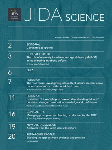
n intrabony defects – a defect that is defined as ‘within or inside the bone’ and can be further classified based on several characteristics (Figure 1); and, n furcation defects – affecting the root separation areas of multi-rooted teeth.
Affecting two adjacent teeth (inter-root):3
n crater – a bowl-shaped defect with loss of interproximal bone between two adjacent teeth but the buccal and lingual alveolar crests are more coronal; n ramp – interproximal bone loss between two adjacent teeth and buccal/lingual bone loss (so the buccal and lingual alveolar crests are not at the same levels); and,
Dr Varkha Rattu
Periodontology Unit
Centre for Host-Microbiome Interactions
Dental Institute
King’s College London United Kingdom
n to demonstrate the concept of MINST; and,
n to outline the clinical research supporting MINST as a treatment approach in the management of intrabony defects.
n plane – interproximal bone loss between two adjacent teeth with buccal and lingual bone loss (so the buccal and lingual alveolar crests are at the same level) – this is usually defined as a zero-wall defect, as there are no bony walls coronal to the defect.
Intrabony defects are classified using the following:
Number of bony walls housing the defect (Figure 1):
n one-wall;
n two-wall;
n three-wall; or,
n combinations.
Prof. Luigi Nibali
Periodontology Unit
Centre for Host-Microbiome Interactions
Dental Institute
King’s College London United Kingdom
Figure 1: Schematic diagram describing the number of walls in intrabony defects. A: one-wall intrabony defect; B: two-wall intrabony defect; C: three-wall intrabony defect; D: interproximal crater. (Figure reproduced with permission.)14
Corresponding author:
Dr Varkha Rattu, Periodontology Unit, Centre for Host-Microbiome Interactions, Dental Institute, King’s College London, United Kingdom E: varkha.rattu@kcl.ac.uk
Extension of the defect around the surfaces of the tooth:
n interproximal only;
n trench – the defect extends to the buccal and/or lingual/palatal surfaces of the same tooth but there is still an outer bony wall;
n dehiscence – extends to the buccal and/or lingual/palatal surface of the tooth with complete loss of bone over the respective root surface; or,
n moat – the defect extends circumferentially around the whole tooth.
Width – the width of the defect based on the long axis of the tooth
It is important to note that different thresholds have been used in the literature:
n narrow – ≤25°;
n average – 26°-36°; and, n wide – ≥37°.
Depth:
n shallow – <3mm; and, n deep – ≥3mm.
Intrabony defects and regenerative surgery
Surgical regenerative techniques aim to restore the attachment apparatus lost as a consequence of periodontitis (alveolar bone, periodontal ligament and cementum) and form part of the third step of periodontal therapy. 4 These approaches typically incorporate biomaterials, such as enamel matrix derivative (EMD), bone replacement grafts (BRGs) and barrier membranes, to enhance outcomes.5 Regenerative procedures rely heavily on the stability of the blood clot within the defect, as it serves as the foundation for new tissue formation and successful healing. This stability is influenced by the containment provided by sufficient bony walls and an intact overlying soft tissue profile. Bony walls act as a natural scaffold to support the blood clot, ensuring that it remains stable within the defect, while the soft tissue compartment helps to protect the clot from external disturbances and promotes primary intention healing. Together, these factors create a conducive micro-environment for cellular proliferation, angiogenesis, and tissue regeneration. 6,7 Therefore, defects with adequate containment (increasing number of walls, minimal extension, narrow width, and adequate depth) are more likely to achieve predictable regenerative outcomes. To optimise blood clot containment, there is growing emphasis on the importance of preserving and optimising both the hard and so tissue architecture during surgical procedures. Consequently, surgical techniques have evolved with a growing emphasis on papilla preservation techniques. They prioritise maintaining the so tissue compartment surrounding the defect to provide stability to the flap and aid primary intention healing – principles that are fundamental in regeneration. With this in mind, avoiding incisions or flap elevation, such as in minimally invasive non-surgical therapy (MINST), theoretically creates the most favourable so tissue environment to naturally support and enhance the regenerative process.
MINST
MINST is a precision-driven non-surgical approach designed to manage intrabony defects while minimising trauma to the surrounding tissues. MINST is particularly effective for deep intrabony defects with favourable bony wall configurations, as it enhances healing through minimally invasive means while leveraging the body’s
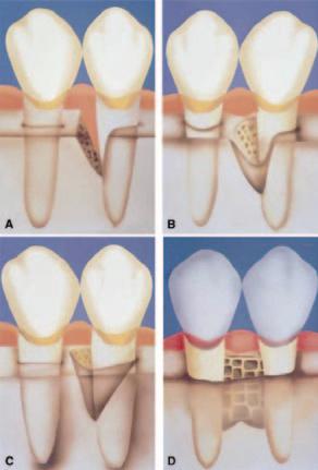
NSK:
n G16 tip (as per image)
n P20 tip (as per image)
n P21R + P21L
n P25R + P25L
EMS: n PS tip (as per image)
natural regenerative capacity. This approach represents a shi towards preserving and utilising the intrinsic healing potential of periodontal tissues. Research detailing MINST consistently highlights core principles:8-10
n incorporating magnification, such as loupes, to enhance precision; n the use of ultrasonic devices (with a particular focus on piezons) ± minicurettes with fine and delicate tips (Figure 2); n thorough root surface debridement of the affected areas, ensuring effective biofilm removal; and,
n exercising care to preserve so tissue stability and minimise trauma to the so tissue.
To reduce the risk of damage to the so tissue, it is advisable to utilise subpapillary access with a focus on minimising trauma to the most coronal part of the interdental papilla.11 The use of a local anaesthetic without a vasoconstrictor has been advocated, to encourage the defect to fill with blood following the procedure and support a stable blood clot.11 However, the relative importance of subpapillary access and anaesthetic without a vasoconstrictor has not been specifically investigated.
MINST has emerged as a promising approach for the management of intrabony defects. It is noteworthy that regeneration in these cases has been assessed clinically and radiographically, without histological confirmation. Therefore, true tissue regeneration cannot be guaranteed. However, this limitation also applies to most commonly performed regenerative procedures in clinical practice. Across various studies (Table 1), MINST has consistently demonstrated its ability to promote periodontal regeneration while preserving so tissue stability. Randomised controlled trials (RCTs) and retrospective studies highlight MINST’s effectiveness in reducing probing pocket depths (PPDs) and improving clinical attachment levels (CALs). For instance, studies have reported PPD reductions and CAL gains comparable to surgical techniques, over follow-up periods of 12 to 24 months, with some improvements sustained for up to five years. The
Figure 2: Examples of delicate tips for piezon handpieces.
Study (study Intervention
Table 1: Summary of clinical parameters assessed in various MINST-related studies.
design) (patients’ age) used (final follow-up)
Ribeiro et al Intrabony T: 13 (13; Microscope, Monthly
cohort) access, piezo-electric devices with thin tips
12
Abbreviations: C – control; EMD – enamel matrix derivative; LA – local anaesthetic; M-MIST – modified-minimally invasive surgical technique; SFA – single flap approach; T – test.
incorporation of adjunctive treatments, such as EMD, has shown comparable outcomes to utilising minimally invasive techniques alone, further supporting MINST’s versatility. A key advantage of MINST is its ability to achieve these outcomes with minimal so tissue recession (0.1-0.7mm) and significantly reduced chair time, ranging from 10 to 29 minutes, compared to 55-61 minutes for traditional minimally invasive surgical techniques.8,12,13 Collectively, these findings position MINST as an effective and efficient, albeit not always predictable, technique for intrabony defect management, emphasising its longterm stability and patient-centred advantages. Direct comparisons with other versions of non-surgical therapy are still lacking in the literature.
2. Papapanou PN, Sanz M, Buduneli N, et al. Periodontitis: consensus report of workgroup 2 of the 2017 World Workshop on the Classification of Periodontal and Peri-Implant Diseases and Conditions. J Periodontol. 2018;89 (Suppl. 1):s173-s182.
3. Nibali L, Cortellini P. Periodontal osseous defects: a treatment-oriented classification to guide regenerative treatment planning. Int J Periodontics Restorative Dent. 2025;45(3):301-315.
4. Sanz M, Herrera D, Kebschull M, et al. Treatment of stage I-III periodontitis – The EFP S3 level clinical practice guideline. J Clin Periodontol. 2020;47 (Suppl. 22):4-60.
5. Nibali L, Koidou VP, Nieri M, Barbato L, Pagliaro U, Cairo F. Regenerative surgery versus access flap for the treatment of intra-bony periodontal defects: a systematic review and meta-analysis. J Clin Periodontol. 2020;47 (Suppl. 22):320-351.
6. Polimeni G, Xiropaidis AV, Wikesjö UM. Biology and principles of periodontal wound healing/regeneration. Periodontol 2000. 2006;41:30-47.
7. Sculean A, Chapple IL, Giannobile WV. Wound models for periodontal and bone regeneration: the role of biologic research. Periodontol 2000. 2015;68(1):7-20.
8. Ribeiro FV, Casarin RC, Palma MA, Júnior FH, Sallum EA, Casati MZ. Clinical and patient-centered outcomes aer minimally invasive non-surgical or surgical approaches for the treatment of intrabony defects: a randomized clinical trial. J Periodontol. 2011;82(9):1256-1266.
9. Nibali L, Pometti D, Chen TT, Tu YK. Minimally invasive non-surgical approach for the treatment of periodontal intrabony defects: a retrospective analysis. J Clin Periodontol. 2015;42(9):853-859.
10. Nibali L, Yeh YC, Pometti D, Tu YK. Long-term stability of intrabony defects treated with minimally invasive non-surgical therapy. J Clinical Periodontol. 2018;45(12):14581464.
11. Nibali L, Koidou V, Salomone S, et al. Minimally invasive non-surgical vs. surgical approach for periodontal intrabony defects: a randomised controlled trial. Trials. 2019;20(1):461.
12. Mehta J, Montevecchi M, Garcia-Sanchez R, et al. Minimally invasive non-surgical periodontal therapy of intrabony defects: a prospective multi-centre cohort study. J Clin Periodontol. 2024;51(7):905-914.
13. Aimetti M, Ferrarotti F, Mariani GM, Romano F. A novel flapless approach versus minimally invasive surgery in periodontal regeneration with enamel matrix derivative proteins: a 24-month randomized controlled clinical trial. Clin Oral Investig. 2017;21(1):327-337.
14. Papapanou PN, Tonetti MS. Diagnosis and epidemiology of periodontal osseous lesions. Periodontol 2000. 2000;22:8-21.
15. Anoixiadou S, Parashis A, Vouros I. Enamel matrix derivative as an adjunct to minimally invasive non-surgical treatment of intrabony defects: a randomized clinical trial. J Clin Periodontol. 2022;49(2):134-143.
Quiz
Submitted by Dr Patrick Quinn, Health Service Executive and Cork University Dental School and Hospital.
Jack, who is nine years of age, is brought to his dental appointment by his grandmother, who would like him to have a dental check-up and is also wondering about the need for fissure sealants as other children in his class have had them done.
Questions
1. How would you obtain a legally valid consent in this case?
2. Having explained the requirements for a legally valid consent, Jack’s grandmother informs you that she is his legal guardian. How would you satisfy yourself that this is the case?
3. If the scenario was different and Jack was brought to the clinic by his school teacher having avulsed an upper central incisor, how would you approach the issue of consent?
4. If Jack was brought to the clinic by his teacher following a fall at school and he appeared to have an enamel fracture on his upper central incisor, would you approach things differently?
Answers on page 18
Dr Hannelie Edgar Dental Core Trainee Northern Ireland Medical and Dental Training Agency
Dr Amanda Willis Consultant in Oral Medicine
School of Dentistry
Royal Victoria Hospital
Belfast
Prof. John Marley Consultant Oral Surgeon/Honorary Professor School of Dentistry
Royal Victoria Hospital
Belfast
Corresponding author: Dr Hannelie Edgar E: hannelieedgar@hotmail.com
The root cause: investigating intermittent inferior alveolar nerve paraesthesia from a multi-rooted third molar
Précis: Pericoronitis-associated inferior alveolar nerve paraesthesia was resolved following coronectomy of a morphologically complex mandibular third molar in intimate relationship with the inferior alveolar nerve.
Abstract
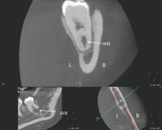
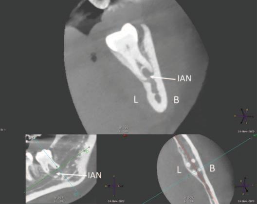
This paper describes a case of intermittent inferior alveolar nerve (IAN) paraesthesia temporally related to episodes of pericoronitis affecting a mandibular third molar in close association with the IAN. A 23-year-old female patient was referred to oral surgery by oral medicine with a two-month history of intermittent alternating numbness and pain associated with the lower left lip and chin. Clinical and radiographic investigations confirmed a partially erupted left mandibular third molar in close association with the IAN. Subsequent conebeam computed tomography (CBCT) revealed that the lower left 8 (LL8) had two mesial and two distal roots. The IAN pathway grooved the distal roots and perforated the mesial. A coronectomy was completed, resulting in the resolution of symptoms.
Journal of the Irish Dental Association Science October/November 2025;1(1):7-10
Introduction
Mandibular third molars are innervated by the inferior alveolar nerve (IAN), part of the third branch of the trigeminal nerve, which arises deep to the lateral pterygoid muscle and passes through the inferior alveolar foramen to enter the mandible. The IAN is a mixed nerve with its sensory component supplying sensation to the mandibular teeth, lower lip and chin.1 Paraesthesia is a transient or chronic sensory abnormality of the nervous system, which can manifest as a tingling, sharp or numb sensation. Systemic causes of paraesthesia include connective tissue diseases, diabetes, neurodegenerative disorders and metastatic disease, whereas local factors include mechanical, thermal and toxic insults.2 IAN paraesthesia is a known risk of mandibular third molar surgery, occurring temporarily in up to 2% of patients and permanently in 0.5% of patients.3 Coronectomy is an effective management option to reduce the risk of nerve injury when managing mandibular third molars closely associated with the IAN canal. Careful case selection is crucial, as there are several contraindications relating to both tooth and patient. The guideline relating to the management of mandibular third molars by the Royal College of Surgeons, England, contraindicates coronectomy for third molars with caries into the pulp, apical disease or mobility. Patient contraindications include those who are immunocompromised or at increased risk of osteoradionecrosis.4 Horizontal impaction is also considered a relative contraindication for coronectomy due to the potential risk of inadvertent damage to the IAN when cutting inferiorly for decoronation.5 Renton et al. further caution against coronectomy in third molars with conical roots due to the increased risk of root mobilisation and failure.6
Case history
A 23-year-old female patient was referred to the oral medicine department by her general dental practitioner querying atypical facial pain, with a two-month history of intermittent numbness of the lower le lip and chin region, with occasional pins and needles sensation. Medically, the patient had well-controlled asthma and no known drug allergies. She reported multiple episodes of numbness affecting the le side of her lower lip and chin occurring daily for approximately 20 minutes with complete resolution aerwards. The patient had a normal gross cranial nerve examination with no sensory deficit noted on testing on the day of examination. The area of paraesthesia was described by the patient as confined to the le aspect of the lower lip and chin, never crossing the midline and extending to the lower border of the mandible.
Blood investigations were completed to evaluate potential deficiencies as a cause for the patient’s symptoms, including a full blood count, iron, folate, B12, and thyroid function tests. Systemic causes of neuropathy, including diabetes and
connective tissue diseases such as scleroderma and Sjögren’s syndrome, were evaluated with a blood glucose, glycosylated haemoglobin and connective tissue screen. All results were within normal ranges, encouraging further exploration of local causative factors.
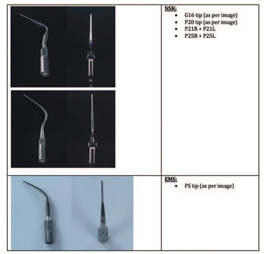
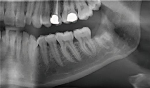
A partially erupted lower le third molar with a prominent operculum was noted during the intraoral examination. The orthopantomogram taken suggested a close relationship of the tooth with the IAN due to a relative rarefaction associated with the distal root, indicating proximity of the nerve canal with little or no bone interposition (Figure 1). Caries was also noted occlusally on the lower le second molar and a deep restoration on the upper le first molar. Following the exclusion
of a systemic cause by the oral physicians, a referral was made to oral surgery to assess the possible correlation of these clinical and radiographic findings with the presenting complaint.
At the oral surgery consultation, the patient also noted a four-week history of intermittent discomfort in the LL8 region, described as throbbing pain with associated swelling of the gum, made worse by chewing and applying pressure to the tooth. This presentation was in keeping with recurrent pericoronitis. A cone-beam computed tomography (CBCT) scan was undertaken, which identified four roots of LL8 and associated loss of IAN canal cortication. The IAN pathway grooved the distobuccal root and passed between/penetrated the mesial roots (Figures 2 and 3).
FIGURE 1: Orthopantomogram showing the partially erupted lower left third molar.
FIGURE 2: Association of IAN with mesial roots (L = lingual, B = buccal, IAN = inferior alveolar nerve).
FIGURE 3: Association of IAN with distal roots (L = lingual, B = buccal, IAN = inferior alveolar nerve).
Given the temporal association between the history of paraesthesia, episodes of pericoronitis and the clinical findings, it was agreed that there was a likely causative association between the pericoronitis and the intimate relationship of LL8 with the IAN. Complete removal of the LL8 was deemed high risk for iatrogenic IAN damage. To reduce this risk, the patient was consented for a coronectomy. The coronectomy was undertaken with a short general anaesthetic without complication, and at the two-month postoperative review, the patient had complete resolution of symptoms and no postoperative sensory deficit. The orthopantomogram taken at this review shows no evidence of periapical infection and the distal retained root is satisfactorily subcrestal (Figure 4). It is acknowledged that following the coronectomy, the mesial root is not at the optimum subcrestal level and has a small radiodense area, which may be residual enamel. This necessitates close follow-up.
Discussion
This case report describes a rare example of IAN sensory disturbance that was temporally associated with and likely caused by recurrent episodes of pericoronitis around a partially erupted lower wisdom tooth. When considering differentials for paraesthesia, one should consider potential systemic and local causes. Paraesthesia of the lip and chin has been associated with the term ‘numb chin syndrome’, for which there are several underlying pathologies suggested, including neuropathies related to diabetes and multiple sclerosis, intracranial lesions, and benign and malignant tumours.7 Cranial nerve neuropathies occur more frequently in patients with diabetes compared with the general population.8 This patient reported no symptoms suggestive of diabetes, which was further excluded by a normal blood glucose and glycosylated haemoglobin level. Connective tissue diseases including scleroderma, rheumatoid arthritis and Sjögren’s syndrome can also be associated with trigeminal sensory neuropathy manifesting as numbness or pain.9 Multiple sclerosis is a chronic inflammatory neurodegenerative disease, which can have orofacial manifestations, one of the most common being trigeminal sensory neuropathy.10 The patient exhibited no features of systemic disease, and all bloodwork investigations were normal.
In the absence of an odontogenic cause of IAN paraesthesia, clinicians should strongly consider exploring if the symptom could be a neurological manifestation of malignancy. Primary and metastatic lesions local to the nerve can cause paraesthesia due to perineural spread or compression of nerve tissue.11 Trigeminal nerve paraesthesia has been found in patients diagnosed with peripheral nerve sheath neoplasia, osteosarcoma of the mandible,
multiple myeloma, and schwannoma lesions.12 Numbness of the chin has also been identified as a sign of relapse and progression of metastatic disease in breast cancer and lymphoma patients.13
Paraesthesia of the IAN has been noted in the literature in instances of periapical lesions affecting mandibular teeth. A case series by Devine et al. 14 discusses 22 cases of patients experiencing numbness, pain and/or paraesthesia of the lip and chin resulting from periapical lesions in premolars and molars. Complete resolution of symptoms was achieved in those patients treated with extraction or root canal treatment of the affected teeth. These findings have also been reflected in a recent case report by Censi et al., which discusses two instances of infection-induced IAN paraesthesia manifesting as numbness of the chin and lip.15 In both patients, there was radiographic evidence of periapical pathology near the mental foramen and patient symptoms resolved following root canal therapy or extraction.
In the current case, there was no radiographic evidence of periapical infection. The intermittent numbness described by the patient preceded the pericoronitis symptoms by a few months; however, the enlarged operculum and impacted mandibular third molar were noted at the initial consultation. The conclusion is drawn from the temporal association with the pericoronitis events and the relationship of the IAN with the tooth, that inflammation around the crown of the partially erupted LL8 and surrounding periodontal tissues led to pressure effects with episodic compression/tension. This compression may have resulted in distortion of the IAN canal with subsequent paraesthesia. A similar case of transient paraesthesia of the lower lip and chin associated with chronic pericoronitis has been previously highlighted in a case report by Di Lauro et al.16 involving a mandibular third molar in a contiguous relationship to the IAN canal. A coronectomy was completed, resulting in a significant reduction of paraesthesia.
IAN injury is avoidable and the risk can be mitigated with thorough case history, clinical examination and radiographic assessment, with consequent appropriate treatment planning. Failure to adequately assess mandibular third molars and their proximity to the IAN can cause postoperative trigeminal sensory neuropathy resulting in long-term chronic pain and disability for up to 70% of patients.17 On radiographic plain films, diversion of the canal, darkening of the root where crossed by the canal, narrowing of the canal and interruption of the canal walls are noted to significantly increase the risk of damage during third molar surgery.18 Consequent further characterisation of canal integrity by CBCT is highly sensitive for predicting intraoperative IAN exposure and the associated increased risk of postoperative IAN paraesthesia.19 However, CBCT should not be the first-line imaging modality of choice, but rather applied in instances where suspicions are raised on 2D imaging. 4 Alternative treatment options should then be explored in cases demonstrating these radiographic features.
The deliberate retention of pathology-free mandibular third molar roots through coronectomy reduces the risk of IAN injury with no adverse impact on the incidence of dry socket or periapical infection of the retained roots.6 It is possible with time that retained roots can migrate away from the IAN, allowing for their extraction under local anaesthetic if indicated, with lower risk of nerve injury. In this particular case, migration of the retained roots may result in future paraesthesia if the intimately involved IAN moves with them. This finding was reported by Renton and Drage (2002), where a patient experienced a return of paraesthesia eight years post coronectomy of a right mandibular third molar perforated by the IAN. 20 Radiographic
FIGURE 4: Postoperative radiograph at two months.
investigation revealed unusual superior migration of the IAN canal as well as the retained roots. Longer-term follow-up is planned to thoroughly evaluate this case.
Conclusion
This case report highlights an unusual presentation of intermittent IAN
References
1. Carter RB, Keen E. The intramandibular course of the inferior alveolar nerve. J Anat. 1971;108(Pt 3):433-440.
2. Giuliani M, Lajolo C, Deli G, Silveri C. Inferior alveolar nerve paresthesia caused by endodontic pathosis: a case report and review of the literature. Oral Surg Oral Med Oral Pathol Oral Radiol Endod. 2001;92(6):670-674.
3. NICE. Guidance on the extraction of wisdom teeth. 2000. https://www.nice.org.uk/guidance/ta1. Accessed June 20, 2024.
4. Royal College of Surgeons, England. Parameters of care for patients undergoing mandibular third molar surgery. 2020. https://www.rcseng.ac.uk//media/files/rcs/fds/guidelines/3rd-molar-guidelines—april-2021.pdf. Accessed June 20, 2024.
5. Pogrel MA, Lee JS, Muff DF. Coronectomy: a technique to protect the inferior alveolar nerve. J Oral Maxillofac Surg. 2004;62(12):1447-1452.
6. Renton T, Hankins M, Sproate C, McGurk M. A randomised controlled clinical trial to compare the incidence of injury to the inferior alveolar nerve as a result of coronectomy and removal of mandibular third molars. Br J Oral Maxillofac Surg. 2005;43(1):7-12.
7. Assaf AT, Jürgens TP, Benecke AW, et al. Numb chin syndrome: a rare and oen overlooked symptom. J Oral Facial Pain Headache. 2014 ;28(1):80-90.
8. Tracy JA, Dyck PJ. The spectrum of diabetic neuropathies. Phys Med Rehabil Clin N Am. 2008;19(1):1-26.
9. Smith JH, Cutrer FM. Numbness matters: a clinical review of trigeminal neuropathy. Cephalalgia. 2011;31(10):1131-1144.
10. Chemaly D, Lefrancois A, Pérusse R. Oral and maxillofacial manifestations of multiple sclerosis. J Can Dent Assoc. 2000;66(11):600-605.
CPD questions
To claim CPD points, go to the MEMBERS’ SECTION of www.dentist.ie and answer the following questions:
1. A potential systemic cause of inferior alveolar nerve paraesthesia could include:
paraesthesia associated temporally with pericoronitis of a partially erupted mandibular third molar where the IAN perforates one root and is in close proximity to the other. Treatment planning was guided by evidence-based CBCT characterisation to reduce the risk of iatrogenic IAN injury, resulting in resolution of the patient’s symptoms to date, with no postoperative sensory deficit.
11. Colella G, Giudice A, Falcone U, Siniscalchi G, Guastafierro S. Chin numbness: a symptom that should not be underestimated: a review of 12 cases. Am J Med Sci. 2009;337(6):407-410.
12. Carter E, Yilmaz Z, Devine M, Renton T. An update on the causes, assessment and management of third division sensory trigeminal neuropathies. Br Dent J. 2016;220(12):627-635.
14. Devine M, Yilmaz Z, Hirani M, Renton T. A case series of trigeminal nerve injuries caused by periapical lesions of mandibular teeth. Br Dent J. 2017;222(6):447-455.
15. Censi R, Vavassori V, Borgonovo AE, Re D. Infection related inferior alveolar nerve paresthesia in the lower premolar teeth. Case Rep Dent. 2016;2016:2623507.
16. Di Lauro AE, Boariu M, Sammartino P, et al. Lower third molar inclusion associated with paraesthesia: a case report. Exp Ther Med. 2021;22(2):826.
17. Renton T, Yilmaz Z. Profiling of patients presenting with posttraumatic neuropathy of the trigeminal nerve. J Orofac Pain. 2011;25(4):333-344.
18. Rood JP, Shehab BN. The radiological prediction of inferior alveolar nerve injury during third molar surgery. Br J Oral Maxillofac Surg. 1990;28(1):20-25.
19. Park W, Choi JW, Kim JY, Kim BC, Kim HJ, Lee SH. Cortical integrity of the inferior alveolar canal as a predictor of paresthesia aer third-molar extraction. J Am Dent Assoc. 2010;141(3):271-278.
20. Drage NA, Renton T. Inferior alveolar nerve injury related to mandibular third molar surgery: an unusual case presentation. Oral Surg Oral Med Oral Pathol Oral Radiol Endod. 2002;93(3):358-361.
2. Which of the following is considered a relative contraindication in coronectomy treatment of mandibular third molars?
3. Permanent inferior alveolar nerve damage following surgical removal of mandibular third molars occurs in what percentage of patients?
l A. Osteoporosis
l B. Myasthenia gravis
l C. Scleroderma
l A. Mesiangular impaction
l B. Horizontal impaction
l C. Distoangular impaction
l A. 0.5%
l B. 2.0%
l C. <0.1%
Marc Lewis Emrys Edwards Centre for Dentistry
Queen’s University Belfast United Kingdom
Shamira McKenna School of Psychology
Queen’s University Belfast United Kingdom
Maria Hussain Centre for Dentistry
Queen’s University Belfast United Kingdom
Corresponding author: Marc Lewis Emrys Edwards, Centre for Dentistry, Grosvenor Road, Royal Victoria Hospital, Belfast BT12 6BP
E: Marc.edwards@qub.ac.uk
Evaluation of a workshop to develop dental undergraduates’ behaviour change conversation knowledge and confidence
Précis: A workshop on the Dental RECUR Brief Negotiated Interview builds dental undergraduates’ knowledge of, and confidence delivering, a behaviour change conversation with a dental patient.
Abstract
Introduction: The General Dental Council’s Safe Practitioner framework of behaviours and outcomes for dental professional education outlines the need for dental undergraduates to learn evidence-based approaches to clinical practice. The Dental RECUR Brief Negotiated Interview (DR-BNI) is an effective oral health behaviour change intervention that draws upon psychological frameworks of disease prevention, behaviour change science, and patient-facing communication skills. Training in this approach involves developing participants’ knowledge of childhood caries, and confidence applying personalised preventive advice to clinical practice via supervised role-play.
Materials and methods: This study explored the effectiveness of a DR-BNI training workshop on developing dental undergraduates’ behaviour change conversation knowledge and confidence. Seventeen participants completed a pre-post evaluation, which assessed their knowledge of DR-BNI-related topics and confidence in applying relevant skills to clinical practice.
Results: Participants’ knowledge of all topics, including the development of dental caries in children, motivational interviewing, and behaviour change theory, significantly increased following the workshop. Participants’ confidence in applying skills for delivering a behaviour change conversation with a dental patient also significantly increased.
Conclusion: The DR-BNI represents a useful model for developing dental undergraduates’ behaviour change conversation knowledge and confidence.
Journal of the Irish Dental Association Science October/November 2025;1(1):11-15
Introduction
The relevance and value of behavioural sciences in dentistry have been increasingly recognised in undergraduate curricula in recent decades.1,2 The latest General Dental Council’s (GDC) Safe Practitioner framework of behaviours and outcomes for dental professional education outlines the need for graduates to be able to: “Explain and evaluate psychological and sociological concepts and theoretical frameworks of health, illness, behavioural change and disease and how these can be applied in clinical practice (C1.7)”.3 Additionally, they must be able to: “Provide patients/carers with comprehensive, personalised preventive advice, instruction and intervention in a manner which is accessible, promotes self-care and motivates patients/carers to comply with advice and take responsibility to maintain and improve oral health (C2.5.1)”. Developing appropriate and engaging teaching methods to meet these objectives can be a challenge, especially given an educational context in which some undergraduate dental students view communication skills and patient adherence components of the behavioural sciences curriculum to be ‘more boring’ and ‘less relevant’ than their other, more clinicalbased, training.4 A possible reason for this is that there is typically less teaching time dedicated to behavioural sciences topics in the dental education curriculum,5 and few dental schools have a specifically appointed lecturer with psychology expertise to deeply integrate those topics with other curricular themes in dedicated modules. While some dental schools may teach dental undergraduates individual evidence-based behavioural approaches to disease prevention, such as the Irish Health Service Executive (HSE) Making Every Contact Count Programme, few actively train students in specific behavioural interventions. Discussions centred on behavioural modification represent one preventive strategy employed by dental care practitioners to encourage patients to adopt healthier habits and routines. Motivational interviewing (MI) is a behaviour change conversation technique that involves assessing individuals’ readiness to change, enhancing their self-efficacy, and facilitating adherence to healthcare recommendations.6 Dentists generally do not have MI as part of their compulsory training, but may learn this skill as part of continuing personal development. Recent research has highlighted the
potential benefits of behaviour change conversations in promoting better oral health, suggesting that they can be effective in managing dental diseases,7 enhancing patient motivation to adopt healthier oral hygiene behaviours,8 and building clinicians’ communication strategies to improve patient engagement.9 Despite the benefits of behaviour change conversations in the promotion of better oral health, focus groups involving various members of the dental team in England (dentists, practice managers, dental therapists/hygienists, dental nurses) report numerous challenges to supporting dental patients’ behaviour change.10 In particular, many struggle with conversational elements, including building rapport and expressing empathy with parents of children with tooth decay. Instead, they report mostly relying on an information-giving approach, contrary to growing evidence that motivational approaches to behaviour change (i.e., eliciting change) can be especially effective at preventing oral disease in children.11 Training dental students in the application of behaviour change skills provides a strong educational tool based on psycho-educational theories.12 Theory-informed training can effectively increase dental care professionals’ motivation to discuss behaviour change with patients.13 Although numerous studies have assessed the impact of general communication training on dental undergraduates’ ability to interact with patients effectively,14-16 to the best of our knowledge, no studies to date have evaluated the training of dental students in a specific behaviour change conversation intervention.
The Dental RECUR Brief Negotiated Interview (DR-BNI) is a behavioural intervention for caries prevention, with strong evidence of its effectiveness (29%) in the reduction of new dental caries in children aged seven to nine years.11 The approach involves dental nurses applying motivational interviewing skills and behaviour change techniques in a 30-minute behaviour change conversation to motivate parents of children with caries to develop one or two personalised goals based on national preventive recommendations.17
The DR-BNI draws upon frameworks of behaviour change science, clinical communication skills, and disease prevention. As such, it has strong potential as an educational model for dental undergraduates to develop knowledge and competency in engaging a dental patient in a behaviour change conversation. DRBNI-related communication skills could be implemented by dental students during both simulated and real interactions with dental patients throughout their training and in future practice. There is flexibility in how, and to whom, the DR-BNI is taught; a DR-BNI hands-on workshop has been successfully delivered to behavioural scientists, academics, and professionals.18 Typically, DR-BNI training involves teaching participants about the problem of child dental caries, motivational interviewing, behaviour change science, and information on how to deliver the DRBNI in clinical practice. This is followed by supervised role-play, during which participants practice DR-BNI skills and receive feedback from a DR-BNI trainer. All components of the DR-BNI training could be readily adapted for delivery to dental undergraduates, as students are regularly taught information and skills that they then implement in practice under the supervision of an academic or clinician. Brief hands-on workshops delivered to a small number of participants can be effective as a teaching and learning methodology for communication skills training,19 knowledge and confidence development,20 and as a means of identifying challenges for informing future practice.21 As the DR-BNI training has not previously been delivered to dental undergraduates, a brief hands-on workshop format would help to identify the potential benefits and challenges of adapting it for this demographic before it can be evaluated more widely within the undergraduate behavioural sciences curriculum. Therefore, this study aimed to develop dental undergraduates’ behaviour change conversation skills in a training workshop on the DR-BNI.
Method and materials
The study was reviewed and approved by the Faculty of Medicine, Health and Life Sciences Research Ethics Committee (Faculty REC) in accordance with the Proportionate Review process at Queen’s University Belfast (Faculty REC Reference Number: MHLS 24_121).
Participants
Undergraduate dental students across years one to five were invited to take part in an in-person behaviour change conversation skills workshop via an invitation email. Participants were 17 dental undergraduates enrolled in the five-year Bachelor of Dental Surgery (BDS) programme at Queen’s University Belfast during the 2023-2024 academic year, who had registered to participate in the DR-BNI workshop via an online form. Participants were between 19 and 26 years of age, and none had completed a previous degree. Participants were composed of students from year one (52.9%), year two (5.9%), year four (35.3%), and year five (5.9%) of their studies. There were a range of ethnicities, with most identifying as ‘White British’ (17.6%), ‘Irish’ (17.6%), ‘Any other ethnic group’ (17.6%), or ‘Pakistani’ (11.8%). Participants were compensated with a £10 Amazon voucher and provided with a completion certificate following attendance at the workshop. Participants’ identifiable information, including email addresses, were kept confidential and stored in a password-encrypted university database. When participants completed the consent form, they were asked to create a nonidentifiable unique ID code, which they used when completing all questionnaires.
Procedure
Pre-evaluation
Participants who consented to participate in the evaluation of the DR-BNI training workshop were sent a ‘pre-evaluation questionnaire’ via Microso Forms in advance to be completed and returned to the Chief Investigator before the workshop. This questionnaire collected participants’ demographic information, their current knowledge of DR-BNI-related topics such as oral health behaviour change and motivational interviewing, and how confident they are in applying relevant skills to clinical practice. This questionnaire also collected qualitative data relating to participants’ expectations for the training.
Workshop
During the 90-minute workshop, a behavioural scientist trained in the DR-BNI intervention (the Chief Investigator, ME) delivered a brief talk on how it was developed and evaluated. This was followed by hands-on training including supervised role-play facilitated by the Chief Investigator (ME) and the Co-Investigator (MH).
The role-play involved participants working in pairs, with one participant roleplaying as a dental student delivering the DR-BNI, and the second participant role-playing as a parental caregiver receiving the DR-BNI. Finally, there was a wholegroup discussion, during which participants shared whether the training met their expectations and if they felt they had successfully met the learning objectives.
Post evaluation
A 13-item post-evaluation questionnaire was developed using the four-level Kirkpatrick Model22,23 to assess: how engaging and relevant participants found the training (Level 1: Reaction); the extent to which they acquired the intended knowledge, skills, and confidence (Level 2: Learning); the extent to which they were able to apply what they learned (Level 3: Behaviour); and, whether training contributed to the learning objectives of the dental undergraduate curriculum
Item Kirkpatrick
I was satisfied with the DR-BNI training overall.
I successfully learned how to deliver an oral health behaviour change conversation.
I feel confident I can now deliver a behaviour change conversation with a dental patient.
This workshop provided content that is relevant to my current training.
The training materials provided me with enough information to deliver the DR-BNI.
Level 1 (Reaction) 4.88 0.34
Strongly agree
Level 2 (Learning) 4.69 0.48 Strongly agree
Level 3 (Behaviour) 4.31 0.48 Somewhat agree
Level 4 (Results) 4.94 0.25 Strongly agree
Level 2 (Learning) 4.44 0.89 Strongly agree I would recommend the workshop to others.
The workshop was a worthwhile use of my time.
Level 1 (Reaction) 4.88
Level 1 (Reaction) 4.88
Strongly agree
Strongly agree I am already seeing positive results from the workshop.
Level 4 (Results) 4.06
agree I am expecting positive results from the workshop in the future.
The information in the workshop is applicable to my training.
The presentation style of the facilitator(s) contributed to my learning experience.
Level 4 (Results)
Level 4 (Results)
Level 2 (Learning)
agree
agree
Strongly agree I gained useful patient-facing communication skills in the workshop.
Level 2 (Learning)
Strongly agree I understood what was expected of me during the workshop.
Level 1 (Reaction) 4.69
Strongly agree
Note: *significant at p < 0.05.
(Level 4: Results). This questionnaire was completed by participants one week following the workshop to allow time for participants to practice using MI communication skills with either a dental patient or friend/family member.
Results
Post-training evaluation
Participants indicated on a five-point Likert scale (1 = strongly disagree, 5 = strongly agree) the extent to which they agreed with the statements, with participants either somewhat or strongly agreeing with all post-training evaluation questionnaire items (M = 4.31, M = 5.00) across all four domains of Kirkpatrick’s Model (Table 1).
Pre-post evaluation: knowledge
Participants reported their knowledge of DR-BNI-related topics on a five-point Likert scale (1 = not at all knowledgeable, 5 = extremely knowledgeable), with a paired-sample t-test finding that participants’ knowledge of all DR-BNI-related topics and skills had significantly increased following the training (Table 2).
Pre-post evaluation: confidence
Participants reported their confidence in applying DR-BNI-related skills to clinical practice on a five-point Likert scale (1 = not at all confident, 5 = extremely confident), with a paired-sample t-test finding that participants’ confidence had significantly increased following the training (Table 3).
Discussion
This study aimed to evaluate the effectiveness of a DR-BNI workshop on dental undergraduates’ behaviour change conversation knowledge and confidence, and to identify the benefits and challenges to embedding training in this approach within the undergraduate behavioural sciences curriculum. The findings of this study demonstrate the potential of DR-BNI training to enhance dental undergraduates’ knowledge of, and confidence in, delivering a behaviour change conversation with a dental patient. The results of this study may benefit both dental educators and students. The learning objectives of DR-BNI training align closely with those of the GDC’s Safe Practitioner framework, meaning that this may be an effective educational tool
Table 1: Participants’ post-evaluation agreement with statements about the training, including relevant Kirkpatrick Model
Table 2: Pre-post training knowledge.
General communication with a dental patient
Note: *significant at p < 0.05.
for developing students’ communication skills for delivering personalised preventive advice, and their knowledge and application of theoretical frameworks of health, illness, behavioural change and disease. Previous research in this area has recommended better equipping dental students with the necessary tools to adopt a more holistic and person-centred approach to improving patient health outcomes.24 Incorporating DR-BNI training more comprehensively within the dental curriculum has the potential to effectively address the existing gaps in students’ exposure to behavioural sciences and, more specifically, oral health behaviour change conversations.
The potential benefits of DR-BNI training extend beyond the immediate educational context to broader public health outcomes. Equipping future dental practitioners with communication skills to effectively engage patients in an oral health behaviour change conversation could result in reduced disease prevalence. Increasingly, non-judgemental and empathetic communication skills are considered effective at fostering a deeper understanding of patients’ psychological and social contexts, ultimately promoting more equitable and patient-centred care to prevent disease progression.25
Limitations and future directions
The DR-BNI training was delivered as a standalone workshop rather than embedded within the broader behavioural science curriculum. Achieving competence in communication and interpersonal skills requires the iterative process of experiencing, reflecting, thinking, and acting.8 However, this may not have been possible in the week between the workshop and the post-evaluation questionnaire in the present study. The condensed format of a workshop may have limited the depth of training, leaving gaps in areas such as motivational interviewing and the full application of behaviour change science to the management of children’s oral health. Delivering the training as part of a combined lecture and workshop series in a behavioural science module across an academic term with formative and summative assessment of skills would allow for iterative learning and practice.
References
1. Piko BF, Kopp MS. Paradigm shis in medical and dental education: behavioural sciences and behavioural medicine. Eur J Dent Educ. 2004;8 (Suppl. 4):25-31.
Participants were mostly composed of students from the first two years of their training. While these students have fewer ‘real’ clinical/patient interactions than year three-five students, patient-facing communication skills and behavioural sciences are taught during these years and they frequently have opportunities to practice communication skills in simulated scenarios, so did have some knowledge and experience in advance of participation (albeit to a lesser extent than their senior colleagues).
Future studies may wish to examine the practical application of DR-BNI-related skills in real versus simulated dental patient interactions. The use of fidelity monitoring checklists or structured proformas could provide deeper insights into how effectively students apply these skills in practice. Additionally, exploring the long-term impact of such training on patient outcomes and health behaviours would further validate the public health significance of incorporating the DR-BNI approach.
Despite dental caries being the most preventable childhood disease, there remains a significant health burden on individuals, their families, and the National Health Service (NHS), with 25% of five-year-old children affected.17 It is vital that dental students are trained in, and are confident delivering, a range of approaches to prevent and treat this disease by the time they begin practising, whether as a ‘safe beginner’ (as per the GDC), or as an independent provider (as per the Dental Council of Ireland).
Conclusion
Training dental undergraduates in effective evidence-based approaches to prevention has the potential to benefit both students in the present and patients in the future. Behaviour change conversations are ideal for teaching students about psychological frameworks of health, illness, and disease, and how to provide personalised preventive advice in clinical practice. The DR-BNI intervention has potential as an educational model for developing dental undergraduates’ behaviour change conversation skills; future research should investigate the utility of its inclusion within the dental undergraduate curriculum.
2. Schou L. The relevance of behavioural sciences in dental practice. Int Dent J. 2000;50:324-332.
Table 3: Pre-post training confidence.
3. General Dental Council. The Safe Practitioner: A framework of behaviours and outcomes for dental professional education. Published 2023. https://www.gdcuk.org/news-blogs/news/detail/2023/11/09/gdc-launches-new-safe-practitioner-fr amework-and-consultation-outcomereport#:~:text=The%20new%20framework%20ensures%20that,common%20acro ss%20all%20professional%20groups. Accessed October 2024.
4. Neville P, Waylen A. That ‘mushy boxed fog feeling’: dental students’ evaluations of the social and behavioural sciences in dental education. MedEdPublish. 2016;5:144.
5. Centore L. Trends in behavioral sciences education in dental schools, 1926 to 2016. J Dent Educ. 2017;81(8):66-73.
6. Hinz JG. Teaching dental students motivational interviewing techniques: analysis of a thirdyear class assignment. J Dent Educ. 2010;74(12):1351-1356.
8. Gillam DG, Yusuf H. Brief motivational interviewing in dental practice. Dent J (Basel). 2019;7(2):51.
9. Gallagher JE, Godson JH, Marshman Z. How to have healthy conversations: contemporary evidence and behaviour change tools in support of delivering better (oral) health. Prim Dent J. 2024;13(1):32-37.
10. Kitsaras G. Dental teams struggle with behaviour change conversations: focus group findings. [Conference abstract]. 2024 IADR/AADOCR/CADR General Session and Exhibition; March 2024, New Orleans, Louisiana, US.
11. Pine CM, Adair PM, Burnside G, et al. Dental RECUR randomized trial to prevent caries recurrence in children. J Dent Res. 2020;99(2):168-174.
12. Rutter L, Duara R, Vinall-Collier KA, et al. Experiences of newly qualified dentists in delivering oral health advice to parents/caregivers of young children – challenges and solutions. Front Oral Health. 2023;4:1079584.
13. McGoldrick PM, Pine CM, Mossey PA. Teaching dental undergraduates behaviour change skills. Eur J Dent Educ 1998;2(3):124-132.
14. Mathew T, Shetty A, Shetty C, Narasimhan D, Shetty S, Hegde MN. Comparison of communication skills between undergraduate dental students with and without
CPD questions
To claim CPD points, go to the MEMBERS’ SECTION of www.dentist.ie and answer the following questions:
1. What was the primary aim of the study?
l A. To assess dental students' ability to diagnose dental diseases.
l B. To evaluate the effectiveness of a DR-BNI workshop in developing behaviour change conversation skills.
l C. To replace traditional dental education with behaviour change training.
l D. To introduce a new dental qualification at Queen’s University Belfast.
prior training in effective communication. J Health Allied Sci NU. 2015;5(2):8-11.
15. Buduneli N. Communication skills of the clinician and patient motivation in dental practice. Curr Oral Health Rep. 2020;7:202-207.
16. Khalifah AM, Celenza A. Teaching and assessment of dentistpatient communication skills: a systematic review to identify bestevidence methods. J Dent Educ. 2019;83(1):16-31.
17. Public Health England. Delivering better oral health: an evidence-based toolkit for prevention. Published 2021. https://www.gov.uk/government/publications/delivering-better-oral-health-anevidence-based-toolkit-for-prevention. Accessed October 2024.
18. Edwards M, Adair P, Pine C, McKenna S, McCabe D. Parenting intervention for caries prevention: The Dental Recur Brief Negotiated Interview (DR-BNI). [Conference hands-on workshop]. 2024 IADR/AADOCR/CADR General Session & Exhibition, March 13, 2024, New Orleans, Louisiana, US.
19. Bylund CL, Brown RF, di Ciccone BL, Levin TT, Gueguen JA, Hill C, Kissane DW. Training faculty to facilitate communication skills training: development and evaluation of a workshop. Patient Educ Couns. 2008;70(3):430-436.
20. Admane MR, Mondhe PJ. Skill development of students through hands-on workshop. Journal of Engineering Education Transformations. 2021;34:250.
21. Ørngreen R, Levinsen K. Workshops as a research methodology. The Electronic Journal of e-learning. 2017;15(1):70-81.
22. Kirkpatrick D, Kirkpatrick J. Transferring Learning to Behavior: Using the Four Levels to Improve Performance. Berrett-Koehler Publishers, 2005.
23. Kirkpatrick D, Kirkpatrick J. Evaluating Training Programs: The Four Levels (3rd ed.). Berrett-Koehler Publishers, 2006.
24. Neville P, Waylen A. Why UK dental education should take a greater interest in the behavioural and social sciences. Br Dent J. 2019;227(8):667-670.
25. Javed MQ, Ahmad Z, Muhammad M, Ali K, Riaz A, Glanville R. Beyond the drill: understanding empathy among undergraduate dental students. Eur J Dent Educ. 2025;29(1):116-123.
2. Which of the following statements bests describes an outcome of the study?
l A. Participants disagreed that the workshop was beneficial to them.
l B. Participants received a qualification following the training.
l C. Participants practiced delivering the DR-BNI with a dental patient.
l D. Participants’ knowledge of, and confidence applying, DR-BNI skills improved following the training.
3. How could integrating DR-BNI training more comprehensively into the dental curriculum potentially benefit students?
l A. By focusing solely on motivational interviewing instead of behaviour change conversations.
l B. By allowing for iterative learning, practice, and assessment of communication skills.
l C. By replacing clinical training with theoretical behavioural science lectures.
l D. By eliminating the need for self-reflection in learning communication skills.
Managing postoperative bleeding: a refresher for the GDP
Prevention of postoperative bleeding can be aided by planning and a thorough medical history. Good lighting, suction and clot evacuation aid assessment.
Postoperative bleeding is a known risk of dental extraction, with varied incidence. Bleeding is a normal response to trauma and may continue for up to 24 hours. Reactionary haemorrhage can also occur in the two to three hours post extraction. Normal haemostasis is composed of three main components: local measures (e.g., vasoconstriction); primary haemostasis (formation of platelet plug); and, secondary haemostasis (coagulation cascade). Several systemic factors (Table 1) can affect haemostasis, along with medication-induced changes. When in doubt, further information should be sought from the patient’s GP or referral to secondary care considered.
Guidance is available around management of medications, and these should be considered.1 This article outlines the common bleeding disorders, and the management strategies that can be adopted in primary care.
Assessing bleeding risk
Consideration must be given to the level of bleeding risk associated with intended procedures (Table 2). Individual patient bleeding risk should be assessed and will depend on prescribed medications, as well as medical conditions previously discussed. Exploration of patient bleeding history can be a useful indicator of potential response to invasive dental treatment.
Systemic causes:
Disorders of coagulation cascade
Platelet disorders
Vascular disorders
Dr Eoin Gough
BDS MFD RCSI PgCert TLHE PgDip ClinEd
Specialty trainee in oral surgery, Department of Dental Surgery, Cork University Dental School and Hospital
Haemophilia A, B von Willebrand disease
Factor XI deficiency
Liver disease
Vitamin K deficiency
Aplastic anaemia
Leukaemia
Immune thrombocytopenia
Wiskott-Aldrich syndrome
Marfan syndrome
Ehlers-Danlos syndrome
Vitamin C deficiency
Arteriovenous malformation
Dr Síofra Murphy
BDS MFDS RCSEd MMEd
Specialty trainee in oral surgery, Department of Dental Surgery, Cork University Dental School and Hospital
Clinical tip:
A patient’s previous bleeding experience with invasive dental treatment is a useful indicator for future experience and should be explored as part of a thorough history-taking.
Liaising with the patient’s pharmacist/GP can clarify any uncertainty around medications and dosing. Many patients with cardiac arrhythmias or atherosclerosis are medicated to reduce their risk of thrombosis. Antiplatelet drugs affect platelet aggregation and activation, and therefore primary haemostasis, while anticoagulants inhibit the production and/or activity of various clotting factors within the coagulation cascade and impair secondary haemostasis. Anticoagulant or antiplatelet therapy should not be stopped for patients with prosthetic metal heart valves or coronary stents, patients who have had a pulmonary embolism or deep vein thrombosis in the last three months, or patients on anticoagulant therapy for cardioversion.
Treatment carrying a low or higher risk of bleeding should be scheduled for sufficient time, and early in the day and week, to facilitate management of potential adverse bleeding events.
Clinical tip:
Schedule the patient for early in the day, early in the week, and allow time for postoperative haemostatic management.
Table 2: Level of bleeding risk associated with intended procedures.
Low risk
Higher risk
Simple extraction(s), one to three teeth Adjacent extractions, surgical extractions, more than three teeth at once
Incision and drainage of intraoral abscess Procedures involving raising a mucoperiosteal flap
Dean and Head of School, Consultant Oral Surgeon, Cork University Dental School and Hospital
Corresponding author:
Dr Eoin Gough, Oral Surgery Department, Cork University Dental School and Hospital, Wilton, Co. Cork. E: eoin.gough@ucc.ie
Table 1: Systemic factors that can affect haemostasis.
Patient assessment
n ABCDE
n PMHx
Visualisation
n Lighting
n Suction
n Saline flush
n Identify source
Serious bleeding events aer dental procedures are rare and bleeding can generally be controlled with local haemostatic measures as outlined in Figure 1 Less than 1% of patients who continue their anticoagulant therapy are likely to require hospitalisation for management.
Direct oral anticoagulants
For patients on direct oral anticoagulants (DOACs) undergoing low-bleedingrisk procedures, treatment can proceed without interrupting medication, while taking the appropriate local measures. For those undergoing higher risk procedures, it is advised that they miss or delay the morning dose before treatment. For those taking twice-daily DOACs (apixaban or dabigatran), the morning dose should be skipped and the usual evening dose taken post treatment. For those taking once-daily medications (rivaroxaban or edoxaban), the morning dose should be delayed and re-started four hours after haemostasis is achieved.
Warfarin
Warfarinised patients should ideally have their INR assessed within 24 hours before treatment, and no longer than 72 hours beforehand. If INR is less than 4, treat without interrupting the medication regimen. If INR is greater than 4, delay treatment until INR stabilises or refer if treatment is deemed urgent.
Antiplatelets
For patients taking antiplatelets, treatment may proceed without interruption of the medication scheduling. Expect prolonged bleeding in patients taking clopidogrel or on dual antiplatelet therapy, and consider staging extensive treatment, ensuring that local measures are undertaken.
Local infiltrations of LA with vasoconstrictor
20-30 mins firm pressure with gauze soaked in LA/saline/tranexamic acid
Pack
n Oxidised regenerated cellulose
n Haemostatic sponge
n Bone wax
Electrocautery
Suturing
Referral
n A&E
n Oral and maxillofacial surgery
Follow-up
n Review of healing
n GP investigations
Management of a bleed
1. Assess the patient
Ensure that the patient is haemodynamically stable, and cycle through ABCDE (airway, breathing, circulation, disability, exposure). Reassure the patient and reconfirm their medical and social history, recording what attempts the patient has made to control bleeding thus far and if blood has been ingested. If the patient is unstable or the airway is compromised, consider early referral to hospital.
2. Assess bleeding
Sit the patient upright and, with good lighting, use suction to remove excess blood, saliva and ‘liver clots’, utilising saline flush and forceps. Clot removal will allow better visualisation of the bleeding source (bone, so tissue).
Clinical tip
Access is vital. Ensure a clear field with good suction, lighting and retraction of tissues if needed to help identify the source of the bleeding.
3. Control bleeding
Once the source is established, consider a combination of the following:
n infiltrations with local anaesthetic containing adrenaline;
n 20-30 minutes of firm pressure with saline/local anaesthetic + adrenaline/tranexamic acid-soaked gauze;
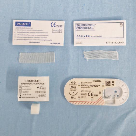
n pack socket with oxidised regenerated cellulose/haemostatic sponge ( Figure 2 );
n electrocautery of vessel;
n bone wax placement; and, n tight sutures, e.g., ‘figure of 8’ technique, can be helpful.
FIGURE 1: Local haemostatic measures.
Clinical tip
Use the suturing technique that works well in your hands. Multiple tight, simple, interrupted sutures are preferrable to horizontal or vertical mattress sutures that are loose.
4. Referral and follow-up
If haemostasis cannot be achieved, the patient should be referred onwards to the emergency department for further management.
Final
note
Dentists should undertake a comprehensive medical history, and ensure that haemostasis is achieved post extraction. Prolonged bleeding for more than 24 hours is abnormal, and time and patience need to be adopted initially when managing this. If in doubt, refer patients to the nearest emergency department and ensure that follow-up investigations are arranged in those with no prior bleeding history.
1. According to the National Consent Policy, when a child is under 16 years of age the consent of parents who are legal guardians or other legal guardians is both necessary and legally effective. In this case, if a parent or legal guardian cannot attend in person, consent can be obtained by telephone or electronic means. Where a parent or legal guardian presents in person, the person should be asked to confirm that they are a parent or legal guardian. This should be recorded in the healthcare record, and where practicable this conversation should be witnessed by another healthcare worker. If the consent of a parent or legal guardian cannot be established, the appointment should be deferred.
2. If the grandmother states that she is a legal guardian, you should request to see the court order appointing her as the legal guardian, or other legal or court documents appointing her as the legal guardian.
3. The avulsion of a permanent tooth is a time-sensitive emergency. All reasonable efforts should be made to contact and seek the consent of a parent or legal guardian, and if this is not possible, emergency care immediately required to prevent a serious detriment to the health of the child should be provided (in this case, reimplantation and stabilisation of the tooth). Even aer the intervention, continued efforts must be made to contact the parent or legal guardian. The clinician should at all times act in the best interests of the child and the circumstances of the emergency. The basis for the decision to treat, and the efforts made to contact the parent or guardian should be documented in the healthcare record.
4. As the management of an enamel fracture would not generally be considered time sensitive, the child and teacher should be reassured and treatment deferred until valid consent can be obtained.
Questions on page 6
FIGURE 2: Local haemostatic agents.
Endodontic treatment modifies circulatory inflammatory mediator levels: a systematic review with meta-analysis
Jakovljevic A, Fransson H, Bakhsh A, et al.
Background: There is limited and conflicting data on the reduction of circulatory inflammatory mediators in patients with apical periodontitis (AP) following endodontic treatment.
Objective: To answer the following research question: in adult healthy patients with AP [Population (P)], is there a difference before [Comparator (C)] and after various endodontic treatments (nonsurgical, surgical or retreatment) [Intervention (I)] on systemic levels of inflammatory biomarkers [Outcome (O)] in the follow-up period [Time (T)]?
Methods: An electronic literature search was conducted in the databases Scopus, PubMed, Clarivate Analytics’ Web of Science, Cochrane Database of Systematic Reviews, and grey literature from inception to July 2024 with no language restrictions. Observational studies examining changes in serum levels of inflammatory mediators were included. Two independent reviewers selected studies, extracted data and critically appraised the included studies. Qualitative and quantitative (meta-analysis) data synthesis methods were employed. The Newcastle-Ottawa Scale was used to assess the quality of the included studies.
Results: Sixteen studies met the inclusion criteria, of which six were included in the meta-analysis. These studies were published between 1992 and 2024, involving a total of 596 patients (54% females) aged between 16 and 75 years. The meta-analysis of pooled data showed a significant decrease in highsensitive C-reactive protein (hs-CRP) levels in the serum of patients with AP six months after treatment [2.26±1.76 versus 1.28±1.06mg/L, (Z=2.03, p=0.04)] and a decrease in interleukin-1 (IL-1 ) levels 12 months after treatment [13.01±5.95 versus 10.86±3.52pg/mL, (Z=3.72, p<0.01)]. One study was assessed as poor quality, while all others were considered high quality.
Discussion: Despite the differences in methodologies across the included studies, it has been established that effective endodontic treatment leads to a reduction in systemic inflammatory biomarkers in the body.
Conclusion: Following effective endodontic treatment in patients with AP, the systemic levels of hs-CRP and IL-1 exhibit a significant reduction at six and 12 months, respectively. Further clinical studies should investigate whether effective endodontic treatment and reduced levels of investigated biomarkers may change the clinical presentation of systemic diseases.
Int Endod J . 2025;58(2):171-192.
Clinical and radiographic failure of non-surgical endodontic treatment and retreatment using single-cone technique with calcium silicate-based sealers: a systematic review and meta-analysis
Sabeti MA, Karimpourtalebi N, Shahravan A, Dianat O.
Introduction: This systematic review and meta-analysis aimed to evaluate the clinical and radiographic failure of non-surgical endodontic treatment and retreatment for mature permanent teeth with or without apical periodontitis using the single-cone (SC) obturation technique with calcium silicate-based bioceramic (CSBC) sealers, and to compare these failure rates to other sealer materials and obturation techniques.
Methods: A comprehensive search was conducted using MEDLINE (PubMed), Embase, Cochrane Library, Scopus, Web of Science, and grey literature up to March 2023. Two reviewers assessed the eligibility of the included studies. Eligible studies were critically appraised for risk of bias and quality of evidence. Subsequently, a meta-analysis of pooled data was conducted utilising the RevMan soware (p<0.05) to evaluate the failure rate of non-surgical root canal therapy using CSBC sealers and SC obturation procedures. Separately, another analysis was conducted to compare those results with the outcome of non-surgical root canal therapy using alternative obturation materials and methods.
Results: Five studies were included. The pooled failure rate for non-surgical endodontic treatment and retreatment combined using CSBC sealers and SC obturation technique was 6.8% [95% confidence interval (CI) = (3%-12%), I2=46%]. A second analysis was conducted on the three included clinical trials to compare the outcomes of the intervention (CSBC sealers and SC obturation technique) and control groups (other sealers and other obturation techniques). This analysis found no significant difference between the two groups regarding clinical and radiographic failure of endodontic treatment and retreatment [Risk ratio = 0.71, 95% CI = (0.33, 1.51), I2=0]. This difference was also not statistically significant when the failure rate of primary root canal treatment and retreatment were separately analysed [combined risk ratio of primary root canal treatment = 0.94, 95% CI = (0.46, 1.93), I2=0%; combined risk ratio of retreatment = 0.21, 95% CI = (0.001, 75.46), I2=0%].
Conclusions: The overall results of this systematic review and meta-analysis demonstrate that the SC obturation technique with CSBC sealer provides similar results compared to other obturation materials and techniques in facilitating the healing of apical periodontitis.
J Endod. 2024;50(6):735-746.e1.
Bridging the gap between evidence and practice
Prof. Sayaka Tada is Assistant Professor at the National University of Singapore Faculty of Dentistry, and is a member of the International Editorial Board of JIDA Science. Her research focuses on oral health in older adults.
Can you tell us about your background and what led you to dentistry, and to dental research?
I grew up in Osaka, Japan, and trained as a prosthodontist. Early in my career, I was captivated not only by the technical and artistic aspects of prosthodontics, but also by the profound impact that oral health rehabilitation can have on overall well-being, especially among older adults. Caring for my grandmother made me acutely aware of the importance of maintaining oral function. At the same time, I encountered many clinical questions that remained unanswered, which inspired me to pursue research as a way to generate knowledge that could directly improve patient care.
You spent some time at University College Cork. Can you tell us about that?
My postdoctoral experience at UCC was pivotal in broadening my perspective. I met mentors such as Prof. Finbarr Allen, who encouraged me to look beyond prosthodontics to the broader social and population context of oral health. Collaborating with inspiring peers like Prof. Gerald McKenna, Prof. Martina Hayes, and Dr Cristiane da Mata fuelled my growing interest in gerodontology. I also discovered the excitement of working with people from diverse backgrounds and perspectives to address shared challenges. One of my happiest memories was cheering for Cork in the hurling final, caught up in the laughter, shouts, and cheers of passionate fans.
Can you share the primary focus of your recent research and what inspired you to pursue this topic?
My recent research focuses on oral health among care-dependent older adults, particularly in long-term care settings, alongside health systems research. Understanding the health system is crucial because even the most effective interventions cannot succeed without a supportive infrastructure and policy.
What challenges did you encounter during your study, and how did you overcome them?
Research in long-term care settings presents unique challenges, including ethical considerations, logistical constraints, varying staff awareness levels, and residents’ health limitations. Building trust with both staff and residents was essential. It was also critical to establish ourselves as partners with Government agencies and policymakers to ensure that research findings could translate into practical solutions.
How do you see your research contributing to clinical practice or patient outcomes in dentistry?
By highlighting the oral–systemic link and identifying barriers to care, we seek to inform strategies that better integrate dental services into long-term care
systems and foster interprofessional collaboration. Ultimately, the goal is to empower dental professionals, caregivers, medical and eldercare staff, and policymakers to work together in practical and sustainable ways, improving oral health management for older adults and enhancing their quality of life in the later stages of life.
Are there any misconceptions in the field that your research helps to clarify?
A common misconception, especially among non-dental professionals, is that poor oral health is an inevitable part of ageing. By challenging this belief, I aim to elevate oral health as a priority in older adult care.
Which emerging technologies or methods do you believe will most significantly shape the future of dental research?
Digital technologies, such as intra-oral scanning and AI-based diagnostic tools, are transforming how we assess and monitor oral health. Combined with telehealth and mobile health platforms, these tools can expand access to care, particularly for individuals with limited mobility. However, technology alone cannot ensure better health or well-being. Meaningful human connections, mutual understanding, and collaboration remain essential for these tools to have real impact in people’s lives.
How important is interdisciplinary collaboration in your work, and can you give an example where it made a key difference?
Even brief conversations can lead to profound changes: during my interview study with medical professionals who initially had limited interest in oral health, I observed a noticeable shi in awareness aer just 40-60 minutes of discussion. They began reflecting on what they could do differently to support oral health.
What advice would you give to young dental researchers just starting out?
I encourage young researchers to stay curious and embrace both clinical and scientific perspectives. Seek multiple mentors and peers with complementary strengths, engage in collaborations with people from diverse backgrounds, step outside your comfort zone, and support one another. Research is another way of helping patients, creating a lasting impact beyond the dental chair.
Outside of your research, how do you maintain balance and well-being in such a demanding field?
Outside of work, I treasure spending time with my dog Viola. Travelling, reading, and experiencing diverse cultures help me recharge. Cultivating curiosity, creativity, and a sense of wonder is essential for maintaining energy, perspective, and resilience in a demanding career.