

youth science journal
ISSUE 2.1 | MAR. 2024 | SCRIPPS RANCH HIGH SCHOOL 11 | | | | | A Brief Review of the Peripheral Nervous System 19 | | | | | The Biological Structure of Neurons 27 | | | | | Stress and Your Skin | | | | | The Effect of Antidepressants on Antibiotic Resistance 03

l e t t e r f r o m t h e e d i t o r i n c h i e f

c n t e n t s
03
Exploring the Effect of Antidepressants on Antibiotic Resistance via Facilitated Horizontal Gene Transfer and ROS Enhancement
Avaneesh Tisgaonkar
A Brief Review of the Peripheral Nervous System
Harry Jang
19
27
The Biological Structure of Neurons
Arjun Dasgupta
Stress and Your Skin
Ryan Min
| | | | | | | |
| | | | | | | | | | | | | | | |
| | | | | | | |
11
| | | |
| | | | | | | |
|
| | | | | | |
02
Exploring the Effect of Antidepressants on Antibiotic Resistance via Facilitated Horizontal Gene Transfer and ROS Enhancement
WRITER: AVANEESH TISGAONKAR | EDITOR: ANDREW YEOAbstract
Antidepressants are a subtle disruptor in the medical landscape; they are not antibiotics, yet they unwittingly contribute to the rise of antibioticresistance.Antidepressantspromote antibiotic resistance through enhancing horizontal gene transfer (HGT), or, more specifically, conjugation, in bacteria. The enhanced conjugation is predominantly a resultofantidepressantsinducinganincrease in reactive oxygen species (ROS) levels. ROS degrades a bacteria’s exterior layers, rendering the membrane increasingly permeable, and facilitating conjugation. Antidepressants also cause upregulation of genesgoverningcellmembranepermeability, expediting conjugation. The potential for a greaterspreadofantibioticresistancegenes, duetoenhancedconjugation,isconfirmed,as antidepressantshavebeenconsistentlylinked to increases in both ROS and antibiotic resistance overall. Antidepressants’ effect on antibiotic resistance should be considered throughouttheindustry.

keywords
Antibioticresistance,Horizontal genetransfer(HGT),Reactive oxygenspecies(ROS),minimum inhibitoryconcentration(MIC)
03
i n t r d u c t i o n
UNDERSTANDING ANTIBIOTIC RESISTANCE & ANTIDEPRESSANTS
Overview of Antibiotic Resistance
Antibioticresistanceistheevolution of bacteria so that antibiotics are no longer effective against them [3]. Antibioticresistancearisesfromselection andmutationsofspecificbacteriastrains.
Let’s consider a situation where a disease “DiseaseMania” is caused by a bacteria“BacterioMania,”withstrains“X” and “Y.” Strain Y is simply a mutated version of Strain X. Suppose scientists wereunawareoftheexistenceofStrainY, whileStrainXwasever-present.Assuch, scientistsdevelopedadrug“AntiMania”to counterStrainX.Overthenextfouryears, AntiMania was used throughout the United States excessively, nearly wiping out Strain X. However, AntiMania was ineffectiveagainstthemutatedStrainY, and the once-uncommon Strain Y becamewidespread.Assuch,despitethe efficacy of AntiMania against Strain X, DiseaseManialivedon.Inanutshell,thisis antibiotic resistance. A bacterial change (inthiscase,StrainY’smutation)andthe resulting natural selection after the addition of an antibiotic (AntiMania) yielded a bacterial population (mostly StrainY)resistanttoaspecificantibiotic (AntiMania).
Overview of Antidepressants
Antidepressants are drugs administered to reduce the effects of depression,byenhancing neurotransmitter activityinthebrain[4].TheWorldHealth Organization estimates that 3.8% of the world’s population and 5% of all adults suffer from depression; this translates to approximately300millionpeopleglobally [5]. Such high depression rates warrant widespread treatment, which is most often given through antidepressants. In England, for example, antidepressant prescriptions have consistently risen for thepastsixyears,withanoverallincrease of35%[6].
Antidepressants, by definition, are notantibiotics,astheyarenotmeantto kill or inhibit bacteria growth. Therefore,

e f f e c t o n a n t i b i
Horizontal gene transfer (HGT) is thetransferringofgeneticmaterialfrom one organism to another; in the case of bacteria, the primary HGT pathway is plasmidconjugation[1].Theresultisthat both the bacteria donor and bacteria recipient will contain the transferred plasmid, thus propagating the genes contained in the plasmid {see Figure 1}. Antidepressants have been found to facilitateHGTofantibiotic-resistantgenes (ARGs)inbacteria.

In2018,ResearchersfromAustralia’s University of Queensland found that antidepressants promote the transfer of plasmids across bacteria [1]. Multiple conjugation assays were conducted, involving donor bacteria with and receiving bacteria without plasmid RP4 {Figure 2A}. Of the six antidepressants tested, five (sertraline, duloxetine, fluoxetine, bupropion, and escitalopram) significantly enhanced the transfer of plasmidRP4betweenbacteria{figure2B} [1].Evenlowconcentrations, (1.0mg/L)of sertraline, for example, proved to significantly promote conjugation (adjusted p-value < 0.01) {figure 2B} [1]. Notably,allfivesignificant antidepressants in the study fall within the six most administered antidepressants in the UnitedStates[8].Wecanonlyimaginethe widespread increase in conjugation that these five antidepressants in the United States contribute to. The study’s finding emphasizessuchawidespreadeffectthat even lower concentrations of antidepressantsenhanceconjugation.
The increased levels of bacterial conjugationarethoughttobearesultof antidepressants’enhancementofreactive
05
Figure1:Bacterialplasmidconjugation; Credit:BYJU’s

t i c r e s i s t a n c e

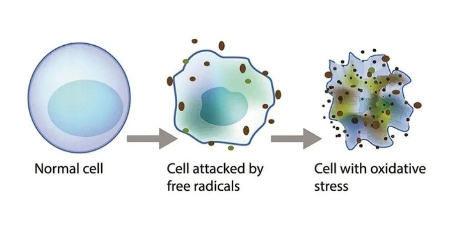
oxygen species (ROS) production. In the same study, the University of Queensland’s team found that the increaseinconjugationcorrespondedwith an increase in ROS production [1]. At higher concentrations (50 mg/L) antidepressantsfluoxetine,sertraline,and duloxetineallcauseda100-foldchangein ROSproductioninrecipientbacteria,and nearing 1000-fold in donor bacteria [1]. ExternalchemicalssuchasROSincrease cell permeability; ROS breaks down bacterial cell walls by oxidizing carbohydratesinthecellwallandloosens the cell membrane through lipid peroxidation [9, 10]. Figure 3 shows a simplification of such a process. Such a looseningofthebacterialexteriorlayers should contribute to an increase in conjugation.

06 i
Figure2:ResultsofUniversityof Queensland’sResearchTeam;Credit:[1]
news-medical.net
Figure3:ROSEffectonacell;Credit:
Antidepressantsalsoenhancedthe expression of membrane porin genes, outer membrane channel genes, and transmembrane transporter genes [1]. Membrane porins are small holes in the membrane that allow molecules to pass through. By increasing the potential for moleculestopassthroughthemembrane, upregulation of membrane porin genes would increase membrane permeability and therefore conjugation. Similarly, upregulation of the outer membrane channelandtransmembranetransporter genes, which also serve to regulate the flowofmoleculesacrossthemembrane, would increase membrane permeability andthepotentialforconjugationaswell.
Increasedconjugationtranslatesto apotentialforincreasedspreadofARGs. Transconjugant bacteria (the receiving bacteria after the transferred plasmid is incorporated into its genome) have the resistancegenesandmechanismsofboth the donor and initial recipient cells [1]. Therefore, after selective pressure from antidepressants, transconjugants would survive better (compared to their plasmid-free counterparts) and ARGs spread,throughreplication[1,11].
Spreading of ARGs means an overall increaseinantibioticresistance.
A subsequent study published in early 2023 confirmed an increase in antibiotic resistance. The addition of antidepressants like sertraline, after a 60-dayincubationperiod,causedan


07
Figure4;C
cTHE EFFECT OF ANTIDEPRESSANTS ON ANTIBIOTIC RESISTANCE
a grave problem for modern medicine, and excessive human use of antidepressants is adding to it. Antidepressants have the effect of increasing plasmid conjugation in bacteria, which leads to an increase in antibiotic resistance. While it is not possible to completely avoid using antidepressants, it is possible to restrict their administration. Reducing dose sizes and only prescribing antidepressants when absolutely necessary would decrease the increase in antibiotic resistance.Additionalresearchisrequired to determine a more specific effect of antidepressants on antibiotic resistance for bacteria in humans; hopefully, this research will provide a clearer path toward regulating antidepressant use. Regardless, antidepressants’ effect on antibiotic resistance must be taken into account throughout the healthcare industry.

09
w o r k s c i t e d
1.Ding,P.,Lu,J.,Wang,Y.,Schembri,M.A.,& Guo,J.(2022).Antidepressantspromotethe spread of antibiotic resistance via horizontally conjugative gene transfer. EnvironmentalMicrobiology.https://doi.org/ 10.1111/1462-2920.16165
2.Wang, Y., Yu, Z., Ding, P., Lu, J., Mao, L., Ngiam, L., Yuan, Z., Engelstädter, J., Schembri, M. A., & Guo, J. (2023). Antidepressants can induce mutation and enhance persistence toward multiple antibiotics. Proceedings of the National AcademyofSciences,120(5).https://doi.org/ 10.1073/pnas.2208344120
3.WorldHealthOrganization.(2021,November 17).Antimicrobialresistance.Who.int;World Health Organization: WHO. https://www.who.int/ news-room/fact-sheets/detail/antimicrobial -resistance
4.Antidepressant Medication. (n.d.). CAMH. https://www.camh.ca/en/health-info/menta l-illness-and-addiction-index/antidepressan t-medications
5.WorldHealthOrganization.(2023,March31). Depressive disorder (depression). World HealthOrganization.https://www.who.int/ news-room/fact-sheets/detail/depression
6.Burns, C. (2022, July 8). Antidepressant prescribingincreasesby35%insixyears. The Pharmaceutical Journal. https://pharmaceuticaljournal.com/article/news/antidepressant-pr escribing-increases-by-35-in-six-years
7.Starr,M.(2018,September10).OneofThe MostWidelyUsedAntidepressantsHasJust Been Implicated in Breeding Antibiotic Resistance.ScienceAlert.
https://www.sciencealert.com/antidepressantmedication-fluoxetine-prozac-may-raise-antib iotic-resistance
8.Most commonly prescribed antidepressants. (2023, June 7). Definitive Healthcare.
https://www.definitivehc.com/resources/health care-insights/top-antidepressants-by-prescrip tion-volume
9.Huang,H.,Ullah,F.,Zhou,D.-X.,Yi,M.,&Zhao,Y. (2019,June25).MechanismsofROSRegulation of Plant Development and Stress Responses. Frontiers.
https://www.frontiersin.org/articles/10.3389/fpl s.2019.00800/full
10.Juan,C.A.,PérezdelaLastra,J.M.,Plou,F.J.,& Pérez-Lebeña, E. (2021). The Chemistry of Reactive Oxygen Species (ROS) Revisited: Outlining Their Role in Biological Macromolecules(DNA,LipidsandProteins)and Induced Pathologies. International Journal of Molecular Sciences, 22(9), 4642.
https://doi.org/10.3390/ijms22094642
11.MatthewDeFurio,SangJoonAhn,Burne,R.A., & Hagen, S. J. (2017). Oxidative Stressors ModifytheResponseofStreptococcusmutans toItsCompetenceSignalPeptides.Appliedand Environmental Microbiology, 83(22).
https://doi.org/10.1128/aem.01345-17
12.Microbiology guide to interpreting minimum inhibitory concentration (MIC). (n.d.). Idexx. Retrieved November 19, 2023, from https://idexx.com/files/microbiology-guide-int erpreting-mic.pdf
13.Soto,S.M.(2013).Roleofeffluxpumpsinthe antibioticresistanceofbacteriaembeddedina biofilm. Virulence, 4(3), 223–229.
https://doi.org/10.4161/viru.23724
10
A Brief Review of the Peripheral Nervous System
WRITER: HARRY JANG | EDITOR: ADRIAN MENDEZAbstract
The peripheral nervous system (PNS) is an importantlatticeworkofnervesconnectingthe centralnervoussystemtotherestofthebody. Comprising sensory and motor neurons, the peripheral nervous system facilitates communication between the brain and spinal cord with peripheral tissues. Sensory neurons conveyenvironmentalstimuliforinterpretation, while motor neurons transmit commands for muscleandglandularresponses.Theperipheral nervous system is subdivided into the somatic and autonomic systems, governing voluntary and involuntary functions, respectively. This intricate system enables adaptive responses, ensuringadynamicinteractionwiththeexternal environment and the maintenance of physiologicalbalancethroughoutthebody.


11
peripheral nerv us system
The peripheral nervous system (PNS)isacrucialintegrantofthenervous system,responsibleforthefundamental functions of regulating neuron transmissions between the central nervoussystem(CNS)andtherestofthe body. The intricate peripheral nervous system structure extends throughout mostofthehumanbody,andcomprises nerves and ganglia, which serve as an indispensable conduit for sensory and motorsignals.
The peripheral nervous system bisects into the autonomic nervous systemandthesomaticnervoussystem, which govern unconscious physiological maneuvers and voluntary muscular processes respectively. The autonomic nervoussystemisoftenfurtherdissected intothesympatheticnervoussystemand the parasympathetic nervous system, which conducts the body during acute stress responses and tranquil, undisturbed responses, and digestive affairscorrespondingly.Furthermore,the somatic nervous system diverges once more into the sensory system and the motorsystem.


12
autonomic nerv us system
Theautonomicnervoussystemisa sectoroftheperipheralnervoussystem thatgovernsunconsciousactivities,such as heart rate, respiration, digestion, urination,sexualarousal,bloodpressure, perspiration, salivation, pupillary reflex, somnolence, metabolism, etc.. The autonomicnervoussystempossessestwo separate systems in different times of need,whicharethesympatheticnervous systemandtheparasympatheticnervous system.
When the sympathetic nervous system dominates a human's subconsciousendeavors,itismostoften during instances where the particular individual is under hyperarousal response, more idiomatically labeled as the “fight or flight” response, when an individualisunderhighamountsofstress.

Thesympatheticnervoussystemis composed of detailed neural pathways thatpreservethephysiologicintegrityof several critical body components. The preganglionic neurons that are directly attached to the spinal cord range from vertebrae1Tto2L.Itshouldbenotedthat theinnervationofnervesinthe1Tto2L vertebrae region are affixed to most visceral organs in the thoracic cavity, where most of the sympathetic responses occur. In the sympathetic nervous system, the first order neurons are miniature prior to synapsing on postsynaptic neurons found within sympatheticganglia.Liketherestofthe peripheral nervous system, the sympatheticnervoussystemmainlyuses acetylcholine neurotransmitters to communicate signals between the centralnervoussystem.
The sympathetic nervous system performsavarietyoffunctionswithina human’s body. During a sympathetic response,theheartwillaccelerate,dilate the pupil, alleviate stress on the bronchioles, constrict the digestive system,activatesweatglands,suppress the penis from acquiring an erection, manage the kidney, and assist the nervoussystem.
13
b e h
a
v i o r d u r i n g s y m p a t h e t i c r e s p o n s e
Organ/System Response Purpose
Heart Acceleratespulseand increasesbloodpressure
Eyes
Dilatespupils
Lungs
Enlargedbronchioles
Digestivesystem Slowsdowndigestion
Sweatglands
Delivershemocytesandoxygen tonecessaryorgansand musclestoreducefatigueand increaseefficiency.
Allowsmorelightintopupils, amelioratinghyperopia eyesight.
Allowslungstoacquireamore andbetterflowofoxygen.
Digestionrequiresprecious energy;slowsdowndigestionto preserveenergy.
Producessweat Regulatesbodytemperature; higherpulsewillcausethebody toheatup.
Reproductionsystem Suppressesthepenisfrom acquiringanerection
Anerectionoccurswhenblood flowsintothepenis;subduing anerectionmeansbloodwill flowtoother,moredemanding organs.
Kidney Mediateswaterexpulsion Waterisacriticalcomponentin everycell;regulatingwater expulsionsaveswater.
14

a
p a r a s y m p a t h e t i c r e s p o n s e
The parasympathetic nervous system performs actions that directly contrast the sympatheticnervoussystem.
Organ/System Response Purpose
Heart Deceleratespulseand bloodpressure Bodydoesnotneedanyexcess oxygenatedblood;theheartwill pulseatanormalrate
DigestiveSystem Re-enablesnormal digestiveprocess Digestingfoodprovides nutrientsandenergyforthe body
Lungs
Eyes
Constrictsbronchioles Bodyisinacalmandpassive state;lungsdonotneedtobe efficient.
Constrictspupil Reduceslightentry,which improvesmyopia.
SalivaryGlands Salivaryglandswill producesaliva
MucusGlands Mucusglandsproduce mucus
Salivalubricatestheinsideof themouth.Eatingfoodiseasier withsaliva.
Lubricatestissuesinthenasal cavitytopreventtissuesfrom dryingout
16
b e h
v i o r d u r i n g
somatic nervo s system
While the autonomic nervous system is responsible for involuntary, subconscious measures primarily in the internal organs, the somatic nervous system is quite the opposite. All fine motor movements, aroma, vision, auditory feedback, and taste are controlled by a separate system called thesomaticnervoussystem.Thesomatic nervoussystemisbrokendownintotwo more systems, the sensory system and themotorsystem.
The sensory system processes information that derives from an individual’s perception of their current situation and environment. These sensations include vision, sight, temperature, pain, awareness of movement, pressure, vibration, hearing, smell, and taste. The sensory system is made up of neurons and neural pathways.Afferentneuronscarrysignals from the sense receptors toward the centralnervoussystem.
The motor system is an intricate system that participates in both the central nervous system and the peripheral nervous system. The central nervous system component sustains the cerebralcortex,brainstem,spinalcord, and cerebellum, while the peripheral nervous system includes structures such asmotornerves.Efferentneuronscarry motor information from the central nervous system to the muscles to inaugurateactions.

17
w o r k s c i t e d
1. Das,JoeM,andMark NAlshak. “Neuroanatomy,Sympathetic NervousSystem-Statpearls-NCBI Bookshelf.” NationalLibraryof Medicine,2023, www.ncbi.nlm.nih.gov/books/NBK5 42195/.
2. Hurst,JohnWillis,andWilburDallas Hall.“68.” ClinicalMethods:The History,Physical,andLaboratory Examinations.3rdEdition, LexisNexisUK,Boston, Massachusetts,1990.
3. Koop,LindseyK,andPrasanna Tadi.“Neuroanatomy,Sensory Nerves.” NationalLibraryof Medicine,24July2023, www.ncbi.nlm.nih.gov/books/NBK5 39846/.
4. “NerveChart.” BackDoctors,Back Doctors,8May2018, thebackdoctorsonline.com/nerve-c hart
5. “ThePeripheralNervousSystem.”
ThePeripheralNervousSystem| SEERTraining, training.seer.cancer.gov/anatomy/ nervous/organization/pns.htm. Accessed11Dec.2023.
8.ClevelandClinicmedical.
“ParasympatheticNervousSystem (PSNS):WhatItIs&Function.” Cleveland Clinic,6June2022, my.clevelandclinic.org/health/body/2326 6-parasympathetic-nervous-system-psns
9.ClevelandClinicmedical.“Peripheral NervousSystem(PNS):WhatItIs& Function.” ClevelandClinic,25May2022, my.clevelandclinic.org/health/body/2312 3-peripheral-nervous-system-pns.
10.ClevelandClinicmedical.“Sympathetic NervousSystem(SNS):WhatItIs& Function.” ClevelandClinic,6June2022, my.clevelandclinic.org/health/body/2326 2-sympathetic-nervous-system-sns-fight -or-flight.
11.Tindle,Jacob,andPrasannaTadi. “Neuroanatomy,Parasympathetic NervousSystem-Statpearls-NCBI Bookshelf.” NationalLibraryofMedicine, 2022, www.ncbi.nlm.nih.gov/books/NBK553141/.
18
The Biological Structure of Neurons
WRITER: ARJUN DASGUPTA |
EDITOR: RYAN MIN
Abstract
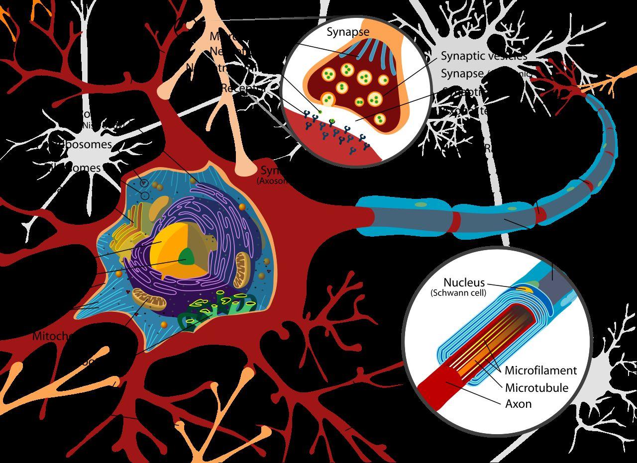
As nerve cells, neurons share the fundamental cell organelles (e.g. the cell membrane, nucleus, cytoplasm, etc.) with manyothercells,yetalsocontain andaxonsthatfacilitateitssalient intercellular communication. At synapses, the presynaptic neuron transmits informationthroughtheterminalendofits axon to the postsynaptic neuron or effector. Neural impulses are imparted acrossthesynapticcleftwiththerelease neurotransmittersthattranspiresdueto fusion of the presynaptic membrane and thesynapticvesiclesandwiththebinding of neurotransmitters to specific receptors onthepostsynapticmembrane.


KEYWORDS: Perikaryons, Axons, and Action Potentials
19
neurons & neurotransmitters
Neurons are composed of a perikaryon (also known as soma or cell body), dendrites, and an axon. The perikaryon of a differentiated neuron houses the nucleus, nucleolus, (both the rough and smooth) endoplasmic reticulum,golgiapparatus,mitochondria, cytoplasm, lysosomes, cell membrane, and microtubules similar to most cells, yethave“deactivated”centrosomesthat typicallydonothavecentriolesandergo cannotundergocellularmitosis.
Locatedatthejunctioninbetween the axon and the perikaryon, the axon hillock is the integration point of excitatory and inhibitory signals in a neuronanddeterminesthecreationofan action potential. The axon itself is a single, elongated extension from the perikaryon.Actionpotentialsarewaves ofelectricitythattraveldowntheaxonof a neuron and are caused by transient rises (and subsequent falls) in the membrane potential of the neuron. The membranepotentialischaracterizedby the difference in voltage between the areas inside and outside of the cell membrane, and its resting value is approximately-70millivolts.
Excitatory neurotransmitters bind toligand-gatedionchannelsthatlinethe cell membrane and engender the openingoftheirpores,whichenablesan influxofpositivelychargedionsinthecell membrane. Inhibitory neurotransmitters counteracttheirexcitatorycounterparts anddiscommodeanotherwiseincessant generation of action potentials by binding to ligand-gated chloride channelsandcausinghyperpolarization.
However, if there are more excitatory neurotransmitters than inhibitory, the influx in positive ions depolarizes the cell membrane, and, if the membrane potential reaches a certain threshold (typically -55 to -50 millivolts), the voltage-gated sodium channels open and effectuate further depolarizationintheformofawavethat travels the length of the neuron’s axon. The wave of depolarization self propagatesbyactivatingvoltage-gated channels within the adjacent regions of theaxonmembraneuntilitreachesthe axonterminals.
20
m y e l i n s h e a t h s
Furthermore, these electric impulses can be more quickly and efficientlyconductedinaxonsenveloped by myelin sheaths. Produced by special glialcells(oligodendrocytesinthecentral nervoussystemandSchwanncellsinthe peripheral nervous system) and composedoflipidsandproteins,myelin sheaths insulate certain neurons, consequently prevent the leakage of electrical currents, and facilitate saltatoryconduction.Duringsaltatory
conduction, action potentials only regenerate via sodium-gated ion channelsatbreaksinthemyelinsheath (where the axon is exposed) called the nodes of Ranvier, rather than regenerating constantly throughout the axon. This intermittent depolarization can occur due to the myelin sheath’s insulation and is faster than action potential propagation in unmyelinated axonsinadditiontobeingmoreenergy efficient.
21
sodium-potassium pump
It is worth noting that, once an action potential terminates, the neuron undergoes repolarization (and sporadically hyperpolarization) via the opening of voltage-gated potassium channels (that allow positive potassium ions to leave the cell) and the sodium-potassiumpump.Thepumphas threebindingsitesforsodiumionsfacing inward and two binding sites for potassium ions facing outward, and the pump repeatedly uses the energy and inorganic phosphate gathered from the hydrolysisofATPtopromptthepumpto flip (in other words, phosphorylation), taking sodium ions out of the cell while bringing in potassium ions (that are promptly removed through the voltage-gatedpotassiumchannels)
Once the action potential reaches the axon terminal, voltage-gated calcium channels open (due to the depolarizationoftheaxonterminal)into the extracellular fluid, which occasions the fusion of synaptic vesicles (small, membrane bound sacs that have neurotransmitters in them) and presynapticmembraneanditsresulting release of neurotransmitters. Before vesiclefusioncantranspire,vesiclesmust “dock”onpartsofthetargetmembrane via the SNARE (Soluble N-ethylmaleimide-sensitive factor attachment protein receptors) proteins present on both the vesicle and target membranes (v-SNARE and t-SNARE, respectively). A combination of the v-SNAREandt-SNAREproteins,more

22
s n a r e p r o t e i n s
specifically syntaxin, SNAP-25, and synaptobrevin,resultsinastableSNARE complex that brings the vesicle and targetmembraneincloseproximityviaa “zippering” mechanism (the SNARE complex has a coiled coil shape - the v-SNARE and t-SNARE proteins get entangled to the point of near convergence). The Munc18 and Munc13 proteins facilitate this entanglement by closingandopeningtheconformationof syntaxin and allowing it to bind to the SNAP-25protein.
Returning to the opening of the voltage-gated calcium channels, the influx in calcium alters the C2 domains (calciumbindingmotifs)ofproteins
involved in the vesicle fusion process, such as synaptotagmin, which induces structuralchangesthatexposeregionsof the proteins and enable the proteins to interactwiththeaforementionedSNARE complex in addition to the presynaptic and vesicle membranes. By penetrating thevesicleandpresynapticmembranes and connecting itself onto the SNARE complex,synaptotagminallowsthelipid bilayersofthevesicleandmembraneto touch and impedes the electrostatic repulsion from the negatively-charged phospholipids,bothofwhichdisruptthe lipidbilayerandcausesthefusionofthe presynaptic membrane and synaptic

23
intercellular transmission
A fusion pore is the result of the aforementioned process and is an openinginthelipidbilayersthatconnects the vesicle interior to the intercellular space, enabling the neurotransmitters storedwithinthevesiclestobereleased intothesynapticcleft(asmall,fluidfilled gap between the axon terminal presynaptic neuron and membrane of the postsynaptic effector), to diffuse across the cleft, and to bind to postsynapticreceptors,typicallyproteins thatareembeddedonthepostsynaptic membrane (which includes dendrites). Each neurotransmitter has a specific correspondingreceptororgroupthereof.

Dendrites emanate from neuronal perikaryons and, as the name implies, resemble branches. Smaller dendritic spines protrude from the dendrites and are where synapses can form. The neurotransmitter receptors at these synapses take both ionotropic and metabotropic forms, the former being directly linked to ion channels and capable of rapidly altering membrane potential,andthelatterbeingconnected to G-proteins that activate or inhibit enzymes that generate second messengermolecules,suchascyclicAMP andinositoltriphosphate,thatcantrigger enzymatic cascades, which can affect ion channels albeit slower and more indirectlythanwithionotropicreceptors.
24
c n c l u s i o n
THE BIOLOGICAL STRUCTURE OF NEURONS
Neurons are unique cells with their inability to undergo cellular mitosis (in general),yetarealsouniqueduetotheir abilitytogenerateandreceiveelectrical impulses, or action potentials, that transmit information. As a result, understandingthestructureofneuronsis requisite for the comprehension of intercellularcommunicationandisatthe core of neuroscience. Intercellular communication facilitates a plethora of physiological processes, such as homeostasis, response to environmental stimuli, immune response, reproduction, metabolic regulation, hormonal regulation,tissuerepair,andmanyother fundamental bodily functions, while neuroscience targets how the brain functions, processes information, and governsbehavior.Anelevated of neuron structure and intercellular communication fosters the creation and advancement of vaccines, potential treatment to mental disorders, diagnostics, neuroimaging, and neuroprosthetics.

25
w o r k s c i t e d
1. Brain Basics: The Life and Death of a Neuron.(2023,March24).NationalInstitute of Neurological Disorders and Stroke. https://www.ninds.nih.gov/health-information/public-education/brain-basic s/brain-basics-life-and-death-neuron
2. Chemical Synapse and Post Synaptic Effects | BISC 106. (n.d.). Wizeprep. Retrieved December 20, 2023, from https://www.wizeprep.com/online-courses /18998/chapter/9/core/4/1
3. How does the sodium-potassium pump work? | Socratic. (2016). How does the sodium-potassiumpumpwork?|Socratic. Socratic.org. https://socratic.org/questions/ how-does-the-sodium-potassium-pumpwork
4. SNAREopathies in epilepsies and neurodevelopmentaldisorders|Beyondthe Ion Channel. (2023, May 2). http://epilepsygenetics.net/2023/05/02/sn areopathies-in-epilepsies-and-neurodevel opmental-disorders/
5. File:Completeneuroncelldiagramen.svg. (2010, June 30). Wikipedia. https://en.wikipedia.org/wiki/File:Complete _neuron_cell_diagram_en.svg
6. Chen,I.,&ForshingLui.(2019,August27). Neuroanatomy, Neuron Action Potential. Nih.gov; StatPearls Publishing. https://www.ncbi.nlm.nih.gov/books/NBK5
46639/
7. Cherry, K. (2019). How neurons transmit information throughout the body. VerywellMind. https://www.verywellmind.com/ what-is-a-neuron-2794890
8. Fang,Q.,&Lindau,M.(2014).HowCould SNARE Proteins Open a Fusion Pore? Physiology, 29(4), 278–285.
https://doi.org/10.1152/physiol.00026.2013
9. Fukuda, M. (2002). Vesicle-associated Membrane Protein-2/Synaptobrevin Binding to Synaptotagmin I Promotes O-Glycosylation of Synaptotagmin I. Journal of Biological Chemistry, 277(33), 30351–30358.
https://doi.org/10.1074/jbc.m204056200
10. Goda, Y. (1997). SNAREs and regulated vesicle exocytosis. Proceedings of the National Academy of Sciences of the UnitedStatesofAmerica,94(3),769–772.
https://www.ncbi.nlm.nih.gov/pmc/articles /PMC33653/
11. Ludwig, P. E., Reddy, V., & Varacallo, M. (2020). Neuroanatomy, Neurons. PubMed; StatPearls Publishing.
https://www.ncbi.nlm.nih.gov/books/NBK4 41977/
12. Nalefski,E.A.,&Falke,J.J.(1996).TheC2 domain calcium-binding motif: Structural and functional diversity. Protein Science, 5(12), 2375–2390.
https://doi.org/10.1002/pro.5560051201
13. Purves,D.,Augustine,G.J.,Fitzpatrick,D., Katz, L. C., Anthony-Samuel LaMantia, McNamara,J.O.,&SMarkWilliams.(2010). Nerve Cells. Nih.gov; Sinauer Associates.
https://www.ncbi.nlm.nih.gov/books/NBK111 03/
14. Ricci,G.,Volpi,L.,Pasquali,L.,Petrozzi,L.,& Siciliano, G. (2009). Astrocyte–neuron interactions in neurological disorders. JournalofBiologicalPhysics,35(4),317–336.
https://doi.org/10.1007/s10867-009-9157-9
15. Vandergriendt,C.(2018,July20).WhatAre Neurons? Healthline; Healthline Media.
https://www.healthline.com/health/neuron s#research
9.
1. 2. 3. 4. . 5. . 6. . 7. . 8.
26


keywords
psychological stress, epidermis, proliferation, cortisol, hypothalamic-pituitary-adrenal (HPA) axis, sympathetic- adrenal medullary (SAM) axes, differentiation, acne, substance P, psoriasis, atopic dermatitis, hyporesponsive, upregulation, stratum corneum, ceramides, murine, p53
Stress & Your Skin
WRITER: RYAN MIN | EDITOR: HARRY JANGAbstract
With the modern focus on cosmetics and appearances, skincare is becoming increasingly important. However, the exploration of skincare has largely been limited to the skin itself. While topical ointments and moisturizers have proven their worth, they disregard a major factor determining skin health—the brain. Clinical observationshavedemonstratedaclearlink between psychological processes and the skin, revealing the need for further exploration of the intricacies and possible applications of this relationship in maintaining skin health. This article will summarize recent findings on the relationshipbetweenthebrainandtheskin, starting with reception and response betweenthetwoorgans,andmovingonto theroleofstressincausingskinconditions, reduced barrier and healing effectiveness, andlong-termskindamage.
27
i n t r d u c t i o n
OVERVIEW OF THE SKIN’S ROLE AND PATHOLOGICAL STRESS
Role of the Skin
The skin plays a number of important roles in the body, primarily forming a barrier against pathogens, chemicals, and physical injury. This physical outer barrier is formed by the stratumcorneum(SC),alayeroftheskin containing protein-enriched corneocytes andlipid-enrichedintracellulardomains.
The skin itself is composed of the epidermis, dermis, and hypodermis, in order from outermost to innermost. The varioustight,gap,andadherensjunctions oftheepidermisfurthercontributetothe skin’s barrier functions. The skin also containsstressreceptors,whichtransmit information to the brain via the spinal cord,triggeringresponsesthatdictatethe
Effects from Pathological Stress
When the brain is under psychological stress, which occurs when anindividualfeelsunabletoadaptquickly enough to match environmental demands, leading the body to employ stress-reducing physiological and behavioral changes. In the skin, psychologicalstresscantriggerorworsen skin conditions like acne, psoriasis, and atopicdermatitis(eczema).
Aside from skin conditions, psychological stress has been found to reduceepidermalbarrierfunctionality,as well as hamper epidermal proliferation (the production of skin cells to replenish the outer layer of skin). Psychological stress has been shown to cause a

28
neurobiological receptio
Thepathwaybetweentheskinand the brain begins with stress receptors. Located in the skin, these receptors fall into three categories: thermoreceptors, nociceptors, and mechanoreceptors, which take in information about temperature, pain, and touch, respectively. These receptors then transmit signals to the brain through variousmethods.

Source:HPA
Thermoreceptors come in two types—warmandcold—andeachhavea specific range of temperatures at which theycontinuouslyputoutdischarge.When they experience a temperature outside their range, these receptors halt discharge, signaling a change in temperature. Nociceptors are only activated when there is risk of tissue damage, such as extreme temperatures or dangerous chemicals. A-delta and C fibers then relay information to the nervous system, with each type of fiber specializingincertainexternalthreatsto thebody.Asformechanoreceptors,there are six types (Pacinian corpuscles, Meissner corpuscles, Merkel complexes, Ruffini corpuscles, and C-fiber low threshold mechanoreceptors), which are activated by physical stimuli like touch, pressure,andstretch,andtransmitsignals through A fiber beta-type nerves. Together,thesereceptorsformthebasis oftheskin-brainpathway.
Thebrainrespondstothesesignals in various ways. For example, one response utilizes the hypothalamic -pituitary-adren-al (HPA) axis, in which thehypothalamusreleasescorticotropin
Figure1:AdepictionoftheHPAaxisasastress responsesystem.CredittoGettyimages29
on & response in the skin
-releasinghormone(CRH).Thistriggersa series of secretions in the brain, which ultimately triggers production of glucocorticoids(GC),particularlycortisol. Cortisolisahormonethatimpactsalmost the entirety of the human body, as it regulatesresponsestostress.Therelease of excess cortisol disrupts natural oscillations of cortisol levels, which are normallyincadencewiththebody’sinner circadian clock, generally resulting in suppressionoftheimmunesystem.
Upon receiving stress signals, the brain can also release catecholamines (hormones produced by the adrenal glandsinresponsetostress) throughthe sympathetic-adrenal medullary (SAM) axes.Theskinitselfcontainsaperipheral catecholamine system that can alter epinephrineproduction.Epinephrine,also known as adrenaline, is a key first messenger and hormone that primarily controls the human “fight or flight” response to danger. In the skin, altered epinephrine levels as a result of stress affects epidermal proliferation, differentiation (the process in cells determinetheirfunction),fibroblast
migration,andcollagenproduction(both essential to the healing of wounds), demonstrating an important relationship betweenthebrainandskin.


30
s t r e s s & s k i n
Psychological stress can induce or exacerbate a number of skin conditions. Acne, or acne vulgaris, is one such skin condition. Characterized inflammation and pain caused by the plugging of sebaceoushairfollicleswithoilanddead skin cells, studies have found that acne, and specifically its pathogenesis (development), are influenced by stress. For instance, CRH promotes the productionofIL-6andIL-11,cytokinesthat cause inflammation. Additionally, acne patientsexperienceanincreaseinnerve fibers near acne-afflicted regions which canacceptsubstanceP,aninflammatory neuropeptide related to stress, demonstratingalinkbetweenstressand inflammationintheskin.

Studies have also found links between psychological stress and psoriasis, a chronic disease in which an overactive immune system causes skin cells,particularlykeratinocytes,tomultiply faster than normal. The resulting inflammation generally causes painful rashesandscalesthattendtoappearon the elbows, knees, and scalp. Stress has been found to make the HPA axis hyporesponsive (having a reduced function), limiting the body’s cortisol responseandinturncausingupregulation (an increase in receptors for a certain substance)ofinflammatoryagents.Stress inducedbypsoriasiscanalsocontributeto theincreasedproductionofinflammatory cytokines,creatingacycleofincreasingly severesymptoms.
Figure2:TheskeletalformulaofsubstanceP,a neuropeptideinvolvedininflammation.Creditto Fvasconcellos,sourcedfrom https://en.wikipedia.org/wiki/Substance_P
31
n c o n d i t i o n s
Atopic dermatitis, more commonly known as eczema, is thought to be correlatedwithstressaswell.Eczemaisa chronic skin disease which causes reducedepidermalbarrierfunctionandin turn inflammatory rashes and scales, which cause pruritus (itching) that can prompt detrimental scratching and itch relief.Similarlytothecasewithpsoriasis, stress,whichisofteninducedbyeczema, cancauseHPAaxishyporesponsiveness, whichhasbeenlinkedtoimmunesystem dysregulation, allergic inflammation, and otherexacerbationsofthedisease.Stress canalsocausetheSAMaxestobecome excessivelyreactive.
Manyeczemapatientsexperiencea mutation in the β2-adrenoceptor gene thatactivatesToll-likereceptor2(TLR-2), a type of receptor that recognizes cell wall components of bacteria. When the adrenoceptor mutation and TLR-2 activation occur, they can change the receptor’s recall memory response, releasing pro-inflammatory cytokines. Thus,anexcessofcatecholamines,paired withtheseresponsesintheSAMaxes,can contribute to increased inflammation in eczemapatients.

32
l a s t i n g e f f e c t s o f
The stratum corneum is a protein-lipidbarrierthatisresponsiblefor theskin’sepidermalbarrierfunction,and maintains proper hydration and protection against microbial threats. Disruptions or damage to this layer can cause dry skin, infection, and can contributetomanyoftheskinconditions mentioned previously, such as eczema andpsoriasis.
Studies have shown that stress altersthelipidcompositionofthestratum corneum.Astudyonovercrowdinginmice indicatedalinkbetweenstressandhigher transepidermal water loss, lower water retention, exfoliation, and wrinkle formation.Furtherstudieshaverevealed
the critical role of lipids, such as ceramides, in maintaining a functioning epidermal barrier. Stress can inhibit the production of lipids, thus lending importance to lipid-based treatments in improvingstratumcorneumhealth.
Stress can also impair the body’s abilitytohealwounds.Astudyregarding final exam stress in students inhibited permeabilitybarrierrecovery,andastudy oninterviewstressindicatedacorrelation withincreasedcortisollevelsandbarrier functionrecoverydelay.Onestudyeven demonstrated a 20% increase in time neededforwoundstohealastheresultof stress from caring for relatives with dementia.

33 Figure3:Therelationshipbetweenstressandcortisol levelsinahumanstudyontheeffectsofstress manipulation.CredittoLempert,K.M.,McGuire,J.T., Hazeltine,D.,Phelps,E.A.,Kable,J.W.,
s t r e s s o n t h e s k i n
Stress has been linked to DNA damageandcanceraswell.Inamurine study (study conducted on mice) examining the effect of fox urine (a natural predator), the group which was placed under stress exhibited faster developmentofskintumorsandalower survival rate, which later studies have determined was likely caused by a stress-induced decrease in IFN-γ, an immune marker responsible for tumor recognition and elimination. Stress-related hormones like epinephrine, norepinephrine, and cortisol increase damage to DNA, undermine DNA repair, and modify transcriptional regulation of thecellcycle.
Stress-induced stimulation of catecholamines over a long period can also result in DNA damage, namely by instigating the degradation of tumor proteinp53.P53isacrucialregulatorthat prevents overproliferation of cells by either initiating DNA repair or causing a damaged cell to undergo apoptosis (self-destruction) to prevent the propagation of DNA damage. By degrading p53 and contributing to DNA damageaccumulation,stresscontributes to skin aging and increases the risk of cancer.
Therelationshipbetweenstressand reduced wound healing capabilities is likelytheresultofavarietyoffactors.For instance, stress can delay immune cell infiltration and lower TNF-α (tumor necrosis factor alpha) levels at a wound site, both of which are crucial to the regular healing process. Stress also impairs the body’s defense against infection by decreasing antimicrobial peptide expression, further delaying the healingprocess.Finally,stresscaninduce epinephrineproductionthroughtheSAM pathway, resulting in stabilization of the actin cytoskeleton and increased focal adhesion formation, both of which prevent proper migration of substances and structures involved in the wound healingprocess.
34
c n c l u s i o n
STRESS & YOUR SKIN
The skin is a complex organ that serves a number of important functions, namely providing a barrier against microbial,chemical,andphysicalthreats and maintaining proper hydration of the skin.Therefore,thebrain-skinpathwayis important to explore when trying to examine methods for improving skin health.Whileemphasisisoftenplacedon the transmission of information from the skintothebrainthroughstressreceptors (thermoreceptors, nociceptors, and mechanoreceptors), it is the brain’s response which begs examination. This response often takes place via the HPA axis or SAM axes, and usually involves catecholamines and hormones such as epinephrine,norepinephrine,andcortisol.
When the body is placed under stress,signalingfromthebraintotheskin canhaveadverseeffects.Actingthrough theaforementionedpathways,stresscan contribute to skin diseases like psoriasis, acne,andatopicdermatitis.Thiseffectis often the product of a stress-induced declineintheskin’sbarrierfunction.Many ofthesediseasesinducegreaterstressin turn,creatingaviciouscycleofescalating stressandworseningsymptoms.Stress
canalsohavelong-termdetrimental effects on skin health, having been found to contribute to decreased woundhealingabilitiesandincreased DNA damage and accumulation. Further study of these relationships, particularly through reverse engineering, may be the key to maintainingskinhealth.Forinstance, ceramidesandotherlipidshavebeen adopted by CeraVe and skincare brands due to the decreased presence of ceramides in patients with stress-induced decreased epidermal barrier function. Conductingresearchintotheefficacy of new treatment plans involving catecholamine and hormone regulation may be the key to remedying prevalent skin diseases andissues,openingthedoortoanew futureforskinhealth.
35
w o r k s c i t e d
1. Chen,Y.,&Lyga,J.(2014).Brain-Skin Connection: Stress, Inflammation andSkinAging.Inflammation& Allergy-DrugTargets,13(3),177–190.
https://doi.org/10.2174/1871528113666 140522104422
2. Pincelli,C.,&Bonté,F.(2003).The “beauty” of skin neurobiology. JournalofCosmeticDermatology, 2(3-4), 195–198.
https://doi.org/10.1111/j.1473-2130.200
4.00061.x
3. Cleveland Clinic. (2021). Cortisol: WhatItIs,Function,Symptoms& Levels.ClevelandClinic;Cleveland Clinic.
https://my.clevelandclinic.org/healt h/articles/22187-cortisol2611-epinep hrine-adrenaline
4. Cellular Differentiation - an overview | ScienceDirect Topics. (n.d.). ScienceDirect.
https://www.sciencedirect.com/topi cs/biochemistry-genetics-and-mole cular-biology/cellular-differentiation
5. NIAMS.(2017,April12).Psoriasis. NationalInstituteofArthritisand MusculoskeletalandSkinDiseases.
https://www.niams.nih.gov/health-t opics/psoriasis
6. Davis,C.(n.d.).MedicalDefinitionof Upregulation. RxList.
https://www.rxlist.com/upregulation /definition.htm
7. EczemaandStress:What’stheLink? | Pfizer. (2020). Pfizer.com.
https://www.pfizer.com/news/articl es/eczema_and_stress_what_s_th e_link
8. CatecholamineTests:MedlinePlus Medical Test. (n.d.). Medlineplus.gov.RetrievedFebruary 18, 2024, from https://medlineplus.gov/lab-tests/c atecholamine-tests
9. TP53gene:MedlinePlusGenetics. (n.d.). Medlineplus.gov.
https://medlineplus.gov/genetics/ge ne/tp53
10. Why do scientists use mouse studies? (2023, February 28). Www.medicalnewstoday.com.
https://www.medicalnewstoday.co m/articles/mouse-studies
11. WhatAreCeramides?|SkinCare| CeraVe. (n.d.). CeraVe.
https://www.cerave.com/about-cer ave/the-ceramides-difference
36


s o c i a l s m e e t i n g s scrippsysj@gmail.com hyuk.l Fridays in Room 252 i n t e r e s t e d ?
thanks to Vianna Loam for leading Graphic Design!
Big