9 minute read
Icon resin infiltration


CASE 2. 2
Icon resin infiltration.
Advertisement
Gabriela Caldeira Andrade Americano, Prof. Dr. Vera Mendes Soviero

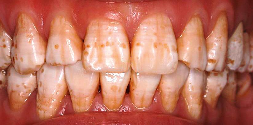
• Fig. 1-2: Through a clinical exam, it was diagnosed that all of permanent anterior teeth, which were erupted, had fluorosis. However, the teeth 11 and 21 were more severely affected according to Thylstrup and Fejerskov index [12].
Abstract
Aesthetic problems due to Fluorosis can occur in children and adolescentes. The aim was to describe a case report about the use of infiltrant resin to mask diffuse opacities. A male patient aged 12 years attended the Paediatric Dentistry clinic of the Rio de Janeiro State University, Rio de Janeiro, Brazil. Through a clinical exam, it was diagnosed that incisors had fluorosis. The teeth 12, 11, 21 and 22 were treated with infiltrant resin (Icon, DMG, Hamburg, Germany). All procedures were done in accordance with manufacturer instructions. Furthermore, Icon-Etch and Icon-Dry were applied three times in order to enhance the masking of the defects. The immediate result as well as 1 week and 4 months after the treatment were satisfactory. The use of infiltrant resin (Icon) can mask diffuse opacities improving the esthetics without a significant loss of tooth tissue.
Introduction
Aesthetic problems due to enamel developmental defects can occur in children and adolescentes. Fluorosis is a defect of enamel mineralization, characterized by porosity of the enamel subsurface [1]. Clinically, fluorosis can be seen as slight accentuation of the perikymata, diffused opacities with a opaque white appearance or chalky white enamel with some yellow to brown staining and pitting [2]. There are several treatment options for aesthetic problems due to fluorosis, such as bleaching, microabrasion and restorative techniques. Bleaching therapy has been reported by being able to mask the blemishes and providing a more uniform appearance [3,4]. Microabrasion works well for shallow defects, but it can result in some reduction of enamel [5, 6.] Treatment with resin composites can correct or improve enamel imperfections [7], however this procedure also ends up in a loss of tooth tissue. Infiltrant resin has masked white spot lesions [8, 9], because this resin has a refractive index similar to apatite crystals. Thereby, light refraction and, consequently, the colour differences of enamel are reduced [10]. As the fluorotic enamel is porous [11], the same as the white spot lesions, the resin infiltration can be a good alternative to mask the opacities. Thus, this paper aimed to describe a case report about the use of infiltrant resin (Icon, DMG, Hamburg, Germany) to mask diffuse opacities in permanent anterior teeth.
Case Report
The patient male, 12 years old, has been assisted at the Paediatric Dentistry clinic of the Rio de Janeiro State University, Rio de Janeiro, Brazil.

• Fig. 3: Before the treatment with Icon, the teeth 12, 11, 21 and 22 were cleaned and a rubber dam was placed.

• Fig. 4

• Fig. 6

• Fig. 8 • Fig. 5


• Fig. 7

• Fig. 9
• Fig. 4-5: Icon-Etch was applied on the buccal surfaces of the upper incisors for 2 minutes. • Fig. 6: Once the teeth were rinsed for 30 seconds and air-dried, Icon-Dry was used for 30 seconds. After the first acid-etching, part of the white diffuse opacities seemed masked when Icon-Dry was applied, but not the yellowish ones. • Fig. 7: A second application of Icon-Etch was done for 2 minutes, followed by dry air and Icon-Dry. However, the yellowish opacities were still visible. Hence, a third application of Icon-Etch was done for 2 minutes. This time a gentle friction was done using Icon-Etch applicator on the yellowish areas. Finally, the yellowish opacities seemed masked when they were wetted by Icon-Dry. • Fig. 8: All the surfaces were dried again, and Icon-Infiltrant was applied. The excess material was removed with gauzes. • Fig. 9: First the infiltrant set for 3 minutes and then light-curing each tooth for 40 seconds.
Discussion
Whenever an aesthetic procedure is recommended, it should be based on patient’s demand. Aesthetic perception is very much subjective and individual. An enamel defect can be an aesthetic problem for dentists, but not for patients. Furthermore, it can be argued that girls may be more concerned with their appearance than boys [6]. In the present case, the patient was a boy and felt really upset about the appearance of his teeth. Thus, the decision to treat the upper incisors aesthetically came from the patient’s wish to have non-discolored teeth. Thereby, as the infiltrant resin has masked white spot lesions [8, 9], it was decided to use the Icon to mask the diffuse opacities. The colour difference of enamel between white spot lesions and sound enamel occurs because the refractive indices of enamel, water and air are different [9]. If lesion pores are filled with water or air, in other words, if lesions are wet or dried, they will appear opaque, because the refractive indices of water and air are smaller than the enamel refractive index. When pores are filled with infiltrant resin, lesions are masked because the refractive indices of sound enamel and infiltrant are similar [9, 13]. As fluorotic enamel has a porous subsurface in the enamel below a well-mineralized surface [11] similar to white spot lesions, the infiltrant can behave in the same way as in white spot lesions. Diffuse opacities were well masked by the infiltrant in this clinical case. The application of Icon-Etch for three times was necessary to achieve a complete erosion of the surface layer allowing the infiltrant to penetrate as it happens in caries lesions [8, 14]. Compliance with manufacturer instructions on how to use the material may have contributed for the treatment success, for instance, the polishing of tooth surfaces. The polishing of treated areas enhances the colour stability of the masking probably due to reduction of the roughness. Clinical conditions, such as type of opacity and infiltration depth,

• Fig. 10

• Fig. 11: The final aspects one week after the treatment. • Fig. 10: According to manufacturer instructions, the application of Icon-Infiltrant was repeated for 1 minute. To finalize the treatment, the tooth surfaces were polished with composite resin polishing discs.

Fig. 12: Follow-up of 4 months. The guardians signed an informed consent form regarding all the procedures.
complete or incomplete infiltration, polymerization shrinkage as well as resin colour, can also interfere in the final result [10]. In this case, the rubber dam hampered Icon to set in the gingival margin. Nonetheless, even with slight blemishes in the gingival margin of the upper incisors, the patient was very satisfied with the treatment. Icon-Infiltrant has a lot advantages over other treatment techniques. The infiltrant can mask deeper lesions [9 ]without a significant loss of tooth tissue, which the microabrasion [5, 6] and restorations with resin composites are not able to do. Moreover, a resin layer is not necessary, once the material penetrates into the enamel [9]. The removal of the excess material with gauzes also retains the surface shape [15]. In contrast to the bleaching therapy, which can reduce the microhardness of demineralized enamel surfaces [16], the infiltrant resin can strengthen the enamel structure mechanically [17].
Conclusion
The use of Icon-Infiltrant can mask diffuse opacities improving the esthetics without a significant loss of tooth tissue.
Key Learnings
• The polishing of treated areas enhances the colour stability of the masking probably due to reduction of the roughness. • In contrast to the bleaching therapy, which can reduce the microhardness of demineralized enamel surfaces [16], the infiltrant resin can strengthen the enamel structure mechanically [17]. • The use of infiltrant resin (Icon) can mask diffuse opacities improving the esthetics without a significant loss of tooth tissue.
References
1. Fejerskov O, Johnson NW, Silverstone LM. The ultrastructure of fluorosed human dental enamel. Scand J Dent Res. 1974;82:357-72. 2. Møller IJ. Fluorides and dental fluorosis. Int Dent J. 1982;32(2):135-47. 3. Wright JT. The etch-bleach-seal technique for managing stained enamel defects in young permanent incisors. Pediatr Dent 2002;24:249-52. 4. Bussadori SK, do Rego MA, da Silva PE, Pinto MM, Pinto AC. Esthetic alternative for fluorosis blemishes with the usage of a dual bleaching system based on hydrogen peroxide at 35%. J Clin Pediatr Dent 2004;28:143-6. 5. Dalzell DP, Howes RI, Hubler PM. Microabrasion: effect of time, number of applications, and pressure on enamel loss. Pediatr Dent 1995;17:207-11. 6. Wong FS, Winter GB. Effectiveness of microabrasion technique for improvement of dental aesthetics. Br Dent J 2002;193:55-8. 7. Dietschi D. Optimizing smile composition and esthetics with resin composites and other conservative esthetic procedures. Eur J Esthet Dent 2008;3:14-29. 8. Paris S, Meyer-Lueckel H. Masking of labial enamel white spot lesions by resin infiltration – a clinical report. Quintessence Int 2009;40:713-8. 9. Kim S, Kim EY, Jeong TS, Kim JW. The evaluation of resin infiltration for masking labial enamel white spot lesions. Int J Paediatr Dent 2011;21:241-8. 10. Paris S, Schwendicke F, Keltsch J, Dörfer C, Meyer-Lueckel H. Masking of white spot lesions by resin infiltration in vitro. J Dent 2013;41:28-34. 11. Newbrun E, Brudevold F. Studies on the physical properties of fluorosed enamel I. Microradiographic studies. Arch Oral Biol 1960;2:15-20. 12. Thylstrup A, Fejerskov O. Clinical appearance of dental fluorosis in permanent teeth in relation to histological changes. Community Dent Oral Epidemiol. 1978;6:329-37. 13. Hosey MT, Deery C, Waterhouse PJ. Paediatric Cariology. London: Quintessence Essentials 2004. 14. Knösel M, Eckstein A, Helms HJ. Durability of esthetic improvement following Icon resin infiltration of multibracket-induced White spot lesions compared with no therapy over 6 months: A single-center, split-mouth, randomized clinical trial. Am J Orthod Dentofacial Orthop 2013;144:86-96. 15. Mueller J, Meyer-Lueckel H, Paris S, Hopfenmuller W, Kielbassa AM. Inhibition of lesion progression by the penetration of resins in vitro: influence of the application procedure. Oper Dent 2006;31:338-45. 16. Basting RT, Rodrigues Júnior AL, Serra MC. The effect of 10% carbamide peroxide bleaching material on microhardness of sound and demineralized enamel and dentin in situ. Oper Dent 2001;26:531-9. 17. Robinson C, Brookes SJ, Kirkham J, Wood SR, Shore RC. In vitro studies of the penetration of adhesive resins into artificial caries-like lesions. Caries Res 2001;35:136-41. 18. Meyer-Lueckel H, Paris S. Improved resin infiltration of natural caries lesions. J Dent Res 2008; 87:1112-6.