12 minute read
Resin infiltration as a micro invasive treatment for fluorosis


CASE 2. 6
Resin infiltration as a micro invasive treatment for fluorosis.
Advertisement
Prof. Dr. Leandro Augusto Hilgert, Marília Bizinoto Silva Duarte

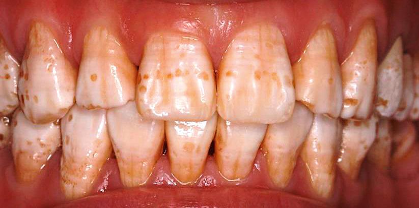
• Fig. 1 • Fig. 2 A 26-year old female patient presenting mild to moderate fluorosis looked for esthetic treatment. Her main complaint regarded the whitish diffuse opacities that affected her smile.
Fluorosis is observed, clinically, from mild diffuse white opacities on the enamel to severe whitish/brownish staining end enamel surface malformation. Those conditions may compromise esthetics according to its severity. For light to moderate fluorosis, the most common cases, there are reports that resin infiltration may successfully mask the opacities improving esthetics with very low enamel wear. The aim of this case report is to present a step by step description of the resin infiltration technique as a microinvasive alternative for the esthetic treatment of fluorosis. Considerations on how to diagnose depth of the opacities and on how many times to etch the enamel to improve results predictability are presented. Main features of the resin infiltration technique and other established esthetic treatments for fluorosis are discussed.
Introduction
Fluorosis is characterized by subsurface enamel hypomineralization (porosities) caused by excessive fluoride intake during enamel development [1, 2]. In mild to moderate cases of fluorosis, the lower refractive index (RI) of the porosities contents gives the enamel a diffuse whitish opaque appearance that, for some patients (according to fluorosis severity), may be aesthetically unpleasant. Many treatment options are available for fluorosis as: (a) bleaching, that can possibly reduce the contrast between whitish opacities and sound enamel; (b) microabrasion, in which the surface and subsurface of the affected enamel are worn out by a combination of acids and abrasives, exposing the underlying sound enamel; (c) macroabrasion, where a preparation is performed on the affected fluorotic areas followed by a restoration; and, (d) resin infiltration, a technique that involves a very mild wear of the surface enamel, exposing the porous subsurface that is subsequently infiltrated by a low-viscosity resin that has a RI more similar to sound enamel [3, 4]. Usually, bleaching alone is not capable of providing a complete optical blending of the fluorotic to the sound enamel. Micro and macroabrasion techniques are effective, but require a more invasive approach, removing the whole affected enamel. Resin infiltration appears as a suitable alternative that combines good results with a very low invasiveness. The aim of this case report is to describe in details the resin infiltration protocol on the esthetic treatment of a mild to moderate fluorosis case.
Case Report
A 26-year old female patient presenting mild to moderate fluorosis looked for esthetic treatment. Her main complaint regarded the whitish diffuse opacities that affected of her smile (Fig. 1 and 2). The patient, a dentist, was questioned on how glad she was with the shade of her teeth and she answered that she would like a more natural, less white appearance. This is a crucial question since after treating mild/moderate fluorosis teeth will become more chromatic and presenting less value. Therefore, patients that enjoy a very white shade should be counseled to bleach before treating the fluorotic lesions. The patient was very pleased with the esthetic result that was obtained with a microinvasive approach.

• Fig. 3

• Fig. 4
• Fig. 5

• Fig. 3: To evaluate the depth of the affected enamel a clinically useful tool is to perform transillumination. Specific equipment or even a simple light curing unit can be positioned on the lingual surface and commanded to emit light. When the transmitted light is not or is only slightly blocked by the opacities, the lesions (enamel porosities) are shallow and the probability of less invasive therapies to be effective is higher. In the present case, this was the situation and the fluorotic lesions were judged shallow, indicating a good prognosis for resin infiltration.
• Fig. 4: Next step is isolating the teeth that will be infiltrated. Field isolation and protection of the gingival tissue can be obtained using rubber dam or a liquid light-cured resin dam (Top Dam, FGM, Brazil) together with cheeks, lips and tongue retractors. While the liquid dam is usually faster and easier to apply, rubber dam may present a more intense gingival retraction, that may improve results in the cervical area. In this case, the liquid dam was selected and carefully applied to cover as little enamel as possible.


• Fig. 6
• Fig. 5-6: Resin infiltration protocol begins with etching of the surface enamel using a 15 % hydrochloric acid gel (Icon-Etch, DMG, Germany) that should stay in contact for 2 minutes aiming to wear the surface layer and expose the porous hypomineralized subsurface. After suctioning the acid gel, rinsing and air-drying the enamel a drop of ethanol (Icon-Dry, DMG, Germany) is applied on the etched surface and the observed optical aspect of the white opacities should be already minimized. If the opacities are still very visible when the ethanol is applied, a second (or even a third) etching steps is indicated. In the present case, in Figure 5, it is possible to observe the aspect after a single etching procedure. Since the opacities were still very visible it was decided to repeat the etching step once more. In Figure 6, it is possible to see the difference between the aspect after one and two etching steps as a drop of Icon-Dry was applied onto the etched enamel surface. Since the result was satisfactory, the ethanol was let on the surface for 30 s to promote a thorough desiccation of the enamel, followed by air-drying.


• Fig. 7 • Fig. 8
• Fig. 7-8: On the etched and dry enamel the low-viscosity infiltrant (Icon-Infiltrant, DMG, Germany) is applied and should remain for at least 3 minutes to achieve maximal infiltration depth into the porosities of the hypomineralized enamel. In Figure 7 it is possible to observe the aspect after infiltrant application on the right upper teeth, while the left have not received the resin yet. When all teeth received the infiltrant and the 3 minutes waiting period was respected, obvious excess can be removed using a gauze and light-curing is performed (Fig. 8). It is paramount to execute a thorough polymerization using adequate irradiance and exposure time (40 seconds per tooth). A second application of the infiltrant should be performed for 1 minute followed by excess removal and light-curing.

• Fig. 9: It is possible to see the immediate aspect after infiltrant polymerization. It is normal to observe a shiny and irregular appearance due to excess of the infiltrant covering the surface. These material is easily removed with polishing instruments as abrasive disks, spirals or rubber cups. • Fig. 10: In the present case polishing was performed using disks and spirals (Sof-lex, 3M, USA).


• Fig. 11 • Fig. 12 After polishing, the result of the treatment is shown on Figure 11 and 12. Almost all white opacities disappeared, indicating a satisfactory infiltration of the fluorotic enamel.
Discussion
An ideal esthetic treatment is the one that can please the demands of the patient, that require very little wear of sound enamel (low biological cost), that can be simply and quickly executed and that lasts. Patients that present fluorotic enamel may not require any kind of esthetic treatment, especially if presenting a mild severity of the lesions [5]. However, whenever a treatment is required, the dentist should be able to offer treatment options that present efficacy, low invasiveness and durability. Resin infiltration is a technique based on the acid dissolution of the well-mineralized surface layer of the enamel (with a thickness of around 30-40 μm) [6], exposing the porous hypomineralized enamel of the subsurface. After thorough drying, a low-viscosity resin is infiltrated into the porosities of the enamel by capillary forces filling the spaces with a material that has a closer refractive index to sound enamel. Therefore, the optical appearance of the infiltrated enamel blends with the sound enamel, significantly improving the esthetic harmony of the smile [7-9]. For the infiltration process to be effective the first step is to diagnose the kind of white opacity. Deeper lesions, that are very opaque to transillumination (as in some molar-incisor hypomineralization cases) usually do note present the best results for any kind of less invasive treatment and may require some localized tooth preparation. Shallow to medium depth lesions, as those depicted in the case report, that clinically do not block the light passage during transillumination (see Fig. 3) have usually a favorable prognosis for resin infiltration. Next, another fundamental step for a successful resin infiltration is adequate removal of the well-mineralized surface layer therefore exposing the porous subsurface. If an adequate access for the resin to infiltrate the porosities is not achieved, the technique will not present the best results. A very effective way of testing if the surface layer was removed after the acid etching step is observing what happens when a drop of ethanol is applied on the etched enamel. If the optical result already looks good, surface layer was properly removed. If the white opacities are still very visible, a new etching step should be performed (see Fig. 6, that depicts the difference between one and two etching steps). An easy and simple method to decide if re-etching is necessary before the drying and infiltration steps. It is imperative for the dentist to realize that the main difference from resin infiltration to microabrasion is that in the first method the porous enamel is preserved and infiltrated while the later method esthetic success is based on the complete removal of the affected enamel. That is why the technique is indicated for lesions no deeper than 0.2 to 0.3 mm (200 to 300 μm) [10]. Therefore, it is clear that microabrasion is a more invasive alternative, requiring much more enamel wear to present pleasant results. The color stability of the resin infiltrated enamel has been tested in vitro,(8) in clinical studies [11] and presented in numerous case reports [4, 12-15]. So far results are positive and very promising. This hybrid structure of enamel/infiltrant (the inifltrated enamel) can be successfully daily submitted to the »polishing« of oral hygiene and able to be polished by the dentist in routine clinical sessions.
Based on the substantial amount of available scientific evidence, clinical reports and our clinical experience of almost eight years conducting resin infiltration treatments, this approach has become our standard of care in treating light to moderate fluorosis. Important to state that for many patients that desire to treat the fluorotic opacities and to have whiter teeth, a bleaching procedure is usually performed before resin infiltration [16].
Conclusion
Resin infiltration seems to be a successful microinvasive treatment for the esthetic treatment of light to moderate fluorosis.
Key Learnings
• Resin infiltration is a microinvasive approach for the treatment of slight to moderate fluorosis; • Additional acid etchings may be necessary to improve resin infiltration. Observing the visual aspect when applying Icon-Dry may be a good way of determining the need for repeating the etching step; • Removal of Icon-Infiltrant excesses before light-curing and adequate finishing and polishing after light-curing are important steps to promote a nice surface texture.
References
1. Fejerskov O, Manji F BV. The nature and mechanism of dental fluorosis in man. J Dent Res. 1990;69(Spec Iss):692–700. 2. Aoba T, Fejerskov O. Dental fluorosis: chemistry and biology. Crit Rev Oral Biol Med . 2002 Mar;13(2):155–70. 3. Duarte MBS, Hilgert LA. Infiltração resinosa: tratamento microinvasivo para melhoria estética de lesões cariosas e hipomineralizadas de esmalte. In: Monte Alto R. Reabilitação Estética Anterior. São Paulo:Napoleão; 2017. 4. Hilgert LA, Leal SC. Resin Infiltration: A Microinvasive Treatment for Carious and Hypomineralised Enamel Lesions. In: Eden E. Evidence-Based Caries Prevention. Springer; 2017. p. 123–41. 5. Nair R, Chuang JCP, Lee PSJ, Leo SJ, Yang NQY, Yee R, et al. Adult perceptions of dental fluorosis and select dental conditions-an Asian perspective. Community Dent Oral Epidemiol. 2016;44(2):135–44. 6. Meyer-Lueckel H, Paris S, Kielbassa AM. Surface layer erosion of natural caries lesions with phosphoric and hydrochloric acid gels in preparation for resin infiltration. Caries Res. 2007;41(3):223–30. 7. Paris S, Meyer-Lueckel H. Masking of labial enamel white spot lesions by resin infiltration--a clinical report. Quintessence Int. 2009;40(9):713–8. 8. Paris S, Schwendicke F, Keltsch J, Dörfer C, Meyer-Lueckel H. Masking of white spot lesions by resin infiltration in vitro. J Dent. 2013;41(5):e28-e34. 9. Torres CRG, Borges AB, Torres LMS, Gomes IS, De Oliveira RS. Effect of caries infiltration technique and fluoride therapy on the colour masking of white spot lesions. J Dent. 2011;39(3):202–7. 10. Benbachir N, Ardu S, Krejci I. Indications and limits of the microabrasion technique. Quintessence Int (Berl). 2007;38(10):811–5. 11. Eckstein A, Helms H-J, Knösel M. Camouflage effects following resin infiltration of postorthodontic white-spot lesions in vivo: One-year follow-up. Angle Orthod. 2015 May;85(3):374–80. 12. Muñoz MA, Arana-Gordillo LA, Gomes GM, Gomes OM, Bombarda NHC, Reis A, et al. Alternative Esthetic Management of Fluorosis and Hypoplasia Stains: Blending Effect Obtained with Resin Infiltration Techniques. J Esthet Restor Dent. 2013 Feb;25(1):32–9. 13. Cocco A, Lund R, Torre E, Martos J. Treatment of Fluorosis Spots Using a Resin Infiltration Technique: 14-month Follow-up. Oper Dent. 2016;41(4):357–62. 14. Torres C, Borges A. Color Masking of Developmental Enamel Defects: A Case Series. Oper Dent. 2015;40(1):25–33. 15. Tirlet G, Chabouis HF, Attal J-P. Infiltration, a new therapy for masking enamel white spots: a 19-month follow-up case series. Eur J Esthet Dent. 2013;8(2):180–90. 16. Gugnani N, Pandit IK, Gupta M, Gugnani S, Soni S, Goyal V. Comparative evaluation of esthetic changes in nonpitted fluorosis stains when treated with resin infiltration, in-office bleaching, and combination therapies. J Esthet Restor Dent. 2017;29(5):317–24.