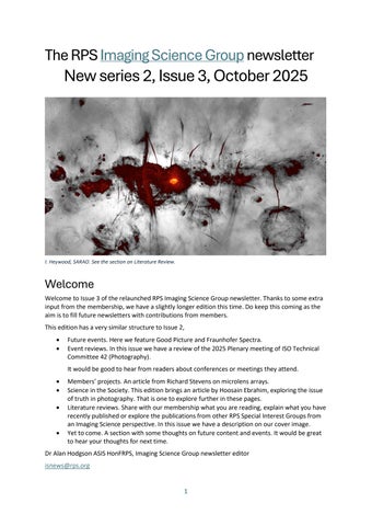
I. Heywood, SARAO. See the section on Literature Review
Welcome
Welcome to Issue 3 of the relaunched RPS Imaging Science Group newsletter. Thanks to some extra input from the membership, we have a slightly longer edition this time. Do keep this coming as the aim is to fill future newsletters with contributions from members.
This edition has a very similar structure to Issue 2,
• Future events. Here we feature Good Picture and Fraunhofer Spectra.
• Event reviews. In this issue we have a review of the 2025 Plenary meeting of ISO Technical Committee 42 (Photography).
It would be good to hear from readers about conferences or meetings they attend.
• Members’ projects. An article from Richard Stevens on microlens arrays
• Science in the Society. This edition brings an article by Hoosain Ebrahim, exploring the issue of truth in photography. That is one to explore further in these pages.
• Literature reviews. Share with our membership what you are reading, explain what you have recently published or explore the publications from other RPS Special Interest Groups from an Imaging Science perspective. In this issue we have a description on our cover image
• Yet to come. A section with some thoughts on future content and events. It would be great to hear your thoughts for next time.
Dr Alan Hodgson ASIS HonFRPS, Imaging Science Group newsletter editor isnews@rps.org
Future events
Good Picture online 2025
The Imaging Science Group is holding the first of its "new look" online Good Picture symposiums on Saturday 18th October. Due to financial restrictions affecting face-to-face meetings we have decided to hold regular Zoom meetings, free to all, with 3 talks over an afternoon. We are hoping this will be of interest and will allow members throughout the UK, and maybe even abroad, to hear some talks and discussions on a wide range of Imaging subjects. The flyer on the next page has all the details of this inaugural event, please contact me if you are interested in enrolling.
The flier can also be found on the Imaging Science Group website here.
Dr Mike Christianson, Organiser.
Fraunhofer spectra – their place in the evolution of photography
A Fraunhofer spectrum from 1894
This on-line meeting is now open for registration here. It will take place at 7pm UK time (GMT) on December 2nd, 2025. For those for whom the timing is inconvenient a recording of the presentation will be made available to those registered for the event – the same will apply for the Good Picture event.
This will be a new initiative in that it is a joint event with the RPS Historical Group. I am in the process of writing a book on some aspects of scientific photography where the topic of Fraunhofer spectra seems a recurrent theme. The aim of this presentation is to explain the topic for a nontechnical audience and to discuss areas deserving further research.
Colour is a persistent theme in this work, both in colour photography and printing. It begins with the 1848 Becquerel colour process but also takes in photographic lens design and astrophotography. As an additional historical perspective these spectra reveal hints on the characteristics of photographic plates and create a perspective on the history of photographic imaging science.
Dr Alan Hodgson
If you attend any of these meetings we would like to have a review from you. It would be great to feature multiple perspectives of the same meeting in this newsletter.
Good Picture – Online – 2025
An RPS Symposium
For over 20 years the Imaging Science Group of the RPS has hosted an annual series of 1 day symposia under the title “Good Picture” on selected technical aspects of Imaging. Unfortunately, due to ever rising costs, the Group is unable to continue to sponsor these meetings without unacceptably large increases in delegate’s fees. Therefore the Group committee has decided to replace them with a series of shorter meetings via Zoom. These presentations and discussions will have three speakers providing photographic practitioners, keen amateurs and students with insights into Imaging. The Group is planning to run at least 2 of these meetings per year and they will befreeto attend.
This event is scheduled for Saturday 18th October 2025, 1.00pm – 5.00pm, if you would like to register yourinterest pleasecontact theorganiser Dr Mike Christianson at pandm.christianson@gmail.com
Programme
DrAvijit Datta
York St John University
Two Adjacent London Societies and Their Members' Influence on Colour Theory and Culture
Dr Datta will discuss the immense influence of polymath members of the Royal Society and adjacent Pall Mall club have had on the development of colour theory, art and culture. New techniques such as Hyperspectral Imaging and the useof lasers to uncover aperceived new colour will also beexamined.
AdrianDavies MSc,ARPS
Freelance Photographer, Lecturer and Author
Photographing Plant Behaviour.
In a previous talk at this event Adrian showed how reflected and fluorescence UV photography could reveal invisible signals on flowers and other plants, to attract pollinators. In this talk Adrian will show how a range of photographic techniques can reveal various plant behaviours such as spore and seed dispersal, plant growth and movement.
DrAlan Hodgson ASIS FRPS
Alan Hodgson Consulting Ltd
Photography by Synthetic Aperture Radar
Synthetic Aperture Radar has an interesting history as a photographic imaging technique, ranging from military to cultural heritage use. This presentation will explain the technique in a non-mathematical fashion, drawing on parallels with more familiar techniques such as aerial photography, holography and medical dopplerultrasound.
Event Review
If you attend any imaging-based event we would like to have a review from you. Equally, if you hear of (or are working on) an event that you feel would be of interest to the membership do pass on the details. In this edition we review an International Standards meeting on Photography.
ISO Technical Committee 42 Plenary Meeting
ISO, the International Organization for Standardization creates International Standards documents that affect many aspects of our lives. ISO creates these documents in specific topic areas and organises Technical Committees to conduct this work. ISO Technical Committee 42 has been designated the task of creating and maintaining International Standards documentation specifically for Photography.
ISO TC 42 has been in existence since 1947 so has a long history of this work. There are currently 216 published standards to support Photography and another 16 currently under development. As a result of this long history and current activity there are International Standards covering legacies such as the dimensions of photographic glass plates out to the latest technologies in digital images, such as HDR and wide colour gamut encoding
ISO TC 42 meets as a whole group for 5 days at a Plenary Meeting every 2 years. The meeting was held this June in Berlin and was hosted by DIN, the German National Standards organisation. 35 delegates registered from Australia, China, Finland, Germany, Japan, Switzerland, UK, USA. A truly international meeting with most of the major countries from the industry participating.
Some countries only allow industry experts or industry nominated experts, others allow individual experts or those from public institutions and universities. Each Technical Committee is hosted by a national standards organisation and in the case of ISO TC42 this is ANSI, the American National Standards Institute, who support the meetings and the project management. ISO publishes and maintains the standards. TC 42 experts generally contribute to several standards in their committee.
The Plenary Meeting is also a welcome occasion to meet long-term colleagues. In general this meeting splits into 2 tracks, with communal sessions to complete the formal business at the beginning and end of the week. The 2 tracks are concerned with what can be broadly described as the capture and output of photographs.
The capture track now works almost exclusively on digital technologies, including camera colour encoding and data exchange and has recently moved into consideration of machine vision applications, in addition to consumer digital and mobile phone cameras. Thus the large corporations that manage the immense quantity of the world’s shared phone images are part of this group. However, it is recognised that TC 42 is also responsible for a number of legacy camera standards that originate from analogue camera days. As a result the capture group have also taken on the maintenance of these for the benefit of photographers in general. Examples of this work include the maintenance of the standards that govern the camera flash connection and tripod thread.
Due to the long history of photography there are still silver halide-based capture technologies with a long history and currently still in use, such as photographic glass plates and camera films. The expertise on these now rests predominantly within the output group as the same chemistry is used in silver halide-based printing papers. Materials utilising silver halide and inkjet technologies take up most of the effort here but plastics and glass plates figure too.
We both participated in the output track and we are part of a diverse group from N. America, Europe and Asia.
The main focus of the output materials group is to develop standards for test method that allow the quantitative comparison of photographic print performance across the industry. Indeed, that today’s photographic print performance is at its best, concerning image quality and permanence is also due to the standards developed in this group. Important parameters are image quality such as gloss and print homogeneity, durability such as scratch and tear resistance and stability against the four environmental factors light, heat, air pollutants and humidity. After years of failures, the committee no longer attempts to combine those factors into one print life specification. The question of ‘how long does it last’, cannot be answered by any of the current standards
There is also a group on museum and cultural heritage standards pertaining to photography. As photographic technology is very mature, the focus turns to how to maintain those materials in longterm preservation. While standards on scanning requirements for the digitization of art and cultural heritage materials are developed in the capture community, environmental storage requirements are covered by the output materials group, reinforced by museum experts from many of the National Archives and important photographic collections. Photography has been recognized art form and an important part of a country’s historical record for the last 50 years. Keeping and sharing these memories is more challenging than keeping a book archive. A fine balance has to be found between the long-term preservation and the cost for specialised facilities and digitisation and the complexity of mixed media storage.
Dimensions sounds like a dull topic but it is key to making photography work. Films in sheets and rolls need to fit into cameras and sheets of photographic papers need to fit into frames. Documents covering and defining these need periodic review, if only to keep the terminology used in them congruent with current use.
ISO 18948 is newly issued and contains a set of test methods for the permanence and durability of photobooks. Most photographic output today is in the form of photobooks printed in centralized specialised printing houses. The majority of high-quality photobooks are made on traditional silver halide materials, the others are printed on a liquid electrophotography press. Printing just one copy of a book is thus possible.
Both areas of TC 42 have various liaisons into other standards groups and they work together in joint committees in related fields. ISO TC 130 is responsible for graphic technology and a long-term partner for cooperation. Strong ties also exist into other standards organisations such as CIE and IEC concerning colour, lighting and electronics.
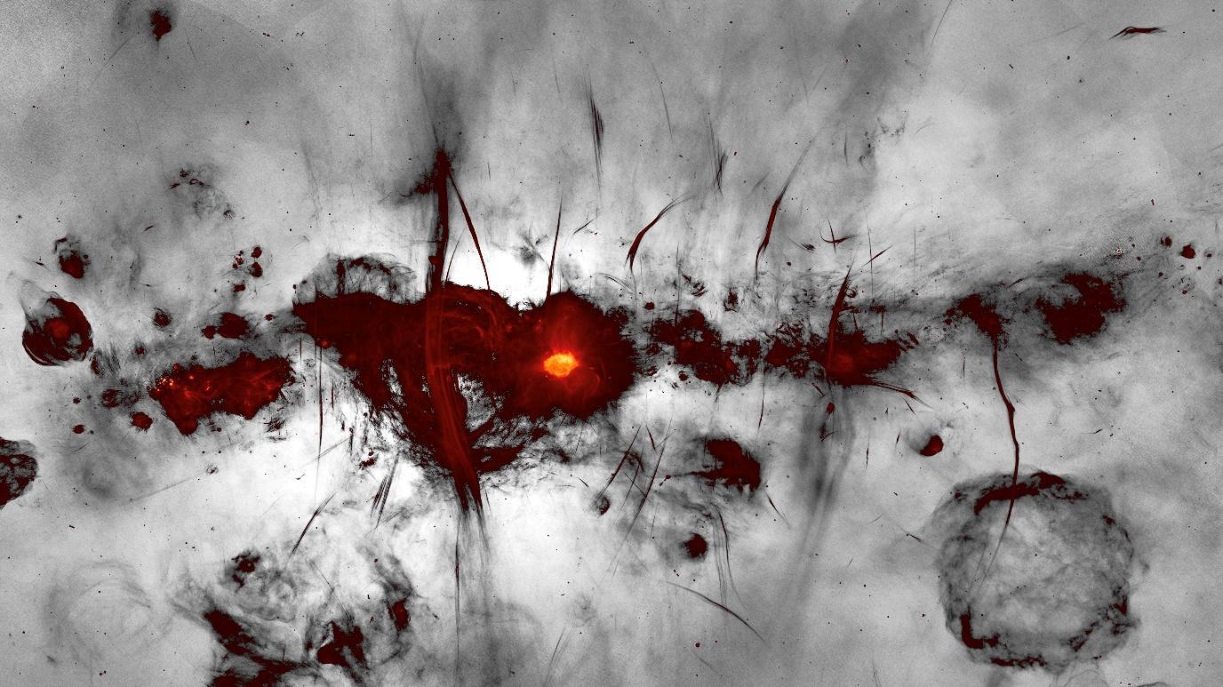
So if you have ever wondered why the speed setting on your camera or smartphone is labelled “ISO” you now know of another area where TC 42 is working for you!
Rita Hofmann-Sievert HonFRPS & Alan Hodgson ASIS HonFRPS
Literature reviews
Colour palettes in image rendition
I have seen our cover image in a number of publications over the past few months and decided to take a closer look. It could easily be interpreted as a piece of conceptual art but the attribution gives this away. SARAO is the South African Radio Astronomy Observatory and this is a radio frequency image of the centre of the Milky Way.
I first came across this image in the April edition of Physics world and it caught my attention for a number of reasons.
1. The MeerKAT telescope array used to generate this image is sensitive to the frequencies used in another of my interests, Synthetic Aperture Radar (see Good Picture above). By definition, radio frequencies are outside the visible spectrum and as a result an algorithm must be chosen to create a visual representation of the image. Hence the use of a specific colour palette.
2. The choice of colour palette was interesting here, probably driven by the fact that MeerKAT can generate High Dynamic Range imagery. For the purposes of illustration they have chosen to use colour to indicate the brightest areas of radio emission, with the lower intensities rendered in grey scale. This is a palette choice I had not seen before but I would be interested in comments from the readership on this.
The image also appeared in a different form twice in the July edition of Sky at Night magazine. One would appear to be the same image but rendered in a different colour palette, presumably to illustrate the elements involved in the radio emissions. However the second went in a somewhat different direction.
2025 marks the 25th anniversary of operations of a very different telescope, the Chandra X-ray observatory. As Earth’s atmosphere is effectively opaque at X-ray wavelengths Chandra was placed into Earth orbit. To celebrate this anniversary NASA have curated an on-line exhibition of the finest of the Chandra images, available here
I. Heywood, SARAO
One of these anniversary images is shown below and it combines X-ray images from Chandra with the radio frequency image from MeerKAT Once again all this image information is outside of the human visual range so a colour palette must be chosen. In this case the X-rays from Chandra are rendered in orange, green, blue, and purple while the radio image from MeerKAT is in lilac An interesting choice but one that renders to a visually pleasing image that is close to expectation of a deep sky image.
Colour palette choice would be an interesting one to debate in future newsletters as it could embrace various perspectives from aesthetics to communication. It can also bring in the philosophical debate on truth – how “accurate” is an image rendition? Both the above images could be considered either true or false but with no attempt to deceive.
Does it matter and is it worthy of debate? There is another perspective in the article that follows. Over to you…
Alan Hodgson
Science in the Society
Has digital technology subverted the truth in photographic evidence?
Photographic truth is being entirely challenged by the emerging technology of digital image manipulation and synthesis. Photographs can be altered in ways that are virtually undetectable and photorealistic synthesized images are becoming increasingly difficult to distinguish from actual photographs.
Image-processing software can rearrange and otherwise transform the elements of a scene. The same software can combine fragments of different images into a new image. Digital images make photographic falsehoods quicker and easier – and often more difficult to trace. The question of how to distinguish visual fact from fiction is becoming increasingly important as we witness the explosive proliferation of digital imaging technology. The fact that an image presents a seamless surface no longer provides evidence for concluding that it has not been manipulated. We must look for other clues such as internal consistency, documentable provenance, and consistency with beliefs. In general, the more information there is in an image, the more difficult it is to change without introducing detectable inconsistencies.
The provenance of a conventional photograph is often easy to trace whereas digital imaging technology eliminates negatives, it can replicate files in seconds, digital images can be transmitted rapidly and invisibly through computers and networks.
The subtlest challenge is posed by images that show no detectable signs of tampering and no obvious internal inconsistencies and yet contradict our established beliefs.
There is no doubt that it is tempting to adjust or modify digital image files. Many such manipulations, however, constitute inappropriate changes to the original data and making such changes can be classified as scientific misconduct. Good science in areas of Medicine, Dentistry and Forensic Imaging require reliable data. Consequently, to protect the integrity of research, the scientific community takes strong action against perceived scientific misconduct. Radiographic software packages allow for image enhancement but do not modify the content of the radiograph. Image manipulation is inadmissible evidence. Admissible evidence must be properly authenticated and continuity of evidence must be demonstrated. Tampering with truthful representation happens at the outset in photography when three dimensional objects are represented in two dimensions. Software packages enable radiologists to manipulate digital radiographs without the patient being exposed to additional radiation. In some countries falsified images were printed and presented to Insurance Companies as evidence for an expensive dental restorative treatment. Insurance companies and experts could demand that digital radiographs are always evaluated in the original software and under predefined conditions.
Modern software has also contributed to the manipulation of digital radiographs highlighting the difficulty in distinguishing the original from the fraudulent one. The concept of hiding data in other data is called steganography. Different steganographic techniques can be divided into two main groups according to the embedding domain of the image, namely, the spatial domain approach and the frequency domain approach.
Professional responsibility for the ethical issues involved in image manipulation is an important area of concern for the forensic photographer. Credibility and integrity of the medical photographer will support the credibility and integrity of the image. The integrity of evidence is an inseparable part of
the code of ethics followed by the medical photographer, guided by procedures and protocols to ensure truthfulness.
Digital imaging technology can provide openings for principled resistance to established social and cultural practices and at the same time it can create possibilities for cynical subversion of those practices. Digital images have less standardized production processes than conventional photographs. These processes are less subject to institutional policing of uniformity, offer more opportunities for human intervention, are far more complex and varied in their range of possible representational commitments. A universal Behavioural Code that the integrity of an image must be respected and adhered to.
Question: Can a scanner manufacturer or imaging processing software developer be held liable for copyright infringement done by someone else using its products?
Hoosain M Ebrahim. ASIS FRPS
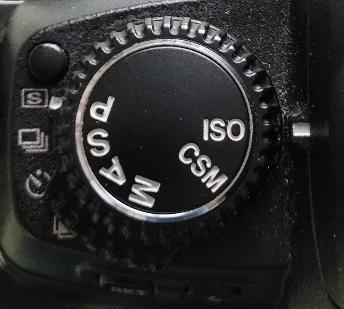
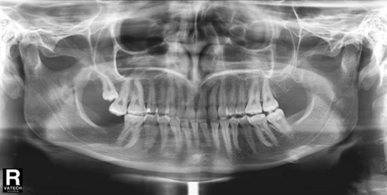
Panoramic Tomograph
Figure 1
Impacted tooth
Digital Manipulation – impacted tooth removed
Same image with adjustment to the red colours to make them more yellow
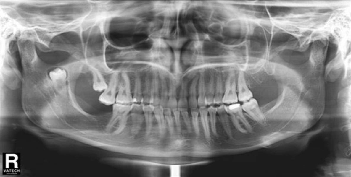
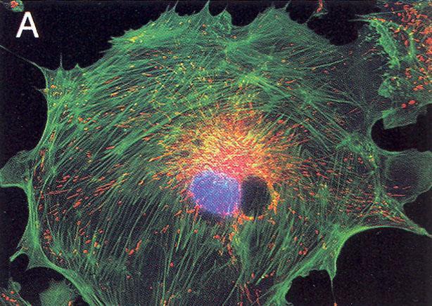
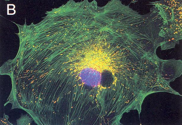
“This would be the visual result of an experiment where the mitochondria (red structures) showed positive labelling with two fluorochromes. This is an unacceptable use of digital manipulation. The client believed that the colour was actually more yellow in the original and wanted the reds shifted in hue independently of the green.”
(Figure 2A and 2B) – James E Hayden, The Journal of Biocommunication, Vol 27. No:1 2000
Figure 2
Fluorescence of a triple labelled epithelial cell
Members’ projects
Following on from the event review on ISO Technical Committee 42 (Photography) we have an article on microlens arrays by Richard Stevens, illustrating the work of ISO Technical Committee 172 (Optics and photonics).
Lenses, Microlenses and International Standards
Photography is largely about collecting light with lenses and mirrors and generating images which we record on a two-dimensional photosensitive surface. A generation or so ago, lens design was carried out by painstakingly tracing ray paths and manually calculating angles of refraction at successive optical boundaries. Materials for making lenses were generally limited to crown and flint glasses and lenses were made by laboriously grinding glass blanks to produce the lens elements with spherical surfaces. While most cameras used one lens with several axially-spaced elements, spatial arrays of lenses were used in special photographic applications such as high-speed cameras and various forms of stereo and 3D imaging.1 In 1941, Dennis Gabor patented an idea that used arrays of cylindrical lenses to generate a “superlens” and simulate the effect of a much thicker conventional lens.2 In one application he envisaged replacing bulky Fresnel lenses, such as those used in lighthouses, with lighter and thinner lens arrays.
Lens design has progressed rapidly with the aid of modern computers that calculate the paths of light rays through multiple components and allow designers to decide on the optimum form of a lens surface to suit the design constraints such as physical size, weight, spectral transmission and image size and position. Materials for making lenses now include plastics with a range of values of refractive index which can be readily moulded with spherical and aspherical surfaces. An example is the mobile phone camera which uses lenses made from several elements formed in moulded plastic. These lenses are generally a few millimetres in diameter and mobile phone cameras may have two or more lens systems for different functions.
While progress in lens design and materials has resulted in more advanced optics, in some applications individual lenses have become much simpler in form. These are applications that require very small lenses, with dimensions less than 1 mm and often as small as 10 µm. They are referred to as microlenses and may be used individually or in arrays of multiple lenses on a common substrate, to generate images or collect light for other purposes. Arrays are usually formed on a plane substrate and each lens may be of a simple form such as a concave or convex spherical surface or, depending on the method of fabrication, a more advanced aspherical shape.
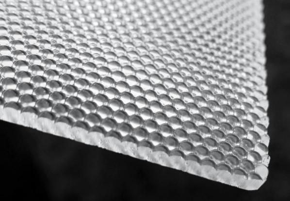
Figure 1. Two views of an array of convex lenses with lens spacing 2.5 mm, moulded in polycarbonate.
Figure 1 shows a close-packed array of plano-convex lenses. The lenses are moulded in polycarbonate and in this case the lens spacing is 2.5 mm and they focus a distant object onto the flat rear surface of the substrate. Similar arrays but with lenses and spacings less than 1 mm or so are known as microlens arrays. Although microlenses may be formed with more than one layer, for example as doublets to correct chromatic aberration, some optical aberrations are less of a problem with small lenses. Effects such as spherical aberration decrease rapidly with decreasing radius of the lens. However, aberrations may be introduced by the substrate which may be relatively thick compared to the lens dimensions and this must be borne in mind.
Robert Hooke and Antonie van Leeuwenhoek were 17th century microscopists who both made their own very small lenses for use in simple microscopes. Hooke melted filaments of glass into globules, relying on surface tension to form a spherical surface.3 More recently an established technique for making microlenses in arrays has been to melt small islands of photoresist formed by lithography on a plane substrate, again relying on surface tension forces to form the optical surface.4 The surface profile of the microlens array is then replicated in a more durable and optically suitable material.
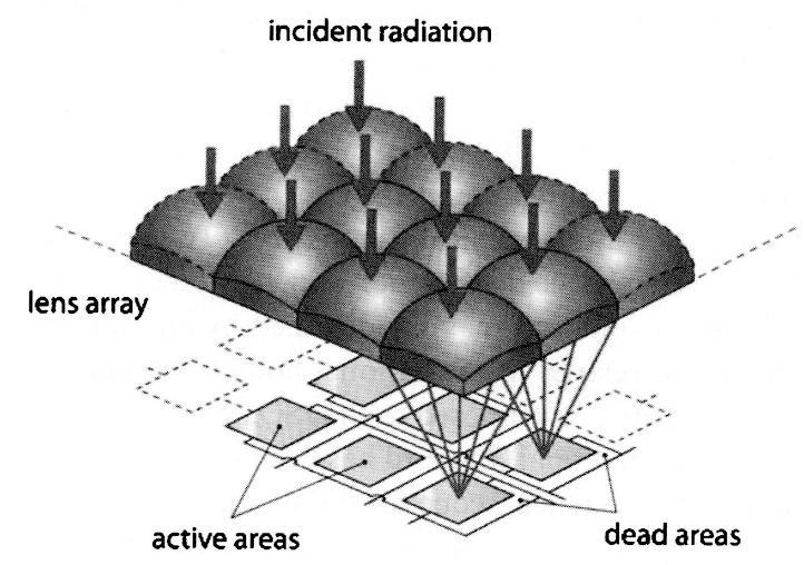
Figure 2 Photodetector array illuminated via a microlens array to enhance the optical efficiency
A common application for microlenses is found in the image sensor of a digital camera, as shown in figure 2. A sensor is formed of an array of photodetectors on a substrate, each spaced to allow the electrical signal to be extracted from the array. To avoid wasting light that falls on the gaps between the detectors, it may be captured by a microlens and focused to the detector element. A closepacked array of microlenses aligned with the detector array thus increases the overall optical efficiency. Obviously to match every element in a modern detector array requires many millions of microlenses.
Microlens arrays may also be used to analyse the wavefront emitted by a source or reflected by an object. Instead of forming an image with a single relatively large lens, the wavefront shape can be examined by dissecting it with a microlens array and detector array. If the wavefront is from a distant point object, as in astronomy, each microlens will produce a small focal spot and the transverse position of the spot will be a function of the angle of incidence of that part of the wavefront. By detecting the lateral positions of the spots in the array the wavefront slopes and hence the shape may be calculated. The technique is used in astronomy to measure atmospheric distortion of star images and to provide feedback to optical components that correct for the atmospheric distortion.
If the wavefront is from an extended 3D object, each microlens will produce a small image of the object as seen from a different viewpoint. Recording and replaying those small images allows the original 3D image to be reconstructed. This is the basis of a photographic technique proposed by
Gabriel Lippmann in 1908 which he called integral photography.5 It was developed more recently in electronic form as light-field photography. 6
Microlenses have been developed that operate by refraction, diffraction or a combination of the two. Diffracting structures can be fabricated using photolithographic techniques used to make microchips. The increasing number of applications includes use with optical fibres, confocal microscopes, digital displays, photocopiers, and 3D displays. They are also used to create novel optical effects such as moire patterns and images which are used in product security and enhancement devices in packaging, banknotes and passports. Moire effects generated by microlens arrays may also be used to inspect manufactured objects with similar periodic structures.7
Because of the small apertures and relatively thick substrates involved it is generally not possible to use traditional definitions and test methods for the parameters involved. This is why it is useful to define common terms and practices. ISO, the International Organization for Standardization, has developed the ISO 14880 series of standards to assist manufacturers and users to specify and assess microlenses and microlens arrays.8 The standards are regularly reviewed and updated and ISO 14880-1 Vocabulary is currently under revision.9 Other parts of the standard are ISO 14880-2:2024 which covers test methods for wavefront aberrations, ISO 14880-3:2024 which covers test methods for optical properties other than wavefront aberrations and ISO 14880-4:2024 which covers test methods for geometrical properties. ISO/TR 14880-5:2010 is a technical report that gives guidance on testing.
References
1) Stevens R Davies N, “Lens Arrays and Photography”. The Journal of Photographic Science, 39, 199-208, 1991
2) Gabor D, “Improvements in or relating to optical systems composed of lenticules”. UK patent 541753, 1941
3) Hooke R, preface to Micrographia. The Royal Society of London, 1665.
4) Daly D, Stevens R, Hutley M, Davies N, “The manufacture of microlenses by melting photoresist”. Meas. Sci. Technol. 1990, 1, 759-766.
6) Ng, R et al. “Lightfield photography with a hand-held plenoptic camera”. Stanford Tech Report CTSR 2005-02
7) Stevens R, “Optical inspection of periodic structures using lens arrays and moire magnification”. Imaging Science Journal 47, 173-179, 2000
8) Stevens R, Miyashita T, “Review of standards for microlenses and microlens arrays”. Imaging Science Journal 58, 202-212, 2010
9) ISO 14880-1:2019 - Optics and Photonics - Microlens Arrays - part 1: Vocabulary
Dr Richard Stevens (UK) and Dr Takaaki Miyashita (JP) are joint project leaders in Working Group 7 of ISO committee TC 172/SC 9 Laser and Electro-optical Systems, responsible for developing the microlens array standards described above.
Richard Stevens 8 July 2025
Yet to come
Between this issue and the next we have two events of group members: Good Picture and the Fraunhofer spectrum presentation Details of these are included in the Future Events section Ideas for future meetings are always welcome – send them to me and I will pass these on. Especially if you are willing to present or organise!
We are starting to build some breadth into the contributors to this newsletter. In this edition I am grateful for contributions from Mike Christianson, Rita Hofmann-Sievert, Richard Stevens and Hoosain Ebrahim. It would be great to add your name to the list for future editions.
As mentioned last time I am interested in including imaging science perspectives on articles in the newsletters of other RPS Special Interest Groups. If you come across any of these and would like to offer your thoughts do send me a note. I previously mentioned an article in the Digital Imaging Group publication DIGIT #104, discussing the use of the Freeware program Sequator to stack sky images. I have started exploring this and would be grateful for other perspectives.
There continues to be a lot in the popular press on images and the rapidly blurring differentiation between “real” and “synthetic” imagery. We touch upon this from two perspectives in both the Literature review and Science in the Society sections above. This would be an interesting one to explore from an Imaging Science perspective – further contributions would be most welcome. It would be good to hear from readers about conferences or meetings they attend. We also intend that this newsletter will provide a forum for members to show their work, completed or in progress. All for now – see you next time! Don’t forget to send me your content and thoughts for the next issue. At the moment we are aiming for a January 1st publication so the edition will be assembled mid-December.
Dr Alan Hodgson ASIS HonFRPS, Imaging Science Group newsletter editor isnews@rps.org
Past newsletters
The Group is starting to create an archive of these newsletters. Issues #1 and #2 are now available on the Group website as pdf files