7 minute read
Clinical precision


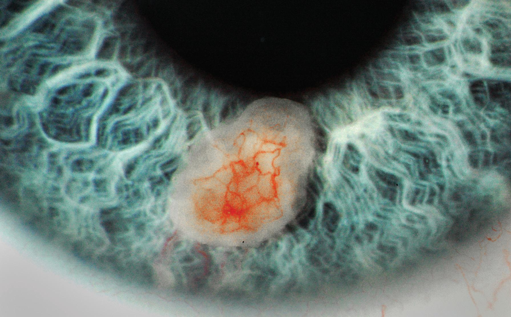
Anterior segment image of a metastatic deposit on the iris Courtesy: Bob Tapper, Royal London Hospital, London, UK
Professor afzal ansary asIs frPs
It may be a little-known discipline, but clinical photography plays a huge role in medicine and its teaching, explains professor Afzal Ansary
Today, visual documentation of patients is integral to healthcare in the UK. But even these days many patients ask why they’re being photographed. There is a lack of understanding, it seems, among some patients as well as the public at large, of the role of clinical photography in healthcare. Application of photography to medicine is almost as old as photography itself. Soon after photography was invented, French neurologist Duchenne de Boulogne documented facial expressions triggered by electric stimulation to study ‘physiology of emotion’. Duchenne was convinced that the ‘truth’ of his pathognomic experiments could only be effectively rendered by photography, the subject’s expressions being too fleeting to be drawn or painted. ‘Only photography,’ he wrote, ‘as truthful as a mirror, could attain such desirable perfection’. In the early days patients were photographed either by the high street portrait photographer or the doctor treating the patient, but these days most large university teaching and NHS Trust hospitals have a department of medical illustration, providing a variety of services including clinical photography. The types of skills required to take these photographs have also moved on. Besides being a technically competent photographer, clinical photographers are required to have a basic knowledge of radiological orientation, human anatomy and physiology, pathology, surgical procedures and medical terminology. They need to have an understanding of the sort of work done in specialist areas of medicine,
surgery and pathology - from ophthalmology and paediatrics, to neurosurgery and immunology - as well as of the terminology clinicians use. The wide range of diseases and conditions a clinical photographer may come across requires an arsenal of techniques; some specialised, for example the use of a ring flash, retractors and dental mirrors in intra-oral photography. Despite the technicalities, however, the clinical photographer’s most useful tool is the ability to put patients at ease, ensuring they are calm, comfortable and cognisant of the consent needed to have their photographs taken. When asked by the patient as to why he/she is being photographed, the clinical photographer should be able to explain this in a simple language without discussing the patient’s diagnosis, prognosis, aetiology or management of treatment. The important consideration in clinical photography is the patient and the paramount purpose of any hospital is to provide care and treatment. While the in-hospital studio, with its consistency of lighting and background material, is the preferred location for clinical photography, there will be many cases where the photographer is required to work elsewhere – in wards, clinics, the intensive care unit (ICU), mortuary, isolation wards, laboratories and out-patient department. This range of locations demands a good understanding of the Health and Safety at Work Act, and of regulations regarding handling of infectious material, culture plates and specimens. Infection prevention and control is a very serious issue in all hospitals and so is the Control of Substances Hazardous to Health (COSHH). All members of staff, including clinical photographers, are given training on these issues. To work in an operating theatre requires strict protocol rules to be observed as it is a sterile area and any contamination can cause harm. Again, strict rules apply when working in the autopsy room during post-portems being performed by the pathologist or forensic pathologist. In many cases, the photographer might need to take images several times at various intervals - the essence of his or her work being the accurate and repeatable pictorial documentation of patients, throughout various stages in the history of disease. The object, in most cases, is to assist the clinicians in assessing progression or regression in the condition of the patient, so accurate anatomical standardisation is important. The resulting images will often go further than the clinic itself, providing material for teaching, research and publications. Clinical photographs may also be used for medico-legal purposes and produced in a law court as evidence. Therefore, these have to be authentic and accurate in scale.
In research terms, the application of imaging in medical research is vast and varied - for clinical research photography, visible and invisible sections of the electromagnetic spectrum can be used to record what can and cannot be seen by the human eye. Many different techniques are used including, for example, high-speed video recordings to analyse the biomechanics of the human body in sports medicine, and time-lapse u
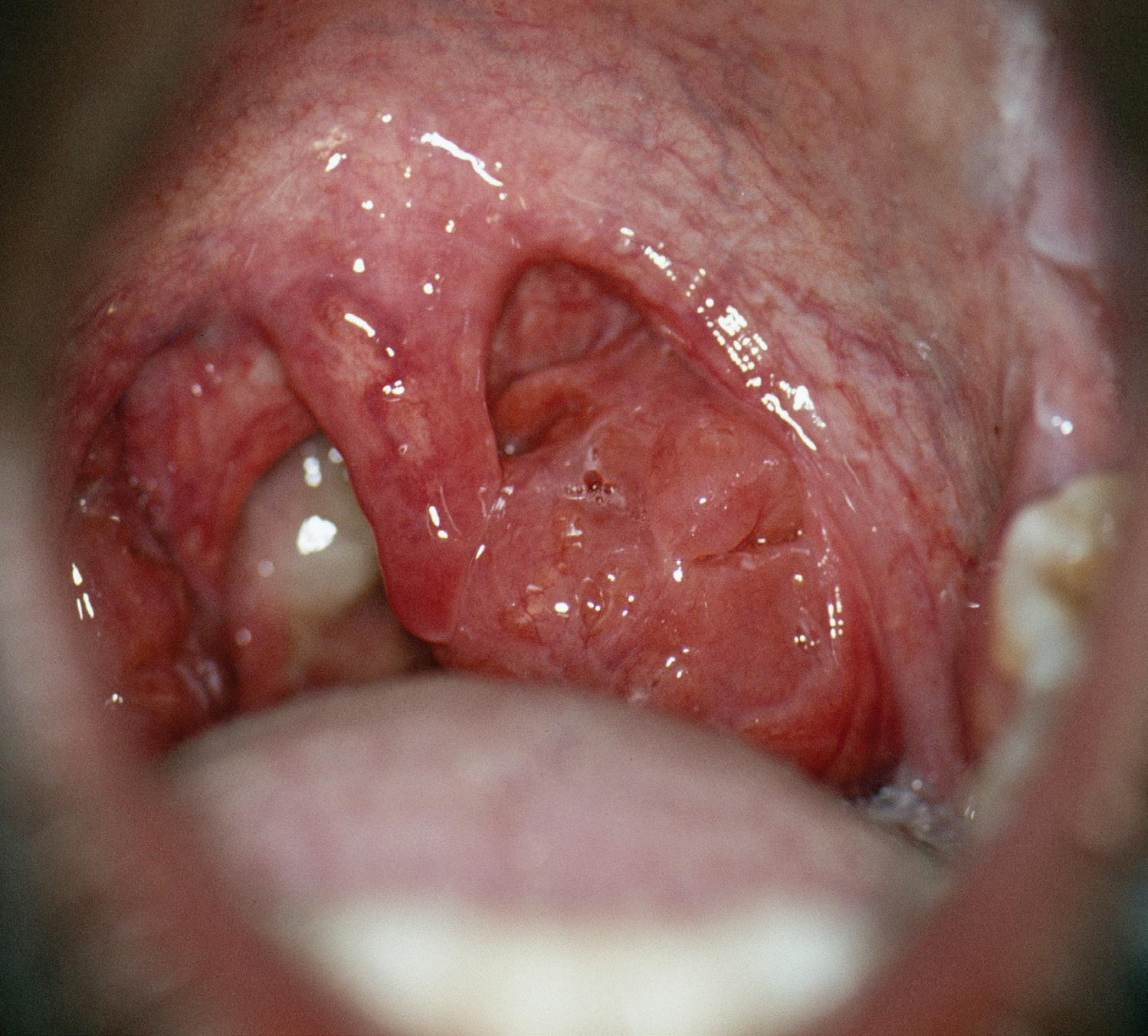
Tonsil hypertrophy: Enlarged tonsils of an HIV positive patient.
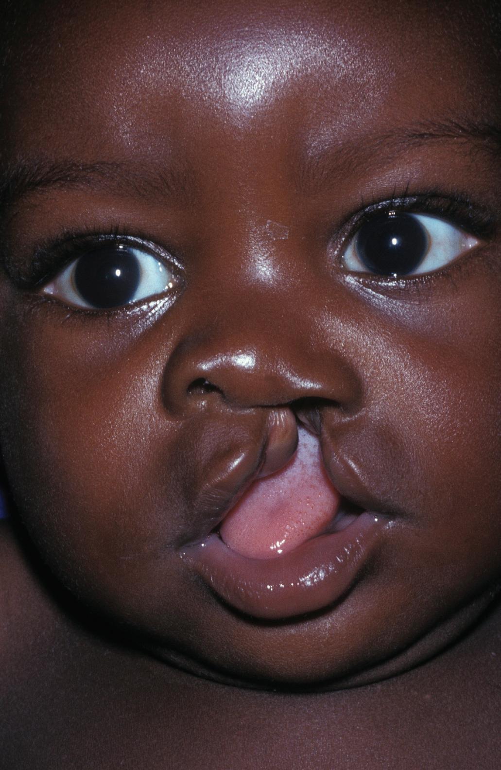
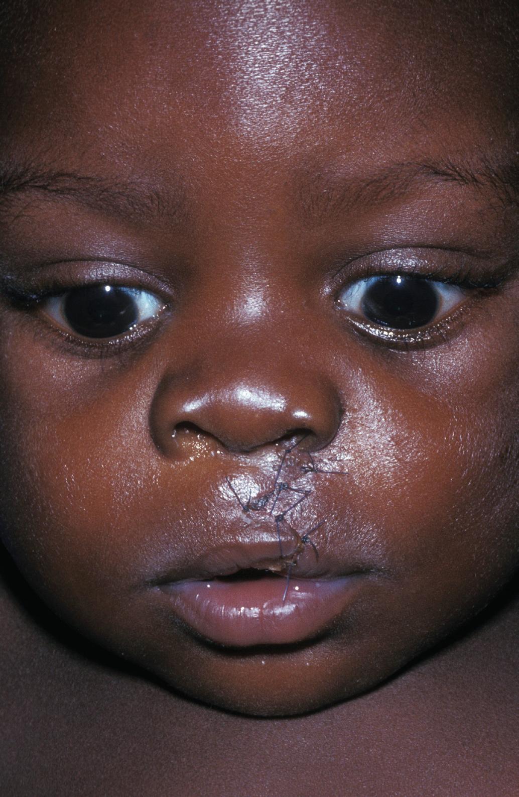
Face of a baby with a cleft, or hare, lip (left). This is a birth defect caused by a failure of the face to fuse together correctly during the embryonic stage of development. The image on the right shows the same patient after surgical repair.
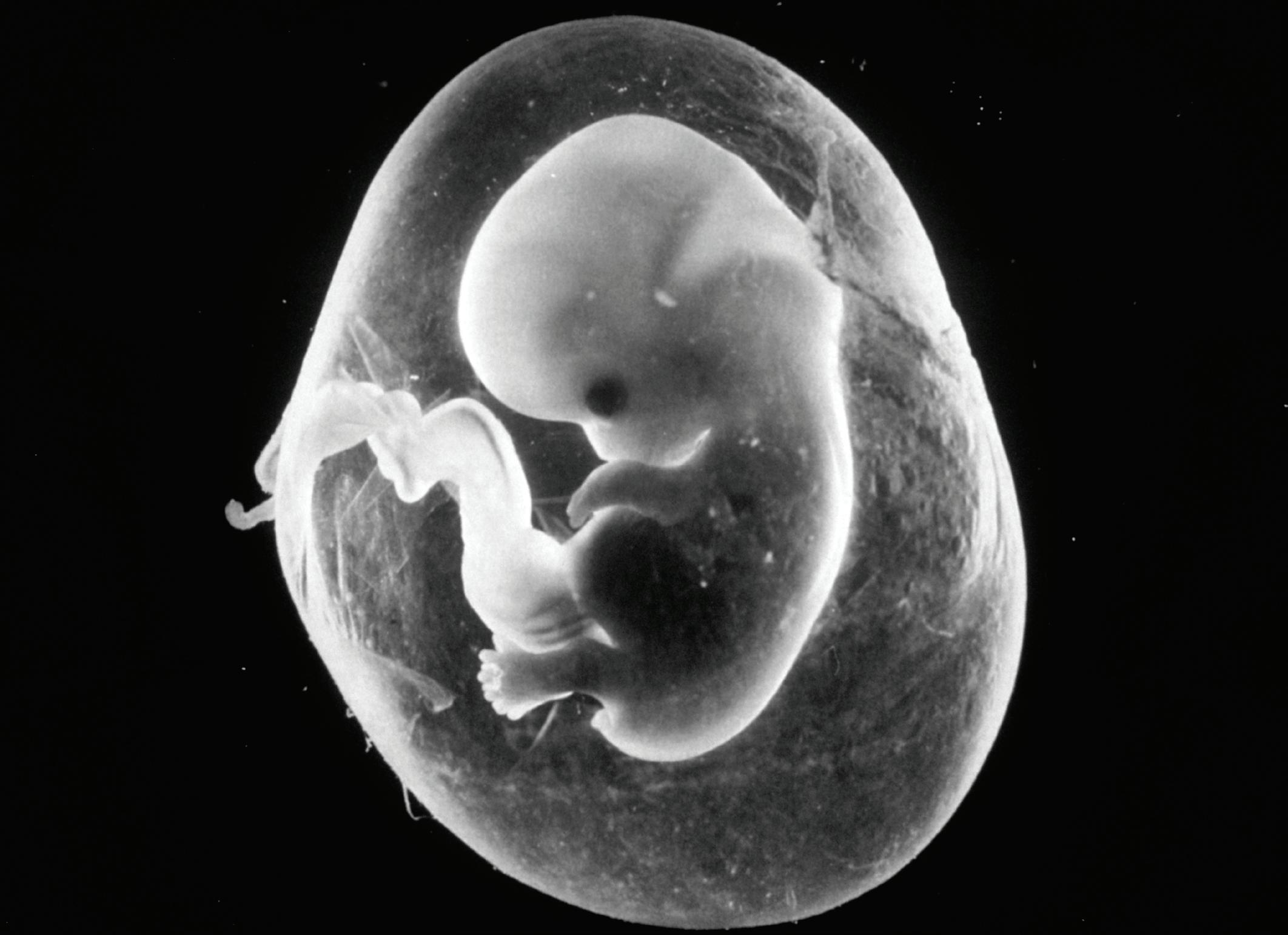
A human foetus after eight weeks development, surrounded by an intact, fluid-filled amniotic sac. The end of the eighth week marks the point at which an embryo becomes known as a foetus.
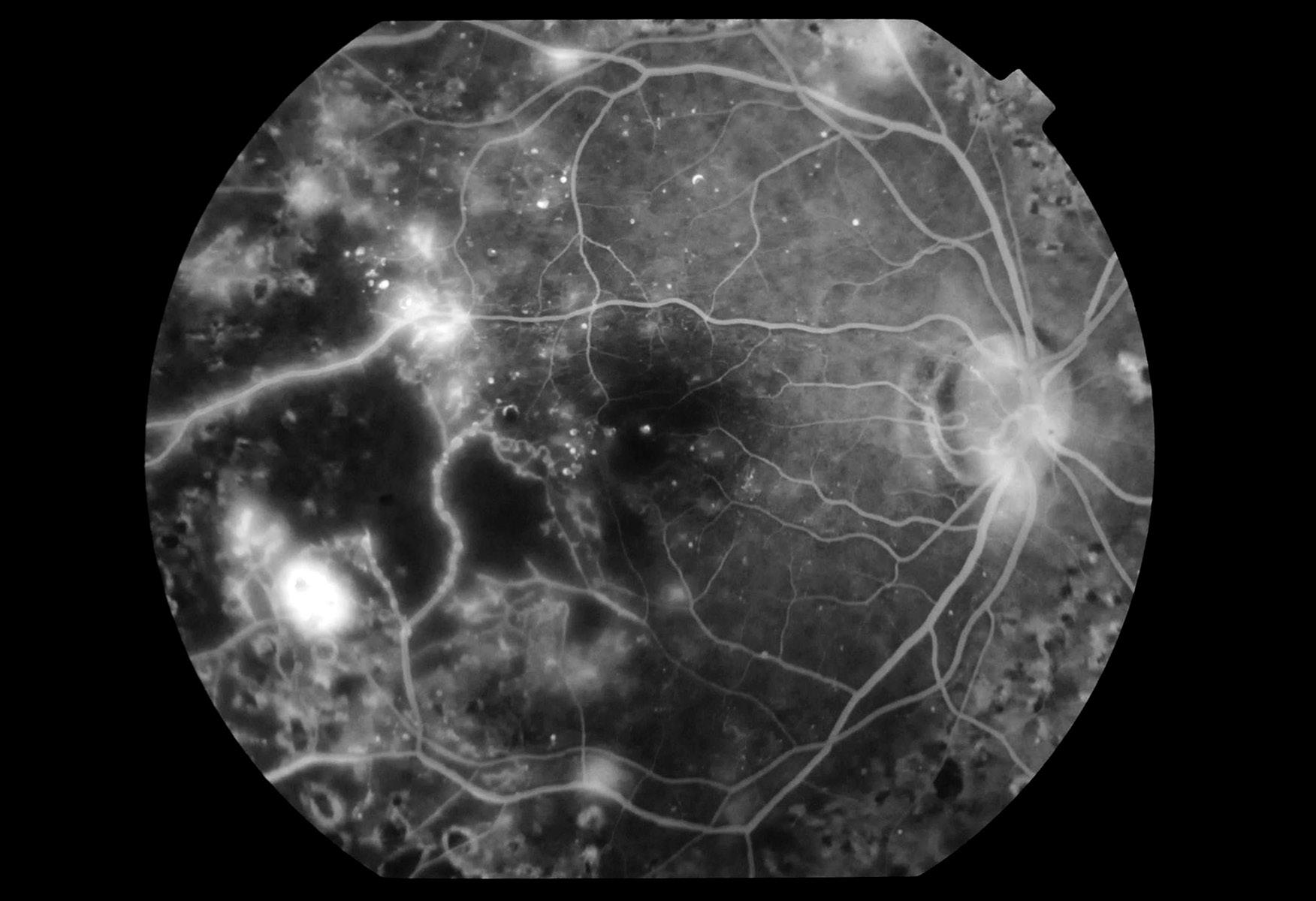
video recording to study the growth of bacteria in microbiology. In the mid-sixties, for example, Dr Peter Cardew of St. Mary’s Hospital Medical School in London devised equipment that was attached to a 16mm cine-camera to carry out direct and indirect cinelaryngoscopy for the examination of the larynx (vocal chords) in patients with myxedema. As innovative imaging techniques in medicine are being developed, the old ones are finding new applications. For example, The Telegraph reported (telegraph.co.uk - 17 May 2013) on how Professor Simon Fishel, one of the world’s leading fertility specialists, used time-lapse imaging to help predict which embryo had the greatest chance of a successful birth. There are a number of applications of photography in medicine such as micro and macrophotography, ultraviolet (UV) and infrared (IR) photography, time lapse and high speed, fluorescein angiography and Ocular Coherence Tomography (OCT), retinal photography, dermatoscopic imaging. These provide visual data for patient-care and research. Future health professionals, too, benefit from the images taken by clinical photographers. Clinical photographic database is a crucial resource for the teaching medicine at undergraduate, postgraduate and continuing medical education (CME). Visual aids are used to support medical teaching, and a teacher may, for example, include medical art, graphics, still images and motion media in developing their teaching materials – for example, a surgical procedure such as kidney removal (nephrectomy) may be best illustrated in video format. At senior level, a clinical photographer may also play a role in the design, development and evaluation of the material, to ensure it meets its objectives. Medicine is a very visual subject and will always need images and imaging. Pick up any medical journal and you will be hard pushed to find articles without images. 3D imaging is already finding innovative use in planning of surgical procedures. Virtual reality (VR) is using images to find more and better applications in medicine, including the teaching of keyhole surgery. With the availability of digital imaging tools and techniques the taking of pictures has become much easier and instantly viewable. Often doctors may take photographs themselves using special equipment – a gastroenterologist, for example, would take pictures during an endoscopic examination, using a camera that allows sight inside the Gastro-Intestinal Tract of the patient. Similarly, a pathologist may take pictures through a microscope during the examination of normal and abnormal cells, and other material. As time goes on, the application of imaging will only expand and it will need active, innovative and imaginative collaboration with the clinicians, clinical scientists and educators to take it forward. Therefore, it seems that highly qualified and experienced clinical photographers will have a place in medicine in the future. However, they will need to expand their skills beyond taking clinical pictures and harness new media such as information technology and digital communication to provide for the demand of the client and the challenges of medicine and its teaching l
ABoUt tHe AUtHoR
Accredited Senior Imaging Scientist Professor Afzal Ansary ASIS FRPS has more than half a century’s experience in medical and scientific photography and has been awarded six Fellowships in his field including two Honorary Fellowships. Former Chair of the Medical Group, he is a member of the Science Committee and Chair of the Imaging Scientist Qualification Board, and established and coordinated the first ever International Images for Science Exhibitions in 2011 and 2013. He is also the Chair of the Combined Royal Colleges Medal Awards Committee. He won the RPS Medical Group Lancet Award in 1990, and in 2013 was awarded Fenton Medal and Honorary Life Membership of The Society. In 2020 he was awarded the President’s and Trustee’s Commendation Award for his meritorious service to the RPS. He is an Honorary Professor at the Postgraduate Institute of Medical Education and Research, Chandigarh, India, and also an Honorary Professor at the University of Nottingham, UK.