Importance of multimodality imaging in
diagnosing in a young woman BREAST CANCER
- a pictorial assay with a case report.
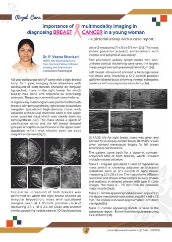
circle-2 measuring 7.1 x 5.6 x 5.9 mm (2c). The mass shows posterior acoustic enhancement with internal and peripheral vascularity.
Dr. P. Veena Shankari MBBS, MD (Radiodiagnosis)., Post-Doctoral Fellow in Breast Imaging and Intervention Consultant Radiologist

y p IVF came with a right breast lump for 1 year, imaging done elsewhere with ultrasound of both breasts revealed an irregular hypoechoic mass in the right breast for which biopsy was done and reported as sclerosing adenosis.Thepatientcamewithpainoverthelump.
A digital x-ray mammogram was performed for both breasts with tomosynthesis, right breast showed an irregular spiculated high-density mass with adjacent architectural distortion seen in the upper inner quadrant (1a,c) which was clearly seen on tomosynthesis (1e,f) The mass shows a speck of calcification within and the left breast showed grouped amorphous calcification in the upper outer quadrant which was clearly seen on spot magnificationviews(1g,h).


Few prominent axillary lymph nodes with nonuniform cortical thickening were seen, the largest measuring 4 mm with preserved fatty hilum (2d).
Left breast ultrasound showed a heterogeneous non-mass area involving a 12-2 o'clock position with few dilated ducts showing internal echogenic contents with no suspicious vascularity (2e).

Correlative ultrasound of both breasts was performed on which the right breast showed an irregular hypoechoic mass with spiculated margins seen at 1 O'clock position circle-2 measuring 2.5 x 1.8 x 2.4 cm (2a,b) and another similar appearing nodule seen at 1 O'clock position
BI-RADS IVa for right breast mass was given and advised for re-biopsy and left breast BI-RADS IV was given advised stereotactic biopsy for left breast amorphouscalcifications
The patient came back for a dynamic contrastenhanced MRI of both breasts, which revealed multiplemassesasbelow:
Mass 1 - Irregular spiculated T1 and T2 hypointense mass which is showing adjacent architectural distortion seen at 12-1 o'clock of right breast measuring 2 2 x 2 8 x 3 cm The mass shows diffusion restriction and shows enhancement in early phases and washout in delayed phases with type III curve images The mass is 7 5 mm from the pectoralis majormuscle(3a,b)
Mass2–Similarappearingnoduleisseenadjacentto the above-mentioned mass measuring 5 9 x 6 8 x 7 5 mm The nodule is located approximately 1 1 cm from thenipple(3c)
Mass 3 –Similar appearing nodule is seen in the subareolar region ~ 8 mm from the nipple measuring 4x4 3x6mm(3d)
contact@royalcarehospitals.in 0422 - 2227000
14
Mass 4 - Area of non-mass enhancement with clustered ring appearance seen involving the upper outer quadrant This area shows diffusion restriction On post-contrast images, there is a progressive enhancement(3e).
The left breast showed a suspicious area of nonmass enhancement seen in segmental distribution with clustered ring pattern involving predominantly upper outer quadrant involving 1 – 3 o’clock position. These areas showed diffusion restriction, and on postcontrast imaging, showed progressive enhancement for a span of 7 6 cm with extension up to the nipple with a type I kinetic curve The area was seen to have suspicious continuity with the pectoralis muscle behindwithsubtleenhancement (3f,g,h)

Histopathology revealed right breast invasive breast carcinoma with ER, PR, HER-2 positive, and Ki 67 - 23% Histopathology of the left breast showed atypical ductal proliferation
Whole body PET scan was performed and showed uptake in the right breast mass with ipsilateral nodal metastasis(5a,5b)


MRI was reported as irregular spiculated mass with enhancement and washout on post-contrast images – Right breast (BIRADS 5) & Suspicious nonmass enhancement in the segmental distribution in the upperouterquadrantoftheleftbreast(BIRADS4)
Ultrasound-guidedbiopsywasdonefrombothbreasts and core samples were sent for histopathological evaluation(4a-rightbreast)(4b-leftbreast).
the patient has been advised 4 cycles of neoadjuvant chemotherapy followed by right breast MRM with nodal clearance and close follow-up for leftbreastatypicalproliferation
Discussion:
Multimodality imaging of HER2-positive breast cancer before and after neoadjuvant chemotherapy provides varied accuracy and predictive value for estimating remaining disease compared with the standard of pathologic analysis, which is a consideration for treatment planning The most common manifestation of HER2-positive breast cancer at mammography or US is an irregular mass with spiculated margins that often contain calcifications; at MRI, HER2-positive breast cancer m a y a p p e a r a s a m a s s o r a s n o n m a s s enhancement. HER2-positive breast cancers are often of intermediate to high nuclear grade at histopathologic analysis, with an increased risk of local recurrence and metastases and a poorer overall prognosis. women are now potentially able to undergo breast conservation therapy and sentinel lymph node biopsy versus mastectomy and axillary lymph node dissection Thus, the radiologist’s role in assessing the extent of localregional disease and response to neoadjuvant treatment at imaging is important for surgical planning and adjuvant treatment However, assessment of treatment response remains difficult, with the potential for different imaging modalities to result in underestimation or overestimation of disease to varying degrees when compared with surgical pathologic analysis Breast MRI findings remain the best predictor of pathologic response

contact@royalcarehospitals.in 0422 - 2227000
15
THERAPEUTIC PLASMA EXCHANGE ( TPE)A PROMISING TREATMENT MODALITY IN NEUROLOGICAL & NON- NEUROLOGICAL PATIENTS
Prof. Dr. P. Chinnaswamy

Ph.D., FICS., MAACC (USA)., FIFCC (CLINICAL CHEM)., FNACB (USA)., Chief of Laboratory Medicine and blood bank
INTRODUCTION:
Therapeutic plasma exchange (TPE) is an extracorporeal patient therapy involving the separation and removal of the plasma in the blood using a cell separator machine, in order to remove disease causing substances circulating in the plasma
The cellular components (RBC,WBC, and platelets) are returned to the patient, along with a prescribed replacement fluid The patient is connected to the machine using central vein preferably The blood in the extra corporeal circuit is anticoagulated using citrate and the administration of this is controlled by the equipment and the operator. The machine separates the blood into its component parts allowing the plasma (containing the disease causing agent) to be drawn off and replacement fluid has to be added to the returning red and white blood cells.The purpose is to remove the agent in the plasma, such as an antibody, toxin or abnormal protein that are causing the clinical symptoms.
PRINCIPLE:
TPE is often used to modulate the level of circulating antibodies, antigen-antibody complexes, complement components, cytokines, abnormal plasma proteins, cholesterol-containing lipoproteins, plasma-bound toxins and drugs Also it has an immunomodulatory effect beyond the removal of Ig which includes T-cell modulation with a shift from in the Th1/Th2 balance with a shift toward Th2, suppression of IL-2 and IFN-γ production
Dr. M.R. Jeeva Priya

M.D (Immuno-Hematology & Blood Transfusion), DDVL. Consultant Transfusion Medicine and Blood Bank Medical Ocer
CAUSATIVE AGENT DISEASES
AUTOANTIBODY TTP, Myasthenia Gravis, Neuromyelitisoptica, Anti - GBM, ANCA associated Vasculitis
MultipleSclerosis, GBS, CIDP
AG-ABCOMPLEX HCV Vasculitis,SLE
ALLOANTIBODY Transplant sensitisation, Transplant rejection (humoral), Transfusion reaction
PARAPROTEINS
NON-Ig PROTEINS
ENDOGENOUS TOXINS EXOGENOUS POISONS
REPLENISHMENT
Waldenstrom’s macroglobinemia, Hyperviscosity, Light chain neuropathy, Light chain glomerulopathy, Myeloma cast nephropathy
FSGS
Hyperlipidemia,Liver failure, Sepsis
Amanita, Drugs
TTP( ADAMTS13), MPGN( complement factor H)
Methods&mechanismofplasmaremoval:
Methodsavailable:
1. Centrifugal TPE ( cTPE).
2. Membranous TPE( mTPE) (By dialysis machine)
The best method used for TPE is by CENTRIFUGAL TPE METHOD.
Whole blood is pumped into a rapidly rotating separation chamber. Components separate into layers based upon their density, with the most dense element, RBCs, migrating the furthest from the axis of rotation and the least dense portion, plasma, layering closest to the axis of rotation. Intermediate layers, moving from the axis of rotation outward, are platelets, lymphocytes, and granulocytes. In TPE, the plasma layer is removed and discarded and the remaining cellular elements are mixed with a replacement fluid and returned to the patient.
contact@royalcarehospitals.in 0422 - 2227000
16
Difference between Centrifugal TPE ( cTPE) & Membranous TPE( mTPE)
2.Time
3.Anticoagulation
standalone treatment or in conjunction with other modes of treatment.
Category II : Disorders for which apheresis is accepted as second-line therapy, either as a standalone treatment or in conjunction with other modes of treatment
4
for removal of High molecular weight substances(IgM),immunecomplexes
5.Risk of hemolysis with high membrane pressure
Ref:
1. Kes et al A randomized crossover study comparing membrane and centrifugal therapeutic plasma exchange procedures, Volume 56, December 2016 TRANSFUSION
2. Centrifugal and Membrane Therapeutic Plasma Exchange – A Mini-review : European Oncology & Haematology, 2018;14 (2) : 105–9
Howmuchplasmaistoberemoved?
Routine practice is to exchange only 1-1.5 plasma volumes during a TPE. For each 1-1.5 plasma volume e x c h a n g e d , a p p r o x i m a t e l y 6 0 % - 7 0 % o f substances present in the plasma at the start of that plasma volume will be removed.
The efficacy of TPE depends on the Plasma Volume (PV) removed in relation to the patient’s total PV, the distribution of the pathogenic substance removed between intravascular and extravascular spaces, and the synthesis and equilibrium rate of that substance between the compartments.
ReplacementFluids:
Examples of physiological fluids used for replacement during TPE include fresh frozen plasma (FFP), 5% human albumin solution (5% HAS), colloids (Gelofusine) and crystalloids (0.9% normal saline).
INDICATIONS
Guidelines on the Use of Therapeutic Apheresis in Clinical Practice– Evidence-Based Approach from the Writing Committee of the American Society for Apheresis (ASFA): J Clin Apher. 2019;34:171–354.
Categories- ASFAGuidelines
Category I : Disorders for which apheresis is accepted as first-line therapy, either as a primary
Category IV: Disorders in which published evidence demonstrates or
Category III : Decision making should be individualized. suggests apheresis to be ineffective or harmful.
CATEGORY FREQUENCY OF PROCEDURES
NEUROLOGY
1. Myasthenia Gravis Cat-I- Acute-short term treatment Cat- II - Long term treatment
Daily/Every other day
2. Guillian Barre syndrome Cat-I Every other day- 5 to 6 cycles
3. Acute Inflammatory Demyelinating Polyradiculoneuropathy (AIDP)
4. Chronic Inflammatory Demyelinating Polyradiculoneuropathy (CIDP)
5. Paraproteinemic demyelinating neuropathiesIgG/IgA/IgM
6. N.Methyl-D -Aspartate receptor antibody encephalitis
7. Multiple Sclerosis-Acute attacks/Relapse
8. Neuromyelitis optica spectrum disorder
9. Paediatric Autoimmune neuropsychiatric disorders associated with streptococcal infections ( PANDAS)Exacerbation
10.Voltage Gated K channel antibody related diseases
11. Hashimoto’s encephalopathy
12.Lambert Eaton myasthenic syndrome
13.Acute Disseminated encephalomyelitis ( steroid refractory)
1. Thrombotic thrombocytopenic purpura
2. Hyperviscosity in hypergammaglobinemiaSymptomatic/prophylaxis
3.Catastrophic AntiPhospholipid antibody syndrome
4.Hereditary hemochromatosis
5.Severe cold agglutinin disease
6.Thrombotic microangiopathy-Complement mediated-Factor H auto antibodies
Cat-I
other day- 5 to 6 cycles
cycles/week
other day- 5 to 6 cycles
HEMATOLOGY
contact@royalcarehospitals.in 0422 - 2227000
17 PARTICULARS cTPE mTPE
eciency (PRE
Higher80% (75-85%) Lower35% (30-50%)
1.Plasma Extraction ratio or Plasma removal
: percentage of plasma removed vs. plasma processed)
duration
Procedure) Less( 102±25min) More(157 ±25min)
(Set up,Priming,
Citrate( No risk of systemic
Heparin( risk of
anticoagulation)
systemic anticoagulation present)
High Low
Eectiveness
Less More INDICATION ASFA
Every
Cat-I
2-3
Cat-I Every
other day- 5 to 6 cycles
Cat-I Every
5 to
Cat-II Every other day -
7cycles
Cat-II Every other day- 5 to 10
cycles
Cat-II Daily/Every other day3 to 6cycles
Cat II Every other day- 5 to 7
cycles
Cat-II Every other day- 3 to 9 cycles y/Every other day3 to 6 Daily/Every other dayDaily/Every other dayEvery other day-
Cat-II Dail
variable
Cat-II Every other day-
cycles
Cat-I Daily- variable
Cat-I Daily- 1 to 3
cycles
Cat-I variable
Cat-I variable
Cat-II Daily/
variable
Cat-I Daily- variable
RHEUMATOLOGY
1. Vasculitis-ANCA associated Cat-I
2.SLE-with Severe complications Cat-II
3. Vasculitis due to HBVPolyarteritis nodosa Cat-II
TreatmentOutcome :
TPE is a well-established treatment modality for many of the above said diseases and its exacerbation Rebound overproduction of antibodies occurs because of sudden removal of antibodies from the circulation. So, concurrent use of immunotherapy is advisable along with TPE.
Daily/Every other day7-12 cycles
Daily/Every other day3-6 cycles
Daily/Every other day9-12 cycles
4. Scleroderma Cat III 1 -3 cycles/week
NEPHROLOGY
1. Renal Transplantationi)ABO compatibleDesensitization for a) living
b) deceased donor c) Antibody mediated rejection
ii) ABO- incompatible
a)Desensitization -LD, b)Antibody mediated rejection
2.ANCA associated Rapidly Progressive GlomeruloNephritis (Cr > 5.7 mg/dl) and Dialysis dependence with DAH
3. Anti-Glomerular Basement Membrane Disease (GoodPasture syndrome) - DAH, dialysis independence
a)Cat I, b)Cat III, c)Cat-I
Every other dayvariable
In a country like ours, cost of the therapy is an important factor while choosing treatment option. TPE is relatively cheaper mode of treatment as compared to IVIG. Although studies showed equal efficacy of IVIG and TPE , an expert consensus suggests that plasma exchange is more effective and works more quickly in the treatment of impending or manifest diseases.
Complications:
Although complication can occur, most of these are mild,rapidly recognized and reversed and are rarely serious.(4% to 6%)
a)Cat I, b)Cat- II
Cat-I
Cat-I
4. Focal Segmental glomerulosclerosis Cat-I
5.IgA NephropathyCrescentic, chronic progressive Cat III
OTHERS
1. Issues related to vascular access is common.
1. TPE with adsorbant columns for treatment for familial hypercholesterolemia.
Cat-I
2.Wilson’s disease - Fulminant Cat-I
3.Thyroid storm Cat-II
4.Drug overdosage Cat-III
5.Refsum’s disease Cat-II
6.MushroomPoisoning Cat-II
7.Envenomation Cat-III
8.Sepsis with multiorgan failure Cat III
9. Pruritus due to hepatobiliary diseases Cat III
10.Hypertriglyceridemic pancreatitis-severe Cat III
11. Acute liver failure I.High volume TPECat I
ii.Routine TPECat III
12. Complex regional pain syndrome - Chronic Cat III
13. Severe pemphigus vulgaris Cat III
Daily/Every other day7-12 cycles
Daily/Every other dayvariable
Daily/Every other dayvariable
Every other day-6-9 cycles
Every 1 -2 weeks
Daily/Every other dayvariable
Daily/Every other dayvariable
Every other dayvariable
Every other dayvariable
Every other dayvariable
Every other dayvariable
Every other dayvariable
Weekly-thrice
Every other day (1-3 cycles)
Every other dayvariable
Every other day - 5 - 7 cycles
Daily/Every other dayvariable
2.Symptomatic hypocalcemia due to citrate anticoagulation typically characterized by perioral and digital paresthesiae,nausea Calcium supplementation may alleviate symptoms of citrate toxicity.
3. Allergic reactions are most common with plasma replacement characterised by urticaria and cutaneous flushing. Severe allergic reactions can involve the respiratory tract with dyspnea, and wheezing. Most allergic reactions respond quickly to IV diphenhydramine.
4.Hypotension during apheresis can be a sign of citrate toxicity, hypovolemia, or vasovagal, allergic, drug, or transfusion reaction.
5.TRALI/TACO are rare.
6. Bleeding due to coagulation factor deficiency is rare,unless replaced with only albumin.
7.Albumin-bound drugs are removed by TPE.Drugs should be administered after TPE procedure to avoid impairing their effectiveness.
w Before the procedure, following parameters are checked- Complete Hemogram, blood urea, serum creatinine, liver function test, serum electrolytes, coagulation profile, and vital parameters.
w TPE consent is taken from the patient/patient’s relatives before the procedure.
w TPE is performed using a Spectra Optia Apheresis system ( Terumo Penpol) through
contact@royalcarehospitals.in 0422 - 2227000 18
TPEinRoyalCareSuperSpecialityHospital:
-
Cat-II Every other day- 3 to 8 cycles 8. Thrombotic microangiopathy -Ticlopidine induced Cat-II Daily/Every other dayvariable
HELLP Syndrome Cat III Daily-variable 10. Refractory Immune thrombocytopenia Cat III Every other day- 6 cycles
7. Cryoglobulinemia
Symptomatic/severe
9.
central line access using 11.5 F double lumen dialysis catheter.
w The amount of plasma to be exchanged was determined by following formula: Estimated plasma volume (EPV) = (0.065 × weight [kg]) × (1 Hematocrit as a fraction) (NADLER’S equation, 1962).
w TPEisdoneonalternatedaybasisfor8–10days
w The treating Consultant decides the number of cycles depending on the clinical outcome.
w Anticoagulation with citrate (ACD) is used systemically.
w Exchange of 1-1.5 PV given to the patients and replaced with Isotonic saline, albumin and fresh frozen plasma (FFP).
w Isotonic saline is used 30% and human albumin and fresh frozen plasma 70% are added to complete it. Albumin only issued for patients who experience adverse reactions with FFP.
w For every 15-30 minutes intervals, the blood pressure and pulse, changes in appearance, development of symptoms like lightheadedness, nausea, paresthesia and overall status are closely monitored and untoward events are identified and reverted by rational interventions.
w The duration of procedure varies from one to three hours depending upon the amount of plasma exchange.
w All TPE procedures are carried out in ICU by blood bank technicians trained in TPE under the supervision of Consultant Transfusion medicine/blood bank medical officer

w Indications for TPE, number of cycles , duration of each session, volume of plasma exchanged and patient tolerance to the procedure are systematically recorded.
w To avoid citrate toxicity, 10 ml of 10% calcium gluconate is infused throughout the procedure.
w After each session, outcomes in terms of clinical improvement is measured.
OTHER PROCEDURES DONE WITH THE SAME EQUIPMENT REDCELLEXCHANGES
1. RBC Exchange for methemoglobinemia,Sickle cell disease( Cat-II),acute sickle cell crisis, removal of donor RBC from the bone marrow grafts in major ABO-incompatible allogeneic hematopoietic stem
cell transplantation to avoid immediate hemolysis,severe infections with intraerythrocytic pathogens such as malaria ( Cat III) or babesiosis
2. Erythrocytapheresis for polycythemia vera( Cat-I) and secondary erythrocytosis.( Cat-III)
STEMCELLCOLLECTION
1. Mononuclear Cell (MNC) Collection


2 Continuous Mononuclear Cell Collection (CMNC)

3. Granulocyte (PMN) Collection
DEPLETION
1. Leukocytapheresis- White Blood Cell Depletion (WBCD)- initial management of leukostasis in patients with hyperleukocytosis in acute leukemias, particularly myeloid leukemias.( Cat-II)
2. Platelet Depletion- for severe thrombocytosis( Cat-II)
PROCESSING
Bonemarrow processing
contact@royalcarehospitals.in 0422 - 2227000 19
Spectra Optia Apheresis system - For all therapeutic exchange procedures, Various cell collection and processing.
Rapid clinical outcome after 5 cycles of Therapeutic plasma exchange for Myasthenia gravis patient on crisis with respiratory failure
Accident & Emergency
Anesthesiology & Pain Clinic
Blood Bank
Cardiology & Interventional Cardiology

Cardiothoracic Surgery
Critical Care Medicine
Dermatology
Dental & Maxillofacial Surgery
Endocrinology & Endocrine Surgery
Endogynecology (Laparoscopy)
ENT, Head & Neck Surgery
General & Laparoscopic Surgery
Fertility Care Clinic
Internal Medicine & Diabetology
Interventional Pulmonology
Interventional Radiology
Interventional Neuro Radiology
Master Health Checkup
Medical Gastroenterology
Medical Oncology
Minimally invasive spine surgery
Nephrology
Neurology
Neurosurgery
Nuclear Medicine
Obstetrics & Gynecology

Orthopaedic & Trauma Surgery

Ophthalmology
Plastic, Reconstructive & Cosmetic Surgery
Physical Medicine and Rehabilitation
Paediatric & Neonatal Surgery
Psychiatry & Clinical Psychology
Radiology & Imaging Sciences
Radiation Oncology
Renal Transplant Unit
Rheumatology
Surgical Gastroenterology
Surgical Oncology
Spine Injury Rehabilitation Centre
Urology
Vascular Surgery
ROYALCARE SUPER SPECIALITY HOSPITAL LIMITED
No
: 0422 - 222 7000, 22 77 000
69331 No : 1/520, Neelambur, Sulur Taluk, Coimbatore - 641 062. Ph : 0422 - 222 7000, 22 77 000 E-mail : contact@royalcarehospitals.in
: 1/520, Neelambur, Sulur Taluk, Coimbatore - 641 062. Ph
ROYALCARE SUPER SPECIALITY HOSPITAL LIMITED Mob : 73977
www.royalcarehospitals.in
H-2022-0901


 Dr. B. Paranthaman Sethupathi Medical Director & Consultant Psychiatrist

 "Success is not final, failure is not fatal, it's the courage to continue that counts "..


 Dr. K. Vijayan
MBBS, MD, DNB, DM (Neurology), ASN (USA)., Consultant Neurologist & Neuro Sonologist
Dr. K. Vijayan
MBBS, MD, DNB, DM (Neurology), ASN (USA)., Consultant Neurologist & Neuro Sonologist


 The following picture shows the principles of Sentinel Lymph node Biopsy.
Images of some of the Sentinel Node Biopsies done by us are shown below
Peri Aeriolar Injection of Blue Dye Lymphatics reaching Sentinel Node
Two Sentinel Nodes seen
Sentinel Nodes stained blue Biopsied
Same Sentinel Node being hot on Gama Probe
The following picture shows the principles of Sentinel Lymph node Biopsy.
Images of some of the Sentinel Node Biopsies done by us are shown below
Peri Aeriolar Injection of Blue Dye Lymphatics reaching Sentinel Node
Two Sentinel Nodes seen
Sentinel Nodes stained blue Biopsied
Same Sentinel Node being hot on Gama Probe