3 minute read
Importance of multimodality imaging in


diagnosing in a young woman BREAST CANCER
- a pictorial assay with a case report.
Advertisement
circle-2 measuring 7.1 x 5.6 x 5.9 mm (2c). The mass shows posterior acoustic enhancement with internal and peripheral vascularity.
Dr. P. Veena Shankari MBBS, MD (Radiodiagnosis)., Post-Doctoral Fellow in Breast Imaging and Intervention Consultant Radiologist

y p IVF came with a right breast lump for 1 year, imaging done elsewhere with ultrasound of both breasts revealed an irregular hypoechoic mass in the right breast for which biopsy was done and reported as sclerosing adenosis.Thepatientcamewithpainoverthelump.
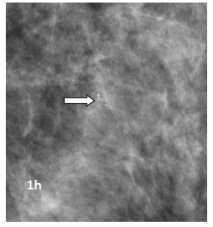
A digital x-ray mammogram was performed for both breasts with tomosynthesis, right breast showed an irregular spiculated high-density mass with adjacent architectural distortion seen in the upper inner quadrant (1a,c) which was clearly seen on tomosynthesis (1e,f) The mass shows a speck of calcification within and the left breast showed grouped amorphous calcification in the upper outer quadrant which was clearly seen on spot magnificationviews(1g,h).

Few prominent axillary lymph nodes with nonuniform cortical thickening were seen, the largest measuring 4 mm with preserved fatty hilum (2d).
Left breast ultrasound showed a heterogeneous non-mass area involving a 12-2 o'clock position with few dilated ducts showing internal echogenic contents with no suspicious vascularity (2e).

Correlative ultrasound of both breasts was performed on which the right breast showed an irregular hypoechoic mass with spiculated margins seen at 1 O'clock position circle-2 measuring 2.5 x 1.8 x 2.4 cm (2a,b) and another similar appearing nodule seen at 1 O'clock position
BI-RADS IVa for right breast mass was given and advised for re-biopsy and left breast BI-RADS IV was given advised stereotactic biopsy for left breast amorphouscalcifications
The patient came back for a dynamic contrastenhanced MRI of both breasts, which revealed multiplemassesasbelow:
Mass 1 - Irregular spiculated T1 and T2 hypointense mass which is showing adjacent architectural distortion seen at 12-1 o'clock of right breast measuring 2 2 x 2 8 x 3 cm The mass shows diffusion restriction and shows enhancement in early phases and washout in delayed phases with type III curve images The mass is 7 5 mm from the pectoralis majormuscle(3a,b)
Mass2–Similarappearingnoduleisseenadjacentto the above-mentioned mass measuring 5 9 x 6 8 x 7 5 mm The nodule is located approximately 1 1 cm from thenipple(3c)
Mass 3 –Similar appearing nodule is seen in the subareolar region ~ 8 mm from the nipple measuring 4x4 3x6mm(3d)
Mass 4 - Area of non-mass enhancement with clustered ring appearance seen involving the upper outer quadrant This area shows diffusion restriction On post-contrast images, there is a progressive enhancement(3e).
The left breast showed a suspicious area of nonmass enhancement seen in segmental distribution with clustered ring pattern involving predominantly upper outer quadrant involving 1 – 3 o’clock position. These areas showed diffusion restriction, and on postcontrast imaging, showed progressive enhancement for a span of 7 6 cm with extension up to the nipple with a type I kinetic curve The area was seen to have suspicious continuity with the pectoralis muscle behindwithsubtleenhancement (3f,g,h)
Histopathology revealed right breast invasive breast carcinoma with ER, PR, HER-2 positive, and Ki 67 - 23% Histopathology of the left breast showed atypical ductal proliferation
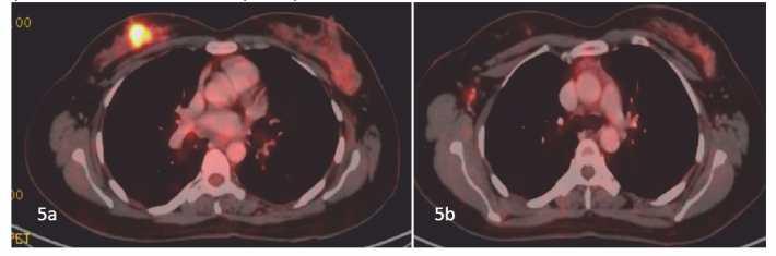
Whole body PET scan was performed and showed uptake in the right breast mass with ipsilateral nodal metastasis(5a,5b)


MRI was reported as irregular spiculated mass with enhancement and washout on post-contrast images – Right breast (BIRADS 5) & Suspicious nonmass enhancement in the segmental distribution in the upperouterquadrantoftheleftbreast(BIRADS4) the patient has been advised 4 cycles of neoadjuvant chemotherapy followed by right breast MRM with nodal clearance and close follow-up for leftbreastatypicalproliferation
Ultrasound-guidedbiopsywasdonefrombothbreasts and core samples were sent for histopathological evaluation(4a-rightbreast)(4b-leftbreast).
Discussion:
Multimodality imaging of HER2-positive breast cancer before and after neoadjuvant chemotherapy provides varied accuracy and predictive value for estimating remaining disease compared with the standard of pathologic analysis, which is a consideration for treatment planning The most common manifestation of HER2-positive breast cancer at mammography or US is an irregular mass with spiculated margins that often contain calcifications; at MRI, HER2-positive breast cancer m a y a p p e a r a s a m a s s o r a s n o n m a s s enhancement. HER2-positive breast cancers are often of intermediate to high nuclear grade at histopathologic analysis, with an increased risk of local recurrence and metastases and a poorer overall prognosis. women are now potentially able to undergo breast conservation therapy and sentinel lymph node biopsy versus mastectomy and axillary lymph node dissection Thus, the radiologist’s role in assessing the extent of localregional disease and response to neoadjuvant treatment at imaging is important for surgical planning and adjuvant treatment However, assessment of treatment response remains difficult, with the potential for different imaging modalities to result in underestimation or overestimation of disease to varying degrees when compared with surgical pathologic analysis Breast MRI findings remain the best predictor of pathologic response