1 minute read
SENTINEL LYMPH NODE BIOPSY IN EARLY-STAGE


Dr. Shiva Kumar Kuppuswamy MBBS, MS(Gen.Surg), M.Ch (Surgical Onco)., Consultant Surgical Oncologist

Advertisement
“Treatment without Prevention is simply unsustainable” - Bill Gates
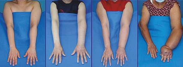
Lymphedema occurs when the load exceeds the transport capacity of the lymphatic system, which leads to the accumulation of protein-rich fluid (lymph) and fibro adipose tissue in the interstitium. Symptoms of lymphedema include limb swelling, skin changes, discomfort, and restricted range of motion. Breast cancer and its treatments are one of the most common causes of secondary peripheral lymphedema. The incidence of upper extremity lymphedema in breast cancer survivors ranges from 5 to 40 percent. Incidences in the higher range occur in patients who have undergone complete axillary dissection combined with radiation therapy.
Breast cancer-associated lymphedema (BCAL) is primarily due to obstruction of the lymphatic channels located in the axilla, most commonly from lymphadenectomy or radiation therapy, but can also occur as a result of infiltration of the lymphatic vessels by tumor cells (lymphangitic carcinomatosis)
Primary prevention of lymphedema in patients undergoing treatment for breast cancer relies on surgical techniques that limit node dissection (eg, sentinel lymph node biopsy) or, less commonly, techniques that repair or bypass injured lymphatics, and limit radiation exposure. Sentinel lymph node biopsy has been instrumental in decreasing the incidence of lymphedema
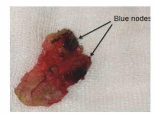
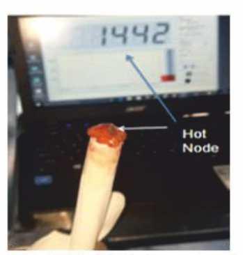
Clinically relevant lymphedema occurs in 5 to 9 percent of patients who undergo sentinel node biopsy alone compared with approximately 40 percent in patients undergoing axillary lymph node dissection. Using advanced radiation therapy (RT) techniques that limit radiation may also be helpful. Sentinel lymph node biopsy is a procedure used to detect the spread of cancer from the primary tumor to the lymph nodes. The sentinel lymph node is the first lymph node that cancer cells reach when they spread from the primary tumor. This node is most likely to contain cancer cells, and if the sentinel lymph node is cancer-free, it is unlikely that other lymph nodes are affected. During a sentinel lymph node biopsy, we will locate and remove the sentinel lymph node, which is usually located in the axilla. A small amount of blue dye or radioactive material is injected into the breast area near the primary tumor. This dye or radioactive material will travel through the lymphatic system and collect in the sentinel lymph node. The surgeon will use the dye or radioactive material to locate the sentinel lymph node and then remove it. The lymph node is then sent to a lab for a frozen section examination. If cancer cells are found in the sentinel lymph node, it may mean that cancer has spread to other lymph nodes and we proceed with formal axillary lymph nodal dissection.