


Delegates gathered in the Main Arenas on Tuesday morning to witness the opening presentations of this year’s Leipzig Interventional Course. The next two days will keep this momentum, continuing the packed agenda of presentations showcasing the latest in vascular medicine. Don’t miss the first-time data sets, live cases and ‘@ LINC’ sessions, offering diverse regional insights alongside posters, dynamic debates, and ‘Speaker’s Corner’

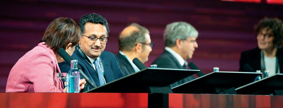


Adecade of insights from the Global Registry for Endovascular Aortic Treatment (GREAT) will be brought into focus in the opening session of Main Arena 2 this morning, spotlighting the prospective observational multicentre cohor t registry that was initiated in November 2011.1
Santi Trimarchi will present – in collaboration with Chiara Lomazzi (University of Milan, Italy) – the aims of GREAT, relaying its contemporary realworld data on aortic endografts in the treatment of multiple aortic pathologies.
Drs Trimarchi and Lomazzi spoke to LINC Today to dive deeper into GREAT, offering a preview of some of the insights they will be sharing today at LINC.
What prompted the creation of the GREAT Registry, and how would you sum it up to someone not familiar with its inception?
The GREAT Registry is a post-marketing registry launched by W.L. Gore 10 years ago, with the aim to better understand the follow-up of patients affected by aortic diseases, at any level, treated with Gore aortic endografts. Although this did not include imaging, several variables were utilised to check the results of these interventions. In particular, it was of interest to adopt a new consideration about the definition/importance of endoleaks, as these were defined as an issue that included both imaging evidence and clinical problems that necessitated additional hospital recovery.
What can you tell us about its design?
The GREAT Registry has enrolled up to 5,000 patients in about 110 centres across four different continents. It employs a multifaceted approach to data integration, combining clinical, demographic, and procedural information, with specific technical note
LINC
E: contact@captivatemedia.co.uk
W: www.captivatemedia.co.uk
Course Chairman
Dierk Scheinert
Editors Tatum Anderson
Design
Copyright

“The GREAT Registry exemplifies the importance of collaborative research efforts in advancing medical knowledge, highlighting how shared data and insights can lead to improved clinical practices, enhanced patient outcomes, and a more robust understanding of vascular interventions.”
Santi Trimarchi and Chiara Lomazzi
and measures collected for planning (diameter, angle, extension, ancillary procedures on visceral vessels and sizing of device used) to create a holistic view of patient outcomes, thereby facilitating nuanced analyses that inform best practices in endovascular aortic treatment.
What were the early insights that set the scene in the last decade?
The GREAT Registry not only collects extensive clinical data but also plays a relevant role in identifying trends and patterns that can lead to improved treatment protocols and enhanced patient outcomes in real-life aortic disease management. The results, reported in several papers, observe a worldwide daily practice associated with the increasing experience of physicians when using these stent grafts, facing challenging anatomy – mostly out of instructions for use – in the setting of staged and emergent treatment.
But, above all, the registry focuses on assessing long-term patient outcomes which are vital for understanding the effectiveness of various endovascular treatments over time. By systematically gathering postoperative data, the GREAT Registry aims to provide insights into
the durability of interventions (not only standard procedures), and inform future clinical practices for improved patient safety and care.
During follow-up, we noted that the number of patients decreased dramatically for mortality, primarily due to ageing or patients no longer being able to go to the doctor for checkup. However, despite this, a consistent number of patients have been followed over 10 years.
The big question: Now at 10 years, with over 30 publications stemming from the registry, what are the key clinical insights or practice-changing findings that stand out to you?
With 5,000 patients managed with aortic endovascular procedures worldwide, GREAT has a consistent number to lend strength to analyses of specific topics, subgroups or less-conventional treatments. Examples we like to share include the initial experiences with feasibility of ascending thoracic endovascular aortic repair (TEVAR), different TEVAR results based on gender, different outcomes and follow-up in patients treated for thoracic aneurysms versus acute dissections, and the results of EVAR in patients with angulated or inverted conic proximal
abdominal aortic necks.
Obviously, the manuscripts that reported these experiences went through the usual peer-review process, which not only improved the qualit y of individual studies, but also contributed to the overall advancement of knowledge in the field, encouraging innovative approaches and methodologies in research.
Overall, data from the registry has had constructive feedback. As is often the case, the publications have encouraged dialogue and critique from the research community, allowing for real-time feedback and adaptation of published work, thus keeping the scientific discourse dynamic and responsive to new findings, insights, collaborations and – last but not least – networking.
Have there been key challenges?
The GREAT Registry exemplifies the importance of collaborative research efforts in advancing medical knowledge, highlighting how shared data and insights can lead to improved clinical practices, enhanced patient outcomes, and a more robust understanding of vascular interventions.
The importance of Core Labs, and/or the availability of imaging to review specific issues or queries is one of the Achille’s heels of GREAT. In particular, a lesson learned for future registries is that imaging is definitely of primary importance.
What role do you see registries like GREAT playing in the future of research, and what is important to focus on next?
Registries have limit s in terms of evidence – that is clear. However, in the setting of aortic diseases, in particular those which need an emergent/ urgent approach, registries can provide comprehensive databases that enable healthcare professionals to analyse treatment outcomes, identify effective interventions, and implement evidencebased practices. Their reflections on daily practice ultimately lead to enhanced patient care and safety in endovascular aortic treatments.
Finally, long-term results need to be investigated intently, as we treat with endovascular therapies more often in young patients, thus we need to include more information about stent graft durability and effects of interaction with aorta and body structures, like dynamics, haemodynamics, and changes in aortic stiffness.
Reference 1.
New insights from the TOGETHER Aor tic Registry will be explored this morning at LINC, detailing the latest updates in collecting realworld data for endovascular aortic devices in standard medical practice.
LINC Today spoke to K ak Khee Yeung, from Amsterdam University Medical Center, the Netherlands, to learn more about the TOGETHER registry, its trajectory, and where it sits following recent work in this arena.
Can you introduce the TOGETHER registry, in a nutshell?
The TOGETHER registry builds on the experience of the GREAT registry. The GREAT registry is the largest registry and resulted in over 30 publications in peerreviewed journals. There were a lot of lessons learned there. The TOGETHER Aor tic Registry addresses a significant gap in the field of vascular medicine by serving as a comprehensive platform for real-world data collection and analysis including image analysis with long-term follow-up of 10 years and including patient-centred outcomes such as quality of life and all-cause mor tality. It also includes device-specific outcomes, including the novel products of the Gore Aortic portfolio, including devices such as the GORE® TAG® Conformable Thoracic Stent Graft with ACTIVE CONTROL System, GORE® TAG® Thoracic Branch Endoprosthesis (TBE), and GORE® EXCLUDER® Conformable AAA Endoprosthesis and Iliac Branch Endoprosthesis (IBE).
How does TOGETHER compare to other aortic registries in terms of patient demographics and procedural characteristics?
TOGETHER aims to provide critical insights into device safety, durability, and clinical outcomes while accommodating the specific regulatory demands of various markets. By spanning a wide range of geographies (Asia, Australia/New Zealand, Europe and USA) and patient populations, TOGETHER delivers an unparalleled understanding of device efficacy across diverse healthcare settings. The registry’s robust data collection will include over 100 sites across the US, Europe, and A sia-Pacific regions and its framework includes variables across the preprocedural, procedural, and post-procedural phases, with standardised outcome measures such as endoleak rates, branch patency, reintervention rates, and longterm durability.
A very important point is that it will collect imaging data, which broadens the research possibilities using new techniques like ar tificial intelligence. Additionally, it evaluates patient-centred outcomes such as quality of life and all-cause mor tality, ensuring a comprehensive assessment of device impact. By including international sites, TOGETHER accounts for variations in healthcare systems and regional practices, enhancing the applicability of its findings across diverse clinical environments.
While we will need to wait to learn about the longterm durability of Gore aortic endovascular devices in TOGETHER, what does GREAT allude to?
In the GREAT registry, freedom from aor tic-related mortality is 95% in endovascular aortic repair (E VAR), and just below 90% in thoracic E VAR, which is already good. As we are extending the indications for
“[TOGETHER]
will answer questions related to endoleaks, optimal choice of devices, sac regression (and growth), patient selection (durability also including the analysis of all-cause mortality), quality of life of the patient, costeffectiveness, personalised predictions, development or adjustment of devices/ perioperative protocols/ follow-up and much more!”
Kak Khee Yeung
endovascular surgery nowadays and really pushing the limits of performing endovascular repair in difficult anatomies, we will know with TOGETHER how this will turn out, as the registry will investigate images and device-specific and aor tic segment-specific outcomes.
Thus far, have you observed any trends in the use of different endovascular techniques or device configurations?
Currently we have of them (87) are in group, with 24 in the registry has almost know more about t
How might early signals from TOGETHER influence clinical decision-making or guideline development for aortic interventions?
We do not have enough yet, but by filling gaps post-market surveillance, the registry supports refinement of therapeutic strategies, informs device labelling, and guides procedural innovations. Its alignment with global regulatory standards, such as the European Medical Device Regulation (MDR) and US Food and Drug Administration (FDA) post-market requirements,
underscores its role in enhancing patient safety and promoting transparency in device performance evaluation. The modular design of the registry is particularly noteworthy, as it allows for detailed, device-specific analyses while facilitating cross-device comparisons. This adaptability enhances the scientific and clinical relevance of TOGETHER, solidifying its role as a foundational tool in evidence-based aortic care.
Has the registry provided any new insights into the optimal follow-up protocols for patients after endovascular aortic repair?
The TOGETHER registry will lead to new insights into optimal follow-up protocols and even more personalised protocols based on the outcomes of the patients included and developing prediction models using their preoperative imaging and patient characteristics.
What challenges have you encountered in collecting and analysing real-world data through the TOGETHER registry?
Despite its strengths, the observational nature of the registry limits its ability to establish causal relationships. Attrition during long-term follow-up poses another challenge, potentially affecting data completeness. TOGETHER addresses these limitations through advanced statistical methods, rigorous site training, and interim analyses to ensure data quality and integrity over time.
What areas of endovascular aortic repair do you believe require further research or improvement? There is a need to analyse data from different regions worldwide to provide personalised therapy, and to look at patient-centred outcomes instead of outcomes that are of interest for the industry or clinician. I think it is great that the TOGETHER registry is addressing the

How do you envision the TOGETHER registry evolving in the coming years to address emerging questions in aortic endovascular therapy? ollection, patientaortic alysis and alysis, we will related to choice of egression (and selection including -cause quality of tient, cost-effectiveness, personalised predictions, development adjustment devices/ perioperative protocols/ followand much more!
JET@LINC: Unravelling the essence of lesion prep and finalisation: how can we achieve optimal endovascular treatment for femoropopliteal lesions? Main Arena 1 Tuesday 09:15
Maximising effort s to reduce residual stenosis and dissections: the role of atherectomy and the hope for FCAT prox imal protection
Osamu Iida Osaka Keisatsu Hospital, Osaka, Japan
Background
Atherectomy devices have been utilised for the treatment of severely calcified lesions in the femoral-popliteal arter y. The types of atherectomy devices include rotational atherectomy and directional atherectomy, and endovascular treatments supported by these devices have repor ted:
1. Increased lumen gain
2. Reduced incidence of severe dissections
3. Lower rates of provisional stenting
4. Higher patenc y rates and freedom from reinterventions.
The use of atherectomy devices for severely calcified lesions can lead to distal embolisation, adversely affecting limb outcomes. In the REALITY study evaluating direct atherectomy plus drug-coated balloon treatment, despite the use of embolic protection devices (EPDs) in 97.1% (99/102) of cases, the incidence of distal embolisation was 12.8% (11/86), with aspiration performed in 45.5% (5/11) and stenting in 9.1% (1/11). It is evident that EPD placement alone cannot completely prevent distal embolisation.
In a report by Dr Brodmann and Dr Lichtenberg evaluating the efficacy and safety of the Jetstream for in-stent restenosis, it was found that the rate of distal embolisation was 3% with the use of an EPD, compared to 21% in cases where an EPD was not utilised. In a procedure conducted at LINC last year to ensure complete prevention of distal embolisation, a sheath was placed retrogradely from the infrapopliteal arter y, and aspiration therapy was performed through the sheath during the atherectomy procedure.
These findings suggest that the more aggressively atherectomy devices are employed, the higher the risk
FCAT stands for flow-controlled atherectomy treatment. During endovascular treatment of the femoral-popliteal artery, distal embolisation occurs when debris travels downstream with antegrade blood flow, potentially occluding macro and micro vessels. By blocking antegrade blood flow, the risk of distal embolisation can be mitigated.
FCAT is inspired by transcarotid artery revascularisation in the treatment of carotid arter y stenosis. In carotid stenting, a proximal protection sheath is placed in the common carotid arter y to establish reverse flow, preventing plaque from embolising distally during ballooning or stenting, thereby reducing the incidence of cerebral infarction. Similarly, by placing a proximal protection sheath at the lesion site, blood flow can be occluded, preventing the migration of embolic material during atherectomy.
In vitro testing evaluated debris collection at three distinct locations: (1) direct aspiration using a 20 cc syringe from the Optimo catheter immediately postJetstream use; (2) the Jetstream aspiration bag; and (3) the distal portion of the artificial lesion, presumed to represent infrapopliteal runoff. In vitro experiments revealed no debris in the distal portion of artificial lesions following atherectomy under proximal protection, and debris was successfully captured via direct aspiration from the sheath and the Jetstream collection bag.
Between June and October 2024, FCAT procedures were conducted on 18 patients with symptomatic peripheral artery disease due to severely calcified femoropopliteal lesions. The mean lesion length was 195 ± 111 mm, with chronic total occlusion and concomitant popliteal artery disease present in 42% and 63% of cases, respectively. Peripheral arterial

“Implementing proximal protection during Jetstream atherectomy may significantly reduce the risk of distal embolisation.”
Osamu Iida
No adverse events, such as worsening limb ischaemia or embolisation due to thrombosis, were observed with lower extremity ischaemia caused by proximal protection. The technical success rate was 100%, with a distal embolisation rate of 0%. Debris was successfully captured via aspiration from the Optimo

‘Yes,
Access techniques and approaches in complex peripheral procedures will open Wednesday’s programme in Main Arena 1, with chronic total occlusions (CTOs), entry tips and crossing challenges all taking centre stage. In his presentation, Grzegorz Halena (Medical University of Gda ´ nsk, Poland) will tackle simplified retrograde access for superficial femoral artery (SFA) occlusions, taking the position that they are always possible to cross.
What are the primary challenges in managing SFA occlusions?
The first and foremost is successful crossing the occlusion. Sometimes long CTOs are surprisingly easy to cross, while crossing shor ter occlusions proves to be time-consuming and challenging In either case the operator should have a plan in mind when antegrade crossing fails.
Retrograde access is often seen as a secondary approach. What led you to focus on simplifying this technique, and how does it change the paradigm for treating SFA occlusions?
Retrograde access should always be a secondary approach. My technique has evolved over the years. I remember how impressed I was when I saw live cases performed here in Leipzig, probably more than 10 years ago. Everybody, including me, has moved in the same direction, which is to minimise the diameter of retrograde access. Sheathless techniques are the way to go
Retrograde access can pose risks such as vessel injury or infection. How does your simplified technique mitigate these risks?
using the tools available on the shelf. Using the same guidewires, support catheters, and imaging (X-ray in my case) leads to a repeatable and safe technique. Tactile feedback can only be developed when using this technique on a regular basis. It is never merely an act of ‘pushing’.
Computed tomography angiography analysis (when available) is also very helpful. Whenever I see a calcified occlusion, I am assessing vessels distal to the occlusion, looking for potential retrograde access sites. And I always prep the whole limb. Those two elements remove the initial ‘mental block ’ that simply means that I am ready to do it rather sooner than later. Poking from above for another 15 minutes can be daunting and pointless if you see no progress. I have never seen infection of a vessel injury.
Your presentation title is intriguing – do you really mean ‘yes, you can always cross,’ and how does this mindset influence procedural success?
Actually, I remember a very similar title from another conference a couple of years ago. Yet still some operators are hesitant to embrace the technique described in detail by Leipzig group years ago. An inability to cross should not determine the fate of the patient, because yes, you can always cross!
It is really a small evolution of the local Leipzig technique. Attend my lecture, and maybe this time I will be able convince you that it is easy!
For interventionalists new to retrograde techniques, how steep is the learning curve for your simplified approach?
I might be mentally easier for vascular

“For complex SFA lesions you can’t always guarantee good long-term patency rates. Every journey begins with a single step. Here, that first step is crossing.”
Grzegorz Halena
The beauty of the procedure is that in the vast majority of cases I only use a standard armamentarium of tools: nothing fancy.
The only valid point is crossing. It does not matter whether you achieve it from above or below. If you are comfortable with re -entry devices and your success rate is 99%, stick with the
Are there clinical studies or data that support the efficacy and safety of your approach? How does it compare to standard antegrade techniques in terms of outcomes?
There are plent y of data coming from Europe, USA and Japan. You should of course expect worse patency in patients where retrograde access was used, but it has nothing to do with the technique. Long calcified lesions that are difficult to cross will result in more re -occlusions than ‘easier’ lesions.
What’s next?
Despite the rapid development of specialty guidewires and new re-entry devices, retrograde access is here to stay. Do not hesitate to use it I am hoping to see improvement in crossing rates in future trials. With the widespread use of retrograde access or re-entry devices it should really be at least 95%. Some recent publications present much worse crossing rates.
For complex SFA lesions you can’t always guarantee good long-term patency rates. Every journey begins with a single step. Here, that first step

Transradial access for endovascular therapy (EVT) is gaining traction as a viable alternative to traditional femoral access, offering distinct advantages in certain cases. That will be the message today for Masahiko Fujihara (Kishiwada Tokushukai Hospital, Kishiwada, Osaka, Japan) who shares his insights on procedural times, techniques, and the future potential of transradial EVT, drawing on findings from recent study, and his extensive clinical experience.
When asked how procedural times for transradial EVT compare to traditional femoral access, Dr Fujihara emphasised his patient selection approach. “Since we have primarily selected simple aortoiliac lesions for transradial procedures, there has been no significant difference in procedural time compared to traditional femoral access,” he explained.
Efficiency in transradial endovascular therapy relies heavily on optimised devices and techniques. “Most of the time, I use the Cordis system (RADIANZ), a guiding catheter, and a 0.018-inch system, performing the procedure from the left radial artery,” Dr Fujihara shared. These tools allow for precise and effective interventions, even in challenging cases.
A previous study, AVOCADO, led by Dr Fujihara, evaluated the safety and efficacy of the VIABAHN® VBX Balloon Expandable Endoprosthesis (W.L. Gore & Associates) in treating complex aortoiliac artery disease. The primary endpoint was one-year primary patency, while secondary endpoints included

“We need to accumulate more expertise on complications and preoperative evaluations.”
Masahiko Fujihara
even for specialists less familiar with the technique. “Radial puncture is relatively easy for all specialties,” he explained. “Once operators become comfortable with device length considerations, they can quickly overcome the learning curve.”
Radiation exposure remains an area for future research, continued Dr Fujihara. “While there was no difference in the amount of contrast agent used, radiation exposure may slightly increase with the transradial approach,” he noted.
However, transradial access offers significant advantages in terms of recovery. “The ‘time’ in endovascular therapy consists of three components: procedural time, haemostasis time, and resting time,” Dr Fujihara explained. “In one case study, we saw a 4.5-hour difference in total time from puncture to ambulation, demonstrating significant benefits in patient comfort and healthcare cost reduction.”
Looking ahead, Dr Fujihara is optimistic about the potential of transradial access. “Since the VIABAHN VBX stent graft became available for transradial use, we are entering an era where more complex endovascular therapies can be per formed via transradial access. This will likely allow us to take on more challenging cases with a higher risk of complications,” he said.
predominantly came from cases using transfemoral access, but Dr Fujihara sees the potential for transradial access in similar scenarios. “Now that the VIABAHN VBX stent graft can also be
endovascular therapy. “Considering aortic branches and cur vatures is essential, and we avoid shaggy aortas based on preoperative computed tomography angiography,” he noted.
To further optimise the approach, Dr Fujihara believes device development and evidence-based research will be critical. “We need to accumulate more expertise on complications and

An exploration of the covered endovascular reconstruction of the aor ta and side branches (CERAS) technique for treating aortic stenosis was laid bare on Tuesday by Olaf Bakker (St Antonius hospital, Utrecht, the Netherlands).
Not to be confused with CERAB – a distal aortic and iliac pathology approach which is well-established and proven –CERAS is completely different. As Dr Bakker explained, it is designed for treating stenosis of the aor ta at the level of renal and mesenteric ar teries, a condition known as coral reef aorta due to the distinct, very calcified lesions.
Aortic stenosis at this level presents a unique set
of challenges. According to Dr Bakker, the calcified plaques, often accompanied by similar lesions in the renal and mesenteric ar teries, can cause a spectrum of symptoms. “Patients may present with heart failure, pulmonary oedema, hypertension, renal failure, intestinal ischaemia, intermittent claudication, or chronic limbthreatening ischaemia. Some very unfortunate patients suffer from nearly all of these symptoms simultaneously,” he noted.
The CERAS procedure involves parallel stenting of the aorta and its side branches with covered balloon-expandable stent grafts. This approach mitigates the risk of plaque shift, which can occlude renal
“Even in young patients (who are often also fragile) open surgery has considerable disadvantages [compared to CERAS].”
Olaf Bakker

or mesenteric ar teries during treatment. “These plaques are gigantic,” Dr Bakker emphasised. By stenting the side branches as well, assurance can be made that they remain patent. The stents are deployed in a ‘kissing’ manner, facing upward and simultaneously.
Navigating calcified occlusions in this delicate area is no small feat. Dr Bakker described the technical demands: “The calcified occlusions can be as hard as concrete, and crossing them requires specific skills and tools,” he said. These include 0.014” chronic total occlusion (CTO) wires for mesenteric and renal arteries, and 8 Fr guiding catheters and 0.035” stiff wires for the aor tic lesion. “What further complicates the procedure is the fact that recanalisation of calcified CTOs can easily lead to perforation,” he added.
Other challenges include imaging difficulties due to extensive occlusions, respiratory movement, and patient-specific factors such as obesity. To address this, Dr Bakker and colleagues often use extreme C-arm angulation and cannulate all target vessels before deploying stents when complications are anticipated.”
international,
The study, conducted at the University Hospital Leipzig and the St Antonius Hospital in the Netherlands, included 18 patients. Dr Bakker credited Professor Andrej Schmidt, who developed the technique, pioneering its use in Leipzig. “Most doctors will recognise these lesions, as they occasionally encounter them, but they are still a rare entity,” Dr Bakker commented.
The CERAS procedure was performed on patients presenting with a wide variety of indications, reflecting the complex nature of aor tic stenosis involving renal and mesenteric ar teries. These indications ranged from intermittent claudication and therapy-resistant hypertension to more severe conditions like hear t failure, mesenteric ischaemia, and pulmonary
“CERAS is a procedure that needs to be performed in specialised centres.”
Olaf Bakker
oedema. Each case was assessed individually to determine the suitability of CERAS as a treatment option.
The decision to perform CERAS was heavily influenced by the severity of symptoms and the complexity of the procedure. Dr Bakker highlighted the delicate balancing act clinicians face, noting most patients having considerable comorbidities.
“The difficult question beforehand is, will this patient survive the procedure and improve?” he said. “Or is this patient too fragile and should we accept the situation? We have decided not to perform CERAS in quite a considerable percentage of patients, although this is often more difficult than to push through with the intervention.”
For patients deemed unsuitable for CERAS, the alternative is open surgery, which involves a range of highly invasive techniques such as aortic endarterectomy or extraanatomical bypasses. These procedures are fraught with challenges and risks.
Open surgery has a mortality rate of around 10% and major complications occur in 30% of patients, Dr Bakker noted. “These operations require a thoracophrenico laparotomy, which is maximally invasive,” he added. The aor ta and its major side branches must be clamped, but extensive calcifications often make this impossible. The mesenteric and renal arteries, being small, fragile, and heavily calcified, present further obstacles.
In any case, results from the
study have been promising. The technical success rate was high, with only minor complications reported. Over a mean follow-up of 27 months, all aor tic stents remained patent, and only two needed re-interventions for edge stenosis.
The learning curve for CERAS is difficult to quantify, Dr Bakker admitted, but there are specific reasons why it is complex. It involves mastering various steps, from transaxillary access and crossing calcified occlusions (perhaps the most expertise-driven aspect) to stent deployment. “Trying to open a completely occluded, hard-to-see small mesenteric or renal artery through a wall of calcium is challenging,” he said. “Treating occluded mesenteric and renal arteries through walls of calcium is par ticularly challenging and requires experience. Therefore, it is my opinion that this is a procedure that needs to be performed in specialised centres.”
Despite its complexity, CERAS is emerging as the future of coral reef aor ta treatment.
“The difference with open surgery is enormous,” said Dr Bakker. “Even in young patients (who are often also fragile) open surgery has considerable disadvantages.”
While lithotripsy could play a role in reducing the need for extensive stenting, for now, CERAS offers a minimally invasive solution with promising outcomes, he said in closing.
@ LINC 2025
Take a glimpse of upcoming lectures –and get a flavour of what you may have missed – with SNAPSHOTS @ LINC 2025.
Mortality rIsK in patients undergoing ENdovascular therapy for lower extremity artEry disease and taKing a renin-angiotensinaldosterone s ystem inhibitOr: MIKENEKO multicentre study

Kazuki Tobita Shonan Kamakura General Hospital, Kamakura, Japan
Background
Lower extremity artery disease (LEAD) is a strong risk factor of all-cause death. The ASPARAGUS study revealed the hazard ratio for mortality in LEAD patients was 3.0 compared with coronary artery disease patients. On the other hand, data on medication is currently scarce in the field of LEAD.
Renin-angiotensin-aldosterone system (RAAS) inhibitors are known to improve the prognosis of patients with heart failure and myocardial infarction. In addition, suppression of RAAS is also expected to reduce the development of atherosclerosis. The importance of

“Our study revealed the clinical impact of renin-angiotensinaldosterone system inhibitors on mortality risk, even for lower extremity artery disease patients.”
Kazuki Tobita

guideline-directed medical therapy was reported even for LEAD patients, however there were few data on RAAS inhibitors.
More detail
A total of 1,081 patients were treated with a RAAS inhibitors, whereas the remaining 1,030 patients were not . Propensity score matching extracted 853 pairs. The RAAS inhibitor group had a higher overall survival (80.0% versus 72.3% at 36 months; P = 0.001). The rates of primary patency, freedom from target lesion revascularisation and limb salvage were comparable between the groups (P = 0.48, P > 0.99, and P = 0.30, respectively). No baseline characteristics had any significant
interaction effect on the association between RAAS inhibitor use and mortality risk (P > 0.05 overall).
Conclusion
Our study revealed the clinical impact of RAAS inhibitors on mortality risk, even for LEAD patients. Effectiveness showed independence, showing no significant interaction effect between baseline characteristics and RAAS inhibitor on mortality risk. No inhibitory effect on limb events could be demonstrated.

Registration hours
Tuesday, 28 January 07:00 – 18:00
Wednesday, 29 January 07:30 – 18:00
Thursday, 30 January 07:30 – 18:00
Your name badge
Your name badge must be worn at all times for admittance to all LINC sessions, including the exhibit area. No new badge will be printed in case of loss. No exceptions will be made.
Internet access
Internet is available for free in the exhibit area during the entire duration of the congress.
Network: Leipziger Messe, Username: linc2025, Password: leipzig
Exhibit hours/Program hours
Tuesday, 28 January 08:00 – 18:00
Wednesday, 29 January 08:00 – 18:00
Thursday, 30 January 08:00 – 18:00
Your certificate
Your certificate will be available for download in your personal registration account about one week after the congress.
Congress bags/Welcome bags
The bags can be collected in front of Multi purpose area 3 on level 0 in the CCL. 1 bag per participant.
Media restrictions
LINC strictly prohibits recording or streaming any of the scientific sessions and live cases in the plenary rooms.
The qr-code on your badge contains your encoded contact details. Only exhibitors who have the lead retrieval qr-code reader will be able to obtain them from you. By letting exhibitors scan your badge, you give your consent to share your data.
We will be taking photography throughout the event for use in future marketing and communication activities. If you DO NOT wish your photo to be included, please pick up a RED lanyard from the registration desks so that you can be identified by our photographer.
For your convenience we have arranged an information desk in hall 2 and CCL level 0. You will be able to obtain information/make reservations regarding local restaurants, hotels, ticket sales, etc.
Meeting rooms
All meeting rooms are located in the CCL. Please follow the signs from hall 2 to the CCL. A concierge at the information meeting rooms counter will show you the location of your meeting room.
At LINC you have the opportunity to visit the Self-Service Restaurant Snack Point, located at level 0, between hall 2 and the CCL.
Opening hours from Tuesday to Thursday: 08:00–17:00
Challenging cases: EVAR Speakers Corner Thursday 12:30
Skyi Yin Chun Pang Queen Mary Hospital, Hong Kong
Background
Type IV endoleak , not uncommon after endovascular repair, is usually self-limiting. Close monitoring and accepted algorithms. seldom required.
Endovascular IV endoleak could since defining leakage is difficult. of aor tic stent endovascular
More detail
We had a pa with an juxta-renal abdominal aorta aneurysm suffering from a rupture shortly after elective inner branch endovascular repair due to


Different managing endoleak-related rupture demonstrated.
Conclusion endoleak is not benign. High s essential. be familiar salvage techniques management.

“Type IV endoleak is not always benign. High vigilance is essential. We should be familiar with salvage techniques for timely management.”
Skyi
Yin Chun Pang
LEIPZIG INTERVENTIONAL COURSE 28–30 January 2025, Leipzig, Germany

As a medical professional it can be difficult to keep track of the broad landscape of related first-class vascular events. With PEO you will have a simple tool to overview all upcoming educational live events such as webinars, conferences and workshops. Furthermore PEO offers the possibility to easily find interesting on-demand content and podcasts.
· Tag an event that you don’t want to miss
· Add it to your personal calendar
· Get your reminding notification
· Share it with your colleagues
You will find many helpful features to keep informed about all professional updates of your special field. The PEO congress app is available in the app store for free


Original abstracts and clinical studies: BTK, CLTI Speakers Corner Wednesday 09:30

Efrat Gilat Sheba Medical Center, Israel
Introduction
Innovations in endovascular equipment and techniques have expanded the options for recanalising occluded arteries in patients with peripheral artery disease. These advancements include the ability to directly cross occlusions with a guidewire or bypass them through the subintimal space and re -entering the lumen. The BeBack™ crossing catheter (Cook Medical) is a relatively new device designed for both endoluminal crossing and re-entry, demonstrating promising outcomes in treating chronic limb-threatening ischaemia (CLTI). When a traditional approach is not possible, a retrograde approach through the ipsilateral anterior tibial artery is a viable alternative. The BeBack catheter’s compatibility with a 4 Fr sheath over 0.014“ or 0.018” wires allows access to various sites, including the tibialis anterior artery, enhancing its versatility in complex revascularisation procedures. However, data specifically detailing the outcomes of recanalisation procedures performed via tibial access with the BeBack catheter remain limited.
Insights
This presentation focuses on our local experience utilising the BeBack catheter via tibial artery

Figure 1. Retrograde recanalisation of an occluded femoropopliteal segment via tibialis anterior single access, using the BeBack catheter for luminal re-entry: (A) 0.018” wire in the subintimal space of the superficial femoral artery; (B) directing the BeBack catheter towards the lumen of the patent part of the proximal artery; (C) advancement of the wire into the lumen; (D) balloon-assisted recanalisation of the occluded segment; (E) completion angiogram post-stent placement of the treated segment.
“The BeBack catheter appears to be an effective and safe option for treating complex CTOs, particularly in cases requiring retrograde tibial artery access when traditional antegrade approaches are not feasible.”
Efrat Gilat
access as a single access site. Tibial artery access has become an essential technique for treating chronic total occlusions (CTO) in patients with CLTI, particularly when traditional antegrade approaches are not feasible due to lesion characteristics, complex anatomy, or patient comorbidities. When used for recanalisation of lower limb CTOs via single tibial artery access, the BeBack catheter demonstrated a high technical success rate (achieving recanalisation), a high procedural success rate as evidenced by resolution of stenosis and improved ankle-brachial index outcomes, and a low complication rate, highlighting its safety and efficacy in this subset of procedures.
The BeBack catheter appears to be an effective and safe option for treating complex CTOs, particularly in cases requiring retrograde tibial artery access when traditional antegrade approaches are not feasible.


Original abstracts and clinical studies: BTK, CLTI Speakers Corner Wednesday 11:00
Comparison of balloon sizes for inframalleolar lesions in patients with chronic limb-threatening ischaemia – re sults from the MAVERIC study


Introduction
Inframalleolar (IM) disease is known to negatively impact limb outcomes in patients with chronic limbthreatening ischaemia (CLTI). However, the indications and optimal strategy for IM revascularisation remain undefined and controversial. A recent study reported vessel diameter in IM lesions using intravascular ultrasound. The present study investigates the clinical outcomes associated with different of 230 patients who had CLTI who than 2.5 mm, and the ‘small balloon
“Ensuring an adequate lumen area is crucial, and the outcomes of endovascular treatment for IM lesions that can be significantly dilated are acceptable.”
Riho Suzuki
72.9% vs 70.8, P=0.95; and 67.8% vs 67.7%, P=0.64, at 1 year, respectively). On the other hand, freedom from reintervention was significantly higher in the large balloon group than in the small balloon group (72.9% vs 44.6, P=0.02). Multivariate analysis showed that dilatation larger than 2.5 mm was the only positive factor associated with 1-year freedom from reintervention.
Take-home message


GORE® VIABAHN® VBX
Balloon Expandable Endoprosthesis (VBX Stent Graft)

With a 1 Fr profile reduction on most sizes, the VBX Stent Graft enables you to confidently address complex aortoiliac occlusive disease with greater versatility.
89.5%
primary patency at 5 years per lesion1
of evaluated (n=28) patients improved ≥ 1 Rutherford category from baseline*, † ,1 100%
See what’s new
freedom from target lesion revascularization (fTLR) at 5 years per subject1 89.1%
* 59 subjects participated and 28 were available through the end of the study at 5-year follow-up.
† Based on prior clinical data. New evaluation of reduced profile delivery is underway.
1. Holden A, Takele E, Hill A, et al. Long-Term Follow-up of Subjects With Iliac Occlusive Disease Treated With the Viabahn VBX Balloon-Expandable Endoprosthesis. Journal of Endovascular Therapy 2023;0(0). doi:10.1177/15266028231165723
Refer to Instructions for Use at eifu.goremedical.com for a complete description of all applicable indications, warnings, precautions and contraindications for the markets where this product is available.
Products listed may not be available in all markets.
GORE, Together, improving life, VBX, VIABAHN and designs are trademarks of W. L. Gore & Associates.
© 2024 W. L. Gore & Associates, Inc. 24PL2169-EN01 NOVEMBER 2024