Classification of malocclusion
Malocclusion can be defined as an appreciable deviation from the ideal that may be considered aesthetically or functionally unsatisfactory. Malocclusion has been described in numerous ways, ranging from specific classifications to indices of treatment need and outcome. Unlike a disease process, when the presence of specific features classifies the disease, a wide range of occlusal traits can constitute a malocclusion. However, within this spectrum, certain features can be identified for the purpose of classification, which allows communication and a basis for diagnosis. For any classification to be of use it needs to be simple, objective and reliable.
Molar classification
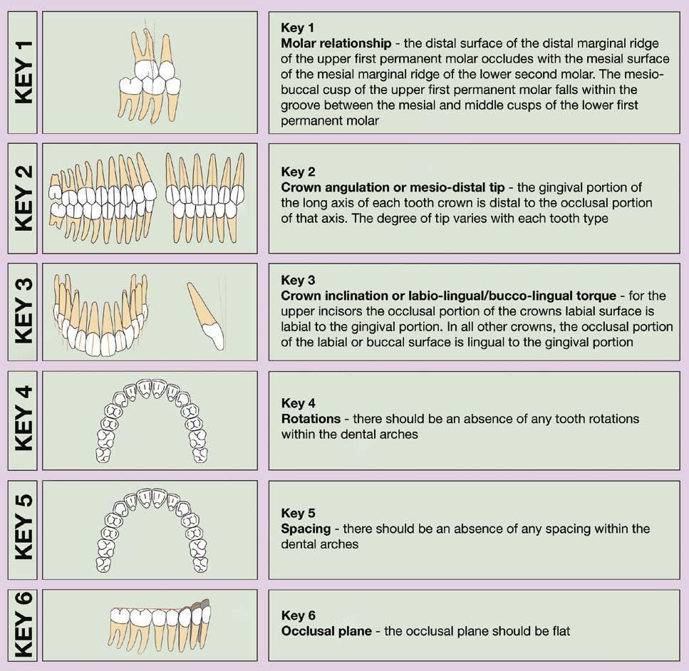
Angle classified occlusion according to the molar relationship and this remains the most internationally recognized classification of malocclusion. When looking at ideal occlusion, Angle found that the mesiobuccal cusp of the upper first permanent molar should occlude with the sulcus between the mesial and distal buccal cusps of the lower
Box 1.2 Andrews Six Keys of Occlusion
first permanent molar (Fig. 1.2). He therefore based his classification of occlusion on this relative mesiodistal position:
l Class I—the position of the dental arches is normal, with first molars in normal occlusion.
l Class II—the relations of the dental arches are abnormal, with all the mandibular teeth occluding distal to normal. Angle recognized two subdivisions under class II:
l Class II division 1—upper incisors are protruding;
l Class II division 2—upper incisors are lingually inclined.
l Class III—the relations of the dental arches are also abnormal, with all mandibular teeth occluding mesial to normal.
In clinical practice, it is common to describe molar relationships in terms of half or even a third of a tooth unit of a class II or class III relationship (Fig. 1.2). However, a
Box
1.3 How important is an ideal functional occlusion?
Advocates of an ideal functional occlusion claim it is necessary to avoid temporomandibular dysfunction, periodontal breakdown and long-term occlusal instability. Indeed, it has been suggested that orthodontic treatment is indicated in all young adults in whom the occlusion is not functionally optimal. These criteria would mean treating most of the population, as an ideal functional occlusion is not very common. For example, as many as 75% of subjects have been described as having non-working side contacts (Tipton & Rinchuse, 1991), whilst a difference of greater than 2 mm has been reported between RCP and ICP for up to 40% of orthodontic patients (Hidaka et al., 2002). So does this matter? Whilst artificially creating non-working side interferences can increase the signs and symptoms of temporomandibular dysfunction (Christensen and Rassouli, 1995), the results of occlusal equilibration, when an idealized functional occlusion is created, are equivocal. Canine guidance has been reported to reduce electromyographic (EMG) activity of the muscles of mastication (Christensen and Rassouli, 1995) but the reproducibility of EMG is open to question (Cecere et al., 1996). There does appear to be a relationship between temporomandibular dysfunction and large slides from RCP into ICP (Solberg et al., 1979) although the correlations between other traits of malocclusion and temporomandibular dysfunction are generally weak (Egermark-Eriksson et al., 1981). So by treating to an ideal functional occlusion does it eliminate or reduce temporomandibular dysfunction? Unfortunately, there is a lack of evidence to support this, or the claim that it results in greater long-term stability (Luther, 2007a, b). Therefore, while any treatment should aim for an ideal functional occlusion, if it is not achieved, there do not appear to be long-term serious consequences to the patient.
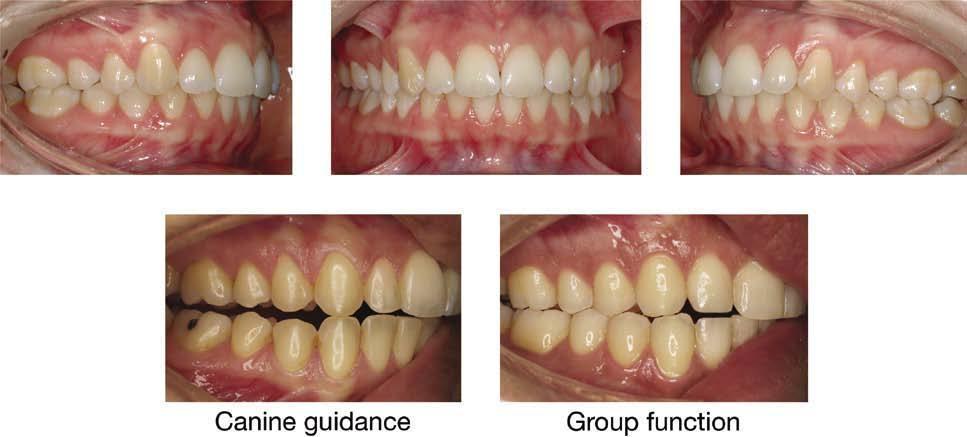
Figure 1.1 Ideal untreated occlusion. The incisor, canine and molar relationship are class I, the dental arches are well aligned and there are no transverse discrepancies. In lateral excursion there should be either canine guidance or group function.
Mesiobuccal cusp
Anterior buccal groove
Figure 1.2 The Angle molar classification. The buccal segment occlusion can be further defined in relation to the degree of mesial or distal occlusion and this is usually measured in units of tooth space.
basic premise of the Angle classification is that the first permanent molars hold a fixed position within the dental arch, which is not necessarily the case. Early loss of deciduous teeth can influence their position and distort the molar relationship and this classification can also be difficult to apply when there is an asymmetric molar relationship. These problems can lead to low levels of inter-examiner agreement (Gravely and Johnson, 1974).
Canine classification
The canine relationship also provides a useful anteroposterior occlusal classification:
l Class I—the maxillary permanent canine should occlude directly in the embrasure between mandibular canine and first premolar.
l Class II—the maxillary permanent canine occludes in front of the embrasure between mandibular canine and first premolar.
l Class III—the maxillary permanent canine occludes behind the embrasure between mandibular canine and first premolar
Similarly to the molar relationship, the severity of the canine relationship can also be described in terms of tooth units and can be inappropriately influenced by local factors such as crowding (Fig. 1.2).
Incisor classification
A more clinically relevant method of classifying malocclusion is based upon the relationship of the maxillary and mandibular incisors. This represents a truer reflection of the underlying skeletal base relationship and also highlights what is often of most concern to the patient. It is essentially the Angle classification, as applied to the incisor
Cingulum plateau
Incisal edge
Figure 1.3 British Standards Institute incisor classification.
teeth, and is defined upon the relationship of the mandibular incisor tip to the cingulum plateau of the maxillary central incisors (Fig. 1.3), being included in the British Standards Institute’s Glossary of Dental Terms:
l Class I—the lower incisor tips occlude or lie below the cingulum plateau of the upper incisors
l Class II—the lower incisor tips occlude or lie posterior to the cingulum plateau of the upper incisors. This classification is further subdivided into:
l Class II division 1—the overjet is increased with upright or proclined upper incisors;
l Class II division 2—the upper incisors are retroclined, with a normal or occasionally increased overjet.
l Class III—the lower incisor tips occlude or lie anterior to the cingulum plateau of the upper incisors.
Confusion can arise when the upper incisors are upright or retroclined, but with an increased overjet. This has led to the introduction of a class II intermediate classification (Williams and Stephens, 1992):
l Class II intermediate—the lower incisor edges lie posterior to the cingulum plateau of the upper central incisors. The upper incisors are upright or slightly retroclined and the overjet lies between 5 and 7-mm.
In reality, an increased overjet with retroclined upper incisors is within the descriptive range of class II division 2.
Prevalence of malocclusion
Malocclusion has been described as a disease of Western societies, and certainly within developed polygenic societies, certain occlusal traits such as crowding are more common. Indeed, from data generated by population studies, the presence of one or more traits of malocclusion is very common. In the USA, noticeable incisor irregularity is present in the majority of adults, with only 34% having well-aligned mandibular incisors and 45% well-aligned maxillary incisors. In addition, about 20% of the American population has a marked deviation from the ideal sagittal jaw relationship, with 2% of these being disfiguring and at the limit for orthodontic correction (Proffit et al, 1998). Within the UK, the last Child Dental Health Survey found around 35% of 12
year olds with a definite need for orthodontic treatment on dental health or aesthetic grounds, which increased to 43% when those already in treatment were included (Chestnutt et al, 2006).
Ethnicity also has a significant bearing on malocclusion. Class II problems are commoner in white populations of northern European descent, whilst class III malocclusion is a common trait amongst Chinese and Japanese societies. Amongst AfricanCaribbean populations, anterior open bite is more common than in Caucasians who, in turn, have a greater proportion of increased overbite.
Aetiology of malocclusion
A malocclusion should be regarded as a developmental condition and does not represent a single entity. Rather, it is the sum of a number of complex occlusal traits, which demonstrate multifactorial inheritance. Although in certain cases specific factors and pathologies can be identified as the cause of a malocclusion; in the majority, the aetiology is less clear. In each individual there is a close interaction between genetics and the environment during development and growth of both the jaws and dentition; it is at this interface that the aetiology of malocclusion lies (Box 1.4).
Evolutionary trends
Comparison of large population studies with archaeological records confirms that malocclusion has become more common over the past 1000 years. In fact, epidemiological data show that the increase in human occlusal variation has been rapid, taking place within a couple of generations, occasionally even from one generation to the next (Weiland et al, 1997). A rapid change such as this would imply a significant contribution from a changing environment, such as has occurred with increasingly urbanized and industrialized societies (Corruccini, 1984). It has been hypothesized that
Box 1.4 Nature versus nurture?
How much a malocclusion is due to the genetic makeup of an individual or the environmental influence upon growth and dental development is the key to understanding the aetiology of malocclusion. The forefathers of modern orthodontics thought that malocclusion was a disease of civilization and that by re-establishing normal jaw function and occlusion; a stable treatment result would be achieved. With a greater understanding of genetics and inheritance, as well as the introduction of cephalometric radiography, it was thought that malocclusion resulted from inherited factors. Therefore, treatment became directed at correcting malocclusion within the existing facial skeleton and soft tissue envelope; both cephalometric and clinical treatment goals were developed, often based around the position of the mandibular incisor teeth. More recently, as science has moved away from simple Mendelian genetics, there has been a shift back to examining the environmental causes of malocclusion. This has also led to renewed interest in treatments that attempt to modify jaw growth.
Figure 1.4 Well-interdigitated class I dentition showing diet-related occlusal interproximal wear.
dietary changes in modern societies, with increased consumption of soft, energy-rich food, has resulted in less interproximal wear between the teeth. Research on aboriginal and stone aged populations has demonstrated this lack of attrition as a possible cause of malocclusion, particularly crowding (Begg, 1954). However, it has been shown that the amount of tooth material lost in each quadrant by interproximal wear is not more than 2 to 3-mm (Fig. 1.4).
A soft diet may also result in underdevelopment of the jaws and a lack of arch space, leading to crowding. According to this hypothesis, hard diet requires vigorous mastication, stimulating the growth of facial bones, particularly in the transverse dimension of the maxilla and mandible. Tooth wear is merely a by-product, brought about by diet-related attrition and high masticatory activity, and has only a minor effect on tooth alignment. Experimental studies have shown that dietary consistency and masticatory activity affect not only the masticatory muscles, but also many aspects of bone growth, including bone size and mass, internal bone structure, and craniofacial size and morphology (Varrela, 2006).
Genetic influences
Genetically homogenous societies exhibit low levels of malocclusion compared to heterogeneous societies and a significant genetic component appears to exist for many individual dental and occlusal anomalies. Early animal experiments initially put forward a compelling argument for a genetic component to malocclusion, based upon inbreeding of dogs, culminating in gross facial deformity. It later emerged that these studies were flawed, merely segregating mutations for specific traits such as achondroplasia, which are present in many breeds of small dog, but rare in humans.
Until recently, most information on the relative contribution of genetic factors to malocclusion has been gained from family studies and the twin method. Monozygotic twins are genetically identical, whereas dizygotic twins only share 50% of their genetic makeup. Therefore, by comparing the differences in occlusal traits between pairs in both groups, some indication of the genetic influence on a particular trait is given; the larger the difference, the greater the genetic effect (Corruccini et al, 1990). This assumes that the environmental effects are similar for both groups.
Many developmental dental anomalies have been shown to occur together and have a strong familial trend. An example of this is development of a palatally impacted maxillary canine, which is more common in females and certain ethnic groups and is
often associated with microdont or absent lateral incisors (Peck et al, 1994). Similarly, jaw growth appears to be mostly genetically determined. A higher correlation has been shown between patients and immediate family than in unrelated subjects for class II division 1 malocclusion, which supports a polygenetic inheritance, particularly in relation to mandibular retrognathia. However, environmental factors, such as lower lip position and digit sucking, can also play a part. Mandibular prognathism, found in class III malocclusions, seems to have a high genetic predisposition, as demonstrated by the high familial inheritance and variation amongst different ethnic groups. More robust evidence for this exists from studies of siblings and first degree relatives (Litton et al, 1970; Watanabe et al, 2005).
Dental arch size and form seems to be more subject to environmental influences (Cassidy et al, 1998). Dental crowding represents a discrepancy between the size of the teeth and the size of the dental arch. Tooth development, including the size, form and presence of teeth within a dentition, is under strong genetic influence. However, the main aetiological factor in crowding appears to be arch size as opposed to tooth size (Howe et al, 1983).
Large population studies have also investigated the influence of population admixture and inbreeding on malocclusion. Generally, the results of these epidemiological studies have shown a greater genetic influence on skeletal relations and arch size and a lower heritability of dental variables such as overbite, molar relationships and crowding, suggesting a greater environmental influence. The importance of hereditary factors also appears to increase with severity of the malocclusion. Although craniofacial form and growth may be under genetic control, the reason that siblings often present with similar malocclusions is probably related to their similar responses to environmental influences. Therefore, while malocclusion appears to be acquired, the underlying genetic control of craniofacial form will tend to divert siblings into similar physiological responses, resulting in the development of similar malocclusions (King et al, 1993).
These studies have also shown that malocclusion does not follow simple Mendelian inheritance, but rather polygenetic or epigenetic transmission, when the interaction of genes with each other and the environment during development determine the phenotypic variation of the trait. Therefore, each would have an additive effect, showing variation along a continuous scale for traits of a malocclusion, which is exactly what happens. Theoretically, in genetically isolated communities, alleles for these traits may be expressed more frequently, giving an indication which have a greater genetic component. Island studies investigating the effects of inbreeding on malocclusion support this polygenetic theory of transmission for certain traits such as overjet and overbite (Lauc et al, 2003).
Environmental factors
The developing dentition is under the influence of resting soft tissue pressure form, and function: lying in a position of muscular balance or equilibrium (Proffit, 1978). Teeth erupt under the influence of the lips and cheeks on one side and the tongue on the other. Abnormal soft tissue patterns seen in those with persistent digit-sucking habits or lip incompetence, with the lower lip trapped behind the upper incisors in function, may predispose to an increased overjet. An alteration in tooth position can also arise when there is a change in this balance of force. Possible causes may be physiological, habitual or pathological and may impact on the lips, cheeks, tongue and periodontal tissues.
Physiological factors
A physiological adaptation can take place in the presence of a skeletal base discrepancy. When teeth erupt, they do so under the influence of soft tissue pressure from the lips, cheeks and tongue. There is a tendency, most notably in the labial segments, for them to upright or procline towards teeth in the opposing arch. This is most often seen in class III skeletal cases, with proclination of the upper incisors and retroclination of the lowers (Fig. 1.5).
Soft tissue envelope
The zone of balance between the lips and cheeks and tongue can in part dictate where the teeth sit. If the forces are imbalanced it can result in tooth movement. Many children have lip incompetence:
l If the lower lip rests behind the upper incisor, this may predispose to an increased overjet and is described as a lip trap (Fig. 1.6).
l In a small percentage of patients there appears to be hyperactivity of the mentalis muscle, resulting in retroclination of the lower incisors and described as a strap-like lower lip (Fig. 1.7).
l Similarly, a high lower lip position is thought to contribute to retroclination of the upper incisors in a class II division 2 relationship (Fig. 1.8).
In cases with anterior open bite, an anterior oral seal on swallowing is created by the tongue coming forward to fill the gap. This is an adaptive behaviour, secondary to the malocclusion. Occasionally, a tongue thrust is the primary cause of the malocclusion: the so-called endogenous (primary) tongue thrust. Although often described,
1.5 Class III malocclusion showing incisor dentoalveolar compensation. The mandibular incisors have retroclined in an attempt to achieve a class I incisor relationship in the presence of a class III skeletal base.
Figure
Figure 1.6 Lip trap contributing to an increase in overjet.
this is a rare phenomenon and is probably related to the anterior resting position of the tongue as opposed to excessive activity.
Mouth breathing
Children with nasopharyngeal obstruction associated with enlarged adenoids have been shown to have longer faces and smaller mandibles compared to controls (Fig. 1.9) (Linder-Aronson, 1970).
Neonates by necessity are nasal breathers to allow suckling but most pre-adolescent children adopt a posture with their lips habitually apart at rest. This resolves in many cases, due to greater vertical growth of the lips compared to the lower facial skeleton, particularly in boys. Total experimental obstruction of the nasal airway in primates and humans results in a change of head posture, with the neck being extended and a downward and backward growth rotation of the mandible occurring. Following adenoidectomy for children with severe nasopharyngeal obstruction, a greater horizontal rather than vertical growth pattern has been described (LinderAronson et al, 1986; Woodside et al, 1991). In humans, total obstruction of the nasal airway is rare and many children, even with some nasal blockage and lip incompetence,
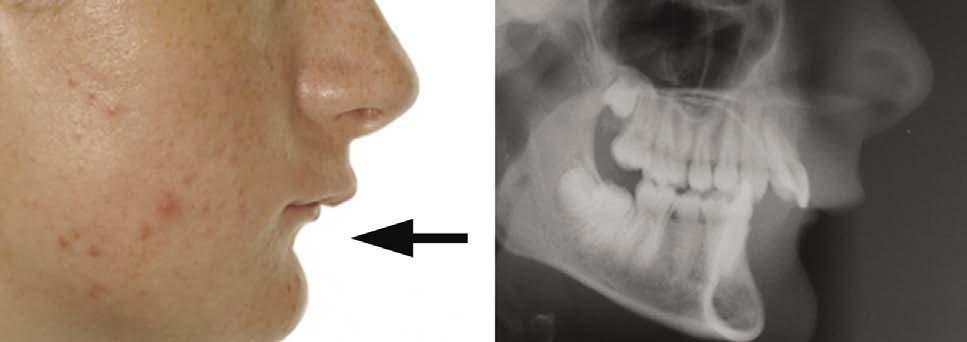
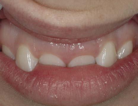
Figure 1.8 A high lower lip position retroclining the upper central incisors and proclining the upper lateral incisors in a class II division 2 incisor relationship (the upper lip has been retracted).
Figure 1.7 Strap-like lower lip.
Figure 1.9 Child with increased facial height and lip incompetence with a history of nasal blockage and mouth breathing. This appearance has been described as an adenoidal face.
do breathe through their noses. It is therefore impossible to generalize on a normal population using data gathered from extremes. The question still remains whether partial nasal blockage is an aetiological factor in malocclusion, as the situation appears more complex than a simple form–function interaction (Vig, 1998).
Muscular activity
Conditions associated with a loss of muscle tone, such as muscular dystrophy and certain types of cerebral palsy, result in a downward and backward rotation of the mandible, an increased lower face height and an anterior open bite (Fig. 1.10). Adults with increased anterior face height have reduced bite force and also a different composition of muscle fibres in the masseter, which implicates muscles as a primary cause of malocclusion (Hunt et al, 2006). However, in children with similar skeletal makeup, the bite force is the same as children with normal face height, implying that the loss in force may develop with, as opposed to causing, a malocclusion (Proffit & Fields, 1983).
Sucking habits
Children can indulge in a variety of non-nutritive sucking habits during their early years, which in the majority of cases involve the use of dummies and/or digits. Dummy sucking is more common in the first few years of life but quite rare beyond the age of five years. In contrast, digit sucking is more prevalent in children over five, being seen in around 10% of this population (Brenchley, 1991). Both these habits can influence the developing dental arches and occlusion if continued beyond the second year of life, with the severity of the effects being related primarily to the type, frequency, intensity and duration of the habit:
• Increased maxillary arch length and prognathism;
• Narrowing of the maxillary arch and widening of mandibular arch width;
• Posterior crossbite;
• Maxillary incisor proclination, spacing and increased overjet;
• Reduced overbite and anterior open bite; and
• Class II buccal segments.
In general terms, dummy sucking is more commonly associated with a symmetrical open bite and posterior crossbite, having a greater effect on the deciduous dentition than digit sucking (Duncan et al, 2007). Digit sucking tends to produce an asymmetric open bite and increased overjet and can have a more significant influence on the mixed and permanent dentitions (Fig. 1.10). The changes in overjet and overbite arise because of the direct affect of the habit on incisor position; whilst a lowering of tongue position away from the upper arch, increased pressure from the cheeks and an absence of tooth contact in the buccal segments contribute to the development of a posterior crossbite. Significantly, many of these occlusal changes can persist well beyond cessation of the habit and if it is continued into the mixed dentition they can be permanent.
Pathology
A number of pathological conditions can contribute directly to a malocclusion, causing either skeletal discrepancies or more local effects upon the dentition.
Childhood fractures of jaws
The condyle is the commonest site of fracture in the mandible during childhood and many go undiagnosed. In severe cases with bilateral fracture and dislocation from the glenoid fossa, an anterior open bite can be one of the presenting features due to a loss in ramus height. A long-term sequelae of early trauma to the mandibular condyle can be asymmetry, with an ipsilateral decrease in ramus height and deviation of the chin point to the affected side (Fig. 1.11). The severity of outcome is in part related to the age at the time of injury. However, a high percentage of children sustaining a condylar fracture have normal mandibular growth due to the reparative capacity of the condyle, even when displaced from the glenoid fossa.
Juvenile rheumatoid arthritis
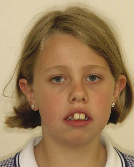
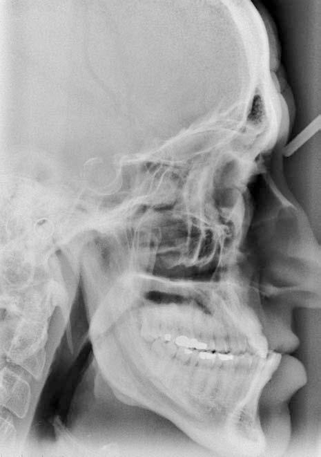
An inflammatory arthritis occurring before the age of 16 years and involving the temporomandibular joints can result in the development of a severe class II malocclusion due to restricted growth of the mandible (Fig. 1.12).
Figure 1.10 Anterior open bite related to cerebral palsy (left) and persistent digit-sucking habit (right).
Figure 1.11 Mandibular asymmetry in an adult following fracture of the condyles as a child.
Excessive growth hormone
Figure 1.12 Lateral skull radiograph of patient with a history of juvenile rheumatoid arthritis.
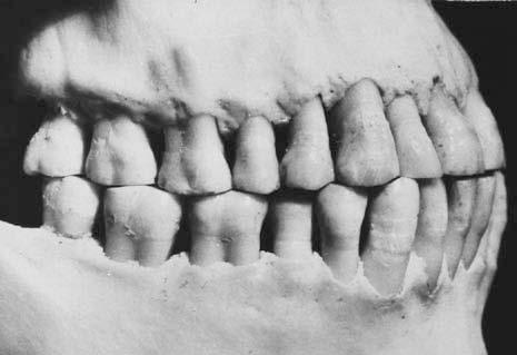
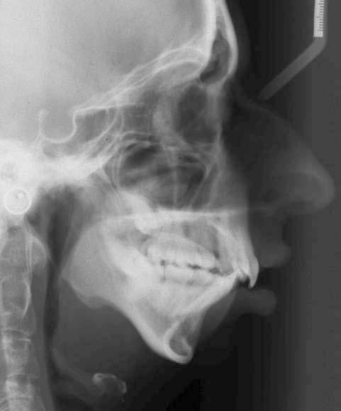
Overproduction of growth hormone from an anterior pituitary tumour causes gigantism in children and acromegaly in adults. In both circumstances, the patient presents with a worsening class III malocclusion characterized by mandibular excess (Fig. 1.13).
Periodontal disease
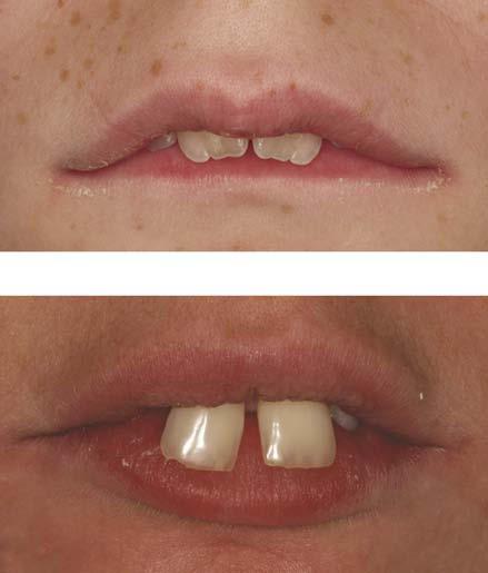
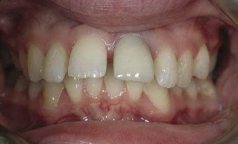
With the loss of alveolar bone that occurs due to periodontal disease, teeth become more susceptible to influence from the soft tissue envelope that surrounds them. Any change in this balance that occurs with age can result in tooth movement. This is commonly seen when upper incisors escape control of the lower lip, resulting in an increase in the overjet and spacing (Fig. 1.14).
Dentoalveolar trauma
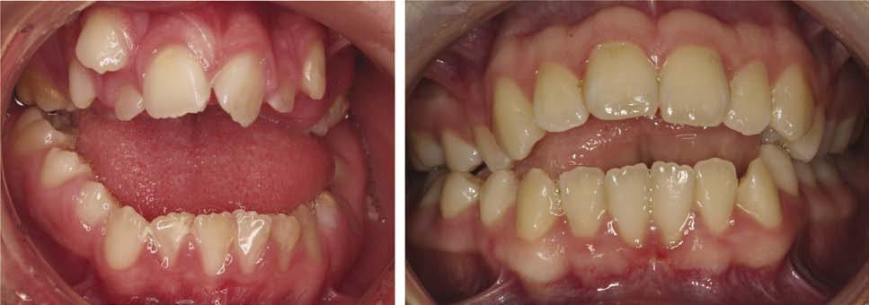
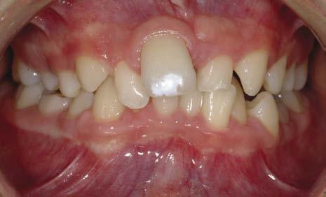
Trauma to the primary maxillary incisors can result in displacement of the tooth into the developing tooth bud of the permanent successor. Damage to the crown or dilaceration of the root can occur, resulting in failure of eruption and impaction of the tooth. Loss of a permanent incisor due to trauma can result in space loss and shift in the dental centre line in crowded dentitions (Fig. 1.15).
Early loss of primary teeth
Although water fluoridation and dental education has significantly reduced the incidence of caries in children, the enforced loss of primary teeth due to caries still remains a major aetiological factor in the development of a local malocclusion. In a crowded dentition, the early loss of deciduous teeth can result in space loss, increased crowding and deviations of the dental centre lines.
Figure 1.14 Proclination and spacing of the upper labial segment resulting from periodontal bone loss.
Figure 1.13 Lateral skull radiograph showing a class III malocclusion in an adult resulting from excessive growth hormone secondary to a pituitary tumour. Note the indistinct and enlarged borders of the pituitary fossa.
Figure 1.15 Loss of UL1 through trauma with subsequent space loss and a shift in the dental centreline.
Benefits of orthodontic treatment
For any elective medical intervention there should be a clear list of benefits for the patient and these should outweigh any potential risks. It is clear that orthodontic treatment can provide significant cosmetic advantages to a patient. However, it has proved difficult to provide strong evidence in support of the widely assumed belief that treatment can also improve the oral health and psychological well-being of an individual.
Resistance to caries and periodontal disease
Dental caries is endemic in most developed societies and the primary aetiological factors are the presence of cariogenic flora in dental plaque and the frequent
intake of refined sugars. The disease process can be controlled with good diet and oral hygiene and is unrelated to the presence or absence of a malocclusion. There is some evidence that straight teeth and a normal overjet are easier to keep clean (Addy et al, 1986; Davies et al, 1988) and that recipients of orthodontic treatment have lower plaque scores (Davies et al, 1991), but this may be more related to the modification of behaviour during treatment rather than the actual presence of straight teeth.
The primary aetiological factor in periodontal disease is dental plaque and the principle way to avoid this condition is maintaining good oral hygiene, not orthodontic treatment. However, there are two specific areas where orthodontic treatment can help to prevent periodontal breakdown:
l Correction of an anterior crossbite with associated recession on a lower incisor; and
l Correction of a deep and traumatic overbite.
Improved masticatory efficiency
Evidence to suggest that having a class I occlusion improves masticatory efficiency is weak. It is perfectly possible to survive without teeth on a Western diet, as many people do, and whilst orthodontic treatment can be beneficial in correcting functional problems such as crossbites, it is unlikely to make a significant difference to masticatory efficiency. One exception is the correction of an anterior open bite when patients are unable to incise food except by biting into it with their posterior teeth.
Prevention or cure of temporomandibular joint dysfunction
The aetiology of temporomandibular joint dysfunction remains controversial, which explains in part the large variety of modalities used to treat it. At the very least, the aetiology is considered to be multifactorial. The relationship between malocclusion and temporomandibular dysfunction has been explored extensively, mostly in large epidemiological studies, and whilst some traits of a malocclusion have been shown to have a correlation with the signs and symptoms of joint dysfunction (Table 1.2), these are very weak (Egermark-Eriksson et al, 1983).
Improvement in speech
Speech patterns are established very early in life and in most cases a long time before eruption of the permanent dentition. Some speech problems are related to certain traits of a malocclusion, such as anterior open bite and a lisp, but treating the malocclusion will not guarantee resolution of the problem.
Table 1.2 Occlusal features associated with temporomandibular dysfunction
• Anterior open bite.
• Deep overbite.
• Class II and III molar relationships.
• Posterior crossbite with displacement.
Prevention of trauma
An increased overjet is a risk factor for trauma to the upper incisors (Jarvinen, 1978). As a consequence, a high percentage of patients with a class II division 1 incisor relationship present with damaged upper incisors. Correction of the incisor relationship will theoretically reduce the vulnerability of these teeth to damage following trauma. Unfortunately most incidences of trauma occur soon after eruption of the permanent incisors and prior to the age when orthodontics is usually started.
Psychological benefits
In its original constitution the World Health Organization defined health as ‘a state of complete physical, social and mental well-being and not merely the absence of disease and infirmity’. Therefore, even though a malocclusion is not a disease state, the benefits of treating it should be considered in terms of both the social and mental wellbeing of an individual. Certain occlusal traits, such as an increased overjet, can lead to a reduction in self-esteem and can be a target for teasing. More severe malocclusions associated with facial disfigurement, such as those seen in cleft lip and palate, have been shown to have a profound and long-lasting psychological impact. However, longitudinal studies have demonstrated little objective evidence to support the assumption that orthodontic treatment can improve long-term psychological health of the individual (Kenealy et al, 2007).
More recently work has focused on the impact malocclusion can have on quality of life for individuals. Certain occlusal traits such as an increased overjet and spacing appear to have some negative impact in children and their families (Johal et al, 2007), whilst in adults severe skeletal problems that require surgical correction can have a profound impact on individual quality of life.
Risks of orthodontic treatment
Orthodontic treatment is not without risk. These risks can arise as a direct consequence of placing an appliance or be secondary to the treatment itself.
Risks from appliances
The principle risks arise from the use of fixed appliances and these can affect the teeth, periodontium and soft tissues.
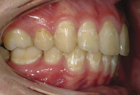
Enamel decalcification
The incidence of demineralization during fixed appliance therapy is high and can result in the development of enamel opacities on the labial surfaces of the teeth. Incidences of up to 50% of patients undergoing fixed appliance therapy have been reported (Gorelick et al, 1982). The main aetiological factors are poor oral hygiene and a diet high in refined sugars. In combination and over the long-term, these factors will inevitably result in demineralization and permanent marking of the teeth (Fig. 1.16). Excellent oral hygiene and a non-cariogenic diet are therefore a prerequisite to orthodontic treatment involving fixed appliances. During treatment, the chances of developing enamel opacities can be reduced by the regular use of topical fluoride supplements. The use of a 0.05% sodium fluoride mouthwash on a daily basis will significantly reduce the incidence of white spot lesions (Benson et al, 2005) and fluoride-releasing
Figure 1.16 Generalized demineralization following orthodontic treatment with fixed appliances.
bonding agents such as glass ionomer will reduce caries levels experienced during treatment (Derks et al, 2004).
Enamel fracture
The removal of a fixed appliance bonded to enamel carries a small risk of fracture at the enamel–dentinal junction if bracket bond strengths are too high. In reality, bond strengths used are considerably lower than this and at debond, failure usually occurs at the bracket base–cement junction. An exception to this proved to be some early ceramic bracket systems; manufacturers were concerned with failure of the bracket bond during treatment and enhanced the mechanical bonding chemically. This resulted in excessive bond strengths and a significant risk of enamel fracture on debonding. Modern ceramic bracket bases are designed with features that facilitate easier debonding, which reduces the risk of enamel fracture.
Root resorption
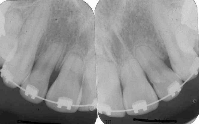
External root resorption is an almost universal finding following orthodontic treatment, but this is usually not clinically significant and has no influence on long-term health of the teeth. Severe root resorption, when more than a quarter of the root length is lost, has been reported to occur in less than 3% of orthodontic patients (Fig. 1.17) (Sameshima and Sinclair, 2004). The greatest amount and severity of root resorption is seen in the anterior maxillary region, especially the maxillary lateral incisors. There is a genetic tendency and ethnic susceptibility, with Asian patients having a lower incidence. The greatest association with root resorption appears to be the duration of treatment and the distance the teeth must move (Linge and Linge, 1991; Segal et al, 2004). Other risk factors associated with a higher incidence of root resorption include:
l Unusually shaped roots, including blunted, pipette-shaped and short roots;
l History of dentoalveolar trauma;
l Excessive orthodontic force;
l Movement of teeth without occlusal contact;
l Intrusive forces;
l Reduction of large overjets by distal movement of anterior teeth; and
l Pushing apices of teeth into cortical bone.
Pain and damage to the pulp
Figure 1.17 Severe root resorption during orthodontic treatment.
Orthodontic treatment, especially with fixed appliances, can be painful. However, this pain usually subsides within a few days of appliance activation and can be controlled with analgesia. The use of excessive force or pushing the apex of teeth through the cortical plate can result in a loss of vitality. Teeth with a history of trauma are more susceptible to vitality loss during treatment but in most cases there is no obvious cause. Fortunately, loss of vitality is a rare complication of orthodontics.
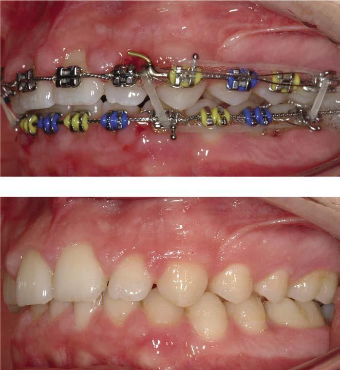
Gingivitis
Gingival irritation is inevitable with the use of fixed appliances, especially the placement of bands and this is exacerbated by poor oral hygiene, which can result in gingival hyperplasia. Gingival health improves significantly following the removal of appliances, with a reduction in probing depths mainly due to shrinkage of hyperplastic tissues (Fig. 1.18). Certain medications such as antiepileptic drugs and immunosuppressants in combination with poor oral hygiene can result in extensive gingival hyperplasia that can require gingival surgery following appliance removal.
Alveolar bone loss
A small loss of alveolar bone height following orthodontic treatment has been reported in relation to teeth adjacent to extraction sites but there appears to be no longterm effect on periodontal health from orthodontic treatment (Zachrisson and Alnaes, 1974). An exception to this is orthodontic treatment in patients with active periodontal disease because this can rapidly increase bone loss. Periodontal disease should be treated, stable and well maintained in these patients prior to commencing orthodontic treatment.
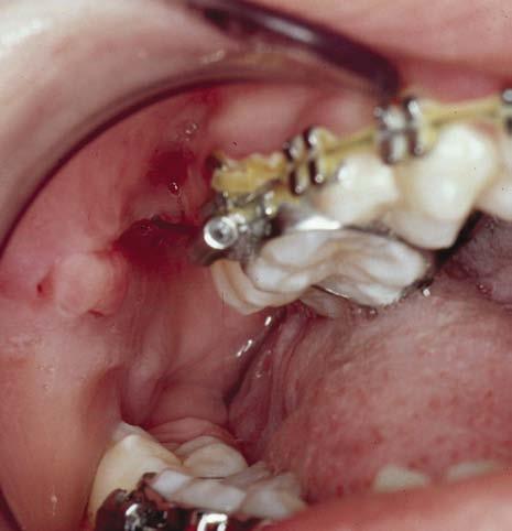
Oral ulceration
Aphthous ulceration in susceptible individuals is common with fixed appliances, particularly during the early stages of treatment. This can be exacerbated if archwires are not cut or bent back and left protruding from molar tubes (Fig. 1.19).
1.18 Gingival hyperplasia during orthodontic treatment and subsequent improvement on removal of the appliance.
Figure
Figure 1.19 Oral ulceration from a fixed appliance.
Allergic reaction
Orthodontic wires and brackets contain nickel and nickel allergy is increasing in frequency. Its prevalence has been reported to be approximately 10% in the USA and Europe, being more common in females. It is usually a Type IV allergic reaction related to wearing jewellery or watches and body piercing. Fortunately oral reactions are rare although prolonged exposure to nickel-containing oral appliances may increase sensitivity to nickel (Bass et al, 1993). Intraoral signs are non-specific and have been reported to include erythematous areas and severe gingivitis despite good oral hygiene.
Headgear injury
A number of intra- and extraoral injuries have been reported with the use of headgear, particularly the risk of ocular penetration. The majority of these injuries occur as a result of the inner bow of the headgear detaching from the molar bands at night. Headgear injury is discussed further in Chapter 5.
Generalized risks associated with orthodontic treatment
A number of more general risks have been proposed with regard to orthodontic treatment, in particular causing damage to the facial profile or temporomandibular joints. A great deal of controversy surrounds these claims and currently there is little robust evidence in the literature to support them. In contrast, it is well recognized that the final tooth positions achieved following orthodontic treatment can relapse and it is important for any orthodontic patient to understand that absolute stability cannot be guaranteed without permanent retention.
Facial aesthetics
The position of the dentition within the soft tissues of the face has an impact on facial aesthetics. Over-retraction of the incisor dentition, especially in relation to mid-arch premolar extractions, has been criticized for flattening facial profiles, especially in relation to the position of the lips. Conversely, excessive proclination of the incisor teeth in association with arch expansion can result in a poor facial appearance. The relationship between incisor movement and soft tissue changes are complex. There have been numerous, mostly retrospective studies, assessing facial change following orthodontic treatment, and although the extraction of teeth produces slightly more retrusive profiles than non-extraction treatment, in the majority of cases the facial changes are seen as beneficial by both lay and professional judges, irrespective of whether teeth were extracted.
Temporomandibular
joint dysfunction
There have been claims and successful litigation in relation to orthodontic treatment and the exacerbation of symptoms associated with temporomandibular joint dysfunction. However, there is currently a lack of robust evidence linking orthodontics and particularly the extraction of permanent teeth to these signs and symptoms. Orthodontic treatment has not been shown to be a causative factor in the development of temporomandibular dysfunction later in life, regardless of whether teeth are extracted (Dibbets and van der Weele, 1991; Kremenak et al, 1992a, b; Mohlin et al, 2004; Sadowsky et al, 1991). However, it is important that any signs and symptoms of temporomandibular dysfunction are recorded prior to treatment and that attention is paid to the functional occlusion at the end of treatment.
Relapse
Longitudinal studies have shown a high potential for relapse following the correction of certain occlusal traits, which include:
l Rotated teeth;
l Lower incisor crowding;
l Changes in position of the lower incisors;
l Expansion of the lower intercanine width; and
l Spacing.
The appearance of increased dental crowding later in life has been found to occur in untreated individuals and as such, should be regarded as an age-related change rather than relapse.
Failure of treatment
Successful orthodontic treatment requires significant cooperation and compliance, which some patients find difficult. This is less of a problem with adult patients who are generally highly motivated towards treatment, but in children and adolescents high discontinuation rates have been reported. Clearly, a patient who fails to complete a course of treatment may end up with an occlusal result that is unsatisfactory or even worse than the presenting malocclusion, particularly if permanent teeth have been extracted and space closure has not been completed.
Provision of orthodontic treatment
Given the high prevalence of malocclusion within the general population and increasing demand for treatment, attempts have been made to develop indices for prioritization of orthodontic treatment provision within healthcare systems (Box 1.5). This is especially relevant where dental health services are subsidized by the government as part of a national health service, such as in the UK and Scandinavia.
Index of Treatment Need (IOTN)
The IOTN was developed within the UK (Brook and Shaw, 1989) where the majority of orthodontic treatment has been provided within a state-funded health service and this has proved to be the most widely used and recognized index. Based on the Swedish National Board for Welfare Index (Linder-Aronson, 1974), it defines need for treatment, both in terms of dental health benefits and aesthetic handicapping and has been shown to be reproducible and reliable over time (Cooper et al, 2000). The IOTN is split into Dental Health and Aesthetic components.
Dental Health Component (DHC)
The DHC has five categories, defining treatment need from none (Grade 1) to a great need (Grade 5). The following characteristics are scored for each individual:
l Missing teeth;
l Overjet;
l Crossbites;
l Displacement of contact points (crowding); and
l Overbite.
Box 1.5 Limitations of occlusal indices
The use of indices to quantify the need for orthodontic treatment is controversial and far from being universally accepted. Indeed, the American Association of Orthodontics does not recognize any index as a scientifically valid measurement of the need for orthodontic treatment (Shaw et al., 1995).
An ideal index has a number of requirements:
l Reliability and reproducibility;
l Validity;
l Acceptability to both professionals and the public;
l Objectivity; and
l Simple to apply.
Certainly the Index of Treatment Need (IOTN) has been shown to be reproducible and simple to apply; however, criticism regarding its validity has been made. Does it measure the need for orthodontic treatment? The perceived need depends upon many factors, only one of which is the malocclusion. These factors can include the country of origin of the clinician and the system of remuneration under which they are employed (Richmond and Daniels, 1998a). Similar differences have been found regarding what constitutes acceptable treatment (Richmond and Daniels, 1998b). The Dental Health Component (DHC) reflects our current understanding of the health risks of a malocclusion, although the correlations between dental disease and certain traits of a malocclusion are very weak. In addition, little allowance is made for facial aesthetics and the psychological impact of a malocclusion, both of which are often reasons that treatment is sought. The Aesthetic Component, which should in part allow for these factors, although validated among professionals, correlates poorly with lay opinion as to what constitutes a need for treatment (Hunt et al., 2002). As the desire for orthodontic treatment is primarily driven by the perception of a patient regarding their own dental aesthetics, future indices may well incorporate patient factors into their scoring systems.
The DHC is hierarchical, for each individual the highest score is found and recorded, irrespective of any other features within the malocclusion. The five categories are further subdivided using letters, which describe the feature of the malocclusion that has been scored (Table 1.3).
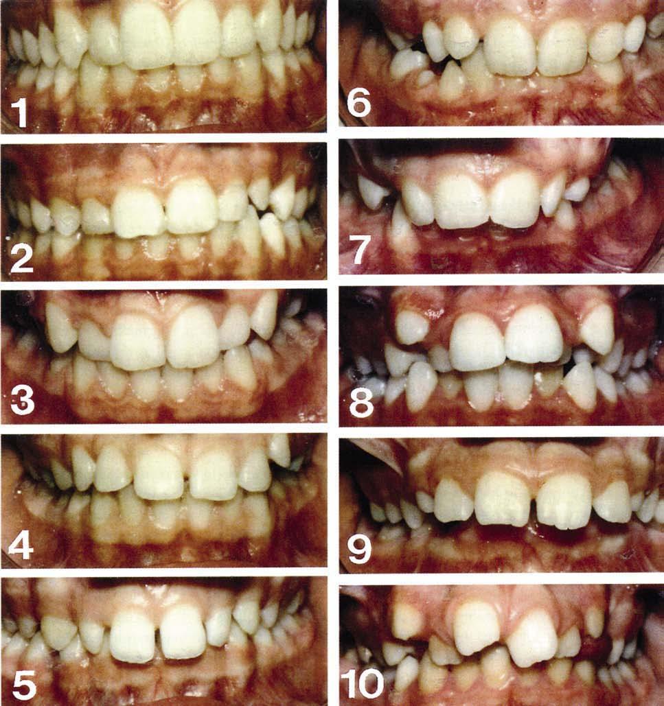
Aesthetic Component
This records the aesthetic handicapping of the malocclusion and is based on a series of ten photographs, which show a graduated decrease in dental aesthetics (Fig. 1.20). A score is given from 0 to 10 based upon the perceived aesthetic impairment of an individual’s malocclusion, not morphological similarities with the photographs. A highlevel agreement has been found between scores given by professionals and those given by patients.
Table 1.3 Dental health component of the IOTN
Grade 1—No treatment required
1.Extremely minor malocclusions, including displacements less than 1 mm
Grade 2—Little need for treatment
2.aIncreased overjet > 3.5 mm but ≤ 6 mm (with competent lips)
2.bReverse overjet greater than 0 mm but ≤ 1 mm
2.cAnterior or posterior crossbite with ≤ 1 mm discrepancy between RCP and ICP
2.dDisplacement of teeth > 1 mm but ≤ 2 mm
2.eAnterior or posterior open bite > 1 mm but ≤ 2 mm
2.fIncreased overbite ≥ 3.5 mm (without gingival contact)
2.g Prenormal or postnormal occlusions with no other anomalies (up to 1 2 a unit of discrepancy)
Grade 3—Borderline need for treatment
3.aIncreased overjet > 3.5 mm but ≤ 6 mm (incompetent lips)
3.bReverse overjet greater than 1 mm but ≤ 3.5 mm
3.cAnterior or posterior crossbites with > 1 mm but ≤ 2 mm discrepancy between RCP and ICP
3.dDisplacement of teeth > 2 mm but ≤ 4 mm
3.eLateral or anterior open bite > 2 mm but ≤ 4 mm
3.fIncreased and incomplete overbite without gingival or palatal trauma
Grade 4—Treatment required
4.aIncreased overjet > 6 mm but ≤ 9 mm
4.bReverse overjet > 3.5 mm with no masticatory or speech difficulties
4.c Anterior or posterior crossbites with > 2 mm discrepancy between RCP and ICP
4.dSevere displacements of teeth > 4 mm
4.eExtreme lateral or anterior open bites > 4 mm
4.fIncreased and complete overbite with gingival or palatal trauma
4.hLess extensive hypodontia requiring pre-restorative orthodontics or orthodontic space closure to obviate the need for a prosthesis
4.lPosterior lingual crossbite with no functional occlusal contact in one or more buccal segments
4.mReverse overjet > 1 mm but < 3.5 mm with recorded masticatory and speech difficulties
4.tPartially erupted teeth, tipped and impacted against adjacent teeth
4.xExisting supernumerary teeth
Grade 5—Treatment required
5.aIncreased overjet > 9 mm
5.hExtensive hypodontia with restorative implications (more than one tooth missing in any quadrant requiring pre-restorative orthodontics)
Table 1.3 Dental health component of the IOTN—cont’d
5.iImpeded eruption of teeth (apart from 3rd molars) due to crowding, displacement, the presence of supernumerary teeth, retained deciduous teeth and any pathological cause
5.mReverse overjet > 3.5 mm with reported masticatory and speech difficulties
5.p Defects of cleft lip and palate
5.sSubmerged deciduous teeth
Figure 1.20 The aesthetic component of the IOTN. The SCAN scale was first published in 1987 by the European Orthodontic Society (Evans R and Shaw W, Preliminary evaluation of an illustrated scale for dental attractiveness. Eur J Orthod 9: 314–318). IOTN aesthetic and dental health components reproduced courtesy of Orthocare.
1.21 Nomogram of pre- and post-treatment PAR scores.
Monitoring orthodontic treatment
The outcome of orthodontic treatment can be recorded in terms of occlusal changes and an attempt has been made to give this an objective numeric score using the Peer Assessment Rating (PAR) (Richmond et al, 1992a, b). This gives an accumulative score, indicating the extent of deviation from a normal functioning occlusion assessed from dental study casts. There is no maximum cut-off level, and the pre- and post-treatment models should be assessed, which gives a percentage score for the change with treatment. A reduction in the weighted PAR score of less than 30% is considered to show occlusal changes that are worse or no different. A reduction of greater than 30% shows improved occlusal changes, whilst a PAR reduction of 22 points or greater indicates a greatly improved occlusal result. This can be plotted on a nomogram (Fig. 1.21), which is divided into three sections: upper (worse–no difference), middle (improved) and lower (greatly improved). This is useful when looking at the outcome of multiple patients, as it gives an indication of the quality of treatment an individual or group of individuals is providing.
Index of Complexity and Orthodontic Need (ICON)
Based on the IOTN and PAR indices a single index, the Index of Complexity and Need (ICON), has been developed to measure both treatment need and outcome of treatment (Daniels and Richmond, 2000). By combining five occlusal traits (IOTN Aesthetic Component, crossbite, upper arch crowding and spacing, buccal segment anteroposterior relationships, and anterior vertical relationship) with different weightings, a numeric score is given that can be used to ascertain need for treatment, the complexity of the treatment and the improvement resulting from treatment. This has been shown to be reproducible for treatment need and complexity but less so for outcome, due to low levels of agreement between examiners as to what constitutes acceptable treatment (Richmond and Daniels, 1998a, b).
Figure
Further reading
CLARK JR AND EVANS RD (2001). Functional occlusion: I. A review. J Orthod 28: 76–81.
DAVIES S AND GRAY RM (2001). What is occlusion? Br Dent J 191: 235–8, 241–245.
DAVIES SJ, GRAY RM, SANDLER PJ ET AL (2001). Orthodontics and occlusion. Br Dent J 191: 539–542, 545–549.
References
ADDY M, DUMMER PM, GRIFFITHS G ET AL (1986). Prevalence of plaque, gingivitis and caries in 11–12-year-old children in South Wales. Community Dent Oral Epidemiol 14: 115–118.
ANDREWS LF (1972). The six keys to normal occlusion. Am J Orthod 62: 296–309.
ANGLE EH (1899). Classification of malocclusion. Dental Cosmos 41: 248–264.
BASS JK, FINE H AND CISNEROS GJ (1993). Nickel hypersensitivity in the orthodontic patient. Am J Orthod Dentofacial Orthop 103: 280–285.
BEGG PR (1954). Stone age man’s dentition. Am J Orthod 40: 298–312.
BENSON PE, SHAH AA, MILLETT DT ET AL (2005). Fluorides, orthodontics and demineralization: a systematic review. J Orthod 32: 102–114.
BRENCHLEY ML (1991). Is digit sucking of significance? Br Dent J 171: 357–362.
BROOK PH AND SHAW WC (1989). The development of an index of orthodontic treatment priority. Eur J Orthod 11: 309–320.
CASSIDY KM, HARRIS EF, TOLLEY EA ET AL (1998). Genetic influence on dental arch form in orthodontic patients. Angle Orthod 68: 445–454.
CECERE F, RUF S AND PANCHERZ H (1996). Is quantitative electromyography reliable? J Orofac Pain 10: 38–47.
CHESTNUTT IG, BURDEN DJ, STEELE JG ET AL (2006). The orthodontic condition of children in the United Kingdom, 2003. Br Dent J 200: 609–612;quiz 638.
CHRISTENSEN LV AND RASSOULI NM (1995). Experimental occlusal interferences. Part I. A review. J Oral Rehabil 22: 515–520.
COOPER S, MANDALL NA, DIBIASE D ET AL (2000). The reliability of the Index of Orthodontic Treatment Need over time. J Orthod 27: 47–53.
CORRUCCINI RS (1984). An epidemiologic transition in dental occlusion in world populations. Am J Orthod 86: 419–426.
CORRUCCINI RS, TOWNSEND GC, RICHARDS LC ET AL (1990). Genetic and environmental determinants of dental occlusal variation in twins of different nationalities. Hum Biol 62: 353–367.
DANIELS C AND RICHMOND S (2000). The development of the index of complexity, outcome and need (ICON). J Orthod 27: 149–162.
DAVIES TM, SHAW WC, ADDY M ET AL (1988). The relationship of anterior overjet to plaque and gingivitis in children. Am J Orthod Dentofacial Orthop 93: 303–309.
DAVIES TM, SHAW WC, WORTHINGTON HV ET AL (1991). The effect of orthodontic treatment on plaque and gingivitis. Am J Orthod Dentofacial Orthop 99: 155–161.
DERKS A, KATSAROS C, FRENCKEN JE ET AL (2004). Caries-inhibiting effect of preventive measures during orthodontic treatment with fixed appliances. A systematic review. Caries Res 38: 413–420.
DIBBETS JM AND VAN DER WEELE LT (1991). Extraction, orthodontic treatment, and craniomandibular dysfunction. Am J Orthod Dentofacial Orthop 99: 210–219.
DUNCAN K, MCNAMARA C, IRELAND AJ (2007) Sucking habits in childhood and the effects on the primary dentition: findings of the Avon Longitudinal Study of Pregnancy and Childhood. Int J Paediatr Dent 18: 178–188.
EGERMARK-ERIKSSON I, CARLSSON GE AND INGERVALL B (1981). Prevalence of mandibular dysfunction and orofacial parafunction in 7-, 11- and 15-year-old Swedish children. Eur J Orthod 3: 163–172.
EGERMARK-ERIKSSON I, INGERVALL B AND CARLSSON GE (1983). The dependence of mandibular dysfunction in children on functional and morphologic malocclusion. Am J Orthod 83: 187–194.
GORELICK L, GEIGER AM AND GWINNETT AJ (1982). Incidence of white spot formation after bonding and banding. Am J Orthod 81: 93–98.
GRAVELY JF AND JOHNSON DB (1974). Angle’s classification of malocclusion: an assessment of reliability. Br J Orthod 1: 79–86.
HIDAKA O, ADACHI S AND TAKADA K (2002). The difference in condylar position between centric relation and centric occlusion in pretreatment Japanese orthodontic patients. Angle Orthod 72: 295–301.
HOWE RP, MCNAMARA JA Jr AND O’CONNOR KA (1983). An examination of dental crowding and its relationship to tooth size and arch dimension. Am J Orthod 83: 363–373.
HUNT N, SHAH R, SINANAN A ET AL (2006). Northcroft Memorial Lecture 2005: Muscling in on malocclusions: Current concepts on the role of muscles in the aetiology and treatment of malocclusion. J Orthod 33: 187–197.
HUNT O, HEPPER P, JOHNSTON C ET AL (2002). The Aesthetic Component of the Index of Orthodontic Treatment Need validated against lay opinion. Eur J Orthod 24: 53–59.
JARVINEN S (1978). Incisal overjet and traumatic injuries to upper permanent incisors. A retrospective study. Acta Odontol Scand 36: 359–362.
JOHAL A, CHEUNG MY AND MARCENE W (2007). The impact of two different malocclusion traits on quality of life. Br Dent J 202: E2.
KENEALY PM, KINGDON A, RICHMOND S ET AL (2007). The Cardiff dental study: a 20-year critical evaluation of the psychological health gain from orthodontic treatment. Br J Health Psychol 12: 17–49.
KING L, HARRIS EF AND TOLLEY EA (1993). Heritability of cephalometric and occlusal variables as assessed from siblings with overt malocclusions. Am J Orthod Dentofacial Orthop 104: 121–131.
KREMENAK CR, KINSER DD, HARMAN HA ET AL (1992a). Orthodontic risk factors for temporomandibular disorders (TMD). I: Premolar extractions. Am J Orthod Dentofacial Orthop 101: 13–20.
KREMENAK CR, KINSER DD, MELCHER TJ ET AL (1992b). Orthodontics as a risk factor for temporomandibular disorders (TMD). II. Am J Orthod Dentofacial Orthop 101: 21–27.
LAUC T, RUDAN P, RUDAN I ET AL (2003). Effect of inbreeding and endogamy on occlusal traits in human isolates. J Orthod 30: 301–308; discussion 297.
LINDER-ARONSON S (1970). Adenoids. Their effect on mode of breathing and nasal airflow and their relationship to characteristics of the facial skeleton and the denition. A biometric, rhino-manometric and cephalometro-radiographic study on children with and without adenoids. Acta Otolaryngol Suppl 265: 1–132.
LINDER-ARONSON S (1974). Orthodontics in the Swedish Public Dental Health Service. Trans Eur Orthod Soc: 233–240.
LINDER-ARONSON S, WOODSIDE DG AND LUNDSTROM A (1986). Mandibular growth direction following adenoidectomy. Am J Orthod 89: 273–284.
LINGE L and LINGE BO (1991). Patient characteristics and treatment variables associated with apical root resorption during orthodontic treatment. Am J Orthod Dentofacial Orthop 99: 35–43.
LITTON SF, ACKERMANN LV, ISAACSON RJ AND SHAPIRO BL (1970). A genetic study of Class 3 malocclusion. Am J Orthod 58: 565–577.
LUTHER F (2007a). TMD and occlusion part I. Damned if we do? Occlusion: the interface of dentistry and orthodontics. Br Dent J 202: E2; discussion 38–39.
LUTHER F (2007b). TMD and occlusion part II. Damned if we don’t? Functional occlusal problems: TMD epidemiology in a wider context. Br Dent J 202: E3; discussion 38–39.
MOHLIN BO, DERWEDUWEN K, PILLEY R ET AL (2004). Malocclusion and temporomandibular disorder: a comparison of adolescents with moderate to severe dysfunction with those without signs and symptoms of temporomandibular disorder and their further development to 30 years of age. Angle Orthod 74: 319–327.
PECK S, PECK L AND KATAJA M (1994). The palatally displaced canine as a dental anomaly of genetic origin. Angle Orthod 64: 249–256.
PROFFIT WR (1978). Equilibrium theory revisited: factors influencing position of the teeth. Angle Orthod 48: 175–186.
PROFFIT WR AND FIELDS HW (1983). Occlusal forces in normal- and long-face children. J Dent Res 62: 571–574.
PROFFIT WR, FIELDS HW Jr AND MORAY LJ (1998). Prevalence of malocclusion and orthodontic treatment need in the United States: estimates from the NHANES III survey. Int J Adult Orthodon Orthognath Surg 13: 97–106.
RICHMOND S AND DANIELS CP (1998a). International comparisons of professional assessments in orthodontics: Part 1—Treatment need. Am J Orthod Dentofacial Orthop 113: 180–185.
RICHMOND S AND DANIELS CP (1998b). International comparisons of professional assessments in orthodontics: Part 2—treatment outcome. Am J Orthod Dentofacial Orthop 113: 324–328.
RICHMOND S, SHAW WC, O’BRIEN KD ET AL (1992a). The development of the PAR Index (Peer Assessment Rating): reliability and validity. Eur J Orthod 14: 125–139.
RICHMOND S, SHAW WC, ROBERTS CT ET AL (1992b). The PAR Index (Peer Assessment Rating): methods to determine outcome of orthodontic treatment in terms of improvement and standards. Eur J Orthod 14: 180–187.
SADOWSKY C, THEISEN TA AND SAKOLS EI (1991). Orthodontic treatment and temporomandibular joint sounds—a longitudinal study. Am J Orthod Dentofacial Orthop 99: 441–447.
SAMESHIMA GT AND SINCLAIR PM (2004). Characteristics of patients with severe root resorption. Orthod Craniofac Res 7: 108–114.
SEGAL GR, SCHIFFMAN PH AND TUNCAY OC (2004). Meta analysis of the treatment-related factors of external apical root resorption. Orthod Craniofac Res 7: 71–78.
SHAW WC, RICHMOND S AND O’BRIEN KD (1995). The use of occlusal indices: a European perspective. Am J Orthod Dentofacial Orthop 107: 1–10.
SOLBERG WK, WOO MW AND HOUSTON JB (1979). Prevalence of mandibular dysfunction in young adults. J Am Dent Assoc 98: 25–34.
TIPTON RT AND RINCHUSE DJ (1991). The relationship between static occlusion and functional occlusion in a dental school population. Angle Orthod 61: 57–66.
VARRELA J (2006). Masticatory function and malocclusion: A clinical perspective. Semin Orthod 12: 102–109.
VIG KW (1998). Nasal obstruction and facial growth: the strength of evidence for clinical assumptions. Am J Orthod Dentofacial Orthop 113: 603–611.
WATANABE M, SUDA N AND OHYAMA K (2005). Mandibular prognathism in Japanese families ascertained through orthognathically treated patients. Am J Orthod Dentofacial Orthop 128: 466–470.
WEILAND FJ, JONKE E AND BANTLEON HP (1997). Secular trends in malocclusion in Austrian men. Eur J Orthod 19: 355–359.
WILLIAMS AC and STEPHENS CD (1992). A modification to the incisor classification of malocclusion. Br J Orthod 19: 127–130.
WOODSIDE DG, LINDER-ARONSON S, LUNDSTROM A ET AL (1991). Mandibular and maxillary growth after changed mode of breathing. Am J Orthod Dentofacial Orthop 100: 1–18.
ZACHRISSON BU AND ALNAES L (1974). Periodontal condition in orthodontically treated and untreated individuals. II. Alveolar bone loss: radiographic findings. Angle Orthod 44: 48–55.
This page intentionally left blank