Basic Medical Endocrinology
Fourth Edition
H. Maurice Goodman Department of Physiology
University of Massachusetts Medical School
AMSTERDAM • BOSTON
NEW YORK
OXFORD
HEIDELBERG • LONDON
PARIS
SAN DIEGO
SAN FRANCISCO • SINGAPORE • SYDNEY • TOKYO
Academic Press is an imprint of Elsevier
Cover Credits:
Background: FIGURE 3-2 Histology of the human thyroid. Simple cuboidal cells (arrows) make up the follicles. C _ thyroid colloid (thyroglobulin), which fills the follicles. (From Borysenko, M. and Beringer, T. (1979) Functional Histology, 312. Little, Brown, Boston by permission of Lippincott, Williams and Wilkins, Philadelphia.)
Black/green: FIGURE 7-11 Confocal fluorescent microscope images of cultured mouse adipocytes that were transfected with a GLUT4-enhanced green fluorescent protein fusion construct and then incubated in the absence (A) or presence (B) of insulin for 30 min. Insulin stimulation results in the translocation of GLUT4 from intracellular storage sites to the plasma membrane. (From Watson, R.T., Kanzaki, M., and Pessin, J. (2004) Regulated membrane trafficking of the insulin-responsive glucose transporter 4 in adipocytes. Endocr. Revs. 25: 177–204, by permission of The Endocrine Society.)
Blue figure: FIGURE 10-14 Low-power photomicrograph of a portion of the thyroid gland of a normal dog. Parafollicular (C) cells are indicated in the walls of the follicles. (From Ham, A.W. and Cormack, D. H. (1979) Histology, 8th Edition, 802, by permission of Lippincott, Williams and Wilkins, Philadelphia.)
Red, white and blue: FIGURE 11-3 Schematic representation of the tibial epiphyseal growth plate. (Modified from Nilsson, O., Marino, R., De Luca, F., Phillip, M., and Baron, J. (2005) Endocrine regulation of the growth plate. Hormone Research 64: 157–165 by permission of S. Karger AG, Basel.)
Pink, yellow, white: FIGURE 12-1 Histological section of human testis. The transected tubules show various stages of spermatogenesis. (From di Fiore, M.S.H. (1981) Atlas of Human Histology, 5th Edition, 209. Lea & Febiger, by permission of Lippincott, Williams and Wilkins, Philadelphia.)
Academic Press is an imprint of Elsevier 30 Corporate Drive, Suite 400, Burlington, MA 01803, USA 525 B Street, Suite 1900, San Diego, California 92101-4495, USA 84 Theobald’s Road, London WC1X 8RR, UK
Copyright © 2009, Elsevier Inc. All rights reserved.
No part of this publication may be reproduced or transmitted in any form or by any means, electronic or mechanical, including photocopy, recording, or any information storage and retrieval system, without permission in writing from the publisher.
Permissions may be sought directly from Elsevier’s Science & Technology Rights Department in Oxford, UK: phone: ( 44) 1865 843830, fax: ( 44) 1865 853333, E-mail: permissions@elsevier.com. You may also complete your request online via the Elsevier homepage (http://elsevier.com), by selecting “Support & Contact ” then “Copyright and Permission” and then “Obtaining Permissions.”
Library of Congress Cataloging-in-Publication Data
Application Submitted
British Library Cataloguing-in-Publication Data
A catalogue record for this book is available from the British Library.
ISBN: 978-0-12-373975-9
For information on all Academic Press publications visit our Web site at www.elsevierdirect.com
Printed in China
09 10 9 8 7 6 5 4 3 2
This volume is dedicated to my children’s children: Dylan, Adam, Rebecca, and Joshua
This page intentionally left blank
Preface
Preface
Preface
Preface
List of Figures
CHAPTER 1
Fig. 1. Chemical communication between cells.
Fig. 2. Levels at which hormone actions are considered.
Fig. 3. Composition of DNA.
Fig. 4. Complementary base pairing.
Fig. 5. Transcription and RNA processing.
Fig. 6. Alternative splicing.
Fig. 7. Translation: Alberts et al. Molecular Biology of the Cell, New York: Garland Publishing, 1994; reprinted by permission of Garland Publishing, Inc.
Fig. 8. Post-translational processing.
Fig. 9. Exocytosis.
Fig. 10. Hormone binding to plasma proteins.
Fig. 11. Specificity of hormone signaling.
Fig. 12. Receptor components.
Fig. 13. General scheme of steroid hormone action.
Fig. 14. Schematic view of a nuclear receptor.
Fig. 15. Activation of steroid hormone receptors.
Fig. 16. An unactivated G-Protein coupled receptor.
Fig. 17. Activation of G-protein coupled receptor.
Fig. 18. Formation and degradation of cyclic adenosine monophosphate (cyclic AMP).
Fig. 19. Formation of inositol 1,4,5 triphosphate (IP3) and diacylglycerol (DAG).
Fig. 20. Effects of cyclic AMP.
Fig. 21. (IP3) diacylglycerol/(DAG) second messenger system.
Fig. 22. DAG lipase to releases precursors of the prostaglandins and leukotrienes.
Fig. 23. Signaling through tyrosine kinase.
Fig. 24. Signaling through JAK/Stat.
Fig. 25. Components of a hormone response system.
Fig. 26. Negative feedback of hepatic glucose production by glucagon.
Fig. 27. Negative feedback regulation of blood glucose concentration by insulin and glucagon.
Fig. 28. Positive feedback regulation of oxytocin secretion.
Fig. 29. Competing reactions that form the basis of the radioimmunoassay.
Fig. 30. Sandwich type assay.
Fig. 31. Patterns of hormone concentrations in blood: A. Bremer et al., J. Clin Endocrinol. Metab., 56: 1278, 1983; reprinted by permission of the Endocrine Society; B. Yamaji et al., Endocrinology, 90: 771, 1972; reprinted by permission of the Endocrine Society; C. Hwang et al., Proc. Natl. Acad. Sci. USA, 68: 1902–1971, reprinted with permission.
CHAPTER 2
Fig. 1. Human pituitary gland and adjacent hypothalamic structures: Netter, F.H., Atlas of Human Anatomy, 2nd ed. Novartis Summit New Jersey; reprinted by permission of Elsevier.
Fig. 2. Vascular supply of the human pituitary gland: Netter, F.H., Atlas of Human Anatomy, 2nd ed. Novartis Summit New Jersey; reprinted by permission of Elsevier.
Fig. 3. The glycoproteins.
Fig. 4. The growth hormone/prolactin family.
Fig. 5. Proteolytic processing of pro-opiomelanocortin (POMC).
Fig. 6. Development of the principal cell types of the anterior pituitary gland.
Fig. 7. Midsagittal section of the human hypothalamus and pituitary: Netter, F.H., Atlas of Human Anatomy, 2nd ed. Novartis Summit New Jersey; reprinted by permission of Elsevier.
Fig. 8. Regulation of anterior pituitary hormone secretion.
Fig. 9. Structures of the neurohypophysial hormones.
Fig. 10. Regulation of oxytocin secretion.
Fig. 11. Regulation of vasopressin secretion.
CHAPTER 3
Fig. 1. Gross anatomy of the thyroid gland: Netter, F.H., Atlas of Human Anatomy, 2nd ed. Novartis Summit New Jersey; reprinted by permission of Elsevier.
Fig. 2. Histology of the human thyroid.
Fig. 3. Thyroid hormones: Braverman, L.E., and Utiger, R.D., eds. Werner and Ingbar’s The Thyroid, 8th ed., Lippincott Williams and Wilkins, Philadelphia; reprinted by permission of Lippincott, Williams and Wilkins, Philadelphia.
Fig. 4. Thyroid hormone biosynthesis and secretion: Balasse, P.D., Rodesch, F.R., Neve, P.E. et al. (1972) C. R. Acad .Sci. [D] (Paris), 274: 2332; reprinted by permission of Publies Avec Le Concours Du Centre National.
Fig. 5. Hypothetical coupling scheme for intramolecular formation of T4.
Fig. 6. Scanning electron micrographs of the luminal microvilli of dog thyroid.
Fig. 7. Rate of loss of serum radioactivity after injection of labeled T4 or T3.
Fig. 8. Metabolism of thyroxine.
Fig. 9. Effects of thyroid therapy on growth and development of a child: Wilkins, L. (1965) The Diagnosis and Treatment of Endocrine Disorders in Childhood and Adolescence, Charles C. Thomas, Springfield, Illinois; reprinted by permission of Charles C. Thomas Publishing.
Fig. 10. Effects of thyroxine on oxygen consumption by various tissues: Barker, S.B. and Klitgaard, H.M. (1952) Metabolism of tissues excised from thyroxine-injected rats. Am. J. Physiol. 170: 81; reprinted by permission of the American Physiological Society.
Fig. 11. Effects of glucose and T3 on the induction of malic enzyme (ME): Mariash, G.N. and Oppenheimer, J.H. (1982) Thyroid hormone-carbohydrate interaction at the hepatic nuclear level Fed. Proc., 41: 2674; reprinted by permission of the Federation of American Societies for Experimental Bio (FASEB).
Fig. 12. Feedback regulation of thyroid hormone secretion.
Fig. 13. Effects of TRH, T3, and T4 on the thyrotrope.
Fig. 14. Effects of on the response to thyrotropin releasing hormone (TRH): Snyder, P.J., and Utiger, R.D. (1972) Inhibition of thyrotropin response to thyrotropin-releasing hormone by small quantities of thyroid hormones. J. Clin. Invest. 52: 2077; reprinted by permission of the American Society of Clinical Investigation.
Fig. 15. Models of the Effects of thyroid hormone receptor (TR) on gene transcription.
CHAPTER 4
Fig. 1. Anatomy and histology of the adrenal glands.
Fig. 2. The principal adrenal steroid hormones.
Fig. 3. Conversion of cholesterol to pregnenolone.
Fig. 4. Biosynthesis of adrenal cortical hormones: Rainey, W.E., Carr, B.R., Sasano, H., Suzuki, T., and Mason, J.I. (2002) Dissecting human adrenal androgen production. http://www.sciencedirect. com/science/journal/10432760. Trends in Endocrinology and Metabolism 13: 234–239; reprinted by permission of Elsevier.
Fig. 5. The principal estrogens.
Fig. 6. Stimulation of steroidogenesis by ACTH in zona fasciculata cells.
Fig. 7. Plasma concentrations of cortisol and dehydroepiandrosterone sulfate.
Fig. 8. Stimulation of aldosterone synthesis by angiotensin II (AII).
Fig. 9. The cortisol-cortisone shuttle.
Fig. 10. Oxidation of cortisol to cortisone prevents binding to mineralocorticoid receptor.
Fig. 11. Hemiacetal protects aldosterone from oxidation by HSD.
Fig. 12. Extra-adrenal synthesis of testosterone and estrogens from DHEAS.
Fig. 13. Effects of continuous administration of aldosterone to a normal man: August, J.T., Nelson, D.H., Thorn, G.W. (1958) Response of normal subjects to large amounts of aldosterone. J. Clin. Invest. 37: 1549−1559; reprinted by permission of the American Society of Clinical Investigation.
Fig. 14. Proposed mechanisms of action of aldosterone in the kidney.
Fig. 15. Dual negative feedback control of aldosterone secretion.
Fig. 16. Effects of glucocorticoids on metabolism of body fuels.
Fig. 17. Synthesis and structures of some arachidonic acid metabolites.
Fig. 18. Effects of interleukin -1(IL-1).
Fig. 19. Anti-inflammatory actions of cortisol.
Fig. 20. Cortisol inhibits proliferation of T cells.
Fig. 21. Negative feedback control of glucocorticoid secretion.
Fig. 22. Hormonal interactions that regulate ACTH secretion by pituitary corticotrope.
Fig. 23. Plasma concentrations of ACTH and cortisol at different times of day: Matsukura, S., West, C.D., Ichikawa, Y., Jubiz, W., Harada, G., Tyler, F.H. (1971). A new phenomenon of usefulness in the radioimmunoassay of plasma adrenocorticotropic hormone. J. Lab. Clin. Med., 77: 490–500; reprinted by permission of Elsevier.
Fig. 24. Feedback regulation of the hypothalamic-pituitary-adrenal axis by cytokines.
Fig. 25. Consequences of a partial block of cortisol production.
Fig. 26. Biosynthesis of epinephrine and norepinephrine in adrenal medullary cells.
Fig. 27. Catecholamine degradation: Cryer. In: Endocrinology and Metabolism, 2nd ed., edited by Felig et al. McGraw Hill, New York, 1987; with permission of the McGraw-Hill Companies.
Fig. 28. Secretion of epinephrine and norepinephrine in response to hypoglycemia: Garber, A.J., Bier, D.M., Cryer, P.E., Pagliara, A.S. (1976) Hypoglycemia in compensated chronic renal insufficiency. Substrate limitation of gluconeogenesis. J. Clin. Invest. 58: 7–15; reprinted by permission of the American Society of Clinical Investigation.
CHAPTER 5
Fig. 1. Integration at the cellular level.
Fig. 2. Synergistic effects of growth hormone and glucocorticoid: Gorin, E., Tai, L.R., Honeyman, T.W., and Goodman, H.M. (1990) Evidence for a role of protein kinase C in the stimulation of lipolysis by growth hormone and isoproterenol. Endocrinology 126: 2973, 1990; reprinted by permission of the Endocrine Society.
Fig. 3. Determinants of the magnitude of a hormonal response.
Fig. 4. Determinants of the duration of a hormonal response.
Fig. 5. Relationship between concentration and response at different sensitivities.
Fig. 6. Concentration response relationships showing different capacities to respond.
Fig. 7. Effects of receptor number on sensitivity to hormonal stimulation.
Fig. 8. Spare receptors.
Fig. 9. Redundant mechanisms to stimulate hepatic glucose production.
Fig. 10. Redundant mechanisms to activate glycogen phosphorylase.
Fig. 11. Effects of epinephrine and growth hormone on plasma free fatty acids.
Fig. 12. Push:pull mechanism.
Fig. 13. Push:pull mechanism on glycogen phosphorylase and glycogen synthase.
CHAPTER 6
Fig. 1. The gastrointestinal tract.
Fig. 2. Gastric glands.
Fig. 3. Schematic representation of the enteric nervous system: Johnson, L.R., Essential Medical Physiology, 3rd ed., Elsevier, Academic Press, San Diego, 2003, p. 469; reprinted by permission of Elsevier.
Fig. 4. Vago-vagal reflexes.
Fig. 5. Progastrin, procholecystokinin(CCK), and their posttranslational processing.
Fig. 6. Stimulation of gastric acid secretion.
Fig. 7. Cellular actions of gastrin, acetylcholine and histamine on the parietal cell.
Fig. 8. Actions of gastrin and PACAP in ECL cells.
Fig. 9. Direct and indirect feedback regulation of gastrin secretion.
Fig. 10. Somatostatin (SST) secretion in the gastric mucosa.
Fig. 11. Effects of a meal on secretion of cholecystokinin, gall bladder contraction, and pancreatic chymotrypsin: Liddle, R.A., Goldfine, I.D., Rosen, M.S., Taplitz, R.A., and Williams, J.A., Cholecystokinin activity in human plasma. molecular forms, responses to feeding. And relaionship to gall bladder contraction, J. Clin. Invest. 75: 1144–1152, 1985; reprinted by permission of the American Society of Clinical Investigation and Owyang, C., Louie, D.S., and Tatum, D. Feedback regulation of pancreatic enzyme secretion. Suppression of cholecyctokinin release by trypsin. J. Clin. Invest. 77: 2042–2047, 1986; reprinted by permission of the American Society of Clinical Investigation.
Fig. 12. Actions of CCK on pancreatic secretion and bile flow.
Fig. 13. Regulation of CCK secretion.
Fig. 14. The secretin/glucagon family of peptides.
Fig. 15. Effects of secretin on bicarbonate secretion by pancreatic and bile ducts.
Fig. 16. Synergistic effects of secretin and CCK on bicarbonate secretion: Refeld, J.F., Best Practice and Research in Clinical Endocrinology and Metabolism 18: 569–586, 2004; reprinted by permission of Elsevier.
Fig. 17. Actions of secretin and feedback regulation of its secretion.
Fig. 18. The incretin effect: McIntyre, N., Holdsworth, C.D., and Turner, D.S. Intestinal Factors in the control of insulin secretion. J. Clin. Endocrinol. Metab. 25: 1317–, 1965; reprinted by permission of the Endocrine Society.
Fig. 19. Effects of nutrients on secretion of incretin hormones: Elliott, R.M., Morgan, L.M., Tredger, L.A., Deacon, S., Wright, J., Marks, V., Glucagon-like peptide-1 (7–36)amide and glucose-dependent insulinotropic polypeptide secretion in response to nutrient ingestion in man: acute post-prandial and 24-h secretion patterns. J. Endocrinol. 138: 162, 1993; reprinted by permission of the Society of Endocrinology.
Fig. 20. Post-translational processing of proglucagon.
Fig. 21. Effects of GLP-1 gastric emptying and acid: Nauck, M.A., Niedereichholz, U., Ettler, R., Holst, J.J., Orskov, C., Ritzel, R., Schmiegel, W.H., Glucagon-like peptide 1 inhibition of gastric emptying outweighs its insulinotropic effects in healthy humans. Am. J. Physiol. Endocrinol. Metab. 273: E981−E988, 1997; reprinted by permission of the American Physiological Society.
Fig. 22. The ileal brake.
Fig. 23. Amino acid sequences of the PPY (PPfold) family of peptides.
Fig. 24. Effects of test meals on plasma concentrations of neurotensin.
Fig. 25. The motilin ghrelin family: Rosell, S. and Rökaeus, Ä. The effect of ingestion of amino acids, glucose and fat on circulating neurotensin-like immunoreactivity (NTLI) in man. Acta. Physiol. Scand. 107: 263−267, 1979; reprinted by permission of Wiley-Blackwell Publishing.
Fig. 26. Effects of motilin on gastric muscle tone: Cuomo, R. Vandaele, P., Coulie, B., Peeters,T., Depoortere, I., Janssens, J., an Tack, J., Influence of motilin on gastric fundus tone and on meal-induced satiety in man: role of cholinergic pathways. Am. J. Gastroenterol. 101: 804–811, 2006; reprinted by permission of Wiley-Blackwell Publishing.
Fig. 27. Average plasma ghrelin concentrations during a 24-h period: Cummings, D.E., Purnell, J.Q., Frayo, R.S., Schmidova, K., Wisse, B.E., and Weigle, D.S. A preprandial rise in plasma ghrelin levels suggests a role in meal initiation in humans. Diabetes 50: 1714−1719, 2001; reprinted by permission of Elsevier.
CHAPTER 7
Fig. 1. Cytoarchitecture of a typical human pancreatic islet: Cabrera, O., Berman, D.M., Kenyon, N.S., Ricordi, C., Berggren, P.O., and Caicedo, A. (2006) The unique cytoarchitecture of human pancreatic islets has implications for islet cell function. Proc. Nat. Acad. Sci. USA 103: 2334−2339; Copyright 2006: National Academy of Sciences, USA.
Fig. 2. Biochemical pathways of glucose metabolism in hepatocytes.
Fig. 3. Role of protein kinase A in glycogen metabolism.
Fig. 4. Regulation of fructose-1,6-bisphosphate metabolism by protein kinase A.
Fig. 5. Regulation of phosphoenol pyruvate (PEP) formation by protein kinase A.
Fig. 6. Protein kinase A indirectly stimulates ketogenesis.
Fig. 7. Stimulatory and inhibitory signals for glucagon secretion.
Fig. 8. Post-translational processing of preproinsulin.
Fig. 9. Idealized glucose tolerance tests in normal and diabetic subjects.
Fig. 10. Carbohydrate and lipid metabolism in adipose tissue: Watson, R.T., Kanzaki, M., and Pessin, J. (2004) Regulated membrane trafficking of the insulin-responsive glucose transporter 4 in adipocytes. Endocr. Revs. 25: 177–204; reprinted by permission of The Endocrine Society.
Fig. 11. Confocal fluorescent microscope images of cultured mouse adipocytes .
Fig. 12. Metabolism of carbohydrate and lipid in muscle.
Fig. 13. Effects of insulin on protein turnover in muscle.
Fig. 14. Insulin limits availability of glucose and ketone precursors.
Fig. 15. Responses to a carbohydrate meal: Taylor, R., Magnusson, I., Rothman, D.L., Cline, D.W., Caumo, A., Cobelli, C., and Shulman, G.L. (1996) Direct assessment of liver glycogen storage by 13C nuclear magnetic resonance spectroscopy and regulation of glucose homeostasis after a mixed meal in normal subjects. J. Clin. Invest. 97: 126–132; reprinted by permission of the American Society of Clinical Investigation.
Fig. 16. Effects of insulin on glucose metabolism in hepatocytes.
Fig. 17. Effects of insulin on lipogenesis in hepatocytes.
Fig. 19. Simplified model of insulin signaling pathways.
Fig. 20. Changes plasma glucose, glucagon, and insulin throughout the day: Tasaka, Y., Sekine, M., Wakatsuki, M., Ohgawara, H., and Shizume, K. (1975) Levels of pancreatic glucagon, insulin and glucose during twenty-four hours of the day in normal subjects. Horm. Metab. Res. 7: 205–206; reprinted by permission of Thieme Medical Publishers Inc.
Fig. 21. Insulin secretion by isolated human pancreatic islets: Henquin, J.C., Dufrane, D., and Nenquin, M. (2006) Nutrient control of insulin secretion in isolated normal human islets. Diabetes 55: 3470–3477; reprinted by permission of Elsevier.
Fig. 22. Metabolic, hormonal, and neural influences on insulin secretion.
Fig. 23. Triggering of insulin secretion by glucose.
Fig. 24. Acute cellular actions of incretins.
CHAPTER 8
Fig. 1. Storage and utilization of biological fuels.
Fig. 2. Intra-organ flow of substrate and the glucose-fatty acid cycle.
Fig. 3. Actions of AMP activated protein kinase (AMPK) in muscle
Fig. 4. Multiple effects of AMP activated kinase (AMPK).
Fig. 5. Interaction of hormones to maintain the blood glucose concentration.
Fig. 6. Counter-regulatory hormonal responses to insulin-induced hypoglycemia: Sacca, L., Sherwin, R., Hendler, R., Felig, P. (1979) Influence of continuous physiologic hyperinsulinemia on glucose kinetics and counterregulatory hormones in normal and diabetic humans. J. Clin. Invest. 63: 849–857; reprinted by permission of the American Society of Clinical Investigation.
Fig. 7. Synergistic effects of cortisol, glucagon, and epinephrine: Eigler, N., Sacca, L., and Sherwin, R.S. (1979) Synergistic interactions of physiologic increments of glucagon, epinephrine, and cortisol in the dog. J. Clin. Invest. 63: 114–123, reprinted by permission of the American Society of Clinical Investigation.
Fig. 8. Hormonal effects on FFA production.
Fig. 9. Effects of metabolic hormones on adipose tissue.
Fig. 10. Effects of metabolic hormones on skeletal muscle.
Fig. 11. Effects of metabolic hormones on the liver.
Fig. 12. Substrate turnover in the basal state after fasting for 24 hr [-1800 Calories]: Cahill, G.F. Jr. (1970) Starvation in man. N. Eng. J. Med. 282: 668–675; Copyright 1970: Massachusetts Medical Society. All rights reserved.
Fig. 13. Changes in plasma glucose and metabolic hormones during fasting.
Fig. 14. Effects of 1 day of fasting on GH secretion: Ho, K.Y., Veldhuis, J.D., Johnson. M.L., Furlanetto, R., Evans, W.S., Albereti, K.G., and Thorner, M.O. (1988) Fasting enhances growth hormone secretion and amplifies the complex rhythms of growth hormone secretion in man. J. Clin. Invest. 81: 968; reprinted by permission of the American Society of Clinical Investigation.
Fig. 15. Role of GH in maintaining plasma glucose during fasting: Merimee, T.J., Felig, P., Marliss, E., Fineberg, S.E., Cahill, G.F. Jr. (1971) Glucose and lipid homeostasis in the absence of human growth hormone. J. Clin. Invest. 50: 574–582; reprinted by permission of the American Society of Clinical Investigation.
Fig. 16. Changes in fuel utilization during prolonged exercise: Ahlborg, G., Felig, P., Hagenfeldt, L., Hendler, R., and Wahren, J. (1974) Substrate turnover during prolonged exercise in man. Splanchnic and leg metabolism of glucose, free fatty acids, and amino acids. J. Clin. Invest. 53: 1080–1090; figure adapted from data.
Fig. 17. Changes in plasma hormone concentrations in prolonged moderate exercise: Davis, S.N., Galassetti, P., Wasserman D.H., and Tate, D. (2000) Effects of gender on neuroendocrine and metabolic counterregulatory responses to exercise in normal man. J. Clin. Endocrinol. Metab. 85: 224–230; reprinted by permission of the Endocrine Society.
Fig. 18. Postulated interactions of exercising and resting muscle via the Cori cycle: Ahlborg, G., Wahren, J., and Felig, P. (1986) Splanchnic and peripheral glucose and lactate metabolism during and after prolonged arm exercise. J. Clin. Invest. 77: 690–699; reprinted by permission of the American Society of Clinical Investigation.
Fig. 19. Adipocyte differentiation.
Fig. 20. Changes in body weight and fat content with aging: Forbes, G.B., Reina, J.C. (1970) Adult lean body mass declines with age: some longitudinal observations. Metabolism 19: 653–663; reprinted by permission of Elsevier.
Fig. 21. Changes in energy expenditure after increase or decrease of body weight.
Fig. 22. Hypothetical system for maintaining constancy of adipose mass.
Fig. 23. Principal neurons that control fuel consumption and energy utilization.
Fig. 24. Effects of leptin in leptin-deficient mice: Levin, N., Nelson, C., Gurney, A., Vandlen, R., de Sauvage, F. (1996) Pair-feeding studies provide compelling evidence that the ob protein exerts adiposereducing effects in excess of those induced by reductions in food intake. Proc. Natl. Acad. Sci. USA 93: 1726–1730; Copyright 1996: National Academy of Sciences, USA.
Fig. 25. Leptin concentrations in blood plasma correlate with body fat content: Caro, J.F., Sinha, M.K., Kolaczynski, J.W., Zhang, P.L., Considine R.V. (1996) Leptin: the tale of an obesity gene. Diabetes 45: 1455–1462; reprinted by permission of Elsevier.
Fig. 26. Daily profiles of plasma glucose and insulin in obese and normal subjects: Polonsky, K., Given, B.D., and Van Cauter, E. (1988) Twenty-four-hour profiles and pulsatile patterns of insulin secretion in normal and obese subjects. J. Clin. Invest. 81: 442–448, 1988; reprinted by permission of the American Society of Clinical Investigation.
Fig. 27. Overall regulation of energy balance.
Fig. 28. Major routes of communication in the regulation of energy balance.
CHAPTER 9
Fig. 1. Distribution of body water and principal electrolytes.
Fig. 2. Schematic representation of renal tubules and their component parts: Kriz, W. (1988). A standard nomenclature for structures of the kidney. Am. J. Physiol. 254: F1–F8; reprinted by permission of the American Physiological Society.
Fig. 3. The countercurrent multiplier in the loop of Henle.
Fig. 4. Vasapressin signaling via V1 and V2 receptors.
Fig. 5. Principal cells of the collecting duct before and after ADH.
Fig. 6. Relation between ADH and osmolality in plasma of unanesthetized rats.
Fig. 7. Relation of the circumventricular organs to ADH-producing cells: A. Netter, F.H. In: Netter’s Atlas Of Human Neuroscience, David L. Felten and Ralph Jozefowicz, eds., Icon Learning Systems; Teterboro, NJ, 2003; reprinted by permission of Elsevier.
Fig. 8. Relation of ADH to blood osmolality, pressure, or volume: Dunn, F.L., Brennan, T.J., Nelson, A.E., and Robertson, G.L. The role of blood osmolality and volume in regulating vasopressin secretion in the rat. J. Clin. Invest. 52: 3212, 1973; reprinted by permission of the American Society of Clinical Investigation.
Fig. 9. Effects of blood volume on ADH responses to changes in osmolality: Robertson, G.L., and Berl, T. In: The Kidney, 5th ed, edited by Brenner and Rector. Saunders, Philadelphia, 1996, reprinted by permission of Elsevier.
Fig. 10. Two step formation of angiotensin II.
Fig. 11. The juxtaglomerular apparatus: Davis, J.O. (1975) Regulation of aldosterone secretion. In: Handbook of Physiology, sect 7: Endocrinology, Vol. IV: Adrenal Gland. American Physiological Society, Washington DC; reprinted by permission of the American Physiological Society.
Fig. 12. Angiotensin II increases sodium reabsorption by proximal tubular cells.
Fig. 13. Actions of angiotensin.
Fig. 14. Negative feedback control of renin and angiotensin secretion.
Fig. 15. ANF secretory granules in atrial myocytes: De Bold, A.J., and Bruneau, B.G. Natriuretic Peptides in Fray J.C.S. ed. (2000) Handbook of Physiology Section VII The Endocrine System, Volume 3 Endocrine Regulation of Water and Electrolyte Balance, American Physiological Society/Oxford University Press, pp. 377–409): reprinted by permission of the American Physiological Society.
Fig. 16. The natriuretic peptides.
Fig. 17. Actions of atrial natriuretic factor.
Fig. 18. Direct and indirect actions of ANF on the kidney.
Fig. 19. Negative feedback regulation of ANF secretion.
Fig. 20. Hormonal responses to hemorrhage.
Fig. 21. Hormonal responses to dehydration.
Fig. 22. Responses of normal subjects to low or high intake of sodium chloride.
CHAPTER 10
Fig. 1. Daily calcium balance in a typical adult.
Fig. 2. Section of the tibia illustrating cortical and trabecular bone: Fawcett, D.W. (1986): A Textbook of Histology, 11th ed., Saunders, Philadelphia; reprinted by permission of Elsevier.
Fig. 3. Cross section through a bony trabecula.
Fig. 4. Differentiation and activation of osteoclasts: Khosla, S. (2001) Minireview: The OPG/RANKL/RANK system. Endocrinology 142: 5050–5055; reprinted by permission of the Endocrine Society.
Fig. 5. Daily phosphorus balance in a typical adult.
Fig. 6. Drawing of a section through a human parathyroid gland: Borysenko and Beringer (1984)
Functional Histology 2nd ed. Little, Brown, Boston; reprinted by permission of Lippincott, Williams and Wilkins, Philadelphia.
Fig. 7. A. Post-translational metabolism of PTH.
Fig. 8. Effects of PTH on bone.
Fig. 9. Effects of PTH on the principal cells in the distal nephron.
Fig. 10. Effects of PTH on proximal tubule cells.
Fig. 11. Relation between plasma ionized calcium concentration and PTH secretion: Brown, E.M. (1983) Four parameter model of the sigmoidal relationship between parathyroid hormone release and extracellular calcium concentration in normal and abnormal parathyroid tissue. J. Clin. Endocrinol. Metab. 56: 572–581; reprinted by permission of the Endocrine Society.
Fig. 12. Regulation of PTH secretion.
Fig. 13. Regulation of parathyroid hormone secretion by calcium (Ca2 ).
Fig. 14. Low-power photomicrograph of a portion of the thyroid gland.
Fig. 15. Alternate splicing of calcitonin/calcitonin gene related peptide (CGRP).
Fig. 16. Biosynthesis of 1α,25 dihydroxycholecalciferol (1,25(OH)2D3).
Fig. 17. Effects of 1,25(OH)2D3 on intestinal transport of calcium.
Fig. 18. Multiple negative feedback loops iregulating of 1,25(OH)2D3 synthesis.
Fig. 19. Overall regulation of calcium balance by PTH, calcitonin and 1,25(OH)2D3.
Fig. 20. Regulation of calcium reabsorption in the thick limb of Henle’s loop.
Fig. 21. Relation of estrogens to cytokines and growth factors in bone.
CHAPTER 11
Fig. 1. Hormonal regulation of growth at different stages of life.
Fig. 2. Typical growth curves for boys and girls.
Fig. 3. The tibial epiphyseal growth plate: Nilsson, O., Marino, R., De Luca, F., Phillip, M., and Baron, J. (2005) Endocrine regulation of the Growth Plate, Hormone Research 64: 157–165; reprinted by permission of S. Krager AG, Basel.
Fig. 4. Growth in response to insulin-like growth factor-I and growth hormone (GH): Guevara-Aguirre, J., Rosenbloom, A.L., Vasconez, O., Martinez, V., Gargosky, S., Allen, L., Rosenfeld, R. (1997) Two-year treatment of growth hormone (GH) receptor deficiency with recombinant insulin-like growth factor-I in 22 children: Comparison of two dosage levels and to GH-treated GH deficiency. J. Clin. Endocrinol. Metab. 82: 629–633; figure adapted from data.
Fig. 5. Roles of GH and IGF-I in promoting growth.
Fig. 6. Structures of proinsulinand the insulin-like growth factors.
Fig. 7. Effects of GH on nitrogen, sodium, potassium, and phosphorus balances: Hutchings, J.J., Escamilla, R.F., Deamer, W.C., et al. (1959): Metabolic changes produced human growth hormone in a pituitary dwarf. J. Clin. Endocrinol. Metab. 19: 759–764; reprinted by permission of the Endocrine Society.
Fig. 8. Daily variations in plasma GH in a normal man and a normal woman: Asplin, C.M., Faria, H.C.S., Carlsen, E.C., et al. (1989) Alterations in the pulsatile mode of growth hormone release in men and women with insulin-dependent diabetes mellitus. J. Clin Endocrinol. Metab. 69: 239–245; reprinted by permission of the Endocrine Society.
Fig. 9. Relation between the integrated plasma concentration of GH and age: Zadik, Z., Chalew, S.A., McCarter, R.J. Jr., Meistas, M., Kowarski, A.A. (1985) The influence of age on the 24-hour integrated concentration of growth hormone in normal individuals. J. Clin. Endocrinol. Metab., 60: 513–516; reprinted by permission of the Endocrine Society.
Fig. 10. Changing patterns of GH secretion with age: Robinson, I.C.A.F., Hindmarsh, P.C. (1999) The growth hormone secretory pattern and statural growth. In: Kostyo, J.L., Ed. Handbook of Physiology Section 7. The Endocrine System, Vol. V Hormonal Control of Growth, Oxford University Press, New York, pp. 329–396; reprinted by permission of the American Physiological Society.
Fig. 11. Acute changes in plasma GH in response to insulin-induced hypoglycemia: Roth, J., Glick, S.M., Yalow, R.S., and Berson, S. (1963) Hypoglycemia: A potent stimulus to secretion of growth hormone. Science 140: 987–989; reprinted with permission from AAAS.
Fig. 12. Effects of IGF-I on GH secretion in normal fasted men: Hartman, M.L., Clayton, P.E., Johnson, M.L., Celniker, A., Perlman, A.J., Alberti, K.G., and Thorner, M.O. (1993) A low dose euglycemic infusion of recombinant human insulin-like growth factor-I rapidly suppresses fasting-enhanced pulsatile growth hormone secretion in humans. J. Clin. Invest. 91: 2453–2462; reprinted by permission of the American Society of Clinical Investigation.
Fig. 13. Regulation of growth hormone (GH) secretion.
Fig. 14. Effects of GHRH, IGF-I, somatostatin, and ghrelin on the somatotrope.
Fig. 15. Effects of thyroxine on plasma GH in a hypothyroid boy in early puberty: Chernausek, S.D., and Turner, R. (1989) Attenuation of spontaneous nocturnal growth hormone secretion in children with hypothyroidism and its correlation with plasma insulin-like growth factor I concentrations.
J. Pedatr. 114: 965–972; reprinted with permission by Elsevier.
Fig. 16. Effects of thyroxine on the plasma concentrations of IGF-I and IGF-II: Miell, J.P., Zini, M., Quin, J.D., Jones, J., Portioli, I., and Valcavi, R. (1994) Reversible effects of cessation and recommencement of thyroxine treatment on insulin-like growth factors (IGFs) and IGF-Binding proeins in patients
with total thyroidectomy. J. Clin. Endocrinol. Metab. 79: 1507–1512; reprinted by permission of the Endocrine Society.
Fig. 17. Responses to GHRH in hypothyroid, normal, and hyperthyroid individuals: Valcavi, R., Zini, M., Portioli, M. (1992) Thyroid hormones and growth hormone secretion. J. Endocrinol. Invest. 15: 313–330; reprinted by permission of Italian Society of Endocrinology.
Fig. 18. Requirement of insulin for normal growth in response to GH: Scow, R.O., Wagner, E.M., and Ronov, E. (1958) Effect of growth hormone and insulin on body weight and nitrogen retention in pancreatectomized rats. Endocrinology 62: 593–604; reprinted by permission of the Endocrine Society.
Fig. 19. Growth curves of men with deficiency of estrogen receptors or aromatase: Smith, E.P., Boyd, J., Frank, G.R., Takahashi, H., Cohen, R.M., Specker, B., Williams, T.C., Lubahn, D.B., Korach, K.S. (1994) Estrogen resistance caused by a mutation in the estrogen-receptor gene in a man. N. Engl. J. Med. 331: 1056–1061; Copyright 1994: Massachusetts Medical Society. All rights reserved and Morishima, A., Grumbach, M.M., Simpson, E.R., Fisher, C., Qin, K. (1995) Aromatase deficiency in male and female siblings caused by a novel mutation and the physiological role of estrogens. J. Clin. Endocrinol. Metab. 80: 3689–3698; reprinted by permission of the Endocrine Society.
Fig. 20. Changes in plasma IGF-I and GH concentrations in peripubertal boys: Juul, A., Dalgaard, P., Blum, W.F., Bang, P., Hall, K., Michaelsen, K.F., Muller, J., Skakkebaek, N.E. (1995) Serum levels of insulinlike growth factor (IGF)-binding protein-3 (IGFBP-3) in healthy infants, children, and adolescents: the relation to IGF-I, IGF-II, IGFBP-1, IGFBP-2, age, sex, body mass index, and pubertal maturation. J. Clin. Endocrinol. Metab. 80: 2534–2542; reprinted by permission of the Endocrine Society and Martha, P.M. Jr., Rogol, A.D., Veldhuis, J.D., Kerrigan, J.R., Goodman, D.W., Blizzard, R.M. (1989) Alterations in pulsatile properties of circulating growth hormone concentrations during puberty in boys. J. Clin. Endocr. Metab. 69: 563–570; reprinted by permission of the Endocrine Society.
Fig. 21. Effects of testosterone in a boy with short stature and delayed puberty: Link, K., Blizzard, R.M., Evans, W.S., Kaiser, D.L., Parker, M.W., Rogol, A.D. (1986) The effect of androgens on the pulsatile release and the twenty-four-hour mean concentration of growth hormone in peripubertal males. J. Clin. Endocrinol. Metab. 62: 159–164; reprinted by permission of the Endocrine Society.
Fig. 22. Effects of cortisone on growth in hypophysectomized rats given GH: Soyka, L.F., and Crawford, J.D. (1965) Antagonism by cortisone of the linear growth induced in hypopituitary patients and hypophysectomized rats by human growth hormone. J. Clin. Endocrinol. Metab. 25: 469–475; reprinted by permission of the Endocrine Society.
Fig. 23. Effects of hormones on the epiphyseal growth plate.
CHAPTER 12
Fig. 1. Histological section of human testis: di Fiore, M.S.H. (1981) Atlas of Human Histology, 5th ed., Lea & Febiger, Philadelphia; reprinted by permission of Lippincott, Williams and Wilkins, Philadelphia.
Fig. 2. Diagrammatic representation of the human testicular tubules: Netter, F.H. Atlas of Human Anatomy, 2nd ed., Novartis, East Hanover, 1997; reprinted by permission of Elsevier.
Fig. 3. The formation of mammalian germ cells.
Fig. 4. Ultrastructure of the Sertoli cell and its relation to the germ cells: Fawcett, D.W. (1986) A Textbook of Histology, 11th ed. W.B. Saunders, Philadelphia; reprinted by permission of Elsevier.
Fig. 5. Biosynthesis of testicular steroids.
Fig. 6. Actions of FSH and LH on the testis.
Fig. 7. Metabolism of testosterone.
Fig. 8. Action of testosterone.
Fig. 9. Development of the testes and ovaries from common precursors.
Fig. 10. Development of the male and female internal genitalia: Jaffe, R.B. (1986) Disorders of Sexual Development. In: Reproductive Endocrinology, edited by, Yen, S.C., and Jaffe, R.B., 2nd ed., W.B. Saunders, Philadelphia; reprinted by permission of Elsevier.
Fig. 11. Normal development of the male and female reproductive tracts: from Jost, A. (1971) Embryonic sexual differentiation. In: Hermaphroditism, Genital Anomalies and Related Endocrine Disorders, 2nd ed. edited by Jones, H.W., and Scott, W.W., Lippincott, Williams & Wilkins, Baltimore; reprinted by permission of Lippincott, Williams and Wilkins, Philadelphia.
Fig. 12. Antimüllerian hormone (AMH) signaling pathway.
Fig. 13. Anomolies in male sexual development due to single gene mutations.
Fig. 14. LH secretory pattern observed in a normal 36 year old man: Crowley, W.F. Jr. (1985) In: Current Topics in, Endocrinology and Metabolism, edited by Krieger, D.T., and Bardin, C.W., Marcel Decker, New York. Reprinted by permission of Taylor and Francis Group, LLC.
Fig. 15. Electrical activity in arcuate nuclei and plasma LH concentrations: Wilson, R.C., Kesner, J.S., Kaufman, J.N. et al. (1984) Central electrophysiologic correlates of pulsatile luteinizing hormone secretion in the rhesus monkey. Neuroendocrinology 39: 256; reprinted by permission of S. Karger, AG, Basel.
Fig. 16. Structures of activins and inhibins.
Fig. 17. Negative feedback regulation of testicular function.
Fig. 18. Nocturnal pulsatile secretion of GnRH in a pubertal 14-year-old boy: Boyer, R.M., Rosenfeld, R.S., Kapen, S., et al. (1974) Simultaneous augmented secretion of luteinizing hormone and testosterone during sleep. J. Clin. Invest. 54: 609; reprinted by permission of the American Society for Clinical Investigation.
CHAPTER 13
Fig. 1. Human ovary showing progression of follicular and luteal development: Netter, F.H. Atlas of Human Anatomy, 2nd ed., Novartis, Hanover, NJ. 1997; reprinted by permission of Elsevier.
Fig. 2. Development of ovarian follicles: Erickson, G.F. Endocrinology and Metabolism, 3rd ed. pp. 973–1015. McGraw Hill, New York, 1995, with permission of the McGraw-Hill Companies.
Fig. 3. Follicular development at various stages of a woman’s life: McGee, E.A., and Hsueh, A.J.W. Endocr. Rev. 21: 200–214, 2000; reprinted by permission of the Endocrine Society.
Fig. 4. Ovulation in a rabbit: Hafez, E.S.E., and Blandau, R.J. (1969) Gamete transport-comparative aspects. In: The Mammalian Oviduct, edited by Hafez, E.S.E., and Blandau, R.J.; reprinted by permission of University of Chicago Press, Chicago.
Fig. 5. Uterus and associated female reproductive structures: Netter, F.H. Atlas of Human Anatomy, 2nd ed., Novartis, Hanover, N.J., 1997; reprinted by permission of Elsevier.
Fig. 6. Biosynthesis of ovarian hormones.
Fig. 7. Theca and granulosa cell cooperation in estrogen synthesis.
Fig. 8. Proliferation of granulosa cells during follicular development.
Fig. 9. Endometrial changes during a typical menstrual cycle: Netter’s Atlas of Human Physiology, Hansen, J.T., and Koeppen, B.W., eds., Icon Learning Systems, Teterboro, N.J., 2002; reprinted by permission of Elsevier.
Fig. 10. Mean daily values of reproductive hormones in ovulatory menstrual cycles: Groome, N.J., Illingworth, P.J., O’btien, M., Pal, R., Faye, E.R., Mather, J.P., and McNeilly, A.S. Measurement of dimeric inhibin B throughout the human menstrual cycle. J. Clin Endocrinol. Metab. 81: 1401–1405. 1996; reprinted by permission of the Endocrine Society.
Fig. 11. Ovarian-pituitary interactions at various phases of the menstrual cycle.
Fig. 12. Concentrations of reproductive hormones at the luteal-follicular transition: Welt, C.K., Martin, K.A., Taylor, A.E., Lambert-Messerlian, G.M., Crowley, W.F. Jr., Smith J.A., Schoenfeld, D.A., Hall, J.E., Frequency modulation of follicle-stimulating hormone (FSH) during the luteal-follicular
transition: evidence for FSH control of inhibin B in normal women. J. Clin. Endocrinol. Metab. 1997; 82: 2645–2652; reprinted by permission of the Endocrine Society.
Fig. 13. Induction of ovulation with pulsatile GnRH hypogonadotropic women: Crowley, W.F. Jr., Filicori, M., Spratt, D.J., et al. (1985) The physiology of gonadotropin-releasing hormones (GnRH) secretion in men and women. Rec. Prog. Horm. Res., 41: 473; reprinted by permission of Elsevier.
Fig. 14. Effects of frequency of GnRH pulses on FSH and LH secretion: Wildt, L., Häusler, A., Marshall, G., Hutchison, J.S., Plant, T.M., Belchetz, P.E., and Knobil, E., Frequency and amplitude of gonadotropinreleasing hormone stimulation and gonadotropin secretion in the Rhesus monkey. Endocrinology 109: 376–379, 1981 reprinted by permission of the Endocrine Society.
CHAPTER 14
Fig. 1. Actions of estrogen to promote gamete transport: Netter, F.H. Atlas of Human Anatomy, 2nd ed., Novartis, Hanover, NJ, 1997; reprinted by permission of Elsevier.
Fig. 2. Relation of events of early pregnancy and steroid hormone concentrations.
Fig. 3. Implantation: Khong, T.Y. and Pearce J.M. Development and investigation of the placenta and its blood supply. in: Lavery, J.P. ed. The Human Placenta: Clinical Perspectives, Aspen Publishers Rockville, MD, 1987; reprinted by permission of Wolters Kluwer.
Fig. 4. Schematic representation of the human placenta.
Fig. 5. Maternal responses to hCG.
Fig. 6. Concentrations of pregnancy-related hormones during normal gestation: Freinkel, N., and Metzger, B.E. (1992) Metabolic changes in pregnancy. In: Williams Textbook of Endocrinology, 8th ed. edited by Wilson, J.D., and Foster, D.W., Saunders, Philadelphia, 1992; reprinted by permission of Elsevier; and Rigg, L.A., Lein, A., Yen, S.C.C., Pattern of increase in circulating prolactin levels during human gestation, Am. J. Obstet. Gynecol. 129: 454–456, 1977; and Johnson, M.R., Abbas, A.A., Allman, A.C., Nicolaides, K.H., Lightman, S.L. The regulation of plasma relaxin levels during human pregnancy, J. Endocrinol. 142: 261–265, 1994; and Carr, R.B., Parker, C.R., Jr., Madden, J.D., MacDonald, P.C., and Porter, J.C., Maternal plasma adrenocorticotropin and cortisol relationships throughout human pregnancy. Am. J. Obstet. Gynecol. 39: 416–422, 1981; and Fuglsang, J., Skjaerbaek, C., Espelund, U., Frystyk, S., Fisker, A., Flyvbjerg, A., and Ovesen, P. Ghrelin and its relationship to growth hormones during pregnancy. Clin. Endocrinol. (Oxf.) 62: 554–559, 2005; reprinted by permission of Elsevier.
Fig. 7. Progesterone synthesis by the trophoblast.
Fig. 8. Biosynthesis of estrogens during pregnancy.
Fig. 9. Effects of estrogens on production of placental steroid hormones.
Fig. 10. Cardiovascular changes in normal pregnancies.
Fig. 11. Relation of plasma osmolality to ADH concentrations in pregnant women: Goodman, H.M. in Johnson, L.R. Essential Medical Physiology 3rd ed., Academic Press, reprinted by permission of Elsevier.
Fig. 12. Summary of cardiovascular and renal changes in pregnancy.
Fig. 13. Regulation of calcium balance during pregnancy.
Fig. 14. Summary of maternal adaptations to pregnancy.
Fig. 15. Effects of cortisol in late pregnancy.
Fig. 16. Maternal plasma concentrations of CRH and CRH-BP in late pregnancy: McLean, M., Bisits, A., Davies, J., Woods, R., Lowry, P., and Smith, R., A placental clock controlling the length of human pregnancy. Nature Med. 1: 460–463, 1995; reprinted by permission of Nature Publishing Group.
Fig. 17. Positive feedback cycles that contribute to initiation of parturition.
Fig. 18. Schematic views of the breast.
Fig. 19. Relation of hormonal events in lactation to calcium metabolism.
Fig. 20. Control of oxytocin secretion during lactation.
Fig. 21. Relation of blood oxytocin concentrations to suckling: McNeilly, A.S., Robinson, I.C.A., Houston, M.J., et al. (1983) Release of oxytocin and prolactin in response to suckling. Br. Med. J. 286: 257; with permission by the BMJ Publishing Group.
Fig. 22. Plasma prolactin concentrations during nursing and anticipation of nursing: Noel, G.L., Suh, H.K., and Franz, A.G. (1974) Prolactin release during nursing and breast stimulation in postpartum and non-postpartum subjects. J. Clin. Endocrinol. Metab. 38: 413; reprinted by permission of the Endocrine Society.
Fig. 23. Control of prolactin secretion.
Fig. 24. Cellular events in the regulation of prolactin secretion.
Fig. 25. Around-the-clock prolactin concentrations in normal women: From Yen, S.C. and Jaffe, R.B. (1999) Prolactin in human reproduction. In: Reproductive Physiology, 4th ed., edited by Yen, S.C., and Jaffe, R., p. 268, Saunders, Philadelphia; reprinted by permission of Elsevier.
This page intentionally left blank
Preface to the Third Edition
Nearly a decade elapsed between publication of the second and third editions of Basic Medical Endocrinology due in large part to the turmoil in the publishing industry brought on by massive consolidation. Although this edition is new and the publisher is new, the aims of earlier editions of this work are unchanged. Its focus remains human endocrinology with an emphasis on cellular and molecular mechanisms presented in the context of integration of body functions. The intent is to provide a sufficient level of understanding of normal endocrine physiology to prepare students to study not only endocrine diseases, but also the cellular and molecular alterations that disrupt normal function. Such understanding is a prerequisite for institution of rational diagnostic procedures, therapeutic interventions, and research strategies. It is further hoped that this text provides the necessary background to facilitate continuing self-education in endocrinology.
A decade is a long time in this remarkable era of modern biology. Whole new vistas of inquiry have been opened since the previous edition of this text appeared, and new discoveries have mandated reinterpretation of many areas that were once thought to be solidly understood. Much of the progress of the past decade must be credited to ingenious application of rapidly evolving technology in molecular biology. Studies of gene expression and the charting of the genomes of several species, including our own, has provided a deluge of new information and new insights. The exquisite sensitivity and versatility of this technology has uncovered both hormone production and hormone receptors in unexpected places and revealed hitherto unappreciated roles for classical hormones. Classical techniques of organ ablation and extract injection
have been reapplied using the once unthinkable technology of gene ablation or overexpression to explore the functions of individual proteins instead of individual glands. The decade has also witnessed the discovery of new hormones and expanded our appreciation of the physiological importance of extraglandular metabolism of hormones. The understanding of hormone actions has grown enormously and spawned the quest for “designer drugs” that target particular, critical, biochemical reactions in combating disease.
In light of these and many other developments, every chapter of this text has been extensively revised to present the well-established factual basis of endocrinology enriched by exciting, rapidly unfolding new information and insights. The challenge has been to digest and reduce the massive literature to illuminate the regulatory and integrative roles of the endocrine system without overloading the text with arcane detail. However, the text is designed to provide somewhat more than the minimum acceptable level of understanding and attempts to anticipate and answer some of the next level of questions that might occur to the thoughtful student.
Looking back over 40 years of teaching endocrine physiology, one cannot fail but to marvel at how far we have come and how resourceful is the human mind in probing the mysteries of life. As has always been true of scientific inquiry, obtaining answers to long-standing questions inevitably raises a host of new questions to challenge a new generation of endocrinologists. It is my hope that this text will provide a foundation for students to meet that challenge both in the clinic and in the laboratory.
H. Maurice Goodman 2002
This page intentionally left blank
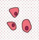
As animals evolved from single cells to multicellular organisms, individual cells took on specialized functions and became mutually dependent on each other to satisfy their own needs and the needs of the whole organism. Survival thus hinged on integration and coordination of their individual specialized functions. Increased specialization of cellul ar functions was accompanied by decreased tolerance for variations in the cellular environment. Control systems evolved that allowed more and more precise regulation of the cellular environment, which in turn permitted the development of even more highly specialized cells, such as those of higher brain centers, whose continued function requires that the internal environment be maintained constant within narrow limits, no matter what conditions prevail in the external environment. Survival of the individual requires a capacity to adjust and adapt to hostile conditions in the external environment, and survival of the species requires coordination of reproductive function with those factors in the internal and external environment that are most conducive to survival of the offspring. Crucial to meeting these needs for survival as a multicellular organism is the capacity of specialized cells to coordinate their activities through some sort of communication. Cells communicate with each other by means of chemical signals. These signals may be simple molecules such as modified amino or fatty acids, or they may be more complex peptides, proteins, or steroids. Communication takes place locally between cells within a tissue or
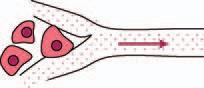
organ and at a distance to integrate the activities of cells or tissues in other organs. For communication between cells whose surfaces come in direct contact with each other, these signals may be substances that form part of the cell surface, or they may be molecules that pass from the cytosol of one cell to another through gap junctions. For communication with nearby cells and also between contiguous cells chemical signals are released into the extracellular fluid and reach their destinations by simple diffusion through extracellular fluid. Such communication is said to occur by paracrine, or local, secretion. Sometimes cells respond to their own secretions, and this is called autocrine secretion. For cells that are too far apart for the slow process of diffusion to permit meaningful communication, the chemical signal may enter the circulation and be transported in blood to all parts of the body. Release of chemical signals into the bloodstream is referred to as endocrine, or internal, secretion, and the signal secreted is called a hormone. We may define a hormone as a chemical substance that is released into the blood in small amounts and that, after delivery by the circulation, elicits a typical physiological response in other cells target cells (Figure 1.1). Often these modalities are used in combination such that paracrine and autocrine secretions provide local fine-tuning for events that are evoked by a hormonal signal that arrives from a distant source.
Because hormones are diluted in a huge volume of blood and extracellular fluid, achieving meaningful concentrations (10 10 to 10 7 M)
FIGURE 1.1 Chemical communication between cells. A: Local. Secretory product, shown as red dots, reaches nearby target cell by diffusion through extracellular fluid ( paracrine or autocrine communication).
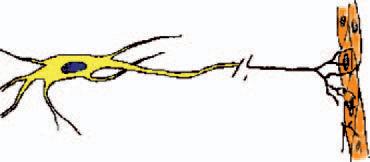
Juxtacrine: Communication by physical contact via signaling molecules in the membrane of one cell activating membrane receptor molecules in an adjacent cell. B: Endocrine. Secretory product reaches distant cells by transport through the circulation. C: Secretory product released from terminals of long cell processes reaches target cells distant from the nerve cell body by diffusion across the synaptic cleft.
It is only recently that endocrinologists have embraced the large number of locally produced hormone-like agents called growth factors and cytokines that regulate immune cell functions, cell division, differentiation, and even programmed cell death (apoptosis). These agents act locally in a paracrine or autocrine manner, but may also enter the circulation and affect the functions of distant cells, and hence behave as hormones. Many of these secretions produce effects that impinge upon actions of the classical hormones. Conversely, some of the classical hormones also act as local paracrine or autocrine factors and may be produced by cells that are unrelated to the endocrine glands that are usually associated with their secretion. Rapidly accumulating information about protein and gene structure has revealed relationships among these compounds, which can be grouped into families or superfamilies. Some hormones, such as growth hormone and prolactin, belong to the same superfamily of proteins as some of the cytokines, whereas the insulin-like growth factors are closely related to insulin. At the molecular level, production, secretion, and actions of cytokines and growth factors are no different from those of the classical hormones, and so our narrow definitions of endocrinology and hormones must be broadened to accommodate the wide range of communication by chemical messengers.
usually requires coordinated secretion by a mass of cells, an endocrine gland. The secretory products of endocrine glands are released into the extracellular space and diffuse across the capillary endothelium into the bloodstream, which gives rise to the terms ductless glands and internal secretion. In contrast, exocrine glands deliver their products through ducts to the outside of the body or to the lumen of the gastrointestinal tract. Classical endocrine glands include the pituitary, thyroid, adrenals, parathyroids, gonads, and islets of Langerhans. It has become apparent, however, that this list is far too short. Virtually every organ, including brain, kidney, heart, and even fat has an endocrine function in addition to its more commonly recognized role. Many aspects of gastrointestinal function are governed by hormones produced by cells of the gastric and intestinal mucosa, and so the intestinal tract is also an enormous endocrine gland. In fact, the word hormone was coined to describe a duodenal product, secretin (Chapter 6), that regulates pancreatic fluid secretion. Consequently, our notions of what constitutes an endocrine gland must be broadened to include any group of cells that secrete a hormone.
Another mechanism has also evolved to breach the distance between cells and allow rapid communication. Some cells developed the ability to release their signals locally from the tips of long processes that extend great distances to nearly touch their target cells. This mechanism, of course, is how nerve cells communicate with each other or with effector cells. By releasing their signals (neurotransmitters) so close to receptive cells, nerve cells achieve both exquisite specificity and economy in the quantity of transmitter needed to provide a meaningful concentration within a highly localized space, the synapse. Although use of the action potential to trigger secretion is not unique for nerve cells, the electrical wave that travels along the axons enables these cells to transmit information rapidly over great distances between the perikarya and the nerve terminals. Despite these specialized features of nerve cells, it is important to note that the same cellular mechanisms are used for signal production and release as well as for reception and response during neural, endocrine, and paracrine communication.
Distinctions between the various modes of communication are limited only to the means of signal delivery to target cells, and even these distinctions are blurred in some cases. Neurotransmitters act in a paracrine fashion on postsynaptic cells and in some cases may diffuse beyond the synaptic cleft to affect other nearby cells or may even enter the blood and act as hormones, in which case they are called neurohormones. Moreover, the same chemical signals may be secreted by both endocrine and nerve cells and even in very
small amounts by other cells that use them to communicate with neighboring cells in a paracrine or autocrine manner. Nature is parsimonious in this regard. Many peptides that are regarded as hormones or neurohormones may also serve as paracrine regulators in a variety of tissues. Although adequate to cause localized responses, the minute quantities of these substances produced extraglandularly are usually too small to enter the blood and interfere with endocrine relationships.
Clearly, the boundaries between endocrinology and other fields of modern biology are both artificial and imprecisely drawn. Endocrinology has benefited enormously from recent advances in other fields, particularly immunology, biochemistry, cell biology, and molecular biology. Early insights into endocrine function were gained from “experiments of nature.” Injury or inborn errors produced some pathological conditions that were traced to defects in hormone secretion or action. Conversely, hormone-secreting tumors or deranged regulatory mechanisms produced early insights into the consequences of excess hormone production. Early endocrinologists were able to create similar experiments by excising a gland or administering glandular extracts and observing the consequences. Progress in biochemistry made it possible to study pure hormones, and application of immunological techniques allowed identification and measurement of various molecular species.
The introduction of techniques of molecular biology brought breakthroughs in the understanding of hormone actions, and curiously brought us full circle back to the early approaches of studying the consequences of eliminating the source of a signaling molecule or administering an excess to gain insight into function. It is now possible to overexpress a hormone or other molecule by inserting its gene into developing mice to make them transgenic. Conversely it is possible to disrupt or “knock out ” a particular gene and study the consequences of the lack of its protein product(s) in otherwise intact mice. It is even possible to limit expression of transgenes to particular organs or cells and evoke their expression at desired stages of life. Similarly, it is now possible to knock out genes in particular organs and at particular times of life or to transiently interfere with their expression. In discussing hormone actions in subsequent chapters it will be necessary to refer to all these experimental techniques and many others.
In this text we concentrate on the integrating function of the endocrine system and focus our discussion principally on that aspect of cellular communication that is carried out by the classical endocrine glands and their hormones (Table 1.1). We first present some basic information about various endocrine glands and their hormones, and then consider interactions of hormones and the integration of endocrine function to produce homeostatic regulation. Such regulation throughout the body is achieved by regulation of cellular functions, which in turn are achieved by actions of hormones
TABLE 1.1 Endocrine Glands Considered in This Text
The Classical Endocrine Glands
Pituitary gland
Thyroid gland
Parathyroid gland
Islets of Langerhans
Adrenal glands
Gonads (testes and ovaries)
Placenta
Organs with Endocrine Functions
Brain
Heart
Liver
Gastrointestinal tract
Kidneys
Fat
WHOLE BODY LEVEL
Regulation and integration of:
Ionic and fluid balance
energy balance (metabolism) coping with the environment growth and development reproduction
MOLECULAR LEVEL
Regulation of:
Gene transcription protein synthesis and degradation enzyme activity protein conformation and protein:protein interactions
HORMONE ACTIONS
CELLULAR LEVEL
Regulation of:
Cell division differentiation death (apoptosis) motility secretion nutrient uptake
on molecules within those cells. We therefore consider the actions of hormones on all three levels (Figure 1.2).
Throughout this text, emphasis is on normal function, and reference to disease is limited to those aspects that are logical extensions of normal physiology or that facilitate understanding of normal physiology. Endocrine disease is not simply a matter of too much or too little hormone; rather, disease occurs when there is an inappropriate amount of hormone for the prevailing physiological situation or when there is an inappropriate response by target tissues to a perfectly appropriate amount of hormone. Some aspects of endocrine disease are still too poorly understood to be put in the context of normal physiology and are best left for a more detailed text of pathology or medicine.
Endocrinology is a subject that unfortunately involves a sometimes bewildering array of facts, not all of which can be derived from basic principles. To help organize and digest this necessarily large volume of material, the student might find the following outline of goals and objectives helpful.
FIGURE 1.2 Levels at which hormone actions are considered.
Goals and Objectives
A. The student should be familiar with
1. Essential features of feedback regulation
2. Essentials of competitive binding assays
B. For each hormone, the student should know:
1. Its cell of origin
2. Its chemical nature, including
a. Distinctive features of its chemical composition
b. Biosynthesis
c. Whether it circulates free or bound to plasma proteins
d. How it is degraded and removed from the body
3. Its principal physiological actions
a. At the whole body level
b. At the tissue level
c. At the cellular level
d. At the molecular level
e. Consequences of inadequate or excess secretion
4. What signals or perturbations in the internal or external environment evoke or suppress its secretion
a. How those signals are transmitted
b. How that secretion is controlled
c. What factors modulate the secretory response
d. How rapidly the hormone acts
e. How long it acts
f. What factors modulate its action
BIOSYNTHESIS OF HORMONES
The classical hormones fall into three categories ( Table 1.2):
● Derivatives of the amino acid tyrosine
● Steroids, which are derivatives of cholesterol
Epinephrine
Norepinephrine Estradiol Vasopressin
● Peptides/proteins, which comprise the largest and most diverse class of hormones
Many small molecules, including nitric oxide, derivatives of amino acids and fatty acids, function as neurotransmitters or paracrine signals, but usually are not considered to be hormones, and so are discussed only when pertinent to the actions of hormones. Relevant details of hormone synthesis and storage, particularly for the amino acid and steroid hormones, are presented with the discussion of their glands of origin, but steps in biosynthesis, storage, and secretion common to all protein and peptide hormones are sufficiently general for this largest class of hormones to warrant some discussion here. A brief review of these steps also provides an opportunity for a general consideration of gene expression and protein synthesis, and provides some background for understanding hormone actions. In-depth consideration of these complex processes is beyond the scope of this text, and is best left for the many excellent texts of cellular and molecular biology.
Protein and peptide hormones are encoded in genes, with each hormone usually represented only once in the genome. Information determining the amino acid sequence of proteins is encoded in the nucleotide sequence of DNA (deoxyribonucleic acid) (Figure 1.3). Nucleotides in DNA consist of a five-carbon sugar, deoxyribose, in ester linkage with a phosphate group, and attached in N-glycosidic linkage to one of four organic bases: adenine (A), guanine (G), thymidine (T), or cytidine (C). The ability of the purine bases A and G to form complementary pairs with the pyrimidine bases T and C (Figure 1.4), respectively, on an
Glucagon
Dopamine Progesterone Angiotensin Adrenocorticotropic hormone
Triiodothyronine Cortisol Melanocyte-stimulating hormone Thyroid-stimulating hormone
Thyroxine Aldosterone Somatostatin
Secretin Vitamin D Thyrotropin-releasing hormone Motilin
Gastrin Follicle-stimulating hormone
Cholecystokinin
Luteinizing hormone
Gonadotropin-releasing hormone
Growth hormone
Prolactin
Corticotropin-releasing hormone
Growth hormone-releasing hormone
Parathyroid hormone
Calcitonin
Chorionic gonadotropin
Choriosomatomammotropin
TABLE 1.2 Chemical Nature of the Classic Hormones
FIGURE 1.3 Composition of DNA. DNA is a polymer of the five-carbon sugar, deoxyribose, in diester linkage with phosphate forming ester bonds with hydroxyl groups on carbons 3 and 5 on adjacent sugar molecules. The purine and pyrimidine bases are linked to carbon 1 of each sugar. The numbering system for the five carbons of deoxyribose are shown at the top of the figure. The chemical bonds forming the backbone of the DNA chain are highlighted in blue. The 5’ or 3’ ends refer to the carbons in deoxyribose.
adjacent strand of DNA is the fundamental property that permits accurate replication of DNA and transmission of stored information from generation to generation. A single strand of DNA consists of a chain of millions of nucleotides linked by phosphate groups that form ester bonds with hydroxyl groups at carbon 3 of one deoxyribose and carbon 5 of the next deoxyribose. The DNA in each chromosome is present as a pair of long strands oriented in opposite directions and is organized into nucleosomes, each of which consists of a stretch of about 180 nucleotides tightly wound around a complex of eight histone molecules. The nucleosomes are linked by stretches of about 30 nucleotides, and the whole double strand of nucleoproteins is tightly coiled in a higher order of organization to form the chromosomes. Instructions for protein structure are transmitted from the DNA to cytoplasmic sites of protein synthesis, the
FIGURE 1.4 Complementary base pairing by the formation of hydrogen bonds between thymine and adenine and between cytosine and guanine. RNA contains uracil in place of the thymine found in DNA. Uracil and thymine differ in structure only by the presence of the methyl group (CH3) found in thymine.
ribosomes, in the messenger ribonucleic acid (mRNA) template. RNA differs in structure from DNA only in having ribose instead of deoxyribose as its sugar and uridine (U) instead of thymidine as one of its pyrimidine bases. The nucleotide sequence of the mRNA precursor is complementary to the nucleotide sequence of DNA. Messenger RNA synthesis proceeds linearly from an upstream “start site” designated by a particular sequence of nucleotides in DNA in a process called transcription. The start site is located downstream from the promoter region, which contains sequences to which regulatory proteins can bind, and a short sequence where RNA polymerase II and a large aggregate of proteins, the general transcription complex, binds. The DNA that is transcribed is comprised of segments that encode structural and regulatory information called exons separated by intervening sequences of DNA with no coding function, called introns (Figure 1.5).
Transcription is regulated by nuclear proteins called transcription factors or transactivating factors, which bind to regulatory sites that are usually upstream from the promoter and stimulate or repress gene transcription. These proteins form complexes with multiple other transcription factors and proteins called coactivators or corepressors, which not only govern attachment and activity of the general transcription complex, but control the “tightness” of the DNA coil and hence the accessibility of genes to the transcription apparatus. Transcription proceeds from the start site through the introns and exons and a downstream flanking sequence
and coactivators
FIGURE 1.5 Transcription and RNA processing. The DNA strand contains all the stored information for expression of the gene including the promoter, distant regulatory elements (not shown), binding sites (response elements) for regulatory proteins, and the coding for the sequence of the protein (exons) interrupted by intervening sequences of DNA (introns). Exons are numbered 1–5. The primary RNA transcript contains the complementary sequence of bases coupled to a poly A tail at the 3’ end and a methyl guanosine cap at the 5’ end. Removal of the introns and splicing the remaining exons together produces the messenger RNA that contains all the information needed for translation, including the codons for the amino acid sequence of the protein and untranslated regulatory sequences at both ends.
FIGURE 1.6 Alternative splicing of mRNA can give rise to different proteins. Numbers indicate exons. Exon 1 is untranslated. N amino terminus; C carboxyl terminus.
where a long polyadenine (polyA) tail is added. A special “cap” structure containing methylated guanosine added to the opposite end of the RNA transcript permits its export from the nucleus after it is modified further by removal of the introns and attachment of the exons to each other in a process called splicing. Under some circumstances the splicing reactions may bypass some exons or parts of exons, which are then omitted from the final mRNA transcript. Because of such alternate splicing, a single gene can give rise to more than one mRNA transcript, and hence more than one protein product (Figure 1.6). Multiple mRNA transcripts may also be produced from some genes that have more than one site at which transcription can start. Transcription
Upon export from the nucleus, the mRNA transcripts attach to ribosomes where they are translated into protein (Figure 1.7). Ribosomes are large complexes of RNA and protein enzymes that “read ” the mRNA code in triplets of nucleotides called codons. The translation initiation site begins with the codon for methionine. Each codon designates a specific amino acid. Triplets of complementary nucleotides (anticodons) are found in small RNA molecules called transfer RNA (tRNA), each of which binds a particular amino acid and delivers it to the ribosome. Alignment of amino acids in the proper sequence is achieved by the complementary pairing of anticodons in the tRNA with codons in the mRNA. The tRNA thus delivers the correct amino acid to the carboxyl terminus of the growing peptide chain and holds it in position so that ribosomal enzymes can release it from the tRNA and link it to the peptide. Once the peptide bond is formed, the empty tRNA is released and the ribosome moves down the mRNA to the next codon where the next tRNA molecule charged with its amino acid waits to bind to its complementary codon. Elongation of the chain continues until the ribosome reaches a “stop” codon at which time it dissociates from the mRNA. As each ribosome moves down the mRNA, other ribosomes attach behind them to repeat the process. In this way a single mRNA molecule may be translated over and over again to yield many copies of a protein before it is degraded.
AAGACG
FIGURE 1.7 Translation. A molecule of transfer RNA (tRNA) charged with its specific amino acid, phenylalanine, and already linked to the growing peptide chain, is positioned on the mRNA by complementary pairing of its triplet of nucleotides with its codon of three nucleotides in the mRNA. A second molecule of tRNA charged with its specific amino acid, tryptophan, has docked at the adjacent triplet of nucleotides and awaits the action of ribosomal enzymes to form the peptide bond with phenylalanine. Linking the amino acid to the peptide chain releases it from its tRNA and allows the empty tRNA to dissociate from the mRNA. A third molecule of tRNA, which brought the preceding molecule of leucine, is departing from the left, while a fourth molecule of tRNA, carrying its cargo of glutamine, arrives from the right and waits to form the complementary bonds with the next codon in the mRNA that will bring the glutamine in position to be joined to tryptophan at the carboxyl terminus of the peptide chain. The ribosome moves down the mRNA adding one amino acid at a time until it reaches a stop codon. (Adapted from Alberts et al. (1994) Molecular Biology of the Cell New York: Garland Publishing.)
Protein and peptide hormones are synthesized as larger molecules (prohormones and preprohormones) than the final secretory product. Proteins destined for secretion have a hydrophobic sequence of 12 to 30 amino acids at their amino terminals (Figure 1.8). This signal sequence is recognized by a special structure that directs the growing peptide chain through a protein channel in the endoplasmic reticular membrane and into the cisternae of the endoplasmic reticulum. Postsynthetic processing begins in the endoplasmic reticulum and continues as hormone precursors are translocated to the Golgi apparatus for final processing and packaging for export. Processing begins even as the peptide chain is still elongating, and includes cleavage to remove the signal peptide. Interactions with intrinsic endoplasmic reticulum proteins facilitate proper folding and catalyze formation of disulfide bonds linking cysteine residues. Other processing of peptide hormones may include glycosylation (addition of carbohydrate chains to asparagine residues) or coupling of subunits that are products of different genes, as seen with the pituitary glycoproteins (see Chapter 2). Glycosylation begins in the endoplasmic reticulum and is completed in the Golgi complex, but final processing of peptide chains takes place in the secretory granules.
FIGURE 1.8 Post-translational processing. The leader sequence or signal peptide of proteins destined for secretion enters the cisternae of the endoplasmic reticulum even as peptide elongation continues. In the endoplasmic reticulum (1) the leader sequence is removed, (2) the protein is folded with the assistance of protein chaperons, (3) sulfhydryl bridges may form, and (4) carbohydrate may be added (glycosylation). The partially processed protein (5) is then entrapped in vesicles that bud off the endoplasmic reticulum and (6) fuse with the Golgi apparatus, where glycosylation is completed, and (7) the protein is packaged for export in secretory vesicles in which the final stages of processing take place.
The hormones, along with trypsin-like peptidases called hormone convertases, carboxypeptidase, amidating enzymes, and other peptide processing enzymes, are packaged into immature secretory vesicles that bud off from the Golgi stacks. Other proteins that are incorporated into secretory vesicles include one or more proteins of the family of secretogranins. These large acidic proteins contribute to the sorting of hormone into the immature vesicles, facilitate cleavage reactions at appropriate sites in the prohormone, and organize condensation of the hormone and associated proteins into dense granules. Proton pumps in the vesicle membrane acidify vesicular fluid, which activates convertases and promotes extrusion of water. Cleavage of the prohormones removes those amino acid sequences that may have functioned to target the peptides to the secretory granules or to orient folding of the molecule so that disulfide bridges form in the right places. Cleavage of the prohormones by hormone convertases may yield more than one biologically active peptide from a single precursor, as seen with the ACTH and glucagon families of hormones (see Chapters 2 and 6). Amidation of the carboxyl terminus using glycine as the amino donor is a common feature of the final maturation of many hormones. Because these processing reactions take place in secretory granules, peptide fragments, enzymes