The Aestheticians Journal Digital November '2025 Issue
Q-Switched
Nd:YAG Laser in Hyperpigmentation: Safety, Efficacy and Clinical Applications in Darker Skin Phototypes
Optimizing
Outcomes
in Pattern Hair Loss: Clinical Insights into Implanter-Assisted Hair Transplantation
Management of Facial Melasma and Skin Aging with Glutathione and Red Orange Complex (ROC) Therapy— A Combined Multimodal Approach
The Use of Novel Regenerative Molecules in Aesthetic Dermatology: Exosomes, PDRN, Polynucleotides and Hyaluronic Acid-Based Micro-Filler in Clinical Practice
Chemical Peel for Hyperpigmentation Part-II
EXECUTIVE EDITOR & PUBLISHER
Dom Daniel CORPORATE OFFICE
22, Shreeji Bhavan, 275-279, Samuel Street, Masjid Bunder (W), Mumbai-4000 03, INDIA.
EMAIL: theaestheticiansjournalindia@gmail.com
Website: theaestheticiansjournal.com
Printed, Published, Edited and Owned by Dom Daniel
Printed at Swastik Printer, Gala No.9 & 10, Vishal Industrial Estate, Bhandup (West), Mumbai- 400078. Published at 22 Shreeji Bhavan, 275/279, Samuel Street, Masjid Bunder (West), Mumbai - 400003. India.
“The Aestheticians Journal” takes no responsibility for unsolicited photographs or material
ALL PHOTOGRAPHS, UNLESS OTHERWISE INDICATED, ARE USED FOR ILLUSTRATIVE PURPOSE ONLY.
Views expressed in this Journal are those of the contributors and not of the publisher. Reproduction in whole or in parts of texts or photography is prohibited. Manuscripts, Photographs and art are selected at the discretion of the publisher free of charge (advertising excluded). Whether published or not, no material will be returned and remains the property of the publishing house, which may make use of it as seen fit. This may include the withdrawal of publication rights to other publishing houses.
All rights reserved. Reproducing in any manner without prior written permission prohibited.
Published for the period of November -2025
The Art and Science of Modern Aesthetics
This era of Aesthetic Dermatology is defined by continuous innovation—where scientific advancement seamlessly merges with artistry to enhance both appearance and confidence. Emerging technologies and novel therapeutic approaches are transforming dermatologic practice, pushing the boundaries of non-surgical rejuvenation and regenerative care.
In this issue, we present a collection of insightful, evidence-based articles highlighting recent advancements in aesthetic dermatology. The feature article, “Management of Facial Melasma and Skin Aging with Glutathione, Red Orange Complex (ROC) Therapy—A Combined Multimodal Approach,” emphasizes synergistic antioxidant therapy. “The Use of Novel Regenerative Molecules in Aesthetic Dermatology” explores exosomes, PDRN, polynucleotides, and hyaluronic acid-based microfillers for skin rejuvenation. “Optimizing Outcomes in Pattern Hair Loss” discusses implanter-assisted hair transplantation for superior results. “Q-Switched Nd:YAG Laser in Hyperpigmentation” and “Chemical Peels for Hyperpigmentation” offer practical insights for safe pigment correction and tone enhancement, reflecting precision, innovation, and scientific excellence in aesthetic dermatology.
The Aestheticians Journal remains dedicated to showcasing the dynamic intersection of science and aesthetics—continuously advancing the field and inspiring Dermatologist to refine their practice with innovation, expertise, and artistry.
Hope you have a great read!
Thanks & Cheers
- Dom Daniel
Executive Editor & Publisher
08 Case Study: Management of Facial Melasma and Skin Aging with Glutathione and Red Orange Complex (ROC) Therapy— A Combined
Multimodal Approach
Dr. Rekha Singh, MD, DVL
Editorial Board
Dr. Rekha Singh
MD, DVL
Consultant Dermatologist
Oliva Skin & Hair Clinic
Hyderabad
Dr. Dipak Patel
MD, DVD
Aesthetic Dermatologist & Cosmetologist
Neel Aesthetics
Surat
Dr. Komal Jerath
MD (Dermatology)
Cosmetic Dermatologist and Trichologist
Komal Skin and Laser Clinic
Amritsar, Punjab
Dr. Gaurav Nakra
MD (Dermatology)
Consultant Dermatologist
Centre for Skin
Delhi
Dr. Shalini Malhotra
MBBS, DNB (Dermatology)
MNAMS, ABHRS
Hair Transplant Surgeon and Consultant Dermatologist
SM Hair Restoration & Aesthetic Clinic
New Delhi
Case Study: Management of Facial Melasma and Skin Aging with Glutathione and Red Orange Complex (ROC) Therapy— A Combined Multimodal Approach
Dr. Rekha Singh MD, DVL
Consultant Dermatologist
Oliva Skin & Hair Clinic
Hyderabad
Introduction
Skin aging and hyperpigmentation are interrelated processes that share common molecular pathways, primarily driven by oxidative stress and melanogenic overactivity. Excessive generation of reactive oxygen species (ROS) accelerates collagen degradation, disrupts dermal matrix integrity, and stimulates melanogenesis, resulting in uneven tone, laxity, and textural deterioration. Targeting oxidative stress through integrated antioxidant and cellular renewal strategies has therefore become a cornerstone of modern aesthetic dermatology. The synergistic use of antioxidants and vitamins helps reduce pigment formation, prevent premature cellular senescence, and promote collagen
The rising demand for skinlightening and anti-aging therapies has brought glutathione, a tripeptide with potent antioxidant and melanogenesis-regulating ...... properties, into the spotlight. Glutathione neutralizes free radicals and inhibits melanin synthesis by shifting the balance of pigment production from eumelanin toward pheomelanin, resulting in a brighter and more uniform complexion. It is also a proven anti-aging agent that enhances epidermal turnover and stimulates collagen synthesis, leading to visible skin renewal with improved texture and elasticity. The co-administration of vitamin C and other antioxidants further augments these effects
by protecting against UVinduced oxidative damage and preserving dermal integrity. Notably, red orange complex and glutathione exhibit synergistic antioxidant activity, as each regenerates the other in its reduced form, thereby amplifying their overall protective and rejuvenating potential.6,7
This case study evaluates the efficacy of a glutathione and red orange complex in demelanising and anti-aging therapy, demonstrating excellent clinical outcomes.
Case Presentation 1
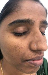
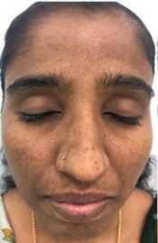
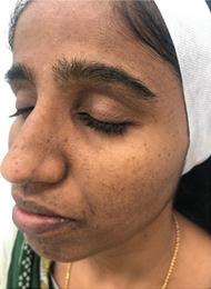
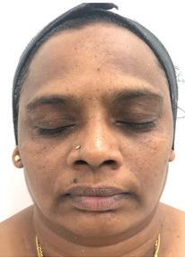
A 28-year-old female presented with progressive facial pigmentation for the past 1.5 years, primarily involving the cheeks, nose, and forehead. The pigmentation intensified following prolonged sun exposure and during the summer months. The patient reported occasional dullness and uneven skin tone but denied any itching, burning or scaling. She had no history of thyroid dysfunction, hormonal therapy, or recent pregnancy. Her menstrual cycles were regular. There was no significant family history of melasma. She had been using overthe-counter fairness creams intermittently without noticeable improvement. On cutaneous examination, the patient had Fitzpatrick Skin Type IV. Lesions were distributed bilaterally over the malar region, nose bridge, and forehead. The lesions presented as irregularly shaped hyperpigmented macules with
mild background tanning. The skin surface appeared mildly coarse, without any signs of inflammation or scaling. No associated findings such as acne, erythema, or post-inflammatory hyperpigmentation were observed. Based on the clinical features—ill-defined, light to dark brown macules symmetrically distributed over the malar area and forehead—the diagnosis of epidermal melasma with associated tanning was made.
Treatment Plan
A combined antioxidant and cellular renewal regimen was initiated, aiming to restore redox balance and enhance dermal remodeling:
1. Oral therapy:
o Glutathione: 500 mg/day in divided doses for its depigmenting and antioxidant effects through inhibition of tyrosinase and melanin synthesis.
o Red Orange Complex (ROC) combats photoaging by reducing oxidative stress and inflammation, inhibiting melanogenesis and collagen degradation, and restoring collagen synthesis for improved skin health and radiance.
2. Topical Therapy
• Retinol Cream (Night Application): To enhance epidermal turnover and promote dermal collagen regeneration.
• Broad-Spectrum Sunscreen (SPF 50+): Reapplied every 3–4 hours during the day to prevent UV- and visible light–induced melanogenesis.
• Moisturizing Antioxidant Serum (Morning): Containing Vitamin C and botanical polyphenols to combat oxidative stress and provide photoprotection.
3. Lifestyle Modifications
• Adherence to strict photoprotection with the use of widebrimmed hats and physical barriers.
• Maintenance of adequate hydration and consumption of an antioxidant-rich diet (fruits, vegetables, and green tea).
• Avoidance of irritants (harsh scrubs, chemical peels) and unnecessary exfoliation.
Follow-Up and Treatment Outcome
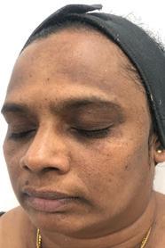
The patient was followed up every 4 weeks over a 12-week period, showing progressive improvement throughout the treatment duration.
• At 4 weeks:
o Noticeable reduction in pigmentation intensity and facial tan
o Improved skin hydration.
• At 8 weeks:
o Smoother skin texture.
o Improved overall skin tone uniformity and luminosity.
• At 12 weeks:
o Marked reduction in pigmentation.
o Restored luminosity with visibly improved skin firmness and elasticity.
• Photographic Evidence:
o Before-and-after images
demonstrated significant improvement in melasma and facial tanning.
o Enhanced skin clarity and texture observed.
• Safety and Tolerability:
o No adverse effects such as irritation or post-inflammatory changes were reported.
• Patient Feedback:
o The patient expressed high satisfaction with the overall treatment outcome.
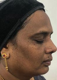
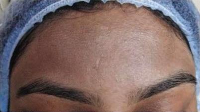
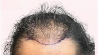
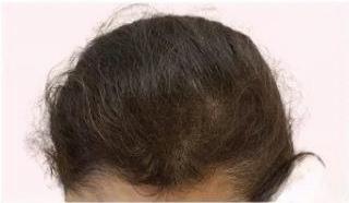
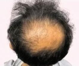
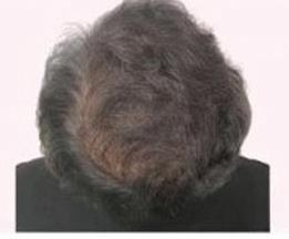
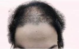
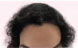
After treatment
12
Figure 1: Clinical results after
weeks of consistent use of glutathione and red orange complex showing noticeable reduction in pigmentation intensity, facial tan and improved skin texture. Before treatment Case Study: Management
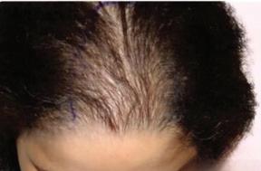
Case Presentation 2
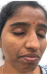
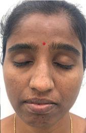
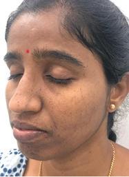
A 50-year-old menopausal woman presented with complaints of gradually progressive facial pigmentation and dullness persisting for the past two years. She reported frequent sun exposure and inconsistent use of sunscreen. There was no history of hormonal therapy, systemic illness, or topical steroid application. Her primary concern was pigmentation involving the malar and forehead regions, associated with loss of skin luminosity and uneven texture. Based on clinical findings and history, a diagnosis of Acanthosis Nigricans with Melasma was made. On examination, the patient had Fitzpatrick Skin Type IV. Symmetrical hyperpigmented patches were observed over the forehead, malar regions, and upper lip, accompanied by mild background tanning. The skin appeared coarse with a dull texture and reduced elasticity. Additionally, subtle velvety hyperpigmentation with skin thickening was noted over the neck folds, suggestive of acanthosis nigricans. No erythema, active inflammation, or postinflammatory hyperpigmentation was evident. The pattern and distribution of pigmentation were consistent with chronic ultraviolet exposure and oxidative stress–induced dermal damage, compounded by metabolic and hormonal influences contributing to acanthosis nigricans.
Treatment Protocol
A comprehensive multimodal treatment plan was initiated targeting both melanogenesis and the underlying metabolic component, with emphasis on antioxidant protection, dermal renewal, and lifestyle optimization:
• Oral Glutathione: Administered for its dual role in inhibiting melanin synthesis, combating oxidative stress and providing potent antioxidant effects.
• Red Orange Complex (ROC): Mechanistically, ROC reduces inflammation, regulates melanogenesis, restores collagen synthesis, and improves skin firmness and integrity.
• Topical Depigmenting and Antioxidant Agents: Formulated to suppress melanogenesis, neutralize free radicals, and improve overall skin barrier function.
• Topical Retinol (Night Application): Introduced gradually to accelerate epidermal turnover, refine texture and stimulate dermal remodeling.
• Broad-Spectrum .......... Sunscreen (SPF 50+): Advised for daily use with frequent reapplication to prevent UV-induced pigmentation recurrence.
• Lifestyle and Metabolic Management: Dietary modifications, weight reduction, and incorporation of regular physical activity
were emphasized.
• Insulin Sensitizers: Considered to address underlying insulin resistance contributing to acanthosis nigricans.
Follow-Up and Treatment Outcome
The patient was monitored at four-week intervals over a 12-week period. Gradual yet consistent improvement was observed throughout the treatment course. After the first month, a visible decrease in pigmentation intensity and facial tanning was noted, accompanied by enhanced hydration and smoother appearance. By the eighth week, the complexion had become more uniform, with a noticeable boost in luminosity and refinement of skin texture.
At the end of 12 weeks, there was a marked reduction in melasma pigmentation, restoration of skin radiance, and perceptible improvement in firmness and elasticity. The transformation was clearly evident in the before-andafter clinical photographs, which demonstrated significant lightening of hyperpigmented areas and improved overall clarity. The treatment was well tolerated, with no reports of irritation, erythema, or post-inflammatory changes. The patient expressed high satisfaction with both the aesthetic and textural outcomes achieved through the therapy.
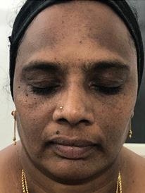
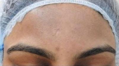
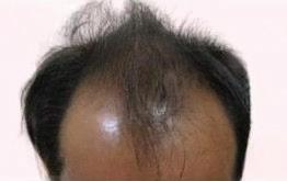
After treatment
Figure 2: At the end of 12 weeks, there was a marked reduction in melasma pigmentation, restoration of skin radiance, and perceptible improvement in firmness and elasticity.
Discussion
Melasma is a common acquired hypermelanosis of the face, characterized by symmetric, reticulated hyperpigmented macules and patches, predominantly over the centrofacial and malar regions. It is more prevalent among females and individuals with darker Fitzpatrick skin types. Multiple etiological factors contribute to its pathogenesis. Histopathologically, melasma demonstrates increased epidermal and/or dermal melanin, melanocyte hyperactivity, and solar elastosis—features that closely resemble those seen in photoaged skin. The overlapping mechanisms of melanogenesis and photoaging highlight oxidative stress as a central pathogenic factor linking both conditions. Skin aging is a multifactorial biological process influenced by intrinsic factors
such as genetic and hormonal alterations, and extrinsic factors including UV radiation, pollution, and lifestyle stressors. These factors collectively induce the overproduction of reactive oxygen species (ROS), leading to DNA damage, lipid peroxidation, and the degradation of collagen and elastin fibers within the dermal matrix. Clinically, this manifests as wrinkles, laxity, volume loss,
Before treatment
Case Study: Management of Facial Melasma and Skin Aging with Glutathione and Red Orange Complex (ROC) Therapy—
and uneven pigmentation. Because oxidative stress underlies both pigmentation and aging, antioxidant-based therapies have emerged as a rational and effective treatment strategy.8,9,10,11
Mechanisms and Therapeutic Role of Key Agents
Glutathione has gained global recognition as a potent antioxidant and skin-lightening molecule. In its reduced form (GSH), glutathione directly scavenges free radicals, inhibits lipid peroxidation, and downregulates melanogenesis by interfering with tyrosinase activity—the rate-limiting enzyme in melanin synthesis. Furthermore, it shifts melanin production from the dark eumelanin toward the lighter pheomelanin pathway, resulting in gradual depigmentation and skin brightening. Glutathione also participates in redox cycling, maintaining cellular homeostasis and enhancing tissue repair. It can be administered via oral, topical, or parenteral routes, with oral and topical formulations demonstrating significant improvements in skin tone and oxidative balance upon consistent use.11,12,13
Red Orange Complex (ROC) is a potent antioxidant formulation derived from red oranges, naturally enriched with polyphenols and vitamin C. It contains a high concentration of anthocyanins, hydroxycinnamic acids, flavanones, and ascorbic acid, which together provide strong free radical–scavenging activity. This unique phytocomplex helps combat oxidative stress, thereby protecting and revitalizing the skin during the aging process. Clinical studies have demonstrated that ROC effectively reduces hyperpigmentation, enhances skin tone and hydration, and minimizes the appearance of fine lines and wrinkles, making it a valuable natural adjunct for managing melasma and photoaging.14 Vitamin C complements glutathione by supporting its regeneration and amplifying its antioxidant effects. It inhibits melanogenesis by interacting with copper ions at the tyrosinase active site, thereby reducing melanin formation. Vitamin C further protects skin structures from UVinduced ROS damage, stimulates fibroblast activity, and promotes collagen synthesis by hydroxylation of proline and lysine residues. Its additional anti-inflammatory and photoprotective effects make it a valuable adjunct in both melasma management and anti-aging protocols.8,15
Retinol (Vitamin A derivative) is one of the most extensively studied and widely used topical agents for anti-aging and pigmentation disorders. It enhances epidermal turnover, facilitates even melanin dispersion, and stimulates dermal fibroblasts to produce collagen and elastin. Retinol also downregulates matrix metalloproteinases (MMPs), particularly MMP-1, which are responsible for collagen degradation during photoaging. Compared to its active metabolite, tretinoin, retinol offers similar benefits with a lower risk of irritation, making it suitable for long-term maintenance therapy.8
Combined Approach
The combination of glutathione, red orange complex and retinol offers a synergistic therapeutic effect by addressing
multiple pathogenic pathways simultaneously—antioxidant defense, inhibition of melanogenesis, and dermal remodeling. Glutathione and ROC restore redox balance and regulate melanin production, while retinol promotes epidermal renewal and collagen synthesis. Collectively, these agents not only lighten hyperpigmentation but also enhance skin texture, firmness and radiance. Additionally, Red Orange Complex (ROC), rich in anthocyanins, polyphenols, and vitamin C, further strengthens this approach by combating oxidative stress and downregulating matrix metalloproteinases (MMPs) to preserve collagen integrity and prevent photoaging.
In the present case, consistent use of this combination over 12 weeks resulted in marked improvement in pigmentation and visible rejuvenation, reflecting the efficacy of an integrated antioxidant and cellular renewal regimen. This multimodal approach represents a safe, effective, and evidencebased strategy for managing melasma and age-related skin changes, especially in patients seeking both depigmentation and rejuvenation outcomes.
Conclusion
The combination of glutathione, red orange complex and retinol proved effective in addressing both hyperpigmentation and early signs of aging. By targeting oxidative stress and enhancing epidermal renewal, this multimodal regimen achieved significant clinical improvement within 12 weeks, demonstrating its efficacy and safety as an
integrated demelanising and anti-aging therapy. A multimodal approach combining oral antioxidants, topical retinoids, and rigorous photoprotection offers a safe and effective option for managing melasma and facial tanning. Regular follow-up and patient education on maintenance therapy are crucial for sustained results.
References
1. Hussen, N. H. A., Abdulla, S. K., Ali, N. M., Ahmed, V. A., Hasan, A. H., & Qadir, E. E. (2025). Role of antioxidants in skin aging and the molecular mechanism of ROS: A comprehensive review. Aspects of Molecular Medicine, 5, 100063. https:// doi.org/10.1016/j.amolm.2025.100063
2. Chaudhary, M., Khan, A., & Gupta, M. (2020). Skin Ageing: Pathophysiology and Current Market Treatment Approaches. Current aging science, 13(1), 22–30. https://doi.org/10.2174/15672050166 66190809161115
3. Zhang, S., & Duan, E. (2018). Fighting against Skin Aging: The Way from Bench to Bedside. Cell transplantation, 27(5), 729–738. https://doi. org/10.1177/0963689717725755
4. Thawabteh, A. M., Jibreen, A., Karaman, D., Thawabteh, A., & Karaman, R. (2023). Skin Pigmentation Types, Causes and Treatment-A Review. Molecules (Basel, Switzerland), 28(12), 4839. https://doi. org/10.3390/molecules28124839
5. Nautiyal A, Wairkar S. Management of hyperpigmentation: Current treatments and emerging therapies. Pigment Cell Melanoma Res. 2021; 34: 1000–1014. https://doi.org/10.1111/pcmr.12986
6. Alzahrani, T. F., Alotaibi, S. M., Alzahrani, A. A., Alzahrani, A. F., Alturki, L. E.,
Alshammari, M. M., Alharbi, R. A., Alanazi, S. I., Alshammari, W. Z., & Algarni, A. S. (2025). Exploring the Safety and Efficacy of Glutathione Supplementation for Skin Lightening: A Narrative Review. Cureus, 17(1), e78045. https://doi.org/10.7759/ cureus.78045
7. Lee, E., Park, H. Y., Kim, S. W., Kim, J., & Lim, K. (2023). Vitamin C and glutathione supplementation: a review of their additive effects on exercise performance. Physical activity and nutrition, 27(3), 36–43. https:// doi.org/10.20463/pan.2023.0027
8. Ogbechie-Godec, O. A., & Elbuluk, N. (2017). Melasma: an Up-to-Date Comprehensive Review. Dermatology and therapy, 7(3), 305–318. https://doi. org/10.1007/s13555-017-0194-1
9. Jo JY, Chae SJ, Ryu HJ. Update on Melasma Treatments. Ann Dermatol. 2024 Jun;36(3):125-134. https://doi. org/10.5021/ad.23.133
10. Ganceviciene, R., Liakou, A. I., Theodoridis, A., Makrantonaki, E., & Zouboulis, C. C. (2012). Skin anti-aging strategies. Dermato-endocrinology, 4(3), 308–319. https://doi.org/10.4161/ derm.22804
11. Sonthalia, S., Jha, A. K., Lallas, A., Jain, G., & Jakhar, D. (2018). Glutathione for skin lightening: a regnant myth or evidence-
based verity?. Dermatology practical & conceptual, 8(1), 15–21. https://doi. org/10.5826/dpc.0801a04
12. Mohan S, Mohan L, Sangal R, Singh N. Glutathione for skin lightening for dermatologists and cosmetologists. Int J Res Dermatol 2020;6:284-7.
13. Sharma DK, Sharma P. Augmented Glutathione Absorption from Oral Mucosa and its Effect on Skin Pigmentation: A Clinical Review. Clin Cosmet Investig Dermatol. 2022;15:1853-1862. https:// doi.org/10.2147/CCID.S378470
14. Kim, Y. H., Lim, C. Y., Jung, J. I., Kim, T. Y., & Kim, E. J. (2023). Protective effects of red orange (Citrus sinensis [L.] Osbeck [Rutaceae]) extract against UVA-B radiation-induced photoaging in Skh:HR-2 mice. Nutrition research and practice, 17(4), 641–659. https://doi. org/10.4162/nrp.2023.17.4.641
15. Sanadi, R. M., & Deshmukh, R. S. (2020). The effect of Vitamin C on melanin pigmentation - A systematic review. Journal of oral and maxillofacial pathology : JOMFP, 24(2), 374–382. https://doi. org/10.4103/jomfp.JOMFP_207_20
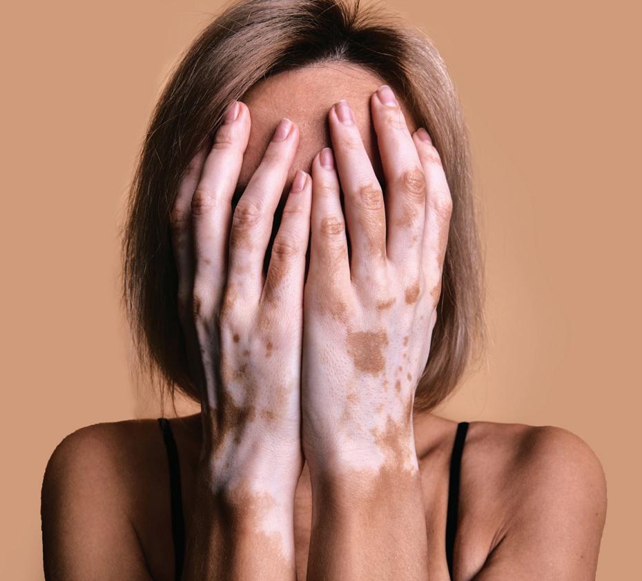
Duration of Vitiligo is Associated with the Severity of Depression
Emotional distress, including anxiety, depression, and stress, is common among individuals with vitiligo and can negatively impact disease perception and treatment adherence. While physical activity (PA) is known to improve psychological outcomes in many chronic conditions, its effect in vitiligo has been unclear. A recent cross-sectional study assessed the association between PA and psychological symptoms in a group of vitiligo patients. Using validated versions of the DASS-21 and IPAQ, alongside the Vitiligo Area Scoring Index (VASI), the study found high rates of psychological distress—more than half of the participants reported stress and depression, and nearly half reported anxiety. However, PA levels showed no significant correlation with psychological measures. Anxiety correlated weakly with disease severity, and longer disease duration was associated with higher depression scores. PA levels were higher in males, while other demographic and clinical variables showed no significant associations. Despite known mental health benefits of exercise, the lack of effect in vitiligo may reflect the condition’s unique psychosocial burden, including stigma and altered self-image, as well as possible neuroinflammatory mechanisms. These findings highlight the need for integrated mental health support in vitiligo management, as lifestyle modification alone may be insufficient to address the psychological comorbidities of the disease.
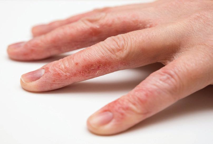
Evaluating Dose-Dependent Skin Changes from Long-Term Isotretinoin Therapy, Including CHE
A recent retrospective study examining oral isotretinoin use for acne over a five-year period primarily identified dermatologic adverse effects, with skin dryness being the most common. This included conditions such as chronic hand eczema, xerosis, cheilitis, and retinoid dermatitis. These dryness-related side effects were dose-dependent, with higher isotretinoin doses associated with a greater incidence of symptoms. Notably, these adverse effects typically manifested early in the treatment course, often within the first week, and peaked around the fourth week of therapy. Age also played a role in the presentation of side effects; younger patients were more susceptible to retinoid dermatitis, whereas older individuals experienced more pronounced desquamation. Although statistically significant, these age-related differences were relatively modest in clinical impact. In addition to cutaneous effects, the study observed metabolic changes, including lipid profile disturbances, particularly elevated LDL cholesterol and thyroid-stimulating hormone levels, which became apparent by the fourth week of treatment and were more prominent among younger patients receiving higher doses of isotretinoin. Despite these findings, isotretinoin was generally well tolerated at moderate cumulative doses. Routine monitoring of lipid and hormonal parameters during therapy is necessary, alongside dose individualization based on patient response and tolerance. Furthermore, early and proactive management of skin dryness using emollients is recommended to mitigate adverse dermatologic effects and improve overall treatment adherence and outcomes.
The Use of Novel Regenerative Molecules in Aesthetic Dermatology: Exosomes, PDRN, Polynucleotides and Hyaluronic Acid-Based Micro-Filler in Clinical Practice
Dr. Komal Jerath
MD (Dermatology)
Cosmetic Dermatologist and Trichologist
Komal Skin and Laser Clinic
Amritsar, Punjab
Abstract
Aesthetic dermatology increasingly adopts regenerative molecules to restore skin health rather than only cosmetic correction. Agents including exosomes (human- and plant-derived), polydeoxyribonucleotides (PDRN), polynucleotides and complex mesotherapy formulas with hyaluronic acid such as hyaluronic acid-based microfiller are gaining attention for skin rejuvenation, scar modulation, and under-eye treatments.
Objective: To present current clinical data, illustrate experience from our clinic and highlight practical considerations for use of these newer molecules in aesthetic practice.
Methods: Literature was reviewed through comprehensive searches of biomedical databases for human clinical studies of these agents, with comparisons of mechanism,
safety and outcomes synthesized. Integrated into this analysis is our clinic’s experience in patient acceptance of PDRN (after test dosing) for undereye rejuvenation, and use of Hyaluronic acid-based microfiller in photodamaged skin.
Results: Exosomes have been evaluated in a limited number of human trials; evidence suggests improved texture, pigmentation, and dermal remodeling. PDRN shows efficacy for wound healing, photo-damage, and under-eye skin laxity; acceptance among patients in our clinic is good after test dosing. Polynucleotides are associated with improvements in elasticity and hydration in small open-label trials. Hyaluronic acid-based microfiller demonstrates benefits in photodamaged skin quality in serial mesotherapy sessions. Safety is acceptable, with mild
transient side effects; however, standardized protocols and larger randomized trials are required.
Conclusion: Clinical data suggests these molecules can play important roles beyond traditional therapies. Clinicians should employ strict selection, test dosing (especially for PDRN), and clear patient counselling. Larger randomized studies are needed to define optimal protocols and long-term safety.
Aesthetic dermatology has traditionally emphasized structural approaches, such as fillers and lasers, to reduce signs of aging. Over recent years, a shift has occurred toward agents that modulate biologic repair, inflammation, oxidative damage, and extracellular matrix regeneration. Regenerative molecules such as exosomes, Polydeoxyribonucleotide (PDRN), polynucleotides, and skin boosters combining noncrosslinked hyaluronic acid (HA) with growth factors and cofactors (for e.g. hyaluronic acid-based micro-filler) offer therapeutic potential for skin rejuvenation, under-eye rejuvenation, photo damage, scar improvement, and possibly hair restoration.
Exosomes are extracellular vesicles, approximately 30 to 150 nanometers in size, containing proteins,
lipids, microRNAs, and other nucleic acids that mediate intercellular communication. They can downregulate matrix metalloproteinases (MMPs), increase fibroblast proliferation, promote collagen I and III expression, and stimulate angiogenesis.1,2 Human mesenchymal stem cell (MSC) derived exosomes and adipose derived stem cell (ADSC) exosomes are the most commonly studied, while plant derived exosome-like nanoparticles from fruit or plant cells have shown antioxidant and anti-inflammatory effects in preclinical models, with early human trials still limited.3
PDRN is a mixture of deoxyribonucleotides, typically derived from marine sources such as trout or salmon DNA. Its mechanism involves activation of the adenosine A2A receptor, stimulation of angiogenesis, increased collagen formation, and tissue repair. In human studies, PDRN has been used to improve photoaged skin, support wound healing, treat pigmentation disorders, and modulate scars. The under-eye region, with its delicate skin and limited treatment options, is a particularly suitable area for PDRN in our clinical practice.4
Polynucleotides are purified DNA fragments in longer chains, formulated for intradermal injection. They act both as scaffolds and as stimuli to dermal fibroblasts, enhancing extracellular matrix deposition, increasing elastin, and improving hydration and firmness. Various open-label studies and small randomized trials suggest improvements in skin tightness, elasticity, and fine
wrinkles.4
Hyaluronic acid-based microfiller is a complex skin booster formula that combines noncrosslinked hyaluronic acid with vitamins, amino acids, minerals, antioxidants, and nucleotides. Delivered via intradermal microinjections, it is intended to improve skin hydration, dermal density, smoothness, and reduce fine lines. Clinical series, including those focusing on photodamaged facial skin, have shown measurable improvements in skin roughness, hydration, and overall quality following serial treatments.5
This article presents available clinical evidence, incorporates insights from our own practice, and explores key challenges along with practical recommendations. While not a traditional review article, it offers a synthesis of established clinical data combined with real-world experience.
Materials and Methods
A comprehensive literature review was conducted through systematic searches of biomedical databases to identify human clinical studies evaluating the efficacy and safety of exosomes, polydeoxyribonucleotide (PDRN), polynucleotides, and hyaluronic acid (HA)based micro-fillers in aesthetic dermatology. Key data regarding mechanisms of action, clinical outcomes, and safety profiles were extracted and synthesized to provide a comparative overview.
Concurrently, this analysis integrates practical insights from our clinical application of these agents, focusing on:
• PDRN for Under-Eye Rejuvenation:
Patients presenting with periorbital skin laxity, dark circles, and thin dermis underwent intradermal PDRN treatment following a test dosing protocol designed to exclude adverse reactions, especially in those with marine allergies.
• Hyaluronic acid (HA)-based micro-fillers for Photodamaged Skin:
Patients with moderate to severe photodamage skin characterized by uneven pigmentation, rough texture, and loss of elasticity were treated using hyaluronic acid (HA)-based micro-fillers delivered via serial mesotherapy. The treatment
Results
Results
protocol typically involved three sessions spaced 3 to 4 weeks apart.
Clinical outcomes were assessed via patient-reported satisfaction and clinician-led visual and tactile evaluations. Safety was monitored by recording any adverse events during and after treatment.
Table 1: Literature Review Findings on Regenerative Agents in Aesthetic Dermatology
Table 1: Literature Review Findings on Regenerative Agents in Aesthetic Dermatology
Agent Number and Type of Human Studies Key Outcomes Reported Side Effects / Safety Notes
Exosomes 2 Case reports and trials (including 21month follow-up studies) 2
PDRN 6 RCTs in photoaging and wound healing; under-eye use mostly experiential 6
Improved pigmentation, skin texture, pore size, and erythema effects were sustained long term in some cases.2
Enhanced skin texture, pigmentation, and reduced healing time.6
Mild discomfort, transient erythema; variability in source and preparation; caution with humanderived sources.2,5
Generally well tolerated; mild transient swelling or injection site redness; caution advised in patients with marine allergies; test dosing recommended, especially in delicate areas like the under-eye region.6
Polynucleotides6 Small RCTs and open-label studies.6
Hyaluronic acid (HA)-based microfillers 7 Serial mesotherapy case series; photodamaged skin studies in various centres.7
Improved skin elasticity, hydration, reduced fine lines; dermal firmness; results accumulate over sessions.6
Improved skin hydration, dermal density, and visible improvement in surface roughness, tone and fine lines especially in photodamaged skin in multiple sessions.7
Possible bruising and mild discomfort. Multiple sessions typically required. Product concentrations vary. Regulatory clearances differ by country.6
Transient redness, swelling; proper technique critical to avoid Tyndall effect or edema; noncrosslinked HA means less risk of filler-type complications 7
Table 2: Clinical Application Outcomes from Clinical Practice
Table 2: Clinical Application Outcomes from Clinical Practice
Treatment
PDRN for Under-Eye Rejuvenation
Hyaluronic acid (HA)based micro-fillers for Photodamaged Skin
Observed Outcomes
Smoother skin texture, reduced fine wrinkling, fresher appearance after 2 –3 sessions
Measurable improvement in skin texture, glow and dermal quality; outcomes confirmed by both patients and clinician visual/tactile assessment.
Practical Considerations for Use
1. Patient selection and test dosing:
Particularly for PDRN and human-derived exosomes, it is advisable to administer a small test dose, especially in sensitive areas such as the under-eye region, and to assess the patient’s medical history for allergies (including marine or DNA-derived products), immune status, and any prior adjuvant therapies.
2. Product source, purity, and standardization:
For exosomes, it is essential to ensure that the manufacturer provides proper characterization, including particle size, cargo content, sterility, and endotoxin levels. For polynucleotides and PDRN, confirm DNA purity, absence of contaminants, and adherence to regulatory compliance in the manufacturing process is important.
3. Dosing schedules and technique:
Multiple sessions, usually 2 to 4 depending on the agent and indication, are often necessary for optimal results. Treatments are typically delivered via intradermal microinjections or mesotherapy, particularly for Hyaluronic acid (HA)-based microfillers and polynucleotides. In the case of exosomes, they are frequently combined with delivery enhancement methods such as microneedling or laser therapy. It is essential to avoid concurrent use of aggressive procedures like deep peels or intense lasers unless the skin has adequately healed.
4. Safety monitoring and swelling management:
Mild swelling and erythema are common post-treatment effects and are usually self-limiting. However, if swelling persists, it may be managed with antihistamines or a short course of corticosteroids. It is important to document treatment outcomes using photographic evidence and subjective assessment scales for both clinical evaluation and patient follow-up.
5. Cost, patient expectations, and informed consent:
Patients should be informed that treatment outcomes are typically modest to moderate and tend to build progressively over multiple sessions. It is also important to explain that while early results are promising, large randomized trials are still limited, and long-term effects are not yet fully established.
Adverse Events
None significant; good patient acceptance
None significant; Patient satisfied with treatment provided.
Challenges, Gaps, and Future Directions
• Standardization of manufacturing and characterization of exosomes is poorly uniform, which makes comparison of trial data difficult.
• There is a need for larger randomized controlled trials comparing these newer agents to established modalities such as HA fillers, retinoids, or lasers to accurately quantify effect sizes and treatment durability.
• Long-term safety data is currently lacking, particularly for human-derived biologics due to potential risks of infection or immunologic reaction.
• The regulatory framework varies significantly across countries, making compliance with biologic or medicinal product regulations is essential, especially for exosomes and PDRN of human origin.
Discussion
Aesthetic dermatology has rapidly evolved within regenerative medicine, attracting a diverse patient base beyond just mature skin concerns. Driven by changing beauty standards and a demand for minimally invasive, long-lasting skin health solutions, the field has shifted from superficial enhancement to biologically driven restoration. Regenerative
agents such as exosomes, polydeoxyribonucleotide (PDRN), polynucleotides, and hyaluronic acid (HA)based micro-fillers are at the forefront of this transition. Complementing these are emerging modalities such as platelet-rich plasma (PRP), which harnesses autologous growth factors to stimulate collagen and angiogenesis for skin rejuvenation and hair restoration. Stem cell therapies hold promise for tissue regeneration and antiinflammatory effects, while peptide-based treatments and laser-assisted protocols increasingly enhance outcomes by promoting cellular turnover and dermal remodeling alongside injectable agents.8
The above study, alongside existing literature, highlights the clinical efficacy of regenerative agents such as exosomes, PDRN, polynucleotides, and HA-based micro-fillers in
References
1. Haykal D, Wyles S, Garibyan L, Cartier H, Gold M. Exosomes in Cosmetic Dermatology: A Review of Benefits and Challenges. J Drugs Dermatol. 2025; 24(1):12-18. doi:10.36849/JDD.8872.
2. Lee YS. Regenerative Skin Remodeling through Exosome-Based Therapy: A Case Study Demonstrating 21-Month Sustained Outcomes in Pore Size, Erythema, and Hyperpigmentation. Dermatol Ther (Heidelb). 2025; 15(10):3055-3064. Doi: 10.1007/s13555-025-01501-3.
3. Vyas KS, Kaufman J, Munavalli GS, Robertson K, Behfar A, Wyles SP. Exosomes: the latest in regenerative aesthetics. Regen Med. 2023; 18(2):181194. Doi: 10.2217/rme-2022-0134.
aesthetic dermatology. These agents have been shown to improve pigmentation, pore size, skin texture, elasticity, and hydration, with PDRN notably enhancing dermal thickness and wound healing in the periorbital region. HA-based micro-fillers, particularly noncrosslinked formulations used in mesotherapy, effectively improve hydration and texture in photodamaged skin while maintaining a favourable safety profile. Our clinical experience aligns with these findings, demonstrating significant improvements in under-eye skin quality and dermal radiance, with both treatments well tolerated and associated with high patient satisfaction. However, clinical adoption is limited by variability in product preparation, small-scale or nonrandomized studies, and inconsistent regulatory classifications. Despite these challenges, with appropriate patient selection and technique, these regenerative therapies represent valuable options for natural skin rejuvenation. Larger randomized controlled trials with extended follow-up are needed to confirm long-term safety and establish standardized treatment protocols.
Conclusion
Clinical data suggests that exosomes, polydeoxyribonucleotides, polynucleotides and mesotherapy formulas such as Hyaluronic acid (HA)-based micro-fillers have roles in aesthetic dermatology beyond conventional treatments. Use of PDRN in the under-eye region and micro-fillers in photodamaged skin in our clinic has yielded favourable results with good patient acceptance, provided that test dosing and careful technique are employed. These agents should be viewed as adjuncts rather than replacements for established therapies. Larger randomized trials are required to define optimal protocols, concentrations, frequency, and long-term safety.
4. Liang C, Yi Y, Li J, et al. Unveiling exosomes in combating skin aging: insights into resources, mechanisms and challenges. Stem Cell Res Ther. 2025; 16(1):474. Published 2025 Aug 29. Doi: 10.1186/s13287-025-04620-y. (MSC)
5. Domaszewska-Szostek A, Krzyżanowska M, Polak A, Puzianowska-Kuźnicka M. Effectiveness of Extracellular Vesicle Application in Skin Aging Treatment and Regeneration: Do We Have Enough Evidence from Clinical Trials? Int J Mol Sci. 2025; 26(5):2354. Published 2025 Mar 6. Doi: 10.3390/ijms26052354
6. Lampridou S, Bassett S, Cavallini M, Christopoulos G. The Effectiveness of Polynucleotides in Esthetic Medicine: A Systematic Review. J Cosmet Dermatol.
2025; 24(2):e16721. doi:10.1111/ jocd.16721.
7. Fanian F, Deutsch JJ, Bousquet MT, et al. A hyaluronic acid-based microfiller improves superficial wrinkles and skin quality: a randomized prospective controlled multicenter study. J Dermatolog Treat. 2023; 34(1):2216323. doi:10.1080/09546634.2023.2216323.
8. Trovato F, Ceccarelli S, Michelini S, Vespasiani G, Guida S, Galadari HI, Nisticò SP, Colonna L, Pellacani G. Advancements in Regenerative Medicine for Aesthetic Dermatology: A Comprehensive Review and Future Trends. Cosmetics. 2024; 11(2):49. https://doi.org/10.3390/ cosmetics11020049.
Q-Switched Nd:YAG
Laser
in Hyperpigmentation:
Safety, Efficacy and Clinical Applications in Darker Skin Phototypes
Dr. Gaurav Nakra
MD (Dermatology)
Consultant
Centre for
Dermatologist
Skin
Delhi Introduction
Cutaneous hyperpigmentation ... is a prevalent dermatological condition that presents significant therapeutic challenges, particularly in individuals with darker skin phototypes (Fitzpatrick III to VI). It results from increased melanin synthesis, dysregulated melanocyte activity, or uneven melanin distribution within the epidermis, dermis, or both. Clinically, it appears as localized or diffuse hyperpigmented macules or patches, often causing considerable cosmetic dissatisfaction and psychological distress due to its chronic and conspicuous nature. The pathogenesis is multifactorial, involving intrinsic and extrinsic factors. In skin of colour, melanocytes exhibit heightened sensitivity to inflammatory stimuli and ultraviolet radiation, leading to more persistent, treatment-resistant pigmentation and a greater risk of adverse outcomes
such as postinflammatory hyperpigmentation, ............... hypopigmentation, and scarring. These visible pigmentary alterations, especially when present on the face, frequently result in substantial psychosocial burden, decreased self-esteem, and impaired quality of life.1, 2
As such, therapeutic interventions in this population necessitate a tailored and judicious approach. Common etiologies of hyperpigmentation include postinflammatory hyperpigmentation following inflammatory dermatoses like acne vulgaris and atopic dermatitis, dermal melanocytosis, chronic ultraviolet exposure, hormonal fluctuations as seen in melasma, and underlying genetic predispositions. While topical depigmenting agents—such as hydroquinone, azelaic acid, kojic acid, and retinoids—remain the first-line treatment, their clinical
efficacy is often limited in cases involving dermal or mixeddepth pigmentation. Procedural options including chemical peels and microdermabrasion may provide adjunctive benefit but are associated with a heightened risk of irritation and pigmentary alteration in darker skin types. Advancements in selective laser technology have expanded the therapeutic arsenal for managing pigmentary disorders. The Q-switched Nd:YAG laser has shown strong safety and effectiveness, especially in individuals with darker skin tones. The laser delivers high-intensity, nanosecondduration pulses that facilitate selective photothermolysis and photoacoustic fragmentation of melanin granules. These pigment particles are subsequently cleared by dermal macrophages through phagocytosis and lymphatic drainage, resulting in progressive pigment reduction with minimal disruption to surrounding tissue. When used with appropriately calibrated energy settings and supported by strict photoprotection and adjunctive topical therapies, Q-switched Nd:YAG laser therapy has shown consistent efficacy in treating recalcitrant hyperpigmentation. Success depends on comprehensive evaluation, accurate pigment depth assessment, and the development of individualized treatment protocols with close clinical monitoring.1, 2
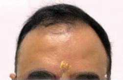
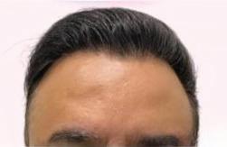
Case report
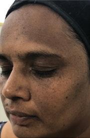
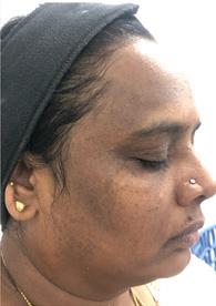
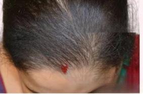
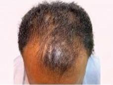
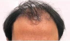
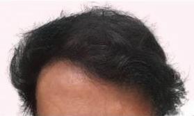
A 28-year-old female presented with persistent forehead hyperpigmentation, significantly affecting her cosmetic appearance and quality of life. She underwent treatment with
Q-switched Nd:YAG laser, with energy settings carefully adjusted to her Fitzpatrick skin type IV to minimize adverse effects. The patient showed marked reduction in pigmentation intensity without complications such as postinflammatory hyperpigmentation or scarring. This case underscores the safety and efficacy of Q-switched Nd:YAG laser therapy in managing refractory forehead hyperpigmentation in darker skin phototypes, highlighting its role as a well-tolerated and effective treatment modality.
Before Treatment
After Treatment
Diagnosis
Prior to Q-switched Nd:YAG laser therapy in darker skin types (Fitzpatrick IV–VI), comprehensive diagnostic evaluation is essential to ensure treatment safety and efficacy. Dermoscopic examination assists in differentiating between epidermal and dermal pigmentation, guiding laser parameters, predicting treatment response, and identifying melanocytic or atypical lesions, which are contraindications for laser use. Histopathological assessment is indicated when the clinical or dermoscopic diagnosis is uncertain or when malignancy is suspected, providing definitive information on pigment location and lesion type.3,4 Wood’s lamp examination further aids in assessing pigment depth; dermal pigmentation
Figure 1: Patient showed significant improvement with forehead hyperpigmentation using Q-switched Nd:YAG laser
typically appears less enhanced under Wood’s light, supporting the indication for 1064 nm Q-switched Nd:YAG laser, which is optimal for targeting deeper pigment while reducing the risk of post-inflammatory hyperpigmentation.5 Patch testing is a recommended pre-treatment step before Q-switched Nd:YAG laser therapy, especially in Fitzpatrick IV–VI, sensitive skin, or prior PIH. A small area is treated using test parameters to assess pigment response.6 Clinical photography is an essential component of preand post-treatment assessment in Q-switched Nd:YAG laser therapy. Standardized photographic documentation provides an objective baseline for evaluating lesion characteristics, treatment response, and progression over time.3 It facilitates accurate comparison across sessions, supports clinical decision-making, and serves as a medicolegal record of the patient's condition and therapeutic outcomes.
Treatment
Q-switched Nd:YAG laser therapy delivers high peak power nanosecond pulses to selectively photothermolytically disrupt melanin or exogenous pigment with minimal thermal damage to surrounding tissue. Preprocedure preparation includes informed consent, standardized clinical photography, and antiseptic cleansing of the treatment area. Topical anaesthesia with lidocaine prilocaine cream. Appropriate wavelength selection, such as 1064 nm for dermal pigment and darker skin
types and 532 nm for superficial epidermal pigment in lighter skin, is guided by chromophore depth. Laser parameters including spot size of 6mm to 10mm, fluence of 4 J/cm2 to 1.5 J/cm2 and repetition rate of 1 to 10 Hz are adjusted based on pigment depth and skin phototype. The laser is applied in a single pass, nonoverlapping manner with the handpiece held perpendicular to the skin. Clinical endpoints include transient whitening or erythema. Use of protective eyewear by both the patient and the operator is essential. Epidermal cooling can help reduce pain and prevent heat-related skin damage. Post treatment care involves bland emollients or topical antibiotics, strict photoprotection, and avoidance of irritants. Sessions are scheduled at 4 to 8 week intervals, with the number determined by clinical indication and treatment response.6, 7
Patient preparation for Q-switched Nd:YAG laser therapy involves obtaining informed consent following a detailed discussion of potential risks, benefits, treatment duration, and expected outcomes. Baseline standardized clinical photographs are taken prior to the initial treatment session. In patients with low pain threshold, topical anaesthesia using a eutectic mixture of lidocaine and Prilocaine under occlusion is applied for 45–60 minutes. Protective intraocular shields are placed on the patient, and the operator wears wavelengthspecific protective eyewear. The treatment site is cleansed and disinfected with an appropriate antiseptic solution, such as isopropyl alcohol, to reduce
infection risk.6, 7
Immediate post-treatment management following Q-switched Nd: YAG laser therapy includes the application of cooling modalities such as cold air or cool compresses to alleviate erythema and discomfort. Topical antibiotics like mupirocin or fusidic acid may be applied if epidermal compromise is present to prevent secondary infection. Strict photoprotection with broad-spectrum sunscreen (SPF 50+) is essential to reduce the risk of post-inflammatory hyperpigmentation (PIH), particularly in darker phototypes. Patients are advised to avoid sun exposure, retinoids, exfoliants, and skin irritants for 3 to 5 days post-procedure, and to refrain from manipulating the treated area. Transient erythema, mild edema, and occasional crusting or scabbing may occur, typically resolving within 1 to 2 days without intervention. Gentle cleansing and moisturization should be maintained until barrier restoration. Treatment intervals range from 4 to 8 weeks, with the number of sessions varying by indication: melasma often requires 6 to 10 low-fluence sessions, nevus of Ota 4 to 8, and tattoos 6 to 12 depending on pigment characteristics. Follow-up includes clinical evaluation, photographic documentation, and parameter adjustments based on pigment response and adverse effects. The laser’s mechanism relies on selective photothermolysis and photoacoustic disruption, fragmenting melanin or ink particles, which are cleared by dermal macrophages and lymphatic drainage with minimal
Q-Switched
collateral damage.6, 7
Discussion
Dark skin, classified within Fitzpatrick phototypes III to VI, exhibits distinct physiological and biochemical traits that shape its response to environmental factors and influence dermatological care. Higher eumelanin content provides enhanced photoprotection against ultraviolet radiation but also predisposes to pigmentary disorders such as melasma and postinflammatory hyperpigmentation, which tend to be persistent and treatmentresistant compared with lighter skin types. These alterations are often conspicuous on cosmetically sensitive areas like the face, generating pronounced aesthetic concerns. Their chronicity and visibility impose a considerable psychosocial burden, contributing to reduced self-esteem, social withdrawal, anxiety, and depression.1,8
Q-switched lasers deliver high-peak power pulses in the nanosecond range, generating photomechanical and photoacoustic effects that selectively fragment melanin granules, enabling pigment clearance with minimal collateral thermal damage. Among them, the Q-switched Nd:YAG laser is especially versatile due to its dual wavelengths. The 1064 nm wavelength penetrates deeply into the dermis with reduced epidermal melanin absorption, making it effective for dermal pigmentary disorders such as nevus of Ota and Mongolian spots, as well as dark tattoos, particularly black and blue, where it offers superior efficacy and a lower scarring risk compared to ruby or alexandrite lasers. In
contrast, the 532 nm wavelength is suited for superficial epidermal lesions, including solar lentigines and ephelides. Epidermal pigmentation usually responds within one to two sessions, whereas dermal pigmentation requires multiple treatments at 4–8 week intervals. In darker skin phototypes, precise fluence adjustment and strict photoprotection are essential to minimize postinflammatory hyperpigmentation. Beyond pigmentation, low-fluence Q-switched Nd:YAG lasers are employed for non-ablative skin rejuvenation, stimulating neocollagenesis and improving photoaging, fine wrinkles, acne scars, and striae distensae without epidermal disruption. Off-label applications include temporary hair growth reduction and treatment of superficial vascular lesions such as telangiectasia and cherry angiomas, though these demand multiple sessions and careful management.1, 8
Adverse effects of Q-switched lasers are usually transient, including erythema, edema, petechiae, and urticaria, but more serious complications such as hypopigmentation, hyperpigmentation, leukoderma, scarring, and HSV reactivation necessitate careful patient selection and parameter optimization. Hypopigmentation is managed by reducing fluence, extending treatment intervals, and using adjunctive agents such as azelaic acid or kojic acid, while hydroquinone is avoided due to its potential to worsen depigmentation. Hyperpigmentation generally responds to conservative topical therapy and strict
photoprotection. Persistent hypopigmentation warrants suspension of further treatment until resolution. Combination therapies enhance outcomes in resistant pigmentary disorders such as melasma. Integrating Q-switched lasers with topical bleaching agents, intralesional tranexamic acid, chemical peels, microdermabrasion, or microneedling provides synergistic benefits by targeting pigment through multiple mechanisms. Treatment efficacy is monitored by clinical assessment and standardized photographic documentation before initiation and after each session. Response is graded on a quartile scale: Grade I (<25%) minimal, Grade II (26–50%) moderate, Grade III (51–75%) marked, and Grade IV (>75%) near-total clearance. This objective grading system guides treatment planning and patient counseling. While Q-switched lasers represent a major advance in managing pigmentary disorders, recurrence and pigmentary complications in darker skin types remain challenges, emphasizing the need for refined protocols and ongoing research.1, 8
Conclusion
Q-switched Nd:YAG laser therapy constitutes a cornerstone in the management of pigmentary dermatoses, offering high specificity for melanin-containing targets with minimal collateral tissue disruption. Its efficacy is underpinned by the principles of selective photothermolysis and photoacoustic fragmentation, allowing for precise chromophore targeting across variable pigment depths. When
employed with appropriate diagnostic stratification and parameter modulation, it enables reproducible clinical outcomes
References
1. Goel A. Clinical applications of Q-switched NdYAG laser. Indian J Dermatol Venereol Leprol 2008; 74:682-686.
2. Hałasiński P, Lubarska M, Lubarski K, Jałowska M. Lasers' Q-switched treatment in skin and subcutaneous lesionsreview. Postepy Dermatol Alergol. 2023; 40(2):181-186. doi:10.5114/ ada.2023.127636.
3. Kaul, Neenu; Kumari, Neeti; Rawat, Shiv D. S.Evaluation of Efficacy of 1064 nm Q Switched Nd:YAG Laser Treatment in the Nevus of Ota: Seven-year Retrospective Study. Journal of Dermatology and Dermatologic Surgery 28(2): p 84-89, Jul–
with a favourable safety profile. As therapeutic paradigms evolve toward minimally invasive, pathophysiology-driven approaches, Q-switched laser technology remains integral to modern dermatologic interventions.
Dec 2024. | DOI: 10.4103/jdds.jdds_14_24
4. Arsiwala SZ, Arsiwala N. Role of Dermoscopy in Laser Therapy. Indian Dermatol Online J. 2023; 14(5):585-593. Published 2023 Aug 29. doi:10.4103/idoj. idoj_325_22.
5. Dyer JM, Foy VM. Revealing The Unseen: A Review of Wood's Lamp in Dermatology. J Clin Aesthet Dermatol. 2022; 15(6):25-30.
6 Hałasiński P, Lubarska M, Lubarski K, Jałowska M. Lasers' Q-switched treatment in skin and subcutaneous lesions - review. Postepy Dermatol Alergol. 2023; 40(2):181186. doi:10.5114/ada.2023.127636
7. Kim YJ, Whang KU, Choi WB, et al. Efficacy and safety of 1,064 nm Q-switched Nd:YAG laser treatment for removing melanocytic nevi. Ann Dermatol. 2012; 24(2):162-167. doi:10.5021/ ad.2012.24.2.162
8. Kaur, J., & Kaur, T. (2020). A study on efficacy of high fluence Q-switched neodymium doped yttrium aluminium garnet laser in macular amyloidosis. International Journal of Research in Dermatology, 6(6), 755–758. https:// doi.org/10.18203/issn.2455-4529. IntJResDermatol20204562.
Chemical Peel for Hyperpigmentation Part-II
Dr. Dipak Patel MD, DVD
Aesthetic Dermatologist & Cosmetologist
Neel Aesthetics
Surat
Newer methods of peeling
1. Progressive peeling
To enhance the intensity of chemical peels through deeper penetration, consider modifying one parameter at a time. Utilizing a higher concentration of the peeling agent can achieve greater efficacy, while applying multiple layers of the peel solution intensifies the treatment's effects. Additionally, implementing rigorous skin priming techniques before peeling prepares and optimizes the skin for treatment. Performing microdermabrasion prior to the chemical peel exfoliates the outer layer, allowing for better penetration of the peel and modify one parameter at a time.1
2. Sequential Peels
Sequential peels involve the use of different chemicals applied one after the other, as their varying pH levels and concentrations prevent them
from being combined into a single solution. Examples of sequential peels include salicylic acid at 20-30% followed by glycolic acid at 35%, salicylic acid at 20-30% followed by trichloroacetic acid at 10-25%, and glycolic acid at 70% followed by trichloroacetic acid at 35%.4
3. Combination of Peels
Combination peels involve the use of different chemical compounds at varying depths and strengths within a single formulation to achieve enhanced skin rejuvenation. Examples of combination peels include the yellow peel and AHA kojic peel. Additionally, combining peeling with other procedures such as mesolift, microdermabrasion, needling and fillers can further enhance treatment outcomes, providing comprehensive benefits for skin texture, tone and overall appearance.5
Table 2: Combination of peels
Peel Contents
Phytic acid combination
Combination peel with GA, LA and mandelic acid
Modified Jessner's formula Lactic acid 17%, salicylic acid 17%, and citric acid 8% in ethanol (95%)
Salicylic-mandelic acid
20% salicylic acid and 10% mandelic acid
Argilac L-arginine 30%, lactic acid 20%, and niacinamide 5%
4. Newer Commercial Peels
i) Black Peel – The black peel is an advanced dermatological formulation primarily used for treating acne-prone and oily skin. It combines the powerful properties of salicylic acid, jasmonic acid, potassium iodide, and black acetic acid, each contributing to its efficacy. Salicylic acid, a beta-hydroxy acid, penetrates the pores to clear out debris, reduce inflammation, and prevent future breakouts. Jasmonic acid, an anti-inflammatory agent, promotes cell turnover and helps in skin healing and renewal. Potassium iodide offers antibacterial and antiseptic benefits, reducing infection risks and supporting clearer skin. Black acetic acid acts as a mild exfoliant, softening the top skin layers and allowing active ingredients to penetrate more effectively. Together, these components create a potent treatment that not only targets acne lesions but also controls oil production, reduces pore size, and improves the skin’s overall texture and tone. The black peel is particularly beneficial for individuals with resistant acne and helps in achieving a smoother, more refined Chemical Peel
Indications
Melasma and acne
Hyperpigmentation, acne
Inflammatory and noninflammatory acne, postacne hyperpigmentation
Acne, erythema, and pigmentation
ii) Yellow Peel: The yellow peel is a specialized treatment formulated to target skin issues associated with photoaging, fine wrinkles, and superficial pigmentation. This peel contains a blend of kojic acid, phytic acid, azelaic acid, stabilized vitamin C (in the form of ascorbyl palmitate), bisabolol, and retinoic acid, each ingredient chosen for its unique skin benefits. Kojic acid and phytic acid work synergistically to reduce melanin production, helping to lighten dark spots and even out skin tone. Azelaic acid further enhances this brightening effect and provides anti-inflammatory properties, which can be beneficial for sensitive or acne-prone skin. Stabilized vitamin C (ascorbyl palmitate) contributes powerful antioxidant protection, guarding the skin against free radical damage and promoting collagen synthesis for firmer skin. This peel is a leave-on treatment, allowing the active ingredients to deeply penetrate the skin over an extended period, with an application time ranging from 30 minutes to as long as 8 hours, depending on skin tolerance and treatment goals. Following application, the skin may experience peeling and redness, which is why a downtime of approximately 5 to 7 days is typical. This controlled exfoliation period allows the skin to renew itself, revealing
a brighter, smoother, and more youthful complexion.7
5. Newer commercial peels
New commercial products contain active ingredients such as azelaic acid, mandelic acid, tranexamic acid, kojic acid, retinal, salicylic acid, ascorbic acid, phytic acid, and pyruvic acid, each offering unique benefits for skin health. Salicylic acid is known for its powerful keratolytic action, targeting superficial layers of the skin, while pyruvic acid provides
Peel Contents
desmoplastic effects. Retinol (or retinal) helps maintain the epidermis in normal physiological conditions and regulates sebum production. These products often feature specific concentrations, such as 10% lactic acid, 10% citric acid, 5% kojic acid, 2% salicylic acid, and 2% alpha-arbutin, to enhance their efficacy. Common base components include aqua (water), propylene glycol, hydroxyethylcellulose, sodium hydroxide, and sodium nitrate. Additionally, formulations may incorporate glycolic acid, trichloroacetic acid, and isopropyl alcohol, alongside ascorbyl glucoside and sodium metabisulfite. To promote hydration and support the skin barrier, many products also contain moisturizing agents like urea, aloe vera, and allantoin, with some formulations featuring up to 20% lactic acid and 20% arginine for optimal skin health.7
Coloured peel
The yellow peel, with retinoic acid, kojic acid, phytic acid, and azelaic acid, targets anti-aging, wrinkles, acne, and melasma. The black peel, containing black acetic acid, salicylic acid, jasmonic acid, biosulfur, and potassium iodide, treats acne, scars, pigmentation, oily skin, and aging. Green peel uses herbs and vitamins to improve sun damage, scars, and hyperpigmentation. The purple peel, with varying TCA levels and retinoic acid, addresses acne scars and deep wrinkles. Red peel variations treat actinic keratosis, fine lines, and photoaging. The pink peel, with Kudzu acids, benefits sundamaged skin, and the blue radiance peel, with salicylic, lactic, and glycolic acids, is ideal for sensitive skin, uneven tone, and sun damage. Each peel addresses specific skin issues effectively.
Yellow Retinoic acid, kojic acid, phytic acid, and azelaic acid
Black Black vinegar (black acetic acid), salicylic acid, jasmonic acid, biosulfur, and potassium iodide
Green Herbs and algae, high in minerals, vitamins, and enzymes. Six herbs, marigold, aloe vera, ribwort, lungwort, pansy, and horsetail
Purple Purple peel 1, 2, and 4 contain TCA 10, 15, and 20%, and retinoic acid 10, 25, and 35%, respectively
Red Red peel 1, 3, and 5 contain TCA 15, 35, and 50%, respectively
Salicylic, lactic acid, glycolic acid, willow bark, and licorice root
TCA: Trichloroacetic acid
Fruits peels
Indications
Anti-aging, wrinkles, acne, fine lines, and melasma
Acne, acne scars, aging, pigmentation, oily skin, and active acne
Sun-damaged skin, scars, striae, and hyperpigmentation
Acne scars, deep wrinkles, and hyperpigmentation
Actinic keratosis, photoaging, fine lines, and wrinkles
Sun-damaged skin, scars, striae, and photoaging
Sensitive skin, uneven tone and texture, hyperpigmentation, and sun-damaged skin
a) Pumpkin peel- The pumpkin peel is a natural peel derived from
pumpkin, featuring a blend of vitamin A derivatives that interact
Table 3: Coloured peel
with retinoic acid receptors, along with fermented enzymes and salicylic acid. This peel is ideal for dry, aging, and sensitive skin, providing an instant glow without any downtime.8
b) Papaya peel-The papaya peel contains the natural enzyme papain, which gently removes dead skin cells and enhances skin texture, making it an effective treatment for achieving smoother, healthier skin.9
c) Berry peel- The berry peel boasts strong antioxidant properties, making it effective for treating skin conditions associated with aging.9
d) Pineapple peel - The pineapple peel contains bromelain enzyme, which aids in exfoliation and brightens the complexion.9
e) Grapefruit peel - The grapefruit peel is rich in AHAs that enhance exfoliation and improve skin texture.10
f) Apple cider vinegar peel - The apple cider vinegar peel, formulated with 2% apple cider vinegar, is packed with antioxidants that help brighten the skin and reduce redness.10
Popular branded peels
Popular branded peels contain a combination of key active agents, including azelaic acid, retinoic acid, kojic acid, ascorbic acid, phytic acid, and arbutin. These components are widely utilized in dermatological treatments for their synergistic effects on skin rejuvenation, hyperpigmentation reduction, and overall skin health. Each agent contributes unique benefits: azelaic acid possesses anti-inflammatory and exfoliating properties; retinoic acid enhances cellular turnover; kojic acid inhibits melanin production; ascorbic acid offers antioxidant protection; phytic acid serves as a gentle exfoliant; and arbutin aids in lightening skin discoloration.11
This comprehensive regimen for skin rejuvenation and hyperpigmentation management combines kojic acid for hyperpigmentation reduction, ascorbic acid as a collagen-boosting antioxidant, and phytic acid for mild exfoliation and brightening, alongside arbutin, a natural hydroquinone derivative targeting dark spots. Phase I involves an intense clinic-based depigmentation mask, followed by a home protocol in Phase II to reduce pigmentation and control melanin overproduction, with a recovery balm in Phase III and pigment control in Phase IV for ongoing color correction and protection. The home-based protocol includes a high-protection pigment control product with antioxidants and UV filters, offering color correction and sun protection to prevent pigmentation recurrence and support daily skin tone management.
The formulation includes ferulic acid, phloretin, retinoids, and fruit acids for antioxidant support, cellular renewal, and gentle exfoliation, addressing signs of aging and optimizing skin tone. A superficial peel with 2% hexylresorcinol, 34% glycolic acid, 5% kojic acid, 10% lactic acid, 3% salicylic acid, and 10% citric acid addresses pigmentary disorders like melasma, promoting exfoliation and a more even skin tone. An advanced at-home peel combines salicylic, glycolic, and lactic acids to smooth texture and brighten the complexion, while a specialized peel with 20% salicylic acid, 10% azelaic acid, 5% urea, and 0.1% allantoin targets striae, skin laxity, and seborrheic
dermatitis through exfoliation, renewal, and anti-inflammatory effects.8,11
Discussion
Chemical peels are a widely utilized therapeutic intervention in dermatology and cosmetic medicine, aimed at enhancing cutaneous aesthetics and addressing a variety of dermatological conditions. A principal indication for chemical peels is the treatment of hyperpigmentation, a common dermatosis characterized by localized or diffuse areas of increased melanin deposition, resulting in dyschromia. Conditions such as melasma, actinic lentigines, and postinflammatory hyperpigmentation can significantly compromise a patient’s cosmetic appearance and self-perception.12
The therapeutic action of chemical peels involves the application of a chemical exfoliant that facilitates the enzymatic and physical sloughing of the corneocytes of the stratum corneum, thereby promoting epidermal turnover. This process leads to the reduction in the appearance of hyperpigmented lesions while simultaneously enhancing overall dermal texture and complexion. The depth of the chemical peel can be meticulously tailored, ranging from superficial to deep penetration, in accordance with the specific clinical presentation and severity of hyperpigmentation, thereby optimizing therapeutic outcomes. Complications of chemical peels may include immediate adverse effects such as pain and burning sensations, pruritus (itching), erythema, edema, chemical
burns, and ocular injuries. Delayed adverse effects can manifest as pigmentary changes, including hyperpigmentation and hypopigmentation, persistent erythema, and demarcation lines. Furthermore, there is a risk of infections, which may involve the activation of the herpes simplex virus as well as bacterial and fungal infections. Scarring may also occur, potentially resulting in hypertrophic scars or keloids.5
Moreover, in addition to their aesthetic benefits, chemical peels play a pivotal role in elevating patient self-esteem and body image. The symptomatic resolution of hyperpigmentation and the resultant improvement in skin clarity often correlate
References
1. Rendon MI, Berson DS, Cohen JL, Roberts WE, Starker I, Wang B. Evidence and considerations in the application of chemical peels in skin disorders and aesthetic resurfacing. J Clin Aesthet Dermatol. 2010 Jul; 3(7):32-43. PMID: 20725555; PMCID: PMC2921757.
2. Krueger L, Saizan A, Stein JA, Elbuluk N. Dermoscopy of acquired pigmentary disorders: a comprehensive review. Int J Dermatol. 2022 Jan; 61(1):7-19. doi: 10.1111/ijd.15741. Epub 2021 Jul 7. PMID: 34235719.
3. O'Connor AA, Lowe PM, Shumack S, Lim AC. Chemical peels: A review of current practice. Australas J Dermatol. 2018 Aug; 59(3):171-181. doi: 10.1111/ajd.12715. Epub 2017 Oct 24. PMID: 29064096.
4. Divyalakshmi C, Hazarika N, Barnwal S. Role of sequential chemical peel in cutaneous amyloidosis: a pilot case series. Clin Exp Dermatol. 2022 Apr; 47(4):743-747. doi: 10.1111/ced.15016. Epub 2021 Dec 7. PMID: 34773296.
5. Nofal E, Nofal A, Gharib K, Nasr M, Abdelshafy A, Elsaid E. Combination chemical peels are more effective than single chemical peel in treatment of mild-to-moderate acne vulgaris: A split face comparative clinical trial. J Cosmet Dermatol. 2018 Oct; 17(5):802-810. doi: 10.1111/jocd.12763. Epub 2018 Sep 10.
with enhanced psychosocial functioning and increased confidence levels among patients. Such psychological benefits may extend to improved social interactions, professional engagement, and overall mental health. Thus, chemical peels not only provide significant dermatologic benefits but also contribute positively to a patient's psychosocial well-being, reinforcing their self-perception and enhancing their quality of life.13
Conclusion
Chemical peels have significantly impacted the fields of dermatology and aesthetics, offering effective solutions for improving skin texture, reducing signs of aging, and treating conditions like acne and hyperpigmentation. Their benefits extend beyond appearance, enhancing self-esteem and overall mental well-being. As awareness of skin health grows, the demand for chemical peels is likely to rise, leading to advancements in formulations and techniques that prioritize safety and efficacy. Future developments may focus on personalized treatments, integrating technology for better results while minimizing side effects. Overall, chemical peels promise to enhance skin health practices and contribute positively to individual wellness.
No conflict of interest
PMID: 30203434.
6. Nikalji, N. S., et al. (2015). Chemical peels in dermatology: An overview. Journal of Clinical and Aesthetic Dermatology, 8(6), 33-43.
7. Puri N, Kumar S, Kaur S, Brar BK. Comparative evaluation of therapeutic efficacy and safety of black peel versus 25% trichloroacetic acid peel in mild to moderate acne vulgaris: a split face study. J Cutan Aesthet Surg. 2024 Jan-Mar; 17(1):11-18. doi: 10.4103/JCAS.JCAS_69_23. PMID: 38736859; PMCID: PMC11086936.
8. Leichtweis MG, Molina AK, Petropoulos SA, Carocho M, Pires TCSP, Dias MI, Calhelha R, Oliveira MBPP, Pereira C, Barros L. Valorization of Pumpkin Peel as a Source of Bioactive Compounds: Optimization of Heat- and Ultrasound-Assisted Extraction. Molecules. 2023 Apr 2; 28(7):3168. Doi: 10.3390/molecules28073168. PMID: 37049931; PMCID: PMC10096157.
9. Easmin S, Bhattacharyya M, Pal K, Das P, Sahu R, Nandi G, Dewanjee S, Paul P, Haydar MS, Roy S, Dua TK. Papaya peel extract-mediated green synthesis of zinc oxide nanoparticles and determination of their antioxidant, antibacterial, and photocatalytic properties. Bioprocess Biosyst Eng. 2024 Jan; 47(1):65-74. doi: 10.1007/s00449-023-02945-7. Epub 2023 Dec 12. PMID: 38086975.
10. Castro-Vazquez L, Alañón ME, Rodríguez-Robledo V, Pérez-Coello MS, Hermosín-Gutierrez I, DíazMaroto MC, Jordán J, Galindo MF, Arroyo-Jiménez Mdel M. Bioactive Flavonoids, Antioxidant Behaviour, and Cytoprotective Effects of Dried Grapefruit Peels (Citrus paradisi Macf.). Oxid Med Cell Longev. 2016; 2016:8915729. doi: 10.1155/2016/8915729. Epub 2016 Jan 21. PMID: 26904169; PMCID: PMC4745316.
11. Dayal S, Sangal B, Sahu P. Ferulic acid 12% peel: An innovative peel for constitutional type of periorbital melanosis-Comparing clinical efficacy and safety with 20% glycolic peel and 15% lactic peel. J Cosmet Dermatol. 2020 Sep; 19(9):2342-2348. doi: 10.1111/ jocd.13292. Epub 2020 Jan 16. PMID: 31944519.
12. Al Aboud DM, Gossman W. Wood's Light. 2023 Aug 28. In: StatPearls [Internet]. Treasure Island (FL): StatPearls Publishing; 2024 Jan–. PMID: 30725878.
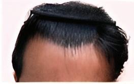
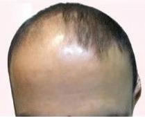
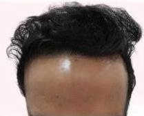
Optimizing Outcomes in Pattern Hair Loss: Clinical Insights into
Implanter-Assisted Hair Transplantation
Dr. Shalini Malhotra
MBBS, DNB (Dermatology), MNAMS, ABHRS
Hair Transplant Surgeon and Consultant Dermatologist
SM Hair Restoration & Aesthetic Clinic
New Delhi
Introduction
Alopecia represents a multifactorial dermatological disorder characterized by pathological hair loss secondary to genetic, hormonal, autoimmune, psychosocial, traumatic, or chemical triggers. The underlying pathogenesis involves dysregulation of follicular homeostasis and progressive follicular miniaturization, ultimately leading to diffuse or patterned scalp hair thinning. Hair follicles, as highly specialized cutaneous appendages, rely on tightly regulated molecular signaling pathways to maintain continuous regenerative cycling essential for sustained hair growth.1, 2
Alopecia comprises a diverse group of disorders broadly classified into non-scarring (non-cicatricial) and scarring (cicatricial) types, distinguished by the potential for follicular regeneration. Non-scarring alopecias, which preserve follicular architecture, include
androgenetic alopecia (AGA), the most prevalent form, characterized by genetically mediated androgen sensitivity predominantly to dihydrotestosterone (DHT). DHT interaction with dermal papilla cells induces follicular miniaturization, marked by shortened anagen phase, prolonged telogen, and transformation of terminal hairs into finer vellus hairs, manifesting as patterned hair thinning primarily on the frontal scalp and vertex. Alopecia areata (AA), another nonscarring entity, results from autoimmune-mediated cytotoxic T-cell attack on anagen follicles, leading to patchy hair loss with potential for spontaneous remission. Telogen effluvium (TE) involves an abnormal shift of hair follicles into the telogen phase following physiological or psychological stressors, causing diffuse shedding usually delayed by 2-3 months. Traction alopecia arises from chronic mechanical stress and tension, often due to hairstyling practices, initially
reversible but potentially leading to scarring with persistent insult. In contrast, scarring alopecias represent irreversible follicular destruction replaced by fibrosis, driven by inflammatory, autoimmune, infectious, or neoplastic processes targeting the follicular stem cell niche in the bulge region. Key cicatricial alopecias include lichen planopilaris (LPP), a lymphocytic inflammatory condition causing perifollicular fibrosis and permanent follicular loss; frontal fibrosing alopecia (FFA), a variant predominantly affecting postmenopausal women with progressive frontotemporal recession and eyebrow loss; and neutrophilic disorders like dissecting cellulitis and folliculitis decalvans, characterized by chronic suppurative inflammation.1, 2
The human hair follicle is a dynamic mini-organ undergoing cyclic phases—anagen, catagen, telogen, and exogen— regulated by intricate molecular signaling governing keratinocyte activity and follicular homeostasis. Structurally, it comprises a permanent upper segment (infundibulum, isthmus) and a transient lower segment (bulb, dermal papilla), with the bulge region acting as a niche for epithelial and melanocyte stem cells essential for regeneration. In androgenetic alopecia (AGA), androgen (DHT) mediated miniaturization results in shortened anagen, prolonged telogen, and terminal to vellus hair transformation due to disrupted dermal papilla stem cell interaction. Hair transplantation exploits the androgen resistant nature of occipital follicles by relocating
them to balding areas, relying on preservation of the bulge DP axis to ensure graft viability and long term aesthetic outcomes.1, 2, 3
We present a case series of 10 patients with varying grades of pattern hair loss, including type III and IV male androgenetic alopecia and type III female pattern hair loss, who underwent hair transplantation using the “Advanced FUE (Follicular Unit Excision) “technique wherein implanters are used to implant grafts. Postprocedure evaluations revealed marked improvement in hair density and scalp coverage. All patients reported satisfactory aesthetic outcomes, underscoring the implanters as a reliable, effective, and well-tolerated approach for carefully selected individuals with pattern hair loss.
Result
Before surgery
After 6 months of surgery
Patient 1
Before surgery After 9
Patient 2
Patient 3
After 7 months of surgery (2500 Grafts)
After 9 months of surgery (2000 Grafts)
Patient 4
After 6 months of surgery (1500 Grafts)
Patient 5 Before surgery After 7 months of surgery (3000 Grafts)
Patient 7
After 9 months of surgery (3000 Grafts)
Patient 6
Patient 8
Patient 9
Patient 10
Diagnosis
A comprehensive evaluation of alopecia requires documentation of onset, duration, pattern and progression, with careful distinction between scarring and non-scarring forms. A detailed history should include family predisposition, systemic illnesses, medications, and prior treatments. Clinical examination must assess scalp laxity and donor density to establish transplantation suitability. Several bedside tests provide diagnostic value. The card test differentiates new anagen hairs (tapered ends) from broken or miniaturized hairs using a dual-colored background. The hair pull test, performed by gently extracting 50–60 hairs, indicates active shedding if more than 5–6 hairs are removed; patients should avoid shampooing for 24 hours beforehand. The tug test evaluates fragility, with mid-shaft breakage suggesting structural disorders. Non-invasive imaging enhances accuracy. Trichoscopy demonstrates miniaturization, broken hairs, black/yellow dots, and perifollicular changes. Hair mount microscopy identifies shaft abnormalities, while scalp biopsy remains the gold standard in uncertain cases; two 4-mm punch biopsies for horizontal and vertical sectioning provide optimal evaluation. Laboratory tests (thyroid profile, ferritin, hormones, autoimmune markers) exclude systemic causes. Advanced modalities such as Optical Coherence Tomography (OCT) and Reflectance Confocal Microscopy (RCM) distinguish scarring from non-scarring alopecia. Genetic testing, cytokine profiling in alopecia areata, automated hair analysis, and standardized photographic documentation further refine diagnosis and treatment monitoring.4,5,6,7,8,9,10
Procedure & Technique Protocol: Hair Transplantation (Advanced FUE using Implanters)
Hair transplantation (HT) involves relocating hair follicles from a donor site, typically the occipital scalp resistant to androgenetic alopecia (AGA), to balding areas, based on the principle of donor dominance. Alternative donor sites such as beard or body hair may be used when scalp donor supply is insufficient. Preoperative evaluation includes complete blood count, coagulation profile, blood glucose, viral serology (HIV, hepatitis B), chest X-ray, ECG and medical clearance. Minoxidil should be stopped two weeks prior; smoking and NSAIDs avoided for one week to reduce bleeding risk. Contraindications include active autoimmune disease, uncontrolled
diabetes or hypertension and lignocaine hypersensitivity.4,11,12
Preoperative Preparation
Before donor harvesting, the scalp is trimmed to ~3 mm to visualize follicular orientation. A Fox test may be conducted by extracting a few units to assess graft quality and ease of extraction; poor grades (Fox 4 or 5) suggest high transection risk and unsuitability for FUE. Local anesthesia is administered via regional nerve blocks and tumescent infiltration, ensuring analgesia, reducing bleeding, and creating a safe dissection plane while preserving neurovascular structures.4,11,12
Donor Harvesting
Advanced Follicular Unit
Excision (FUE) entails the individual removal of follicular units using manual or motorized punches (0.8–1.0 mm). Donor hair is trimmed to visualize shaft angulation, and punches are aligned with follicular axes under counter-traction to reduce transection. Grafts are harvested in a dispersed pattern to preserve donor aesthetics and are stored in chilled isotonic saline. A Fox test may be performed to assess graft quality and extraction ease.4,11,12
Recipient Site Preparation
Recipient site preparation begins with the administration of local anaesthesia to ensure patient comfort. In the Advanced FUE, recipient slits are not created with blade or needle instead grafts are implanted using implanters by one step stick and place technique. Typically, around 250 to 300 micrografts are used to reconstruct a natural frontal hairline, while larger follicular unit grafts are placed
Before surgery
After 7 months of surgery (3000 Grafts)
strategically in the posterior recipient areas to enhance overall density and volume.4,11,12
Graft Placement in Hair
Transplantation: Sharp Implanter and Forceps Techniques
Sharp Implanter Technique:
The latest technology in hair transplantation employs the sharp implanter method, integrating incision and implantation into a single, precise step using the stick-and-place technique without pre-made recipient sites. The needle-tipped implanter, skilfully with a refined three-finger grip, allows for controlled vertical and rectilinear movements, minimizing tissue trauma. Proper bevel orientation ensures graft protection and accurate alignment.13
Implanters feature wide or narrow lumens tailored to graft morphology: wide channels accommodate FUE grafts with protruding epidermis acting as a natural depth limiter, while narrow channels suit FUE grafts fully enclosed within the lumen, requiring meticulous depth control. Selecting the appropriate tip size, ranging from 0.6 to 1.2 mm based on follicular unit dimensions, is critical to maintaining graft viability.13
This technique enhances operative efficiency, allows precise control of implantation angle and depth, and reduces follicular trauma compared to conventional forceps or dull needle methods. There is minimal bleeding during implantation as premade slits are not created at the recipient site. It is particularly advantageous in delicate recipient areas such as eyebrows, moustaches,
and female hairlines, where meticulous angle and direction control is essential to achieve a natural look. By eliminating the need for pre-made incisions, the sharp implanter minimizes scalp injury, promotes graft survival, and enables high-density, cosmetically superior outcomes with reduced operative time.13
Operative Efficiency
The sharp implanter technique significantly optimizes operative efficiency by combining recipient site creation and graft insertion into a single, continuous motion. Implementation of a structured team-based workflow, such as the 2-1-1 or 3-1-1 implanter circulation system, facilitates uninterrupted graft placement, minimizes operator fatigue, and maintains procedural precision. This approach also reduces graft ischemia time, thereby potentially enhancing follicular unit survival. On average, 2,000 to 3,000 grafts can be implanted within approximately two to three hours, contingent upon the surgeon’s expertise, team coordination, and recipient scalp characteristics. Compared to the conventional pre-made slit with forceps insertion method, the implanter technique may reduce total operative time.4,11,12
Postoperative Care and Follow-up
Hair transplantation is typically an outpatient procedure with same-day discharge. Bandaging of the recipient site is usually avoided to prevent graft displacement. Postoperative edema, especially in older patients, can be managed with forehead pressure dressings, short courses of systemic corticosteroids, or intraoperative triamcinolone injections. Patients
should begin gentle scalp cleansing with mild shampoo 2–3 days post-procedure and avoid combing or tight headgear for at least three weeks. Topical minoxidil in strength of 5% for both men and women can be prescribed after 2 weeks and at least for atleast 9 months post procedure. In men 1 mg finasteride may be added. Full results are expected by 7-9 months. Extensive alopecia cases are preferably treated in staged sessions spaced 4–6 months apart to optimize graft survival and scalp healing.4, 11, 12
Discussion
Alopecia is a significant global health concern, impacting both men and women across all ages, with substantial psychosocial and quality-of-life consequences. Androgenetic alopecia (AGA) is the most common form, presenting as progressive thinning and patterned hair loss. Accurate classification systems are essential for diagnosis, management, and surgical planning. Male pattern hair loss (MPHL) classifications have evolved from basic descriptions of frontal and vertex balding, initially based on Caucasian populations, to more inclusive systems accounting for frontoparietal recession, vertex thinning, ethnic variations, and atypical patterns. These systems define distinct stages such as continuous anterior hairline recession or isolated vertex involvement. However, their complexity has limited everyday use, leading to simplified morphology-based and geometric classifications designed for practical clinical application. Female pattern
hair loss (FPHL) classifications have similarly advanced, ranging from crown thinning with preserved frontal hairline to those recognizing frontotemporal recession, male-pattern features, and specific patterns such as the “Christmas tree” configuration. A universal, gender-independent classification, based on anterior hairline shape and density, further aids standardized assessment and surgical planning. Therapeutic options span medical treatments— including topical minoxidil, oral finasteride in men, and hormonal or anti-androgen therapy in women—to adjunctive modalities such as platelet-rich plasma (PRP), low-level laser therapy (LLLT), microneedling, and nutritional supplementation. Hair transplantation remains the gold standard for stable alopecia, while emerging approaches such as JAK inhibitors and novel biologics hold promise for the future.13
Patient selection is crucial for successful hair transplantation. Young patients should first undergo medical therapy to stabilize loss and preserve donor
References
1. Wall D, Meah N, Fagan N, York K, Sinclair R. Advances in hair growth. Fac Rev. 2022; 11:1. Published 2022 Jan 12. doi:10.12703/r/11-1.
2. Liu D, Xu Q, Meng X, Liu X, Liu J. Status of research on the development and regeneration of hair follicles. Int J Med Sci. 2024; 21(1):80-94. Published 2024 Jan 1. doi:10.7150/ijms.88508.
3. Müller Ramos P, Melo DF, Radwanski H, de Almeida RFC, Miot HA. Femalepattern hair loss: therapeutic update. An Bras Dermatol. 2023; 98(4):506-519. doi:10.1016/j.abd.2022.09.006
hair. Diffuse thinning requires counseling and adjunctive treatment. In advanced cases, conservative hairline design, safe donor zone adherence, and judicious harvesting ensure natural outcomes and long-term graft survival.4,14
Hair transplantation using forceps in follicular unit excision (FUE) relies on atraumatic graft placement by holding follicular units through surrounding subcutaneous tissue, minimizing direct root injury. Grafts are placed flush with the scalp to avoid pitting, ridging, or cyst formation, with implantation angles generally ≤45°, and even more acute in temporal triangles for natural contouring. Although widely practiced, forceps placement has limitations: increased graft manipulation, potential follicular trauma, excessive bleeding, reduced precision in angle and depth, and longer surgical times contributing to surgeon fatigue. These drawbacks can cause variable density and direction, compromising natural outcomes. In contrast, sharp implanters integrate site creation and graft insertion in
a single motion, enhancing control of depth, direction, and angulation. This improves graft survival and enables denser, more natural results. Nonetheless, medical therapy remains first-line for progressive alopecia, with transplantation reserved for stable cases. Careful patient selection, preoperative evaluation, and counselling remain critical to achieving durable, aesthetic restoration.4,13,14
Conclusion
Hair transplantation is a wellestablished treatment for androgenetic alopecia, providing durable and natural results in patients with stable hair loss. Achieving optimal outcomes depends on thorough evaluation of donor and recipient sites, meticulous surgical technique including the use of sharp implanters for precise graft placement, and comprehensive patient counseling. Recent advancements such as implanter-assisted implantation improve graft survival and procedural efficiency, making hair transplantation a valuable tool in the management of alopecia.
4. Singh S, Muthuvel K. Practical Approach to Hair Loss Diagnosis. Indian J Plast Surg. 2021; 54(4):399-403. Published 2021 Dec 27. Doi: 10.1055/s-0041-1739240.
5. Issa NT, Tosti A. Trichoscopy for the Hair Transplant Surgeon-Assessing for Mimickers of Androgenetic Alopecia and Preoperative Evaluation of Donor Site Area. Indian J Plast Surg. 2021; 54(4):393-398. Published 2021 Dec 20. Doi: 10.1055/s0041-1739245.
6. Starace M, De Carvalho N, Melo DF, et al. Concordance Among In Vivo Reflectance Confocal Microscopy, Trichoscopy, and
Histopathology in the Evaluation of Alopecia Areata Incognita. Dermatol Pract Concept. 2024; 14(4):e2024229. Published 2024 Oct 30. doi:10.5826/ dpc.1404a229.
7. Ekelem C, Feil N, Csuka E, et al. Optical Coherence Tomography in the Evaluation of the Scalp and Hair: Common Features and Clinical Utility. Lasers Surg Med. 2021; 53(1):129-140. doi:10.1002/ lsm.23243.
8. Rand MR, Yale K, Kato BS, Kim DJ, Birmingham S, Mesinkovska NA. Commonly Associated Disorders with
Complete Scalp Alopecia in Early Childhood: A Review. Int J Trichology. 2023; 15(2):43-49. doi:10.4103/ijt. ijt_70_22
9. Balık AR, Balık ZB, Aktaş A, Neşelioğlu S, Karabulut E, Karabulut AB. Examination of androgenetic alopecia with serum biomarkers. J Cosmet Dermatol. 2021; 20(6):1855-1859. doi:10.1111/ jocd.13732
10. Gudobba C, Mane T, Bayramova A, et al. Automating Hair Loss Labels
for Universally Scoring Alopecia From Images: Rethinking Alopecia Scores. JAMA Dermatol. 2023; 159(2):143-150. doi:10.1001/jamadermatol.2022.5415
11. Khanna M. Hair transplantation surgery. Indian J Plast Surg. 2008; 41(Suppl):S56-S63.
12. Singh M, Acharya A. Overview and Algorithmic Approach to Management of Male and Female Pattern Hair Loss. Indian J Plast Surg. 2021; 54(4):416-421. Published 2021 Dec 14. Doi: 10.1055/s0041-1739256
13. Park JH, Ho YH, Manonukul K. A Practical Guide to Hair Graft Placement Using the Sharp Implanter Method. Clin Cosmet Investig Dermatol. 2023; 16:1777-1785. Published 2023 Jul 11. doi:10.2147/CCID.S411488
14. Gupta M, Mysore V. Classifications of Patterned Hair Loss: A Review. J Cutan Aesthet Surg. 2016; 9(1):3-12. doi:10.4103/0974-2077.178536