Resectable Pancreatic Cancer 1st Edition Matthew H.G. Katz
Visit to download the full and correct content document: https://textbookfull.com/product/multimodality-management-of-borderline-resectable-p ancreatic-cancer-1st-edition-matthew-h-g-katz/
More products digital (pdf, epub, mobi) instant download maybe you interests ...

Pancreatic Cancer With Special Focus on Topical Issues and Surgical Techniques Kim
https://textbookfull.com/product/pancreatic-cancer-with-specialfocus-on-topical-issues-and-surgical-techniques-kim/

Translational Pancreatic Cancer Research From Understanding of Mechanisms to Novel Clinical Trials
Christoph W. Michalski
https://textbookfull.com/product/translational-pancreatic-cancerresearch-from-understanding-of-mechanisms-to-novel-clinicaltrials-christoph-w-michalski/
Pancreatic Cancer With Special Focus on Topical Issues and Surgical Techniques 1st Edition Sun-Whe Kim
https://textbookfull.com/product/pancreatic-cancer-with-specialfocus-on-topical-issues-and-surgical-techniques-1st-edition-sunwhe-kim/
Multimodality imaging guidance in interventional pain management 1st Edition Narouze
https://textbookfull.com/product/multimodality-imaging-guidancein-interventional-pain-management-1st-edition-narouze/
Management of Differentiated Thyroid
Anne T. Mancino
Cancer 1st Edition
https://textbookfull.com/product/management-of-differentiatedthyroid-cancer-1st-edition-anne-t-mancino/

Multidisciplinary Management of Rectal Cancer Vincenzo Valentini
https://textbookfull.com/product/multidisciplinary-management-ofrectal-cancer-vincenzo-valentini/
Management of Hematological Cancer in Older People 1st Edition Ulrich Wedding
https://textbookfull.com/product/management-of-hematologicalcancer-in-older-people-1st-edition-ulrich-wedding/
Breast Cancer Fundamentals of Evidence-Based Disease Management 1st Edition Henderson
https://textbookfull.com/product/breast-cancer-fundamentals-ofevidence-based-disease-management-1st-edition-henderson/
Management of Advanced Prostate Cancer Choung Soo Kim
https://textbookfull.com/product/management-of-advanced-prostatecancer-choung-soo-kim/
Multimodality Management of Borderline Resectable Pancreatic Cancer


Matthew H.G. Katz
Syed A. Ahmad Editors


Multimodality Management of Borderline
Resectable
Pancreatic Cancer
Matthew H.G. Katz • Syed A. Ahmad Editors
Multimodality Management of Borderline Resectable Pancreatic
Cancer
Editors Matthew H.G. Katz
Department of Surgical Oncology
MD Anderson Cancer Center Houston, TX, USA
Syed A. Ahmad Division of Surgical Oncology
The University of Cincinnati Medical Center Cincinnati, OH, USA
ISBN 978-3-319-22779-5
DOI 10.1007/978-3-319-22780-1
ISBN 978-3-319-22780-1 (eBook)
Library of Congress Control Number: 2015956343
Springer Cham Heidelberg New York Dordrecht London © Springer International Publishing Switzerland 2016
This work is subject to copyright. All rights are reserved by the Publisher, whether the whole or part of the material is concerned, specifically the rights of translation, reprinting, reuse of illustrations, recitation, broadcasting, reproduction on microfilms or in any other physical way, and transmission or information storage and retrieval, electronic adaptation, computer software, or by similar or dissimilar methodology now known or hereafter developed.
The use of general descriptive names, registered names, trademarks, service marks, etc. in this publication does not imply, even in the absence of a specific statement, that such names are exempt from the relevant protective laws and regulations and therefore free for general use.
The publisher, the authors and the editors are safe to assume that the advice and information in this book are believed to be true and accurate at the date of publication. Neither the publisher nor the authors or the editors give a warranty, express or implied, with respect to the material contained herein or for any errors or omissions that may have been made.
Printed on acid-free paper
Springer International Publishing AG Switzerland is part of Springer Science+Business Media (www.springer.com)
Preface
Over the past decade, great efforts have been made toward refining the clinical systems used to stage localized pancreatic cancer. As the benefits of the administration of preoperative therapy have increasingly become recognized, and as the performance of vascular resection and reconstruction at pancreatectomy has concurrently become more common, many infiltrative cancers that were historically considered unresectable are now more commonly described as “borderline resectable.” Tumors in this category are those that are technically removable, but which are associated with a significant likelihood of a positive margin when surgery is performed de novo. Given that the overall survival rate of patients who undergo margin-positive operations is similar to that of patients who do not undergo surgery at all, recognition of borderline resectable pancreatic cancer as a unique clinical entity is critical, both for optimal patient care and for the proper evaluation of novel (neo) adjuvant treatment regimens in clinical trials.
In this book, we have assembled an internationally recognized group of clinical experts to compile an up-to-date appraisal of the diagnostic and therapeutic modalities used for patients with borderline resectable pancreatic cancer. The book includes an overview of clinical staging, a review of endoscopic approaches, a summary on the latest clinical research, and a discussion of emerging targeted therapies. We also present several well-illustrated surgical chapters on novel technical strategies and techniques that may be utilized to safely manage this difficult group of patients in the operating room.
We acknowledge Mr. Andy Kwan, Mr. Brian Halm, and Ms. Portia Wong from Springer, whose support of this project was essential for its development and completion.
We would like to thank the physicians who trained us to provide safe, thoughtful, and effective surgical care for patients with pancreatic tumors: Drs. Douglas Evans, Jeffrey Lee, Jason Fleming, Peter Pisters, Michael Bouvet, Andy Lowy, Jeffrey Matthews, Michael Edwards, and the late A.R. Moossa. We would also like to thank our families for their patience during the preparation of this book. Dr. Ahmad would like to acknowledge his wife, Shagufa, and his children, Samar, Ameen, and Saher. Dr. Katz would like to acknowledge his wife, Kristen, and children, Annie and Lucy.
Finally, we would like to dedicate this book to the patients and families who have battled pancreatic cancer. It is their courage and bravery that motivate and inspire us on a daily basis.
Houston, TX
Matthew H.G. Katz Cincinnati, OH Syed A. Ahmad
A. Snyder, Alexander A. Parikh, Kamran Idrees, and Nipun B. Merchant
Lee M. Ocuin, Herbert J. Zeh
Amir H. Fathi, Susan Tsai, Jeffrey E. Lee, Douglas B. Evans, and Kathleen K. Christians
Ken-ichi Okada and Hiroki Yamaue
Shuji Isaji, Masashi Kishiwada, and Hiroyuki Kato
Robert C.G. Martin II and Rachel O’Connor
Nicholas Spinelli
Ismael
Contributors
Ross Abrams, M.D. Department of Radiation Oncology, Rush University Medical Center, Chicago, IL, USA
Brian Badgwell, M.D., M.S. Department of Surgical Oncology, The University of Texas MD Anderson Cancer Center, Houston, TX, USA
Marshall S. Baker, M.D., M.B.A., F.A.C.S. Department of Surgery, NorthShore University HealthSystem, Evanston, IL, USA
Kyuran Ann Choe, M.D. Department of Radiology, University of Cincinnati Medical Center, Cincinnati, OH, USA
Kathleen K. Christians, M.D. Division of Surgical Oncology, Department of Surgery, Pancreatic Cancer Program, Medical College of Wisconsin, Milwaukee, WI, USA
Douglas B. Evans, M.D. Division of Surgical Oncology, Department of Surgery, Pancreatic Cancer Program, Medical College of Wisconsin, Milwaukee, WI, USA
Amir H. Fathi, M.D. Division of Surgical Oncology, Department of Surgery, Pancreatic Cancer Program, Medical College of Wisconsin, Milwaukee, WI, USA
William Hawkins Washington University School of Medicine, St. Louis, MO, USA
Joseph M. Herman, M.D., M.Sc. Department of Radiation Oncology, The Johns Hopkins University School of Medicine, Baltimore, MD, USA
Kamran Idrees, M.D. Division of Surgery Oncology, Vanderbilt University Medical Center, Nashville, TN, USA
Shuji Isaji Department of Hepatobiliary Pancreatic and Transplant Surgery, Mie University Graduate School of Medicine, Tsu, Mie, Japan
Hishaam Ismael, M.D. Department of Surgical Oncology, The University of Texas MD Anderson Cancer Center, Houston, TX, USA
Hiroyuki Kato Department of Hepatobiliary Pancreatic and Transplant Surgery, Mie University Graduate School of Medicine, Tsu, Mie, Japan
Kaitlyn J. Kelly, M.D. Department of Surgery, University of California San Diego, Moores Cancer Center, La Jolla, CA, USA
Masashi Kishiwada Department of Hepatobiliary Pancreatic and Transplant Surgery, Mie University Graduate School of Medicine, Tsu, Mie, Japan
Jörg Kleeff, M.D., F.A.C.S. Department of Surgery, Klinikum rechts der Isar, Technische Universität München, Munich, Germany
Rachit Kumar, M.D. Division of Radiation Oncology, Banner MD Anderson Cancer Center, Gilbert, AZ, USA
Jeffrey E. Lee, M.D. Department of Surgical Oncology, University of Texas M.D. Anderson Cancer Center, Houston, TX, USA
Andrew M. Lowy Department of Surgical Oncology, Universality of California San Diego, La Jolla, CA, USA
Robert de Wilton Marsh, M.B., Ch.B., F.A.C.P. Department of Medicine, Division of Hematology/Oncology, NorthShore University HealthSystem, Evanston, IL, USA
Robert C.G. Martin II, M.D., Ph.D. Division of Surgical Oncology, University of Louisville, Louisville, KY, USA
Nicholas M. McDonald, M.D. Department of Radiology, University of Cincinnati Medical Center, Cincinnati, OH, USA
Nipun B. Merchant, M.D. Department of Surgery/Oncology, Sylvester Comprehensive Cancer Center, University of Miami Miller School of Medicine, Miami, FL, USA
André L. Mihaljevic, M.D., M.Sc. Department of Surgery, Klinikum rechts der Isar, Technische Universität München, Munich, Germany
Girish Mishra, M.D., M.Sc. Internal Medicine-Section on Gastroenterology, Wake Forest School of Medicine, Medical Center Boulevard, Winston-Salem, NC, USA
Rachel O’Connor, B.S. Division of Surgical Oncology, University of Louisville, Louisville, KY, USA
Lee M. Ocuin, M.D. University of Pittsburgh Medical Center, Pittsburgh, PA, USA
Ken-ichi Okada Second Department of Surgery, Wakayama Medical University, Wakayama, Japan
Trailokya Pandit Department of Oncology, Wayne State University School of Medicine, Barbara Ann Karmanos Cancer Center, Detroit, MI, USA
Alexander A. Parikh, M.D., M.P.H. Division of Surgical Oncology, Vanderbilt University Medical Center, Nashville, TN, USA
Rishi Pawa, M.D. Internal Medicine-Section on Gastroenterology, Wake Forest School of Medicine, Medical Center Boulevard, Winston-Salem, NC, USA
Philip A. Philip Department of Oncology, Wayne State University School of Medicine, Barbara Ann Karmanos Cancer Center, Detroit, MI, USA
Matthew J. Reilley Division of Cancer Medicine, MD Anderson Cancer Center, Houston, TX, USA
Lauren M. Rosati, B.S. Department of Radiation Oncology, The Johns Hopkins University School of Medicine, Baltimore, MD, USA
Neilayan Sen, M.D. Department of Radiation Oncology, Rush University Medical Center, Chicago, IL, USA
Milton T. Smith, M.D. Division of Digestive Diseases, University of Cincinnati, Cincinnati, OH, USA
Rebecca A. Snyder, M.D., M.P.H. Department of Surgery, Vanderbilt Medical Center, Nashville, TN, USA
Nicholas Spinelli Washington University School of Medicine, St. Louis, MO, USA
Kyoichi Takaori, M.D., Ph.D., F.A.C.S. Division of Hepatobiliary-Pancreatic Surgery and Transplantation, Department of Surgery, Kyoto University Graduate School of Medicine, Kyoto, Japan
Mark J. Truty, M.D., M.Sc. Hepatobiliary and Pancreatic Surgery, Mayo Clinic College of Medicine, Rochester, MN, USA
Susan Tsai, M.D. Division of Surgical Oncology, Department of Surgery, Pancreatic Cancer Program, Medical College of Wisconsin, Milwaukee, WI, USA
Shinji Uemoto, M.D., Ph.D. Division of Hepatobiliary-Pancreatic Surgery and Transplantation, Department of Surgery, Kyoto University Graduate School of Medicine, Kyoto, Japan
Gauri R. Varadhachary Department of Gastrointestinal Medical Oncology, MD Anderson Cancer Center, Houston, TX, USA
Hiroki Yamaue Second Department of Surgery, Wakayama Medical University, Wakayama, Japan
Herbert J. Zeh III, M.D., F.A.C.S. Division of Gastrointestinal Surgical Oncology, University of Pittsburgh Medical Center, Pittsburgh, PA, USA
Amer H. Zureikat, M.D., F.A.C.S. Division of Gastrointestinal Surgical Oncology, University of Pittsburgh Medical Center, Pittsburgh, PA, USA
Anatomic Definitions of Borderline Resectable Pancreatic Cancer
Rebecca A. Snyder, Alexander A. Parikh, Kamran Idrees, and Nipun B. Merchant
Introduction
Pancreatic cancer is the fourth leading cause of cancer death in the United States [1]. Although the incidence of pancreatic adenocarcinoma is lower than that of other malignancies, mortality rates remain high. In 2014, there were approximately 46,420 new cases of pancreas cancer and 39,590 people died of the disease [2]. The majority of patients present with advanced disease at the time of initial diagnosis; 5-year survival rates are estimated to be 9.9 % in patients with regionally advanced cancers and 2.3 % in those with distant disease [2]. Survival rates are higher for patients with localized tumors (25.8 %); however, fewer than 10 % of patients with pancreatic adenocarcinoma present at an early stage [2].
R.A. Snyder, M.D., M.P.H. Department of Surgery, Vanderbilt Medical Center, 1161 21st AVE S, Nashville, TN 37232, USA e-mail: rebecca.snyder@vanderbilt.edu
A.A. Parikh, M.D., M.P.H. • K. Idrees, M.D. Division of Surgical Oncology, Vanderbilt University Medical Center, Nashville, TN 37232, USA e-mail: alexander.parikh@vanderbilt.edu; kamran.idrees@vanderbilt.edu
N.B. Merchant, M.D. (*) Department of Surgery/Oncology, Sylvester Comprehensive Cancer Center, University of Miami Miller School of Medicine, 1120 N.W. 14th Street, Miami, FL 33136, USA e-mail: nmerchant@med.miami.edu
As with other cancers, surgery offers the only opportunity for cure. Therefore, one of the most important determinants of overall prognosis for patients with pancreatic cancer is resectability. Indeed, a margin-negative resection is considered one of the strongest prognostic factors for longterm survival in patients with pancreatic cancer. Resection margins are classified as having no evidence of microscopic tumor deposits at or within 1 mm of the inked margins (R0) or as having microscopic tumor deposits but no gross tumor at the margins (R1). Discrimination between R0 and R1 margins is made on the basis of observations made by both the surgeon and the pathologist. Grossly positive resections (R2), which are now rare due to advances in preoperative staging, usually occur as a result of perineural or lymphatic invasion at the retroperitoneal margin within the neural plexus surrounding the SMA.
A number of studies have demonstrated that the median survival of patients who undergo margin-negative (R0) resection is significantly better (17–26 months) than that of patients who undergo a margin-positive (R1 or R2) resection (8–12 months) [3–9]. In fact, the median overall survival duration of patients who undergo R2 resection is no different than that of patients with locally advanced disease who are treated with palliative chemotherapy and/or chemoradiation. This fact emphasizes the critical need to determine each patient’s likelihood of undergoing a marginnegative resection early in the development of the
3 © Springer International Publishing Switzerland 2016
M.H.G. Katz, S.A. Ahmad (eds.), Multimodality Management of Borderline Resectable Pancreatic Cancer, DOI 10.1007/978-3-319-22780-1_1
treatment plan [4, 6, 10, 11]. Unfortunately, fewer than 20 % of patients with pancreatic cancer present with disease amenable to R0 resection.
Historically, surgeons determined whether or not a patient had resectable cancer in the operating room at the time of laparotomy. In patients without evidence of liver or peritoneal metastases, division of the pancreas and stomach was performed to determine the relationship of a tumor within the pancreatic head or uncinate process to the mesenteric vessels. Over time, improvements in imaging capabilities, including computed tomography (CT), magnetic resonance imaging (MRI), and endoscopic ultrasound (EUS), has allowed clinicians to better determine a patient’s candidacy for a margin-negative resection preoperatively. At present, a triple-phase contrasted CT with thin cross-sectional cuts (≤3 mm) and sagittal and coronal reconstructions is the best preoperative imaging modality to characterize a patient’s tumor, specifically with regard to its relationship to the surrounding vascular structures.
In the past, patients were considered to have resectable disease if the tumor had no direct contact with the celiac axis, hepatic artery (HA), superior mesenteric artery (SMA), superior mesenteric vein (SMV), or portal vein (PV). Radiographic tumor involvement of these vessels, in any capacity, was considered to represent locally advanced and unresectable cancer, as it was thought that a negative margin resection was not feasible in the setting of tumor involvement of the major mesenteric vasculature. However, patients whose tumors involve the SMV/PV and who receive modern multidisciplinary treatment regimens including pancreatectomy with concomitant vascular resection have a similar outcome to patients who do not require venous resection and reconstruction at the time of surgery [12–15]. Survival following pancreatectomy with concomitant resection of major arteries, on the other hand, is less encouraging. Although survival following pancreatectomy with concomitant arterial resection can be associated with reasonable rates of survival in highly selected patients, such operations are typically associated with prohibitive rates of perioperative morbidity and mortality [16–18].
R.A. Snyder et al.
Additionally, tumors with vascular involvement have historically been considered to have a fundamentally aggressive disease biology irrespective of treatment. For this reason, too, patients with tumors that involved major vascular structures were traditionally offered only palliative chemotherapy or chemoradiation therapy. With advances in systemic therapy, however, a subset of patients with historically unresectable tumors that have demonstrated an indolent disease process have been shown to benefit from surgical intervention.
These results have ultimately led to the development of the clinical stage of “borderline resectable” pancreatic cancer (BRPC). This stage designation categorizes a distinct subgroup of patients with localized tumors who are nonetheless at high risk for a margin-positive resection and early therapeutic failure when surgery is used as an initial treatment strategy. The administration of preoperative chemotherapy and/or chemoradiation therapy to patients with this stage of disease provides an opportunity for R0 resection. Furthermore, the administration of neoadjuvant therapy improves patient selection for surgery and helps surgeons avoid pancreatectomy for patients with biologically unfavorable disease.
Importance of a Definition
Because one of the critical determinations in the work-up of each patient with localized pancreatic cancer is the potential of undergoing a R0 resection, localized pancreatic tumors are generally divided on the basis of CT images into three clinical stages: resectable, borderline resectable, and locally advanced. Use of these descriptors—and specifically the use of the borderline resectable category—is helpful not only to understand a patient’s prognosis, but also to determine the best treatment algorithm. Given their high likelihood of treatment failure with a surgery-first approach, patients with BRPC—in contrast to patients with resectable tumors—may benefit from multimodality therapy prior to intended resection.
One of the difficulties in attempting to precisely define BRPC is that the definition of what
constitutes a tumor in which an R0 resection can be achieved is highly variable and subjective amongst surgeons. In addition, clinical trials that have attempted to study BRPC have included patients with locally advanced disease, making not only defining BRPC challenging, but also making the interpretation of overall prognosis and treatment options very difficult. Only one multi-institutional prospective trial has been attempted to specifically study patients with BRPC; however, it closed prematurely from a lack of accrual due to a poorly defined study population that included patients with BRPC and locally advanced disease, and a lack of therapeutic and surgical standards [19]. A more standardized definition is clearly needed to allow accurate and meaningful investigation of the clinical management of this subset of patients.
Borderline Resectable Disease and Staging
Contemporary clinical staging for pancreatic cancer is outlined by the American Joint Committee on Cancer (AJCC) 7th edition using the TNM format. However, the subgroup of borderline resectable tumors is not well delineated within the current AJCC staging system. In that system, the primary tumor (T) stage is determined by size and extension beyond the pancreas. T3 tumors are those that extend beyond the pancreas without involvement of the celiac axis or SMA. In contrast, T4 tumors involve the celiac
Table 1.1 Resectable pancreatic cancer definitions
axis or SMA. Stage III cancer comprises T4 tumors with or without lymph node (LN) metastases. Stage III is considered to represent locally advanced, unresectable disease. Based on this system, tumors with <180° CA or SMA involvement on imaging—which would typically be considered borderline resectable using modern clinical staging—would be classified as unresectable. AJCC staging is therefore of limited clinical relevance in this patient population.
Relevant Anatomy
The fundamental .anatomic relationships relevant to the clinical staging of tumors of the pancreatic head include those between the primary tumor and the common hepatic artery (CHA), the SMA; and the (SMV), portal vein (PV), and SMV–PV confluence. In addition, the relationship of the tumor to the inferior vena cava (IVC) is often considered. For tumors of the pancreatic neck and body, the relationships between the tumor and the celiac axis (CA) and aorta are also relevant.
In an attempt to gain some clarity toward defining which patients have resectable, borderline resectable, or locally advanced disease, several entities have provided definitions for these categories of patients including MD Anderson Cancer Center (MDACC), the National Commission on Cancer Network (NCCN), and a joint consensus statement issued by the AHPBA, SSO, and SSAT medical societies (Table 1.1).
MDACC AHPBA/SSO/SSAT NCCN
Celiac axis (CA) No extension
Common hepatic artery (CHA) No extension
Superior mesenteric artery (SMA)
Superior mesenteric vein-portal vein (SMV–PV) confluence
No extension; normal fat plane between tumor and SMA
Abutment or encasement with patent vessels (no occlusion)
Clear fat plane around CA No contact
Clear fat plane around CHA No contact
Clear fat plane around SMA No contact
No abutment, distortion, tumor thrombus, or encasement
No contact OR ≤180° contact without vein contour irregularity
MDACC MD Anderson Cancer Center, AHPBA/SSO/SSAT Americas Hepato Pancreato-Biliary Association/Society of Surgical Oncology/Society for Surgery of the Alimentary Tract, NCCN
National Comprehensive Cancer Network
Definitions of Resectable Disease
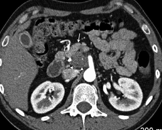
Uniformly, resectable disease includes tumors that do not appear to contact the CA, HA, SMA, or SMV given that a margin-negative resection can often be achieved without any prior therapy in patients with these tumors. More recently, several groups have also considered as resectable tumors with limited involvement of the SMV–PV confluence in which an R0 resection is still possible, albeit with vascular resection and reconstruction. For example, the definition used at MDACC considers tumors resectable if there is abutment of the SMV–PV with patent vessels (no occlusion) [20]. The NCCN definition also classifies as resectable any tumor with ≤180° contact of the SMV–PV but without any vein contour irregularity [21] (Table 1.1, Fig. 1.1).
Definitions of Locally Advanced Disease
Definitions of locally advanced disease (LAD) also vary, although in general, LAD characterizes those patients in whom the likelihood of response to non-operative therapy sufficient to allow for a subsequent margin-negative resection is nearly
Fig. 1.1 CT scan demonstrating resectable pancreatic adenocarcinoma of the head with SMV abutment of less than 180° and a clear fat plane between the tumor and the SMA R.A.
zero. For example, according to the MDACC definition, locally advanced disease consists of tumors that involve the SMA greater than 180°, those that encase the CA or CHA without a technical option for reconstruction, and those which occlude the SMV–PV with no technical option for reconstruction [20].
The NCCN considers tumors of the pancreatic head and uncinate process locally advanced if the tumor demonstrates contact with the first jejunal vein draining into the SMV or an unreconstructable SMV–PV confluence. Tumors with >180° contact with the SMA, CA, or contact the first jejunal arterial SMA branch are also considered locally advanced. Body and tail tumors are considered locally advanced if the SMV–PV confluence is involved and unreconstructable or if there is contact of >180° with the SMA or HA, or with the CA and aorta (Table 1.2, Figs. 1.2 and 1.3).
Definitions of Borderline Resectable Disease
The definition of borderline resectable (BRPC), first termed marginally resectable, was first described in 2001 in a prospective case series by Mehta et al. and was intended to describe patients

Table 1.2 Locally advanced (unresectable) pancreatic cancer definitions
Celiac axis (CA)
Common hepatic artery (CHA)
Superior mesenteric artery (SMA)
Superior mesenteric vein-portal vein (SMV–PV) confluence
MDACC
Encasement
Encasement with no technical option for reconstruction
Encasement >180°
Occluded and no technical option for reconstruction
AHPBA/SSO/SSAT NCCN
Abutment or encasement Contact >180°
Encasement with extension to celiac axis
Encasement >180°
Occlusion without options for reconstruction
Contact with extension to CA or bifurcation
Contact >180° or contact with first jejunal SMA branch
Unreconstructible due to tumor involvement or occlusion, contact with proximal jejunal branch
MDACC MD Anderson Cancer Center, AHPBA/SSO/SSAT Americas Hepato Pancreato-Biliary Association/Society of Surgical Oncology/Society for Surgery of the Alimentary Tract, NCCN National Comprehensive Cancer Network
Fig. 1.2 CT scan of a locally advanced pancreatic adenocarcinoma with obliteration of the SMV at the base of the mesentery, leaving no distal venous target for reconstruction

Fig. 1.3 CT scan of a locally advanced pancreatic adenocarcinoma with encasement of the SMV as well as the CHA (common hepatic artery)

at high risk of grossly positive margins with immediate resection [22]. Patients were treated with 5-FU and radiation therapy and then reevaluated for resection; 9 of 15 patients subsequently underwent resection with negative margins [22]. In 2006, the NCCN first adopted the term “borderline resectable” to characterize the group of patients at high risk for a marginpositive resection and for whom administration of neoadjuvant therapy should be considered.
Over the past decade, a number of different radiographic classification schemes have been subsequently developed to describe which patients are considered borderline resectable, including consensus statements and guidelines from not only the NCCN, but also the International Study Group of Pancreatic Surgery (ISGPS); MDACC; Americas Hepato Pancreato-Biliary Association (AHPBA), Society of Surgical Oncology (SSO), and Society for Surgery of the Alimentary Tract (SSAT) [20, 23, 24]. A definition has also been established within the context of a now-completed multi-institutional prospective trial, the Intergroup borderline resectable pilot study (Alliance A021101) [25]. These criteria have been endorsed by the NCCN and are summarized in Table 1.3 and shown with representative CT scans in Figs. 1.4, 1.5, and 1.6. To date, no single definition has been used uniformly.
The most recent NCCN guidelines outline a definition of BRPC as tumor demonstrating radiographic contact with the SMV–PV of >180°, or contact of ≤180° with contour irregularity or thrombosis of the vein but with suitable vessel proximal and distal to the site of involvement to allow for adequate resection [21]. Solid tumor contact with the IVC is also considered borderline resectable. Regarding arterial involvement, NCCN considers tumor contact with CHA without extension to CA or HA bifurcation and ≤180° contact with SMA to be borderline resectable for pancreatic head lesions. A tumor within the body or tail is considered borderline resectable if there is contact ≤180° with CA or >180° of CA without involvement of aorta and with an uninvolved, intact GDA.
The MDACC definition published in 2006 allows for short segment occlusion of the SMV–
R.A. Snyder et al.
PV as long as a suitable vessel is available above and below the involved segment for reconstruction [20]. Additionally, tumor abutment of ≤180° of the circumference of the SMA and short segment encasement or abutment of the CHA (usually at the GDA origin) is considered potentially resectable. In follow-up work, Katz et al. elaborated on these definitions further, to not only account for anatomical feasibility but also clinical appropriateness for pancreatectomy [26]. MDACC categorized patients into three subsets: Group A comprised patients meeting the anatomic criteria listed above; Group B consisted of patients with preoperative work-up suggestive but not diagnostic of metastasis; and Group C was made up of patients with comorbidities or those with a marginal, but potentially reversible, performance status (typically ECOG 2-3). Group B patients had CT findings suspicious for but not diagnostic of metastatic disease (indeterminate subcentimeter liver lesions or peritoneal or omental nodules too small for biopsy) or known N1 disease as determined by EUS-FNA or pre-referral laparotomy.
In 2008, the AHPBA, SSO, and SSAT held a Consensus Conference to outline a uniform definition of borderline resectable disease [25]. Published in 2009, the proceedings delineated the following criteria to define BRPC: tumorassociated deformity of the SMV–PV, abutment of the SMV–PV ≥180°, short-segment occlusion of the SMV–PV amenable to resection and reconstruction, short-segment involvement of the HA or its branches amenable to resection and reconstruction, and abutment of the SMA (<180°).
The ISGPS published a consensus statement in 2014 intended to promote an internationally agreed upon definition for the subset of patients with BRPC [24]. This group endorses the NCCN criteria for the definition, based on preoperative CT imaging performed within 4 weeks of consideration for resection. Consistent with NCCN, the ISGPS classification of BRPC allows for SMV–PV occlusion if reconstruction is possible, encasement of the GDA up to the HA with encasement of HA without extension to the CA, and abutment of the SMA <180°.
Most recently, members of several cooperative groups, including the Southwest Oncology
Table 1.3 Borderline resectable pancreatic cancer defi nitions
Intergroup (Alliance A021101)
Interface between tumor and vessel <180 ° circumference vessel wall
Reconstructible, shortsegment interface between tumor and vessel of any degree
Moffi tt
NCCN/ISGPS
Contact ≤ 180 ° or contact >180 ° with uninvolved GDA Not specifi ed
AHPBA/SSO/SSAT
MDACC
Celiac axis (CA) Abutment No abutment or encasement
Encasement of GDA up to origin of CHA
Short segment abutment or encasement amenable to reconstruction GDA encasement to hepatic artery with short segment encasement or direct abutment without extension to hepatic bifurcation or celiac axis
Abutment or short segment encasement
Common hepatic artery (CHA)
Abutment <180 ° Abutment <180 ° Contact ≤ 180 ° Circumferential tumor abutment <180 ° Interface between tumor and vessel measuring <180 ° circumference of vessel wall
Superior mesenteric artery (SMA)
Interface between tumor and vessel measuring 180 ° or greater of circumference of vessel wall, and/or reconstructable occlusion
Circumferential abutment or encasement amenable to resection and reconstruction
Contact of >180 ° , contact of ≤ 180 ° with irregularity of vein or thrombosis amenable to resection and reconstruction
Abutment >180 ° or occlusion amenable to resection and reconstruction
Short segment occlusion amenable to resection and reconstruction
Superior mesenteric vein-portal vein (SMV–PV) confl uence
MDACC MD Anderson Cancer Center, AHPBA/SSO/SSAT Americas Hepato Pancreato-Biliary Association/Society of Surgical Oncology/Society for Surgery of the Alimentary Tract, NCCN National Comprehensive Cancer Network, ISGPS International Study Group of Pancreatic Surgeons
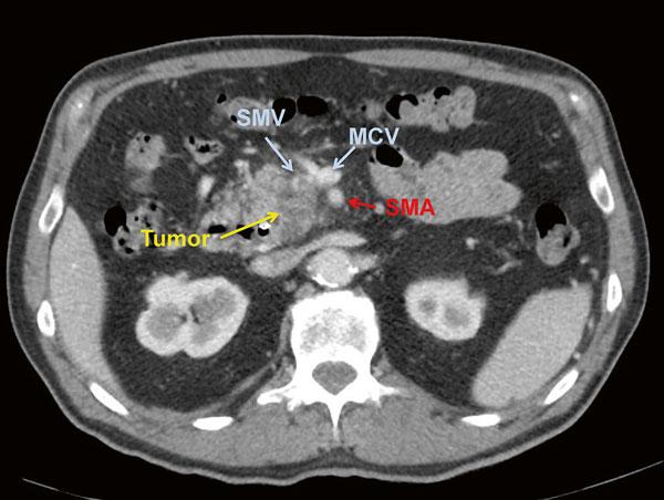
Fig. 1.4 CT scan demonstrating a borderline resectable pancreatic adenocarcinoma with SMV abutment of approximately 180° and subtle haziness posterior to the SMA
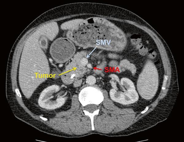
Fig. 1.5 CT scan demonstrating a borderline resectable pancreatic adenocarcinoma with tumor thrombus within the SMV
Group, (SWOG) Alliance for Clinical Trials in Oncology, Eastern Cooperative Oncology Group (ECOG), and Radiation Therapy Oncology Group (RTOG), proposed a precise definition for use in a now-completed pilot study for patients with BRPC. The Alliance Trial (A021101) was a multi-institutional single-armed trial designed to evaluate the feasibility of multi-institutional
study of BRPC using a modified regimen of FOLFIRINOX (oxaliplatin, irinotecan, leucovorin, and 5-FU) followed by 5040 Gy external beam radiation therapy prior to intended surgery [26]. Here, the investigators advocated for an easily reproducible definition based on objective data derived from standard CT imaging and suggested avoidance of subjective or imprecise
R.A. Snyder et al.
Fig. 1.6 CT scan demonstrating a borderline resectable pancreatic adenocarcinoma with SMV abutment of approximately 180° at the base of the mesentery and haziness around the SMA
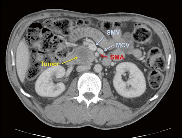
assessments of “impingement” or “abutment.”
This definition consisted of the following: (1) an interface between the tumor and SMV–PV ≥180° of the vein wall circumference; (2) short-segment occlusion of the SMV–PV with normal vein above and below the obstruction amenable to resection and reconstruction; (3) short-segment interface of any degree between tumor and HA with normal artery proximal and distal to the interface amenable to arterial resection and reconstruction; and (4) interface between the SMA and CA measuring <180° of the circumference of the artery.
Classifying Venous Involvement
In addition to the definitions described above, there have been efforts to describe the extent of venous involvement based on preoperative imaging in order to more accurately predict the likelihood of R0 resection and define borderline resectable disease. In 1991, Ishikawa et al. published a classification system of SMV–PV involvement based on the portal phase of preoperative arteriography [27]. Invasion of the SMV–PV was classified as Type (I) normal, (II) smooth shift without narrowing, (III) unilateral narrow-
ing, (IV) bilateral narrowing, (V) bilateral narrowing with the presence of collateral veins.
A follow-up study in 2010 at Fox Chase Cancer Center utilized the Ishikawa definitions to evaluate the effect of neoadjuvant chemoradiation therapy among patients with SMV–PV involvement [28]. Preoperative therapy was associated with improved R0 resection rates and overall survival among patients with Type II or III vein involvement, but not in patients with Type IV or V. However, the number of patients with bilateral involvement or occlusion (Type IV or V) was small.
More recently, Tran Cao et al. correlated preoperative CT imaging of the circumferential SMV–PV tumor-vein interface (TVI) with the presence of histologic vein invasion postresection in order to determine the ability of preoperative radiographic criteria to predict the need for vein resection at the time of pancreaticoduodenectomy [29]. The TVI was assigned to the following classifications: (1) No direct interface with either normal pancreas or fat separating the primary tumor from the vessel, (2) ≤180° of the vessel circumference, (3) >180° of the vessel circumference, or (4) vascular occlusion (absence of contrast within the lumen of the vein in association with adjacent tumor). Based on review of
254 patients undergoing pancreaticoduodenectomy, the authors found that the TVI system predicted the need for SMV–PV resection and histologic vein involvement with reasonable accuracy. Specifically, 89.5 % of patients with TVI >180° or occlusion required SMV–PV resection, and 82.4 % of these patients had documented histologic SMV–PV invasion.
Validation of Classification Systems
Although the various anatomic definitions are similar, there are several important differences among them. Unfortunately, it is very difficult to ascertain which definition is most appropriate without comparison or validation studies. Extrapolating from a number of single-institution, retrospective studies investigating neoadjuvant chemotherapy and/or chemoradiation therapy prior to resection in the borderline resectable population, it is possible to perform a limited comparison of resection rates by definition. By definition, a proportion of patients with initially BRPC are expected to have disease progression, but an additional percentage will ultimately represent reasonable candidates for margin-negative resection. However, it is unclear as to what percent of patients that come to resection after neoadjuvant therapy should be considered the benchmark to validate a definition of BRPC.
Among recent studies using the NCCN consensus definition, resection rates range from 46 to 56 % after preoperative therapy [30, 31]. Rates of resection among patients considered to have borderline resectable disease based on the MD Anderson definition range between 18 and 46 % after completion of neoadjuvant therapy [26, 32, 33]. In a study comparing rates using both the AHPBA/SSO/SSAT definition and the MD Anderson definition among the same study population, resection rates differed significantly at 84 % and 78 %, respectively [34]. Given that the AHPBA/SSO/SSAT definition considers abutment or encasement of the SMV–PV to be BRPC, whereas MD Anderson considers those patients candidates for upfront resection, it is not surpris-
R.A. Snyder et al.
ing that resection rates differ. Additionally, MD Anderson definition allows for a greater extent of arterial involvement, specifically abutment of the celiac artery, compared to the AHPBA/SSO/ SSAT definition.
Of equal importance, the neoadjuvant treatment regimens differed significantly across these studies which adds another significant confounder, further limiting comparisons of the various definitions.
Special Considerations: Aberrant Anatomy
As many as 40–45 % of patients have a variation in visceral arterial anatomy, which becomes an important consideration in the surgical evaluation of patients with pancreatic cancer [35]. The most common anomaly is a replaced or accessory right hepatic artery, originating off the SMA in approximately 11–15 % of patients. In this setting, the right HA courses posterior to the head of the pancreas and is positioned lateral to the PV, entering the right side of the hepatoduodenal ligament. Additionally, in approximately 2.5 % of patients, a replaced CHA can arise from the SMA and follow a similar path.
Although not specifically addressed in the contemporary definitions of BRPC, abutment or encasement of either a replaced (not accessory) right HA or replaced CHA should also be considered BRPC, consistent with borderline resectable definitions of CHA involvement.
Summary
The concept of BRPC is a relatively recent, but important development in the treatment of pancreatic cancer. The evolution of this category is two-fold. First, even with modern imaging techniques, a percentage of patients who undergo attempted resection will have microscopic residual disease left behind, primarily adjacent to the mesenteric vasculature. These patients, unfortunately, do not benefit from immediate resection and should be treated instead with systemic and/or
locoregional therapy prior to attempted resection. Second, largely due to advances in our understanding of the biology of the disease and to somewhat improved locoregional and systemic therapies, there appears to be a subset of patients whose tumors do not progress (or may even regress) following neoadjuvant therapy. These patients may represent suitable candidates for resection. These patients are considered to have borderline resectable tumors.
Although conceptually this definition appears fairly straightforward, the precise definition of BRPC remains somewhat subjective. While the various definitions have similar core components, differences exist regarding the precise details of vascular involvement. In addition, with significant institutional differences associated with the treatment of borderline resectable disease, a direct comparison amongst these definitions is nearly impossible. The recently completed Alliance pilot trial used a more objective definition of BRPC with attempted centralized review for rapid evaluation and consistency in a multicenter fashion. The pilot trial has recently been completed, and it is our hope that the success of this trial will pave the way not only for future studies in regard to the best treatment options, but also for a universal definition of BRPC.
References
1. Group USCSW, editor. United States cancer statistics: 1999–2011 incidence and mortality web-based report. Atlanta: U.S. Department of Health and Human Services, Centers for Disease Control and Prevention and National Cancer Institute. 2014. www.cdc.gov/uscs
2. Howlader N, Noone AM, Krapcho M, Garshell J, Miller D, Altekruse SF, Kosary CL, Yu M, Ruhl J, Tatalovich Z, Mariotto A, Lewis DR, Chen HS, Feuer EJ, Cronin KA, editors. SEER cancer statistics review, 1975–2011. 2013 ed. Bethesda: National Cancer Institute; 2013.
3. Millikan KW, Deziel DJ, Silverstein JC, Kanjo TM, Christein JD, Doolas A, et al. Prognostic factors associated with resectable adenocarcinoma of the head of the pancreas. Am Surg. 1999;65(7):618–23; discussion 623–4.
4. Sohn TA, Yeo CJ, Cameron JL, Koniaris L, Kaushal S, Abrams RA, et al. Resected adenocarcinoma of the pancreas-616 patients: results, outcomes, and prognostic indicators. J Gastrointest Surg. 2000;4(6):567–79.
5. Benassai G, Mastrorilli M, Quarto G, Cappiello A, Giani U, Mosella G. Survival after pancreaticoduodenectomy for ductal adenocarcinoma of the head of the pancreas. Chir Ital. 2000;52(3):263–70.
6. Bassi C, Stocken DD, Olah A, Friess H, Buckels J, Hickey H, et al. Influence of surgical resection and post-operative complications on survival following adjuvant treatment for pancreatic cancer in the ESPAC-1 randomized controlled trial. Dig Surg. 2005;22(5):353–63.
7. Richter A, Niedergethmann M, Sturm JW, Lorenz D, Post S, Trede M. Long-term results of partial pancreaticoduodenectomy for ductal adenocarcinoma of the pancreatic head: 25-year experience. World J Surg. 2003;27(3):324–9.
8. Takai S, Satoi S, Toyokawa H, Yanagimoto H, Sugimoto N, Tsuji K, et al. Clinicopathologic evaluation after resection for ductal adenocarcinoma of the pancreas: a retrospective, single-institution experience. Pancreas. 2003;26(3):243–9.
9. Kuhlmann KFD, de Castro SMM, Wesseling JG, ten Kate FJW, Offerhaus GJA, Busch ORC, et al. Surgical treatment of pancreatic adenocarcinoma; actual survival and prognostic factors in 343 patients. Eur J Cancer. 2004;40(4):549–58.
10. Bilimoria KY, Talamonti MS, Sener SF, Bilimoria MM, Stewart AK, Winchester DP, et al. Effect of hospital volume on margin status after pancreaticoduodenectomy for cancer. J Am Coll Surg. 2008;207(4):510–9.
11. Winter JM, Cameron JL, Campbell KA, Arnold MA, Chang DC, Coleman J, et al. 1423 pancreaticoduodenectomies for pancreatic cancer: a single-institution experience. J Gastrointest Surg. 2006;10(9):1199–210; discussion 1210–1.
12. Allema JH, Reinders ME, van Gulik TM, van Leeuwen DJ, de Wit LT, Verbeek PC, et al. Portal vein resection in patients undergoing pancreatoduodenectomy for carcinoma of the pancreatic head. Br J Surg. 1994;81(11):1642–6.
13. Fuhrman GM, Leach SD, Staley CA, Cusack JC, Charnsangavej C, Cleary KR, et al. Rationale for en bloc vein resection in the treatment of pancreatic adenocarcinoma adherent to the superior mesentericportal vein confluence. Pancreatic Tumor Study Group. Ann Surg. 1996;223(2):154–62.
14. Kelly KJ, Winslow E, Kooby D, Lad NL, Parikh AA, Scoggins CR, et al. Vein involvement during pancreaticoduodenectomy: is there a need for redefinition of “borderline resectable disease”? J Gastrointest Surg. 2013;17(7):1209–17; discussion 1217.
15. Tseng JF, Raut CP, Lee JE, Pisters PWT, Vauthey J-N, Abdalla EK, et al. Pancreaticoduodenectomy with vascular resection: margin status and survival duration. J Gastrointest Surg. 2004;8(8):935–49; discussion 949–50.
16. Amano H, Miura F, Toyota N, Wada K, Katoh K-I, Hayano K, et al. Is pancreatectomy with arterial reconstruction a safe and useful procedure for locally advanced pancreatic cancer? J Hepatobiliary Pancreat Surg. 2009;16(6):850–7.
17. Bockhorn M, Burdelski C, Bogoevski D, Sgourakis G, Yekebas EF, Izbicki JR. Arterial en bloc resection for pancreatic carcinoma. Br J Surg. 2011;98(1):86–92.
18. Mollberg N, Rahbari NN, Koch M, Hartwig W, Hoeger Y, Büchler MW, et al. Arterial resection during pancreatectomy for pancreatic cancer: a systematic review and meta-analysis. Ann Surg. 2011;254(6): 882–93.
19. Landry J, Catalano PJ, Staley C, Harris W, Hoffman J, Talamonti M, et al. Randomized phase II study of gemcitabine plus radiotherapy versus gemcitabine, 5-fluorouracil, and cisplatin followed by radiotherapy and 5-fluorouracil for patients with locally advanced, potentially resectable pancreatic adenocarcinoma. J Surg Oncol. 2010;101(7):587–92.
20. Varadhachary GR, Tamm EP, Abbruzzese JL, Xiong HQ, Crane CH, Wang H, et al. Borderline resectable pancreatic cancer: definitions, management, and role of preoperative therapy. Ann Surg Oncol. 2006; 13(8):1035–46.
21. al TME, editor. NCCN Clinical practice guidelines in oncology: pancreatic adenocarcinoma. 2nd ed. National Comprehensive Cancer Network.
22. Mehta VK, Fisher G, Ford JA, Poen JC, Vierra MA, Oberhelman H, et al. Preoperative chemoradiation for marginally resectable adenocarcinoma of the pancreas. J Gastrointest Surg. 2001;5(1):27–35.
23. Bockhorn M, Uzunoglu FG, Adham M, Imrie C, Milicevic M, Sandberg AA, et al. Borderline resectable pancreatic cancer: a consensus statement by the International Study Group of Pancreatic Surgery (ISGPS). Surgery. 2014;155(6):977–88.
24. Abrams RA, Lowy AM, O’Reilly EM, Wolff RA, Picozzi VJ, Pisters PWT. Combined modality treatment of resectable and borderline resectable pancreas cancer: expert consensus statement. Ann Surg Oncol. 2009;16(7):1751–6.
25. Katz MHG, Marsh R, Herman JM, Shi Q, Collison E, Venook AP, et al. Borderline resectable pancreatic cancer: need for standardization and methods for optimal clinical trial design. Ann Surg Oncol. 2013;20(8): 2787–95.
26. Katz MHG, Pisters PWT, Evans DB, Sun CC, Lee JE, Fleming JB, et al. Borderline resectable pancreatic cancer: the importance of this emerging stage of
disease. J Am Coll Surg. 2008;206(5):833–46; discussion 846–8.
27. Ishikawa O, Ohigashi H, Imaoka S, Furukawa H, Sasaki Y, Fujita M, et al. Preoperative indications for extended pancreatectomy for locally advanced pancreas cancer involving the portal vein. Ann Surg. 1992;215(3):231–6.
28. Chun YS, Milestone BN, Watson JC, Cohen SJ, Burtness B, Engstrom PF, et al. Defining venous involvement in borderline resectable pancreatic cancer. Ann Surg Oncol. 2010;17(11):2832–8.
29. Tran Cao HS, Balachandran A, Wang H, NoguerasGonzález GM, Bailey CE, Lee JE, et al. Radiographic tumor-vein interface as a predictor of intraoperative, pathologic, and oncologic outcomes in resectable and borderline resectable pancreatic cancer. J Gastrointest Surg. 2014;18(2):269–78; discussion 278.
30. Chuong MD, Springett GM, Freilich JM, Park CK, Weber JM, Mellon EA, et al. Stereotactic body radiation therapy for locally advanced and borderline resectable pancreatic cancer is effective and well tolerated. Int J Radiat Oncol Biol Phys. 2013;86(3):516–22.
31. Kang CM, Chung YE, Park JY, Sung JS, Hwang HK, Choi HJ, et al. Potential contribution of preoperative neoadjuvant concurrent chemoradiation therapy on margin-negative resection in borderline resectable pancreatic cancer. J Gastrointest Surg. 2012;16(3):509–17.
32. Stokes JB, Nolan NJ, Stelow EB, Walters DM, Weiss GR, de Lange EE, et al. Preoperative capecitabine and concurrent radiation for borderline resectable pancreatic cancer. Ann Surg Oncol. 2011;18(3):619–27.
33. Turrini O, Viret F, Moureau-Zabotto L, Guiramand J, Moutardier V, Lelong B, et al. Neoadjuvant chemoradiation and pancreaticoduodenectomy for initially locally advanced head pancreatic adenocarcinoma. Eur J Surg Oncol. 2009;35(12):1306–11.
34. Katz MHG, Fleming JB, Bhosale P, Varadhachary G, Lee JE, Wolff R, et al. Response of borderline resectable pancreatic cancer to neoadjuvant therapy is not reflected by radiographic indicators. Cancer. 2012; 118(23):5749–56.
35. Balachandran A, Darden DL, Tamm EP, Faria SC, Evans DB, Charnsangavej C. Arterial variants in pancreatic adenocarcinoma. Abdom Imaging. 2008;33(2): 214–21.
Part 2
Staging and Pretreatment Management
Imaging Evaluation of Borderline Pancreatic Cancer
Kyuran Ann Choe and Nicholas M. McDonald
Introduction
Pancreatic adenocarcinoma has been increasing in incidence [1] and it has been estimated to represent 3 % of new cancer diagnoses and 7 % of cancer deaths in 2014 [2]. Most commonly, the staging of pancreatic carcinoma follows American Joint Committee on Cancer (AJCC) guidelines. In the absence of metastatic disease, there is concurrent classification of tumors into resectable, borderline resectable, and unresectable locally advanced disease for the purposes of clinical management [3–6]. Although there is discussion regarding some of the criteria that define borderline resectable and locally advanced pancreatic cancer, both tumor staging and evaluation of local extent of disease for potential resectability are based on findings seen on cross-sectional imaging, primarily contrast-enhanced multidetector computed tomography (MDCT) and magnetic resonance imaging (MRI).
Ultrasound
Transabdominal ultrasound is frequently the initial examination that is performed in a patient with jaundice and it is sensitive for the detection of biliary ductal dilation. However, the etiology of the biliary obstruction can be difficult to elucidate and visualization of the entirety of the pancreas is difficult due to patient body habitus and interference from overlying bowel gas which limits tumor detection. In addition, when a tumor is present, complete assessment of local tumor extension is limited [7]. The finding of biliary and/or pancreatic ductal dilation on ultrasound commonly prompts further imaging evaluation by CT or MRI. The role of endoscopic ultrasound will be discussed in Chaps. 3 and 4
Multidetector Computed Tomography
K.A. Choe, M.D. (*) • N.M. McDonald, M.D. Department of Radiology, University of Cincinnati Medical Center, 234 Goodman St. ML 0761, Cincinnati, OH 45267, USA
e-mail: ann.choe@uc.edu; mcdonana@ucmail.uc.edu
Optimal imaging of the abdomen and pelvis by CT for the evaluation of patients with pancreatic carcinoma is performed using multidetector scanners which provide thin slices and isotropic data for multiplanar reformatted images and 3D reconstructions. Rapid image acquisition allows for multiple phases of image acquisition with contrast enhancement optimized for tumor detection, vascular assessment, and evaluation for metastatic disease. Initial noncontrast imaging of the
17 © Springer International Publishing Switzerland 2016 M.H.G. Katz, S.A. Ahmad (eds.), Multimodality Management of Borderline Resectable Pancreatic Cancer, DOI 10.1007/978-3-319-22780-1_2
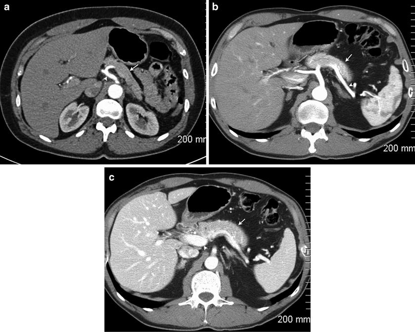
Fig. 2.1 Normal multiphasic appearance of the pancreas and adjacent structures. The arterial phase image (a) demonstrates dense arterial enhancement of the celiac artery (arrowhead) and mild enhancement of the pancreas (arrow). The pancreatic parenchymal phase (b) shows excellent arterial enhancement of the splenic artery
abdomen is not required but is recommended in the 2012 National Comprehensive Cancer Network (NCCN) guidelines [8]. Negative enteric contrast (usually water) is used to avoid obscuration of the vasculature particularly if 3D reconstructions are utilized [9, 10]. Following intravenous high-iodine (>300 mg I/mL) contrast administration at 3–5 mL/s [11], images may be acquired in the angiographic arterial phase, pancreatic parenchymal phase [12], and portal venous (hepatic) phase (Fig. 2.1). Images are acquired in at least two of these phases, one of which is the portal venous (hepatic) phase for the detection of metastatic disease [13, 14]. At our institution, the portal venous phase extends through the pelvis to
(arrowhead) with enhancement of the pancreas (arrow). In the portal venous phase (c), there is dense enhancement of the portal vein (arrowhead) with persistent enhancement of the pancreas (arrow). There is parenchymal enhancement of the liver
assess for metastatic disease if a recent CT has not been performed. The arterial angiographic phase (Fig. 2.1a) will optimally demonstrate the arteries with less background solid organ enhancement (for 3D volume rendered images), but has less sensitivity for lesion detection due to less image contrast between the tumor and normal parenchyma [13]. Some authors advocate image acquisition in the late arterial/pancreatic parenchymal phase (Fig. 2.1b) to optimize lesion detection and local arterial vascular involvement since the image contrast between the tumor and normal parenchyma is greater in this phase in addition to greater arterial enhancement [13, 15, 16]. The timing of image acquisition is dependent
K.A. Choe and N.M. McDonald
2.2 Pancreatic adenocarcinoma in the uncinate process (arrowhead) appears hypodense in comparison to the normal parenchyma (arrow)
upon the rate of intravenous contrast administration which is 3–5 mL/s. The arterial phase of enhancement is at 30, 25, and 20 s following the start of peripheral intravenous contrast administration at an injection rate of 3, 4, and 5 mL/s, respectively [13]. Similarly, the normal pancreas demonstrates peak parenchymal enhancement at 50, 45, and 40 s, respectively [13]. Images in the portal venous phase are acquired between 60 and 70 s [13]. Images are typically reconstructed at slice thicknesses of 3 mm or less [3]. The thinner slice data (1.0 mm or less) is used for multiplanar and 3D reconstruction. If the initial examination is not of diagnostic quality to perform adequate tumor and vascular assessment, it should be repeated using a dedicated pancreas cancer protocol [11]. Preferably, imaging is performed prior to biliary stent placement since the stent may cause streak artifact at the level of the pancreatic head limiting evaluation of the local extent of tumor and secondary pancreatitis may obscure the primary lesion and complicate vascular assessment [3, 14].
Pancreatic adenocarcinoma is typically isodense to the normal pancreas on the noncontrast images, limiting their utility for the purposes of staging. Most pancreatic adenocarcinoma tumors enhance less than the normal pancreatic parenchyma and appear hypodense on contrast-
enhanced images (Fig. 2.2). The sensitivity for the detection of tumor is greatest in the pancreatic parenchymal phase in comparison to the arterial and portal venous phases due to maximal differential enhancement and image contrast between tumor and normal parenchyma [13]. Isoattenuating and isoenhancing tumors (Fig. 2.3) comprise a small portion of tumors—5.4 % of pancreatic adenocarcinoma tumors evaluated in a recent study [17] and 11 % in an older study [18]. A higher percentage of smaller (2 cm or less) tumors have been shown to be isoattenuating [19]. These tumors are difficult to detect but may be inferred by secondary signs including mass effect of the tumor on the contour of the pancreas or on adjacent structures or by the level of biliary and/or pancreatic ductal obstruction [17, 18]. When tumors are isoattenuating, evaluation of local extent of disease can be difficult; particularly problematic is the assessment of abutment or encasement of the intrapancreatic portion of the portal vein and superior mesenteric vein in the absence of distortion.
Evaluation of the local extent of the tumor requires careful evaluation of the local vasculature, including the celiac artery and branches, superior mesenteric artery, superior mesenteric vein, and portal vein. Evaluation for aberrant vasculature such as a replaced or accessory right
Fig.
Another random document with no related content on Scribd:
microscope. On blood serum and potato it forms a grayish white layer.
Pathogenesis. The bacillus is inoculable on other pigeons and as it usually appears in the young birds in the nest, still fed by the parent bird, it is probable that no inflammation nor abrasion is necessary to make it take. Pure cultures inoculated in the mouth gave rise to the usual local type of the disease. When inoculated subcutem it caused a local necrotic inflammation.
In mice subcutaneous injections proved fatal in five days with general dissemination of the bacillus. There are congested and hemorrhagic spots on the lungs, enlarged spleen, and the liver is marbled by numerous necrotic white masses, in the centre of which the capillaries are found to be blocked with the bacilli. This is so pathognomonic that Löffler looks on the inoculation of mice as the best means of diagnosis.
Inoculated rabbits showed inflammation in the seat of inoculation and sometimes fibrinous peritonitis and enlarged spleen. Inoculation on the cornea produced a false membrane.
In Guinea pigs induration and ulceration occurred in the seat of inoculation but recovery followed in 14 days.
Sparrows inoculated in the pectoral muscles died in three days with yellowish necrotic tissue highly charged with bacilli.
Inoculation of the chicken by Löffler and Megnin produced a circumscribed redness which soon disappeared. On the other hand Krajewski, Colin, Loir and Ducloux seem to have inoculated chickens successfully, and Cadeac says that the cultures are infecting for sparrows, pigeons, turkeys, chickens and ducks. It rests uncertain therefore whether the pseudomembranous pharyngitis of hens is a distinct disease as alleged by Löffler and Megnin or if the chickens used by these observers were not already immune by reason of a prior attack.
Löffler’s experiments showed that dogs and rats were immune. Loir and Ducloux failed to infect cattle.
In infected dove-cots a comparative immunity is attained by the older pigeons, which continue to harbor the germ, but do not suffer materially from its presence. They however communicate it to the susceptible young in the milky secretion produced in the crop and
with which they feed them, and these accordingly perish in large numbers. Thus pigeons that are themselves in fine condition become the propagators of the bacillus to the more impressible.
Sparrows and other small birds are also held to be common propagators of the germ, and if they too can secure an individual immunity and yet harbor the bacillus, their passage from yard to yard may be attended with great danger. The grains soiled by their bills and not swallowed are common media of transmission.
Loir and Ducloux found the affection transmissible between man and pigeon. The identity of the bacillus with that of genuine diphtheria in man appears to have been thoroughly disproved by the observations of Roux and Yersin.
The following differential characters have been noted:
Bacillus Diphtheriæ (KlebsLöffler).
1. In gelatine cultures grows only above 23°C.
2. Kills Guinea-pig and dog.
3. Mice immune.
4. Does not grow on potatoe.
Bacillus Diphtheriæ Columbarum
1. In gelatine cultures grows at 15–17°C.
2. Guinea-pig and dog nearly immune.
3. Mice usually die with hepatic necrosis.
4. Grows luxuriantly on potatoe.
It may be accepted as demonstrated that the common diphtheria of birds is essentially distinct from the genuine diphtheria of man, and that when such diphtheria of the bird is conveyed to man as has been often alleged (Richter, Gips, Bonig, Gerhart, etc.), it is one of the forms of pseudo diphtheria that is produced, and not that which is caused by the Klebs-Löffler bacillus. Dr. V. A. Moore, who has cultivated specimens of the bacillus diphtheriæ Columbarum obtained from Germany, considers the germ as belonging to the group of the bacillus coli communis, and as not the cause of the chicken diphtheria in America. Further investigation must settle whether the bacillus diphtheriæ Columbarum is the one cause of this affection in Europe, and what is the microbian cause or causes of the disease in America.
Incubation. This is very variable. False membranes may form in twenty-four hours in some cases; in other cases they may be delayed from four to fourteen days (Colin, Babes, Puscarin, Marinescu).
Symptoms. There is dullness, prostration, sunken head, ruffled feathers, altered hoarse voice, drooping wings, wheezing breathing, difficult deglutition, sneezing, and patches of dark red congestion in the fauces covered with a thin film, at first translucent, but soon becoming dense, adherent, opaque, whitish or yellowish. As it becomes older this deposit becomes granular, wrinkled, dry and friable. It is more adherent in chickens than in pigeons and causes bleeding when detached. Necrotic changes may take place in the mucosa leading to considerable loss of tissue, and even to perforations of the soft palate, pharynx or œsophagus. It may remain circumscribed by the region of the mouth and end in an early recovery, or it may extend to the organs of the chest and abdomen, or the germs may proliferate largely in the blood and induce fatal results. On the other hand it may become subacute or even chronic, and, as already noted in the case of the parent pigeons, it may persist as an infecting disease without materially injuring the general health of a comparatively immune animal.
The affected nasal passages become filled by frothy liquid and blocked by false membranes, so that the bird is driven to breath through the open mouth. The skin around the nares, and eyelids and the cavity beneath the eye may be covered with the false membrane, by the increase of this product the bones may be driven out of place, so that the palatines press downward, the eyeball is pressed outward and the root of the beak may seem swollen. The false membranes that form on the skin or reach the surface are soft, creamy, cheesy, or dry, granular and friable.
When the eye is specially affected there are swelling of the lids, profuse lachrymation, closure of the lids by adhesion, and formation around their borders or on their inner surface and on the membrana nictitans of false membranes which press the lids outward more or less unevenly, and may be easily recognized when the lid is everted. The cornea and even the interior of the eye may suffer, leading to perforation, internal tension, and in some cases atrophy, with permanent blindness.
The tongue may suffer on the tip as in pip, or on its dorsum, from which the disease extends to the larynx, trachea and even the air sacks, which become filled with false membranes, that are coughed up, and decomposing in the mouth, add to the infection and fœtor. Dyspnœa and cyanosis of comb and wattles are marked features.
The extension may take place downward along the alimentary track, the false membranes forming on the gullet or crop and interfering with swallowing or digestion, or on the intestine and determining a fœtid, often greenish or bloody diarrhœa with indications of false membranes. Vomiting may be a marked symptom.
The skin is usually attacked secondarily around the margin of the beak, the eyelids, the nares, the ears, the comb, the wattles, the anus, but it may develop at any point where the infecting material has touched an abraded surface.
Trinchera found that in acute cases the acme was reached in fifteen days after which improvement might be looked for. A chronic form affecting the gullet might however persist indefinitely in pigeons without proving incompatible with good health.
Paralysis of the wings or limbs may remain after the healing of the local lesions.
Mortality. Prognosis. The disease is very fatal to both pigeons and chickens, 50, 70 or even 100 per cent. perishing when a flock is attacked for the first time. In flocks that have previously suffered, on the other hand, a large number are practically immune, and even if they contract the disease it assumes a mild form, and they survive but may retain the germ and continue to communicate it to others. Even the young of such immune flocks suffer less severely, coming as they probably do from less susceptible and therefore surviving birds, or having already perhaps contracted a mild (non-fatal) type of the disease from their parents.
Differential Diagnosis. From psorospermosis (coccidiosis) it is distinguished by its origin on the mucous membranes, and not on the skin, the skin lesion being a secondary one. In psorospermosis the primary lesion is usually on the skin, from which it extends to the mouth and especially along its floor. In psorospermosis the morbid deposit assumes the form of rounded warty-like masses, on comb or
wattles; is easily propagated by inoculation, is promptly checked by antiseptics, does not tend to produce internal extension nor generalization, and on microscopic examination shows numerous spheroidal coccidia intermingled with the epidermic cells and possessing amœboid movement. By virtue of this automatic movement they make their way between and into the epidermic cells in which they multiply.
From the croupous angina of Rivolta it is distinguished by the absence of the infusoria (monocercomonas gallinæ) to which he, Delprato and Pfeiffer attributed that affection. The monocercomonas is a flagellate organism 14 μ to 25 μ in length and 5 μ to 7 μ in breadth. Its rounded end bears one flagellum as long as the body, and its acute end three flagella which give it active motions. These are found in the yellowish white swellings of the mucosa, which vary in size from a millet seed to a pea, covering a hyperæmic spot and composed of epithelial cells, blood globules—white and red,— leucocytes, granules and the infusoria. The false membrane is remarkable for its lack of consistency and its tendency to invade the mouth and gullet rather than the air passages. These infusoria are not colored by picrocarminate of ammonia, but stain by methylviolet and then appear as round or slightly irregular hyaline bodies.
From aspergillus disease of pigeons, by the absence of the characteristic, miliary, white nodule of that disease showing caseated contents intermixed with an abundant mycelium of aspergillus fumigatus. The aspergillus disease attacks especially the mouth but may also implicate the gullet, lungs, liver, intestine and kidneys. The microscopic examination of the exudate is conclusive, by reason of the presence of the bacillus diphtheriæ columbarum, and the comparative absence of the filamentous mycelium.
Treatment. This is mainly prophylactic. The first step must be to separate the sick and healthy, destroying the former, or shutting them up in a special enclosure apart from all other birds. In the case of valuable chickens, their eggs may be set under other hens and the young raised apart from the suspected flock. This may even be attempted in pigeons, the common eggs being removed and the valuable ones put in their place under a healthy sitting dove. In the case of pigeons that have been recently through the disease they should be kept strictly by themselves, even though they may appear
to have regained perfect health. The dead bodies must be burned or deeply buried. Sparrows and even rabbits dying in the vicinity must be similarly disposed of, and where the disease prevails sparrows and small birds may be exterminated as probable bearers of infection.
The purchase of strange birds must be carefully guarded, none being taken that show weeping eyes, nasal discharge, labored or wheezing breathing, and all new birds should be placed by themselves in quarantine for ten to fifteen days. Finally a thorough disinfection of the place where the sick have been is of first importance. Thorough cleaning of the poultry house, followed by a coat of white-wash, every gallon of which contains four ounces of chloride of lime, or one drachm of mercuric chloride will usually prove effective. The poultry runs should be liberally sprinkled with a solution of sulphuric or hydrochloric acid, one part to 1000. The same may be used on the building, which may further be fumigated by burning sulphur.
Poultry shows should be kept under the most rigorous sanitary supervision.
Curative treatment is only profitable in the case of specially valuable birds, and even then only, as a rule, when the disease is confined to the nose, mouth, larynx and pharynx. The affected parts may be brushed with a solution of chloride of iron (1 dr. of the tincture to 1 oz. water), nitrate of silver (2 grs. to 1 oz. water), sulphide of calcium (½ dr. to 1 oz. water), tannin (10 grs. to 1 oz.). Tincture of iodine may be applied direct, or a solution of carbolic acid or of creosote or creolin (1 part to 50) will often succeed. Thomassen recommends the removal of the false membranes and the application of boric acid followed by dry sulphur. Benoist says the majority recover when made to inhale the fumes of oil of turpentine evaporated at a gentle heat twice a day.
As internal medication, or to correct the intestinal affection, sulphate of iron may be dissolved in the drinking water, or salicylic acid may be given in pill form with molasses.
CHRONIC PHARYNGITIS.
Sequel of acute: or subacute from the first. Due to œstrus, in cattle to summer catarrh, tubercle or actinomycosis. Lymphatic horses predisposed; attends chronic indigestion; in swine tonsilitis. Symptoms: chronic cough, easily roused, wheezy or mucous; nasal discharge; low condition; lack of spirit. Lesions: congestion; softenings; erosions; cicatrices; tonsilitis; abscesses; specific deposits. Treatment: hygienic; antiparasitic; astringent; antiseptic; derivative; counter-irritant; tonic inhalations and electuaries. Bitters. Iron.
Causes and Nature. Chronic pharyngitis in animals may be a simple continuation of the acute, in a milder form, or it may assume a subacute or chronic type from the first and never rise to the intensity that would characterize the acute. It may be a simple catarrhal affection or it may become more or less follicular or glandular. Again in horses it is not infrequently a result of the hibernation form of the œstrus (bots) attached to the delicate pharyngeal mucosa, and in cattle from the extension of the chronic summer catarrh, or from the local development of tubercle or actinomycosis in the walls of the pharynx or in the adjacent lymph glands. Horses of a soft, lymphatic constitution, with a heavy coat, confined in close warm stalls, and which perspire abundantly are especially liable to the affection. It may also be an accompaniment and result of chronic gastric indigestion. In swine the affection is commonly associated with tonsilitis.
Symptoms. In many cases the main symptom is a chronic cough which is aroused by any cause of irritation, feed, especially dry or fibrous fodder, cold drinking water, sudden passing from the hot stable to the cold outer air, reining in, pressure on the throat, or sudden active exertion. If the cartilages are calcified it may be impossible to rouse the cough by pressure. The cough is often dry and wheezy, rather than soft and gurgling as in the second stage of acute pharyngitis, and is repeated several times paroxysmally. In the intervals there is more or less stertor or wheezing, or a distinct rattle especially when the neck is curved by drawing the nose inward. Deglutition may be interfered with but this shows most with the first swallow, which in the case of liquids may be returned through the
nose, whereas those that follow go down without difficulty. A lateral swelling of the parts above the larynx or a bulging of the parotids is not uncommon. Discharge from the nose of a mucopurulent character is usually present, but often so scanty as to be overlooked. There is usually loss of flesh and lack of vigor even if the subject is well fed.
Lesions. In the simple catarrhal form the mucous membrane of the lateral pharyngeal walls, the posterior pillars of the palate and the back of the soft palate, is red, congested, with arborescent vessels, thickening, and puckering into rugæ. The epithelium has lost its translucency, become opaque and granular, and its desquamation in spots and patches may leave erosions, ulcers more or less deep, and white drawn cicatrices. When the follicles and mucous crypts are involved (follicular) they stand out like millet seed, peas or beans, and may show ulceration or minute abscess. In pigs especially, tonsilitis is liable to be present, and the tonsillar follicles are filled and distended with tenacious mucous, a caseous granular debris, or even a cretaceous material. In the vicinity of the tonsils, minute abscesses may exist in or beneath the mucosa.
Ulceration may be the result of tubercle, glanders, actinomycosis, aspergillus, sarcoma, or some local infection, and attendant symptoms of one or other of these diseases will guide the diagnosis. Thus in tubercle there will be the implication of the adjacent lymph glands and usually of distant ones; in glanders the deposits in the nose, submaxillary lymph glands and lungs will enable one to diagnosticate; in actinomycosis the hardness of the neoplasm and the presence of the yellowish tufts which present under the microscope the concentrically arranged club-shaped elements, will show its nature; and in sarcoma or carcinoma the structure of the new tissue will decide its character. The pharyngeal muscles are the seat of granular or fatty degeneration or of fibroid change. Friedenreich speaks of a fold from the vault of the pharynx which had nearly closed the passage and had killed the horse by inability to swallow.
Treatment. Chronic pharyngitis is usually a very obstinate affection and demands careful hygienic as well as medicinal treatment. Hot, foul stables, unduly thick coats, unwholesome food, irregular feeding, excessive meals at long intervals, overwork, undue
exposure to cold and wet, lack of sunshine or of grooming are to be corrected. Next, the removal of mechanical irritants such as pharyngeal bots, actinomycosis growths, etc., will be in order. Then the use of astringents and antiseptics internally and of derivatives externally will be demanded. An occasional embrocation of mustard, or the application of ammonia and oil, will often serve a good purpose, and in obstinate cases the hot iron in points will sometimes prove effective.
Internally the inhalation of the fumes of tar, carbolic acid, creolin, oil of turpentine, or of burning sulphur kept up continually or frequently repeated. Giving all drink in the form of tar water will often have a good effect. Electuaries made with boric acid, salicylate of soda, ammonium chloride or iodide, borax, with honey, molasses, liquorice, Iceland moss, or gum arabic will often prove beneficial. Agents that stimulate the mucosa may follow, such as balsams of Peru or Tolu, copaiba, cubebs, pilocarpin, wild cherry bark, or these may be combined with the former. Finally a course of tonics are usually of the first importance; iron sulphate, copper sulphate, arsenious acid, arsenite of strychnia may furnish examples.
DEPRAVED APPETITE. STUMP SUCKING. PICA. LICKING DISEASE.
Common features of group. Ruminants; depraved appetite; objects swallowed: hair balls. Sheep eating wool in winter. Pigs eat bristles. Puppies swallow marbles, etc., wantonly. Solipeds swallow hair, plaster, earth, sand, and lick manger or rack. Fowls eat their feathers. Causes: soil exhaustion, lack of lime, soda, potash, phosphorous; relation to osteo malacia; granitic or sandy soils, peat, muck, causative; digestive disorder; faulty food; yearly breeding and heavy milking; constant stabling; dry seasons. Course: chronic. Lesions; emaciation; anæmia; serous exudate; catarrh of the bowels. Treatment: soil; good fodder; salts of soda, potash and lime, phosphates; tonics; apomorphine. Wool eating; example: digestive disorders; emaciation. Treatment: open air; good fodder; salts of the bones and soft tissues; clip nurses; apomorphine.
Definition. We have here a class of morbid habits, which cannot be referred to any constant lesion or group of lesions, and which appear in certain cases to result from example and to constitute nothing more than a bad habit.
Symptoms. Ruminants without any appreciable cause, lick the clothes of their care-takers, chew and swallow articles of clothing of all kinds, bones, old shoes, gloves, socks, cuffs, collars, small forks, pocket-knives, nails, wires, needles, coins, stones, lumps of clay, hair, which may give rise to secondary troubles of a more or less serious kind. Pregnant cows are especially subject to this infirmity. The small pointed objects like pins, needles, ends of wires, etc., which are mostly taken by accident with the food are especially apt to be entangled in the alveoli of the reticulum and make their way to the heart, with fatal effect, or through the abdominal walls creating a fistula. Hair aggregates with saliva, mucus and phosphates, to form balls in the first two or three stomachs. Other indigestible objects may also become encrusted and prove sources of irritation. Licking the skin of another animal is doubtless at times encouraged by the taste of the salts of perspiration, but in other cases it has all the appearance of a mutual kind service as the cow with itching head will walk up and present it to its fellow which rarely fails to respond to the invitation. Stump licking is not uncommon.
Sheep shut up in the winter get in the habit of chewing each other’s wool, thus virtually depilating their fellows and accumulating wool balls in their stomachs.
Pigs when running at large eat human fæces often infecting themselves with the cysticercus cellulosa, and devour their own or their fellows’ bristles, which form ovoid and irritating aggregations in the stomach.
Puppies are proverbial for swallowing every small object that comes in their way, coal, pebbles, marbles, leather, hair, etc., with the result of inducing nausea and vomiting, or more seriously, wounds of the stomach, gastritis and enteritis. In older dogs the habit is more likely to imply rabies.
Solipeds will lick and swallow each others hair, eat off the hair from each other’s tails and manes, eat their clothing, lick the wall plaster, earth or sand, and even the manger or rack. The last named habits are usually connected with disease.
Fowls can digest almost anything they swallow, but if they take to picking their feathers, they create serious injury to the skin and indirectly to the general health.
Causes and Nature. In general terms it may be said that the causes of depraved appetite are very numerous, so that the trouble must be looked upon as a symptom of many morbid conditions in place of a disease sui generis.
Heredity has been invoked as a cause, mainly, it would appear, because the disease appears enzootically on certain exhausted soils, or in herds kept in the same unhygienic conditions. In such cases the real cause is usually to be found in faulty conditions of soil, water, buildings, food, etc., on the correction of which the trouble disappears. When, however, from a long continuance of unhygienic conditions, a weakness of constitution is transmitted from parent to offspring, such hereditary debility may be accepted as a predisposing factor.
An exhausted soil, lacking especially the elements of lime and phosphorus, is a common cause, though by no means the only one. Nessler who analyzed the hay and water, furnished to cattle suffering from this disease in the Black Forest found a notable absence of the soda salts. In others in which osteo malacia was the prominent
symptom the lack was in phosphate of lime as well. In the nature of things the soil that has been continuously cropped to exhaustion is robbed of both earthy and alkaline salts, and the animals fed on its exclusive products suffer not only as regards the nutrition of the bone, but also of the soft parts. Hence Trasbot says that in osteo malacia, pica is never absent. Roloff and Röll hold that it is the first symptom of osteomalacia. In South Africa where the land has been cropped with oats year after year without manure and as long as it will bear, the disease became prevalent in the street car horses fed on the oats, and was corrected by the addition of phosphates, or phosphate bearing food, to the ration. In the older dairying farms of New York which have been kept under grass for a great length of time, and all the milk products sold off, depraved appetite in all its forms is quite frequent. Where the land is originally light and sandy and naturally deficient in lime, osteo malacia is often a concurrent disorder. The two conditions may however occur independently of each other, and especially may pica appear alone, in keeping with the greater solubility of the soda and potash salts and the readiness with which these can be washed out of the soil, while the less soluble lime salts in part remain.
Lemcke, Haubner and Siedamgrotzky attribute the disease to a nervous disorder. Lemcke indeed traces the disorder to a lack of phosphorus, and claims that osteomalacia only supervenes where the rheumatic diathesis is also present.
It may be shortly stated that the disease prevails especially on granitic or sandy soils, or on those which are mainly composed of organic debris (peat, muck). Limestone soils and those which contain any considerable proportion of potash or soda are usually exempt.
Digestive disorder though starting from a different point may tend to the same end. A hyperacidity of the stomach has been observed to coincide with the malady, and by interfering with easy and normal digestion, it may stand in the way of such assimilation as is necessary to vigorous health.
Faulty food operates in a similar manner. The exhausted soils, and their products deficient in alkaline and earthy salts have been already referred to; we must also note the evil effect of fibrous fodders, the main nutritive elements of which have been washed out by intemperate weather after they were cut, the rank aqueous
products of wet or swampy soils, the fibrous and siliceous plants (rushes, carex, equisetums, etc.) which grow on poor, wet or soured soils, the innutritious and fermented products of beet sugar factories, and generally the spoilt food which has undergone fermentation.
Yearly breeding and constant milking, by undermining the general health, predisposes so strongly that in many cases the affection is seen in dairy cows, while oxen and young cattle escape. The last period of gestation when the demands for the growing calf are greatest, is the period of especial danger.
Permanent stabling which denies the invigorating influence of sun, exercise and pure air contributes toward the general debility and therefore, in animals that are closely stabled for the winter the spring is especially to be feared, when compulsory inactivity, poor feeding, gestation and milking have combined to reduce the system.
Dry seasons have been noticed to increase the affection manifestly by reducing the supply of food.
Course. The affection is chronic and unless arrested by the supervention of more favorable conditions, may last for a year or more. Spontaneous recovery may set in when turned out to pasturage and open air life, and especially if a rich grain feeding is added. Without change in the conditions however, the tendency is to a fatal result.
Lesions. The victims of the disorder are emaciated, the fatty tissue contains a yellow serum, there is little blood, and that is thin and watery and coagulates loosely, the muscles are pale and flabby, and the gastro-intestinal mucous membrane is the seat of catarrh.
Treatment. To treat rationally and successfully we must adapt the measures to the obvious causes. When the soil has been scourged and exhausted, a change of pasture, and of land used for hay or soiling crops is the first consideration. If these cannot be secured then grain and seeds rich in protein, and alkaline and earthy salts should be added to the ration. Wheat bran, middlings, peas, beans, cotton seed meal, linseed meal, rapecake may be named among available resorts, or in their absence, daily doses of phosphate of lime, and sodium chloride or bicarbonate, or potash salts may be allowed, or even bone dust.
If imperfect digestion is a manifest factor, sodium chloride, or potassium chloride, calcium phosphate, iron and bitters will serve a good end. In hyperacidity, limewater, chalk, or magnesia may be given. If the digestion is torpid, hydrochloric acid with bitters may be resorted to.
Feser and especially Lemcke strongly recommend apomorphia. It is used hypodermically in doses of 2 grains for horse or cow repeated daily for three days.
Secondary Symptoms in Wool-eating Lambs. Lambs from two to six weeks old especially such as suck ewes with woolly udders (merino, Cotswold) first swallow the wool inadvertently, and then acquire a liking for the saline matters in the abundant yolk (merino), till finally the accumulating wool balls produce digestive and nervous disorder and a craving for the indulgence. Thus the breed must be considered in estimating the symptoms. For the same reason the wool about the hips or elsewhere soiled with salts of the urine or liquid fæces prove attractive to the victim. The proximity of other wool eaters is another cause which starts others to follow the bad example. The general conditions of debility, exhausted soil, and the absence of alkaline and earthy salts must be borne in mind. So too with prolonged confinement indoors in winter, the absence of invigorating exercise and the restriction of the animals (dams) to food which is deficient in saline matters.
Beyond the mere eating of the wool and the destruction of fleeces, the lambs do not usually suffer seriously. But if the consumption of wool is excessive the accumulating balls of the size of marbles in the stomach, and the blocking of the pylorus and small intestine, may give rise to intermittent constipations and diarrhœas, deranged digestion, muco-enteritis, mucous covered stools, loss of condition, emaciation and retarded development.
Treatment consists first in the securing of a more healthy regimen. This is but one of the evils of the close winter confinement of an animal preëminently adapted to freedom and exercise. Turning out in a wide range, especially if pasture is available, is a prime consideration. The separation from the flock, of the first wool eaters, will check the propagation of the vice by imitation. Food that is defective in one or more constituents must be supplemented by that which will correct the deficiency. Salt, potassic salts and above all
phosphate of lime or bone meal will sometimes benefit. May recommends the separation of the lambs from the ewes except when nursing, three times a day. Finally Lemcke claims for apomorphia the same curative effect as in other animals. The dose is 2 grains, subcutem, as in the cow and may be repeated three days in succession.
LACERATION OF THE PHARYNX.
Trauma of pharynx from objects swallowed; from whip or other instrument in choking; lesions. Symptoms: swelling; rapidly extending; dysphagia; salivation; retching; dyspnœa; roaring; asphyxia. Treatment: as for pharyngitis; open pouch, suture laceration; use antiseptics; liquid diet.
Laceration of the velum palati has been already referred to, and the remaining walls of the pharynx sometimes suffer in the same way and from identical causes. Pins, needles, and other sharp pointed bodies taken with the food sometimes perforate the walls and determine an advancing ulceration which furnishes a way for their escape externally in the region of the throat. In other cases a rigid staff, a whip, or even a probang introduced to overcome choking, is forced through the walls of the pharynx forming a pouch for the accumulation and septic fermentation of ingesta, and extensive ulcerative and gangrenous lesions.
Lesions and Symptoms. These depend mainly on the extent of the laceration. If there is a mere abrasion, superficial laceration or prick of the mucosa, it determines a prompt inflammation, with exudation which covers or closes the wound and a speedy healing may ensue. When, however, the whole thickness of the mucosa has been extensively lacerated and a pouch has been formed beneath it, it becomes filled with decomposing mucus and ingesta, and the resulting septic products determine ulceration, abscess, or gangrene. The result is too often a general and fatal septic infection.
In the milder forms there are only the common indications of a moderate pharyngitis. In the more severe form, the throat swells at first on the lacerated side and later all around. This swelling soon fills the intermaxillary space and extends over the face and the entire head. From the first, deglutition is extremely difficult or impossible, liquids are returned through the nose and saliva flows abundantly from the mouth. Retching is not uncommon and saliva mixed with alimentary matters is discharged by the nose (solipeds) or mouth (other animals). The swelling of throat and head has a doughy, œdematous feeling, it is very tender, and soon causes rattling, wheezing breathing, roaring, dyspnœa and asphyxia.
Necropsy shows the general œdematous exudate, the laceration of the pharyngeal walls, and the collection of debris and pus in the lacerated cavity. The pus may have extended between the muscles following the course of the gullet and trachea as far as the chest. Extensive patches of necrosis may also be shown. Treatment. In the slighter cases the ordinary treatment for catarrhal pharyngitis is demanded. In the more severe the lesions are so redoubtable and their progress so rapid that a fatal result is virtually inevitable. As a desperate resort the septic pouch may be opened from without, its contents removed, the pharyngeal wound sutured if possible, and a thorough irrigation with antiseptics (acetate of aluminium solution) employed at frequent intervals to check if possible the septic process. The animal should be fed with well boiled milk or other liquid which will not add to the fermentation, and this may be given through a stomach tube, or by the rectum when deglutition is impossible.
PARALYSIS OF THE PHARYNX
From nervous bulbar lesion, or toxic from the pathogenic bacteria. Transient or permanent and fatal. Symptoms: dysphagia: liquids expelled by nose or enter lungs. Inhalation pneumonia. Facial palsy. Roaring, laryngeal thrill. Atrophy. Gangrene. Treatment: remove cause: combat bulbar hyperæmia and cephalic congestion: cold: derivatives, electricity: blisters: antiseptics.
This has been described as a rare affection, yet it is often a marked symptom of cerebro spinal meningitis, and has been observed in infectious pneumonia, and influenza of the horse (Cadeac, Palat) as well as in rabies, and traumatic injuries of the brain.
The existence of the condition usually implies disease of the bulb at the roots of the vagus and glossopharyngeal nerves, or swellings affecting these nerves or the sympathetic along its course. The morbid condition may be transient in which case a speedy recovery may follow, or it may be permanent and end fatally.
Symptoms. Swallowing is impossible and the animal refuses food and drink or if the latter is forced on him it is rejected by the nose or mouth when the head is lowered or still worse, it enters the larynx and descends into the lungs. The larynx innervated by the same trunks is usually involved and the alimentary solids and liquids determine gangrenous bronchitis and pneumonia, with labored breathing, fœtid breath and violent dyspnœa. In other cases the facial nerve is involved, the nostrils and lips are flaccid on one or both sides, and the eyelids and ears may droop. There may be snuffling breathing from the closure of the alæ nasi, or roaring from the approximation of the arytenoids and vocal cords. If the affection is unilateral the difficulty of breathing is greatly diminished and even deglutition may be effected with some effort. There is usually, however, the obvious unilateral paralysis of the face, and especially
of the larynx, with the distinct thrill, during inspiration, conveyed to the finger placed on the larynx.
Complications in the form of gangrene and atrophy of parts supplied by the same nerves, or those adjoining the pharynx, have been recognized in different cases.
Treatment. This must depend on the obvious cause of the affection. If due to an infectious disease the first attention must be given to that. If due to tumors or abscesses pressing on the nerves they may be removed. If there is bulbar hyperæmia or effusion attention must be devoted to derivation and other means of combating that. Cephalic congestion and heat may be met by cold applications. Derivation toward the bowels may be secured by eserine, pilocarpin or physostigmine administered subcutem. Reabsorption of exudate may be sought by pilocarpin, or diuretics— the latter administered by the rectum. Electricity in weak current may be tried when the acute febrile symptoms have moderated, accompanied by hypodermic injection of strychnia (2 grs.). Frictions around the throat with essential oils or even a cantharides blister may be used to advantage. Antiseptic washes may be injected into the mouth,—vinegar, boric acid, borax, sulphite of soda. Finally the animal must be nourished by rich gruels and soups given by the rectum, or in the smaller animals by the stomach tube.
TUMORS OF THE PHARYNX.
Varieties of neoplasms. Malignant invade adjacent and distant parts. Symptoms: sore throat; stertor; dyspnœea; dysphagia. In cattle, lymphadenoma, tubercle, actinomycosis. Cause cough, ptyalism, discharge, fœtor, dysphagia. Dogs and pigs vomit. Treatment: medical; surgical.
Tumors of the pharynx are not common in the horse yet they occasionally appear as either primary or secondary neoplasms. They are of various kinds, as, epithelioma (Labat, Mathis), carcinoma (Casper, Dupuy, Mathis), sarcoma (Siedamgrotzky, Johne), lipoma (Fricker), cystoma (Degive) and melicerous (Lesbre). The malignant forms tend to invade the surrounding tissues and spread widely into the nose, palate, tongue, pharyngeal glands, and, secondarily, into the small intestines. The simple tumors like the lipomata and fibromata tend to detach themselves and hang by a pedicle (see pharyngeal polypi). The same is true of the melicerous cyst which originating in an obstructed mucous or salivary duct projects as a mass as large as a hen’s or pigeon’s egg into the fauces or pharynx.
Symptoms. There are symptoms of intense sore throat with stertor continuous or intermittent, increasing to dyspnœa at intervals or on exertion. In case of pediculated tumors these attacks correspond to the displacement of the tumor into the glottis. Deglutition is difficult or impossible, liquids or even solids returning through the nose, mixed with mucus and at times tinged with blood. Nasal discharge and ptyalism are present.
In cattle pharyngeal tumors may be of the same nature as mentioned for the horse, but they are far more frequently lymphadenoma, and above all tubercle or actinomycosis. Zimmer found that of seventy-three such tumors fifty-four were actinomycosis.
The symptoms are wheezing breathing, cough, nasal discharge, ptyalism, bleeding from the nose, fœtid breath, difficult deglutition attended by cough and rejection of the ingesta through the nose, and the presence of a solid body in or on the pharynx which may be manipulated from without or within and tends to increase in size.