
Visit to download the full and correct content document: https://textbookfull.com/product/men-women-and-relationships-making-peace-with-th e-opposite-sex-john-gray/

More products digital (pdf, epub, mobi) instant download maybe you interests ...

Sex Positions for Couples - The Greatest and Easiest Love and Sex Guide for Men and Women. Discover the Secrets of Kamasutra, Tantric Sex and Erotic Game
Sharma Evelyn
https://textbookfull.com/product/sex-positions-for-couples-thegreatest-and-easiest-love-and-sex-guide-for-men-and-womendiscover-the-secrets-of-kamasutra-tantric-sex-and-erotic-gamesharma-evelyn/

A Woman Looking at Men Looking at Women Essays on Art Sex and the Mind Hustvedt
https://textbookfull.com/product/a-woman-looking-at-men-lookingat-women-essays-on-art-sex-and-the-mind-hustvedt/

Biota Grow 2C gather 2C cook Loucas
https://textbookfull.com/product/biota-grow-2c-gather-2c-cookloucas/

Sex and Cardiac Electrophysiology: Differences in Cardiac Electrical Disorders Between Men and Women 1st Edition Marek Malik (Editor)
https://textbookfull.com/product/sex-and-cardiacelectrophysiology-differences-in-cardiac-electrical-disordersbetween-men-and-women-1st-edition-marek-malik-editor/

Enhancing
Sexual Health Self
Identity
and Wellbeing
among Men Who Have Sex With Men A Guide for Practitioners Rusi Jaspal
https://textbookfull.com/product/enhancing-sexual-health-selfidentity-and-wellbeing-among-men-who-have-sex-with-men-a-guidefor-practitioners-rusi-jaspal/

When Art Therapy Meets Sex Therapy Creative Explorations of Sex Gender and Relationships Einat S. Metzl
https://textbookfull.com/product/when-art-therapy-meets-sextherapy-creative-explorations-of-sex-gender-and-relationshipseinat-s-metzl/

The
Economics of Women, Men, and Work Francine D. Blau
https://textbookfull.com/product/the-economics-of-women-men-andwork-francine-d-blau/

The Making and Meaning of Relationships in Sri Lanka
Mihirini Sirisena
https://textbookfull.com/product/the-making-and-meaning-ofrelationships-in-sri-lanka-mihirini-sirisena/

Cheap sex : the transformation of men, marriage, and monogamy 1st Edition Mark Regnerus
https://textbookfull.com/product/cheap-sex-the-transformation-ofmen-marriage-and-monogamy-1st-edition-mark-regnerus/
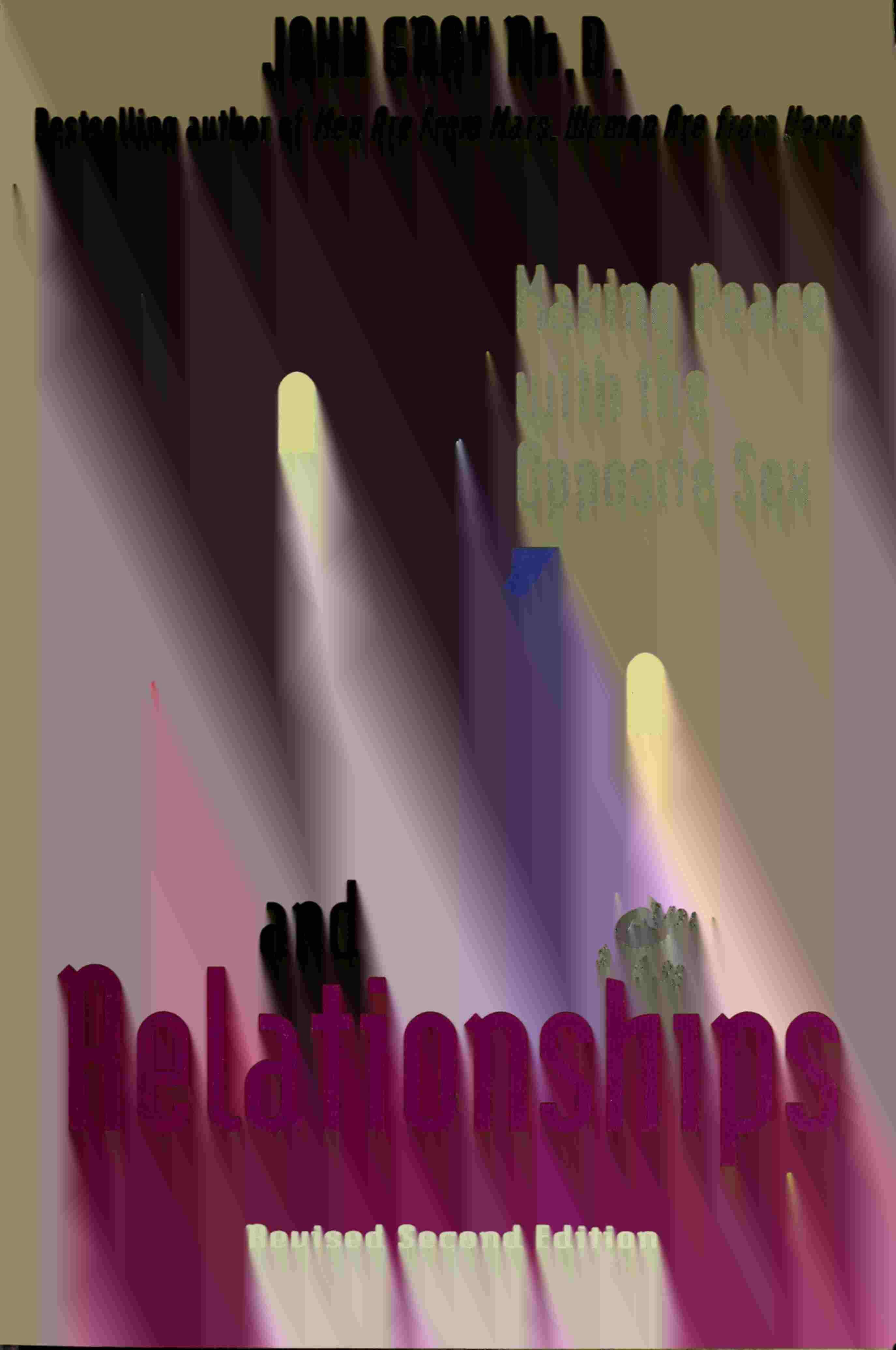
Men,Women andRelationships
MAKINGPEACE WITHTHEOPPOSITESEX
John Gray, Ph.D.


Another random document with no related content on Scribd:

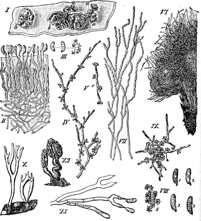
F��. 156.—Chrysomyxa abietis: a leaf of the Fir, with 5 clusters of basidiospores (× 4); b branched rows of teleutospores springing from the mycelium (m).

F�� 157 Cronartium ribicola: a mass of uredospores (× 50); b an uredospore; c a column of teleutospores (× 60); d a small portion of the same more highly magnified, with a basidium and two basidiospores (s)
To the Fungi of which the æcidium is known, whilst the remaining forms are still undetermined, but which are without doubt heterœcious, belong Æcidium elatinum, which produces the enormous “witches’ brooms” and barrel-shaped swellings on stems and branches of Abies alba; and Æcidium strobilinum (Fig 158), which attacks Fir-cones, causing all the scales to become covered with clusters of æcidia opening by a lid Hemileia vastatrix destroyed the coffee plantations in Asia
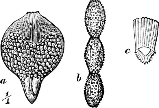
F�� 158 Æcidium strobilinum: a scale of cone of Picea excelsa, with numerous æcidia; b æcidiospores arranged in a series; c a cell of the peridium
Order 2. Auriculariaceæ. The long, transversely divided basidia bear laterally 4 long sterigmata with basidiospores (Fig. 160 B) and are united to form an hymenium on the surface of the fruit-body.
Parasites or saprophytes.
Auricularia sambucina (Auricula judæ), Judas’-ear, has large fruit-bodies, which may attain the size of several inches, resembling an ear or a mussel shell In the moist condition they are flesh-coloured, tough and gelatinous, but when dried, become hard, grey and wrinkled; the exterior is covered with short hairs; while the internal surface bears the hymenium. Habitat: stems and branches of old Eldertrees (Sambucus).
Order 3. Tremellaceæ. The round, pear-shaped, longitudinally divided basidia bear 4 elongated sterigmata, situated apically, and 4 basidiospores (Fig. 160 C, D), and are united into the hymenium on the surface of the fruit-body. The fruit-bodies are frequently gelatinous and quivering; similar fruit-bodies are also found in the Dacryomycetaceæ and Hydnaceæ. Simple conidiophores, which
appear not infrequently in the basidiocarps, before the basidia, are known in many species. Saprophytes.
159.—
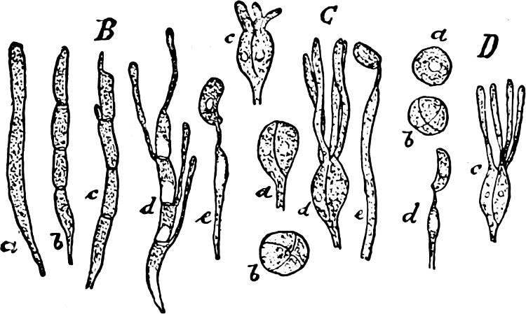
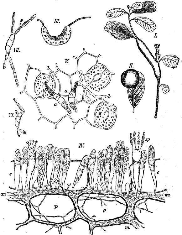
F��. 160. B Auricularia sambucina: a-d basidia in various stages of development; e a sterigma bearing a spore. C Tremella lutescens: a-d basidia seen from various sides (b from above) and in various stages of development; e sterigma with basidiospore (× 400). D Exidia glandulosa: a-c various stages in the development of a basidium; d sterigma with basidiospore (× 350).
Exidia has kidney-shaped, oblong basidiospores, and small, hook-like conidia; E. glandulosa, E. albida, etc., on wood. Craterocolla has conidiocarps; C. cerasi on Cherry-wood. Sebacina incrustans; the yellow, fleshy, or cartilaginous fruitbodies are found in autumn covering the ground in moist woods. Tremella has round basidiospores; T. mesenterica has irregularly-folded, quivering, orange fruitbodies, about one inch in breadth; T. lutescens (Fig. 161) has orange-yellow conidial-and yellow basidial-layers; T. frondosa has fruit-bodies upwards of a foot in breadth.
Order 4. Pilacraceæ. The transversely divided basidia have no sterigmata, but sessile basidiospores, and fill up the cavity of a closed (angiocarpic) fruit-body as a gleba without a regular arrangement (hymenium wanting).
Pilacre fagi on the old stems of the Copper-Beech; P. petersii, on dried branches of the Hornbeam, has stalked, capitate fruit-bodies.
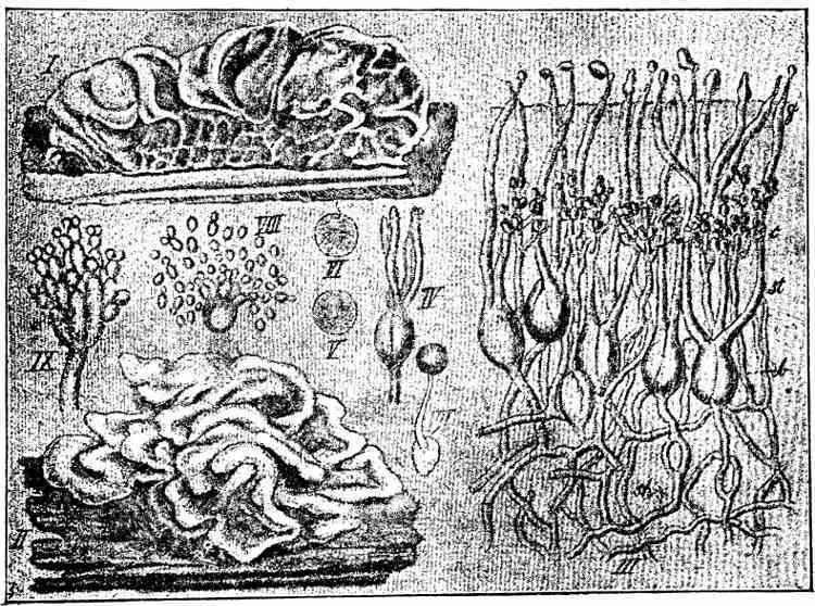
F�� 161 —Tremella lutescens: I and II fruit-bodies (nat size); III vertical section through a fruit-body; b basidia; c conidia; IV-VI basidia; VII basidiospore with a second spore; VIII a basidiospore with yeast-like budding (cultivated); IX a conidiophore. (III-IX about 400.)
Series 2. Autobasidiomycetes.
This second and larger part of the Basidiomycetes is characterised by its more highly differentiated, undivided, clubshaped, or cylindrical basidia, which generally bear 4 (seldom 2, 6, 8) apically-placed sterigmata and basidiospores (Fig. 145). The fruitbodies are partly gymnocarpic (in the first 3 orders and in some Agaricaceæ), partly hemiangiocarpic (in orders 3–6 of the Hymenomycetes and in the Phalloideæ, the fruit-bodies in these orders are in the young conditions more or less angiocarpic, but later on generally open below and bear the hymenium on the under
surface of the fruit-body), partly also angiocarpic (in the Gasteromycetes).
F��. 162. Dacryomyces deliquescens: I fruit-body (nat. size); II vertical section through the hymenium; III germinating basidiospore; IV a portion of mycelium with conidia; V a germinating conidium; VI and VII chains of oidia more or less strongly magnified; VIII basidiospore of D longisporus; IX germinating basidiospore of D ovisporus; X and XI Calocera viscosa; X fruit-body (nat size); XI basidia with basidiospores (highly magnified); XII Dacryomitra glossoides (nat size)
Family 1. Dacryomycetes.
The long, club-shaped basidia bear two tapering sterigmata, which develope remarkably large basidiospores (Fig. 162 II, XI) and form gymnocarpic fruit-bodies with hymenium. 1 order:
Order 1. Dacryomycetaceæ. This order comprises 4 genera of which the first two develope the hymenium on the whole surface of the fruit-body, but the two last only on its apex.
Dacryomyces: the folded, gelatinous, Tremella-like fruit-bodies break out in winter on dried wood (hedges) in the form of red or yellow drops D deliquescens is very common (Fig 121) The following genera have cartilaginous fruit-bodies Calocera (Fig 162), with club-like, simple, or branched, Clavaria-like, fruit-bodies; the orange coloured fruit-bodies of C viscosa grow aggregated together on the wood of Conifers —Guepinia resembles a Peziza, and has the hymenium only on the hollow upper surface —Dacryomitra resembles a Mitrula (Fig 162)
Family 2. Hymenomycetes.
This family is very rich in species (more than 8000 have been described), and to it belong all the “Mushrooms” and “Toadstools.” The fruit-bodies present very various forms; they are generally fleshy, very perishable, seldom leathery or corky, in the last case often perennial. The basidia are more or less cylindrical and bear generally 4 (seldom 2, 6 or 8) sterigmata and basidiospores. The hymenium in the fully-formed fruit-bodies lies free on the surface: in orders 1 and 2 and a portion of order 6 it is from the commencement exposed, fruit-bodies gymnocarpic; orders 3–6 have hemiangiocarpic fruit-bodies (p. 157). In the first order the basidia (or the hymenium) are developed immediately from the mycelium (Fig. 163); the fruit-bodies of orders 2 and 3 present a higher grade of development, and have between the mycelium and hymenium a special hyphal-tissue, a stroma, which is crustaceous, club-like, or coralloid, etc., and in general bears the hymenium on the largest part of the free, smooth surface. In the forms most highly developed (orders 4–6) a new tissue—the hymenophore—is introduced between the stroma and hymenium, which appears on the under side of the fruit-body in the form of warts, projections, tubes, folds or lamellæ (Figs. 166, 167, 174 bc). Paraphyses are frequently found in
the hymenium, among the basidia. In the Hymenomycetes few examples of conidia can be recognised at first. More frequently chlamydospores are found, particularly oidia. The mycelium is richly branched, generally colourless, often perennial; it lives in humus or decaying wood, and is seldom parasitic. The hyphæ generally have clamp-connections and unite, sometimes, to form a rhizomorpha (Fig. 177) or sclerotia with coloured, pseudo-parenchymatous covering.
with basidia in various stages of development; V epidermis with germinating spores; VI and VII spores germinating in water (IV-VII × 620).
Order 1. Tomentellaceæ. To this order belong the simplest of the Hymenomycetes. The basidia (Fig. 145) arise free and irregularly from the mycelium; a hymenium is entirely absent or very slightly formed (in Corticium it attains its highest development); fruit-bodies are also wanting.—In general they form flaky, membranous or leathery coverings on bark and wood. Some are parasites.
Hypochnus without conidia —Tomentella with conidiophores; growing on wood or earth.—Exobasidium vaccinii (Fig. 163), a parasite on Vaccinium, Andromeda, Arctostaphylos, and Rhododendron, forms flaky-powdery, white or red coverings and may cause hypertrophy of the parts attacked. E. warmingii is parasitic on Saxifraga; E. lauri causes outgrowths on the stem of Laurus canariensis as long as a finger, which formerly were regarded as aerial roots. Corticium forms membranous to leathery layers or crusts; C. quercinum on wood and bark, particularly Oak, is flesh-coloured; C. cæruleum has a blue hymenium; C. giganteum on the bark of fallen Pine-trees.
Order 2. Clavariaceæ. The hymenium is situated on a stroma, and either completely covers the smooth surface of the more or less fleshy gymnocarpic fruit-body, or is confined to a tolerably well defined upper portion of it (Typhula). Paraphyses absent. The vertical, white, yellow, or red fruit-bodies are roundish or club-like, undivided or richly branched (Fig. 125). Generally on the ground in woods, seldom on tree-stems, etc.

F��. 164.—Clavaria coralloides (nat. size).
G�����: Clavaria, generally large Fungi with thick, round branches. C. botrytis has a very thick, tubercular stem with numerous short, flesh-coloured branches: it has an agreeable taste C coralloides has a brittle, richly-branched fruit-body (Fig 164); basidia with two large spores C pistillaris consists of a single, undivided club of a yellowish-white colour Sparassis has compressed, leaf-like, curled branches; S crispa has fruit-bodies as large as a white cabbage-head, with an agreeable taste Typhula and Pistillaria are small Fungi with filamentous stalks, terminating in a small club The fruit-bodies of the former often arise from a small, spheroid sclerotium; the latter is distinguished by the basidia bearing only two spores.
Order 3. Thelephoraceæ. The hymenium is placed on a stroma and covers the smooth surface of the leathery hemiangiocarpic fruitbody, generally on its under side. The edge of the stroma, which bounds the hymenium, is sometimes especially developed (Stereum). Saprophytes.
G�����: Thelephora. The fruit-bodies in this genus are brown, very irregularly shaped, and often lobed. The spores too are brown, but in the other genera colourless. The species are found growing on barren soil. T. laciniata (Fig. 165) has imbricate, semicircular, dark-brown pileus, which is jagged at the edge and upper surface. The fruit-bodies are very often raised above the ground, and although this species is not a parasite, yet it destroys young seedlings by growing above and smothering them. Stereum has a stiffer fruit-body, with a distinct, fibrous, intermediate layer. It grows on bark and wood, projecting like a series of imbricate brackets. S. hirsutum is yellow; its free edge is provided with a number of stiff hairs, the upper surface being divided into a number of zones S purpureum has a red-violet hymenium which distinguishes it from the previous species Cyphella has a membranous cup- or bell-shaped fruit-body, often borne on a stalk, the concave surface being covered with the hymenium They are small, white Fungi, growing on Moss and dead stems —Solenia is closely related to Cyphella; its fruit-bodies are smaller and hairy; they are found clustered together forming a crust-like covering on dead wood.—Craterellus has a large, funnel-shaped fruitbody, the hymenium covering the external surface. C. cornucopioides is shaped like a trumpet or a “horn of plenty.” It is dark-grey, several inches in height, and grows gregariously on the ground in forests. It is distinguished by the basidia bearing only two sterigmata.

Order 4. Hydnaceæ. The fruit-body is most frequently fleshy, and varies considerably in shape, the simplest forms being resupinate, [14] the higher ones umbrella-like. The hymenophore is found on the free or downward-turned surface, and always takes the form of soft emergences hanging vertically downwards. The emergencies may be thorn-, awl-, or wart-like. The species are found growing on the soil and on dead wood.
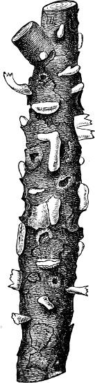
G�����: Hydnum has subulate, distinct emergences H repandum is yellow, the stalk being placed in the centre of the pileus It is an edible species, and often forms “fairy rings” in woods H auriscalpium (Fig 166) is dark-brown, with stalk placed at the edge of the pileus It grows on old Fir-cones H erinaceus grows on old tree-trunks The fruit-body is yellow and very large—as big as a human head— with emergences as much as an inch in length —Irpex has a leathery fruit-body, partly resupinate, partly with free, projecting edge; the under side bears tooth-like emergences which are arranged in rows, and Irpex thus forms a transition to the Agaricaceæ. Phlebia is entirely resupinate, with radially-arranged folds on the free side, and pectinate border.
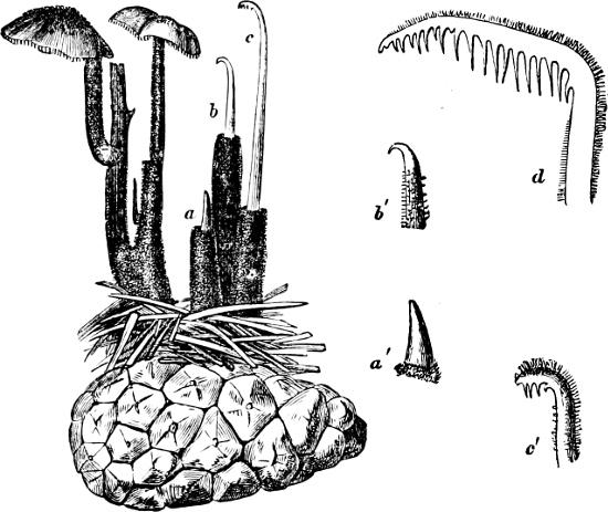
F��. 166. Hydnum auriscalpium, upon a Fir-cone, in different stages of development.
Order 5. Polyporaceæ (Pore-Fungi). An order very rich in species (about 2000 species are described). The fruit-body is of very different forms—resupinate, projecting like a bracket, hoof-like, or umbrella-shaped. In some it is fleshy and edible, in others leathery or corky, persisting for several years. The hymenophore is situated on the under side of the fruit-body, and consists of wide or narrow tubes or pores, whose inner surface is clothed with the hymenium (Fig. 167). In some fruit-bodies large cavities are to be found, which have arisen as interstices between the labyrinthine curved and reticulate folds. Chlamydospores are known in some species. Conidia occur very rarely. Many species work considerable damage: some as parasites on trees, others by destroying timber

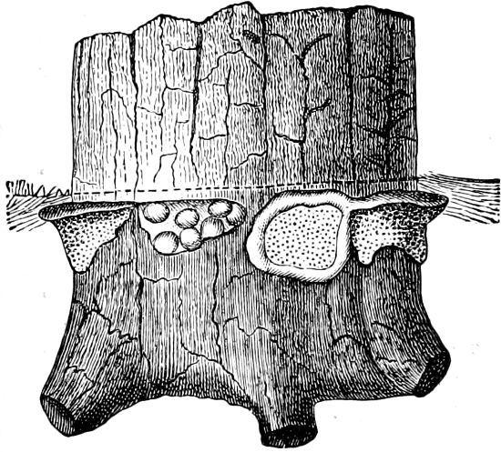
F��. 167. Polyporus igniarius. Section through the under side of the Fungus: h-h is hyphal-tissue between the tubes, formed by irregularly felted hyphæ, many of which are seen cut across; s is the hymenium which covers the walls of the tubes, and from which the basidia with the spores protrude.
G�����. Polyporus (Pore-Fungus). The tubes are narrow, accurately fitted together, and forming a thick layer on the under side of the fruit-body, appearing as a number of fine holes. The fruit-body most frequently resembles a bracket, or is
hoof-shaped, with one side growing from a tree-trunk; it is very often perennial, and a new layer of tubes arises in each succeeding period of vegetation. Strata, corresponding to the periodically interrupted growth, are thus formed in storeys one above the other, and are visible on the upper surface of the fruit-body, as well as in the interior, as a series of concentric belts, sometimes as many as half a score or more in number P fomentarius (Touchwood) attacks trees, especially the Beech The spores germinate on wounds from broken branches, and the hyphæ, following the course of the medullary rays, find their way into the interior of the tree, from whence the mycelium spreads upwards, downwards, and peripherally, so that the wood becomes rotten (“white-rot”) and thick felts of mycelium are formed in radial and tangential directions. A dark line, caused by the youngest parts of the hyphæ containing a brown juice, marks the boundary between the rotten and the unattacked parts of the stem (Fig. 168); at places where the mycelium extends to the bark, the cambium becomes destroyed and further growth is arrested, so that longitudinal furrows arise on the stem. It is at these places, too, that the hoof-shaped, ash-coloured fruit-bodies are developed, which may attain a circumference of upwards of 7 feet. The interior of the fruit-body consists of a dried-up, loosely felted, red-brown mass of hyphæ, which has been used for tinder and as a styptic (“Fungus chirurgorum”). P. igniarius has a harder, dark-brown, more rounded fruit-body; it grows in a similar manner, but especially attacks Oaks, Poplars, and Plum-trees, the wood of which becomes rotten, and is called touchwood P pini (Trametes pini), (Fig 170), a parasite on the stems of Pinus, causes a kind of “red-rot” in the stem P sulphureus has a soft, cheesy, yellow fruit-body; it produces “rot” in Oaks and Apple-trees P officinalis, Larchfungus (“Fungus Laricis” in Pharmocopœia), grows on Larch-trees in the southeast of Europe P versicolor has thin, semicircular fruit-bodies, with zones of various colours on the upper side; it is one of the most frequent species on treestems. P. frondosus grows on soil in woods, and consists of numerous aggregated fruit-bodies, which become very large and fleshy. This species is edible. P. perennis also grows on the soil in woods; it is very leathery, with central stalk, and has concentric zones on the upper surface of the fruit-body. P. vaporarius destroys the wood of living Pines (Pinus silvestris) and Firs (Picea excelsa), causing it to become red-brown; in timber this Fungus causes “red-strip” followed by a “dry-rot.” P. squamosus destroys many Walnut-trees, and is also very destructive to Limes and Elms. P. fulvus causes a “white-rot” in Abies alba.
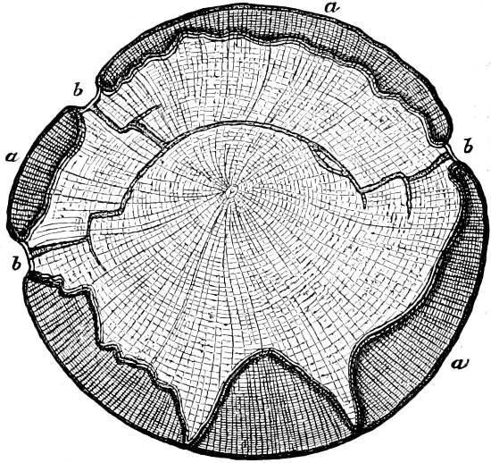
F��. 168. Section of stem of a Beech attacked by P. fomentarius: a non-attacked parts of the stem; b the furrows where the mycelium has reached the bark, and where the thick mycelium-strands reach the exterior (⅙th of the nat size)
F��. 169. Base of a Fir-tree, with a number of fruit-bodies of Heterobasidion annosum just beneath the surface of the soil, indicated by the dotted line (¼th nat. size).