Congenital Heart Diseases:
The Broken Heart
Clinical Features, Human Genetics
Editors
Silke Rickert-Sperling
Cardiovascular Genetics
Charité - Universitätsmedizin Berlin
Berlin Germany
Robert G. Kelly
Developmental Biology
Institute of Marseilles
Aix-Marseille Université
Marseille France
Editorial Assistant
Andreas Perrot
Cardiovascular Genetics
Charité - Universitätsmedizin Berlin Berlin Germany
David J. Driscoll
Department of Pediatrics
Division of Pediatric Cardiology
Mayo Clinic College of Medicine
Rochester, MN USA
ISBN 978-3-7091-1882-5
DOI 10.1007/978-3-7091-1883-2
ISBN 978-3-7091-1883-2 (eBook)
Library of Congress Control Number: 2015958767
Springer Wien Heidelberg New York Dordrecht London © Springer-Verlag Wien 2016
This work is subject to copyright. All rights are reserved by the Publisher, whether the whole or part of the material is concerned, specifically the rights of translation, reprinting, reuse of illustrations, recitation, broadcasting, reproduction on microfilms or in any other physical way, and transmission or information storage and retrieval, electronic adaptation, computer software, or by similar or dissimilar methodology now known or hereafter developed.
The use of general descriptive names, registered names, trademarks, service marks, etc. in this publication does not imply, even in the absence of a specific statement, that such names are exempt from the relevant protective laws and regulations and therefore free for general use.
The publisher, the authors and the editors are safe to assume that the advice and information in this book are believed to be true and accurate at the date of publication. Neither the publisher nor the authors or the editors give a warranty, express or implied, with respect to the material contained herein or for any errors or omissions that may have been made.
Printed on acid-free paper
Springer-Verlag GmbH Wien is part of Springer Science+Business Media (www.springer.com)
This book is dedicated to my mentors Hanno D. Schmidt, Peter E. Lange, and Hans Lehrach. Their training, support, and encouragement have made this book possible.
Silke Rickert-Sperling
Foreword
As is indicated in its title, the book you are about to read is concerned with the congenitally malformed heart. Approximately eight neonates in every thousand born alive present with such a “broken heart”. This number has changed little since Maude Abbott, when describing the first plate in her Atlas devoted to congenitally malformed hearts, commented that “An understanding of the elementary facts of human and comparative embryology is essential to an intelligent grasp of the ontogenetic problems of congenital cardiac disease”. Paul Dudley White, when writing the foreword to her Atlas, commented that it had been left to Abbott to “make the subject one of such general and widespread interest that we no longer regard it with either disdain or awe as a mystery for the autopsy table alone to discover and to solve”. It is perhaps surprising, therefore, to realise that it has taken nearly a century for us to achieve the necessary understanding of the “elementary facts” emphasised by Abbott. Indeed, it is not that long since, in company with my very good friend and collaborator Anton Becker, we suggested that interpretations based on embryology might prove to be a hindrance, rather than a help, in understanding the congenitally malformed heart. The contents of this book show how much has changed in the years that have passed since we made that comment, such that we now need to eat our words.
As is revealed by the multiple chapters of this book, the recent advances made in the fields of cardiac embryology and molecular genetics have been truly spectacular. It was these fields that were expertly summarised in the volumes edited by Rosenthal and Harvey. The details contained in the central part of this book, related to central molecular pathways, recapitulate and extend those reviews. Such extensive knowledge of the genetic and molecular background, however, is of limited value if these interpretations cannot properly be translated into the findings observed on a daily basis by those who diagnose and treat the individual cardiac lesions. The first part of this book, therefore, provides a necessarily brief overview of normal cardiac development, while the final chapters then incorporate the developmental and molecular findings into the clinical manifestations of the abnormal morphogenesis.
I know from my own experience how difficult it is to obtain such chapters from multiple authors, who nowadays are themselves under greater pressure to produce primarily in the peer-reviewed realm. The editors, therefore, are to be congratulated
on assembling such a panoply of authoritative texts. As might be expected, not all of the texts are of comparable length or content. The critical reader will note that several of the topics addressed remain contentious, and that opinions continue to vary between the chosen experts. This is no more than to be expected, since the topics remain very much moving targets. One hopes, therefore, that this is but the first edition of a work which itself, for the first time, seeks to provide in detail the scientific background to the specific lesions that continue to break the normal heart. As the pages of this book demonstrate, we still have much to do if we are fully to understand the mechanics of normal as opposed to abnormal cardiac development.
London, United Kingdom Robert H. Anderson August 2015
Preface
Leonardo Da Vinci made the first drawing of partial anomalous pulmonary venous connection in the fifteenth century, and 300 years later Karl von Rokitansky described ventricular septal defects. Since then the history of clinical recognition, therapeutic opportunities, and understanding of the developmental and genetic origin of congenital heart diseases (CHDs) has evolved rapidly. The first wave of progress was dedicated to the improvement of clinical diagnosis and therapy based on anatomical, physiological, and surgical considerations. Thus, the mortality of patients with CHD declined below 1 in 100,000 cases and a new group of adult patients with corrected and palliated CHD was formed.
A second wave of progress focused on the developmental, genetic, and molecular aspects of CHDs. Here significant insights were gained by studying animal models along with human. A large collection of genes, signaling pathways, and other molecular or hemodynamic insults have been discovered, frequently considering the developmental perspective as a starting point.
After decades of basic research focusing on animal models, the human phenotype will be the central dogma in the following years. This shift is based on significant developments to overcome technological limitations now enabling studies addressing more and more complex biological questions and systems together with the recognition that improving human health is a central aim of life science research. This book brings together clinical, genetic and molecular knowledge starting from the perspective of the observed human phenotype during development and in the disease state. It aims to reach basic scientists as well as physicians and it might contribute to the current third wave of progression where basic science of cardiovascular development is translated into clinical diagnosis and therapy of CHDs.
To reach this goal, this book is structured in three main parts providing an introduction to the development of the heart and its vessels, an overview of molecular pathways affecting the development of multiple cardiovascular structures, and a textbook-like structure focused on the different types of congenital heart diseases with their clinical features, underlying genetic alterations and related animal models and pathways. We are grateful to all the contributors to this volume, who have provided state of the art accounts of their fields of expertise.
Berlin, Germany
Silke Rickert-Sperling Marseille, France Robert G. Kelly Rochester, MN, USA David J. Driscoll October 2015
Amelia E. Aranega and Diego
Rajan Jain, Mudit Gupta, and Jonathan A. Epstein
George A. Porter Jr.
Cornelia Dorn, Marcel Grunert, Ana Dopazo, Fátima Sánchez-Cabo, Alberto Gatto, Jésus Vázquez, Silke Rickert-Sperling, and Enrique Lara-Pezzi
David J. Driscoll
Rabia Khan and Patrick Y. Jay
Patrick Y. Jay, Karl R. Degenhardt, and Robert H. Anderson
Katherina Bellmann, Andreas Perrot, and Silke Rickert-Sperling
Lucile Houyel
David J. Driscoll
Cheryl L. Maslen
27 Molecular Pathways and Animal Models of Atrioventricular Septal Defect .
Andy Wessels
Part VII Total Anomalous Pulmonary Venous Return
28 Clinical Presentation and Therapy of Total Anomalous Pulmonary Venous Return
David J. Driscoll
29 Human Genetics of Total Anomalous Pulmonary Venous Return . .
Robert E. Poelmann, Monique R.M. Jongbloed, Marco C. DeRuiter, and Adriana C. Gittenberger-de Groot
30 Molecular Pathways and Animal Models of Total Anomalous Pulmonary Venous Return .
Robert E. Poelmann, Adriana C. Gittenberger-de Groot, Monique R.M. Jongbloed, and Marco C. DeRuiter
Part VIII Tetralogy of Fallot and Double Outlet Right Ventricle
31 Clinical Presentation and Therapy of Tetralogy of Fallot and Double Outlet Right Ventricle
David J. Driscoll
32 Human Genetics of Tetralogy of Fallot and Double Outlet Right Ventricle
Cornelia Dorn, Andreas Perrot, and Silke Rickert-Sperling
33 Molecular Pathways and Animal Models of Tetralogy of Fallot and Double Outlet Right Ventricle
Robert G. Kelly
Part IX d-Transposition of the Great Arteries
34 Clinical Presentation and Therapy of d-Transposition of the Great Arteries
David J. Driscoll
35 Human Genetics of d-Transposition of the Great Arteries
Patrice Bouvagnet and Anne Moreau de Bellaing
36 Molecular Pathways and Animal Models of d-Transposition of the Great Arteries
Amy-Leigh Johnson and Simon D. Bamforth
David J. Driscoll
Andreas Perrot and Silke Rickert-Sperling
Nikolai T. Klena, George C. Gabriel, and Cecilia W. Lo
and Therapy of Semilunar Valve and Aortic Arch Anomalies
David J. Driscoll 41 Human Genetics of Semilunar Valve and Aortic Arch Anomalies
Matina Prapa and Siew Yen Ho 42 Molecular Pathways and Animal Models of Semilunar Valve and Aortic Arch Anomalies
Amy-Leigh Johnson and Simon D. Bamforth
Part XII Coronary Artery Anomalies
43 Clinical Presentation and Therapy of Coronary Artery Anomalies
David J. Driscoll
Beatriz Picazo and José M. Pérez-Pomares 45 Molecular Pathways and Animal Models of Coronary Artery Anomalies
Juan A. Guadix and José M. Pérez-Pomares
David J. Driscoll
Human Genetics of Truncus Arteriosus
Hiroyuki Yamagishi
48 Molecular Pathways and Animal Models of Truncus Arteriosus
Amy-Leigh Johnson and Simon D. Bamforth
Part XIV Tricuspid Atresia and Univentricular Heart
49 Clinical Presentation and Therapy of Tricuspid Atresia and Univentricular Heart .
David J. Driscoll
50 Human Genetics of Tricuspid Atresia and Univentricular Heart .
Abdul-Karim Sleiman, Liane Sadder, and George Nemer
51 Molecular Pathways and Animal Models of Tricuspid Atresia and Univentricular Heart
Kamel Shibbani and George Nemer
Part XV Ebstein Anomaly
David J. Driscoll
53
Gregor U. Andelfinger 54 Molecular Pathways and Animal Models of Ebstein Anomaly
Gregor U. Andelfinger
Part XVI Hypoplastic Left Heart Syndrome
55 Clinical Presentation and Therapy of Hypoplastic Left Heart Syndrome
Florian Wünnemann and Gregor U. Andelfinger Part
Alexa M.C. Vermeer, Arthur A.M. Wilde, and Imke Christiaans
60 Molecular Pathways and Animal Models
Enkhsaikhan Purevjav
Part XVIII Arrhythmias
61 Clinical Presentation and Therapy of Arrhythmias
David J. Driscoll
62 Human Genetics of Arrhythmias
Erik Schulze-Bahr and Sven Dittmann
63 Molecular Pathways and Animal Models of Arrhythmias
Sara Adelman, Amy C. Sturm, and Peter J. Mohler Perspective
Deepak Srivastava
Index
Contributors
Sara Adelman The Dorothy M. Davis Heart and Lung Research Institute, The Ohio State University Wexner Medical Center, Columbus, OH, USA
Gregor U. Andelfinger Cardiovascular Genetics, Department of Pediatrics, CHU Sainte Justine, Université de Montréal, Montréal, QC, Canada
Robert H. Anderson Institute of Genetic Medicine, Newcastle University, International Centre for Life, Newcastle upon Tyne, United Kingdom
Amelia E Aranega Cardiovascular Research Group, Department of Experimental Biology, University of Jaén, Jaén, Spain
Simon D. Bamforth Institute of Genetic Medicine, Newcastle University, Newcastle upon Tyne, United Kingdom
Katherina Bellmann Cardiovascular Genetics, Charité – Universitätsmedizin Berlin, Berlin, Germany
D. Woodrow Benson Herma Heart Center, Children’s Hospital of Wisconsin, Medical College of Wisconsin, Milwaukee, WI, USA
Patrice Bouvagnet Laboratoire Cardiogénétique, Groupe Hospitalier Est, Hospices Civils de Lyon, Lyon, France
Margaret Buckingham Department of Developmental and Stem Cell Biology, Institut Pasteur, Paris, France
Imke Christiaans Department of Clinical and Experimental Cardiology and Department of Clinical Genetics, Academic Medical Centre, Amsterdam, The Netherlands
Vincent M. Christoffels Department of Anatomy, Embryology, and Physiology, Academic Medical Center, Amsterdam, The Netherlands
Anne Moreau de Bellaing Laboratoire Cardiogénétique, Groupe Hospitalier Est, Hospices Civils de Lyon, Lyon, France
Karl R. Degenhardt Division of Cardiology, Department of Pediatrics, Children’s Hospital of Philadelphia, Perelman School of Medicine at the University of Pennsylvania, Philadelphia, PA, USA
Marco C. DeRuiter Department of Anatomy & Embryology, Leiden University Medical Center, Leiden, The Netherlands
Sven Dittmann Department of Cardiovascular Medicine, Institute for Genetics of Heart Diseases (IfGH), University Hospital Münster, Münster, Germany
Ana Dopazo Cardiovascular Development and Repair Department, Centro Nacional de Investigaciones Cardiovasculares, Madrid, Spain
Cornelia Dorn Cardiovascular Genetics, Charité – Universitätsmedizin Berlin, Berlin, Germany
David J. Driscoll Division of Pediatric Cardiology, Department of Pediatrics, Mayo Clinic College of Medicine, Rochester, MN, USA
Jonathan A. Epstein Department of Cell and Developmental Biology, Institute for Regenerative Medicine and the Cardiovascular Institute, Perelman School of Medicine at the University of Pennsylvania, Philadelphia, PA, USA
Diego Franco Cardiovascular Research Group, Department of Experimental Biology, University of Jaén, Jaén, Spain
George C. Gabriel Department of Developmental Biology, University of Pittsburgh School of Medicine, Pittsburgh, PA, USA
Alberto Gatto Cardiovascular Development and Repair Department, Centro Nacional de Investigaciones Cardiovasculares, Madrid, Spain
Adriana C. Gittenberger-de Groot Department of Cardiology, Leiden University Medical Center, Leiden, The Netherlands
Marcel Grunert Cardiovascular Genetics, Charité – Universitätsmedizin Berlin, Berlin, Germany
Juan A. Guadix Department of Animal Biology, Faculty of Sciences, University of Málaga, Málaga, Spain
Mudit Gupta Department of Cell and Developmental Biology, Institute for Regenerative Medicine and the Cardiovascular Institute, Perelman School of Medicine at the University of Pennsylvania, Philadelphia, PA, USA
Jörg Heineke Experimentelle Kardiologie, Rebirth – Cluster of Excellence, Klinik für Kardiologie und Angiologie, Medizinische Hochschule Hannover, Hannover, Germany
Siew Yen Ho Royal Brompton & Harefield NHS Foundation Trust, London, United Kingdom
Lucile Houyel Department of Congenital Cardiac Surgery, Marie-Lannelongue Hospital – M3C, Paris-Sud University, Le Plessis-Robinson, France
Mary Hutson Department of Pediatrics, Neonatal-Perinatal Research Institute, Duke University Medical Center, Durham, NC, USA
Rajan Jain Department of Cell and Developmental Biology, Institute for Regenerative Medicine and the Cardiovascular Institute, Perelman School of Medicine at the University of Pennsylvania, Philadelphia, PA, USA
Patrick Y. Jay Departments of Pediatrics and Genetics, Washington University School of Medicine, St. Louis, MO, USA
Bjarke Jensen Department of Anatomy, Embryology & Physiology, Academic Medical Center, University of Amsterdam, Amsterdam, The Netherlands
Amy-Leigh Johnson Institute of Genetic Medicine, Newcastle University, Newcastle upon Tyne, United Kingdom
Monique R. M. Jongbloed Department of Cardiology and Department of Anatomy & Embryology, Leiden University Medical Center, Leiden, The Netherlands
Robert G. Kelly Aix Marseille Université, Institut de Biologie du Dévelopment de Marseille, Marseille, France
Rabia Khan Department of Pediatrics, Washington University School of Medicine, St. Louis, MO, USA
Nikolai T. Klena Department of Developmental Biology, University of Pittsburgh School of Medicine, Pittsburgh, PA, USA
Enrique Lara-Pezzi Cardiovascular Development and Repair Department, Centro Nacional de Investigaciones Cardiovasculares, Madrid, Spain
Cecilia W. Lo Department of Developmental Biology, University of Pittsburgh School of Medicine, Pittsburgh, PA, USA
José C. Martín-Robles Department of Animal Biology, Faculty of Sciences, University of Málaga, Málaga, Spain
Cheryl L. Maslen Knight Cardiovascular Institute, Oregon Health & Science University, Portland, OR, USA
Rajiv Mohan Department of Anatomy, Embryology, and Physiology, Academic Medical Center, Amsterdam, The Netherlands
Peter J. Mohler Division of Cardiovascular Medicine and Division of Human Genetics, Department of Physiology and Cell Biology, Department of Internal Medicine, The Dorothy M. Davis Heart and Lung Research Institute, The Ohio State University Wexner Medical Center, Columbus, OH, USA
Antoon F. M. Moorman Department of Anatomy, Embryology & Physiology, Academic Medical Center, University of Amsterdam, Amsterdam, The Netherlands
Ingo Morano Department of Molecular Muscle Physiology, Max-Delbrück Center for Molecular Medicine and University Medicine Charité Berlin, Berlin, Germany
George Nemer Department of Biochemistry and Molecular Genetics, Faculty of Medicine, American University of Beirut, Beirut, Lebanon
José M. Pérez-Pomares Department of Animal Biology, Faculty of Sciences, University of Málaga, Málaga, Spain
Andreas Perrot Cardiovascular Genetics, Charité – Universitätsmedizin Berlin, Berlin, Germany
Beatriz Picazo Hospital Materno Infantil-Hospital Carlos de Haya, Málaga, Spain
Robert E. Poelmann Department of Cardiology and Department of Integrative Zoology, Institute of Biology, Leiden University, Leiden University Medical Center, Leiden, The Netherlands
George A. Porter Jr. Departments of Pediatrics (Cardiology), Pharmacology and Physiology, and Medicine, Cardiovascular Research Institute, University of Rochester Medical Center, Rochester, NY, USA
Matina Prapa St George’s Healthcare NHS Trust, London, United Kingdom
Enkhsaikhan Purevjav Cardiology, Department of Pediatrics, The Heart Institute, University of Tennessee Health Science Center, Le Bonheur Children’s Hospital, Memphis, TN, USA
Silke Rickert-Sperling Cardiovascular Genetics, Charité – Universitätsmedizin Berlin, Berlin, Germany
Liane Sadder Faculty of Medicine, American University of Beirut, Beirut, Lebanon
Fátima Sánchez-Cabo Cardiovascular Development and Repair Department, Centro Nacional de Investigaciones Cardiovasculares, Madrid, Spain
Eric Schulze-Bahr Department of Cardiovascular Medicine, Institute for Genetics of Heart Diseases (IfGH), University Hospital Münster, Münster, Germany
Robert J. Schwartz Texas Heart Institute, Houston, TX, USA
David Sedmera Institute of Physiology, Academy of Sciences of the Czech Republic, Institute of Anatomy, First Faculty of Medicine Charles University, Prague, Czech Republic
Kamel Shibbani Department of Biochemistry and Molecular Genetics, American University of Beirut, Beirut, Lebanon
Abdul-Karim Sleiman Faculty of Medicine, American University of Beirut, Beirut, Lebanon
Deepak Srivastava Gladstone Institute of Cardiovascular Disease, Roddenberry Stem Cell Center at Gladstone, University of California San Francisco, San Francisco, CA, USA
Amy C. Sturm Division of Human Genetics, Department of Internal Medicine, The Dorothy M. Davis Heart and Lung Research Institute, The Ohio State University Wexner Medical Center, Columbus, OH, USA
Bijoy Thattaliyath Department of Pediatrics, Neonatal-Perinatal Research Institute, Duke University Medical Center, Durham, NC, USA
Jesús Vázquez Cardiovascular Development and Repair Department, Centro Nacional de Investigaciones Cardiovasculares, Madrid, Spain
Alexa M. C. Vermeer Department of Clinical and Experimental Cardiology and Department of Clinical Genetics, Academic Medical Centre, Amsterdam, The Netherlands
Jun Wang Texas Heart Institute, Houston, TX, USA
Andy Wessels Department of Regenerative Medicine and Cell Biology, Medical University of South Carolina, Charleston, SC, USA
Arthur A. M. Wilde Department of Clinical and Experimental Cardiology, Academic Medical Center, Amsterdam, The Netherlands
Florian Wünnemann Cardiovascular Genetics, Department of Pediatrics, CHU
Sainte Justine, Université de Montréal, Montréal, QC, Canada
Hiroyuki Yamagishi Division of Pediatric Cardiology, Department of Pediatrics, Keio University School of Medicine, Tokyo, Japan
Abbreviations
22q11DS 22q11 deletion syndrome
AAA Aortic arch anomalies
ACTC1 Cardiac alpha-actin
ACVR Activin A receptor
AD Arterial duct
ADAM19
ADAR
ADAM metallopeptidase domain 19
Adenosine deaminase that acts on RNA
ADP Adenosine diphosphate
AGS Allagile syndrome
AICD Automatic internal cardiac defibrillator
ALCAPA
Anomalous origin of the left coronary artery from the pulmonary artery
AKT V-akt murine thymoma viral oncogene homolog
AngII Angiotensin II
ANP Atrial natriuretic peptide
ANK2 Ankyrin B
ANKRD1/CARP Ankyrin repeat domain 1, cardiac muscle
Ao Aorta
AP Action potential
ARVC Arrhythmogenic right ventricular cardiomyopathy
ASD Atrial septal defect
ATFB Atrial fibrillation
ATP Adenosin triphosphate
AV
AVB
AVC
AVN
AVSD
BAF
Atrioventricular
Atrioventricular bundle
Atrioventricular canal
Atrioventricular node
Atrioventricular septal defect
Brg1-associated factor
BAV Bicuspid aortic valve
BBS Bardet-Biedl syndrome
BET Bromodomain and extra terminal
BMP
Bone morphogenetic protein
BNP
Brain natriuretic peptide
BRAF v-Raf murine sarcoma viral oncogene homolog B
BRG1
SWI/SNF-related, matrix-associated, actin-dependent regulator of chromatin, subfamily a, member 4 (also known as brahma-related gene 1)
BRGDA Brugada syndrome
BWIS Baltimore Washington Infant Study
CAA Coronary artery anomalies
CACN Calcium channel, voltage-dependent, L type
CAD Coronary atherosclerotic disease
CaMK Calmodulin dependent kinase
cAMP Cyclic adenosine monophosphate
CALM Calmodulin
CASQ Calsequestrin
CAT Common arterial trunk
CBP CREB-binding protein
CC Cardiac crescent
CCDC Coiled-coil domain containing
CCS Cardiac conduction system
CCVA Congenital coronary vascular anomalies
CF Cephalic folds
CFC1 Cripto, FRL-1, Cryptic family 1 (CRYPTIC)
CGH Comparative genomic hybridization
CHARGE Coloboma of the eye, Heart defects, Atresia of the nasal choanae, Retarded growth and/or development, Genital and/or urinal abnormalities, and Ear anomalies
CHD Congenital heart disease
CHD7 Chromodomain helicase DNA binding protein 7
CHF Congestive heart failure
ChIP Chromatin immunoprecipitation
CITED2
CNCCs
Cbp/P300-interacting transactivator, with Glu/Asp-rich carboxy-terminal domain 2
Cardiac neural crest cells
CNV Copy number variation
CoA
CPVT
Coarctation of the aorta
Catecholaminergic polymorphic ventricular tachycardia
CRE Cre recombinase
CRELD1
Cysteine-rich protein with EGF-like domains 1
CRISPR Clustered regularly interspaced short palindromic repeats
CTD Conotruncal defects
CTGF Connective tissue growth factor
CTVM
Canine tricuspid valve malformation
CX Connexin
DCM
DGC
Dilated cardiomyopathy
Dystrophin-glycoprotein complex
DGS
DMP
DNAH
DiGeorge syndrome
Dorsal mesenchymal protrusion
Dynein, axonemal, heavy chain
DNMT DNA methyltransferases
DORV Double outlet right ventricle
DPF3
DSC2
DSG2
DSP
Dvl2
E
ECs
ECG
ECM
EGFR
ELC
D4 Zinc and double PHD fingers family 3 (also known as Baf45c)
Desmocollin 2
Desmoglein 2
Desmoplakin
Dishevelled segment polarity protein 2
Embryonic day
Endocardial cushions
Electrocardiogram
Extracellular matrix
Epidermal growth factor receptor
Essential myosin light chain
ELN Elastin
EMT
ENU
ET1
EPDC
ErbB
ERK
ERS
ESC
EVC
FA
FACS
FDA
FGF
FGFR
Epithelial-to-mesenchymal transition
N-ethyl-N-nitrosourea
Endothelin 1
Epicardially derived cells
Erythroblastic leukemia viral oncogene homolog
Extracellular signal-regulated kinase
Early repolarization syndrome
Embryonic stem cells
Ellis-van-Creveld
Folic acid
Fluorescence-activated cell sorting
Food and Drug Administration
Fibroblast growth factor
Fibroblast growth factor receptor
FHF First heart field
FHL1
FISH
Four and a half LIM domains protein 1
Fluorescence in situ hybridization
FOX Forkhead box
FOG2
GATA
GBX
GDF1
Friend of GATA 2
GATA binding protein
Gastrulation brain homeobox
Growth differentiation factor
GFP Green fluorescent protein
GJA5
GPCR
GRP
GWAS
Gap junction protein, alpha 5, 40 kDa (connexin 40)
G-protein coupled receptors
Gastrocoel roof plate
Genome-wide association study
H3K4me3
H3K4me2
H3K4me1
H3K24ac
H3K27ac
H3K27me3
HAND
HAT
HDAC
HCM
HCN4
HE
HES1
HEY
HLHS
HOX
HT
IAA
Trimethylation of histone H3 at lysine 4
Dimethylation of histone H3 at lysine 4
Monomethylation of histone H3 at lysine 4
Acetylation of histone H3 at lysine 24
Acetylation of histone H3 at lysine 27
Trimethylation of histone H3 at lysine 27
Heart and neural crest derivatives expressed
Histone acetyltransferase
Histone deacetylase
Hypertrophic cardiomyopathy
Hyperpolarization activated cyclic nucleotide-gated potassium channel 4
Haematoxylin and eosin
Hes family BHLH transcription factor 1
Hes-related family bHLH transcription factor with YRPW motif
Hypoplastic left heart syndrome
Homeobox genes
Heart tube
Interrupted aortic arch
IC Inner curvature
IGF1
INO80
IP3
IPCCC
iPSCs
IRX
ISL1
IVF
IVS
JAG1
JNK
JUP
KLF
KCNE
KCNJ
KCNQ
LA
LBB
LCC
LEFTY
LEOPARD
Insulin-like growth factor 1
Inositol requiring 80
Inositol-1,4,5-triphosphate
International Pediatric and Congenital Cardiac Code
Induced pluripotent stem cells
Iroquois homeobox
ISL LIM homeobox 1 (Islet 1)
Idiopathic ventricular fibrillation
Interventricular septum
Jagged 1
c-Jun N-terminal kinase
Junctional plakoglobin
Kruppel-like factor
Potassium channel, voltage gated subfamily E regulatory beta subunit
Potassium channel, inwardly rectifying subfamily J
Potassium channel, voltage gated KQT-like subfamily Q
Left atrium
Left bundle branch
Left common carotid
Left-right determination factor
Lentigenes, ECG conduction abnormalities, Ocular hypertelorism, Pulmonic stenosis, Abnormal genitalia, Retardation of growth and sensorineural Deafness
LIF
Leukemia inhibitory factor
LMNA Lamin A/C
lncRNA
LPM
Long non-coding RNA
Lateral plate mesoderm
LLPM Left lateral plate mesoderm
LQTS
LRO
LSA
LTCC
Long QT syndrome
Left-right organizer
Left subclavian artery
L-type calcium channel
LV Left ventricle
LVNC
MAPCA
MBD
MDM2
MED13L
MEF2C
Left ventricular noncompaction
Major aortopulmonary collateral arteries
Methyl-CpG binding domain-based
Murine double minute 2
Mediator complex subunit 13-like
Myocyte enhancer factor 2C
MEK2 kinase 2
MESP1
MHC
Mesoderm posterior 1 homolog
Myosin heavy chain
MI Myocardial infarction
MMP Matrix metalloproteinases
MLC
MLL2
Myosin light chain
Mixed-lineage leukemia protein 2
miRNA MicroRNA
MOs
MPES
Morpholino oligonucleotides
Mid-pharyngeal endothelial strand
MRI Magnetic resonance imaging
mRNA Messenger RNA
MS
MSX
MuRF
MyoD
MYBPC3
MYH6
MYH7
NADPH
NCC
Mass spectrometry
Msh homeobox
Muscle RING finger protein
Myogenic differentiation factor
Cardiac myosin-binding protein C
Cardiac alpha-myosin heavy chain
Cardiac beta-myosin heavy chain
Nicotinamide adenine dinucleotide phosphate
Neural crest cells
NCCM Noncompaction cardiomyopathy
NCX
ncRNA
NF1
NFAT
NGS
NKA
NKX2-5
Sodium/Calcium exchanger
Non-coding RNA
Neurofibromatosis type 1
Nuclear factor of activated T-cells
Next-generation sequencing
Sodium/Potassium ATPase
NK2 homeobox 5
NKX2-6 NK2 homeobox 6
NMD
NMR
NODAL
NOS3
Nonsense mediated decay
Nuclear magnetic resonance spectroscopy
Nodal growth differentiation factor
Nitric oxide synthase 3
NPC Nuclear pore complexes
NPHP
NPPA
NRG1
NR2F2
NVP
OFT
OR
p300
Nephronophthisis
Natriuretic peptide A
Neuregulin 1
Nuclear receptor subfamily2/group F
Nodal vesicular parcels
Outflow tract
Odds ratios
E1A binding protein p300
PA Pulmonary atresia
PAAs
PACHD
PAPVC
PAX3
PCD
PCP
Pharyngeal arch arteries
Pancreatic agenesis and congenital heart defects
Partial anomalous pulmonary venous connections
Paired box 3
Primary ciliary dyskinesia
Planar cell polarity
PCR Polymerase chain reaction
PDA
PDGF
PDK1
PECAM1
PFO
PI3K
PITX2
PKA
PKC
PKD2
PKP2
PLN
PLXN
PRDM16
PTA
PTPN11
PTC
PVCS
QT
RA
RA
RAA
RAAS
Patent ductus arteriosus
Platelet-derived growth factor
Phosphoinositide-dependent kinase1
Platelet endothelial cell adhesion molecule 1
Patent foramen ovale
Phosphoinositide 3-kinase
Paired-like homeodomain 2
Protein kinase A
Protein kinase C
Polycystic kidney disease 2 (polycystin 2)
Plakophilin 2
Phospholamban
Plexin
PR domain containing 16
Persistent truncus arteriosus
Protein tyrosine phosphatase, non-receptor type 11
Premature termination codon
Peripheral ventricular conduction system
QT interval (electrocardiogram)
Right atrium
Retinoic acid
Right aortic arch
Renin-angiotensin-aldosterone system
Another random document with no related content on Scribd:
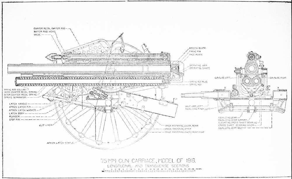
75 MM GUN CARRIAGE MODEL OF 1916 LEFT ELEVATION
75 MM GUN CARRIAGE MODEL OF 1916 RIGHT
ELEVATION
The trail is made in two halves of box section built of bent and riveted steel plate. Each half is bolted to a lug on the equalizing gear, so that it may be rotated horizontally from the junction point of the trail to the point where the trail hits the wheel.
The trails are locked together in traveling position by means of a cone-shaped vertical lug on the lunette bracket which fits in a socket in the trail coupling, and is locked in place by the trail-coupling latch. Trail-coupling latch has a handle and catch with a vertical spindle seated in a socket in the lunette bracket. A handle-return spring is assembled around the spindle and the latch engages a catch on the trail coupling when trails are fixed in the traveling position. Latch is opened by moving handle forward.
Lunette consists of a ring for attaching the carriage to the limber and is bolted through the lunette bracket.
Floats are attached to the bottoms of both trails at their rear ends, consisting of flanged steel plates for the purpose of increasing bearing area of the trails on soft ground.
Spade bearings are riveted to rear of the trails and form bearings for spades in firing position. Spades are driven through the bearings, and their upward movement relative to the trails is prevented by spade latch.
Spade-latch bracket consists of a bronze plate with a cylindrical chamber for a spring and plunger and two bearings for latch-handle pin. Bracket is riveted to the inside top of trail in front of the spade. Spade-latch plunger, with a spring assembled around it, is seated in the chamber and the spade-latch handle is pinned in the bearing. Top of handle extends through the trail and is roughened for use as a foot pedal. Lower part of handle engages with the plunger. When the spade is driven the plunger is forced into a notch in the spade by means of the spring, and the slope on face of plunger allows a downward movement of the spade and prevents upward movement. To release spade the foot pedal on latch handle is pressed down, disengaging plunger from spade, and the spade is removed.
Trail handles are riveted to outside of both trails for lifting trails. Name plate is riveted to outside lower left trail. It is important that the number of carriage on this plate be recorded by the officer in charge of the unit to which it is assigned and that this number be used as a reference in all correspondence. Wheel guards, rear, are plates riveted to the outside lower left of both trails for the protection of trail bodies against contact with limber wheels on short turns. Trail guards are bent plates riveted to the top of trail in front of trailcoupling latch to prevent battering of trails by sledges used for driving the spades.
Sponge-staff fastenings are riveted to tops of both trails. Sponge staffs are inserted in upper rings of staff fastenings and the lower ends are clamped in place. The smallest section of sponge staffs fits in sponge fastenings.
Sledge fastenings are similar to sponge staff fastenings and are riveted to the outside of each trail. Wheel guards (front) are plates
riveted to the outside of trails near the front to prevent contact of trails with wheels when the trails are separated.
Spare parts case is a steel box with a hinged steel cover provided with a bolt snap and padlock riveted to the outside of front left trail. This case contains spare parts for emergency use.
Trail seats are made of formed bent plates riveted to the tops of trails near breech of gun. Oiler support with springs is under the right-hand trail seat. Oiler rests on this support and is held in place by springs.
75-MM. GUN CARRIAGE, MODEL OF 1916. REAR VIEW.
75 MM. GUN CARRIAGE, MODEL OF 1916. PLAN VIEW.
Traveling lock bar consists of a forged steel bar pinned to lock bar bearing on left trail and made to swing across trails in traveling position and along left trail in firing position. In traveling position the socket in the middle of the lock bar engages with the traveling lock stud in the bottom of cradle, and right end of lock bar is held in lock bar clip on right trail by the latch. To disengage the latch for firing, the latch handle is lifted and the lock bar swung to fastening in left trail, where it latches.
To lock the cradle, the gun is brought to 0 azimuth and the traveling lock pointer on right trunnion cap brought to line marked “March.” In this position the traveling lock socket fits over stud, and the lock is latched. The latch consists of a lever pinned at one end to the lock bar with a plunger pinned in center extending through the bar with a spring around the plunger body to hold the latch in place.
Trail connections are riveted to front end of trail and bolted to equalizing pinions.
The cradle comprises the spring cylinder with attached parts.
The spring cylinder is below and shorter than the gun. It is in the form of two cylinders joined at the center, with axes in the same horizontal plane. Above the cylinders are the gun ways, parallel to the cylinders, bronze lined, and opening toward the center line of cylinders. Traveling lock stud is bolted through a lug at the rear and below the cylinders. Firing-shaft bracket is riveted to the left side and range-scale bracket to the right side of the cylinder at its rear end. Shoulder guards are pinned in sockets in both firing-shaft and range scale brackets to prevent contact of the gun during recoil, with the cannoneers. Trunnions are riveted and keyed to the cylinder near center. Elevating arc is bolted to lugs on the bottom of cylinder at trunnions. Piston-rod bracket is riveted to projections on the cylinder above the gun slides near the front end. Cylinder cover is pinned to cylinder clips, which are riveted to the front of spring cylinder. (Note: On some carriages the clips are made integral with the cylinder.)
The recoil mechanism is designed for variable recoil, the length of which is regulated automatically by the elevation of the gun. The following table gives lengths at various elevations: (These lengths are based on theoretical calculations. Actual lengths of recoil between 8’ and 45’ elevation are generally greater.)
Elevation. Length of Recoil.
-7.0 to plus 8.0 degrees 46 inches.
-8.0 to plus 16.47 degrees 46 to 28 inches.
-16.47 to plus 27.20 degrees 28 inches.
-27.20 to plus 36.7 degrees 28 to 18 inches.
-36.7 to plus 53 degrees 18 inches.
The breech of the gun on short recoil will strike the ground at the level with the bottom of the wheels at an elevation of 47 degrees or over.
The recoil mechanism is of the hydraulic spring type, with the recoil cylinder mounted above the gun and the counter-recoil springs in the cradle below the gun. The recoil cylinder is held in place by a slot machined in the gun jacket at the front and rests in the cylindrical opening in the gun lug above the rear of the gun. It is held in place by the cylinder retainer, which screws into the rear cylinder parallel to the center line.
The recoil valve is a cylinder with a collar at the front end and three lands inside and parallel to the bore. Three rows of holes are bored at the lands. The recoil valve fits inside the cylinder, resting on the lands, and is held in place by a collar bearing against the edge of the counterbore in the cylinder at the front, while the rear end of the valve bears against the inside rear end of the cylinder.
The piston is screwed and pinned to the piston rod and is of bronze, slotted to fit lands and grooves in the recoil valve. The piston rod is hollow for almost the entire length. The front end passes through the gland in the cylinder head and piston-rod sleeve. The front of the recoil cylinder is closed by the front cylinder head, which
is screwed in place with a gasket. A bronze gland with four rings of 5/16 inch Garlock packing prevents leakage around the piston rod.
The counter-recoil buffer consists of a buffer rod screwed into the buffer nut at the rear end of the recoil cylinder, and extending through the buffer bushing into the interior of the piston rod. The buffer head is screwed and pinned into the front end of the buffer rod. The buffer head is of two diameters and connected by a short cone. The rear end is the smaller diameter and is threaded inside to screw over the buffer rod. The coned surface contains slots leading to a hollow chamber in front. The front end of head is faced and provided with a central bearing for valve stem. The bearing is supported by webs to main body of guide. Valve stem has a stop on rear and a valve screwed to front. Valve is faced to seat on front of the bearing, webs and circular face of main body of guide.
The counter-recoil springs are assembled around spring rods in spring cylinder. Spring rods fit in gun lug and are fixed in place by taper keys driven diagonally through lug and rod. The rod is hollow for entire length, except at the rear, where the outside diameter is decreased to permit entrance in gun lug. Collars are screwed and pinned to front ends of rods. Three coils of inner counter-recoil springs are assembled over the spring rod, surrounded by three coils of outer springs Inner and outer springs are coiled in opposite direction to prevent nesting, and sets of coils are separated by a bronze separator. Rear ends of cylinder are bushed for spring rods.
The operation of recoil mechanism is as follows:
When the gun is fired it moves back in slides on cradle, carrying with it spring rods, buffer rod, recoil cylinder, and recoil valve. The piston, piston rod, and spring cylinder remain stationary, being fixed to carriage.
75 MM GUN CARRIAGE, MODEL OF 1916
LONGITUDINAL SECTION, RECOIL AND COUNTER RECOIL MECHANISM
The recoil cylinder being full of oil, this oil is forced by the piston through holes in recoil valve in front of piston up into annular space between valve and cylinder and into space behind and vacated by the piston. The hydraulic resistance caused by forcing the oil through the holes in valve absorbs most of the recoil energy of the gun, and the remaining energy is taken up by compression of the counterrecoil springs and friction.
When the gun reaches the end of recoil all of the recoil energy has been absorbed and the counter-recoil springs acting against springrod piston force the gun back to battery position. The purpose of the counter-recoil buffer is to overcome the tendency for gun to return to battery too rapidly, at the same time allowing sufficient speed of counter recoil to permit maximum rapidity of fire. Buffer action is necessary, as the strength of springs required to return the gun to battery at high elevations is greater than is required at lower elevations.
The action of counter-recoil buffer is as follows:
As the buffer rod moves backward in piston rod the valve in bufferrod head is opened by the pressure of oil in back of valve and the vacuum in front, which forces oil into buffer chamber in front of the buffer-rod head. At full recoil the buffer chamber is full of oil and buffer-rod head is inside the rear end of piston rod. When springs force gun back in counter recoil, buffer rod moves forward, compressing oil in chamber and forcing valve closed. This prevents escape of oil through valve and forces oil to throttle between outside surface of buffer-rod head and inside surface of piston rod, offering resistance to spring action and thus easing the gun into battery The inside bore of piston rod is tapered at front end to increase resistance and obtain desired decrease in counter-recoil velocity.
If guns fails to return to battery after a few rounds of rapid firing, it is probably due to expansion of oil. This may be determined and corrected by loosening filling plug. If oil spurts out, allow it to run until gun is back in battery. It may be necessary to relieve oil two or three times immediately after filling. Gun should never be allowed to remain out of battery more than 1 inch on counter recoil without determining and correcting the cause.
If gun remains out of battery and the relief of oil does not cause it to return, it is due to:
(a) Weak or broken springs; (b) piston-rod gland too tight; (c) dirt or lack of lubrication in gun slides; (d) distortion of gun on gun ways; (e) distortion of piston rod due to improper counter recoil action.
The majority of cases are due to (a), (b) and (c).
(a) Can be determined only by removing springs, and should be undertaken only after all other methods have been tried.
(b) Can be determined by loosening piston-rod gland. If gland is too tight, gun will return to battery when it is loosened. If gland cannot be loosened, piston-rod is probably distorted.
(c) Flood slides with oil, and if possible retract gun and examine gun ways and slide for dirt.
(d) If possible allow gun to cool for 15 or 20 minutes. In case of (a), (c) or (d) gun can generally be pushed back into battery by hand.
(e) If piston rod or interior mechanism is distorted, mechanism must be disassembled and defective parts replaced. If distortion has occurred, it can generally be identified by very rapid counter recoil for round on which gun does not return to battery. This may be caused by foreign matter in oil causing buffer valve to stick, or by lack of sufficient oil. If distortion has occurred, it will be near gland and can generally be felt by running hand along rod from bracket to gland.
75 MM. GUN CARRIAGE MODEL OF 1916 VALVE TURNING GEAR AND RECOIL CYLINDER ASSEMBLED.
In case of any improper functioning of recoil mechanism during recoil or counter recoil, cease firing until cause has been determined and corrected. A piece is out of action when recoil mechanism is not operating properly and will almost certainly be damaged seriously if further firing is attempted.
After dismounting any part of recoil mechanism or filling recoil cylinder, gun is to be retracted and released to allow counter recoil if
possible. In performing this test, valve-turning mechanism must be disconnected and valve turned to correspond to an elevation of carriage of 53° before gun is retracted. Gun must not be held out of battery more than 10 seconds before being released.
Variable recoil is obtained by varying the area of effective throttling holes in the recoil valve. An arm on the trunnion cap is connected by means of connecting rod, valve-turning arm, valveturning gear, and a piston-rod gear, to the piston rod itself. As the gun is elevated the relation of the cylinder to the trunnion changes, causing the piston rod to turn by means of the valve-turning mechanism. Slots in the piston engage lands in the valve, causing the valve to turn with the piston. As the cylinder remains stationary the location of the lands inside of the cylinder change with relation to the three rows of holes in the valve, and these rows of holes are covered to produce variations in the length of recoil. At long recoil all the rows are uncovered; at intermediate recoil one row is uncovered; and at short recoil two rows are uncovered. The setting of the valve in degrees elevation is shown by the scale on the piston-rod sleeve and index mark on the edge of the piston-rod bracket bushing at the top of the piston rod.
The top carriage carries trunnions of the spring cylinders and rests on pintle bearing. The top carriage bears on the circular bronze slides in upper part of pintle bearing and is centered on the bronze pintle collar of the pintle bearing.
The pintle bearing carries the top carriage, the equalizing pinions and the equalizing gear, and is supported by the axle arms, which are shrunk in the arms of the pintle bearing. Axle arms bear in the wheels.
The object of the equalizing gear is to increase the stability of the carriage in firing when the wheels are at different elevations. Equalizing gear is an H-section with bevel tooth sector on each end and bronze-bushed bearing in the center It bears over the vertical journal below the pintle bearing and is held in place by equalizinggear support screwed inside the journal. Vertical deflection is prevented by the equalizing-gear bolts which are fixed to the pintle
bearing by means of nut and shoulder, pass through slots in equalizing gear, and support gear on bolt heads. Equalizing pinions are bevel pinions sectors, bronze bushed, bearing over the arms of the pintle bearing, and have the lugs for trail connection bolts. Pinions are held in place by locking rings screwed over axle arms and are free to revolve about the pintle bearing arms.
Equalizing pinions mesh with equalizing gear.
When the carriage is laid with wheels at different elevations, it is more unstable than when wheels are level. If fired under this condition, the force of recoil tends to overturn the carriage. The function of the equalizing gear is to overcome this tendency. When carriage is fired, firing stresses are transmitted to trails, and the side on which the smaller stress is exerted tends to rise. This motion is transmitted through equalizing pinion and equalizing gear to equalizing pinion on other side, applying downward force on this trail and preserving the stability of carriage.
The angle of site mechanism is designed to give the gun a maximum depression of about 6° and a maximum elevation of 11°, independent of the elevating mechanism. The mechanism is operated by two handwheels, one on each side of gun.
75 MM GUN CARRIAGE MODEL OF 1916 DIAGRAM OF ANGLE OF SITE MECHANISM.
Handwheel on right side operates through bevel gear on handwheel shaft and intermediate shaft, both mounted in angle of site bracket, right, and cross shaft mounted in bronze bushings in top carriage. Handwheel on left side operates through bevel gears on handwheel shaft, mounted in angle of site bracket, left, and cross shaft mounted in bronze bushings in top carriage. Bevel gears on ends of both cross shafts mesh with bevel gear on angle of site worm, which is mounted in bushings in top carriage and held in place by angle of site-worm caps. This worm meshes with teeth cut in rocker.
Rocker is a U-shaped piece with bearings at the tops of both arms and teeth cut in bottom of U. The bearings bear over and are free to revolve about trunnions on cradle independent of trunnion bearing in top of carriage. Top half of right bearing is formed by rocker arm, right, which extends back and carries angle of site scale, pointer, rack, and level and forms a bearing for elevating handwheel shaft. Rear of rocker arm, right, is braced by rocker arm brace, a diagonal hollow rod attached to rocker arm and rocker. Top half of left bearing is formed by rocker arm, left, a diagonal arm extending upward to the rear to form a support for sight. Movement of the angle of site mechanism is limited in elevation by the rocker stop bolted to the side of the rocker and in depression by a screw in the arc.
The elevating mechanism is designed to allow an elevation of the gun of 42° independent of the angle of site mechanism. The mechanism is operated by one handwheel on the right side of carriage, which is turned in a clockwise direction to elevate gun.
75 MM GUN CARRIAGE, MODEL OF 1916. ELEVATING MECHANISM.
The elevating mechanism is operated through bevel gears on elevating handwheel shaft mounted on a rocker arm, right, elevating intermediate shaft inside rocker-arm brace, elevating cross shaft, mounted in an elevating cross-shaft bearing bolted to the rocker, and the elevating worm, which bears inside lower part of the rocker. The elevating worm meshes with the elevating arc, which is bolted to the bottom of the spring cylinder.
In indirect fire the angle of site in mils is laid off on the angle of site scale with the pointer and the desired range of graduation brought opposite the pointer by means of the elevating handwheel.
Operation of the Angle of Site and Elevating Mechanism.
The angle of site mechanism is operated by turning handwheel, the movement of which is transmitted through the shafts and gears to the angle of site worm meshing with the rocker. Movement of the rocker is transmitted directly through the elevating worm, elevating arc, and spring cylinder to the gun, and through the rocker arms to the elevating mechanism, gun, cradle, and sights. The elevating mechanism moves only gun and cradle through movement of handwheel shafts, and the elevating worm inside the rocker, which meshes with the elevating arc.
The angle of site scale is graduated in mils from 170 to 500. The range scale is graduated in meters. The zero setting of the gun is with O on the range scale opposite 300 on the angle of site scale and the level bubble on the rocker arm, right, at the center of the tube. This allows the maximum depression of 7 degrees (about 130 mils) or the maximum elevation of 11° of angle of site mechanism to be read on the angle of site scale against the zero of the range scale.
The sight, model of 1916, which acts as a support for the panoramic or peep sight, is attached to the rocker arm, left.
75 MM GUN CARRIAGE, MODEL OF 1916 TRAVERSING MECHANISM.
In direct fire, the axle of the bore is brought on the line of site by operating the angle of site handwheel until the cross hairs of the sight are on the target and the range is laid off independently by bringing the desired range graduation opposite 300 on the angle of site scale. Line of site may be set independent of the range, as there are two angle of site handwheels.
Traversing Mechanism. The total traverse of the gun on the carriage is 800 mils. The traversing handwheel is located on the left side of the carriage and turns in a clockwise direction for left traverse.
The traversing handwheel shaft is mounted in the angle of site bracket, left, and the angle of site bracket cover, left. A bevel pinion on upper end of the shaft meshes with bevel gear on traversing shaft, which bears in angle of site bracket, cover, left and intermediate shaft bearing bolted to top carriage. A bevel pinion at lower end of the intermediate shaft meshes with bevel gear on end of traversing-worm shaft, which is mounted in bearing in top carriage. Traversing worm meshes with traversing rack which is screwed to pintle bearing. Traversing stops are filister head screws between end teeth of traversing racks to limit movement of worm in rack.
The movement of handwheel is transmitted through shafts and bevel gears to worm and rack. Rack is mounted in pintle bearing, which remains stationary, and top carriage moves about its bearing in center of pintle bearing and bronze-lined slides around the outside of pintle bearing. Traversing scale is screwed to pintle bearing above rack, and pointer is formed on traversing worm-shaft bearing.
DISMOUNTING AND ASSEMBLING CARRIAGE.
Note.—The first and most important precaution to be observed in assembling guns and carriages is that all parts must be clean.
Where dismounting but not assembling operation is described, assembling is approximately the reverse of dismounting.
I. To remove recoil cylinder
II. To disassemble recoil cylinder.
III. To assemble recoil cylinder.
IV. To dismount gun.
V To remove counter-recoil spring.
VI. To remove breechblock.
VII. To replace piston rod, gland pkg.
VIII. To remove wheel.
IX. To remove shields.
X. To remove spring cylinder.
XI. To remove sight.
XII. To remove rocker and rocker arms.
XIII. To remove top carriage.
XIV. To remove equalizing gear and pinions.
XV To remove brake mechanism.
XVI. To remove trails.
I. To Remove Recoil Cylinder.
1. Remove valve turning gear cover (take out four ⅜” bolts attaching it to the piston rod bracket).
2. Remove valve turning gear, valve turning arm and connectingrod as a unit by removing split pin, nut, and connecting rod pin from trunnion cap, right.
3. Remove piston rod (remove lash wire and two 3/16 ” split pins) slide piston rod gear forward and remove.
4. Remove ¼” locking screw from top of piston rod bracket.
5. Remove 3/16” cylinder retainer screw and loosen cylinder retainer, but do not remove retainer.
6. Remove brass spring-rod plugs from rear ends of both spring rods.
7. Screw spring compressor eye into rear of left spring rod. Make loop in compressor and attach double sheave close to cradle. Attach single sheave to lunette by means of loose cord of sheave rope.
8. Man pulling rope with from four to six men, retract gun not less than 10”, and secure rope to lunette.
9. Remove cylinder retainer, slide cylinder forward until free of groove in gun and remove cylinder. Handle carefully.
10. Allow gun to return to battery slowly by slacking off on pull rope.
II. To Disassemble Recoil Cylinder.
Note.—The interior parts of recoil cylinder are made with great accuracy to insure proper operation and must be handled with care to avoid injury.
1. Remove recoil cylinder from carriage. (See I.)
2. Drain recoil cylinder by resting on blocks at front and rear, removing both filling plugs and drain plug, and tipping rear end up to allow all oil to flow out of drain-plug hole.
3. Unscrew buffer-rod nut from rear cylinder head, draw out buffer rod until wrench can be applied on flats, and remove nut. Push rod back into cylinder.
4. Remove lower split pin from gland lock, swing gland lock back until free of notches in gland, and loosen gland with gland wrench. Unscrew front cylinder head with special wrench. Threads may be started by striking handle of wrench with soft hammer. Do not hold cylinder in a vise.


