Core Procedures in Plastic Surgery 2nd
Edition
Peter C. Neligan
Visit to download the full and correct content document:
https://ebookmass.com/product/core-procedures-in-plastic-surgery-2nd-edition-peterc-neligan/


Any

Visit to download the full and correct content document:
https://ebookmass.com/product/core-procedures-in-plastic-surgery-2nd-edition-peterc-neligan/
Any
Activate the eBook version of this title at no additional charge.

Expert Consult eBooks give you the power to browse and find content, view enhanced images, share notes and highlights—both online and offline.
Unlock your eBook today.
1 Visit expertconsult.inkling.com/redeem
2 Scratch off your code
3 Type code into “Enter Code” box
4 Click “Redeem”
5 Log in or Sign up
6 Go to “My Library”
It’s that easy!
For technical assistance:
email expertconsult.help@elsevier.com call 1-800-401-9962 (inside the US) call +1-314-447-8200 (outside the US) Scan this QR code to redeem your eBook through your mobile device:
Use of the current edition of the electronic version of this book (eBook) is subject to the terms of the nontransferable, limited license granted on expertconsult.inkling.com. Access to the eBook is limited to the first individual who redeems the PIN, located on the inside cover of this book, at expertconsult.inkling.com and may not be transferred to another party by resale, lending, or other means.
Peter C. Neligan MB, FRCS(I), FRCSC, FACS
Professor of Surgery
Department of Surgery, Division of Plastic Surgery University of Washington Seattle, WA, USA
Donald W. Buck II MD
Assistant Professor of Surgery
Division of Plastic & Reconstructive Surgery
Washington University School of Medicine
St. Louis, MO, USA
For additional online content visit ExpertConsult.com
© 2020, Elsevier Inc. All rights reserved.
First edition 2014
No part of this publication may be reproduced or transmitted in any form or by any means, electronic or mechanical, including photocopying, recording, or any information storage and retrieval system, without permission in writing from the publisher. Details on how to seek permission, further information about the Publisher’s permissions policies and our arrangements with organizations such as the Copyright Clearance Center and the Copyright Licensing Agency, can be found at our website: www.elsevier.com/permissions
This book and the individual contributions contained in it are protected under copyright by the Publisher (other than as may be noted herein).
The following authors retain copyright of the following content:
Video clip 1.1 Periorbital Rejuvenation © Julius Few Jr.
Video clip 2.6 The High SMAS Technique with Septal Reset © Fritz E. Barton Jr.
Video clip 6.3 Post Bariatric Reconstruction-Bodylift © J. Peter Rubin.
Video clip 21.4 DIEP flap breast reconstruction © Philip N. Blondeel. Notices
Practitioners and researchers must always rely on their own experience and knowledge in evaluating and using any information, methods, compounds or experiments described herein. Because of rapid advances in the medical sciences, in particular, independent verification of diagnoses and drug dosages should be made. To the fullest extent of the law, no responsibility is assumed by Elsevier, authors, editors or contributors for any injury and/or damage to persons or property as a matter of products liability, negligence or otherwise, or from any use or operation of any methods, products, instructions, or ideas contained in the material herein.
Library of Congress Control Number: 2018963267
ISBN: 978-0-323-54697-3
Ebook ISBN: 978-0-323-54773-4
Inkling ISBN: 978-0-323-54774-1
Content Strategist: Belinda Kuhn
Content Development Specialist: Nani Clansey
Project Manager: Julie Taylor
Designer: Patrick Ferguson
Illustration Manager: Teresa McBryan
Marketing Manager: Claire McKenzie
Printed in China
Last digit is the print number: 9 8 7 6
When putting together the first edition of Core Procedures in Plastic Surgery, our goal was to provide a quick reference resource for some of the most common plastic surgery procedures performed in day-to-day practice. Recognizing the challenge of having “enough” time – time to balance work and life activities, time to read encyclopedic textbooks – we felt that this text would be useful as an adjunct to its more exhaustive counterparts. When Elsevier contacted us about putting together a second edition for this book, it further solidified the notion that busy surgeons just want important facts, the key information, and technical detail that is easily accessible and will help them day to day. We are humbled and grateful for the opportunity to produce this second edition of Core Procedures in Plastic Surgery. Since our first edition, there have been some new developments within plastic surgery, including new techniques, which we have tried to capture in this edition. In keeping with our original goal, we have kept the focus of this edition on highlighting some of the most common plastic surgery procedures. Likewise, as the companion to the six-volume Plastic Surgery 4th Edition text, we have combed through the various topics and condensed the appropriate chapters into our bulleted, highly focused, high-yield chapters containing only the most pertinent information along with high-quality images and video.
In this new edition, we have compiled 27 chapters in both aesthetic and reconstructive plastic surgery. In addition to the updated original 24 chapters from the first edition, we have added new chapters focusing on forehead rejuvenation, body contouring, liposuction, and fat grafting. Again, reference is made to the chapters in Plastic Surgery 4th Edition from which the content is extracted. Each chapter follows a template, so the text is presented in a consistent and concise manner. Within each topic, the chapters are littered with bulleted pearls of wisdom highlighting key concepts of anatomy, operative technique, complications and outcomes, and preoperative and postoperative considerations. There is generous use of illustrations, schematic diagrams, photographs, as well as videos extracted from the fourth edition text. A short annotated bibliography is at the end of each chapter. An icon appears beside the text or illustration to indicate video content.
We are honored to produce this second edition of Core Procedures and hope you will find this edition as useful as the first and a great addition to your plastic surgery library. We have tried to design it to be intuitive and user-friendly, and we hope that you will appreciate the condensed format that makes for quick review in the OR or clinic.
Peter C. Neligan Donald W. Buck IIThe editor(s) would like to acknowledge and offer grateful thanks for the input of all previous editions’ contributors, without whom this new edition would not have been possible.
Jamil Ahmad, MD, FRCSC
Staff Plastic Surgeon
The Plastic Surgery Clinic
Mississauga, ON, Canada
Robert J. Allen, MD
Clinical Professor of Plastic Surgery
Department of Plastic Surgery
New York University Medical Center
Charleston, SC, USA
Al S. Aly, MD
Professor of Plastic Surgery
Aesthetic and Plastic Surgery Institute
University of California Irvine
Orange, CA, USA
Khalid Al-Zahrani, MD, SSC-PLAST Assistant Professor
Consultant Plastic Surgeon
King Khalid University Hospital
King Saud University
Riyadh, Saudi Arabia
Ryan E. Austin, MD, FRCSC Plastic Surgeon
The Plastic Surgery Clinic
Mississauga, ON, Canada
Sérgio Fernando Dantas de Azevedo, MD Member
Brazilian Society of Plastic Surgery Volunteer Professor of Plastic Surgery
Department of Plastic Surgery
Federal University of Pernambuco
Pernambuco, Brazil
Daniel C. Baker, MD
Professor of Surgery
Institute of Reconstructive Plastic Surgery
New York University Medical Center
Department of Plastic Surgery
New York, NY, USA
Jonathan Bank, MD
Resident, Section of Plastic and Reconstructive Surgery
Department of Surgery
Pritzker School of Medicine
University of Chicago Medical Center
Chicago, IL, USA
Fritz E. Barton Jr, MD Clinical Professor
Department of Plastic Surgery
University of Texas Southwestern Medical Center
Dallas, TX, USA
Brett Beber, BA, MD, FRCSC
Plastic and Reconstructive Surgeon
Lecturer, Department of Surgery
University of Toronto
Toronto, ON, Canada
Miles G. Berry, MS, FRCS(Plast)
Consultant Plastic and Aesthetic Surgeon
Institute of Cosmetic and Reconstructive Surgery
London, UK
Phillip N. Blondeel, MD, PhD, FCCP
Professor of Plastic Surgery
Department of Plastic and Reconstructive Surgery
University Hospital Gent
Gent, Belgium
Kirsty U. Boyd, MD, FRCSC
Clinical Fellow – Hand Surgery
Department of Surgery – Division of Plastic Surgery
Washington University School of Medicine
Saint Louis, MO, USA
Mitchell H. Brown, MD, MEd, FRCSC
Plastic and Reconstructive Surgeon
Associate Professor, Department of Surgery
University of Toronto
Toronto, ON, Canada
Donald W. Buck II, MD
Assistant Professor of Surgery
Division of Plastic & Reconstructive Surgery
Washington University School of Medicine
St. Louis, MO, USA
Charles E. Butler, MD, FACS
Professor and Chairman
Department of Plastic Surgery
Charles B. Barker Endowed Chair in Surgery
The University of Texas M. D. Anderson Cancer Center
Houston, TX, USA
M. Bradley Calobrace, MD, FACS
Plastic Surgeon
Calobrace and Mizuguchi Plastic Surgery Center
Departments of Surgery, Divisions of Plastic Surgery
Clinical Faculty, University of Louisville and University of Kentucky
Louisville, KY, USA
Andrés F. Cánchica, MD
Chief Resident of Plastic Surgery
Plastic Surgery Service Dr. Osvaldo Saldanha
São Paulo, Brazil
Joseph F. Capella, MD
Chief Post-bariatric Body Contouring
Division of Plastic Surgery
Hackensack University Medical Center
Hackensack, NJ, USA
Giuseppe Catanuto, MD, PhD
Research Fellow
The School of Oncological Reconstructive Surgery
Milan, Italy
Robert F. Centeno, MD, MBA
Medical Director
St. Croix Plastic Surgery and MediSpa; Chief Medical Quality Officer
Governor Juan F. Luis Hospital and Medical Center
Christiansted, Saint Croix
United States Virgin Islands
James Chang, MD
Professor and Chief
Division of Plastic and Reconstructive Surgery
Stanford University Medical Center
Stanford, CA, USA
Robert A. Chase, MD
Holman Professor of Surgery – Emeritus
Stanford University Medical Center
Stanford, CA, USA
Philip Kuo-Ting Chen, MD Director
Department of Plastic and Reconstructive Surgery
Chang Gung Memorial Hospital and Chang
Gung University
Taipei, Taiwan, The People’s Republic of China
Mark W. Clemens II, MD
Assistant Professor
Department of Plastic Surgery
Anderson Cancer Center University of Texas Houston, TX, USA
Robert Cohen, MD, FACS Medical Director
Plastic Surgery
Scottsdale Center for Plastic Surgery
Paradise Valley, AZ, and Santa Monica, CA, USA
Amy S. Colwell, MD
Associate Professor
Harvard Medical School
Massachusetts General Hospital
Boston, MA, USA
Mark B. Constantian, MD, FACS Active Staff
Saint Joseph Hospital
Nashua, NH (private practice)
Assistant Clinical Professor of Plastic Surgery
Division of Plastic Surgery
Department of Surgery
University of Wisconsin
Madison, WI, USA
Albert Cram, MD
Professor Emeritus
University of Iowa
Iowa City Plastic Surgery
Coralville, IO, USA
Phillip Dauwe, MD
Department of Plastic Surgery
University of Texas Southwestern Medical School
Dallas, TX, USA
Dai M. Davies, FRCS
Consultant and Institute Director
Institute of Cosmetic and Reconstructive Surgery
London, UK
Michael R. Davis, MD, FACS, LtCol, USAF, MC
Chief Reconstructive Surgery and Regenerative Medicine
Plastic and Reconstructive Surgeon
San Antonio Military Medical Center
Houston, TX, USA
Jorge I. de la Torre, MD
Professor and Chief
Division of Plastic Surgery
University of Alabama at Birmingham Birmingham, AL, USA
Amir H. Dorafshar, MBChB
Assistant Professor
Department of Plastic and Reconstructive Surgery
John Hopkins Medical Institute
John Hopkins Outpatient Center Baltimore, MD, USA
Gregory A. Dumanian, MD, FACS
Chief of Plastic Surgery
Division of Plastic Surgery, Department of Surgery
Northwestern Feinberg School of Medicine
Chicago, IL, USA
L. Franklyn Elliott, MD
Assistant Clinical Professor
Emory Section of Plastic Surgery
Emory University
Atlanta, GA, USA
Marco F. Ellis, MD
Chief Resident
Division of Plastic Surgery
Northwestern Memorial Hospital
Northwestern University, Feinberg School of Medicine
Chicago, IL, USA
Julius W. Few Jr, MD
Director
The Few Institute for Aesthetic Plastic Surgery
Clinical Associate
Division of Plastic Surgery
University of Chicago
Chicago, IL, USA
Neil A Fine, MD
Associate Professor of Clinical Surgery
Department of Surgery
Northwestern University
Chicago, IL, USA
David M. Fisher, MB, BCh, FRCSC, FACS
Medical Director, Cleft Lip and Palate Program
Division of Plastic and Reconstructive Surgery
The Hospital for Sick Children
Toronto, ON, Canada
Jack Fisher, MD
Department of Plastic Surgery
Vanderbilt University
Nashville, TN, USA
Nicholas A. Flugstad, MD
Flugstad Plastic Surgery
Bellevue, WA, USA
Joshua Fosnot, MD
Resident
Division of Plastic Surgery
The University of Pennsylvania Health System
Philadelphia, PA, USA
Ida K. Fox, MD
Assistant Professor of Plastic Surgery
Department of Surgery
Washington University School of Medicine
Saint Louis, MO, USA
Allen Gabriel, MD
Assistant Professor
Department of Plastic Surgery
Loma Linda University Medical Center
Chief of Plastic Surgery
Southwest Washington Medical Center
Vancouver, WA, USA
Michael S. Gart, MD
Resident Physician
Division of Plastic Surgery
Northwestern University Feinberg School of Medicine
Chicago, IL, USA
Günter Germann, MD, PhD
Professor of Plastic Surgery
Clinic for Plastic and Reconstructive Surgery
Heidelberg University Hospital
Heidelberg, Germany
Jazmina M. Gonzalez, MD
Bitar Cosmetic Surgery Institute
Fairfax, VA, USA
Lawrence J. Gottlieb, MD
Professor of Surgery
Department of Surgery
Section of Plastic and Reconstructive Surgery
University of Chicago
Chicago, IL, USA
Barry H. Grayson, DDS
Associate Professor of Surgery (Craniofacial Orthodontics)
New York University Langone Medical Center
Institute of Reconstructive Plastic Surgery
New York, NY, USA
James C. Grotting, MD, FACS
Clinical Professor of Plastic Surgery
University of Alabama at Birmingham;
The University of Wisconsin, Madison, WI;
Grotting and Cohn Plastic Surgery
Birmingham, AL, USA
Dennis C. Hammond, MD
Clinical Assistant Professor
Department of Surgery
Michigan State University College of Human Medicine
East Lansing
Associate Program Director
Plastic and Reconstructive Surgery
Grand Rapids Medical Education and Research
Center for Health Professions
Grand Rapids, MI, USA
Emily C. Hartmann, MD, MS
Aesthetic Surgery Fellow
Plastic and Reconstructive Surgery
University of Southern California
Los Angeles, CA, USA
Vincent R. Hentz, MD
Emeritus Professor of Surgery and Orthopedic Surgery (by courtesy)
Stanford University Stanford, CA, USA
Kent K. Higdon, MD Former Aesthetic Fellow
Grotting and Cohn Plastic Surgery; Current Assistant Professor
Vanderbilt University
Nashville, TN, USA
William Y. Hoffman, MD
Professor and Chief
Division of Plastic and Reconstructive Surgery
University of California, San Francisco San Francisco, CA, USA
Joon Pio Hong, MD, PhD, MMM
Chief and Associate Professor
Department of Plastic Surgery
Asian Medical Center University of Ulsan School of Medicine
Seoul, Korea
Joseph P. Hunstad, MD, FACS
Associate Consulting Professor
Division of Plastic Surgery
The University of North Carolina at Chapel Hill; Private Practice
Huntersville/Charlotte, NC, USA
Ian T. Jackson, MD, DSc(Hon), FRCS, FACS, FRACS (Hon) Emeritus Surgeon
Surgical Services Administration
William Beaumont Hospitals
Royal Oak, MI, USA
Mark Laurence Jewell, MD
Assistant Clinical Professor of Plastic Surgery
Oregon Health Science University
Jewell Plastic Surgery Center
Eugene, OR, USA
Neil F. Jones, MD, FRCS
Chief of Hand Surgery
University of California Medical Center
Professor of Orthopedic Surgery
Professor of Plastic and Reconstructive Surgery
University of California Irvine
Irvine, CA, USA
Ryosuke Kakinoki, MD, PhD
Associate Professor
Chief of the Hand Surgery and Microsurgery Unit
Department of Orthopedic Surgery and Rehabilitation Medicine
Graduate School of Medicine
Kyoto University
Kyoto, Japan
Alex Kane, MD
Associate Professor of Surgery
Washington University School of Medicine
Saint Louis, WO, USA
Jeffrey Kenkel, MD
Professor and Chairman
Department of Plastic Surgery
UT Southwestern Medical Center
Dallas, TX, USA
Marwan R. Khalifeh, MD
Instructor of Plastic Surgery
Department of Plastic Surgery
Johns Hopkins University School of Medicine
Washington, DC, USA
John Y.S. Kim, MD
Professor and Clinical Director
Department of Surgery
Division of Plastic Surgery
Northwestern University Feinberg School of Medicine
Chicago, IL, USA
Steven M. Levine, MD
Assistant Professor of Surgery (Plastic)
Hofstra Medical School, Northwell Health
New York, NY, USA
Frank Lista, MD, FRCSC Medical Director
The Plastic Surgery Clinic
Mississauga, ON, Canada;
Assistant Professor Surgery
University of Toronto
Toronto, ON, Canada
Alyssa Lolofie
University of Utah
Salt Lake City, UT, USA
Susan E. Mackinnon, MD
Sydney M. Shoenberg, Jr and Robert H. Shoenberg Professor
Department of Surgery, Division of Plastic and Reconstructive Surgery
Washington University School of Medicine
Saint Louis, MO, USA
Charles M. Malata, BSc(HB), MB ChB, LRCP, MRCS, FRCS(Glasg), FRCS(Plast)
Professor of Academic Plastic Surgery
Postgraduate Medical Institute
Faculty of Health Sciences
Anglia Ruskin University
Cambridge and Chelmsford, UK;
Consultant Plastic and Reconstructive Surgeon
Department of Plastic and Reconstructive Surgery
Cambridge Breast Unit at Addenbrooke’s Hospital
Cambridge University Hospitals NHS Foundation Trust
Cambridge, UK
Paul N. Manson, MD
Professor of Plastic Surgery
University of Maryland Shock Trauma Unit
University of Maryland and Johns Hopkins
School of Medicine
Baltimore, MD, USA
David W. Mathes, MD
Professor and Chief of the Division of Plastic and Reconstructive Surgery
University of Colorado
Aurora, CO, USA
G. Patrick Maxwell, MD, FACS
Clinical Professor of Surgery
Department of Plastic Surgery
Loma Linda University Medical Center
Loma Linda, CA, USA
Kai Megerle, MD
Research Fellow
Division of Plastic and Reconstructive Surgery
Stanford Medical Center
Stanford, CA, USA
Roberto N. Miranda, MD
Professor
Department of Hematopathology
Division of Pathology and Laboratory Medicine
MD Anderson Cancer Center
Houston, TX, USA
Luis Humberto Uribe Morelli, MD
Resident of Plastic Surgery
Unisanta Plastic Surgery Department
Sao Paulo, Brazil
Colin M. Morrison, MSc (Hons), FRCSI (Plast)
Consultant Plastic Surgeon
Department of Plastic and Reconstructive Surgery
Saint Vincent’s University Hospital
Dublin, Ireland
Hunter R. Moyer, MD
Fellow
Department of Plastic and Reconstructive Surgery
Emory University, Atlanta, GA, USA
John B. Mulliken, MD
Director, Craniofacial Centre
Department of Plastic and Oral Surgery
Children’s Hospital
Boston, MA, USA
Maurice Y. Nahabedian, MD, FACS
Professor and Chief
Section of Plastic Surgery
MedStar Washington Hospital Center
Washington DC, USA;
Vice Chairman
Department of Plastic Surgery
MedStar Georgetown University Hospital Washington DC, USA
Maurizio B. Nava, MD
Chief of Plastic Surgery Unit
Istituto Nazionale dei Tumori
Milano, Italy
Peter C. Neligan, MB, FRCS(I), FRCSC, FACS
Professor of Surgery
Department of Surgery, Division of Plastic Surgery
University of Washington
Seattle, WA, USA
Jonas A. Nelson, MD
Integrated General/Plastic Surgery Resident
Department of Surgery
Division of Plastic Surgery
Perelman School of Medicine
University of Pennsylvania Philadelphia, PA, USA
M. Samuel Noordhoff, MD, FACS Emeritus Superintendent
Chang Gung Memorial Hospitals
Taipei, Taiwan, The People’s Republic of China
Sabina Aparecida Alvarez de Paiva, MD
Resident of Plastic Surgery
Plastic Surgery Service Dr. Ewaldo Bolivar de Souza Pinto São Paulo, Brazil
Angela Pennati, MD
Assistant Plastic Surgeon
Unit of Plastic Surgery
Istituto Nazionale dei Tumori Milano, Italy
Jason Pomerantz, MD
Assistant Professor Surgery
University of California San Francisco
Surgical Director
Craniofacial Center
University of California San Francisco San Francisco, CA, USA
Karl-Josef Prommersberger, MD, PhD
Chair, Professor of Orthopedic Surgery
Clinic for Hand Surgery
Bad Neustadt/Saale Germany
Oscar M. Ramirez, MD, FACS
Adjunct Clinical Faculty
Plastic Surgery Division
Cleveland Clinic Florida Boca Raton, FL, USA
Vinay Rawlani, MD
Division of Plastic Surgery
Northwestern Feinberg School of Medicine Chicago, IL, USA
Dirk F. Richter, MD, PhD
Clinical Director
Department of Plastic Surgery
Dreifaltigkeits-Hospital Wesseling Wesseling, Germany
Eduardo D. Rodriguez, MD, DDS
Chief, Plastic Reconstructive and Maxillofacial Surgery, R Adams Cowley Shock Trauma Center
Professor of Surgery
University of Maryland School of Medicine Baltimore, MD, USA
Rod J. Rohrich, MD, FACS
Professor and Chairman Crystal Charity Ball
Distinguished Chair in Plastic Surgery
Department of Plastic Surgery; Professor and Chairman Betty and Warren Woodward Chair in Plastic and Reconstructive Surgery
University of Texas Southwestern Medical Center at Dallas Dallas, TX, USA
Michelle C. Roughton, MD
Chief Resident
Section of Plastic and Reconstructive Surgery
University of Chicago Medical Center Chicago, IL, USA
J. Peter Rubin, MD, FACS
Chief of Plastic Surgery
Director, Life After Weight Loss Body Contouring Program
University of Pittsburgh
Pittsburgh, PA, USA
Michel Saint-Cyr, MD, FRCSC
Associate Professor Plastic Surgery
Department of Plastic Surgery
University of Texas Southwestern Medical Center
Dallas, TX, USA
Cristianna Bonneto Saldanha, MD
Resident General Surgery Department
Santa Casa of Santos Hospital
São Paulo, Brazil
Osvaldo Ribeiro Saldanha, MD
Chairman of Plastic Surgery Unisanta Santos
Past President of the Brazilian Society of Plastic Surgery (SBCP) International Associate
Editor of Plastic and Reconstructive Surgery
São Paulo, Brazil
Osvaldo Ribeiro Saldanha Filho, MD
São Paulo, Brazil
Renato Saltz, MD, FACS
Saltz Plastic Surgery
President
International Society of Aesthetic Plastic Surgery
Adjunct Professor of Surgery
University of Utah
Past-President, American Society for Aesthetic Plastic Surgery
Salt Lake City and Park City, UT, USA
Paulo Rodamilans Sanjuan, MD
Chief Resident of Plastic Surgery
Plastic Surgery Service Dr. Ewaldo Boliar de Souza Pinto
São Paulo, Brazil
Nina Schwaiger, MD
Senior Specialist in Plastic and Aesthetic Surgery
Department of Plastic Surgery
Dreifaltigkeits-Hospital Wesseling Wesseling, Germany
Jeremiah Un Chang See, MD
Plastic Surgeon
Department of Plastic and Reconstructive Surgery
Penang General Hospital
Georgetown, Penang, Malaysia
Joseph M. Serletti, MD, FACS
Henry Royster-William Maul Measey
Professor of Surgery; Chief
Division of Plastic Surgery
Vice Chair (Finance)
Department of Surgery
University of Pennsylvania Philadelphia, PA, USA
Kenneth C. Shestak, MD
Professor of Plastic Surgery
Division of Plastic Surgery
University of Pittsburgh
Pittsburgh, PA, USA
Navin K. Singh, MD, MSc
Assistant Professor of Plastic Surgery
Department of Plastic Surgery
Johns Hopkins University School of Medicine
Washington, DC, USA
Wesley N. Sivak, MD, PhD
Resident in Plastic Surgery
Department of Plastic Surgery
University of Pittsburgh
Pittsburgh, PA, USA
Ron B. Somogyi, MD, MSc FRCSC
Plastic and Reconstructive Surgeon Assistant Professor, Department of Surgery
University of Toronto Toronto, ON, Canada
David H. Song, MD, MBA, FACS
Cynthia Chow Professor of Surgery
Chief, Section of Plastic and Reconstructive Surgery
Vice-Chairman, Department of Surgery
The University of Chicago Medicine & Biological Sciences
Chicago, IL, USA
Andrea Spano, MD
Senior Assistant Plastic Surgeon Unit of Plastic Surgery
Istituto Nazionale dei Tumori
Milano, Italy
Scott L. Spear, MD, FACS Professor and Chairman
Department of Plastic Surgery
Georgetown University Hospital
Georgetown, WA, USA
Michelle A. Spring, MD, FACS Program Director
Glacier View Plastic Surgery
Kalispell Regional Medical Center
Kalispell, MT, USA
Phillip J. Stephan, MD Clinical Faculty Plastic Surgery
UT Southwestern Medical School; Plastic Surgeon
Texoma Plastic Surgery
Wichita Falls, TX, USA
W. Grant Stevens, MD, FACS Clinical Professor of Surgery
Marina Plastic Surgery Associates; Keck School of Medicine of USC
Los Angeles, CA, USA
Alexander Stoff, MD, PhD Senior Fellow
Department of Plastic Surgery
Dreifaltigkeits-Hospital Wesseling Wesseling, Germany
John D. Symbas, MD
Plastic and Reconstructive Surgeon Private Practice
Marietta Plastic Surgery
Marietta, GA, USA
Jin Bo Tang, MD
Professor and Chair
Department of Hand Surgery; Chair
The Hand Surgery Research Center
Affiliated Hospital of Nantong University
Nantong, The People’s Republic of China
Charles H. Thorne, MD
Associate Professor of Plastic Surgery
Department of Plastic Surgery
NYU School of Medicine
New York, NY, USA
Patrick L. Tonnard, MD
Coupure Centrum Voor Plastische Chirurgie
Ghent, Belgium
Matthew J. Trovato, MD
Dallas Plastic Surgery Institute
Dallas, TX, USA
Francisco Valero-Cuevas, PhD Director
Brain-Body Dynamics Laboratory
Professor of Biomedical Engineering
Professor of Biokinesiology and Physical Therapy
By Courtesy, Professor of Computer Science and Aerospace and Mechanical Engineering
The University of Southern California
Los Angeles, CA, USA
Allen L. Van Beek, MD, FACS
Adjunct Professor
University Minnesota School of Medicine
Division of Plastic Surgery
Minneapolis, MN, USA
Valentina Visintini Cividin, MD
Assistant Plastic Surgeon
Unit of Plastic Surgery
Istituto Nazionale dei Tumori
Milano, Italy
Richard J. Warren, MD, FRCSC
Clinical Professor
Division of Plastic Surgery
University of British Columbia
Vancouver, BC, Canada
Henry Wilson, MD, FACS
Attending Plastic Surgeon
Private Practice
Plastic Surgery Associates
Lynchburg, VA, USA
Scott Woehrle, MS BS
Physician Assistant
Department of Plastic Surgery
Jospeh Capella Plastic Surgery
Ramsey, NJ, USA
Kai Yuen Wong, MA, MB BChir, MRCS, FHEA, FRSPH
Specialist Registrar in Plastic Surgery
Department of Plastic and Reconstructive Surgery
Cambridge University Hospitals NHS Foundation Trust
Cambridge, UK
Alan Yan, MD
Former Fellow
Adult Reconstructive and Aesthetic Craniomaxillofacial Surgery
Division of Plastic and Reconstructive Surgery
Massachusetts General Hospital
Boston, MA, USA
Michael J. Yaremchuk, MD
Chief of Craniofacial Surgery
Massachusetts General Hospital; Clinical Professor of Surgery
Harvard Medical School; Program Director
Harvard Plastic Surgery Residency Program
Boston, MA, USA
Donnie Buck came up with the idea for the first edition of this book and followed through with spearheading this second edition. The Elsevier editorial team who helped me put together the 4th edition of Plastic Surgery fleshed out the details and oversaw the production. The team, led by Belinda Kuhn consisted of Louise Cook, Alexandra Mortimer and Sam Crowe, worked to help make this a reality. Donnie extracted the content from chapters in the big book and re-formatted the information in a condensed form, often combining more than one chapter. The feedback from the first edition was very positive and I hope you will find this second edition equally useful. I am indebted to everyone who helped bring this about. As always, Gabrielle Kane, my wife has supported every part of this project and without her, none of this would happen.
I am incredibly honored for the opportunity to produce this second edition of Core Procedures in Plastic Surgery. When I first approached the folks at Elsevier with my original core
procedure concept in 2011, I couldn’t have imagined that 7 years later I’d be putting the finishing touches on another edition. I must again thank Peter Neligan for believing in my idea and collaborating with me to make it a reality all those years ago. Working on this title with Elsevier and Dr. Neligan has been a tremendous honor, and I cannot thank them enough for their support and guidance throughout the process. Specifically, I owe a debt of gratitude to the wonderful team at Elsevier, especially Belinda Kuhn and Nani Clansey, who have kept me on target and are responsible for making this book a reality. I would also like to thank the authors, all masters of their craft, for contributing the wonderful text, illustrations, photographs, and videos that comprise this book. Finally, none of this would be possible without the love and support of my incredible family. Thank you to Benjamin and Brooke for continuing to inspire me daily, and to my beautiful wife Jennifer for her unwavering love and encouragement.
Chapter 1: Blepharoplasty
1.1 Periorbital rejuvenation
Julius Few Jr. and Marco Ellis
Chapter 2: Facelift
2.1 Anterior incision
2.2 Posterior incision
2.3 Facelift skin flap
Richard J. Warren
2.4 Platysma SMAS plication
Dai M. Davies and Miles G. Berry
2.5 Loop sutures MAC S facelift
Patrick L. Tonnard
From Aesthetic Plastic Surgery, Aston 2009, with permission from Elsevier
2.6 The high SMAS technique with septal reset
Fritz E. Barton, Jr.
2.7 Facelift – Subperiosteal mid facelift endoscopic temporo-midface
Oscar M. Ramirez
2.8 Facelift – Subperiosteal midface lift
Alan Yan and Michael J. Yaremchuk
Chapter 4: Rhinoplasty
4.1 Open technique r hinoplasty
Allen L. Van Beek
Chapter 6: Abdominoplasty
6.1 Abdominoplasty
Dirk F. Richter and Alexander Stoff
6.2 Lipoabdominoplasty (including secondary lipo)
Osvaldo Ribeiro Saldanha, Sérgio Fernando Dantas de Azevedo, Osvaldo Ribeiro Saldanha Filho, Cristianna Bonneto Saldanha, and Luis Humberto Uribe Morelli
6.3 Post bariatric reconstruction – bodylift procedure
J. Peter Rubin
Chapter 7: Body contouring
7.1 Post-bariatric reconstruction: bodylift procedure
J. Peter Rubin and Jonathan W. Toy
Chapter 8: Liposuction and fat grafting
8.1 Str uctural fat grafting
Sydney R. Coleman and Alesia P. Saboeiro
Chapter 10: Local flaps for facial coverage
10.1 Facial artery perforator flap
10.2 Local flaps for facial coverage
Peter C. Neligan
Chapter 11: Cleft lip repair
11.1 Repair of unilateral cleft lip
Philip Kuo-Ting Chen and Samuel M. Noordhoff
11.2 Unilateral clef t lip repair – anatomic subunit approximation technique
David M. Fisher
11.3 Repair of bilateral cleft lip
Barry H. Grayson
Chapter 13: Lower extremity reconstruction
13.1 Alter native flap harvest
Michel Saint-Cyr
Chapter 16: Abdominal wall reconstruction
16.1 Component separation innovation
Peter C. Neligan
Chapter 17: Breast augmentation
17.1 Endoscopic transaxillar y breast augmentation
17.2 Endoscopic approaches to the breast
Neil A. Fine
Chapter 18: Mastopexy
18.1 Circum areola mastopexy
Kenneth C. Shestak
18.2 Preoperative markings for a single-stage augmentation mastopexy
W. Grant Stevens
Chapter 19: Reduction mammaplasty
19.1 Spair technique
19.2 Marking the SPAIR mammaplasty
Dennis C. Hammond
19.3 Breast reduction surger y James C. Grotting
19.4 Ultrasound-assisted liposuction
Charles M. Malata
Chapter 20: Implant-based breast reconstruction
20.1 Mastectomy and expander inser tion: first stage
20.2 Mastectomy and expander inser tion: second stage
Maurizio B. Nava, Guiseppe Catanuto, Angela Pennati, Valentina Visitini Cividin, and Andrea Spano
20.3 Acellular der mal matrix
20.4 Pectoralis muscle elevation
20.5 Sizer
Amy S. Colwell
20.6 Latissimus dorsi flap technique
Scott L. Spear
20.7 Markings
20.8 Intraoperative skin paddles 20.9 Tendon division 20.10 Transposition and skin paddles
20.11 Inset and better skin paddle explanation
Neil A. Fine and Michael Gart
Chapter 21: Autologous breast reconstruction using abdominal flaps
21.1 Pedicle TRAM breast reconstruction
L. Franklyn Elliot and John D. Symbas
21.2 The muscle sparing free TRAM flap
Joshua Fosnot, Joseph M. Serletti, and Jonas A. Nelson
21.3 SIEA
Peter C. Neligan
21.4 DIEP flap breast reconstr uction
Philip N. Blondeel and Robert J. Allan
Chapter 23: Examination of the upper extremity
23.1 Flexor profundus test in a nor mal long finger
23.2 Flexor sublimis test in a nor mal long finger
23.3 Extensor pollicis longus test in a nor mal person
23.4 Test for the extensor digitorum communis (EDC) muscle in a normal hand
23.5 Test for assessing thenar muscle function
23.6 The “cross fingers” sign
23.7 Static two point discrimination test (s-2PD test)
23.8 Moving 2PD test (m-2PD test) per formed on the radial or ulnar aspect of the finger
23.9 Semmes-Weinstein monofilament test
23.10 Allen’s test in a nor mal person
23.11 Digital Allen’s test
23.12 Scaphoid shif t test
23.13 Dynamic tenodesis effect in a nor mal hand
23.14 The milking test of the fingers and thumb in a normal hand
23.15 Eichhoff test
23.16 Adson test
23.17 Roos test
Ryosuke Kakinoki
Chapter 24: Flexor tendon injury and reconstruction
24.1 Zone II flexor tendon repair
Jin Bo Tang
24.2 Incision and feed tendon for ward
24.3 Distal tendon exposure
24.4 Six-strand M-tang repair
24.5 Extension-flexion test – wide awake
Chapter 25: Nerve transfers
25.1 Scratch collapse test of ulnar ner ve
Susan E. Mackinnon and Ida K. Fox
Chapter 26: Tendon transfers in the upper extremity
26.1 EIP to EPL tendon transfer
Neil F. Jones, Gustavo Machado, and Surak Eo
Chapter 27: Extensor tendon injuries
27.1 Sagittal band reconstr uction
27.2 Setting the tension in extensor indicis transfer
Kai Megerle
Robert J. Allen Sr., MD Clinical Professor of Plastic Surgery
Department of Plastic Surgery
New York University Medical Center Charleston, NC, USA
Sergio Fernando Dantas de Azevedo, MD Member
Brazilian Society of Plastic Surgery Volunteer Professor of Plastic Surgery
Department of Plastic Surgery
Federal University of Pernambuco Pernambuco, Brazil
Fritz E. Barton Jr., MD Clinical Professor
Department of Plastic Surgery
UT Southwestern Medical Center
Dallas, TX, USA
Miles G. Berry, MS, FRCS(Plast) Consultant Plastic and Aesthetic Surgeon
Institute of Cosmetic and Reconstructive Surgery
London, UK
Philip N. Blondeel, MD Professor of Plastic Surgery
Department of Plastic Surgery
University Hospital Ghent Ghent, Belgium
Guiseppe Catanuto, MD, PhD Research Fellow
The School of Oncological Reconstructive Surgery Milan, Italy
Philip Kuo-Ting Chen, MD Professor Craniofacial Center
Chang Gung Memorial Hospital
Taoyuan City, Taiwan, The People’s Republic of China
Valentina Visintini Cividin, MD Assistant Plastic Surgeon
Unit of Plastic Surgery
Istituto Nazionale dei Tumori
Milano, Italy
Sydney R. Coleman, MD Assistant Clinical Professor Plastic Surgery
New York University Medical Center
New York; Assistant Clinical Professor Plastic Surgery
University of Pittsburgh Medical Center Pittsburgh, PA, USA
Amy S. Colwell, MD Associate Professor
Harvard Medical School
Massachusetts General Hospital
Boston, MA, USA
Dai M. Davies, FRCS
Consultant and Institute Director
Institute of Cosmetic and Reconstructive Surgery
London, UK
L. Franklyn Elliot, MD
Assistant Clinical Professor
Emory Section of Plastic Surgery
Emory University
Atlanta, GA, USA
Marco Ellis, MD
Director of Craniofacial Surgery
Northwestern Specialists in Plastic Surgery; Adjunct Assistant Professor
University of Illinois Chicago Medical Center
Chicago, IL, USA
Surak Eo, MD, PhD
Chief, Professor
Department of Plastic and Reconstructive Surgery
Dongguk University Medical Center
Gyeonggi-do, South Korea
Julius Few Jr., MD
Director
The Few Institute for Aesthetic Plastic Surgery; Clinical Professor Plastic Surgery
University of Chicago Pritzker School of Medicine
Chicago, IL, USA
Neil A. Fine, MD
President
Northwestern Specialists in Plastic Surgery; Associate Professor (Clinical) Surgery/Plastics
Northwestern University Fienberg School of Medicine
Chicago, IL, USA
David M. Fisher, MB, BCh, FRCSC, FACS Medical Director Cleft Lip and Palate Program Plastic Surgery
Hospital for Sick Children;
Associate Professor Surgery
University of Toronto
Toronto, ON, Canada
Joshua Fosnot, MD
Assistant Professor of Surgery
Division of Plastic Surgery
The Perelman School of Medicine
University of Pennsylvania Health System
Philadelphia, PA, USA
Ida K. Fox, MD
Assistant Professor of Plastic Surgery
Department of Surgery
Division of Plastic and Reconstructive Surgery
Washington University School of Medicine
St. Louis, MO, USA
Michael S. Gart, MD
Resident Physician
Division of Plastic Surgery
Northwestern University Feinberg School of Medicine
Chicago, IL, USA
Barry H. Grayson, DDS
Associate Professor of Surgery (Craniofacial Orthodontics)
New York University Langone Medical Centre
Institute of Reconstructive Plastic Surgery
New York, NY, USA
James C. Grotting, MD, FACS
Clinical Professor of Plastic Surgery
University of Alabama at Birmingham; The University of Wisconsin, Madison, WI; Grotting and Cohn Plastic Surgey
Birmingham, AL, USA
Dennis C. Hammond, MD
Clinical Assistant Professor
Department of Surgery
Michigan State University College of Human Medicine
East Lansing
Associate Program Director
Plastic and Reconstructive Surgery
Grand Rapids Medical Education and Research
Center for Health Professions
Grand Rapids, MI, USA
Neil F. Jones, MD, FRCS
Professor and Chief of Hand Surgery
University of California Medical Center; Professor of Orthopedic Surgery; Professor of Plastic and Reconstructive Surgery
University of California Irvine Irvine, CA, USA
Ryosuke Kakinoki, MD, PhD
Professor of Hand Surgery and Microsurgery, Reconstructive, and Orthopedic Surgery
Department of Orthopedic Surgery
Faculty of Medicine
Kindai University
Osakasayama, Osaka, Japan
Gustavo Machado, MD
Prairie Orthopaedic & Plastic Surgery
Lincoln, NE, USA
Susan E. Mackinnon, MD
Sydney M. Shoenberg Jr. and Robert H. Shoenberg Professor
Department of Surgery, Division of Plastic and Reconstructive Surgery
Washington University School of Medicine
St. Louis, MO, USA
Charles M. Malata, BSc(HB), MB ChB, LRCP, MRCS, FRCS(Glasg), FRCS(Plast)
Professor of Academic Plastic Surgery
Postgraduate Medical Institute
Faculty of Health Sciences
Anglia Ruskin University
Cambridge and Chelmsford, UK; Consultant Plastic and Reconstructive Surgeon
Department of Plastic and Reconstructive Surgery
Cambridge Breast Unit at Addenbrooke’s Hospital
Cambridge University Hospitals NHS Foundation Trust
Cambridge, UK
Luis Humbert Uribe Morelli, MD
Resident of Plastic Surgery
Unisanta Plastic Surgery Department
Sao Paulo, Brazil
Maurizio B. Nava, MD
Chief of Plastic Surgery Unit
Instituto Nazionale dei Tumori
Milano, Italy
Peter C. Neligan, MB, FRCS(I), FRCSC, FACS Professor of Surgery
Department of Surgery, Division of Plastic Surgery
University of Washington
Seattle, WA, USA
Jonas A. Nelson, MD
Integrated General/Plastic Surgery Resident
Department of Surgery
Division of Plastic Surgery
Perelman School of Medicine
University of Pennsylvania
Philadelphia, PA, USA
Samuel M. Noordhoff, MD, FACS
Emeritus Professor in Surgery
Chang Gung University
Taoyuan City, Taiwan, The People’s Republic of China
Angela Pennati, MD
Assistant Plastic Surgeon
Unit of Plastic Surgery
Istituto Nazionale dei Tumori
Milano, Italy
Oscar M. Ramirez, MD, FACS
Adjunct Clinical Faculty
Plastic Surgery Division
Cleveland Clinic Florida
Boca Raton, FL, USA
Dirk F. Richter, MD, PhD
Clinical Professor of Plastic Surgery
University of Bonn
Director and Chief
Dreifaltigkeits-Hospital
Wesseling, Germany
J. Peter Rubin, MD, FACS
Chief Plastic and Reconstructive Surgery
University of Pittsburgh Medical Center
Associate Professor
Department of Surgery
University of Pittsburgh
Pittsburgh, PA, USA
Alesia P. Saboeiro, MD Attending Physician
Private Practice
New York, NY, USA
Michel Saint-Cyr, MD, FRSC(C)
Professor Plastic Surgery
Mayo Clinic
Rochester, MN, USA
Cristianna Bonnetto Saldanha, MD
Plastic Surgery Service Dr. Osvaldo Saldanha
São Paulo, Brazil
Osvaldo Saldanha, MD, PhD
Director of Plastic Surgery Service Dr. Osvaldo Saldanha;
Professor of Plastic Surgery Department
Universidade Metropolitana de Santos – UNIMES
São Paulo, Brazil
Osvaldo Ribeiro Saldanha Filho, MD
Professor of Plastic Surgery
Plastic Surgery Service Dr. Osvaldo Saldanha
São Paulo, Brazil
Joseph M. Serletti, MD, FACS
The Henry Royster–William Maul Measey Professor of Surgery and Chief
Division of Plastic Surgery
University of Pennsylvania Health System
Philadelphia, PA, USA
Kenneth C. Shestak, MD
Professor, Department of Plastic Surgery
University of Pittsburgh Medical Center
Pittsburgh, PA, USA
Andrea Spano, MD
Senior Assitant Plastic Surgeon
Unit of Plastic Surgery
Istituto Nazionale dei Tumori
Milano, Italy
Scott L. Spear, MD (deceased)
Formerly Professor of Plastic Surgery
Division of Plastic Surgery
Georgetown University
Washington, MD, USA
W. Grant Stevens, MD, FACS
Clinical Professor of Surgery
Marina Plastic Surgery Associates; Keck School of Medicine of USC
Los Angeles, CA, USA
Alexander Stoff, MD, PhD
Senior Fellow
Department of Plastic Surgery
Dreifaltigkeits-Hospital Wesseling
Wesseling, Germany
John D. Symbas, MD
Plastic and Reconstructive Surgeon
Private Practice
Marietta Plastic Surgery
Marietta, GA, USA
Jin Bo Tang, MD
Professor and Chair
Department of Hand Surgery; Chair, The Hand Surgery Research Center
Affiliated Hospital of Nantong University
Nantong, The People’s Republic of China
Patrick L. Tonnard, MD
Coupure Centrum Voor Plastische Chirurgie
Ghent, Belgium
Jonathan W. Toy, MD, FRCSC
Program Director, Plastic Surgery Residency
Program Assistant Clinical Professor
University of Alberta
Edmonton, AB, Canada
Allen L. Van Beek, MD, FACS
Adjunct Professor
University Minnesota School of Medicine
Division of Plastic Surgery
Minneapolis, MN, USA
Richard J. Warren, MD, FRCSC
Clinical Professor
Division of Plastic Surgery
University of British Columbia
Vancouver, BC, Canada
Alan Yan, MD
Former Fellow
Adult Reconstructive and Aesthetic
Craniomaxillofacial Surgery
Division of Plastic and Reconstructive Surgery
Massachusetts General Hospital
Boston, MA, USA
Michael J. Yaremchuk, MD
Chief of Craniofacial Surgery
Massachusetts General Hospital; Clinical Professor of Surgery
Harvard Medical School; Program Director
Harvard Plastic Surgery Residency Program
Boston, MA, USA
This chapter was created using content from Neligan & Rubin, Plastic Surgery 3rd edition, Volume 2, Aesthetic, Chapter 9, Blepharoplasty, Julius W. Few Jr. and Marco F. Ellis
■ Blepharoplasty is a vital par t of facial rejuvenation. The traditional removal of tissue may or may not be the preferred approach when assessed in relation to modern cosmetic goals.
■ A thorough understanding of orbital and eyelid anatomy is necessar y to understand aging in the periorbital region and to devise appropriate surgical strategies.
■ Preoperative assessment includes a review of the patient’s perceptions, assessment of the patient’s anatomy, and an appropriate medical and ophthalmologic examination.
■ Surgical techniques in blepharoplasty are numerous and should be tailored to the patient’s own unique anatomy and aesthetic diagnosis.
■ Inter related anatomic structures, including the brow and the infraorbital rim, may need to be surgically addressed for an optimal outcome.
■ The eyelids are vital, irreplaceable structures that serve to protect the globes. Their shutter-like mechanism is essential to clean, lubricate, and protect the cornea. Any disruption or restriction of eyelid closure will have significant consequences for both the patient and the surgeon.
■ Instead of the common practice of excising precious upper and, to a somewhat lesser degree, lower eyelid tissue, it is preferable to focus on restoration of attractive, youthful anatomy.
■ One should first conceptualize the desired outcome, then select and execute procedures accurately designed to achieve those specific goals.
■ Several important principles are advocated (Box 1.1).
■ The orbits are pyramids formed by the frontal, sphenoid, maxillary, zygomatic, lacrimal, palatine, and ethmoid bones (Fig. 1.1)
■ The periosteal covering or periorbita is most firmly attached at the suture lines and the circumferential anterior orbital rim.
■ The investing orbital septum in turn attaches to the periorbita of the orbital rim, forming a thickened perimeter known as the arcus marginalis.
■ This structure reduces the perimeter and diameter of the orbital aperture and is thickest in the superior and lateral aspects of the orbital rim.
■ Certain structures must be avoided during upper lid surgery.
• The lacrimal gland, located in the superolateral orbit deep to its anterior rim, often descends beneath the orbital rim, prolapsing into the postseptal upper lid in many persons.
• The trochlea is located 5 mm posterior to the superonasal orbital rim and is attached to the periorbita. Disruption of this structure can cause motility problems.
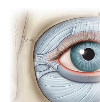
■ Anchored to the lateral orbit is a labyrinth of connective tissues, known as the lateral retinaculum, that are crucial to maintenance of the integrity, position, and function of the globe and periorbital.
• Control of periorbital aesthetics by proper brow positioning, corrugator muscle removal, and lid fold invagination when beneficial.
• Restoration of tone and position of the lateral canthus and, along with it, restoration of a youthful and attractive intercanthal axis tilt.
• Restoration of the tone and posture of the lower lids.
• Preservation of maximal lid skin and muscle (so essential to lid function and aesthetics) as well as orbital fat.
• Lifting of the midface through reinforced canthopexy, preferably enhanced by composite malar advancement.
• Correction of suborbital malar grooves with tear trough (or suborbital malar) implants, obliterating the deforming tear trough (bony) depressions that angle down diagonally across the cheek, which begin below the inner canthus.
• Control of orbital fat by septal restraint or quantity reduction.
• Removal of only that tissue (skin, muscle, fat) that is truly excessive on the upper and lower lids, sometimes resorting to unconventional excision patterns.
• Modification of skin to remove prominent wrinkling and excision of small growths and blemishes.
Supraorbital fissure
Greater wing of sphenoid
Zygomatic
Infraorbital fissure
Zygomaticofacial foramen
Frontal bone
Supraorbital foramen
Superior orbital ridge

Optic foramen
Ethmoid
Lacrimal bone and fossa Maxilla
Medial canthal tendon
Medial check retinaculum
Fossa for lacrimal sac
Medial rectus and sheath
Orbicularis

Lateral canthal tendon
Lateral check retinaculum
Tenon’s capsule
Lateral rectus and sheath Periorbita
Temporalis
Zygomatic bone
check ligaments.
Orbicularis fascia
Lateral orbital thickening
Lateral canthal tendon

Coronoid process of mandible
Maxilla bone
Frontal bone
Tarsal plates
Figure 1.3 Lateral canthal tendon has separate superficial and deep components. The deep
tarsal plates
Infraorbital foramen
■ These structures coalesce at the lateral orbit and support the globe and eyelids like a hammock (Fig. 1.2)
■ The lateral retinaculum consists of the lateral canthal tendon, tarsal strap, lateral horn of the levator aponeurosis, the Lockwood suspensory ligament, Whitnall ligament, and check ligaments of the lateral rectus muscle.
■ They converge and insert securely into the thickened periosteum overlying the Whitnall tubercle.
■ Controversy exists surrounding the naming of the components of the lateral canthal tendon.
■ A superficial component is continuous with the orbicularis oculi fascia and attaches to the lateral orbital
Whitnall tubercle.
periosteum
lateral orbital rim
lateral orbital thickening. Both components are continuous with both superior and inferior lid tarsal plates. (Adapted from Muzaffar AR, Mendelson BC, Adams Jr WP. Surgical anatomy of the ligamentous attachments of the lower lid and lateral canthus. Plast Reconstr Surg. 2002;110(3):873–884.)
rim and deep temporal fascia by means of the lateral orbital thickening.
■ A deep component connects directly to the Whitnall tubercle and is classically known as the lateral canthal tendon (Fig. 1.3)
■ The tarsal strap is a distinct anatomic structure that inserts into the tarsus medial and inferior to the lateral canthal tendon.
■ The tarsal strap attaches approximately 3 mm inferiorly and 1 mm posteriorly to the deep lateral canthal tendon, approximately 4–5 mm from the anterior orbital rim.
■ It shortens in response to lid laxity, benefiting from release during surgery to help achieve a long-lasting restoration or elevation canthopexy (Fig. 1.4)
Whitnall tubercle
Lateral canthal tendon
‘Tarsal strap’
Orbital septum

Tarsus


Posterior limb, medial canthal tendon
Superior limb, medial canthal tendon
Anterior limb, medial canthal tendon
Lacrimal fossa
Anterior and posterior lacrimal crests

■ Adequate release of the tarsal strap permits a tensionfree canthopexy, minimizing the downward-tethering force of this fibrous condensation.
■ This release, along with a superior reattachment of the lateral canthal tendon, is key to a successful canthopexy.
■ A hammock of fibrous condensations suspends the globe above the orbital floor. The medial components of the apparatus include medial canthal tendon, the Lockwood suspensory ligament, and check ligaments of the medial rectus.
■ The medial canthal tendon, like the lateral canthal tendon, has separate limbs that attach the tarsal plates to the ethmoid and lacrimal bones.
■ Each limb inserts onto the periorbital of the apex of the lacrimal fossa. The anterior limb provides the bulk of the medial globe support (Fig. 1.5).
■ The forehead and brow consist of four layers: skin, subcutaneous tissue, muscle, and galea.
■ There are four distinct brow muscles: frontalis, procerus, corrugator superciliaris, and orbicularis oculi (Fig. 1.6)
■ The frontalis muscle inserts predominately into the medial half or two-thirds of the eyebrow (Fig. 1.7), allowing the lateral brow to drop hopelessly ptotic from aging, while the medial brow responds to frontalis activation and elevates, often excessively, in its drive to clear the lateral overhand. Constant contraction of the
Orbicularis oculi
Pretarsal orbicularis
Preseptal orbicularis
Saunders; 2004:13.) Superciliary cor rugator
Frontalis


Procerus

frontalis will give the appearance of deep horizontal creases in the forehead (Fig. 1.8).
■ The vertically oriented procerus is a medial muscle, often continuous with the frontalis, arising from the nasal bones and inserting into the subcutaneous tissue of the glabellar region. It pulls the medial brow inferiorly and contributes to the horizontal wrinkles at the root of the nose. More commonly, these wrinkles result from brow ptosis and correct spontaneously with brow elevation.
■ The obliquely oriented corrugators muscle arises from the frontal bone and inserts into the brow tissue laterally, with some extensions into orbicularis and frontalis musculature, forming vertical glabellar furrows during contraction.

Figure 1.7 The frontalis muscle inserts predominantly into the medial half or two-thirds of the eyebrow. The medial brow responds to frontalis activation and elevates, often excessively, in its drive to clear lateral overhang.
Diagonal lines

Figure 1.8 Frontalis action. The frontalis muscle inserts into the medial twothirds of the brow. Exaggerated medial brow elevation is required to clear the lateral overhang and to eliminate visual obstruction. Constant contraction of the frontalis will give the appearance of deep horizontal creases in the forehead. This necessarily means that when the lateral skin is elevated or excised, the overelevated and distorted medial brow drops profoundly.
■ There is much similarity between upper and lower eyelid anatomy. Each consists of an anterior lamella of skin and orbicularis muscle and a posterior lamella of tarsus and conjunctiva (Fig. 1.9)
Levator palpebrae
Superior rectus

Inferior rectus
■ The orbicularis muscle, which acts as a sphincter for the eyelids, consists of orbital, preseptal, and pretarsal segments.
■ The pretarsal muscle segment fuses with the lateral canthal tendon and attaches laterally to Whitnall tubercle. Medially it forms two heads, which insert into the anterior and posterior lacrimal crests (see Fig. 1.6).
■ The orbital septum originates superiorly at the arcus and forms the anterior border of the orbit. It joins with the levator aponeurosis, just superior to the tarsus. The sling formed by the union of these two structures houses the orbital fat.
■ The levator palpebrae superioris muscle originates above the annulus of Zinn. It extends anteriorly for 40 mm before becoming a tendinous aponeurosis below Whitnall ligament. The aponeurosis fans out medially and laterally to attach to the orbital retinacula. The aponeurosis fuses with the orbital septum above the superior border of the tarsus and at the caudal extent of the sling, sending fibrous strands to the dermis to form the lid crease. Extensions of the aponeurosis finally insert into the anterior and inferior tarsus. As the levator aponeurosis undergoes senile attenuation, the lid crease rises into the superior orbit from its remaining dermal attachments while the lid margin drops.
■ Müller muscle, or the supratarsal muscle, originates on the deep surface of the levator near the point where the muscle becomes aponeurotic and inserts into the superior tarsus. Dehiscence of the attachment of the levator aponeurosis to the tarsus results in an acquired
ptosis only after the Müller muscle attenuates and loses its integrity.
■ In the Asian eyelid, fusion of the levator and septum commonly occurs at a lower level, allowing the sling and fat to descend farther into the lid. This lower descent of fat creates the characteristic fullness of their upper eyelid. In addition, the aponeurotic fibers form a weaker attachment to the dermis, resulting in a less distinct lid fold (Fig. 1.10)
■ The orbital septum has an adhesion to the levator aponeurosis above the tarsus. The septum continues beyond this adhesion and extends to the ciliary margin. It is superficial to the preaponeurotic fat found at the supratarsal crease. The septal extension is a dynamic component to the motor apparatus, as traction on this fibrous sheet reproducibly alters ciliary margin position (Fig. 1.11). The septal extension serves as an adjunct to, and does not operate independent of, levator function, as mistaking the septal extension for levator apparatus and plicating this layer solely results in failed ptosis correction.
■ The anatomy of the lower eyelid is somewhat analogous to that of the upper eyelid.
■ The retractors of the lower lid, the capsulopalpebral fascia, correspond to the levator above.
■ The capsulopalpebral head splits to surround and fuse with the sheath of the inferior oblique muscle. The two heads fuse to form the Lockwood suspensory ligament, which is analogous to Whitnall ligament.
■ It fuses with the orbital septum 5 mm below the tarsal border and then inserts into the anterior and inferior surface of the tarsus.
■ The inferior tarsal muscle is analogous to Müller muscle of the upper eyelid and also arises from the sheath of the inferior rectus muscle. It runs anteriorly above the inferior oblique muscle and also attaches to the inferior tarsal border.
■ The combination of the orbital septum, orbicularis, and skin of the lower lid acts as the anterior barrier of the orbital fat. As these connective tissue properties relax, the orbital fat is allowed to herniate forward, forming an unpleasing, full lower eyelid. This relative loss of orbital volume leads to a commensurate, progressive hollowing of the upper lid as upper eyelid fat recesses.
■ The capsulopalpebral fascia and its overlying conjunctiva form the posterior border of the lower orbital fat. Transection of the capsulopalpebral fascia during lower lid procedures, particularly transconjunctival blepharoplasty, releases the retractors of the lower eyelid, which can reduce downward traction and allow the position of the lower lid margin to rise.
■ A network of ligaments serves as a scaffold for the skin and subcutaneous tissue surrounding the orbit. The
orbital retaining ligament directly attaches the orbicularis at the junction of its orbital and preseptal components to the periosteum of the orbital rim and, consequently, separates the prezygomatic space from the preseptal space. This ligament is continuous with the lateral orbital thickening, which inserts onto the lateral orbital rim and deep temporal fascia. It also has attachments to the superficial lateral canthal tendon (see Figs. 1.3, 1.12, 1.13). Attenuation of these ligaments permits descent of orbital fat onto the cheek. A midfacelift must release these ligaments to achieve a supported, lasting lift.
■ The internal and external carotid arteries supply blood to the orbit and eyelids (Fig. 1.14).
■ The ophthalmic artery is the first intracranial branch of the internal carotid; its branches supply the globe, extraocular muscles, lacrimal gland, ethmoid, upper eyelids, and forehead.
■ The external carotid artery branches into the superficial temporal and maxillary arteries. The infraorbital artery is a continuation of the maxillary artery and exits 8 mm below the inferomedial orbital rim to supply the lower eyelid.
■ The arcade of the superior and inferior palpebral arteries gives a rich blood supply to the eyelids. The superior palpebral artery consists of a peripheral arcade located at the superior tarsal border – the area where surgical dissection occurs to correct lid ptosis and to define lid folds. Damage to a vessel within this network commonly results in a hematoma of Müller muscle, causing lid ptosis for 2–8 weeks postoperatively. Likewise, on the lower lid, the inferior palpebral artery lies at the inferior border of the inferior tarsus.
■ The supratrochlear, dorsal nasal, and medial palpebral arteries all traverse the orbit medially. Severing these arteries during fat removal, without adequately providing hemostasis, may lead to a retrobulbar hematoma, a vision-threatening complication of blepharoplasty.
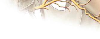
■ The trigeminal nerve, along with its branches, provides sensory innervations to the periorbital region (Fig. 1.15)
■ A well-placed supraorbital block will anesthetize most of the upper lid and the central precoronal scalp.
■ The maxillary division exits the orbit through one to three infraorbital foramina. It provides sensation to the skin of the nose, the lower eyelids, and the upper lid.
■ The facial nerve supplies motor function to the lids (Fig. 1.16)
■ Innervation of facial muscles occurs on their deep surfaces.
■ Interruption of the branches to the orbicularis muscle from the periorbital surgery or facial surgery may result in atonicity due to partial denervation of the orbicularis with loss of lid tone or anomalous reinnervation and possibly undesirable eyelid twitching.
■ The frontal branch of the facial nerve courses immediately above and is attached to the periosteum of


Figure 1.10 The anatomic variations in the upper eyelid displayed by different ethnic groups and the changes associated with senescence within each group allow for a convergence of anatomy. (A) The normal youthful Asian upper eyelid has levator extensions inserting onto the skin surface to define a lid fold that averages 6–8 mm above the lid margin. The position of the levator–skin linkage and the anteroposterior relationship of the preaponeurotic fat determine lid fold height and degree of sulcus concavity or convexity (as shown on the right half of each anatomic depiction). (B) In the case of levator dehiscence from the tarsal plate, the upper lid crease is displaced superiorly. The orbital septum and preaponeurotic fat linked to the levator are displaced superiorly and posteriorly. These anatomic changes create a high lid crease, a deep superior sulcus, and eyelid ptosis. (C) In the aging eyelid, the septum becomes attenuated and stretches. The septal extension loosens, and this allows orbital fat to prolapse forward and slide over the levator into an anterior and inferior position. Clinically, this results in an inferior displacement of the levator skin attachments and a low and anterior position of the preaponeurotic fat pad. (D) The youthful Asian eyelid anatomically resembles the senescent upper lid with a low levator skin zone of adhesion and inferior and anteriorly located preaponeurotic fat. The characteristic but variable low eyelid crease and convex upper eyelid and sulcus are classic. (Adapted from Spinelli HM. Atlas of Aesthetic Eyelid and Periocular Surgery. Philadelphia: Saunders; 2004:59.).
Orbicularis
Orbital septum
Müller muscle
Levator aponeurosis
Septal extension
Tarsus

Figure 1.11 The orbital septum has an adhesion to the levator aponeurosis above the tarsus. The septal extension begins at the adhesion of the orbital septum to the levator and extends to the ciliary margin. It is superficial to the preaponeurotic fat found at the supratarsal crease. (Adapted from Reid RR, Said HK, Yu M, et al. Revisiting upper eyelid anatomy: introduction of the septal extension. Plast Reconstr Surg. 2006;117(1):65–70.)
Cor rugator supercilii
Frontal bone
Orbicularis retaining ligament
Maxilla bone
Nasal bone
Globe
Septum orbitale
Orbitomalar ligament
Prezygomatic space
Orbicularis oculi

Figure 1.13 The orbital retaining ligament (ORL) directly attaches the orbicularis oris (OO) at the junction of its pars palpebrarum and pars orbitalis to the periosteum of the orbital rim and, consequently, separates the prezygomatic space from the preseptal space. (Adapted from Muzaffar AR, Mendelson BC, Adams Jr WP. Surgical anatomy of the ligamentous attachments of the lower lid and lateral canthus. Plast Reconstr Surg. 2002;110(3):873–884.)
Supraorbital ar tery
Superficial temporal ar ter y Lacrimal ar tery
Zygomaticofacial ar ter y
Transverse facial ar ter y
Orbicularis oculi

Lateral orbital thickening
Zygomatic bone
Medial palpebral ar ter y (superior)
1. Peripheral arcade
2. Marginal arcade

Infraorbital ar ter y
Facial ar ter y
Figure 1.14 Arterial supply to the periorbital region.
Supratrochlear ar tery
Dorsal nasal ar ter y
Angular ar ter y
Medial palpebral ar ter y (inferior)
Lateral nasal ar ter y
Inferior palpebral ar ter y
Orbicularis retaining ligament
Figure 1.12 The orbicularis muscle fascia attaches to the skeleton along the orbital rim by the lateral orbital thickening (LOT) in continuity with the orbicularis retaining ligament (ORL). (Adapted from Ghavami A, Pessa JE, Janis J, et al. The orbicularis retaining ligament of the medial orbit: closing the circle. Plast Reconstr Surg. 2008;121(3):994–1001.).
Zygomaticotemporal nerve
Lacrimal nerve
Supraorbital nerve
Supra trochlear ner ve
Zygomaticofacial nerve

Sensory
Infratrochlear nerve
Infraorbital ner ve
Temporal branches (facial nerve VIII)


Zygomatic branches (facial nerve VIII)
Figure 1.16 Anatomy of the brow and temporal region. The light-green opaque area denotes the deep temporal fascia and the periosteum where sutures may be used to suspend soft tissue. Wide undermining, soft tissue suspension, and canthopexy are safely performed here.
the zygomatic bone. It then courses medially approximately 2 cm above the superior orbital rim to innervate the frontalis, corrugators, and procerus muscles from their deep surface.
■ A separate branch travels along the inferior border of the zygoma to innervate the inferior component of orbicularis oculi.
■ The characteristics of youthful, beautiful eyes differ from one population to another, but generalizations are possible and provide a needed reference to judge the success of various surgical maneuvers.
■ Attractive, youthful eyes have globes framed in generously sized horizontal apertures (from medial and lateral), often accentuated by a slight upward tilt of the intercanthal axis (Fig. 1.17)
■ The aperture length should span most of the distance between the orbital rims.
■ In a relaxed forward gaze, the vertical height of the aperture should expose at least three-quarters of the cornea, with the upper lid extending down at least 1.5 mm below the upper limbus (the upper margin of the cornea) but no more than 3 mm. The lower lid ideally covers 0.5 mm of the lower limbus but no more than 1.5 mm.
■ In the upper lid, there should be a well-defined lid crease lying above the lid margin with lid skin under slight stretch, slightly wider laterally.
■ Ideally, the actual pretarsal skin visualized on relaxed forward gaze ranges from 3 to 6 mm in European ethnicities.
■ The Asian lid crease is generally 2–3 mm lower, with the distance from lid margin diminishing as the crease moves toward the inner canthus.

■ Patients of Indo-European and African decent show 1 to 2 mm lower than European ethnicities.
■ The ratio of distance from the lower edge of the eyebrow (at the center of the globe) to the open lid margin to the visualized pretarsal skin should never be less than 3 : 1 (see Fig. 1.1), preferably more.
■ Scleral show is the appearance of white sclera below the lower border of the cornea and above the lower eyelid margin. In general, sclera show is contradictory to optimal aesthetics and may be perceived as a sign of aging, previous blepharoplasty, or orbital disease (e.g., thyroid disease).
■ More than 0.5 mm of sclera show beneath the cornea on direct, forward gaze begins to confer a sad or melancholy aura to one’s appearance.
■ The intercanthal axis is normally tilted slightly upward (from medial to lateral) in most populations.
■ Exaggerated tilts are encountered in some Asian, Indo-European, and African-American populations.
■ A thorough history and physical examination should be obtained – including an ophthalmic history (see Box 1.2).
■ Physical exam should include evaluation for symmetry; globe shape, position, and appearance; signs of aging; lid appearance; lid function; and relative laxity.
■ In the upper lid, excessive skin due to loss of elasticity and sun damage is one of the major causes of an aged appearance in the periorbital area.
■ In addition to relaxed skin changes, excessive fat herniation can cause bulging, resulting in a heavy appearance to the upper lid area.
■ Aging changes in the lower lid include relaxation of the tarsal margin with scleral show, rhytides of the lower lid, herniated fat pads resulting in bulging in one or all of
BOX 1.2 Important information to obtain during history and physical examination
• Medication use: particularly anticoagulants, anti-inflammatory and cardiovascular drugs, and vitamins (especially vitamin E).
• Herbal supplement use: herbs represent risks to anesthesia and surgery, particularly those affecting blood pressure, blood coagulation, the cardiovascular system, and healing.
• Allergies: medication and type.
• Past medical history: especially hypertension, diabetes, cardiovascular and cerebrovascular disease, hepatitis, liver disease, heart disease or arrhythmias, cancer, thyroid disease, and endocrine disease.
• Bleeding disorders or blood clots.
• Psychiatric disease.
• Alcohol and smoking history
• Recreational drug use, which may interact with anesthesia.
• Exposure to human immunodeficiency virus and hepatitis virus.
• Any history of facial herpes zoster or simplex.
the three fat pocket areas, and hollowing of the nasojugal groove and lateral orbital rim areas.
■ Hollowing of the nasojugal groove area appears as dark circles under the eyes, mostly because of lighting and the shadowing that result from this defect.
■ Contact lens wear poses particular risks when eyelid surgery is performed.
■ Long-term contact lens wearing hastens the process of drying out the eyes.
■ Traditional blepharoplasty techniques consistently produce vertical dystopia with increased scleral exposure, making the lens wear difficult if not dangerous.
■ Ptosis and canthopexy surgery may alter the corneal curvature and require that contacts be refitted.
■ The patient should discontinue contact lens wear in the perioperative period to allow healing without the need to manipulate the eyelids.
■ Dry, irritated eyes before surgery will lead to irritated eyes after surgery, and the surgeon may be blamed.
■ Treatment options include artificial tears, ointment, anti-inflammatory drops, and punctal plugs or punctal closure.
■ Exophthalmos, unilaterally or bilaterally, associated with a thyroid disorder, should be completely stabilized for approximately 6 months before elective aesthetic surgery.
■ Eyelid measurements are documented for use during ptosis surgery and, if necessary, for insurance purposes.
■ In the typical person with the brow in an aesthetically pleasing position, 20 mm of upper lid skin must remain between the bottom of the central eyebrow and the upper lid margin to allow adequate lid closure during sleep, a well-defined lid crease, and an effective and complete blink.
■ In the eyelid of the white individual, the aperture (distance between the upper and lower eyelids) average is 10–12 mm.
BOX 1.3 Recommended photographic views
• Full face, upright (at rest) frontal, oblique, and lateral views.
• Full face, upright, and smiling.
• Direct periorbital views in upward gaze and downward gaze and with eyes gently closed.
• A view with a finger slightly elevating the brows with the eyes open and another with the eyes closed.
■ The margin reflex distance (MRD), measured from the light reflex on the center of the cornea to the upper eyelid margin, ranges from 3 to 5 mm.
■ True blepharoptosis is defined by the degree of upper lid infringement upon the iris and pupil.
■ As the MRD decreases toward zero, the severity of blepharoptosis increases.
■ Before method selection, the levator function must be determined by measuring the upper eyelid excursion from extreme downward gaze to extreme upward gaze; it generally ranges from 10 to 12 mm.
■ If ptosis exists, the type of repair depends upon the severity of the ptosis and the reliability of the levator to recreate smooth upper lid elevation.
■ Pseudoptosis occurs when excess upper lid skin covers the eyelid, depressing the eyelashes, forming hooding, and simulating ptosis.
■ Photographic evidence of this is often necessary for insurance purposes when a levator aponeurosis repair or an excisional blepharoplasty is planned.
■ Brow ptosis is a common aspect of facial aging. It adds weight and volume to the upper eyelid to develop, or exacerbate, eyelid ptosis.
■ The ability to differentiate the causes of droopy eyelids – brow ptosis (brow weight resting on the eyelids), dermatochalasis (excess skin), and blepharoptosis (levator attenuation or dehiscence) – will enable the surgeon to select the proper correction.
■ There is a normal 10–12 mm projection of the globe seen in a lateral, as measured from the lateral orbital rim at the level of the canthal tendon to the pupil.
■ Proptosis and enophthalmos are relative anterior and posterior displacement of the globe, respectively. Hertel exophthalmometry can be used to quantitate the degree of relative projection for documentation purposes.
■ Assessment of tear production is a necessary but unreliable task.
■ The Schirmer test:
• Placing filter paper strips in the lateral third of the lower eyelid.
• After 5 min, normal tear production should be greater than 15 mm; 5–10 mm indicates borderline tear secretion, and below 5 mm is hyposecretion.
■ No other area of cosmetic surgery is more dependent on accurate photography than the periorbital region (Box 1.3).
■ Before surgical planning, one must have a meaningful conceptualization of the desired result. Only then can the surgical maneuvers required be organized in a meaningful way (Box 1.4).
The preoperative periorbital plan should include the following:
• The patient’s specific concerns and desires for improvement.
• Brow position.
• Lower eyelid tonicity
• Eyelid ptosis, retraction, or levator dehiscence.
• Exophthalmos or enophthalmos.
• Supraorbital rim prominence or hypoplasia.
• Suborbital malar and tear trough deformities.
• Excision of necessary skin, muscle, and fat – only if necessary.
■ When skin-only excision is elected, it should occur above the supratarsal fold or crease, leaving that structure intact – this retains most of the definition of an existing lid fold.
■ The supratarsal fold is located approximately 7–8 mm above the ciliary margin in women and 6–7 mm in men.
■ The upper marking must be at least 10 mm from the lower edge of the brow and should not include any thick brow skin.
■ The use of a pinch test for redraping the skin is helpful.
■ The shape of the skin resection is lenticular in younger patients and more trapezoid shaped laterally in older patients.
■ The incision may need to be extended laterally with a larger extension, but extension lateral to the orbital rim should be avoided if possible to prevent a prominent scar (Fig. 1.18)
■ Similarly, the medial markings should not be extended medial to the medial canthus because extensions onto the nasal side wall result in webbing.
■ At the conclusion of the case, the patient should have approximately 1–2 mm of lagophthalmos bilaterally. Fig. 1.19 displays the predictable, restorative outcomes that can be achieved with skin excision alone.
■ Anchor blepharoplasty involves the creation of an upper eyelid crease by attaching pretarsal skin to the underlying aponeurosis.
■ Advantages of an anchor blepharoplasty are a crisp, precise, and well-defined eyelid crease that persists indefinitely.
■ Disadvantages are that it is more time-consuming, requires greater surgical skills and expertise, and encourages greater frontalis relaxation as a result of more effective correction of the overhanging pseudoptotic skin.
■ Key components of the anchor blepharoplasty include:
• Minimal skin excision (2–3 mm) extending cephalad from the tarsus.
• A 1–2 mm sliver of orbicularis must be removed in proportion to the amount of skin removed.
• A small pretarsal skin and muscle flap are dissected from the aponeurosis and septum adhesion.
• After sharply disinserting the aponeurosis from the tarsus, pretarsal fatty tissue can be removed to debulk the pretarsal skin.
• Mattressed anchor sutures are placed connecting the tarsus to the aponeurosis and pretarsal skin (Fig. 1.20)
• Finally, a running suture approximates the preseptal skin incision.
■ A relative excess of retroseptal fat may be safely excised through an upper eyelid blepharoplasty incision.
■ A small septotomy is made at the superior aspect of the skin excision into each fat compartment in which conservative resection of redundant fat has been planned.
■ The fat is teased out bluntly and resected using pinpoint cautery.
■ This fat usually includes the medial or nasal compartment, which contains white fat.
■ Yellow fat in the central compartment is usually more superficial and lateral.
■ Gentle pressure on the patient’s globe can reproduce the degree of excess while the patient lies recumbent on the operating room table (Fig. 1.21)
■ Overall, undercorrection is preferred to prevent hollowing, which can be dramatic and recognized as an A-frame abnormality.
■ The attenuated orbital septum may be addressed by using selective diathermy along the exposed caudal septum.
■ Inflammation-mediated tightening can enhance septal integrity.
■ Septal plication aid is unnecessary and may induce a brisk, restrictive inflammatory response.
■ During upper blepharoplasty, with the septum open and the aponeurosis and superior tarsus exposed, there is an ideal opportunity to adjust the level of the aperture.
■ Inappropriate aperture opening can be due to upper lid ptosis or upper lid retraction.
■ True ptosis repair involves reattachment of the levator aponeurosis to the tarsus, with or without shortening of applicable structures (e.g. aponeurosis, Müller muscle, and tarsus).
■ There are a variety of techniques to address blepharoptosis, but they are outside the scope of this chapter. There is a significant learning curve to performing a ptosis repair, and even then, the ability to get perfect symmetry is elusive.
■ In the setting of mild upper eyelid ptosis (~1 mm), where the decision has been made to avoid a formal lid ptosis procedure, selective myectomy of the upper eyelid orbicularis can be performed to widen the lid aperture.
■ The amount of muscle to be resected depends on a host of factors, including the severity of relative lid ptosis,


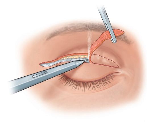
brow position, and fold disparity (Fig. 1.22) and is titrated depending on the amount of effect desired.
■ For 1 mm or less of relative upper lid ptosis, resection of at least 3–4 mm of orbicularis is required.
■ No attempt is made to close orbicularis muscle in this resection, which could increase the risk of lagophthalmos.
The key components of formal lid ptosis correction include:
■ Correct identification of the distal extensions of the aponeurosis and the orbital septal extension.
■ The superior edge of the tarsus is freed from any dermal or tendinous extensions.
■ Leaving a small cuff of filmy connective tissue (~1 mm) on the tarsus will minimize bleeding from the richly vascularized area.
■ Ensure that there is complete hemostasis by use of a fine forceps cautery, lifting all lid tissues away from the cornea and globe before cauterizing.
■ Anchor the upper third of the tarsus to the remaining levator with 5–0 silk suture, placed as a horizontal mattress.
■ The lid should be flipped to ensure that the suture is not exposed posteriorly on the tarsus, which could cause a troublesome corneal abrasion.
■ If performed under sedation or local anesthetic, the level should be checked by having the patient open the eye.


■ For cases under general anesthetic, one should attempt to create one to two times the amount of lagophthalmos relative to the preoperative ptosis.
■ If there is any medial or lateral retraction or ptosis, the central suture should be repositioned medially or laterally as many times as needed, with adjustment to a pleasing lid height and contour.
■ Both sides should be completed before the suture is permanently tied.
■ Lower blepharoplasty has evolved substantially. Although excellent aesthetic results can be achieved with transcutaneous lower blepharoplasty, lid retraction and ectropion are concerning complications. Conservative excisional techniques center on the concept of fat preservation. Transconjunctival lower blepharoplasty, although more conservative, does not eliminate the risk of lid malposition. An effective, lasting procedure should address the extrinsic and intrinsic support of the eye, which is weakened during the aging process.
■ Transconjunctival blepharoplasty is the preferred procedure for fat reduction in patients without excess skin and with good canthal position.
■ It is less likely to lead to lower lid malposition than a transcutaneous approach.
■ It minimizes but does not eliminate postoperative lower lid retraction.
■ Transection of the lower lid retractors can cause a temporary rise in the lid margin, especially if they are suspended during the healing period.
■ Previously suspected septal scarring through transconjunctival fat excision has not been shown to significantly alter lid posture or tonicity.
■ The lower lid retractors (capsulopalpebral fascia and inferior tarsal muscle) and overlying conjunctiva lie directly posterior to the three fat pads of the lower lid.
■ A broad and deep transconjunctival incision severs both conjunctiva and retractors but typically should not incise the orbital septum, orbicularis, or skin.
■ The conjunctival incision is made with a monopolar cautery needle tip at least 4 mm below the inferior border of the tarsus – never through the tarsus (Fig. 1.23)


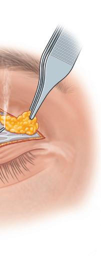


■ Conjunctival closure, when it is elected, is simplified by a monofilament pull-out suture that enters the eye externally, closes the conjunctiva, and exits through the skin and is taped.
■ The incision through the conjunctiva and retractors gives excellent access to the orbital fat.
■ A 6–0 silk traction suture passed through the inferior conjunctival wound and retracted over the globe gives wide access to the orbital fat, even helping to prolapse the fat into the wound. The thin film of synovium-appearing capsule encasing the orbital fat is opened, releasing the fat to bulge into the operative field (Fig. 1.24)
■ Once fat is removed through a transconjunctival incision, excess skin can be removed through a subciliary position.
■ Fat reduction may leave skin excess, leading to wrinkling.
■ A conservative “skin pinch” can be done to estimate skin removal, or alternatively, skin can be tightened by skin resurfacing with chemical or laser peels (Fig. 1.25).
■ One should be careful not to incise the orbital septum, which leads to increased postoperative retraction.

Figure 1.21 Simple skin excision blepharoplasty. (A) The medial fat pad may require digital pressure to expose and grasp; however, care should be taken not to overly resect fat when using digital pressure techniques. (B) Closure may then be performed with a combination of interrupted and running intracuticular sutures.
(Adapted from Spinelli HM. Atlas of Aesthetic Eyelid and Periocular Surgery. Philadelphia: Saunders; 2004:65.)
■ A preseptal approach is obtained by entering the conjunctiva above the level of septal attachment to the capsulopalpebral fascia.
■ A retroseptal approach involves a 1.5- to 2-cm incision lower down in the fornix, and is typically used to excise fat.
■ It is preferable to leave the transconjunctival incision open.
■ Suturing the wound may trap bacteria or cause corneal irritation. A B Medial fat pad removed Intracuticular running suture
■ This procedure works particularly well when there is an isolated fat pad, especially medially, accessed through a single stab incision through the conjunctiva.
■ A subciliary incision can be used to develop a skin flap or a skin–muscle flap.
■ With either method, pretarsal orbicularis fibers should remain intact.
■ For the skin–muscle flap, skin and preseptal orbicularis are elevated as one flap, while with a skin flap, the muscle and its innervation can be preserved.
■ Periorbital fat, muscle, and skin can be addressed with either approach.
■ Once the plane deep to the orbicularis is entered, dissection continues between the muscle and the orbital septum down to the level of the orbital rim.
■ Periorbital fat can be excised through small incisions in the septum.
■ The fat can also be retropositioned using capsulopalpebral fascia placation, or it can be transferred into the nasojugular fold.
Orbicularis muscle fibers and skin can be excised at closure.
■ Care must be taken with muscle excision, which can lead to orbicularis denervation and lid malposition.
■ An alternative to excising prominent orbital fat is to redrape the pedicled fat onto the arcus marginalis.
■ Patients with tear trough deformities who have prominent medial fat pads are excellent candidates.
■ Access to the medial and central fat pads is by the subciliary or transconjunctival incision.
■ The minor degree of lateral fat pad prominence is generally insufficient to affect any change with repositioning.


Figure 1.22 (A,B) Once the upper lid is incised, the levator may be modified (shortened/lengthened) in a number of ways, including simple plication. A suborbicularis skin flap can also be developed, allowing access to preaponeurotic fat. (Adapted from Spinelli HM. Atlas of Aesthetic Eyelid and Periocular Surgery. Philadelphia: Saunders; 2004:69).
■ A supraperiosteal or a subperiosteal dissection of 8–10 mm caudal to the inferior orbital rim permits tension-free placement.
■ The fat can be secured in place with interrupted absorbable sutures.
■ Patients must be warned that various degrees of fat loss and hardening are possible. There is also a rare but described possibility of restrictive strabismus related to aggressive fat mobilization and fixation.
■ In this procedure, the herniated septum is plicated and repositioned to its normal anatomic site within the orbit.
■ The fat is replaced in the retroseptal position to regain its original anatomic integrity (Fig. 1.26).
■ Three to four 5–0 polyglycolic acid sutures are placed in a vertical fashion from medial to lateral.
■ The protruding fat pads are invaginated, and the integrity of the thin, flaccid septum is restored.
■ Additional support may be gained with septo-orbitoperiostoplasty variation, which plicates the flaccid septum and secures it to the periosteum of the inferior orbital rim.
■ Because no disruption of the eyelid anatomy occurs, complications related to lid malposition, such as lid retraction, scleral show, and ectropion, are reduced.
■ The capsulopalpebral fascia can be plicated to the orbital rim either through a transcutaneous or a transconjunctival approach.
■ In the transcutaneous method, dissection is carried out between the orbicularis and the septum down to the orbital rim; the capsulopalpebral fascia is then sutured to the orbital rim.
■ In the transconjunctival method, the capsulopalpebral fascia is divided from the tarsus, and orbital fat is retroplaced, its position maintained by suturing the capsulopalpebral fascia to the periosteum of the orbital rim using a continuous running 6–0 nonabsorbable suture.
■ The conjunctival gap of a few millimeters is allowed to reepithelialize (Fig. 1.27)
■ One advantage of the transconjunctival approach is the division of lower eyelid depressors, which helps maintain the lower eyelid at an elevated level due to the unopposed action of the pretarsal orbicularis.
■ Orbicularis repositioning can be used to eliminate hypotonic and herniated orbicularis muscle, soften palpebral depressions, and shorten the lower lid to cheek distance.


Retroseptal approac h
Pretroseptal (suborbicularis) approach
Nonconductive retractor


Inferior tarsal plate

everted. This
surgeon. (B)
conjunctiva and lower lid retractors are incised just below the tarsal plate entering the suborbicularis preseptal space. This plane is developed to the orbital rim with the assistance of the traction suture and a nonconductive instrument. (C) The conjunctiva and lower lid retractors are incised just below the tarsal plate entering the suborbicularis preseptal space. This plane is developed to the orbital rim with the assistance of the traction suture and a nonconductive instrument. (Adapted from Spinelli HM. Atlas of Aesthetic Eyelid and Periocular Surgery. Philadelphia: Saunders; 2004:86.)
■ The main steps include:
• Elevation of a skin muscle flap.
• Release of the orbicularis retaining ligament and resuspension of the orbicularis – frequently after lateral canthopexy.
• Along the entire infraorbital rim, the orbicularis retaining ligament is divided.
• Additional medial dissection is performed to release the levator labii when a tear trough deformity is present.
• The skin muscle flap is draped in a superior lateral vector rather than a pure vertical vector.
• Excision of skin and muscle are performed by removing a triangle of tissue lateral to the canthus,


Reposition

thereby minimizing the amount of tissue removed along the actual lid margin.
• The lateral suspension of the orbicularis is to the orbital periosteum.
• Lower lid support is gained by resuspension of the anterior (skin and muscle) and posterior lamellae (tarsus by canthopexy).
■ This technique is best suited for patients with scleral show, lid laxity, and a negative vector, which put them at risk for lid malposition in the postoperative period.
■ Disadvantages are it inherently disrupts the orbicularis, which may lead to denervation, and mobilization of the levator labii muscles may put the buccal branch of the facial nerve at risk.