
model


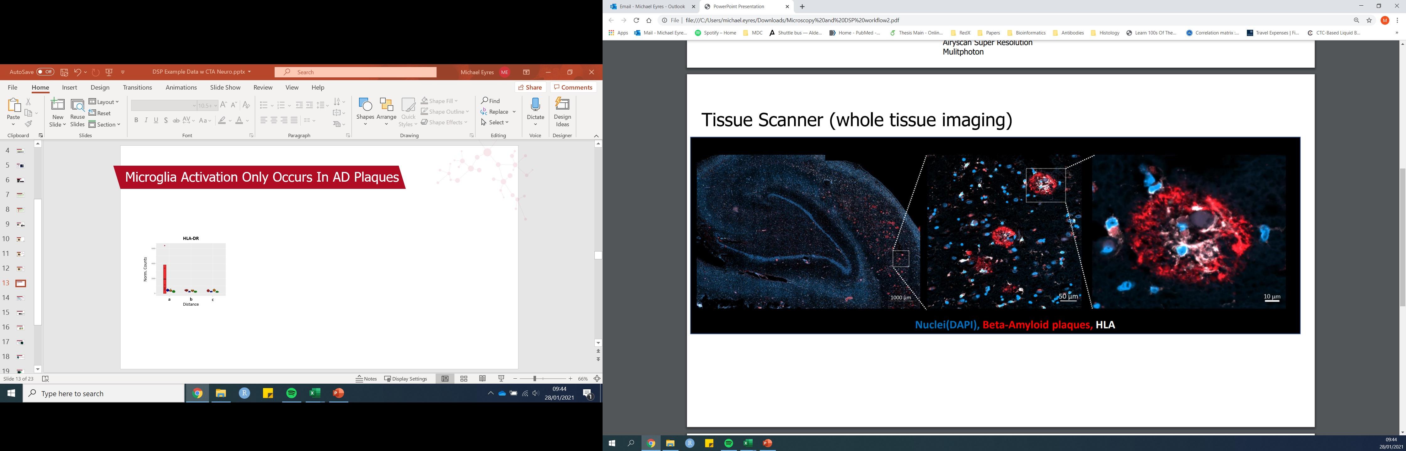
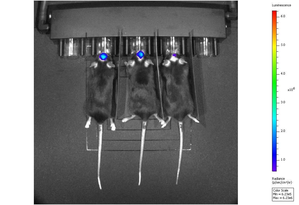
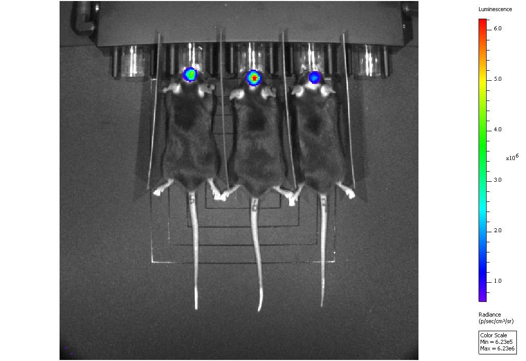
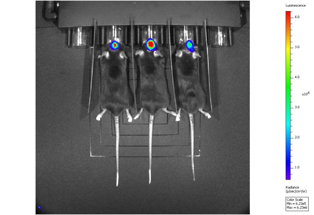
Kerry Shea, Regine Anderson, Michael Eyres, Bruno Bellina, Tara Bowen, Maike Langini, Andrzej Rutkowski, Philippa Hart, Irma Berruta Razo, Lucy Frost, Gayle Marshall Medicines Discovery Catapult, Block 35, Alderley Park, Cheshire, SK10 4ZF, UK. Identification and Profiling of Biomarkers in Neurodegeneration Mass Spectrometry Capabilities • Novel biomarkers are necessary to further the development of treatments for neurodegenerative diseases like Alzheimer’s Disease, Amyotrophic Lateral Sclerosis (ALS) and Parkinson’s Disease with advanced and emerging technologies being crucial for their identification and validation in both tissue and fluid samples • Tissue samples can be spatially profiled using a range of techniques, including the GeoMx Digital Spatial Profiler to examine transcriptomic or proteomic profiles of specific regions and mass spectrometry imaging to examine a range of molecules including metabolites and lipids The outputs from these technologies can be overlaid to examine variations between diseased and control samples • Liquid samples like plasma or cerebrospinal fluid can be used to track biomarker profiles longitudinally and can be interrogated using Luminex or Simoa technology alongside mass spectrometry for a wide range be interrogated for a deeper understanding of drug and disease Introduction 844 Beta-Amyloid HLA-DR Deeper understanding of drug and disease – Tissue Advancing Biomarker Discovery and validation through combining standard and emerging technologies • Highly multiplexed readouts can be interrogated for a deeper understanding of drug and disease • Hypothesis free and hypothesis driven biomarker analysis workflows are being established to compare gold standard methods to emerging multiplexing platforms Lipidomics metabolomics and Proteomics Time (Days) ● RNA ● Protein ● Lipids ● Small Molecules Homogenised Tissue (RNAseq, Nanostring nCounter) Mass Spec Imaging Spatial Multiomics Cell Type Profiling/ Target Engagement (Confocal/ Multiphoton/ SuperResolution microscopy) Gold standard assays (IHC/IF& ISH in vivo Imaging Biodistribution, disease phenotyping, efficacy (PET, CT & IVIS) Biomarker Identification/ Drug distribution (DESI/ MALDI) Tissue Profiling Transcriptomics/ Proteomics DSP/ seq) Histology Pathway Analysis ● Deeper understanding of drug and disease – Circulating Liquid biopsies are just as valuable as they give a real time insight into the patient's disease and response • Opportunity to utilise longitudinally • Additional multiplexed readouts can be interrogated/overlaid to support hypothesis observed • Circulating factors can provide information on the heterogeneity of the disease along with early response or resistance biomarkers, often before clinical responses are observed signal D538G signal RNA Protein Lipids Small Molecules DNA Circulating RNA (RNAseq, nCounter) Circulating Metabolites, proteins and lipids (MS) Circulating Disease DNA/CNV (ddPCR) Circulating soluble markers (Luminex/ Simoa) Circulating Cells (multiple platforms) Single Cell RNAseq (10x Genomics) Sample Acquisition We can run analysis on your own samples, assist with acquiring relevant human samples or run in vivo models to acquire animal samples if required. Samples from in vivo Models In addition to validating new models and extensive experience of neurosurgical techniques we have experience of running numerous mouse models of disease: Human Samples A dedicated samples team that has built connections with various sample providers including biobanks with global networks – this enables us to access ‘hard to find’ samples at competitive rates. • Extensive experience in sourcing a variety of high-quality samples including brain tissue, spinal cord and biofluids from donors with conditions such as Amyotrophic Lateral Sclerosis, Alzheimer’s Disease, Parkinson’s Disease, Huntington’s Disease. • Experience in establishing new bespoke sample collections in a timely manner based on the needs of our collaborators. Our samples team is well versed in managing applications for external ethical approvals to ensure compliance with HTA guidelines. Alzheimer’s
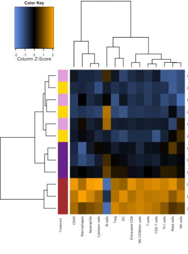
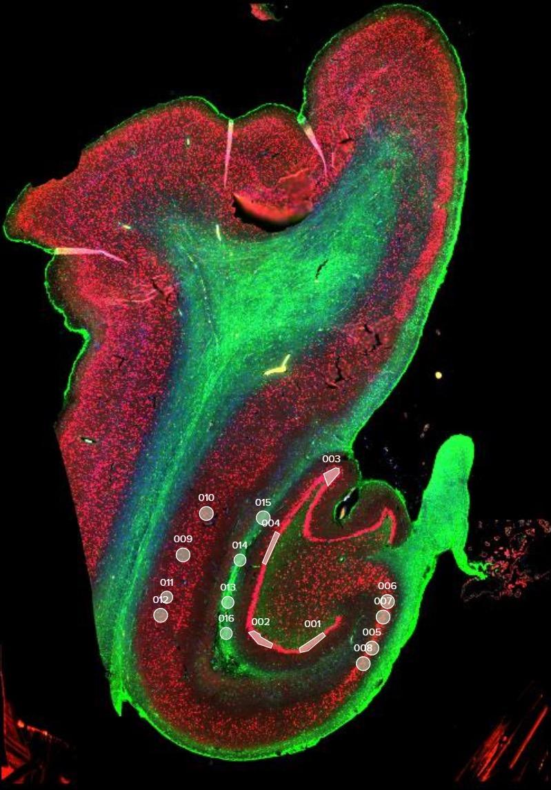
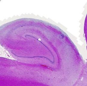
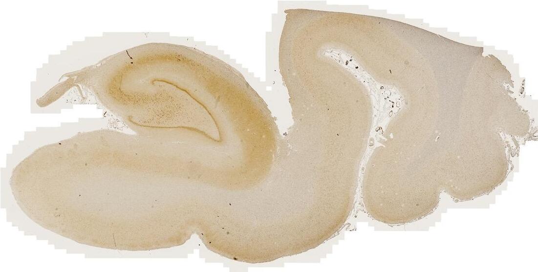
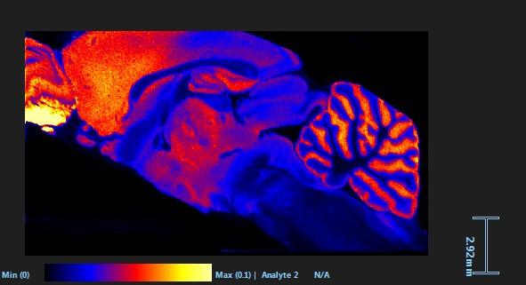
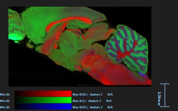
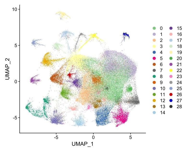
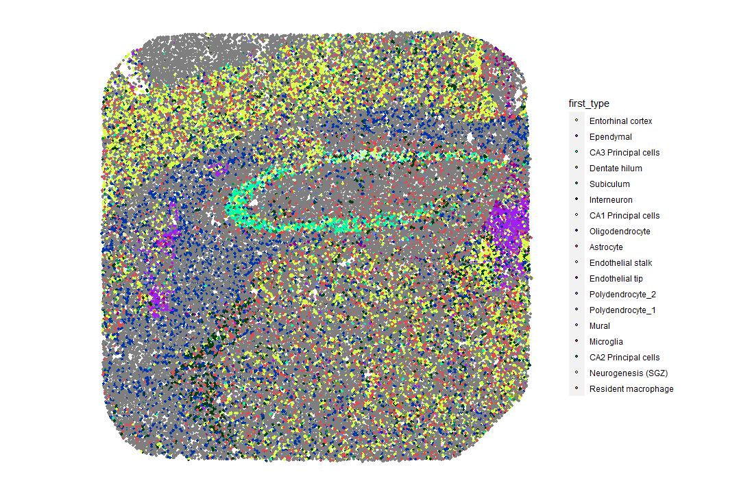
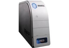
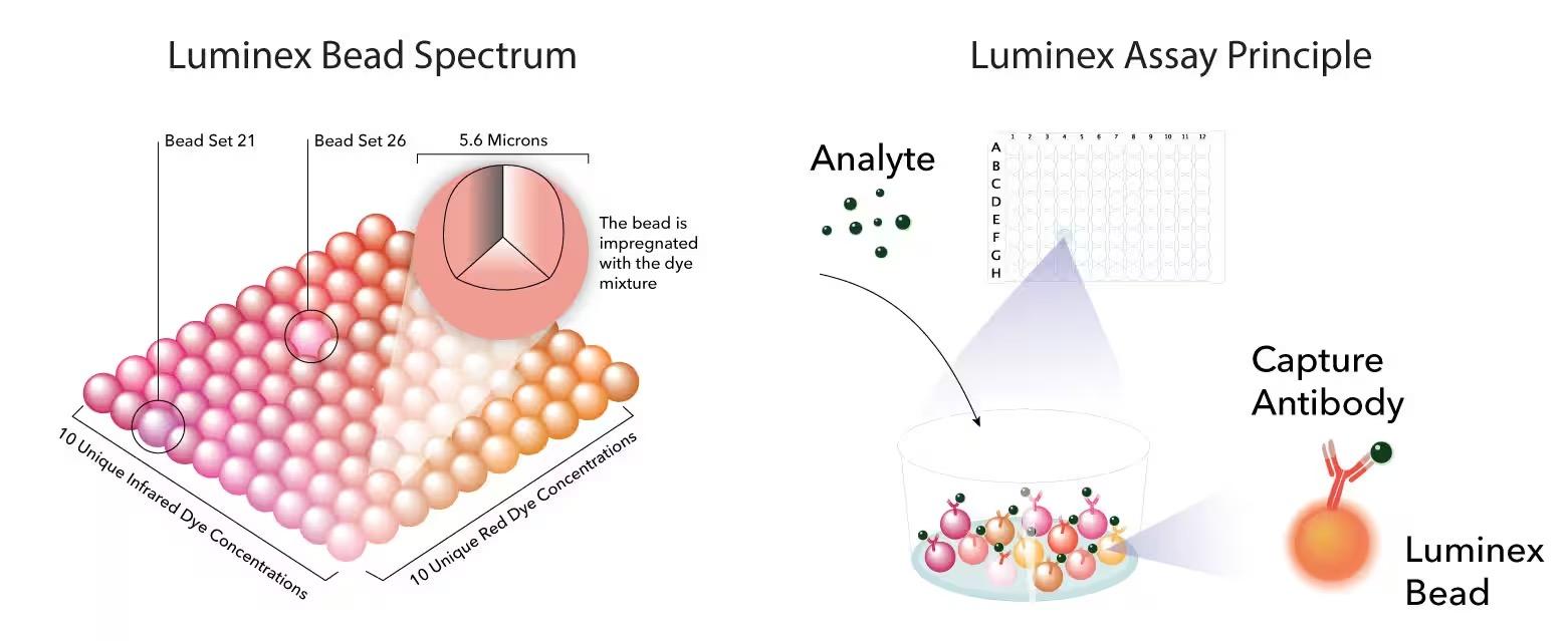
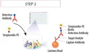
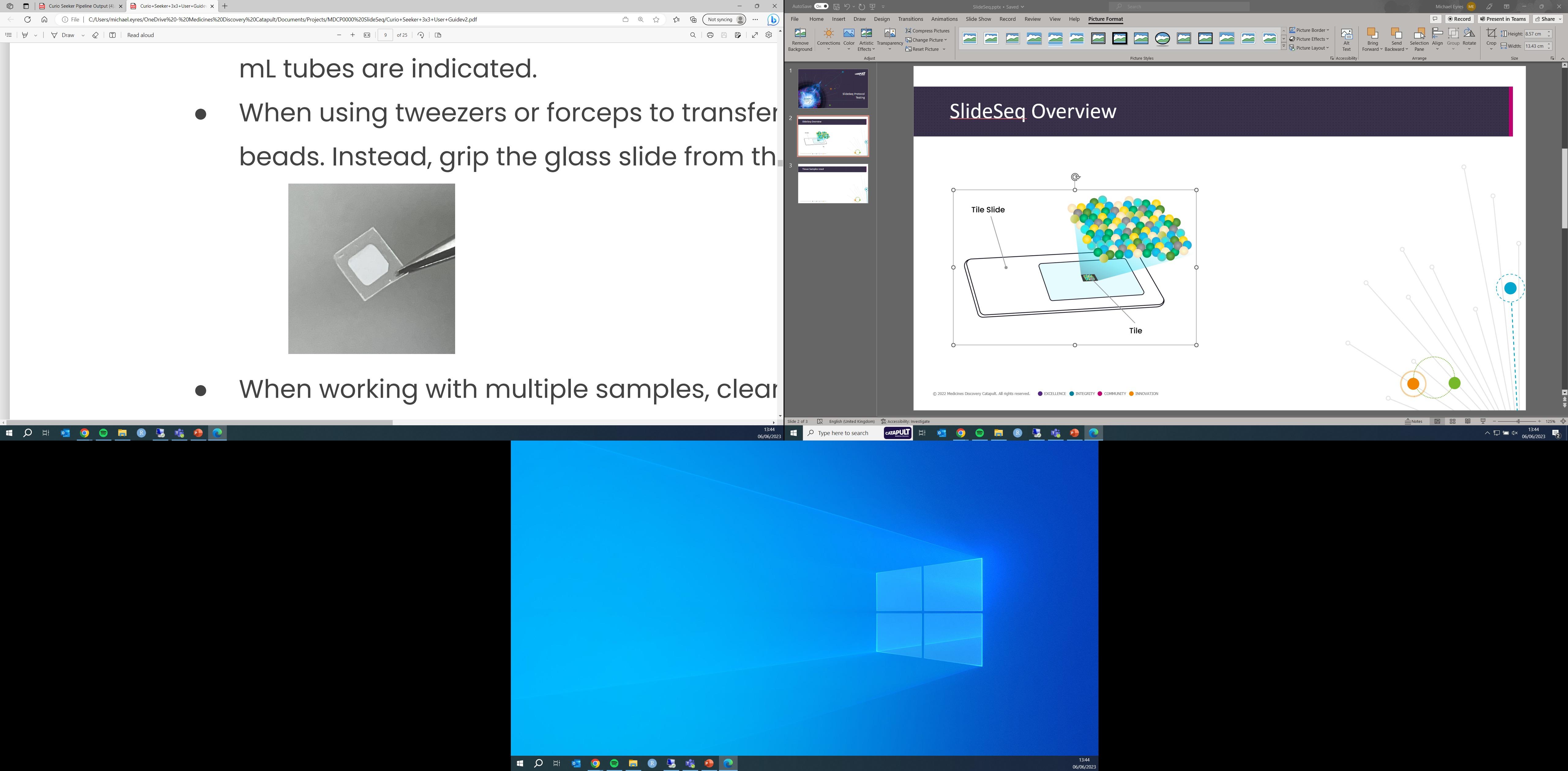
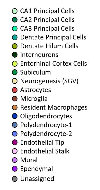
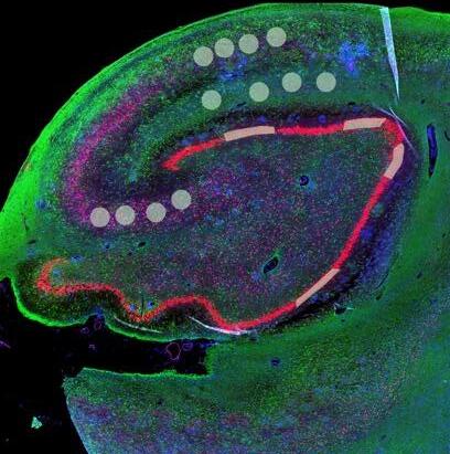
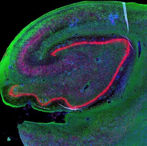
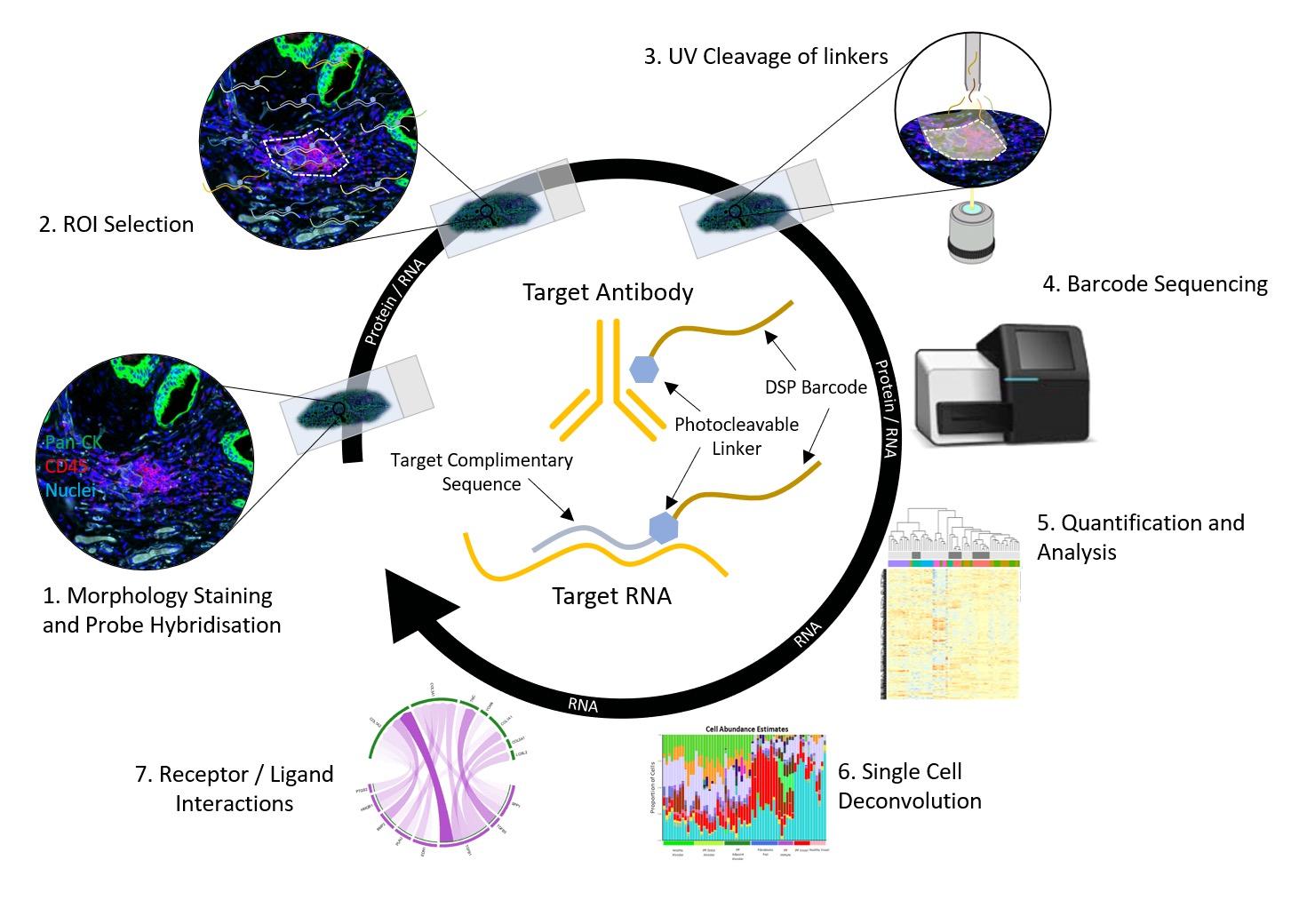
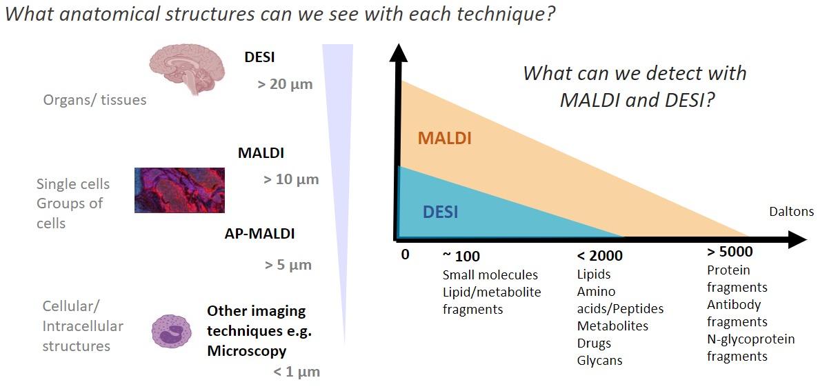
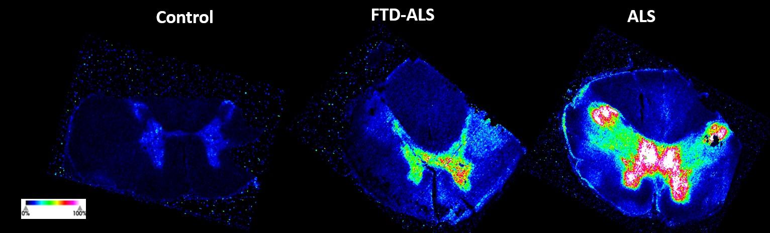
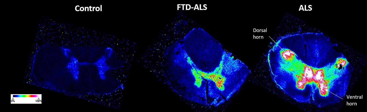
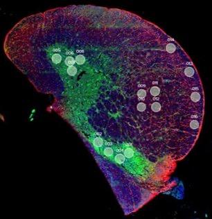
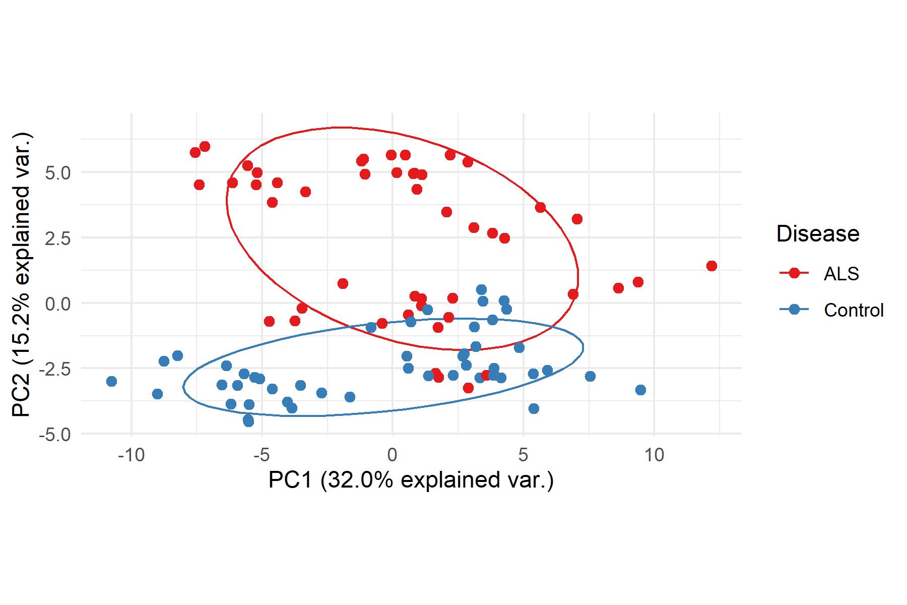
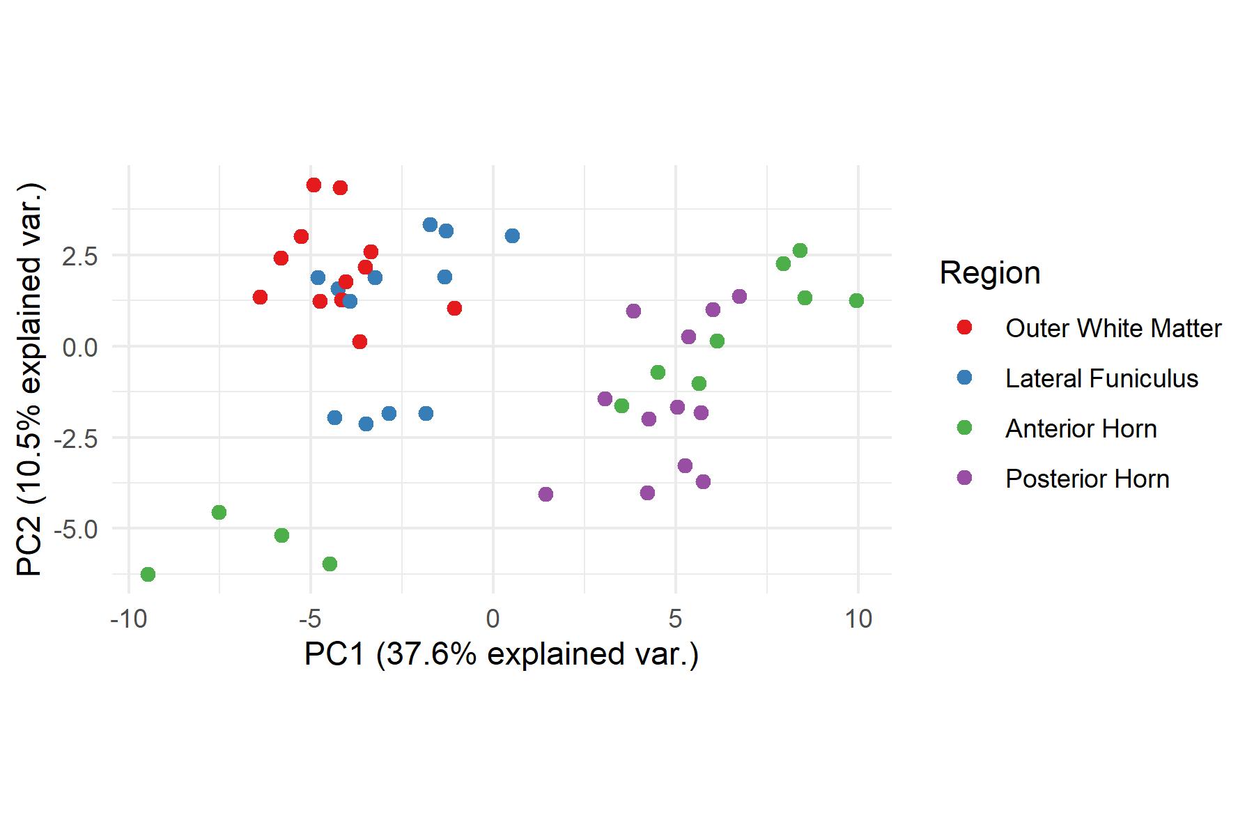
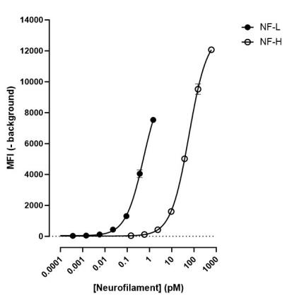
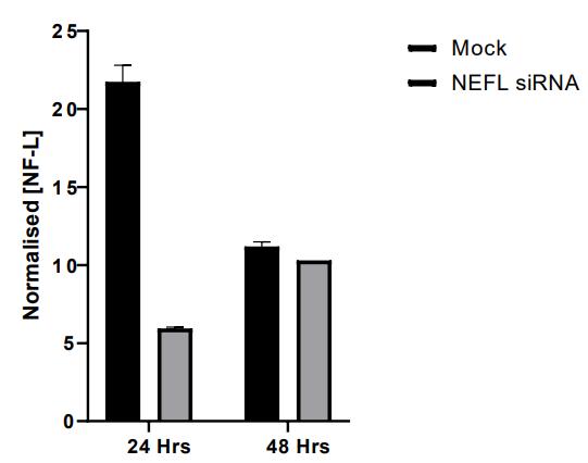
and mouse (and selected canine) probe panels available Quantitative, non-destructive, spatial proteomics and transcriptomics FFPE or fresh frozen tissue sections or TMAs Morphology Markers • Can be selected from a host of pre-validated antibodies (E.g. GFAP (Astrocytes); Iba1 (Microglia), NeuN (Neurons), Amyloid-β (Plaques)) New markers bespoke to each project can additionally be validated in house Can including segmentation and contouring GeoMx® Digital Spatial Profiler • Luminex and Simoa platforms perform bead-based immunoassays, enabling simultaneous multiplex detection of up to 100 analytes Pre-validated Assays • Various commercially pre-validated assays are available and can be run in house on the Luminex MAGPIX instrument Catalogues with >600 validated human analytes (includes several multiplex neuroscience/ neurodegeneration panels) • NF-H (but not NF-L), Tau (Total and phospho (pT181 + pT231)), TDP-43 Customised Assays Assays to targets not commercially available can be built and validated in house (E g NF-L assay validated in house) Several neuro focused assays are currently being validated for use with both human and mouse CSF and plasma samples Protein Multiplexing (Luminex® and Quanterix™ Simoa ® Platforms) NF-L Total Tau pTau (181 or 231) Pre-validated Assays 3mm 3mm 3 x 3mm coverslip coated with a monolayer of 10µm diameter DNA barcoded beads • The unique barcode on each bead enables the location to be determined • A section of frozen tissue is melted onto the tile where the mRNA binds to a poly(dT) tail on each bead, allowing the locations of the RNA transcripts to be deduced by sequencing Clustering analysis identifies groups of cells present with similar expression profiles Robust Decomposition single-cell to identify present in The spatial position of each cell type can then be mapped. Mass Spectrometry Imaging Small molecules Metabolites Lipids • Peptides/proteins MSI Analytes • Label free surface analysis of samples (E g tissue/ dried blood spots) for a variety of analytes Possible to analyse different molecular classes from a single section Relative/semi-quantitation • Can be correlated and overlaid with microscopy or DSP images (on same or consecutive sections) Lipidomic Profiling in Human Spinal Cord Tissue Amyotrophic Lateral Sclerosis (ALS) is a fatal neurodegenerative disorder characterised by the loss of motor neurons in the brain and spinal cord, with approximately 15% of patients additionally developing a form of Frontotemporal Dementia (FTD) • The dysregulation of lipid metabolism is a common hallmark of a range of neurodegenerative diseases, including ALS Mass Spectrometry Imaging (MSI) A collection of lipids and metabolites previously recognised to be associated with anti-inflammatory pathways was spatially profiled across human spinal cord tissue using MSI The snap frozen tissue was additionally H&E stained in order to define the anterior and posterior orientation One of the lipids, the omega-3 polyunsaturated fatty acid Docosahexaenoic acid (DHA), was found to be increased in the grey matter of the spinal cord in ALS and FTD-ALS patients Docosahexaenoic Acid (DHA) MSI Image of the distribution and abundance of DHA across human spinal cord tissue High Abundance Low Abundance Posterior Anterior GFAP • TDP-43 Aβ40 or Aβ42 Transcriptome Profiling • Whole Transcriptome Atlas- profiling of >18,000 protein coding genes Multiplexed Protein Panel Panels of 20 – 80 proteins (Immuno-oncology and neuroscience focused) • Customised assays, barcoding bespoke antibodies can additionally be validated and added into the multiplex panels Profiling Panels Technologies and Platforms Spatial Protein Profiling in Neurodegeneration Luminex Neurofilament- Light Assay • The aim of this project was to examine differences in protein profiles in neurodegenerative disease, (specifically in this case, Amyotrophic Lateral Sclerosis (ALS)), and aging • A panel of 74 proteins was spatially profiled across 6 spinal cord tissues (3 from control donors and 3 from patients suffering from ALS) using the GeoMx DSP platform • Data from 4 Regions of each spinal cord were collected, 2 from the grey matter (posterior horn, anterior horn) and 2 from the white matter (lateral funiculus, outer white matter) Data from the various regions of each tissue Example DSP Image with Selected Regions of Interest Neurofilament light is an established biomarker for neuronal damage in a range of neurodegenerative diseases • A Luminex assay was developed and validated to quantify NF-L in a variety of samples, including cerebrospinal fluid, cell lysates and tissue homogenates • Multiplexing of this assay with a selection of other proteins has additionally been tested and validated Specificity of the assay for NF-L over NF-H Knockdown of NF-L in SH-SY5Y cells BIOLOGICAL SAMPLES Liquid Tissue SAMPLE PREPARATION Automation ASSAY DESIGN Unique MS assays We handle most types of biological samples, ranging from tissue to plasma (CSF, sputum, tumour, serum, etc We wide range of MS analyses compound quantification, (un)targetedomics, heavy labelled molecules, cellbased and screening assays, etc Using novel technologies (acoustic dispenser, multidrop, 384 well-plate), we handle all the sample preparation, optimising your samples for MS analysis (LC)-MS ANALYSIS High-Throughput MS We have developed fast & robust screening assay for plasma proteomics. We are also specialised in HTS MS-assay. Targeted MS We develop/optimise targeted MS assays, improve their throughput and data richness. We run standard LC-MS/MS compound quantification in any sample type. Untargeted MS (-Omics) We are developing methods for biomarkers discovery, focusing on small molecules and proteins using LC-MS/MS approach. W possess dedicated MS laboratory where the latest MS technologies (FAIMS, Ion Mobility) providing unique and novel solutions to specific challenges DATA We have a suite of MS software that allows for fast processing of the data, as well as a dedicated team of bioinformaticians that perform data analysis and visualisation, in line with your research objectives Exploitation
Disease (5xFAD model) • Huntington’s Disease (R6/1 model) Orthotopic Mouse brain tumour model (G261 Acute LPS induced model of acute neuroinflammation U87-luc subcutaneous xenograft
Human