by Hazlin Hassan

In


Last month was the hottest September on record by an “extraordinary” margin as the world approached dangerously close to breaching a key warming limit, the European Union’s climate monitor said on Thursday (Oct 5).
Global average temperatures from January to September were 1.4 degrees Celsius higher
than in 1850-1900, almost breaching the 1.5C warming goal of the 2015 Paris Agreement, the Copernicus Climate Change Service (C3S) monitor said in a report.
That threshold is seen as essential to avoid the most catastrophic consequences of climate change.

10 | 08 | 2023 PIE MAGAZINE’S DAILY CONGRESS NEWS ON THE POSTERIOR SEGMENT posterior segment • innovation • enlightenment 4ISSUE OCTA Through The Years Find out how EURETINA 23 Gisbert Richard Lecture awardee Prof. Cynthia Toth revolutionized eye care and surgery. Rapid-fire RetinaTECH Symposium Shows Shape of the Future The future is now in retinal innovation, and leading technology from the next generation were presented by some of the world’s experts in the field. 04 06 HIGHLIGHTS Matt Young CEO & Publisher Hannah Nguyen COO & CFO Gloria D. Gamat Chief Editor Matt Herman Associate Editor Maricel Salvador Graphic Designer Writers Chow Ee-Tan Hazlin Hassan Nick Eustice Tan Sher Lynn Ruchi Ranga Customer Care International Business Development Brandon Winkeler Robert Anderson Sven Mehlitz Media MICE Pte. Ltd. 6001 Beach Road, #09-09 Golden Mile Tower, Singapore 199589 Tel: +65 8186 7677 Email: enquiry@mediamice.com www.mediaMICE.com Published by Please note that this is an independent publication of Media MICE Pte Ltd (“Media MICE”) in our role as independent media. Media MICE is in no way affiliated to or with any person, organization, or entity mentioned in this publication, nor is this publication published in any way intended to convey any such affiliation. Cont. on Page 3 >>
today’s world where climate change is real, and water is a scarce resource, it has become increasingly critical to make urgent changes to the way we live and work.
Are we at a turning point in retinal vascular disease?
Thank you for attending the BAYER sponsored symposia and the .

We hope you found the information interesting and informative.

Intended for healthcare professionals only. PP-EYL-ALL-1283-1 | September 2023
Cont. from Page 1 >>
EURETINA committed to change
At a time when the Congress of the European Society of Retina Specialists (EURETINA) returns as a live, face-toface event, the Board is committed to reducing waste and carbon emissions.
Some changes have taken place at this year’s congress, including:
– No printed programmes or flyers
– No complimentary delegate bags containing industry flyers
– No plastic bottles
Some of the ways that you can help:
– Bring your own reusable bottle (there are plenty of refilling points)
– Bring your own bag, pen, anything else you may need throughout the day
– Take public transport
– Download the congress App
– Photograph business cards or flyers instead of taking a copy
– Use the congress as an opportunity to arrange as many meetings as you can with international colleagues, thus reducing the need to take separate flights in the future
Sustainability in retina


Retina specialists came together on Day 3 of the congress to share ideas on what measures can be taken in order to boost sustainability in their practice, ranging from reusing equipment to reducing unnecessary visits, and using public transport networks.
A lot of waste is generated when administering intravitreal injections, said Dr. Redmer van Leeuwen, ophthalmologist and vitreoretinal surgeon from University Medical
Center
Utrecht, The
Netherlands. The 10 Rs of waste management are refuse, reduce, re-use, repair, refurbish, remanufacture, re-purpose, redesign, recycle and recover, he emphasized.
Dr. van Leeuwen reminded the audience that equipment used for intravitreal injections can be categorized as essential (face mask, gloves, anesthesia eye drops, povidone iodine drops, sterile eyelid speculum), optional (marker/calliper, cotton buds, gauze, eye ointment), and superfluous (drape, forceps, tablecloth, antibiotics). Hence in one’s clinical practice, here are some questions to ask one’s self, he reminded the audience: Is this intravitreal injection meaningful? Do I really need all these disposables?
Reuse, reuse and reuse
While some may balk at the idea of reusing instruments and probes in order to reduce wastage in vitreoretinal surgery, Dr. Vivek Dave, vitreoretinal surgeon from L.V. Prasad Eye Institute, Hyderabad, India, explained that this is being practiced in India.
“Coming from a developing country, taking care of the overall costs and sustainability becomes a very big issue,” he said. A controversial concept, reusing cassettes, trocar cannulas, cutters, laser probes, and tubings are of great value in developing countries such as India. Reusing tools also reduces wastage in cases where a surgeon needs to open a complete pack of tools but does not use everything in it.
This also reduces the amount of nonbiodegradable plastic waste that is generated for each surgery, thereby saving plastic waste from ending up in landfills or in oceans. By reusing items,
vitreoretinal surgery becomes more affordable.
However, the concept of reusing has its downside. Based on a study in India, some 0.06% of patients encountered post-vitrectomy endophthalmitis, shared Dr. Dave. In India, a cutter can be reused 4.7 times before it is discarded. Surgical attire such as caps, gowns and footwear are washed, sterilized and reused. Cloth drapes are used instead of plastic.
Attitudes on sustainability in retina
The majority of eye surgeons feel that waste generated in the operating theater is excessive, said Dr. Francesc March de Ribot, ophthalmologist, from University Hospital Girona, Spain. Ninety two percent (92%) of eye surgeons felt that OR waste needs to be reduced, he said, while 77% of eye surgeons are willing to reuse surgical supplies. However, he noted there are limitations, due to restrictions on reuse by manufacturers and regulations.
With water being a scarce resource, he suggested the use of alcohol solutions instead of sponges. Alcohol hand rub provides superior disinfection for a longer duration. “If your hands are visibly dirty, use a surgical scrub brush before the first surgery of the day only. If they are not visibly dirty, an alcoholbased wash alone is sufficient,” he said.
Water use per scrub is 18.5 liters, requiring 47.7 to 60.2 liters per surgery. If a surgeon has 15 cataract surgeries a day, that would equal to a savings of 750 liters a day or the equivalent of drinking water for a whole year or 20 showers or one water pool, he explained. A full body drape can be replaced with a face drape, with the smallest face drape measuring 120 x 100 cm. “Twenty percent (20%) of the waste during eye surgery is due to the plastic face drape. Bigger drapes are not safer but create more anxiety (among patients).
3 PIE Magazine’s Daily Congress News on the Posterior Segment
OCTA Through The Years


How Prof. Cynthia Toth revolutionized eye care and surgery

The development of optical coherence tomography (OCT) has advanced in leaps and bounds over the last three decades, resulting in better surgical outcomes for patients with retinal disease.

The EURETINA 23 Gisbert Richard Lecture was delivered yesterday by Prof. Cynthia A. Toth, MD, known worldwide for having revolutionized retinal imaging for both adult and pediatric ophthalmology. A Joseph A.C. Wadsworth Distinguished Professor of Ophthalmology, she is a world expert in a high-resolution, three-dimensional imaging technology and she has pioneered its use during surgery and in pediatric patients.
Her research has improved how ophthalmologists diagnose and predict the course of blinding eye diseases such as macular degeneration and retinopathy of prematurity, one of the most common causes of childhood vision loss.
by Hazlin Hassan
Prof. Dr. Richard Gisbert is chairman and head of the department of ophthalmology at the University Medical Center Hamburg-Eppendorf Germany. He is a professor of ophthalmology at the University Eye Clinic Mainz. A native from Northern Germany, he studied medicine, psychology, and biochemistry at the universities of Munster and Munich. Prof. Gisbert conducted research devoted to diagnostic and therapeutic improvements in diseases of the macula, the retina and the vitreous body. He developed fluorescein videoangiography (on a patent in 1985) which enabled for the first time the quantification of retinal and choroidal circulation and a better understanding of retinal dysfunction.
Prof. Toth was instrumental in developing Duke Eye Center’s Pediatric Retina Clinic, and she is the first female vice chair of clinical research in the Department of Ophthalmology. She is the director of the surgical instrument prototyping Duke Eye Center Biophysics Laboratory, and Chief Medical Officer and co-founder of Theia Imaging, both in North Carolina, United States.
For over 20 years, Prof. Toth has worked with colleagues to improve healthcare for patients with retinal disease through technological advances in handheld and intraoperative OCT. “We recognize the value of OCT data in the care of patients with retinal disease without exclusion,” she said.
Dr. Toth and her longtime collaborator, Duke professor of engineering Joseph Izatt, PhD, have been at the forefront of OCT innovation since its inception. The handheld OCT was developed in
Prof. Dr. Toth received her MD from the Medical College of Pennsylvania followed by an ophthalmology residency at Geisinger Medical Center and vitreoretinal fellowship at University of California. She served as an ophthalmologist in the US Air Force, directed the Retina Service at Wilford Hall USAF Medical Center in Texas, and collaborated in retinal OCT research at Brooks Air Force Base. She joined the Duke University vitreoretinal retina surgical faculty in 1993. In 2005, Dr. Toth became professor of ophthalmology with tenure and professor of biomedical engineering in the Pratt School of Engineering. Ten years later, she was named the Joseph A.C. Wadsworth Distinguished Professor of Ophthalmology. Currently living in North Carolina with her husband, the couple share travel, cooking, the outdoors, family and friends. She loves cappuccino.
order to examine pediatric patients at the bedside. She also touched on the latest generation of intraoperative 3D in near real-time 4D OCT being used at Duke. “Challenges remain to capture, extract and communicate relevant imaging data in real time to improve surgery and outcomes,” she said in concluding her lecture.
08 October 2023 | Issue #4 by 4
Retinal Real Talk on High Myopia
by Matt Herman
The secret is out (and then some) on myopia’s rise across the globe. A Day 3 symposium at EURETINA 2023 showed retinal specialists how high myopia can be handled.
If you’ve been living under a rock for the past half-decade or so, there’s distressing news to share: Myopia rates are exploding worldwide. But for those who make their homes in more conventional places, the focus in retina has now turned to dealing with the myriad complications of high myopia.
Fortunately, the 23rd EURETINA Congress in Amsterdam had doctors’ backs. A dedicated Day 2 session, entitled High Myopia and How to Avoid it in 2023 and cohosted by the European Association for Vision and Eye Research (EVER), went all-in on the implications of high myopia for vitreoretinal specialists and what can be done about it.
Histology pictures worth more than 1000 words
Prof. Jost Jonas (Germany) kicked off the symposium by sharing valuable takeaways from modern imaging and histology of highly myopic eyes.
The first key insight in Prof. Jonas’ talk was about Bruch’s membrane. “[In some highly myopic eyes] there is a dome-shaped macular, which is usually associated with a defective Bruch’s membrane,” Prof. Jonas explained. “This points towards a biomechanical role that Bruch’s membrane may have in the development of high myopia and myopic axial elongation.
Another key point from Prof. Jonas was his observation that nearly all highly myopic eyes have some form of optic nerve damage. Understanding
this phenomenon could be key for finding new treatment pathways. “Nonglaucomatous optic nerve damage may be associated with stretching of the optic nerve due to the elongation and enlargement of the inner ocular surface,” he summarized.

Priceless high myopia pearls from a pro
Prof. Kyoko Ohno-Matsui (Japan) is one of the world’s most respected myopia experts, and she joined the symposium virtually to give invaluable advice to practitioners dealing with high myopia.
Her presentation focused on myopic macular atrophy, retinoschisis, and issues with glaucoma. For retinoschisis, she recommended fovea-sparing internal limiting membrane (ILM) peeling for intervention.
For any maculopathy in highly myopic
anti-VEGF patients, Prof. Ohno-Matsui had one nugget of advice. “After one year of anti-VEGF therapies, we should regularly take autofluorescent images to detect macular atrophy in the patient as well.”
Glaucoma is another important concern with high myopia. “Glaucoma is underdiagnosed and overlooked in pathologic myopia because of the deformity and tilting of the optical disc,” Prof. Ohno-Matsui pointed out. “In our high myopia center, we conduct Goldmann perimetry for all patients once a year,” she said. “This is because glaucoma develops at a very early age in highly myopic eyes, and it is not rare for them to completely lose vision in their productive age.”
Potential solutions and models
The good news is that horror shows like those described by Profs. Jonas and Ohno-Matsui can be nipped in the bud with early intervention in pediatric patients. Prof. Andrzej Grzybowski (Poland) gave a quick rundown of modern myopia control options for younger patients.
Atropine, myopia control contact lenses and spectacles, and orthokeratology comprise the current arsenal in most countries according to Prof. Grzybowski. Another emerging technology out of China is low-level red light therapy, and he quickly ran through a portion of the more than 22 studies on the topic.
But the best option? “Present methods in myopia control are not perfect. Combination therapy might be the future,” he concluded.
And speaking of China, Prof. Calvin Pang (China) finished things off by giving an update on nationwide myopia control efforts in one of the epidemic’s hotspots. According to Prof. Pang, top-down governmentmandated measures are working. He attributed this success to clinician-led, holistic approaches to the problem that include harnessing the power of everything from the education system to NGOs and industry.
5 PIE Magazine’s Daily Congress News on the Posterior Segment
Rapid-fire RetinaTECH Symposium Shows Shape of the Future
by Matt Herman
Augmented reality ORs? Nonstick super surgical tools? The future is now in retinal innovation, and leading technology from the next generation was on display at a notable symposium at EURETINA 2023 yesterday.

these repellent surfaces in nature and found that they all have a microscopic roughness, where air pockets are created in between pillars and a very microscopic layer,” he said, pointing to examples like rice leaves, mosquito and butterfly wings and shark skin.
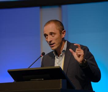
Besides anterior segment uses, Dr. Tarek Hassan (USA) sees a lot of potential in vitrectomies and other retinal surgeries. “What we want is to avoid traction on the vitreous,” he explained. “If we can do that, we can eliminate the mechanism that causes iatrogenic breaks during surgery, and create safer instrumentation in the future.”
Going with the (vitrectomy) flow
Few sessions at a scientific congress generate more buzz than a showcase of cutting edge technology. From incremental qualityof-life improvements to far-flung contraptions ripped straight out of science fiction, innovations on display run the gamut from the practical to the downright implausible.
High-impact and near-term were the words of the day at RetinaTECH, a Day 3 symposium at the 23rd EURETINA Congress showcasing next-gen posterior segment advancements. In this late afternoon session, the panel
of experts gave the audience exactly what they came for: a glimpse into the crystal ball of retinal medicine future.
A not-so-sticky situation
Up first was an innovation in ophthalmic surgical tools with Dr. Tarek Hassan’s unveiling of non-stick surgical tools. The driving force behind this innovation was to create a tool that was both hydrophobic, oleophobic and, crucially, non-coated.
The result was a design inspired by nature. “We looked at the structure of
The next three talks centered around flow and fluidics in vitrectomy, with a special focus on intraocular pressure. It was all about intraocular pressure compensation for Dr. Mario Romano (Italy), who shared his model of vitrectomy flow control and its applications.
Using a model of the vitreous, Dr. Romano tested the EVA phacovitrectomy system (DORC; Zuidland, The Netherlands) and a cutting-edge new automatic IOP compensation (AIC) feature. The team found that using a high-flow (HF) infusion system was generally more effective than standard flow (SF), and that AIC was most effective with 25and 27-gauge cutters.
Prof. David Steel (United Kingdom) took over from there, with his attempt
08 October 2023 | Issue #4 by 6
“We looked at the structure of these repellent surfaces in nature and found that they all have a microscopic roughness, where air pockets are created in between pillars and a very microscopic layer.”
Dr. Tarek Hassan
to quantify the field of effect of vitrectomy cutters. Using particle image velocimetry, his research aimed to map the flow rate and the convective and temporal acceleration around a vitrectomy cutter.
What he found was that there are essentially two zones around the cutter – a high flow zone and a low-flow zone. The central metaphor he used to explain was as space age as it gets: black holes. “If you think of a black hole, you have a low-flow zone where light can escape, but once it goes past the event horizon, it goes into the black hole no matter what,” he began.
“This is like a vitrectomy cutter. Inside the event horizon, fluid is rapidly pulled from the cutter way faster than human reaction times. Whereas outside in the low-flow zone, things take about two seconds to transit across it,” Prof. Steel explained.
So what’s the use? Prof. Steel explained that this can be eventually modeled and used to improve the cutter itself. “We can use this data to model improved cutters. Change in port area, change in port shape, change in port dimensions – this can all be used to design cutters with improved efficiency and safety,” he concluded.
that can swap ports intraoperatively, and it’s easy-to-use SmartIOP ‘cruise control’ feature. She further illustrated the necessity of being able to spike or drop IOP intraoperatively with several surgical videos. “It is important to be able to, by full switch, increase the intraocular pressure. But we also like to have a rapid decrease in intraocular pressure, without the need of having leakage to reestablish the correct pressure,” she explained.
Astounding augmented reality
Virtual reality is drawing oohs and ahhs from artists to gamers the world over, and Dr. Omer Trivizki (Israel) induced a few of his own with a very visual demonstration of the use of augmented reality (AR) in the operating room.
Dr. Trivizki split his presentation up into three potential applications: real-time guidance, personalized patient data integration, and enhanced control. For real-time guidance, he demonstrated the use of a real-time OCT overlay for subretinal injections to visualize exactly where the tool tip is and get measurements on drug delivery.
Speaking of overlays, integrating patient data like topographic maps or retinal exams is also possible intraoperatively with AR. “We can see the actual peeling and also colorization of the different layers, which helps the surgeon be more accurate and safe,” Dr. Travizki stated, referring to a surgical video from Duke Medical School using an intraoperative OCT overlay.
coherence tomography angiography (OCTA) to take understanding of retinal displacement to new heights.
He showed off an algorithm that can be used to compare two OCTA images, giving surgeons a way to look at not only new elements that appear in the scan, but in what direction elements are moving from one scan to the other after something like macular pucker surgery.
Prof. Rossi gave an example of this technology in action when he took a series of angio OCTA images pre- and post-macular pucker surgery and compared them to catch the entire pathology. “What is interesting to know about the amount of motion is that it [the retina] displaces a lot more after surgery than before,” he noted.
“And more importantly, is, if you compare the overall integral of the curve. [This] should be zero if you simply rewind the pathology,” he continued. “This is not the case. The retina is doing different things – going elsewhere.” The applications are endless, according to Prof. Rossi, and like all of the new tech on display – this is just the beginning.
Prof. Grazia Pertile (Italy) brought the fluid fest to a close by extolling the virtues of DORC’s EVA NEXUS phacovitrectomy system. Prof. Pertile explained that she wants a vitrectomy platform that just works, without having to worry about what goes on under the hood.
For her, the key points with EVA NEXUS are a high-flow infusion line
Enhanced control was last on this list, but certainly not least. “We are no longer limited by a microscope,” he said. “We can fully control our device just by using intuitive head gestures, starting from the regular actions like zooming or panning, and including operating the intraoperative OCT.”
Nifty tricks with angio OCT
Prof. Tomasso Rossi (Italy) concluded the symposium with an explanation of how doctors can use optical

7 PIE Magazine’s Daily Congress News on the Posterior Segment
“If you think of a black hole, you have a lowflow zone where light can escape, but once it goes past the event horizon, it goes into the black hole no matter what.”
Dr. Mario Romano
Dr. Tarek Hassan
Dr. Mario Romano

