


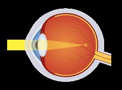
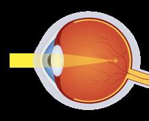
Myopia, commonly known as near-sightedness, is a common cause of refractive error, which impacts a significant proportion of the global adult population. Characterized by the inability to clearly see distant objects, moderate-to-severe myopia not only affects visual acuity but also has broader implications for an individual’s quality of life.
The management of moderate (–3.0 to –6.0 D) myopia has evolved substantially over the past few decades, driven by advancements in technology. Treatment modalities range from conventional methods

like eyeglasses and contact lenses to more advanced surgical interventions such as laser in-situ keratomileusis (LASIK), photorefractive keratectomy (PRK), laser-assisted lenticule extraction (LALEX) and phakic intraocular lenses (IOLs), including Implantable Collamer® Lenses (ICLs). Each of these treatments offers advantages and limitations, necessitating a personalized approach based on the patient’s specific needs, lifestyle and the degree of refractive error.
A consensus group of eleven refractive surgeons and specialists used a combination of an online
survey, online face-to-face interviews and written feedback to provide this guidance on the use of phakic IOLs, with particular consideration of:
• optimal patient identification
• lens power calculation and size selection
• surgical best practice
• postoperative care
• patient expectations, involvement and education

Dr. Robert Ang
Cataract and Refractive Surgery Department, Asian Eye Institute, Makati City, Philippines

Dr. Ik Hee Ryu
B&VIIT Eye Center, Seoul, South Korea; VISUWORKS, Seoul, South Korea

Prof. Zheng Wang
Aier Eye Hospital, Jinan University, Guangzhou, Guangdong Province, China

Dr. Erik Mertens
Medipolis Eye Center, WilrijkAntwerp, Belgium

Prof. Kimiya Shimizu
Department of Ophthalmology, Sanno Hospital, Tokyo, Japan

Dr. Roger Zaldivar Instituto Zaldivar, Mendoza, Argentina

Dr. Morgan Micheletti
Berkeley Eye Center, Houston, TX, USA

Dr. Vance Thompson
Vance Thompson Vision, Sioux Falls, SD, USA

Prof. Xingtao Zhou
Department of Ophthalmology and Vision Science, Eye & ENT Hospital, Fudan University, Shanghai, China; NHC Key Laboratory of Myopia, Fudan University, Shanghai, China; Laboratory of Myopia, Chinese Academy of Medical Sciences, Shanghai, China.

Dr. Neda Nikpoor
Aloha Laser Vision, Honolulu, HI, USA

Prof. Xiaoying Wang
Department of Ophthalmology and Vision Science, Eye and ENT Hospital, Fudan University, Shanghai, China; NHC Key Laboratory of Myopia (Fudan University), Shanghai, China; Laboratory of Myopia, Chinese Academy of Medical Sciences, Shanghai, China
*This activity was supported by an independent education grant from Staar Surgical (Lake Forest, CA, USA) to The Fundingsland Group (Gilroy, CA, USA). The TFG staff, writer, and planners have no financial relationships with ineligible companies. Faculty disclosures were provided as follows: RA: Acevision, Alcon, Bausch and Lomb, BVI, Glaukos, Hoya, Santen, Staar Surgical, Vialase, Twenty Twenty; EM: BVI, CSO, Excel-lens, HOYA, Medicontur, Schwind, Sophi, Staar Surgical, Stroma, Visionix, Zeiss; MM: Alcon, Allergan, Avellino Labs, Bausch & Lomb, BVI, Centricity, Diamatrix, Elios, Glaukos, Johnson & Johnson Vision, Lenstec, New World Medical, RxSight, STAAR, Tarsus, Visus, Zeiss; NN: Alcon, STAAR Surgical, Ziemer, Johnson & Johnson Vision, Bausch & Lomb Surgical, Kala, Allergan, RxSight, SUN, Glaukos, Avellino, Oyster Point, Dompe, Blink, LensAR, Tarsus; IHR: Stock holder, Visuworks Inc; KS: Staar AG.
The high and increasing prevalence of myopia (Box 1) presents significant
BOX 1
requirements and opportunities for myopia-induced refractive error correction.
Patients have access to four primary types of intervention for myopia correction: LASIK, LALEX, PRK and phakic IOLs, each of which have their
The increasing prevalence of myopia
The prevalence of myopia has been rising steadily, making it a public health concern with considerable socioeconomic impact.
As of 2000, approximately:

22.9% (1.406 billion) of the world population was estimated to have myopia with about 2.7% (163.0 million) having higher levels of myopia (more than –5.00 D)
Where do phakic IOLs fit into the refractive surgery armamentarium today?
The expert group were unanimous in the opinion that phakic IOLs should be in every clinic’s armamentarium in order to be able to offer appropriate options to all patients. Most of the experts offered LASIK, PRK, LALEX and phakic IOL options in their practice.
Phakic IOLs have traditionally been reserved for patients with moderateto-high myopia, or for those with contraindications for laser surgery – most frequently a thin or abnormal cornea.
own advantages and disadvantages (Table 1). Familiarity with each intervention and its optimal use, and full awareness of patient suitability for each option, are key to achieving the best possible results for patients.

This trend is expected to increase significantly, with predictions suggesting that by 2050
“ICL surgery is considered the highest level of refractive surgery for patients at this time because of its wide range of correction, reversible surgery and excellent postoperative outcomes.”
— Prof. Kimiya Shimizu (Japan)
around 49.8% (4.758 billion) of the global population will have myopia and 9.8% (938 million) will have a higher degree of myopia1
There was consensus that, in routine practice, there is a ‘growing practical indication’ for phakic IOLs, particularly ICLs, and that patients with myopia of –3.0 D and greater should be considered, even with healthy corneas, based on positive experiences.
In Prof. Shimizu’s clinic in Japan, ICL was the only elective refractive surgery offered, having ceased offering LASIK in 2008 and having offered LALEX from 2010–2015.
Which types of phakic IOL are recommended by the expert group?
Of the available phakic IOL products
(Box 2), the majority of the expert group now only use ICLs and have used each iteration of the Collamer® lens available (Box 3)
Dr. Mertens reported a change in practice: having previously used Artisan (Ophtec) and Implantable Contact Lens V2.0 (IPCL; Care Group) products, he found greater toric stability with ICL,2 and now uses these lenses in almost all phakic IOL procedures. Dr. Thompson has also
moved away from using the more rigid Verisyse (Johnson & Johnson Vision) lens (this is the same lens as the European-marketed Artisan), with a preference for ICL. Dr. Ryu noted that he no longer uses irisclaw lenses, or Eyecryl phakic IOLs (Biotech) which he replaced with ICLs over endothelial cell count loss concerns with the former and inflammation observations with the second; Dr. Ryu uses IPCL for correction of hyperopia.
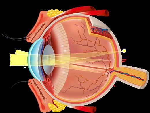
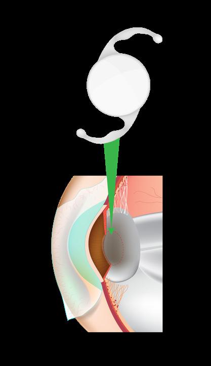
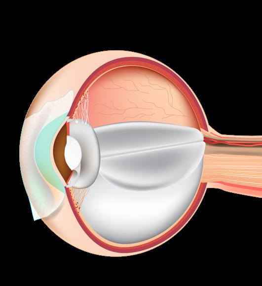
Phakic IOLs are implanted into the eye without removing the natural lens, preserving the eye’s natural accommodation ability.
The development of phakic IOLs was challenged by concerns regarding long-term corneal endothelium cell density and crystalline lens clarity. Over time, improved designs and surgical techniques have enhanced the safety and efficacy of these lenses.3 Several studies have demonstrated the superior visual outcomes of phakic IOLs,4 including when compared with LASIK.5
There are three main types of phakic IOL: anglesupported, iris-fixated and posterior chamber lenses.4,6 Each type has its specific indications, selection criteria and surgical techniques.
The AcrySof Cachet (Alcon) is a single piece foldable hydrophobic acrylic anterior chamber lens that was designed to achieve predictable positioning, stable vaulting and low compression forces on the irido-corneal angle. It is indicated for correcting myopia ranging from –6.00 to –16.50 D, but was voluntarily withdrawn by Alcon due to concerns over endothelial cell loss.
“[In my clinic]…we only use ICL because we trust the material and we have a thirty-year track record with Collamer® that we have witnessed in person.”
— Dr. Roger Zaldivar (Argentina)
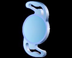
STAAR Surgical introduced a foldable ICL made from Collamer®, a proprietary hydroxyethyl methacrylate (HEMA)/porcine collagen containing biocompatible polymer material, in 1993. The lens features a platehaptic design with a central convex/concave optical zone and now has a central port; the lens incorporates a forward vault to minimize contact of the ICL with the central anterior capsule. See Box 3 for more information.
The IPCL V2.0 is a single-piece posterior chamber phakic IOL, similar to a soft contact lens and can be inserted into the eye through a small incision. The lens is custom-made for each patient, ensuring a precise fit according to the eye’s shape and size. The IPCL V2.0 is available in a wide range of powers, making it suitable for correcting various refractive errors.

A comparative short term study found that both the IPCL and ICL demonstrate similar efficacy and safety profiles. The study included patients who underwent either IPCL or ICL implantation and were followed for over a year. The outcomes measured included corrected and uncorrected visual acuity, refractive predictability, stability and complications. No significant differences were observed between the two groups in terms of visual outcomes and safety, indicating that both IPCL and ICL are effective options for correcting myopia.7
The expert group was unanimous in their positive opinion of the Collamer® material, noting its excellent biocompatibility, ease to unfold in the eye and the exceptional rarity of inflammatory reaction postoperatively. Several experts noted that when ICLs are explanted, usually ahead of cataract surgery, they appear ‘as new’ and show no evidence of inflammation activity.
The benefits of the central hole in the
“Physiologic aqueous flows over the lens, through the central hole and through the pupil. We feel this is better for lens health in the long run.”
— Dr. Vance Thompson (USA)
newer ICL designs were identified as the removal of the need for PIs prior to implant, and the reduction in rate of phakic IOL-associated cataract, to nearly nil. Several experts also suggested that the aqueous flow over the natural lens through the central hole was potentially providing nourishment and contributing to a healthier crystalline lens.
The central hole can be noticed by some patients. For many, neuroadaptation over the first few months following implant overcomes noticeable higher-order aberrations (HOA); however, for some recipients of a central hole ICL, night vision
STAAR Surgical introduced its first foldable posterior chamber phakic ICL made from Collamer® in 1993.
With V2 and V3, the safety and tolerability of the implant improved; however, studies found a small chance of lens opacity under the anterior capsule, which was attributed to contact between the phakic lens and the crystalline lens.8
The Visian ICL V4 was developed to allow more space between the crystalline lens and the ICL, and consequently reduced cataract formation.
aberrations, haloes and glare persist.11,12 HOAs are often described in trial outcomes as ‘acceptable’, 12 and none of the expert group has encountered a request to remove the ICL based on HOA experience.
Recent research has shown that these ICLs provide uncorrected distance visual acuity of 20/20 or better in 87.6% of treated eyes and
The EVO/EVO+ VISIAN ICL™ products (and their toric counterparts, where appropriate) were used in the majority of procedures by all members of the expert group; their input informing subsequent sections relating to ICL use is based on this product. BOX
Two peripheral iridotomies (PIs) were required prior to surgery to reduce the risk of pupillary block glaucoma.
Visian ICL V4c gained the KS-Aquaport, a central hole in the lens that eliminated the need for preoperative PI by allowing aqueous humor to flow freely, reducing the risk of elevated IOP and cataracts.9 V4c has been found to offer the same visual quality as V4.8,10
The EVO/EVO+ ICL are the latest models, including a central hole, and have been designed to treat a wider range of refractive errors.
spherical equivalent within 0.50 D of target in 90.5% of eyes.13 Similar findings have been reported across several markets.14–16 Importantly, in these studies, EVO/EVO+ ICL implant was associated with minimal loss of endothelial cell density; no events of increased IOP secondary to pupillary block, no angle narrowing and no pigment dispersion or intraocular inflammation. No events of visually
significant anterior subcapsular cataract have been reported.16,17
Table 1. A comparison of the common features of laser-based refractive surgeries and phakic IOLs.
Feature Phakic IOLs
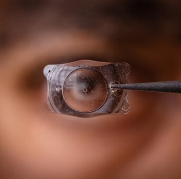
Procedure
Involves implanting a lens inside the eye without removing the natural lens.
The procedure is more complex than LASIK or PRK and until recently was only considered when a patient is not suited to these options due to high refractive errors or cornea abnormalities.
LASIK PRK LALEX
LASIK involves creating a flap in the cornea and reshaping the underlying tissue with a laser. It’s a widely preferred method for its quick recovery and effective results.
The outer layer of the cornea is removed, and the cornea is reshaped with a laser.
Unlike LASIK, no flap is created.
LALEX involves using a laser to create a lenticule within the cornea, which is then extracted through a small incision, reshaping the cornea.
Recovery
Postoperative recovery is very quick and involves monitoring for potential complications like elevated intraocular pressure.
Vision improvement is immediate and stabilizes within a month.
LASIK has a quick visual recovery time, with slow corneal nerve reinnervation. Postsurgery monitoring of surgical temporary ocular discomfort syndrome, corneal health and the integrity of the flap is required.
Vision stabilizes and improves over a few days.
Recovery is slower than LASIK, with blurred vision lasting up to three weeks. A protective lens is worn for a few days postoperatively.
Recovery from LALEX is quicker than PRK but slower than LASIK. Most patients experience improved vision within about a week.
Phakic IOLs provide excellent visual outcomes, including for patients with high levels of myopia.
They have been found to result in better contrast sensitivity and overall satisfaction compared to LASIK in certain studies, and fewer higher order aberrations than LALEX.
Effective for treating myopia, hyperopia and astigmatism, with long-term data available, LASIK also provides rapid visual improvement.
Long-term visual outcomes are similar to LASIK, but the initial recovery period involves more discomfort and slower visual improvement.
Suitable for correcting near-sightedness and astigmatism, LALEX has been associated with fewer higher order aberrations compared with other laser procedures and offers similarly improved visual quality to LASIK later in the postoperative period.
IOL, intraocular lens; LALEX, laser-assisted lenticule extraction; LASIK, laser in-situ keratomileusis; PRK, photorefractive keratectomy.
Who should be offered the option of a phakic IOL?
The expert group were asked ‘What percentage of today’s refractive surgery candidates within the indication do you believe should be considered for phakic IOLs?’
The indication for phakic IOLs varies by product and by applicable guidelines and authorization criteria. Because the expert group were using ICLs as their phakic IOL option in nearly all cases, this question refers to the current ICL indications (Box 4)
The overall expert group opinion was that ICLs should be considered for at least half of those who meet the indication criteria (Figure 1)
However, the experts answered this question from several angles: how many people might be suited to ICLs, how many people would be offered the procedure, and in what proportion of candidates is the surgery actually being performed.
“Every myopic eye is a candidate for ICL implant, unless proven otherwise during examination.”
Dr. Erik Mertens (Belgium)
The proportion of people suitable for ICLs was generally highest in Asian countries (around 90% in China, Japan, South Korea and the Philippines) with experts from the Americas considering around
30–60% of their patients as suitable.
Of these suitable candidates, ICL implant was carried out in 10–20%; this proportion is increasing and is expected to increase further. Dr. Micheletti observed a change in use from around 1% of clients to around 10% this year. Dr. Zaldivar noted that he expects to increase ICL use in moderate myopia (up to –6.0 D) from around 25% of eligible candidates to 50%, based on the quick recovery time and immediate benefit – the ‘wow factor’. He explained that when quality of vision is measured at the retinal plane for either ICLs or LASIK, the improvements are at least five-fold better with ICLs at 4 hours postprocedure. This expectation for greater use of ICLs was common across the expert group.
Question: What percentage of today’s refractive surgery candidates within the indication do you believe should be considered for phakic IOLs?
Average: 52 High: 100
Low: 20
The broadest indication for the EVO and EVO+ ICLs is a correction in adults with –0.5 to –20.0 D of myopia and up to 6.0 D of astigmatism in those requiring a toric lens.22
The indication will vary by region- and country-specific guidelines. The FDA recommendations are for people (21–45 years) with –3.0 to –15.0 D of myopia and up to 2.5 D of astigmatism at the spectacle plane; the toric ICL can be used in those requiring correction of up to 4.0D of astigmatism.
Before undergoing refractive surgery, the OSI test can help determine how reliable the correction will be based on the quality of the tear film and the health of the cornea. It is also used for evaluating DED by analyzing the scattering of reflected light over time, which may help identify people unsuited to LASIK. BOX
The OSI test assesses the quality of a candidate’s vision. This test is particularly useful for evaluating the optical quality of the entire visual system, which includes checking for conditions like cataracts, DED and other factors that can cause refractive errors.

The expert group provided guidance on selecting appropriate candidates for phakic IOL implantation and discussed factors that would contraindicate the procedure. They recommended considering five key candidate characteristics when determining if ICLs are appropriate: the degree of myopia, the candidate age, anterior chamber depth (ACD), the candidate’s refractive history and the health of the cornea.
Degree of myopia, age, ACD and corneal health were agreed with strong (90%+) consensus, with the stability of the candidate’s refractive history also important (Figure 2) Other aspects, such as lifestyle, sports participation, cost and candidate preferences should all also be discussed.
• There was consensus that, for candidates with high myopia, ICLs
would be strongly recommended, unless contraindicated.
• There was also agreement that ICLs would also be recommended for consideration in those with myopia in the –3.0 to –6.0 D range. In this population of candidates, ICLs would be an option alongside the laser surgeries, and the patient would be consulted regarding other aspects of their suitability, including lifestyle, age, cost considerations and eye health.
• Use of ICL in candidates 18–40 years of age, who meet the other indication criteria, was unanimously recommended.
• A difference of opinion in the upper age limit for ICL was apparent among the expert group
refractive lens exchange (RLE) for cataract is needed later in life.
– Dr. Mertens and Dr. Nikpoor noted that they were increasingly using ICLs in people aged 45–55 years, sometimes instead of RLE, in order to minimize dry eye disease (DED) and surgical temporary ocular discomfort syndrome or to preserve the cornea for as long as possible.
– The expert group agreed that the central hole in newer ICLs allows physicians to be more confident in using the lens in older people. In conjunction with findings from the objective scatter index (OSI) test (Box 5), ICLs can be considered in people aged over 40 years.
• At the other end of the age spectrum, Dr. Zaldivar suggested that ICLs could be suitable for younger patients with a need for correction of astigmatism, or with anisometropic myopic amblyopia (Box 6). This is a subtype of amblyopia (‘lazy eye’) characterized by a significant difference in the degree of myopia between the two BOX
– Both Prof. Z Wang and Dr. Thompson advocated for using ICLs as soon as appropriate for the candidate, ensuring the longest enjoyment and benefit from the procedure before
Figure 2. There are several key factors in determining how appropriate a phakic IOL procedure might be for a candidate.
Question: What are your personal demographic criteria for recommending phakic IOL implantation? (Select all that apply)
ACD, anterior chamber depth; IOL, intraocular lens.
Research has looked at the use of posterior-located phakic IOLs and LALEX in managing this condition,23 and the benefits of ICL use in 12 patients with amblyopia were published in 2017.24-26 BOX
greatest degree of myopia, the patient may experience underdevelopment of the visual pathway associated with that eye.
This is a subtype of amblyopia (‘lazy eye’) characterized by a significant difference in the degree of myopia between the two eyes. This discrepancy causes the brain to receive two very different visual images; when the brain ‘ignores’ the image from the eye with the
eyes. Research has looked at the use of posterior-located phakic IOLs and LALEX in managing this condition,23 and the benefits of ICL use in 12 patients with amblyopia were published in 2017.24-26
• The guidelines for the minimum indicated ACD vary by region; however, this is either 3.0 mm (including the FDA recommendation for the USA),27 or 2.8 mm, as Prof. Shimizu confirmed as the Japanese guidance. Both
“We strongly feel that the only suitable modality to correct the refractive errors of people with Avellino corneal dystrophy is phakic IOL implantation.”
— Dr. Ik Hee Ryu (South Korea)
minimum depths are found across other regions.28,29
• There was full consensus about performing the procedure at the depth described by local guidance.
• There was also confidence across the expert group to use the lens at shallower depths.
– In regions with a recommended ACD of 3.0 mm, experts were willing to use the lens at a depth of 2.8 mm in candidates
with open angle and no contraindicating factors.
– Dr. Nikpoor noted that even with a healthy eye and open angle, she would be unwilling to implant at a depth of less than 2.8 mm. Prof. Shimizu and Prof. X Wang felt that in the appropriate eye, a wide angle and with the benefit of the ICL central hole, implant at an ACD of below 2.8 mm could be considered after consultation with the candidate.
• ICLs can be recommended from the age of 18 years, but the indications are from 21 years old. However, several experts noted that the candidate’s myopia may still be developing in their late teenage years, and that there can
be advantages to waiting until the candidate is 20–25 years to help ensure refractive error stability and aid accurate power calculation.
• Overall, the experts agreed that 6 months of stable refractive history is preferred as a pre-operative criterion.
• A candidate’s corneal health – or lack of health and thickness – is a major decision point in selecting a process for refractive correction.
• Because LASIK creates a flap in the cornea and PRK uses corneal ablation to correct refraction, both processes require the candidate to have sufficient corneal thickness, no keratoconus and minimal DED.
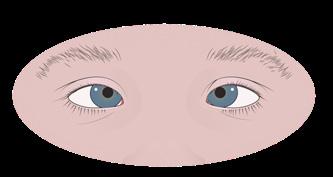
Avellino corneal dystrophy is a rare, inherited eye disorder characterized by the progressive accumulation of gray-white opacities in the stroma of the cornea, which are made of an abnormal protein called keratoepithelin.
• Since phakic IOL implant is performed with no loss of corneal tissue, the expert group noted that it may be a suitable alternative to laser-based therapies in people with suboptimal corneal health.
• Dr. Ryu presented favorable results using an ICL in a patient excluded from laser-based refractive correction on account of Avellino corneal dystrophy, a condition that presents relatively frequently in his practice (Box 7)
Prof. Zhou noted that the indications for ICL surgery are similar to other corneal refractive surgeries with the exception of two factors: the status of the corneal endothelium and the true ACD. Understanding the true ACD means investigating the
Symptoms include decreased night vision and increased sensitivity to light, and patients may progress to a gradual decline in visual acuity. LASIK has been associated with worsening the condition, with recurrence of corneal deposits30 and corneal haze can develop in these eyes after PRK treatment.31 Phakic IOLs offer a suitable alternative for these patients, according to the consensus panel.
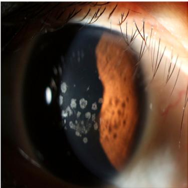
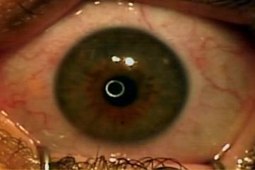
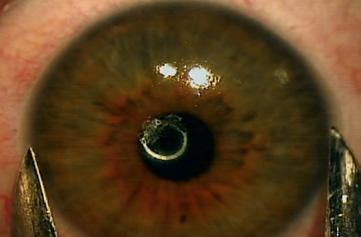
A B
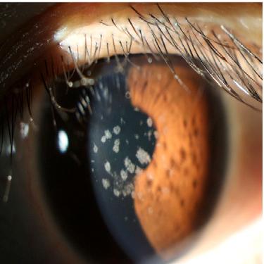
Image of slit lamp assessment, 6 years post-implant of ICLs in a patient with Avellino corneal dystrophy; (A) right eye, (B) left eye.
Image provided by Dr. Ryu, Department of Refractive Surgery, B&VIIT Eye Center, Seoul, South Korea and Research and Development Department, VISUWORKS, Seoul, South Korea.
ICL, Implantable Collamer® Lens.
morphology and width of the ciliary sulcus and the ciliary body. Prof. X Wang explained that there is still work to be done in this area. “There are still things we don’t fully know about in ICL surgery, and improving our understanding and knowledge of the ciliary sulcus will greatly enhance postoperative outcomes,” said Prof. X Wang. Furthermore, he added that ICL focuses more on ACD and endothelial cell density (ECD) requirements, whereas suitability for corneal refractive surgery has greater emphasis on corneal thickness and morphology.
Because the majority of ICL use is in younger people, and the implant acts in an inert, natural manner, the experts noted that contraindications were relatively few.
Most experts were willing to consider ICL procedures in patients with some degree of open-angle glaucoma, if well controlled.
glaucoma (PDG) was generally considered a contraindication; this is a form of glaucoma characterized by the dispersion of pigment granules from the iris into the eye’s anterior segment. These granules can block the trabecular meshwork, leading to increased IOP. The use of phakic IOLs in patients with PDG can be complicated by:
• Increased pigment release: the placement of phakic IOLs can induce further release of pigment granules, exacerbating the blockage of the trabecular meshwork and increasing IOP
Dr. Thompson noted that even in the most optimal situation, an additional PI would be required in patients with PDG even when using an ICL with a central hole.
Dr. Zaldivar identified patients with Marfan syndrome as unsuitable for ICL implant. Marfan syndrome is a genetic disorder affecting the body’s connective tissue; ectopia lentis (dislocation of the lens), myopia and retinal detachment are the most common ocular manifestations. Complications with a phakic IOL could include:
axial length and abnormal anterior chamber depth, which could affect the choice and positioning of a phakic IOL.
Other factors that could make a candidate unsuitable for ICL included: irregular or large degrees of astigmatism, ocular inflammation, autoimmune disease (this would need to be quiescent before consideration for ICL implant), an S-shaped iris, cataract or the presence of retinopathy.
“The posterior chamber morphology, assessed by ultrasound biomicroscopy (UBM) examination, should be considered as the most important indicator of ICL surgery. If an abnormal posterior structure could affect the stability of the phakic lens, then ICL shouldn’t be considered as an option – even if that abnormal structure is physiological,” concluded Prof. Zhou.
Accurate power and size calculations prior to the implant of an ICL are crucial for optimal outcomes. The quality of the measurements and interpretations will affect the patient’s visual acuity and comfort, and are essential in minimizing the risk of complications like cataract formation, elevated IOP or endothelial cell loss.
• Limited anterior chamber space: lenses placed in the anterior chamber of the eye, which may already be compromised in PDG due to pigment accumulation, may have shallower vault than desired.
• Risk of IOP spikes: the surgical procedure to implant phakic IOLs can trigger such spikes, increasing the risk of optic nerve damage.
• Lens instability and dislocation: the zonules, which are the fibers that hold the lens in place in the eye, are often weak or absent in Marfan syndrome. This zonular laxity can lead to lens instability or dislocation, complicating standard IOL implantation.
• Increased risk of retinal detachment: which can be exacerbated by eye surgeries, including IOL implantation.
• Altered eye anatomy: Marfan syndrome can result in altered eye structures, including increased
Calculating the required refractive power for a phakic IOL
The power calculation of an IOL involves several measurements, each contributing to the accuracy of prescription and effectiveness of the lens for the patient. The expert group were asked to prioritize the six most common methods of determining phakic IOL power.32,33
Manifest Refraction: This is the standard process for determining a prescription for eyeglasses or contact lenses. It includes the sphere, cylinder and axis. These values are crucial in determining the
overall refractive error of the eye and are key to calculating the power of the IOL.
Back Vertex Distance (BVD): BVD is the distance between the back surface of the spectacle lens and the front of the cornea. This measurement is important because it affects how the lens power in spectacles translates to the required IOL power.
True ACD: ACD is critical for IOL power calculation as it helps in determining the effective lens position, which is a key factor in predicting how the lens will behave once implanted.
Corneal Thickness: The thickness of the cornea can influence the accuracy of other measurements used in IOL power calculations, such as corneal curvature and ACD.
K1, K2: These are measurements of the corneal curvature in different meridians. They are used to determine the overall curvature of the cornea, which is vital for calculating the refractive power of the
eye and, consequently, the necessary power of the phakic IOL.
Contact lens over-refraction sphere: This is a measurement taken when a patient is wearing their contact lenses. It helps in determining the additional refractive correction needed over the contact lens to achieve optimal vision. This measurement can be particularly useful for patients who have undergone corneal refractive surgery previously.
The expert group were unanimous in the importance of accurate manifest refraction measurement as the cornerstone of all phakic IOL power calculations (Figure 3) It was emphasized that diligence in testing was paramount, and that any unexpected findings should be retested to ensure reliability.
Prof. Z Wang and Prof. Shimizu noted that BVD is of importance in candidates with high degrees of myopia; in routine practice Dr. Ryu felt that BVD can be difficult to determine and interpret, so unlikely to add value for candidates with moderate myopia.
Figure 3. The was unanimous agreement that manifest refraction is the most important parameter in phakic IOL power determination.
Several options were presented for minimizing the effect of accommodation and preventing over-correction. Dr. Zaldivar uses cycloplegic drops in all candidates and considers the manifest refraction pre- and post-administration; Dr. Mertens only uses this approach in those under 30 years of age and finds less value in the process in older patients. Drs. Ang, Micheletti and Nikpoor all advocate a ‘max plus’ or ‘push plus’ approach, using plus-power lenses to help relax the eye’s accommodation and measure the true refractive error without the influence of the eye’s natural focusing efforts. Dr. Thompson uses the Lenstar to supplement manifest refraction with ACD, K1 and K2 measurements.
Accurate sizing of a phakic IOL: Practical experience of the expert group
Correct sizing is essential to maintain the ICL’s position within the eye, preventing rotation or displacement.
Question: For power calculations, what are the most important measurements you recommend for the average refractive surgeon to collect for each phakic IOL patient? (Select all that apply)
All members of the expert group felt that accurate preoperative sizing of the ICL was a critically important aspect of the procedure to ensure optimal outcomes for the recipient. However, there was no consensus on a single approach to sizing, and considerable variation was apparent in approaches to sizing, use of technology and interpretation of findings across the group’s clinical experience and preferences (Figure 4). The FDA approval was based on using white-to-white (WtW) measurement, adjusted as needed for the candidate’s ACD; however, these guidelines recognize that WtW is an indirect measure, and that other techniques may be appropriate to help determine correct lens size.27
Optical coherence tomography (OCT) is a non-contact, non-invasive imaging technique that is particularly useful for assessing anterior chamber depth, WtW distance, scleral spur to scleral spur, and angle-to-angle (AtA) measurements.34 AtA has been associated with potentially greater accuracy of prediction of postoperative vault than WtW distance alone.35
UBM employs high-frequency ultrasound to produce detailed images of the anterior segment of the eye and can visualize structures not easily seen with OCT, such as the ciliary body, zonules and peripheral iris. UBM is essential for measuring the horizontal sulcus-to-sulcus (StS) distance, which has been identified as a valuable measurement for positioning posterior chamber phakic IOLs, because the haptic footplates are ideally located on the ciliary sulcus.34
A common theme through all expert responses was that the accuracy of sizing calculation is directly related to the accuracy of the measurements taken; regardless of the methods used, a reliable and repeatable process is needed to minimize interor intra-observer variations.34
WtW distance was commonly used in some form. Dr. Micheletti tries to give candidates a ‘LASIK-like’ experience and prefers to avoid contact methods where possible; he uses the IOLMaster swept-source OCT and other confirmatory measurements to determine optimal size. Prof. Z Wang, Dr. Ryu and Dr. Ang all use
Figure 4. The expert panel had a range of approaches to sizing phakic IOLs, based on their experience and preferences.
Question: For sizing decisions, what is your primary measurement? (Select all that apply)
corneal topography measurements of WtW (Orbscan) with confirmation using calipers; Prof. Z Wang and Dr. Ryu supplement this with OCT (CASIA2), whereas Dr. Ang has found too much inter-machine variability between OCT readings and does not use this approach. Dr. Thompson uses a comprehensive WtW caliper measurement on anaesthetized and fixated eyes, which he feels is the most appropriate way to understand the limbal anatomy of the candidate; to do this, the measurement is taken in the mid-grey region around the iris (Figure 5). Manual measurement with calibrated calipers can help address the documented over-estimation of WtW distance across the automated options. Several studies have shown that there is significant, but consistent, variation in WtW measurement between calipers and topography systems,36 between OCT apparatus,37 and between biometry and topography systems.38 Care must be taken to use one system constantly and to allow for over-estimation versus measurement with manual calipers when choosing a lens size.
Prof. Z Wang and Prof. Zhou use UBM for StS in every candidate
IOL, intraocular lens; OCT, optical coherence tomography; UBM, ultrasound biomicroscopy; WtW, white-to-white distance. WtW
because they believe AtA is the most critical measurement, with Prof. Zhou noting that a different lens position leads to a different effective lens size, which means the AtA measurement should decide selection of size. Prof. X Wang and Dr. Nikpoor use a combination of AtA (UBM) and WtW distance, the former using Pentacam and the latter using digital calipers for this measurement.
Prof. Shimizu has developed an OCTbased approach to AtA measurement that has proven to be more predictive of final vault than previous WtW measurement.35
“For ICL sizing, there is no universal method.”
—Dr. Robert Ang (Philippines)
Several of the experts are pioneering artificial intelligence (AI) approaches to ICL sizing. Dr. Zaldivar is using an algorithm (ICL Guru: revaicare.com/icl-guru) that combines AtA measurement from UBM images with power calculations, eye morphology and a proprietary
risk assessment algorithm to forecast outcomes with different lens sizes. Dr. Mertens is using the LASSO formula, based in OCT assessment, which has proved to be more predictive of final vault than OCOS and has demonstrated the capacity to estimate postoperative vault to within 500 µm in 100% of trial cases.39 Dr. Ryu has developed the LOOCUS® algorithm based on the OCT approach utilized by physicians in the Republic of Korea, which has proven to be accurate at predicting postoperative vault than the embedded software provided in the OCT apparatus.40
When a candidate appears to be on the borderline between two sizes, there were two schools of thought. Several experts preferred to choose the smaller lens based on reduced concerns with low vault when using an ICL with a central hole; other preferred to use the larger of the size options to allow for the possibility to rotate the lens to reduce the higher vault, as needed.
Figure 5. Currently, WtW distance (A) is the FDA-approved method of sizing an ICL; (B) this can be measured accurately from the mid-gray zone at the 3 o’clock limbus to mid-gray zone at the 9 o’clock limbus bisecting the pupil, with topical anesthesia and lid speculum under an operating microscope; (C) high magnification leads to the most accurate WtW measurements.
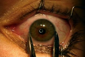
FDA, US Food and Drug Administration; ICL, Implantable Collamer® Lens; WtW, white-to-white.
“Vault” refers to the distance between the posterior surface of the intraocular lens and the anterior surface of the crystalline lens of the eye. This measurement is crucial for the successful implantation of phakic IOLs and is a key factor in avoiding
postoperative complications.
Achieving optimal vault is important. If the vault is too low, there’s a risk of anterior subcapsular cataracts due to direct contact between the intraocular lens and the crystalline lens; a very high vault can lead to angle-closure glaucoma. Anteriorsegment OCT and UBM are used to assess vault.
Figure 6. The expert panel suggest a mean vault range of 213–885 µm was appropriate following ICL implant, but any decisions should be made on a patient-by-patient basis.
If the postoperative vault is found to be significantly outside the ideal range, it may be necessary to rotate, or remove and replace, the phakic IOL.
The collective expert group defined an average ideal vault range of mean 213 µm (minimum, 100 µm) to mean 885 µm (maximum 1500 µm; Figure 6). Question: What is the ideal
A frequent comment from the expert group was that vault considerations are more relaxed in the current era of ICLs with central holes and good biocompatibility. All experts would determine any requirement to act on a vault measurement by considering the individual. “In a patient with a deep anterior chamber, vault can be ideal at a measurement of even greater than 1000 µm, as long as the anterior chamber angle is open,” Prof. X Wang said as an example. Drs. Nikpoor and Mertens added that at higher vaults the combination of vault and angle need to be considered in combination before any decision is made.
Lower vault, even down to 100 µm, was not considered an issue with ICLs with a central hole, and most of the expert group would only intervene if the ICL was touching the crystalline lens. Otherwise, with good aqueous flow over the natural lens, monitoring is preferred. Prof. Z Wang suggested that he might consider lens exchange at very low vaults in young people. There is an evidence base emerging to support changing the indication of ICLs to include an ACD of less than 3.0 mm.16
Vault will change over time, and long-term monitoring at check-ups is advised. The introduction of the central hole ICL has been associated
with smaller changes in vault from week 1 post-implant onwards, compared with the ICL without central hole.41
The experts were asked to share their guidance for optimized ICL implant procedures.
An overview of typical practice is presented in Table 2. In most cases, procedures were carried out bilaterally, with sterilization of the
room and all apparatus between eyes. Dr. Micheletti also uses materials from separate batches for each eye, to further reduce the risk of an unsatisfactory procedure. Dr. Ang noted that he prefers to treat ICL like cataract surgery, and prefers to operate on one eye, send the patient home for a few days, and then perform implant in the second eye.
With recent research observing that, in many cases, ICLs are not anchored in the ciliary sulcus, which may contribute to low vault,44 the expert group was asked for advice on lens positioning during surgery.
Table
Pre-visit considerations:
• discontinuation of contact lens use
• antibiotics
Preoperative steps:
• Prepare as for cataract surgery
• Dilate eye
• Relax/sedate patient as needed
• Make markings for toric positioning as needed
Intraoperative steps:
The key point raised was there is no way to know during surgery whether the footplates are in the sulcus or not – this would be revealed in post-surgery UBM. The expert group shared advice on positioning ICLs:
• Don’t fixate on obtaining a sulcus–sulcus position, most lenses won’t be in this position, but it is not a cause for concern.
• Pressing gently on the footplate and allowing it to glide under the iris will result in a suitable lens position.
• Footplates should be encouraged to locate as shallow as possible,
Comments
1. Small nasal incision (1 mm) for addition of xylocaine/lidocaine and antibiotics
2. Temporal (approx. 2.6 mm) incision
3. Add VE – Inject lens with applicator
4. Locate haptics, distal then proximal, – Position distal haptic through the nasal incision
– Position proximal haptic through main incision
5. Flush VE from the chamber
Assess vault
and therefore close to the iris. This can be done by gently pushing on the rim of the haptic.
• Prof. X Wang advocates using an individualized orientation based on UBM measurements and suggests considering selecting a larger lens in the vertical alignment to allow for rotation to adjust vault, as needed.45,46
The early stages of postoperative care following ICL implant are important (Figure 7) for ensuring the procedure has been successful, and then any adverse events can be identified and managed.
It was recommended that soft contact lenses are discontinued for a week, and hard contact lenses for 3 weeks pre-surgery
Most experts induced dilation in all eyes prior to surgery. In some instances people receiving a toric lens were not dilated in the preoperative setting
Experts recommended using a large enough temporal incision to allow full control of the lens unfolding
There was no consensus on the use of intracameral antibiotics
Prof. X Wang successfully uses a one-step VE technique, without preinjecting VE prior to lens implant42,43
Those who use VE recommend using it sparingly, so that the ‘worms’ are still visible
In most cases, VE should be irrigated until none remains. However, Dr. Nikpoor suggested that where the iris is at risk of prolapse, some VE can be retained to stabilize
Several experts continually assess vault with OCT during the procedure. Prof. Z Wang and Dr. Zaldivar use this technique and can make adjustments ahead of the second implant in a bilateral setting.
Dr. Ryu does not make any amendments to the procedure, noting that the eyes are independent and findings in one eye may not translate to the other in the peri-operational phase
Figure 7. There was broad consensus on: (A) the importance of early postoperative care after an ICL implant and (B) for the need for the patient to be compliant to follow-up visits.
A – Question: On a scale of 1 to 10, how vital do you consider the early postoperative care phase in determining the long-term success of the procedure? (1 being low, 10 being high)
B – Question: On a scale of 1 to 10, how significant is patient compliance in early postoperative care to achieving maximum visual outcomes? (1 being low, 10 being high)
AB
Collectively, the experts suggested a postoperative follow-up schedule comprising:
• Immediately post-surgery
– IOP is the most important measure after ICL surgery, and this should be taken hourly for the 3–4 hours after implant. The FDA guidance is 1-6 hours. Any spike in IOL (e.g. a measurement of more than 20–25 mmHg in most cases) should be observed and potentially treated with IOPlowering drops.
– Ensure all VE has been removed.
• Next day
– IOP check.
– Vault check.
– Assess uncorrected distance
visual acuity (UDVA), corrected distance visual acuity (CDVA), manifest refraction.
– Discussion with the patient about any adverse events or dissatisfaction.
– Prescribe post-operation eyedrops, as needed.
• 1- or 2-week visit
– IOP check.
– Vault check.
– Assess UDVA, CDVA, manifest refraction.
– Discussion with the patient about any adverse events or dissatisfaction.
– Prescribe post-operation eyedrops, as needed.
• 1 month visit, continued for 6 months
– IOP check.
– Vault check.
– Assess UDVA, CDVA, manifest refraction.
– Discussion with the patient about any adverse events or dissatisfaction.
– Prescribe post-operation eyedrops, as needed.
– From 3 months onwards: measure the ECD.
• Annual visits with the patient’s ophthalmologist are also required to monitor the candidate’s myopia, with awareness of change in ECD, assessment of crystalline lens status, and an elevated risk of retinal detachment due to high myopia.
Compliance with this schedule is important but can be challenging when people are satisfied with their surgery, have greatly improved vision and can’t perceive any issues. Physicians should remind patients that they have had a
surgical procedure and that the visits are necessary in preventing infection. Moreover, if they require any adjustment to the ICL, this may be easier to accomplish earlier in the recovery period.
The expert group had little reason to remove ICLs for vault issues or other concerns (Figure 8), and anecdotally, removals and replacements were even less frequent with ICLs with the central hole.
Question: How common is phakic IOL exchange or removal in your practice today due to insufficient/inadequate vault or other concerns?
It’s essential for refractive surgery candidates to have realistic expectations and understand that while the aim of treatment is to reduce dependency on glasses or contact lenses, perfection (such as 20/20 vision without glasses) isn’t guaranteed for everyone. The choice of procedure depends on various factors including the degree of refractive error, corneal thickness, lifestyle and occupational needs. Detailed counseling and a thorough preoperative evaluation are crucial for aligning patient expectations with potential outcomes.
Patients undergoing refractive surgeries like LASIK or ICL implantation often have high expectations regarding the outcomes. These expectations are influenced by several factors, including the cost of the procedures, the desire for spectacle independence and the promotion of these surgeries as life-enhancing.
The expert group emphasized the importance of discussing expectations, potential side effects, cost and the performance of the intervention over time with candidates (Figure 9). In particular, it should be noted that in the case of myopia correction, the treatment will be optimized to offer the greatest degree of improvement immediately,
but over time with aging, the onset of presbyopia, natural thickening of the crystalline lens and the possible progression of myopia, that visual quality and acuity can change over time. A key component in the education on phakic IOLs is that the procedure is reversible and preserves more options for treatment in the future, as required.
All of the experts offered educational materials and consultation time with support staff to candidates to help them make informed decisions. Awareness of non-LASIK procedures is an important consideration. Dr. Nikpoor observed that when some people are initially told that they are not suitable for LASIK, they assume they have no other options.
Figure 9. There was consensus that both (A) and understanding of candidate expectations, and (B) effective communication with candidates were important in obtaining satisfactory results from the procedure.
A – Question: On a scale of 1 to 10, how would you rate the importance of understanding patient expectations in the decision-making process for phakic IOLs? (1 being low, 10 being high)
B – Question: On a scale of 1 to 10, how important are ideal patient communications to maximizing satisfaction with a phakic IOL patient? (1 being low, 10 being high)
The expert group agreed that, in order to help people across the spectrum of myopia and cornea health, ICL should be in a physician’s toolkit – it ‘completes the refractive correction offering’.
As with all procedures, the ‘pros and cons’ must be balanced and discussed with the candidate. The procedure will usually have ‘life changing’ benefits for those with
severe myopia, but the benefits in someone with moderate myopia must be aligned with their expectations and communication is essential in these decisions.
“ICL surgery is not difficult, but it requires excellence at every stage for it to be the greatest success.”
The experts offered reassurance to physicians who might consider ICL procedures as complicated, especially compared with laser-based refractive correction. It was noted that the procedure was more suited
— Dr. Robert Ang (Philippines)
to experienced practitioners, but ultimately ‘no more complicated than LASIK’. Following guidelines on candidate selection and qualification, taking accurate manifest refraction measurements and care in choosing a lens size were identified as the critical steps in achieving a successful outcome.

1. Holden BA, Fricke TR, Wilson DA, et al. Global Prevalence of Myopia and High Myopia and Temporal Trends from 2000 through 2050. Ophthalmology. 2016;123(5):1036-42.
2. Kilic D, Forster A, Mertens E, Dick HB, Taneri S. Rotational Stability After Implantation of Two Different Phakic Toric Intraocular Lenses. J Refract Surg. 2023;39(7):463-472.
3. Chang DH, Davis EA. Phakic intraocular lenses. Curr Opin Ophthalmol. 2006;17(1):99-104.
4. Jonker SMR, Berendschot T, Saelens IEY, Bauer NJC, Nuijts R. Phakic intraocular lenses: An overview. Indian J Ophthalmol. 2020;68(12):27792796.
5. Garcia C, Camps VJ, Caballero MT, et al. Comparison of the optical quality vision between real post-LASIK myopic laser surgery and the simulated implantation of a phakic IOL in low myopia. Sci Rep. 2022;12(1):18942.
6. Pineda R, 2nd, Chauhan T. Phakic Intraocular Lenses and their Special Indications. J Ophthalmic Vis Res. 2016;11(4):422-428.
7. Sachdev GS, Singh S, Ramamurthy S, Rajpal N, Dandapani R. Comparative analysis of clinical outcomes between two types of posterior chamber phakic intraocular lenses for correction of myopia and myopic astigmatism. Indian J Ophthalmol. 2019;67(7):1061-1065.
8. Tian Y, Jiang HB, Jiang J, Wen D, Xia XB, Song WT. Comparison of Implantable Collamer Lens Visian ICL V4 and ICL V4c for high myopia: A cohort study. Medicine (Baltimore). 2017;96(25):e7294.
9. Shimizu K, Kamiya K, Igarashi A, Shiratani T. Early clinical outcomes of implantation of posterior chamber phakic intraocular lens with a central hole (Hole ICL) for moderate to high myopia. Br J Ophthalmol. 2012;96(3):409-412.
10.Naripthaphan P, Pachimkul P, Chantra S. Efficacy and Safety of Hole Implantable Collamer Lens in Comparison with Original Implantable Collamer Lens in Patients with Moderate to High Myopia. J Med Assoc Thai. 2017;100 Suppl 1:S48-55.
11.Zhang H, Gong R, Zhang X, Deng Y. Analysis of perioperative problems related to intraocular Implantable Collamer Lens (ICL) implantation. Int Ophthalmol. 2022;42(11):3625-3641.
12. Wei R, Li M, Niu L, et al. Comparison of visual outcomes after non-toric and toric implantable collamer lens V4c for myopia and astigmatism. Acta Ophthalmol. 2021;99(5):511-518.
13. Packer M. Evaluation of the EVO/EVO+ Sphere and Toric Visian ICL: Six Month Results from the United States Food and Drug Administration Clinical Trial. Clin Ophthalmol. 2022;16:1541-1553.
14. Albo C, Nasser T, Szynkarski DT, et al. A Comprehensive Retrospective Analysis of EVO/EVO+ Implantable Collamer Lens: Evaluating Refractive Outcomes in the Largest Single Center Study of ICL Patients in the United States. Clinical Ophthalmology. 2024;18(null):69-78.
15. Zaldivar R, Zaldivar R, Gordillo CH, Adamek P. Visual Acuity Improvement in Low, Moderate and High Myopia After Posterior-Chamber Phakic Implantable Collamer Lens Surgery in a Large Patient Cohort. Clinical Ophthalmology. 2023;17(null):1179-1185.
16. Kamiya K, Shimizu K, Igarashi A, et al. Posterior Chamber Phakic Intraocular Lens Implantation in Eyes with an Anterior Chamber Depth of Less Than 3 mm: A Multicenter Study. Sci Rep. 2018;8(1):13322.
17. Wannapanich T, Kasetsuwan N, Reinprayoon U. Intraocular Implantable Collamer Lens with a Central Hole Implantation: Safety, Efficacy, and Patient Outcomes. Clin Ophthalmol. 2023;17:969-980.
18. Chang JY, Lin PY, Hsu CC, Liu CJ. Comparison of clinical outcomes of LASIK, Trans-PRK, and SMILE for correction of myopia. J Chin Med Assoc. 2022;85(2):145-151.
19. Chiam NPY, Mehta JS. Comparing Patient-Reported Outcomes of Laser In Situ Keratomileusis and Small-Incision Lenticule Extraction: A Review. Asia Pac J Ophthalmol (Phila). 2019;8(5):377-384.
20. Siedlecki J, Schmelter V, Mayer WJ, et al. SMILE Versus Implantable Collamer Lens Implantation for High Myopia: A Matched Comparative Study. J Refract Surg. 2020;36(3):150-159.
21. Qin Q, Bao L, Yang L, He Z, Huang Z. Comparison of visual quality after EVOICL implantation and SMILE to select the appropriate surgical method for high myopia. BMC Ophthalmol. 2019;19(1):21.
22. Surgical S. EVO Visian Implantable Collamer Lens (EVO ICL) DIRECTIONS FOR USE. Available at: https://edfu.staar.com/edfu/5c784538fd5dd20001d67c89/ ICL%20eDFU’s/eDFU-0016_Rev_01_EVO%20Visian%20ICL.pdf Accessed January 2024.
23. Eissa S, Badr Eldin N. ICL versus SMILE in management of anisometropic myopic amblyopia in children. Can J Ophthalmol. 2018;53(6):560-567.
24. El-Massry A, Said A, Elmasry A. 1 Year Follow up Implantable Collamer Lenses (ICLs) for the Treatment of Pediatric Anisometropic Amblyopia. Adv Ophthalmol Vis Syst. 2017;7(5)
25. Morkos FF, Fawzy NF, El Bahrawy M, Fathy N, Elkitkat RS. Evaluation of the efficacy, safety, and stability of posterior chamber phakic intraocular lenses for correcting intractable myopic anisometropic amblyopia in a pediatric cohort. BMC Ophthalmol. 2021;21(1):311.
26. Wang CY, Wang L, Hua X, Li G, Li Y, Wu LA. Toric implantable collamer lens for the management of pseudophakic anisometropia and astigmatism. Int J Ophthalmol. 2021;14(11):1802-1804.
27.Surgical S. EVO/EVO+ VISIAN Implantable Collamer® Lens (EVO ICL™) for Myopia, DIRECTIONS FOR USE - USA. Availabe at: https://www.accessdata. fda.gov/cdrh_docs/pdf3/P030016S035C.pdf Accessed January 2024
28. Surgical S. EVO EVO+ Visian TORIC Implantable Collamer Lens DIRECTIONS FOR USE. Available at: https://edfu.staar.com/ edfu/5c784538fd5dd20001d67c89/ICL%20eDFU’s/eDFU-0021_Rev_01_EVOEVO+%20Visian%20Toric%20ICL.pdf Accessed January 2024.
29. Surgical S. EVO and EVO+ Visian Implantable Collamer® Lens PATIENT INFORMATION BOOKLET. Available at: https://www.staar.com/file/MKT-0475Rev-1-US-EVO-PIB.pdf Accessed January 2024.
30. Awwad ST, Di Pascuale MA, Hogan RN, Forstot SL, McCulley JP, Cavanagh HD. Avellino corneal dystrophy worsening after laser in situ keratomileusis: further clinicopathologic observations and proposed pathogenesis. Am J Ophthalmol. 2008;145(4):656-61.
31. Moshirfar M, Wang Q, Theis J, et al. Management of Corneal Haze After Photorefractive Keratectomy. Ophthalmol Ther. 2023;12(6):2841-2862.
32. Lee AC, Qazi MA, Pepose JS. Biometry and intraocular lens power calculation. Curr Opin Ophthalmol. 2008;19(1):13-7.
33. Olsen T. Calculation of intraocular lens power: a review. Acta Ophthalmol Scand. 2007;85(5):472-85.
34. Deshpande K, Shroff R, Biswas P, et al. Phakic intraocular lens: Getting the right size. Indian J Ophthalmol. 2020;68(12):2880-2887.
35. Igarashi A, Shimizu K, Kato S, Kamiya K. Predictability of the vault after posterior chamber phakic intraocular lens implantation using anterior segment optical coherence tomography. J Cataract Refract Surg. 2019;45(8):10991104.
36. El-Mekkawi T, Mostafa M, Soliman A, Khedr YAH. A comparative study of the discrepancy in the horizontal white-to-white measurement using Calipers vs Pentacam. Egypt J Hosp Med. 2018;72:4374-4377.
37. Buckenham Boyle A, Namkung S, Shew W, Gokul A, McGhee CNJ, Ziaei M. Repeatability and agreement of white-to-white measurements between slit-scanning tomography, infrared biometry, dual rotating Scheimpflug camera/ Placido disc tomography, and swept source anterior segment optical coherence tomography. PLoS One. 2021;16(7):e0254832.
38. Ang RET, Reyes EKF, Ayuyao FAJ, Jr., Umali MIN, Cruz EM. Comparison of white-to-white measurements using four devices and their determination of ICL sizing. Eye Vis (Lond). 2022;9(1):36.
39. Rocamora L, Orlando JI, Lwowski C, Kohnen T, Mertens E, Van Keer K. Postoperative vault prediction for phakic implantable collamer lens surgery: LASSO formulas. J Cataract Refract Surg. 2023;49(2):126-132.
40. Kamiya K, Ryu IH, Yoo TK, et al. Prediction of Phakic Intraocular Lens Vault Using Machine Learning of Anterior Segment Optical Coherence Tomography Metrics. Am J Ophthalmol. 2021;226:90-99.
41. Chen X, Miao H, Naidu RK, Wang X, Zhou X. Comparison of early changes in and factors affecting vault following posterior chamber phakic Implantable Collamer Lens implantation without and with a central hole (ICL V4 and ICL V4c). BMC Ophthalmol. 2016;16(1):161.
42. Miao HM, Zhao F, Niu LL, Zhao J, Wang XY, Zhou XT. One-step viscoelastic agent technique for ICL V4c implantation for myopia. Int J Ophthalmol. 2021;14(9):1359-1364.
43. Chen Z, Niu L, Zhao J, Yao P, Wang X, Zhou X. One-year Observation of Safety of Implantable Collamer Lens V4c Implantation Without Using an Ophthalmic Viscosurgical Device. Front Med (Lausanne). 2022;9:790137.
44. Yiming Y, Xi C, Huan Y, et al. Evaluation of ciliary body morphology and position of the implantable collamer lens in low-vault eyes using ultrasound biomicroscopy. J Cataract Refract Surg. 2023;49(11):1133-1139.
45. Wei R, Cheng M, Niu L, et al. Outcomes of the EVO ICL Using a Customized Non-horizontal or Horizontal Implanting Orientation Based on UBM Measurement: A Pilot Study. Ophthalmol Ther. 2022;11(3):1187-1198.
46. Wei R, Li M, Aruma A, et al. Factors leading to realignment or exchange after implantable collamer lens implantation in 10 258 eyes. J Cataract Refract Surg. 2022;48(10):1190-1196.