The pandemic has caused us all to get better at communicating in writing –Webex, Zoom, Microsoft Teams, email, social media, and texting. We have also increased virtual visits for patients and perhaps streamlined many aspects of outpatient medicine – especially those areas which involve non-life-threatening emergencies and whatever can be included in the more “routine/non-emergent” aspects of patient care. What remains a challenge may include (but not be limited to) the following: Knowing when someone has a major illness when presenting with a common symptom. For example, we know that the pandemic has resulted in a delay in cancer diagnoses across several disease sites. We have seen patients with the well-studied combination of new onset hyperglycemia and weight loss struggle to get an outpatient appointment with a health care professional – or be hesitant to request one (due to a fear of acquiring COVID). Only to have pancreatic cancer diagnosed when their abdominal pain worsened, or they developed biliary or gastric outlet obstruction. We have seen some patients whose delay in diagnosis was a preventable 6-8 months before we met them. Does the emphasis on virtual visits further threaten the importance of performing a physician examination? Should we become more European in
on another continent several years ago, my invited lecture followed a seminar on the question of how long was appropriate to wait for a medical oncology consult within their national health system. After an interesting discussion, the current wait time of 4-6 weeks was felt to be appropriate as it would facilitate a Darwinian selection whereby those most likely to be helped by active therapy will self-declare themselves as still standing and distinct from the population no longer standing, and most appropriate for just best supportive care. Obviously, this assumes that most of those who declined during the 4-6-week wait would not have been helped by modern medicine. In some areas, cancer and acute care surgery for example, the pandemic has provided us a unique view of delayed diagnoses as well as a renewed importance for taking a good history and
Getting to know our patients and, equally important, their families. I suspect I am not alone in the struggle to determine if patients are “ok” to go home after major surgery when we may have met only one member of their family (if we are lucky). Depending on when, during the day or evening, we make rounds, meeting that one family member may have been a challenge. Most of the time, the intervention performed provides clinical benefit only if the patient fully recovers – the pandemic has made recov-
COVID-19 & Health Disparities: Lessons Learned
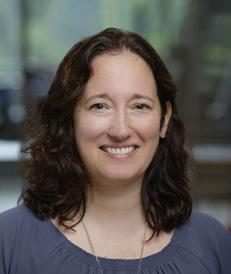
NJoyce L. Sanchez, MD Assistant Professor of Medicine and Surgery Division of Infectious Disease Director, MCW Travel Health Clinic
@joycesanchezmd
othing brings the problem of health care disparities into sharper focus than a global pandemic. I write from the perspective of a clinician who treats the most vulnerable and marginalized members of our society alongside a group of brilliant friends and colleagues with expertise in public health, epidemiology, infection prevention, virology and health disparities.
Infectious diseases do not strike randomly within a population, but rather disproportionately target those with predisposing risk factors, which are all too often not “randomly” distributed. The field of epidemiology aims to identify factors that place certain individuals at greater risk of disease than others. Our cast of characters in this epidemiologic triad includes the antagonist (the pathogen, SARS-CoV-2), the protagonist (the host, an individual) and the stage (the environment).
In the first week of April when elective surgeries were cancelled and units were clearing, infectious disease consults consisted primarily of patients admitted with COVID-19. The overwhelming majority of them were Black. By April 1st, Milwaukee County had seen its first 10 COVID-19 mortalities. All were Black. The 11th death was a Latino man. Against the backdrop of George Floyd’s death, it became impossible to ignore the structural and systemic racism present in our local society and within our healthcare system. Milwaukee County also saw a striking overlap when comparing the clusters of positive cases with the poverty map. Our enterprise’s experience in over 2,500 adults tested for COVID-19 found that zip code of residence explained almost 80% of the overall variance in COVID-19 positivity. COVID-19 positivity was also associated with Black race. Among patients with COVID-19, both race and poverty were associated with higher risk of hospitalization.1 Furthermore, most recognize there is likely considerable underreporting of cases given what we know about lower rates of access to healthcare, lower healthcare utilization, and higher levels of mistrust among communities of color.
To truly understand the scope of a national problem, national data is needed. In 2017, the leading causes of death were heart disease, cancer and unintentional injuries.2 Between February-May 2020, COVID-19 rose as the third leading cause of death.3 Unfortunately, data on the race and ethnicity of COVID-19 victims is incomplete. Even as recently as November 2020, six states and territories
“Whatever houses I may visit, I will come for the benefit of the sick, remaining free of all intentional injustice.”
— Hippocratic Oath
have not published race and ethnicity data for COVID-19 deaths. Of those who report this data, there is variability in how it is collected and categorized. Despite the incomplete data, the COVID-19 death rates in racial and ethnic minority groups are disproportionally higher than compared to Whites.4
The CDC’s updated list of risk factors for severe disease include older age and comorbidities including cancer, chronic kidney disease, obesity, heart conditions, type 2 diabetes and others.5 Many of those comorbidities disproportionally affect non-Hispanic Blacks and Hispanics.6-9 It is imperative to understand that age and health status of individuals is only the tip of the iceberg. Closely intertwined with these are health and social behaviors, including disproportionate barriers to healthcare access and utilization, healthcare literacy, housing and living condition, immigration status, English proficiency and many more.10 As Dr. Leonard Egede so succinctly expanded upon the root cause of health equity at last year’s MCW convocation ceremony, “COVID-19 has further widened pre-existing socioeconomic gaps and alerted us to the vulnerability of our fragile social structures.”
All is not lost. One national multisite study of >11,000 inpatients in 92 hospitals in 12 states (including Wisconsin) between February to May 2020 found that there was no statistically significant difference in risk of mortality between Black and White patients after adjusting for sociodemographic factors and comorbidities.11 If we as a healthcare system, as healthcare policy influencers, and as a united country address these disparities, we have a fighting chance of closing this gap. Equity is not an unattainable dream.
Where does that leave us? This is a call to action. When we recite the Hippocratic oath, we are promising to be part of the solution in this great world’s stage. Representation needs to be reflected at every academic medical center, in every Department and at every level of leadership. Representation needs to be reflected in investigators and study participants across our institutions, not just regarding COVID-19, but in cardiovascular disease, cancer, obesity, trauma and so many others. Inclusion opens the door for individuals to interact in the healthcare system, allows us to determine whether these interventions work
for all, ensures better care in the short- and long-term for all, and gives a reason for hope that these future interventions can and will make a difference for all.
My hope is that this rising generation of students, trainees and faculty members are a better generation, having been taught, mentored and shaped during this monumental time in history for the noble purpose of practicing the art of medicine while addressing health disparities at a time when it is needed more than ever.
FOR ADDITIONAL INFORMATION on this topic, visit mcw.edu/surgery or contact Dr. Joyce Sanchez at jsanchez@mcw.edu.
REFERENCES
1. Muñoz-Price LS, et al. Racial Disparities in Incidence and Outcomes Among Patients With COVID-19. JAMA Netw Open. 2020;3(9):e2021892. https://jamanetwork.com/journals/jamanetworkopen/fullarticle/2770961
2. The CDC at https://www.cdc.gov/nchs/products/ databriefs/db355.htm, Accessed November 2020
3. Zhou Y, Stix G. COVID-19 Is Now the Third Leading Cause of Death in the U.S. Scientific American. 2020 https://www.scientificamerican.com/article/covid19-is-now-the-third-leading-cause-of-death-in-the-us1/, Accessed November 2020
4. The COVID Tracking Project at The Atlantic at https:// covidtracking.com/race, Accessed November 2020
5. The CDC at https://www.cdc.gov/coronavirus/2019ncov/need-extra-precautions/people-with-medicalconditions.html, Accessed November 2020
6. The CDC at https://www.cdc.gov/kidneydisease/ publications-resources/2019-national-facts.html, Accessed November 2020
7. The CDC at https://www.cdc.gov/bloodpressure/ facts.htm, Accessed November 2020
8. The CDC at https://www.cdc.gov/diabetes/data/ statistics-report/appendix.html#tabs-1-3, Accessed November 2020
9. The CDC at https://www.cdc.gov/obesity/data/adult. html, Accessed November 2020
10. Pareek M, et. al. Ethnicity and COVID-19: an urgent public health research priority. The Lancet, 2020: p1421-1422. https://www.thelancet.com/journals/ lancet/article/PIIS0140-6736(20)30922-3/fulltext
11. Yehia BR, et al. Association of Race With Mortality Among Patients Hospitalized With Coronavirus Disease 2019 (COVID-19) at 92 US Hospitals. JAMA Netw Open, 2020;3(8):e2018039. https://jamanetwork. com/journals/jamanetworkopen/fullarticle/2769387
From the Chair, continued
end of the scalpel, a reminder of this basic surgical principle - that our job is not done until the patient fully recovers.
Learning from each other at a national level. This has grinded to a halt - now returning with virtual national meetings and regional CME programs. We have gotten pretty good at producing Department of Surgery Update Conferences (thank you Heidi and many others) and such conferences at a local level may even be more interactive than at a national level, where talks are often pre-recorded and time for questions is limited. What we are missing is the discussion and debate – at the microphone as well as the hotel lobby – on technical surgery, translational research, clinical trial development and other aspects of medicine which enhance our careers both personally and professionally. So much of what we do in the operating room was learned when visiting another institution or talking with colleagues from around the world at national meetings. The financial challenges of the pandemic and the “new normal” threaten widespread physician participation at national meetings – with major implications for our individual development; personally, academically, and as clinicians.
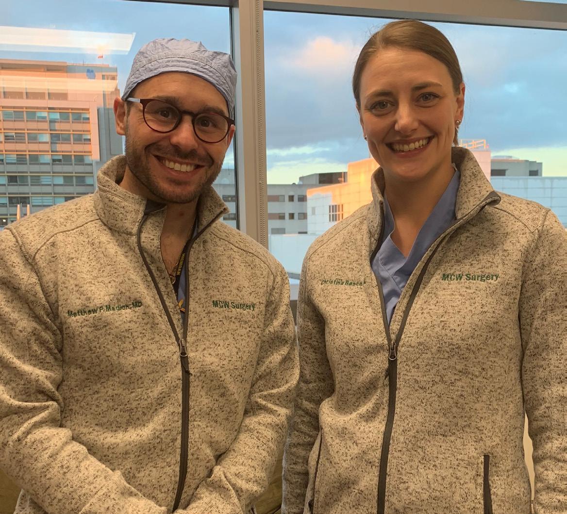
Leadership and management by walking around has largely disappeared. Making all aspects of work-place engagement more difficult and leaving the troops in the trenches feeling somewhat alone. The concern of course is that this may not change – further separating the decision makers from those who are impacted by their decisions. The decision makers (at all levels – almost everyone is a decision maker even if more of a decision receiver) can always get more accomplished on the “to-do” list by not walking around – because the list remains small due to lack of information and does not grow with front-line concerns. We all need to emphasize the importance of immersion; teamwork makes the dream work! Making the dream work in our surgical residency for the next 12 months are our new Administrative Chief Residents, Nina Bence and Matt Madion pictured on the cover. A big thank you as well to all authors who contributed the fantastic articles in this issue of “Leading the Way”.
Nonoperative Management of Uncomplicated Pediatric
Introduction
ADavid R. Lal, MD, MPH Professor of Surgery, Division of Pediatric Surgery
Thomas T. Sato, MD Professor of Surgery, Division of Pediatric Surgery
gical investigators from eleven regional children’s hospitals. The MWPSC has enabled several MCW pediatric surgery faculty and surgical residents to conduct multicenter clinical trials for both rare and common clinical entities.
ppendicitis is the most common etiology for urgent abdominal operation performed on children and adolescents by pediatric surgeons worldwide. On an annual basis, approximately 70,000 children in the United States will be treated for appendicitis. At Children’s Wisconsin, over 60% of cases present with acute, uncomplicated appendicitis, making this one of the most common, urgent clinical entities encountered by the Division. Contemporary management of pediatric appendicitis includes hospitalization, delivery of broad-spectrum intravenous antibiotics, and laparoscopic appendectomy. While perioperative complications are higher for perforated appendicitis, rates of perioperative complications for acute, uncomplicated appendicitis are reported between 5% and 15%, and postoperative recovery inflicts a period of disability for children and parents or caregivers. Similar to many surgical problems, there is much written about childhood appendicitis (case in point – in the past 34 months, there have been 273 peer-reviewed articles on pediatric appendicitis for an average of two publications per week), but there are few evidence-based, controlled trials.
Over the past decade, there are increasing data supporting the treatment of acute appendicitis using antibiotics alone as a safe and effective alternative to appendectomy in selected adult patients. Emerging data from single institutional trials and meta-analysis also provide evidence to support nonoperative management in children and adolescents with acute uncomplicated appendicitis.1,2 Additionally, nonoperative management may be more cost-effective.3 We designed and conducted a multicenter clinical trial evaluating the use of antibiotics alone compared to laparoscopic appendectomy in acute, uncomplicated pediatric appendicitis to determine the success rate of nonoperative management. Additionally, given perioperative complication rates and postoperative recovery disability, we sought to quantify disability days, health-related quality of life, and patient/parent satisfaction for both treatment arms. We utilized the Midwest Pediatric Surgery Consortium (MWPSC: www.mwpsc.org), a collaborative clinical research platform composed of sur-
The study was funded with a $2.875M grant from the Patient-Centered Outcomes Research Institute (PCORI) and designed as a prospective, controlled, nonrandomized multicenter trial utilizing patient/parent choice to determine treatment with either antibiotics alone or laparoscopic appendectomy.4 We hypothesized that nonoperative management would be successful in > 75% of cases with fewer disability days and complications compared to treatment with laparoscopic appendectomy. The two primary study outcomes were success of nonoperative management and disability days of the child at one year.5 A multidisciplinary team of key stakeholders including patients, parents, surgeons, primary care pediatricians, nurses, clinical patient educators, and payors was assembled to determine acceptable thresholds for determining success of nonoperative management, as well as defining perceived treatment-associated disabilities and health-related quality of life. While a randomized clinical trial was attractive from an academic surgical standpoint, non-surgeon key stakeholders felt that patients and families would be unwilling to participate in a randomized trial based upon preconceived treatment preferences. Importantly, while the surgical investigators felt that a minimally acceptable success rate for treatment with antibiotics alone should be greater than 70% and an expectation of 5 fewer disability days, the multidisciplinary key stakeholders indicated a 50% success rate would be entirely acceptable and the threshold clinically important difference in disability was 3 days, demonstrating the differential definition of success based upon perspectives.
Over a 41-month period, a total of 1068 children and adolescents aged 7 to 17 years diagnosed with uncomplicated appendicitis were enrolled using a standard algorithm with the following inclusion and exclusion criteria:
Inclusion Criteria:
1. Image-confirmed uncomplicated appendicitis by ultrasound, CT, or MRI with appendiceal diameter < 1.1 cm with no abscess, fecalith, or phlegmon.
2. WBC between 5000/vl and 18,000/vl
3. Abdominal pain less than 48 hours
Exclusion Criteria:
1. History of chronic intermittent abdominal pain
2. Diffuse peritonitis on clinical examination
3. Positive urinary pregnancy test
4. Communication difficulties
Overall, 88% of eligible patients approached for this study agreed to enrollment. 698 (65%) of the patients
Appendicitis: Perspectives on the Definition of Success
chose surgery and 370 (35%) chose nonoperative management. The surgical group was managed with hospitalization, intravenous antibiotics, and laparoscopic appendectomy within 12 hours of admission. The nonoperative group was managed with hospitalization and a minimum of 24 hours of intravenous antibiotics. Diet was advanced after 12 hours and conversion to oral antibiotics implemented for a total duration of seven days.
The unadjusted success rate of nonoperative management during initial hospitalization was 85.7% (adjusted rate using inverse probability of treatment weighting analysis = 85.4%, 95% CI 81.0% to 88.9%, P < .001). For patients considered non-operative failures crossing over to surgery, 16 failed to improve, 16 had clinical worsening, and 16 had parents that changed initial decision for antibiotics alone to surgery. Excluding these 16 patients who crossed over to surgery due to family decision, the adjusted success rate of nonoperative management during initial hospitalization was 89.3% and at one year was 70.2% (95%CI, 64.8% to 75.1%). For patients managed with surgery during initial hospitalization, the negative appendectomy rate was 7.5%. Patients managed nonoperatively who returned with symptoms of appendicitis were treated with appendectomy. For patients undergoing nonoperative management and ultimately requiring appendectomy either during initial hospitalization or during one year follow up, the negative appendectomy rate was 4.8%.
Over 75% of enrolled patients had 30-day and one year follow up. For patients completing study follow up, the adjusted success rate of nonoperative management at one year was 67.1% (96% CI, 61.5% to 72.3%; P = .86) and the adjusted disability days at one year were significantly fewer compared to the surgery group (6.6 vs 10.9 days; mean difference -4.3 days (99% CI, -6.17 to -2.43; P < .001). There was no significant difference in the rate of complicated appendicitis between groups. Health care satisfaction scores at 30 days were not significantly different between nonoperative management and surgery. Satisfaction with decision scores were very high in both treatment arms but were significantly lower in the nonoperative group. Adjusted health-related quality of life scores reported by patients and caregivers were significantly higher at 30 days in the nonoperative group compared to surgery, but this difference was not significantly different at one year.
Discussion
The results of this study demonstrate two-thirds of children with uncomplicated appendicitis may be safely and effectively treated with antibiotics alone, and there are significantly fewer disability days for the child and family at 30 days and one year. However, the remaining
one-third will likely require readmission and appendectomy within one year. In contrast, children undergoing appendectomy during initial hospitalization had a 6.9% rate of postoperative emergency room visits, a 1.1% postoperative infection rate, and a 2.9% readmission rate. Identification of clinical characteristics in children more likely to fail nonoperative management, as well as the longterm durability of nonoperative management for childhood appendicitis, remain to be determined. This study was limited by its moderately stringent inclusion criteria as only 19.3% of patients with appendicitis qualified for enrollment, as well as potential treatment selection bias given the lack of randomization. We believe areas for future focus include discernment of patient characteristics that may allow recognition of early or delayed failure of nonoperative management, and evaluation of the cost -effectiveness of nonoperative versus laparoscopic appendectomy for uncomplicated pediatric appendicitis across multiple institutions.
Perhaps one of the most powerful academic lessons from this study is the impact of determining the minimal clinically important difference necessary to define treatment success. As surgeons, we estimated sample size using an expected nonoperative success rate of greater than 75%, and we developed surgical consensus of a threshold success rate of 70%. We also expected 5 fewer disability days at one year for the nonoperative group and observed a mean difference of -4.3 days. As the adjusted nonoperative success rate was 67.1% at one year, this did not meet our study’s surgical threshold for success. In contrast, the multidisciplinary team of patients, families, and other medical specialists developed consensus for a nonoperative threshold success rate of 50% and a minimal clinically important difference in disability at 3 days, suggesting this group perceives a 67.1% rate and a mean difference of 4.3 disability days as successful. Additionally, it is clear that families have strong preferences for determining treatment for appendicitis and may not be particularly willing to allow their children to be enrolled in a randomized clinical trial. Ultimately, this trial will better inform future patients and families, allow them to determine their priorities, thresholds for failure and achieve true patient centered care.
FOR ADDITIONAL INFORMATION on this topic, visit mcw.edu/surgery or contact Dr. Lal at dlal@chw.edu or Dr. Sato at ttsato@mcw.edu.
REFERENCES ON PAGE 17
The History of Breast Implants and Breast Implant-
TAmanda L. Kong, MD, MS Professor; Chief, Section of Breast Surgery Division of Surgical Oncology
Erin L. Doren, MD Assistant Professor, Department of Plastic and Reconstructive Surgery
he past fifty years in breast surgery has shown a dramatic de-escalation in surgical procedures, from the modified radical mastectomy to breast conserving surgery and sentinel node biopsy with radiation with no differences in survival. NSABP B-04 demonstrated that radical mastectomies did not confer a survival benefit over total mastectomies1 and B-06 showed that a lumpectomy with radiation was equivalent in survival compared to a total mastectomy.2 Despite these advances, there has been a rise in mastectomy rates. One of the reasons for this rise has been attributed to improvements in breast reconstruction. The majority of patients who undergo mastectomies are now undergoing skin-sparing or nipple-sparing mastectomies with immediate reconstruction.
Patients will choose one of two types of immediate reconstruction, including autologous reconstruction (muscle and tissue flaps) versus implant-based reconstruction. The most common type of reconstruction is implant-based reconstruction. The first implant was designed by Cronin and Gerow in 1963 and was manufactured by Dow Corning.3 It consisted of a thin, smooth silicone elastomer shell containing silicone gel. It was noted that this first generation of implants had a high capsular contracture rate.
In the 1970s, second-generation silicone implants were developed to reduce capsular contractures and were filled
with a less viscous silicone for a more nature feel. Unfortunately, these implants were associated with the leaking of microscopic silicone molecules into the space between the implant and the capsule leaving an oily, sticky residue.4 Third generation implants were developed in the 1980s and these implant shells were composed of multilayered silicone elastomer to prevent leakage of silicone and implant rupture.4 However, in 1982, a series of three case reports emerged that reported patients who had cosmetic breast augmentation with silicone implants had developed autoimmune connective tissue disease (systemic lupus erythematosus, mixed connective tissue disease and rheumatoid arthritis) within two and a half years of surgery.5 Subsequently in 1992, due to these concerns of silicone-related illnesses, the U.S. Food and Drug Administration (FDA) put a moratorium on the use of silicone filled, silicone elastomer shell implants.6
In response to this moratorium, fourth and fifth-generation implants were developed. These silicone gel breast implants were developed with more stringent criteria from the the American Society for Testing Methodology and the FDA for the development of implant shell thickness and silicone gel cohesiveness. Quality control was improved as were surface textures and anatomic implant shapes.4 Two major manufacturers of silicone implants, Mentor and Allergan, began clinical studies in preparation for an application to the FDA. On November 17, 2006 the FDA approved both of their applications to market silicone gel-filled fourth generation breast implants.6 Despite improvements in the safety of silicone implants, these fourth and fifth generation prosthetics were not immune to problems.
In the U.S. alone, over 600,000 breast implants are placed each year.7 Breast implant-associated anaplastic large-cell lymphoma, or BIA-ALCL, is a rare type of T-cell lymphoma that has been found to arise around breast implants. The first case of BIA-ALCL was reported in the literature in 1997.8 This lymphoma is detected in the periprosthetic fluid and scar capsule that forms around breast
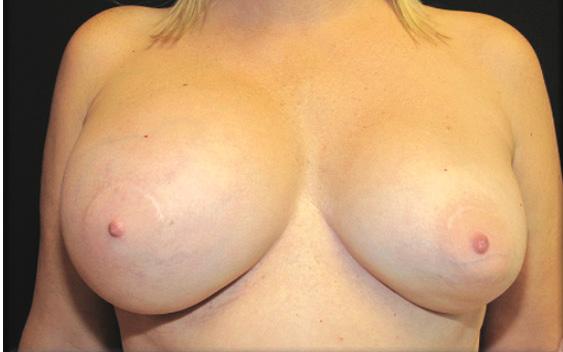
Figure 1: Example of BIA-ALCL presentation with right breast swelling.
Figure 2: Example of a textured and smooth breast implant (Southern Illinois University / Science Source).
Associated Anaplastic Large Cell Lymphoma (BIA-ALCL)
implants. Most cases have been diagnosed after revision surgery for a late-onset (> 1 year) seroma in patients with saline and silicone breast implants, with the mean onset occurring 8 years after implantation (figure 1).9 The FDA first recognized and published a safety communication regarding breast implants and their association with this T-cell lymphoma in 2011.10 Since then, the FDA and American Society of Plastic Surgeons (ASPS) report approximately 343, both suspected and confirmed, cases in the U.S. and a total of 976 worldwide.11
ALCL was first described in 1985 as a novel type of Non-Hodgkin’s Lymphoma. It is characterized by large anaplastic lymphoid cells that express the cell-surface protein CD30, key to its diagnosis. BIA-ALCL is a distinct form of ALCL that arises in the effusion or scar capsule that surrounds a breast implant. To date, all confirmed diagnoses with an adequate clinical history implicate a textured surface breast implant (figure 2).12 Rates of BIA-ALCL have been equally reported in breast augmentation and breast reconstruction patients. In 2017, an estimation of the lifetime prevalence for women with textured breast implants was reported to be 1 in 30,000.13 More recent studies have estimated the risk to be 1:2,207-1:86,029 based upon variable risk with different manufacturer types of textured implants.11
roma or mass should have an ultrasound and fine needle aspiration sent for lymphoma markers.18 Disease-free survival is highest in those who have complete surgical excision which includes a total capsulectomy, explant of the device and oncologic resection of any mass with negative margins.19 The majority of cases can be treated with surgery alone; however, chemotherapy and radiation have been employed for advanced disease. Treatment at a ter-
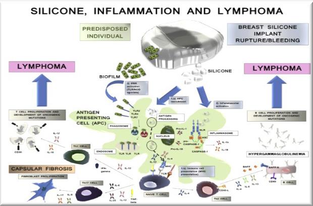
The etiology of BIA-ALCL still largely remains unknown. Most theories assume chronic inflammation plays a part as breast implants are associated with mild to severe scarring leading to capsule formation (figure 3). Additionally, contamination of the implant with bacteria, a biofilm, could elicit a response initiating and maintaining chronic inflammatory responses.14 In 2016, Hu et al. reported that a certain species of non-fermenting gram-negative bacilli, Ralstonia, was found in greater proportion in ALCL breast implant capsules compared to non-tumor capsules where staphylococcus was most common.15 Implants with a textured surface have been found to support a higher bacterial load and therefore higher lymphocytic hyperplasia accounting for this lymphoma’s association with textured implants.16 Not all manufacturer’s implant texturing is created equal. The more aggressive the textured surface the higher the bacterial load.17 One of the three breast implant manufactures in the U.S. recalled their textured devices from the market in July of 2019 in response to an increased incidence of BIA-ALCL with their textured devices (1:2,207). The FDA is not currently recommending the prophylactic removal of textured devices.
Patient and surgeon education are the key to early diagnosis of this very treatable cancer. Any patient who presents with a delayed and persistent peri-implant se-
tiary care center by a multidisciplinary team is critical to improve outcomes.
Moving forward, patient education and informed consent regarding the complications associated with breast implants is imperative. All standard implant consent forms and implant manufacture box warnings include information regarding the risk of BIA-ALCL. The American Society of Plastic Surgeons has created a national breast implant registry to track outcomes, although not all institutions are participating. In addition, there is also a breast implant ALCL registry in the United States. With increasing concerns about breast implant safety, many women are choosing autologous-based reconstruction. However, not all patients are candidates for this procedure. As more data is collected over time, plastic surgeons and their patients will need to weigh the risks and benefits of breast implant reconstruction.
FOR ADDITIONAL INFORMATION on this topic, visit mcw.edu/surgery or contact Dr. Doren at edoren@ mcw.edu or Dr. Kong at akong@mcw.edu
TIMELINE & REFERENCES ON PAGE 8
Figure 3: Inflammatory pathways leading to lymphoma formation around breast implants via Bizjak,M et al. Silicone implants and lymphoma (Journal of Autoimmunity, 65, (2015), 64e7).
The History of Breast Implants and Breast Implant-Associated Anaplastic Large Cell Lymphoma (BIA-ALCL)
1970s
Second generation implants
• thinner shell and less viscous silicone
1950s
First breast augmentations using injections of liquid silicone, paraffin, epoxy resin, petrolatum jelly, glycerine
• “bleed” of silicone into surrounding tissue
1990s
Textured implants introduced to reduce capsular contracture
1995
Dow Corning files for Chapter 11 bankruptcy
1962
Cronin and Gerow develop first silicone implant manufactured by Dow Corning
1980s
Third generation implants
• multilayer shell
• less “bleed” of silicone
1992
FDA issues a voluntary moratorium on silicone implants
• growing concern over siliconeinduced illnesses
• increase in saline implant use
REFERENCES FROM PAGE 5 | BREAST IMPLANTS
1. Fisher B, Montague E, Redmond C et al. Findings from NSABP Protocol No. B-04: comparison of radical mastectomy with alternative treatments. IIl The clinical and biologic significance of medial-central breast cancers. Cancer. 1981: 48 (8): 1863-72.
2. Fisher B, Anderson S, Redmond CK et al. Reanalysis and results after 12 years of follow-up in a randomized clinical trial comparing total mastectomy with lumpectomy with or without irradiation in the treatment of breast cancer. N Engl J Med. 1995 333 (22): 1456-61.
3. Deva A, Cuss A, Magnusson M, et al. The “Game of Implants” : A Perspective on the Crisis-Prone History of Breast Implants. Aesthet Surg J. 2019 39 (suppl_1) S55-S65.
4. Maxwell GP, Gabriel A. The evolution of breast implants. Plst Reconstr Surg 2014 134 (1 Suppl): 12-S-7S.
5. Van Nunen SA, Gatenby PA, Basten A. Post-mammoplasty connective tissue disease. Arthritis Rheum. 1982 25 (6) 694-7.
6. Perry D, Frame JD. The history and development of breast implants. Ann R Coll Surg Engl 2020 102 (7): 478-482.
7. American Society of Plastic Surgeons. Plastic Surgery
2006
Silicone implants reintroduced after manufacturers submit data to the FDA
2000s
2016 World Health Organization designated breast implant associated ALCL as a T-cell lymphoma that can develop following breast implants
2011
FDA issues statement on possible association between development of anaplastic large cell lymphoma (ALCL) and breast implants
2008
DeJong et al. first to publish link between breast implants and anaplastic large cell lymphoma (ALCL)
Fourth generation implants designed with FDA-influenced criteria and American Society of Testing Methodology
2012-2013
Fifth generation implants
• concept of anatomically correct shaped implants
• more cohesive gel
Statistics Report. https://www.plasticsurgery.org/ documents/News/Statistics/2019/plastic-surgerystatistics-full-report-2019.pdf. Accessed October 28, 2020.
8. Keech JA, Creech BJ. Anaplastic T-cell lymphoma in proximity to a saline-filled breast implant. Plast Reconstr Surg. 1997;100(2):554-5.
9. Clemens MW, Miranda RN. Coming of age: Breast implantassociated anaplastic large cell lymphoma after 18 years of investigation. Clin Plast Surg. 2015;42(4):605-13.
10. U.S. Food and Drug Administration (FDA). Anaplastic large cell lymphoma (ALCL) in women with breast implants: preliminary FDA findings and analyses. 2011.
11. American Society of Plastic Surgeons. BIA-ALCL Resources. https://www.plasticsurgery.org/for-medicalprofessionals/health-policy/bia-alcl-physician-resources/ by-the-numbers. Accessed October 28, 2020.
12. Brody GS. Anaplastic large cell lymphoma occurring in women with breast implants: Analysis of 173 Cases. Plast Reconstr Surg. 2015;135(1):695-705.
13. Doren EL, Miranda RN, Selber JC, Garvey PB, et al. U.S. epidemiology of breast implant-associated anaplastic large cell lymphoma. Plast Reconstr Surg. 2017;139(5):10421050.
14. Bizjak M, Selmi C, Praprotnik S, Bruck O, et al. Silicone implants and lymphoma: The role of inflammation. Journal Autoimmun. 2015;65:64-73.
15. Hu H, Johani K, Almatroudi A, Vickery K, et al. Bacterial biofilm infection detected in breast implant-associated anaplastic large-cell lymphoma. Plast. Reconstr Surg. 2016; 137(6):1659-69.
16. Jacombs A, Tahir S, Hu H, Deva AK, et al. In vitro and in vivo investigation of the influence of implant surface on the formation of bacterial biofilm in mammary implants. Plast. Reconstr Surg. 2014;133(4):471-80.
17. Hu H, Jacombs A, Vickery K, Merten SL, et al. Chronic biofilm infection in breast implants is associated with an increased T-cell lymphocytic infiltrate: implications for breast implant-associated lymphoma. Plast. Reconstr Surg. 2015;135(2):31929.
18. U.S. Food and Drug Administration. Questions and answers about Breast Implant-Associated Anaplastic Large Cell Lymphoma (BIA-ALCL). https://www.fda. gov/medical-devices/breast-implants/questionsand-answers-about-breast-implant-associatedanaplastic-large-cell-lymphoma-bia-alcl. Accessed October 29, 2020.
19. Clemens MW, Medeiros LJ, Butler CE, Hunt KK, et al. Complete surgical excision is essential for the management of patients with breast implantassociated anaplastic large-cell lymphoma. J Clin Oncol. 2016;34(2):160-8.
MCW Employee Service Awards
20 YEARS OF SERVICE
Michael Passow Surgery Administration
Mary Ann Rueth Surgery Administration
Jidong Su Research Associate
30 YEARS OF SERVICE
Kara Doffek Research Associate
Anna C. Purdy, MSN, ANP
Nurse Practitioner, Division of Surgical Oncology
Meg Shannon-Stone, RN, MSN, APNP Nurse Practitioner, Division of Cardiothoracic Surgery
35 YEARS OF SERVICE
Ann Kolbach-Mandel Research Associate II
MCW’s Outstanding Medical Student Teachers for 2019-2020
The Curriculum and Evaluation Committee seeks to recognize and affirm those individuals who, through their teaching excellence, advance student learning and provide a “value added” to students’ required medical training.
MCW’s Outstanding Medical Student Teachers for 2019-2020: M3 Surgery Clerkship
Faculty
Dr. Anuoluwapo Elegbede Assistant Professor of Trauma & Acute Care Surgery
Dr. Steven Kappes Clinical Professor, Aurora Health Care
Dr. Caitlin Patten Assistant Professor of Surgical Oncology
Dr. Brian Lewis Professor of Vascular and Endovascular Surgery; Chief, Division of Education
Dr. Betsy Appel Assistant Clinical Professor, Aurora Health Care
Residents/Fellows
Dr. Katherine Flynn-O’Brien Assistant Professor of Pediatric Surgery
Dr. Jacqueline Blank Administrative Chief Resident
Dr. Spencer Klein General Surgery Resident PGY 2
MCW’s Outstanding Medical Student Teachers for 2019-2020: Other Educational Achievements
Faculty
Chandler Cortina, MD Assistant Professor of Surgical Oncology (Foundational Capstone) Advanced Practice Provider Terry Derks, PA-C Physician Assistant, Pediatric Surgery (Health Systems Management & Policy Pathway)
Absorbable Synthetic Mesh: An Alternative to
AMatthew I. Goldblatt, MD Professor of Surgery, Division of Minimally Invasive Gastrointestinal Surgery; Director, Condon Hernia Institute; Director, Surgical Residency Program
bout five years ago, I would consent a patient for a hernia repair with mesh, and after discussing the standard risks and benefits, they would sign it. That is not the case today. After countless ads from national law firms soliciting mesh cases, I now need to spend at least five minutes describing the potential problems with mesh, as well as the significant benefits. I typically tell patients that the biggest issues with mesh started with the nitinol ring in the original Kugel Patch1 and the mesh used in transvaginal urethral slings.2 I then also discuss the voluntary product withdrawal of Physiomesh in 20163 due to a higher incidence of central mesh fractures. It was this last product withdrawal that really spawned the latest round of litigation that has spilled over to all permanent mesh products.
The most logical question from patients when discussing their ventral/incisional hernia is, “Do you need to use mesh?” The data would suggest that for any hernia larger than 1-2 cm in size, the answer is: yes. A systemic review and meta-analysis of primary and incisional ventral hernias comparing primary closure to mesh repair found 63% fewer recurrences when mesh was used.4 After hearing the data on using mesh reinforcement and the relative safety of the mesh products on the market today, most patients are willing to have mesh placed in their body.
Despite the data for the use of mesh, there are still some patients who insist on a hernia repair without mesh. Typically, their reasons are either because of previously bad experiences with permanent mesh, or because of information they have gathered on the internet warning about mesh. What are your options as a surgeon other than primary fascial closure which we know doesn’t work well? In the past 10 years, long-lasting fully absorbable mesh for hernia repairs have come on the market. If an absorbable mesh could hold the patient’s own fascia together long enough to allow it to heal, then maybe it would be a good alternative to permanent mesh. The two most common products on the market are Phasix from BD, and Bio-A (and its most recent upgrade, Enform) from WL Gore. These meshes utilize very different absorbable polymers with unique characteristics that I will outline here. Before these materials are discussed, there needs to be several disclaimers. The first is that I have received research funding from both BD and Gore for research done with these products, and I receive speaking fees from Gore. Most importantly, many surgeons use these materials when their surgical field has some degree of contami-
nation. I will discuss that this may be very appropriate, but any such discussion is considered to be off-label use by the FDA. All mesh products, whether they are permanent synthetic, absorbable synthetic or even biologic, are only approved for use in clean cases.
The first long-term absorbable mesh on the market was Bio-A from Gore. It is a microporous mesh made up of a co-polymer of trimethylcarbonate and polyglycolic acid. In the last few years, they have refabricated it into a softer, stronger, and more pliable material called Enform, but the polymer is the same. The polymer fully resorbs in 6-7 months. The Complex, Open, Bioabsorbable Reconstruction of the Abdominal wall (COBRA) study actually evaluated the use of this product in clean-contaminated or contaminated fields.5 The mesh was placed either in the retro-rectus or intraperitoneal position. All patients achieved primary fascial closure. In other words, the mesh was never used as a bridge. After 24 months, the midline recurrence rate was 17%. The retro-rectus recurrent rate was 13%, whereas the intraperitoneal recurrence rate was 40%. Despite the contaminated fields, none of the meshes had to be removed in these patients.
The other absorbable mesh on the market is Phasix mesh. It is a macroporous mesh made from the polymer poly-4-hydroxybutyrate. The polymer is actually made by bacteria and is extracted and made into a mesh. It takes about 18 months to fully degrade and reabsorb. In the Phasix trial, we looked at long-term recurrence rates when using the mesh in either a retro-rectus position or as an onlay in patients with clean wounds, but high risk for Surgical Site Infections (SSI).6 These risks factors included smoking, obesity, type 2 diabetes, and steroid use. After three years, the hernia recurrence rate was 17.9% with 11.4% in the retro-rectus group and 28.1% in the onlay group. None of the meshes had to be explanted.
The final study is one that we did at MCW. We evaluated the hernia recurrence rate of 55 patients who underwent a Bio-A repair in clean wounds. After 22 months, we found an 8.2% recurrence rate when the mesh was in the retro-rectus position, and a 50% recurrence rate when the mesh was intraperitoneal.7 When evaluating other hernia studies that looked at the long-term outcomes of permanent mesh in the retro-rectus position, they found a 16.9% recurrence rate at 19 months.8
All of these studies share a couple of conclusions. The first is that the long-term outcomes of fully absorbable mesh are comparable to similar studies using permanent mesh. The other conclusion is that these products perform significantly better when placed in the retro-rectus position. Therefore, if absorbable mesh works as well as permanent mesh, but doesn’t have any of the long-term risks associated with a permanent implant, why not use them for every hernia repair? The answer is simply that
Permanent Mesh
they are significantly more expensive than their permanent counterparts. Therefore, I recommend reserving absorbable synthetic mesh for those patients who have unique circumstances such as infection or contamination (off-label), higher risk for SSI, or significant fear of permanent mesh.
FOR ADDITIONAL INFORMATION on this topic, visit mcw.edu/surgery or contact Dr. Goldblatt at mgoldbla@mcw.edu.
REFERENCES
1. FDA Recall Z-0762-06, Feb 22, 2006, Bard Composix Kugel, https://www.accessdata.fda. gov/scripts/cdrh/cfdocs/cfRes/resCollection_2. cfm?ID=44997&CREATE_DT=2006-02-22
2. KC Kobashi, et al, “Erosion of woven polyester pubovaginal sling”, J Urol 1999 Dec;162(6):2070-2
4. T Mathes, et al, “Suture versus mesh repair in primary and incisional ventral hernias: A systemic review and meta-analysis”, World J Surg 2016 Apr;40(4):826-35
5. MJ Rosen, et al, “Multicenter, Prospective, Longitudinal Study of the recurrence, surgical site infection, and quality of life after contaminated ventral hernia repair using biosynthetic absorbable mesh”, Ann Surg 2017 Jan;265(1):205-211
6. JS Roth, et al, “Prospective evaluation of poly-4hydroxybutyrate mesh in CDC class I, high-risk ventral and incisional hernia repair: 3-year followup”, submitted for publication
7. JE Cho, et al, “Retro-rectus placement of bioabsorbable mesh improves patient outcomes”, Surg Endo 2019;33:2629-2634
8. WS Cobb, et al, “Open retromuscular mesh repair of complex incisional hernia: predictors of wound events and recurrence”, JACS 2015;220(4):606-613
2020 Fall Research Symposium Winners
Sarah Suh M4 Student
Nathan Smith, MD
General Surgery Resident PGY 4
Catherine Bodnar M2 Student
Basil Karam, MD
Postdoctoral Research Fellow
“The Influence of Preoperative Carbohydrate Loading on Post-operative Outcomes in Bariatric Surgery Patients: A Randomized, Controlled Trial”
Faculty Mentor: Rana Higgins, MD
“Early Right Ventricular Assist Device and VV ECMO Improve Outcomes in Severe COVID-19 ARDS“
Faculty Mentor: David Joyce, MD
“Association between Neighborhood Socioeconomic Disadvantage and Complicated Appendicitis in Children”
Faculty Mentor: Kyle Van Arendonk, MD, PhD
“Need for Emergent Intervention Within 6 Hours (NEI-6): A novel Prediction Model for Prehospital Trauma Triage”
Faculty Mentor: Rachel Morris, MD
Building International Partnerships to
AMary Elizabeth Schroeder, MD Assistant Professor, Division of Trauma and Acute Care Surgery
s highlighted by Dr. Klinger in the fall edition of Leading the Way, the field of global surgery has evolved significantly. My early global work was largely missionbased and gave me an opportunity to travel to countries throughout Africa, India and South America. Many of these programs involved long-standing relationships, with surgeons and staff returning on a regular basis to provide care and clinical teaching. This work touched the lives of many individual patients but these siloed efforts often lacked sustainability and reproducibility.
In 2015, the Lancet Commission published Global Surgery 203, a groundbreaking paper that made the case that a comprehensive coordinated multidisciplinary effort to provide basic surgical care to all is not only the right thing to do from a humanistic standpoint, it also makes economic sense. The current global burden of surgical disease is overwhelming, with an estimated 5 billion people lacking access to carei and a need for an additional 143 million surgical procedures annually.1 Shrime et al. estimated that in 2010, 16.9 million people died worldwide due to lack of surgical access.2 This is more than four times the annual death toll from HIV/AIDS (1.46 million), tuberculosis (1.2 million) and malaria (1.17 million) combined.1
Since this landmark call to arms, numerous groups have worked to address the at-times overwhelming task of improving access to surgical care to people in low and middle-income countries. There is a recognition that the solutions are not universal as many of the political, economic and geographic issues are region-specific, which require close collaboration with local leaders and experts to have the greatest impact. And often the attempt to find answers leads only to more questions which can be defeating. But in the words of Desmond Tutu, “there is only one way to eat an elephant: a bite at a time.”3
One such partnership project to address this monumental task is the newly formed American College of Surgeons’ Operation Giving Back training hub at Hawassa University Hospital in southern Ethiopia. The Medical College of Wisconsin is an invited participant in this unique multi-institution collaboration in which 14 academic institutions pledged full-time coverage by a U.S. surgeon. The role of the ACS representative is flexible and based on provider specialty and Hawassa’s needs. Initial initiatives have ranged from intra-operative teaching during complex cases, creation of a laparoscopic training center, establish-
ment of a morbidity and mortality conference as well as reconfiguration of the emergency room to provide a resuscitation area for trauma and critically ill patients.
In addition to the above initiatives, Dr. Chris Dodgion and I have developed a research training curriculum for the faculty and staff at Hawassa. In an initial needs assessment of the Hawassa faculty and residents, research was identified by almost all that were surveyed as a high priority. At most medical schools and residency programs in Ethiopia, basic research training is not part of the curriculum. While there are opportunities for highly motivated trainees to pursue advanced training, the majority of surgeons do not feel that they have the skills needed to answer the clinical questions that present in their practice. In addition, it is increasingly recognized that clinically what may be the gold standard in high income countries does not always apply in low and middle-income populations, due to variance in resources as well as inherent differences in the patient population.
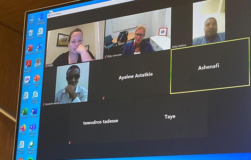
The ACS-Hawassa Research Course was created in collaboration with the School of Public Health at Hawassa University. We utilized their statistical expertise in combination with the surgical research experience of ACS faculty to create a seven-week, web-based, interactive curriculum. Twenty participants (10 faculty and 10 residents) had twice weekly lectures followed by two-hour small group sessions on Zoom each Saturday. The Zoom sessions served to walk the participants through the process of developing their research idea from initial inception to their ethics board proposal. Lecture topics are included in Table 1.
The course is now on hiatus as the participants finalize their ethics board proposal with the assistance of an assigned ACS mentor. Once the participants have received
ilack of access is defined as 2-hour proximity to a center that can perform an exploratory laparotomy, manage an open fracture and perform a caesarian section.
The commute to work at Hawassa University Hospital in southern Ethiopia.
Improve Access to Surgery
approval for their research project, the course will be reconvened to provide teaching in data collection and analysis as well as manuscript writing. We hope that this step-by-step process with individualized mentorship will provide the participants with the skills they need to start addressing the challenges to providing surgical care in their region. The responses from the trainees as well as the involved public health faculty have been very enthusiastic, with interest in making this an ongoing integral part of their resident curriculum.
While there are a handful of web-based global research training programs available online, this is the first to focus on surgeons and surgical disease specifically. We are currently exploring opportunities to disseminate this course to other academic centers in Ethiopia and we are in discussions with the College of Surgeons of East Central and Southern Africa (COSECSA) to integrate it into surgical resident training for the entire region.
With the continued substantial inequity in the provision of global surgical care, MCW’s partnership with Hawassa University Hospital and the American College of Surgeons serves as a model collaboration of sustainability to address these gaps, empowering local providers through knowledge and training. In addition, we are now working to provide global experiences to our trainees through international rotations as well as a global surgery research fellowship that will combine masters-level training in health disparities with the opportunity for international research. I hope that such academic initiatives will help build the next generation of global surgery experts, ready to address global disparities and improve patient outcomes with MCW leading the way.
FOR ADDITIONAL INFORMATION on this topic, visit mcw.edu/surgery or contact Dr. Schroeder at meschroeder@mcw.edu.
Small group sessions on “Zoom,” the video-conferencing software.
Lecture topics
(Table 1)
• Guidelines and regulations of good clinical practice
• The role of IRB and principles of consent
• How to identify a clinical problem for research question
• Formulating structured clinical research questions
• Major clinical study design types
• Factors affecting selection of study design
• How to minimize bias in your study
• Data collection tools
• How to write up a proposal
• How to search the literature
• Sampling techniques and sampling error
• How to calculate sample size
• Critical appraisal of a paper
• How to use reference manager
REFERENCES
1. Meara et al. Global Surgery 2030: evidence and solutions for achieving health, welfare, and economic development. The Lancet 2015; 386:569-624.
2. Shrime MG, Bickler WS, Alkire BC, Mock A. Global burden of surgical disease: an estimation from the provider perspective. Lancet Glob Health 2015; 3: S8–9
3. Lozano R, Naghavi M, Foreman K, et al. Global and regional mortality from 235 causes of death for 20 age groups in 1990 and 2010: a systematic analysis for the Global Burden of Disease Study 2010. Lancet 2012; 380: 2095–128
COMING SOON
The 2020 MCW Surgery Annual Report
Donation after Circulatory Death Heart
David Joyce, MD Associate Professor of Surgery, Division of Cardiothoracic Surgery
Sunday, September 20, 2020
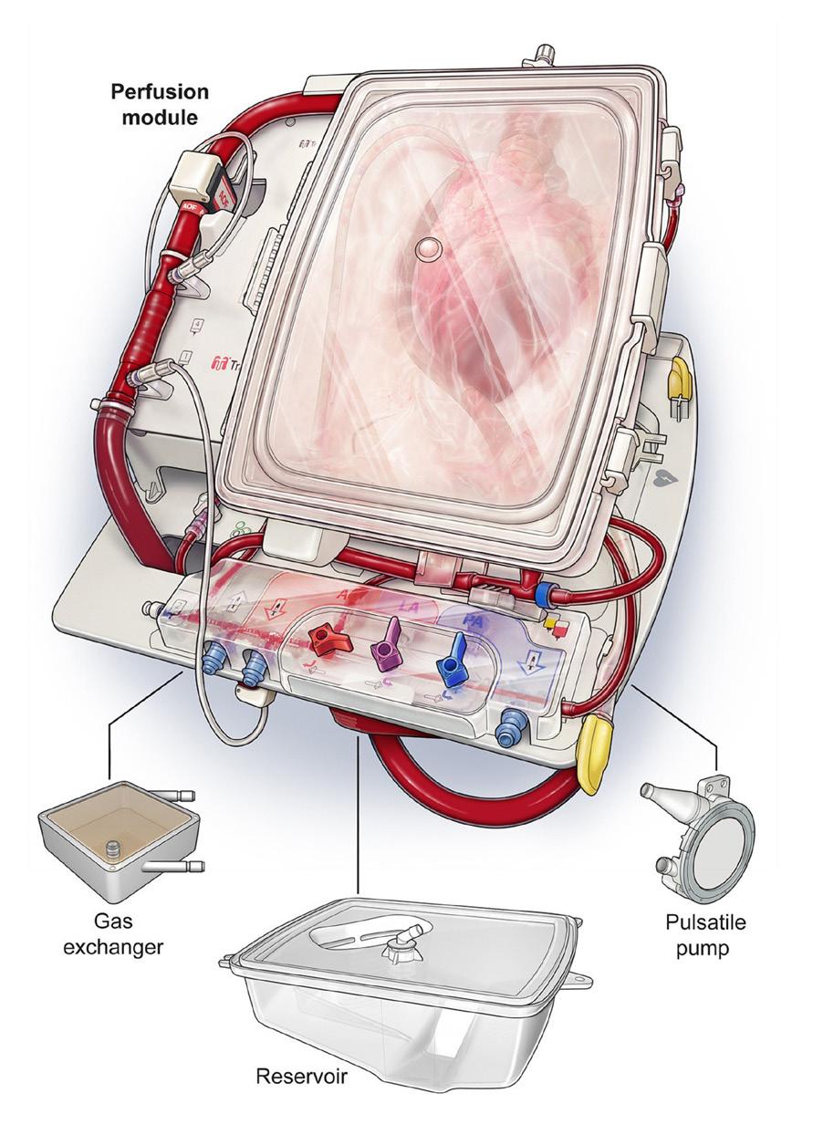
15:53 Months of preparation in the form of presentations, committee meetings, and simulated dry runs culminated in an opportunity to become one of only a few centers in the country to offer a Donation after Circulatory Death (DCD) heart to one of our patients. We’d been down this road before only to come back empty handed. This would be the second time our recipient made a trip to the hospital in expectation of getting a transplant, and his time was running out. At 68 years old, he had already cheated the lethality of cardiogenic shock through the flawless deployment of three novel mechanical circulatory support devices by Drs. Lyle Joyce, Takushi Kohmoto, and Buck Durham. If the TransMedics Organ Care System (OCS) performed as expected, he would be four for four.
Tuesday, September 22, 2020
09:45 Two Nationwide Organ Recovery Transport Alliance vehicles departed from the west entrance of Froedtert Hospital. For the seven passengers (two surgeons, two perfusionists, a preservationist, a heart failure fellow, and a certified surgical assistant), the degree of coordination and communication with the TransMedics staff in Boston and the recipient OR team in Milwaukee resembled the complexity of a Space Shuttle mission. As we drove to the donor hospital, I reviewed each of the details one last time to be sure we hadn’t missed any red flags. Becoming a heart donor is by its very definition an unspeakable tragedy, and in this case that tragedy took the form of a suicide attempt. While “attempt” is the best word we have in the English language to describe what happened, it is far from adequate in explaining the state of limbo that occurs when a patient has been rescued from hypoxia in time to save every organ other than the brain. To be specific, this patient failed to meet the “brain death” definition that was developed by an ad hoc committee at Harvard Medical School in 1968, now established as the legal criteria for declaration. Thanks to the miracle of mechanical ventilation, however, this patient could be kept alive indefinitely despite having no possibility of recovering neurologic status. In this situation, removing the breathing tube offers a compassionate alternative to living in a persistent vegetative state.
14:38 The breathing tube was removed. Our team gathered with the abdominal organ procurement team in an adjacent room, crowding around a monitor that dis-
played the donor’s vital signs. We waited for the heart to stop, and 20 minutes later, there was no longer any electrical activity observable on the monitor. After waiting an additional five minutes, the patient would meet the criteria for cardiac death.
15:40 We started the donor operation. Our ability to successfully retrieve the heart depended on the efficiency of the choreography which followed. In a perfect world, the surgeon would have six hands that could operate in parallel to expose the right atrium, drain off a liter and a half of blood, cannulate the aorta, place the cross clamp, and flush the heart with cardioplegia. As it turned out, a father-son team with over six years of experience working together in the midst of numerous hair-raising operative encounters with the help of a rock-star surgical assistant did the job. We were flushing the heart with two minutes to spare.
15:59 The moment of truth. As we connected the donor heart to the OCS system, oxygenated blood from the circuit filled the aortic root and entered the coronary arteries. It had been well over an hour since this heart had seen normal blood flow and we watched anxiously to see what would happen. As we were preparing to position the electrical paddles in an effort to shock the organ back to life we were halted by a vigorous contraction. Then another. As a cardiac surgeon you observe so many different hearts in your day-to-day work that you begin to assess their quality in the same way that an art dealer would appraise a rare painting. This one was a Rembrandt.
16:43 Group text to the Froedtert team: “Heart looks good. Leaving soon. Wait for lactate before making incision.” Now we just needed to stick the landing. There were different views on how long a heart can be supported on the OCS before it needs to be implanted in the recipient, but the person with the most credibility in this
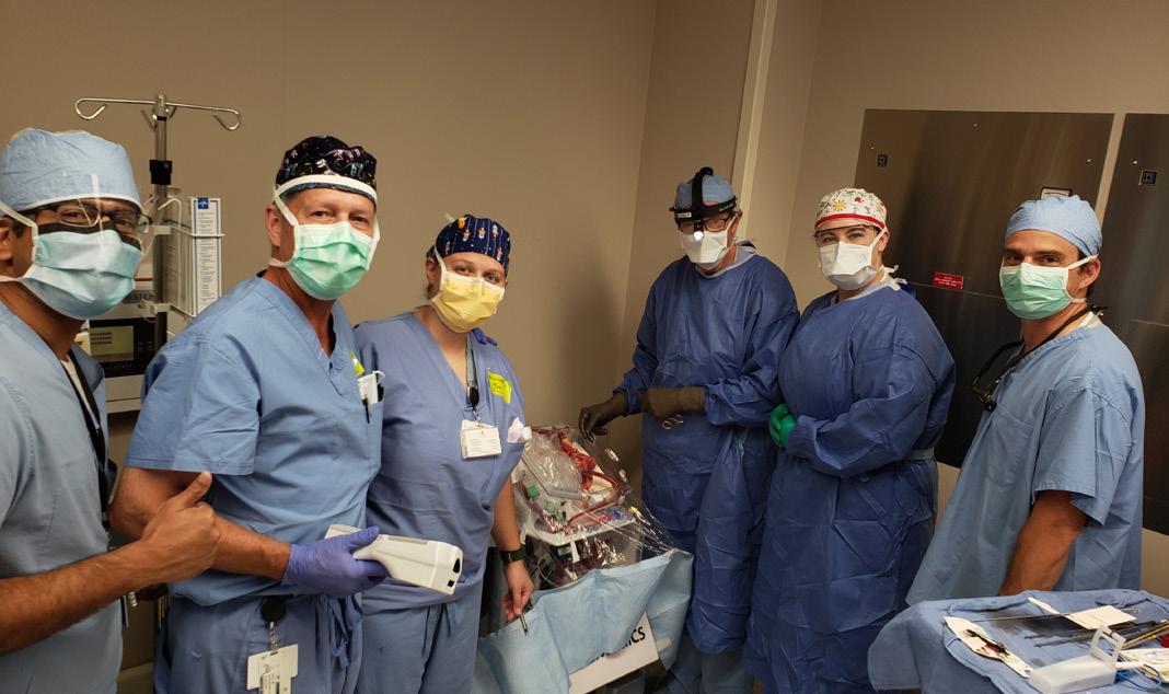
The Froedtert & MCW procurement team (from left): Padmaraj Duvvuri, MD; David Koerten, CCP; Marguerite Wellstein, CCP; Lyle Joyce, MD, PhD; Jackie Hanke, CSA; and David Joyce, MD.
Transplant Comes to Milwaukee
“There was a sudden contraction of the atria, followed quickly by the ventricles in obedient response—then the atria, and again the ventricles. Little by little it began to roll with the lovely rhythm of life.”
— Donald McRae, Every Second Counts: The Race to Transplant the First Human Heart
domain was Professor Steven Tsui at Papworth Hospital in the United Kingdom. Tsui was one of the pioneers who performed much of the animal work that had set the stage for Donation after Cardiac Death (DCD) heart transplants. He came to MCW in October of 2018 to give Surgery Grand Rounds on their clinical outcomes and had become a close friend and trusted advisor in our DCD journey. With his depth of experience, Tsui had given us a set of rules to follow that would ensure a successful outcome for our first patient. One of those rules was to set a perimeter around the city of Milwaukee and not cross that line for any donor offers that were outside that narrow radius. But just like the unexpected plot twists of a “Mission Impossible” movie, Tsui was transparent about the fact that things would come up and even he would occasionally break his own rules to save a patient’s life. Hopefully turning on the lights and sirens would shorten the three-hour drive back to Froedtert…
18:18 In order to minimize the time on the OCS system, we needed to be ready to remove the recipient’s heart the moment our team walked into the operating room. Since this was a difficult redo surgery in which a left ventricular assist device would have to be removed along with the heart, we needed to give Drs. Kohmoto and Durham as much time as possible. After carefully reviewing all of the data with our contact at TransMedics, we decided it was time to push all the chips to the center of the table. The incision was made on the recipient with the stipulation that we would wait to go on cardiopulmonary bypass until our next lactate level came back in one hour. After reviewing the final set of laboratory results, I took one last look to the back seat of the vehicle where our perfusion team was dutifully attending to the heart as it was banging away on the machine. I could tell that my own heart was racing as I anticipated the final stage—sewing it in.
19:53 We arrived in the operating room and obtained one final set of labs before flushing the organ and moving it on to the OR table. While the surgery was unquestionably more difficult than most, what followed was a well-worn routine that our team had executed flawlessly countless times before. As we reperfused the organ with the recipient’s blood, it was clear within seconds that we were on our way to a successful outcome.
As we placed the last of the sternal wires to close the chest, I thought about all the individuals that had performed at such a high level at every step of this journey dating back to the fall of 2018. Just to be included in the trial at all required our team to hold our own with only a small handful of the most reputable solid organ transplant programs in the world. It occurred to me that the level of teamwork involved in this effort was unusual not just for historical human endeavors, but even in the genre of science fiction.
FOR ADDITIONAL INFORMATION on this topic, visit mcw.edu/surgery or contact Dr. Joyce at djoyce@ mcw.edu.
PMarc de Moya, MD Professor and Chief, Division of Trauma and Acute Care Surgery; Milton and Lidy Lunda/Charles Aprahamian Professor of Trauma Surgery
ancreatitis is a common phenomenon occurring in 4.9% of people, 35 per 100,000 of the population, and is increasing due to obesity and incidence of gallstones. The overall mortality for all those with pancreatitis is 1-5%.1
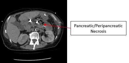
The two main causes of pancreatitis are gallstones that migrate distally in the common bile duct and alcohol induced pancreatitis. There are a number of other causes, but regardless of the cause, there remains a spectrum of disease from a mild form to a life-threatening severe form. This severe form occurs in approximately 20% of the cases and is often associated with a necrotizing component. As the pancreatitis evolves, the degree of necrosis likely secondary to endothelial damage and microvascular thrombosis also typically evolves with extension to the peripancreatic fat (Figure 1).
The inflammatory cascade that ensues often places patients into multi-system organ failure and shock. The degree of complex decision-making and critical care necessary is why these patients are often cared for on the Acute Care Surgery service.
The management of necrotizing pancreatitis has evolved over the last several decades from early operative debridement2 to delayed operative debridement3 to the use of endoscopic and percutaneous drains, and more recently to the sinus tract endoscopic necrosectomy. Mortality associated with early operative debridement ranged from 30% to 70% due to multi-system organ failure. The idea of waiting for the necrosis to mature, and treating with antibiotics if infected until approximately 3-4 weeks after the onset of necrosis, lowered the mortality to 11-15%. However, there remains 1.2% of enteric fistulae, 35-60% with pancreatic fistulae, and 15% who need reoperation. Since this major improvement, there
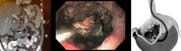
have been further improvements utilizing trans-gastric metal stents to allow for drainage and debridement endoscopically4 via the stent (Figure 2).
In addition, the use of percutaneous retroperitoneal or trans-abdominal drains have been used in a “step-up” approach,5 which involves incremental increases in size of the drains. These drains can then be used as guides to perform video-assisted retroperitoneal dissections (VARDs) or trans-sinus endoscopic debridement.
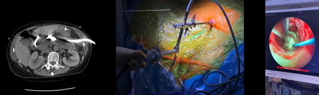
In 2018, Dr. David Milia successfully performed MCW’s first sinus tract necrosectomy. This was done in collaboration with Dr. Peter Fagenholz from the Massachusetts General Hospital, who has helped to develop this technique. In this technique, a wire is placed through the drain under fluoroscopy and the drain is removed over the wire. An expanding dilator is then placed over the wire, and uti-
Figure 1: Peripancreatic necrosis and infection.
Figure 2: Left, CT-scan of transgastric stent into necrotic cavity; middle, endoscopic view of trans-stent debridement; right, cartoon of endoscopic debridement via stent.
Figure 3: Left, CT-scan of percutaneous drain in infected peripancreatic bed used as guide; middle, nephroscope placed into cavity through sheath placed over dilated tract; right, debridement via nephroscope.
lizing a nephroscope with continuous irrigation, the necrotic cavity is entered and debrided (Figure 2).
The necrotic tissue is carefully removed until the viable pink non-infected tissue remains. This often requires multiple trips to the operating room to slowly remove all affected tissue without removing viable or bleeding tissue. The drains are replaced at the end of each operation and, once all necrosis is removed, the drain is left in place to allow the cavity to drain and collapse. The pancreatic fistula incidence is lower using this technique and typically will close spontaneously over time.
This procedure has now been performed a number of times with great success. The case in Figure 3 was recently performed at MCW and the cavity of necrosis cleared. The era of open debridement has come to an end and only remains for the most complicated patients with colonic fistulae or other complication not amenable to a minimally invasive approach. However, over 90% of patients are now managed utilizing percutaneous drains and trans-gastric drains alone. The morbidity and mortality of necrotizing pancreatitis remains high compared to other patient populations but has significantly improved through the years.
FOR ADDITIONAL INFORMATION on this topic, visit mcw.edu/surgery or contact Dr. de Moya at mdemoya@mcw.edu.
REFERENCES FROM PAGE 15 NONOPERATIVE MANAGEMENT OF UNCOMPLICATED ...
1. Minneci PC, Mahida JB, Lodwick DL, Sulkowski JP, Nacion KM, Cooper JN, Ambeba EJ, Moss RL, Deans KJ. Effectiveness of patient choice in nonoperative vs surgical management of pediatric uncomplicated acute appendicitis. JAMA Surg. 2016;151:408-415.
2. Huang L, Yin Y, Yang L, Wang C, Li Y, Zhou Z. Comparison of antibiotic therapy and appendectomy for acute uncomplicated appendicitis in children: a meta-analysis. JAMA Pediatr. 2017;171:426-434.
3. WU JX, Sacks GD, Dawes AJ, DeUgarte D, Lee SL. The cost-effectiveness of nonoperative management versus laparoscopic appendectomy for the treatment of acute, uncomplicated appendicitis in children. J Pediatr Surg. 2017;52:1135-1140.
4. Minneci PC, et al., on behalf of the Midwest Pediatric Surgery Consortium. Multi-institutional trial of non-operative management and surgery for uncomplicated appendicitis in children: Design and rationale. Contemp Clin Trials. 2019;83:10-17.
5. Minneci PC, et al., for the Midwest Pediatric Surgery Consortium. Association of nonoperative management using antibiotic therapy vs laparoscopic appendectomy with treatment success and disability days in children with uncomplicated appendicitis. JAMA. 2020;324:581-593.
2. Fernandez-del Castillo C, et al. Debridement and closed packing for the treatment of necrotizing pancreatitis. Ann Surg. 1998;228(5):676-84.
3. Rodriguez JR, et al. Debridement and closed packing for sterile or infected necrotizing pancreatitis: insights into indications and outcomes in 167 patients. Ann Surg. 2008; 247(2):294-9.
4. Bakker O, van Santvoort H, et al. Endoscopic trans-gastric vs surgical necrosectomy for infected necrotizing pancreatitis. JAMA. 2012;307(10):10531061.
5. Van Santvoort HC, Besselink MG, et al. A step-up approach or open necrosectomy for necrotizing pancreatitis. N Engl J Med. 2010;362:1491-1502.
6. Van Santvoort HC, et al. Videoscopic assisted retroperitoneal debridement in infected necrotizing pancreatitis. HPB (Oxford). 2007;9(2):156-9.
The Word on Medicine COVID-19 episodes:
The Word on Medicine aired a series of weekly special broadcasts that provided accurate information from physicians caring for patients every day. We explained how this virus was transmitted from animals to humans, how human-to-human transmission occurred, the challenges with testing and treatment, and everything you ever wanted to know about vaccines.
The Word on Medicine would like to thank and gratefully acknowledge Drs. Joyce Sanchez, John Fangman, Mary Beth Graham, Njeri Wainaina (Department of Medicine, Division of Infectious Diseases) and Nate Ledeboer (Department of Pathology) for their many contributions during our 11-part series devoted to COVID-19.
Scan the barcode above or visit ihr.fm/2LeatGc to check out The Word on Medicine podcast.
2020 We Care Research Recovery Grants
TGwen Lomberk, PhD Chief, Division of Research; Director, Basic Science Research; Department of Surgery; Associate Professor of Surgery and Pharmacology & Toxicology
Krissa Packard, MS, ACRP-CP Manager, Division of Research Department of Surgery
The COVID-19 pandemic caused massive disturbances and uncertainties around the globe at all levels, and scientific research labs were no exception. The 2020 We Care awards provided a special opportunity for our researchers, as hardships have been faced across the country and world. The necessary implementation of lab hibernation and halting of both basic and clinical research at MCW during the COVID-19 pandemic caused our faculty to lose valuable time, resources, and funds, which threatened to disrupt research programs and significantly delay productivity. The intention of the 2020 We Care Research Recovery Fund was to uniquely help our faculty and their teams get “back to the research benches and bedsides.”
We thank the We Care Committee for their tremendous support of our researchers by funding nine Research Recovery Grants totaling over $175,000.
John E. Baker,
PhD, Professor, Pediatric Congenital Heart Surgery Division
The Baker laboratory is studying why cancer survivors treated with radiation have an increased risk for heart disease, so that innovative interventions or therapies can be developed.
The We Care recovery funds were given to Dr. Baker’s team to help with expenses to re-build their animal colony and perform the necessary experiments to submit competitive grant applications to the NIH and NASA.
Panna A. Codner, MD, Associate Professor, Trauma and Acute Care Surgery Division
Both DNA, the blueprint of our body, and bacteria, which live in our intestines, are affected by the environment in which we live, such as income and poverty level, skin color and exposure to violence. Dr. Codner is actively investigating how these environmental circumstances interact with our DNA and the bacteria living in our intestines to cause anxiety and depression. Dr. Codner’s research was severely impacted by reductions in research staff and temporary discontinuation of services that resulted in direct loss of grant dollars due to the funding end
date occurring during hibernation. The We Care recovery funds were provided to allow processing of these samples to acquire the data Dr. Codner needed for a federal grant submission.
Amanda L. Kong MD, MS, Professor, Surgical Oncology Division
Dr. Kong is a 2017 We Care awardee who is examining the critical unsolved clinical problem of symptomatic cardiovascular toxicity secondary to anti-cancer therapy. In fact, adverse cardiovascular side effects have surpassed cancer recurrence as the number one cause of death in breast cancer survivors. This research project is studying how anti-cancer therapy impairs human microvascular function and how to counteract these adverse effects. Loss of funds, due to continued salaries of non-faculty research personnel during hibernation with reduced or no capacity to continue research operations as well as study participation fees paid for patients who could not remain in the study with the halting of clinical research, were offset by the We Care recovery funds to enroll additional patients to complete the We Care study as designed.
Gwen Lomberk, PhD, Associate Professor, Research Division
The Lomberk lab focuses on uncovering windows of opportunity to provide novel targets and treatment approaches for pancreatic cancer, a painful and deadly disease. Our approach seeks to understand the intersection of normal processes required for cells to duplicate and how they then tolerate rapid division of cancer with the goal of using this knowledge to find vulnerabilities we can target to eliminate pancreatic cancer cells. The We Care recovery funds were given to Dr. Lomberk’s team to aid rebuilding their animal colony that models pancreatic cancer (which was reduced more than half its original size), re-purchasing reagents that expired during hibernation and had to be disposed, and covering expenses incurred for alternative services of experiments impacted by lab hibernation.
Aoy Tomita-Mitchell, PhD, Professor, Pediatric Congenital Heart Surgery Division
Dr. Aoy Mitchell’s lab is interested in explaining cellular processes underlying cardiomyocyte and endothelial cell differentiation during development in order to gain insight into the molecular underpinnings and pathogenetic mechanisms of congenital heart disease (CHD). For this purpose, her team is creating a three-dimensional bioengineered cardiac tissue model in efforts to better mimic physical characteristics of the heart. The Mitchell lab benefited from the We Care recovery funds for salary support of research personnel paid through lab hibernation.
Michael Mitchell, MD, Professor, Pediatric Congenital Heart Surgery Division
The research program of Dr. Michael Mitchell seeks to develop a non-invasive method for monitoring rejection in children and adults with heart transplants. The current gold standard for rejection monitoring is done by heart biopsy, which is accompanied by significant risk, especially for children. The team is working toward a blood test, which is rapid, safe and cost effective to enable sensitive, frequent monitoring with short and long-term benefits for all transplant recipients. The We Care recovery funds were provided to assist Dr. Michael Mitchell’s program to cover salary support for research personnel to salvage delayed grant objectives.
Kirkwood A. Pritchard Jr., PhD, Professor, Pediatric Surgery Division
The long-term goal of Dr. Pritchard’s research program is to determine how people with sickle cell disease suffer from poor vascular function, which increases vaso-congestion. Although vaso-congestion can occur anywhere in the body, when it occurs in the brain of someone who has sickle cell disease, it can cause neurological injuries, such as silent cerebral infarct, stroke, and death. The We Care recovery funds helped Dr. Pritchard’s team to cover lost salary support and expenses to re-build their animal colony for experiments to successfully complete the aims of their NIH funded R01 application.
Raul Urrutia, MD, Professor, Research Division
The research program of the Urrutia lab is focused on effective diagnosis and treatment of devastating diseases associated with the pancreas, such as chronic pancreatitis and pancreatic cancer. These are both painful and incurable diseases; the diagnosis of pancreatic cancer typically occurs when the cancer has advanced, and 5-year survival
is below 10%. Dr. Urrutia’s team is studying how KRAS, the gene most commonly mutated in pancreatic cancer as well as mutated in many other cancer types, triggers changes in the way our DNA functions in efforts to find innovative approaches to counteract KRAS-mediated cancer cell growth. The We Care recovery funds assisted Dr. Urrutia’s program with expenses incurred due to expired reagents and equipment malfunction as a result of lab hibernation.
Amy J. Wagner, MD, Associate Professor, Pediatric Surgery Division
Dr. Wagner’s research program is dedicated to identifying potential genetic variations in gastroschisis, the most common congenital abdominal wall defect which results in intestines herniating outside the baby’s stomach. The purpose of the Gastroschisis Genome Pilot Project is to compare genetic variants between family members affected by gastroschisis, and those who are not. With the help of the We Care recovery funds, Dr. Wagner was able to sequence samples collected from a newborn diagnosed with gastroschisis and the immediate family, consisting of three siblings as well as the mother and father, since the original funding source was eliminated due to COVID-19 financial impact. Importantly, completing this work will establish the proper foundation to process blood samples from gastroschisis affected babies for growing a national bio bank, which will be housed at our institution under Dr. Wagner’s direction.
FOR ADDITIONAL INFORMATION on this topic, visit mcw.edu/surgery or contact Dr. Lomberk at glomberk@mcw.edu.
To refer a patient or request a transfer/consultation, please use the references below:
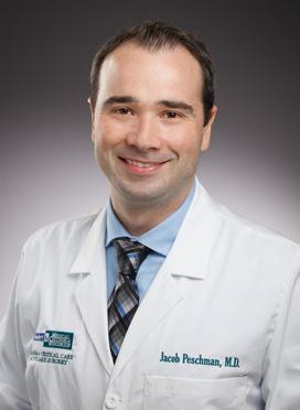
Jacob R. Peschman, MD, will be returning to the Department of Surgery faculty in July 2021 as an Assistant Professor of Surgery from Gundersen Health System in LaCrosse, Wisconsin, where he is currently a general and trauma surgeon and is involved with medical student and resident education. Dr. Peschman earned his medical degree with research distinction from the Medical College of Wisconsin and
also completed general surgery residency at MCW. He completed a Surgical Critical Care fellowship at Mayo Clinic in Rochester, Minnesota. Dr. Peschman will serve as Associate Program Director for the General Surgery Residency program. We are proud of Dr. Peschman’s service to our country as a Lieutenant Commander in the U.S. Navy Reserve Medical Corps and are delighted to welcome him and his family back to our department faculty and to Milwaukee.
DIVISION OF VASCULAR & ENDOVASCULAR SURGERY
Nathan W. Kugler, MD, will join the Department of Surgery faculty in August 2021 as an Assistant Professor of Surgery, following completion of a Vascular Surgery fellowship in our Division of Vascular and Endovascular Surgery. He earned his medical degree from Southern Illinois University School of Medicine and completed general surgery residency at MCW. His clinical interests include all aspects of open and endovascular arterial
Elyan Ruiz Solano, MD, recently joined the Department of Surgery faculty as an Instructor. She earned her medical degree from the University of Santo Domingo in the Dominican Republic and completed a surgical internship in mechanical circulatory support at Deutsches Herzzentrum Berlin, Germany. Dr. Ruiz Solano received a master’s
reconstruction. We are thrilled to have Dr. Kugler and his family remain in Milwaukee to join our department faculty.
DIVISION OF CONGENITAL HEART SURGERY
degree in biomedical research from the University of Seville, Spain. She also completed residency in cardiovascular surgery in Seville, followed by a cardiothoracic surgery clinical fellowship at King’s College Hospital in London and a congenital cardiac surgery clinical fellowship at Evelina London Children’s Hospital in London. Dr. Ruiz Solano will provide clinical care to patients of the Cardiac Surgery and Congenital Heart Surgery services. We are so fortunate to have Dr. Ruiz Solano join our department faculty.
DIVISION OF CARDIOTHORACIC SURGERY
James Mace, MD, joined the Department of Surgery faculty in Feburary through the Joining Forces Program, a civilian-military collaboration with the MCW Comprehensive Injury Center (CIC). The goal is to achieve zero preventable deaths from injury, both in the community and the battlefield. The Medical College of Wisconsin is one of four sites nationally that are partnering with the US Army. Based on the 2017 National Defense Authorization Act, the program is designed to sustain the trauma surgery and resuscitation skills for the surgeons within an US Army Forward Resuscitative Surgical Team (FRST). The surgeons are stationed at MCW for up to three years with the potential for intermittent deployments within that timeframe. They are fully
integrated within the Departments and our campus during their time at MCW. Dr. Mace is a West Point graduate and completed his cardiothoracic fellowship at the University of Alabama, Birmingham (UAB). He is currently an active-duty cardiothoracic surgeon in the US Army. He just returned from a tour in Kuwait. To maintain a high level of readiness and experience in the surgical providers, the Department of Defense developed a program to imbed its specialists in academic medical centers in the USA. Dr. Mace will assist in all aspects of cardiothoracic surgery.
Nathan W. Kugler, MD
Elyan Ruiz Solano, MD
James Mace, MD
THE MEDICAL COLLEGE OF WISCONSIN DEPARTMENT OF SURGERY
SECONDARY FACULTY BY SPECIALTY
Carmen Bergom, MD, PhD, M.Phil.
Radiation Oncology
Meena Bedi, MD
Radiation Oncology
Michael B. Dwinell, PhD
Microbiology and Immunology
Beth Erickson Wittmann, MD
Radiation Oncology
Juan Felix, MD Pathology
James W. Findling, MD Medicine, Endocrinology, Metabolism and Clinical Nutrition
Mary Beth Graham, MD Medicine, Infectious Diseases
Jennifer Geurts, M.S., CGC
Genomic Sciences and Precision Medicine Center
Jaime S. Green, MD Medicine, Infectious Diseases
James B. Gosset, MD Medicine, Cardiovascular Medicine
Michael O. Griffin, Jr., MD, PhD
Radiology, Diagnostic Radiology
William A. Hall, MD
Radiation Oncology
Robert A. Hieb, MD, RVT
Radiology, Vascular and Interventional Radiology
Eric J. Hohenwalter, MD
Radiology, Vascular and Interventional Radiology
Bryon D. Johnson, PhD
Microbiology and Immunology
Mandana Kamgar, MD Medicine, Hematology and Oncology
John A. LoGiudice, MD
Plastic Surgery
Veronica Loy, D.O. Medicine, Gastroenterology
Peter J. Mason, MD, MPH, RPVI
Medicine, Cardiovascular Medicine
GIVE TO THE RESIDENT RESEARCH FUND
The Resident Research fund provides surgical residents an opportunity to initiate and complete research projects related to their professional interests. The objective of the research experience is to create opportunities for residents to gain an understanding of basic, clinical, and translational research methods that inspire them to pursue opportunities for career development as investigators. If you would like to make a gift online using a credit card, please visit our secure site to support the Resident Research Fund, or scan the QR code.
www.mcwsupport.mcw.edu/residentresearchfund
Stacy D. O’Connor, MD, M.P.H., MMSc
Radiology, Diagnostic Radiology
Jong-In Park, PhD
Biochemistry
Parag J. Patel, MD, M.S. Radiology, Vascular and Interventional Radiology
Hershel Raff, PhD
Medicine, Endocrinology, Metabolism and Clinical Nutrition
Honey Reddi, PhD
Pathology/GSPMC
William S. Rilling, MD
Radiology, Vascular and Interventional Radiology
Vice Chair, Clinical Affairs
Evan Robinson, DO Emergency Medicine
Ehab R. Saad, MD Medicine, Nephrology
Michael H. Salinger, MD Medicine, Cardiovascular Medicine
Joyce Sanchez, MD
Medicine, Infectious Diseases
Jennifer J. Schiller, PhD, D(ABHI)
Versiti Blood Center of Wisconsin
Pippa M. Simpson, PhD Pediatrics, Quantitative Health Sciences
Cynthia Solliday-McRoy, PhD Transplant
Parag P. Tolat, MD Radiology, Diagnostic Radiology
Sean M. Tutton, MD
Radiology, Vascular and Interventional Radiology
Njeri. Wainaina, MD Medicine, Infectious Diseases
Sarah B. White, MD, M.S. Radiology, Vascular and Interventional Radiology
MCW SURGERY IN THE MEDIA: CREW IN SAFE HANDS WITH CAPLINGER IN CHARGE
Roger Caplinger, head of the Brewers’ medical operation, survived pancreatic cancer and is setting the tone of the organizational effort to play safely through the pandemic . Read his story online here or scan the QR code.
THE MEDICAL COLLEGE OF WISCONSIN DEPARTMENT OF SURGERY
FACULTY BY SPECIALTY
Bariatric & Minimally Invasive
Gastrointestinal Surgery Matthew I. Goldblatt, MD
Jon C. Gould, MD, MBA
Rana M. Higgins, MD
Andrew S. Kastenmeier, MD
Tammy L. Kindel, MD, PhD
Kathleen Lak, MD
Cardiac Surgery
G. Hossein Almassi, MD
Nilto C. De Oliveira, MD (4/21)
Lucian A. Durham III, MD, PhD
Viktor Hraska, MD, PhD
David L. Joyce, MD, MBA
Lyle D. Joyce, MD, PhD
Takushi Kohmoto, MD, PhD, MBA*
R. Eric Lilly, MD*
James E. Mace MD
Michael E. Mitchell, MD*
Paul J. Pearson, MD, PhD
Elyan Ruiz Solano MD
H. Adam Ubert, MD (8/21)
Ronald K. Woods, MD, PhD*
Colorectal Surgery
Jed F. Calata, MD
Kirk A. Ludwig, MD
Mary F. Otterson, MD, MS
Carrie Y. Peterson, MD, MS*
Timothy J. Ridolfi, MD, MS
Community Surgery
Robert J. Brodish, MD
T. Clark Gamblin, MD, MS, MBA
Dean E. Klinger, MD
Kaizad Machhi, MD
Kevin V. Moss, MD
Eric A. Soneson, MD
Mark A. Timm, MD
Pediatric General & Thoracic Surgery
John J. Aiken, MD*
Casey M. Calkins, MD*
Brian T. Craig, MD
John C. Densmore, MD*
Katherine T. Flynn-O’Brien, MD, MPH
David M. Gourlay, MD*
Tammy L. Kindel, MD, PhD
Dave R. Lal, MD, MPH*
Keith T. Oldham, MD*
Pediatric General & Thoracic Surgery, continued
Thomas T. Sato, MD*
Jack G. Schneider, MD*
Sabina M. Siddiqui, MD
Kyle Van Arendonk, MD, PhD
Amy J. Wagner, MD*
Research Faculty
Mohammed Aldakkak, MD
John E. Baker, PhD
Young-In Chi, PhD
Mats Hidestrand, PhD
Gwen Lomberk, PhD
Angela J. Mathison, PhD
Aoy T. Mitchell, PhD
Kirkwood Pritchard, Jr., PhD
Raul A. Urrutia, MD
Surgical Oncology–Breast Surgery
Chandler S. Cortina, MD
Amanda L. Kong, MD, MS*
Caitlin R. Patten, MD*
Tina W.F. Yen, MD, MS
Surgical Oncology–Endocrine Surgery
Sophie Dream, MD*
Douglas B. Evans, MD*
Tracy S. Wang, MD, MPH*
Tina W.F. Yen, MD, MS
Surgical Oncology–Hepatobiliary and Pancreas Surgery
Editors: Rana Higgins, MD Heidi Brittnacher, 414-955-1831 or hbrittna@mcw.edu
Designer: Liz Chen, echen@mcw.edu
Leading the Way is written for physicians for medical education purposes only. It does not provide a complete overview of the topics covered and should not replace the independent judgment of a physician about the appropriateness or risks of a procedure for a given patient.