Another great edition of “Leading the Way” filled with superb articles by many talented faculty and residents in the Department of Surgery. The first two articles relate to COVID-19; perhaps I can add a couple comments on the lighter side related to the pandemic — which we all hope will be in the rear-view mirror soon!
First, is it just me or do people seem to stare at you from behind their mask — they seem to lock eyes and stare so much more aggressively than without a mask. Why is this and what are they thinking? I suspect it is easier to stare at someone with the protection of a mask? Does the paper mask feel like the mask of a gladiator? We could probably call this the Russell Crowe effect, I’m not sure — and what are they thinking and what do they make you think? When we get stared at we assume some aspect of our clothing is in need of fixing or perhaps zipping up — or did we do something wrong? Were we the third person onto a two-person elevator? Did we not follow the pasted feet on the floor and got too close to someone in front? Is your mask not completely covering your nose? There is also the possibility that the person staring at you cannot figure out who you are? How many times in the last two years has someone said hi to you and you could not figure out who they were — their mask was at their lower eyelids and they had a scrub cap on – their voice was muffled by the mask — you said hi back and wondered who they were. Or, you failed to say hi to someone that actually figured out who you were — all made worse by the growing trend to look down at your phone when walking the halls. Mask up to lower eyelids, cap on, ID badge turned backwards so no name can be seen (the usual position) and eyes directed downward reading the phone — and if you say hi, the eyes stare back at you sometimes with a smile (as best you can tell) or a “hi there” — crazy time indeed. But, stay strong, the mask remains so important right now – and it will be fascinating to see if the stares
MCW Surgery
transcend the life span of the mask — time will tell?? We hope to return to routinely saying hi with more “normal” eye contact sometime very soon!
Second, working from home and the explosion of virtual meetings, will we ever go back? How many of you are now professional remote attendees? - and I know that you all take a break with many of those Zoom and WebEx meetings! You dial in, then put yourself on mute and surf the internet, catch up on email, grab a snack and otherwise get things done - - and you can always tell when one of your colleagues has done this - they get called on to participate in the conversation and they can’t seem to unmute their computer — because we all know that they are clueless as to what has been going on and are too embarrassed to unmute the computer — the perfect time for a technical glitch!! – what will we all ever do when these meetings return to in-person — I suspect we will get a whole lot more accomplished (per meeting)!
Please enjoy this edition of “Leading the Way” — a truly remarkable series of contributions from our amazing faculty and residents.
Cell-Free DNA and COVID-19
Michael E. Mitchell, MD Professor and Chief, Division of Congenital Heart Surgery
Background and Preliminary Data
Early identification of tissue damage and likelihood of progression to severe disease and inflammatory syndrome is critical to effective acute clinical management of patients presenting with COVID-19. High levels of circulating cell-free DNA (cfDNA) have been reported in acute phase COVID-19 patients and persist during prolonged disease. 1,2 As a newly investigated biomarker, cfDNA is found in very low concentrations in plasma of healthy patients due to baseline metabolic activity and natural cell turnover, but rapidly becomes elevated in conditions characterized by inflammation and tissue injury. For instance, in a single institution retrospective study of 87 non-COVID-19 pediatric cardiac transplant patients4 and later confirmed in a cohort including >4000 samples from 388 non-COVID-19 pediatric and adult cardiac surgical and septic intensive care patients, we found plasma total cfDNA level to be an actionable biomarker (3hr turnaround time) predictive of near-term critical clinical events and poorer long-term
outcomes. 5 In both cohorts, cfDNA levels rising above a critical threshold of 50 ng/ml,4 typically before clinical symptoms became apparent, were predictive of disease progression and increased likelihood of adverse clinical events (mechanical circulatory support, cardiac arrest, and/or death) within 30 days (p=0.022). 5
In a pilot IRB-approved blinded retrospective COVID-19 study, we obtained 32 0.5 ml plasma samples from 19 patients who tested positive for SARS CoV-2 RNA and were hospitalized for treatment. Clinical records during hospitalization and convalescence were reviewed by a team physician in a blinded manner in order to assign a numerical score for severity of COVID illness existing at the time of each sample’s collection. A score of 1 represented asymptomatic outpatient convalescence following discharge, 2 representing mild inpatient symptoms improving with therapy, 3 representing moderate inpatient symptoms improving during treatment of initially severe illness, and 4 representing inpatient severe illness requiring high flow oxygen and/or intubation. Plasma concentration of cfDNA (of nuclear origin) was significantly associated with severity of clinical illness (p<0.01 nonparametric Cusick’s test) (Fig. 1).
Hypotheses
We hypothesize that nuclear cell-free DNA (ncfDNA) levels over 50 ng/ml in patients with COVID-19 will predict near-term development of severe disease justifying immediate intensive monitoring, whereas patients with ncfDNA levels under 10 ng/ml are unlikely to progress to severe illness. Although no one biomarker can mandate clinical interventions independent of other clinical indicators, our clinical experience with this assay and recent publications suggest that ncfDNA levels will become an important tool with potential for triage and monitoring of COVID-19 patients.
Technology and Goal
We developed and validated a qPCR protocol for clinical-grade quantification of plasma total (nuclear) cfDNA concentration in a CAP/CLIA-accredited laboratory.
Funding and Current Project Status (Trials and Tribulations)
In the spring of 2020, early in the pandemic, we submitted a grant to the Department of Defense to fund a large prospective observational cohort cell-free DNA study in 250 COVID-19 adult and pediatric inpatients at 3 major hospitals with con-
Fig. 1. Nuclear cell-free DNA vs clinical severity score.
Fig. 2. Total cfDNA linearity 2-1,000 ng/ml. The adjusted linear fit equation is ng/mL (y) = -0.455701 + 1.2255499*Expected ng/mL(x) 3.
firmed SARS-CoV-2 infection in which serial cfDNA levels would be correlated in a blinded fashion with a comprehensive clinical, demographic, laboratory, and physiologic dataset. We also proposed to show equivalency of cfDNA results between our CAP CLIA lab and 3 other accredited labs trained to use a prototype ncfDNA kit developed in our lab. The DoD grant review indicated reviewers scored the project extremely well, but unfortunately administrative priorities prevented funding. In the summer of 2021, the MCW Office of Research provided one year of funding for a similar, but more focused study in adult subjects at Froedtert Hospital to obtain preliminary data in preparation for resubmission of the initial grant. The CTSI grant is currently underway. Without direct clinical access to COVID-19 inpatients, our research group worked with the MCW Tissue Bank to obtain serial blood samples from inpatients who consented to the Bank. At the writing of this article, 31 subjects had provided approximately 130 whole blood samples, which the Bank had processed them to frozen plasma upon receipt. Our next step is to run cfDNA levels on those samples and collect clinical data.
We participated in CTSI COVID-19 R&D Group conference calls, interacting with other local physicians and scientists involved in investigating aspects of COVID-19. Knowing that multiple researchers worldwide as well as at Froedtert and MCW were undertaking COVID-19 research, we did not want to reinvent the wheel relative to designing a COVID-19 clinical database or even collecting data ourselves. We contacted the World Health Or-
ganization (WHO) and they willingly shared data dictionaries of REDCap databases. REDCap is the research database that is available to researchers at MCW through the CTSI, and we have used REDCap many times. Later we learned that Froedtert physicians were participating in the development of a COVID-19 research registry led by Mayo Clinic called VIRUS. We are currently working with the MCW VIRUS research group and the IRB to ascertain if our project will be allowed to utilize COVID-19 clinical data from Froedtert patients that has already been harvested and stored as part of the VIRUS project. Finding an efficient way to set up and manage our data during this proof-of-concept grant period will be invaluable as we prepare to resubmit our project for federal funding.
FOR ADDITIONAL INFORMATION on this topic, visit mcw.edu/ surgery or contact Dr. Mitchell at memitchell@mcw.edu.
REFERENCES
1. Andargie, T.E., Tsuji, N., Seifuddin, F., Jang, M.K., Yuen, P.S., Kong, H., Tunc, I., Singh, K., Charya, A., Wilkins, K., et al. (2021). Cell-free DNA maps COVID-19 tissue injury and risk of death and can cause tissue injury. JCI Insight 6. 10.1172/ jci.insight.147610.
2. Cheng, A.P., Cheng, M.P., Gu, W., Lenz, J.S., Hsu, E., Schurr, E., Bourque, G., Bourgey, M., Ritz, J., Marty, F., et al. (2020). CellFree DNA in Blood Reveals Significant Cell, Tissue and Organ Specific injury and Predicts COVID-19 Severity. medRxiv. 10.1101/2020.07.27.20163188.
3. North, P.E., Ziegler, E., Mahnke, D.K., Stamm, K.D., Thomm, A., Daft, P., Goetsch, M., Liang, H.L., Baker, M.A., Vepraskas, A., et al. (2020). Cell-free DNA donor fraction analysis in pediatric and adult heart transplant patients by multiplexed allele-specific quantitative PCR: Validation of a rapid and highly sensitive clinical test for stratification of rejection probability. PLoS One 15, e0227385. 10.1371/journal. pone.0227385.
4. Scott, J.P., Ragalie, W.S., Stamm, K.D., Mahnke, D.K., Liang, H.L., Simpson, P.M., Dasgupta, M., Katz, R., North, P.E., Tomita-Mitchell, A., et al. (2020). Total Cell-Free DNA Predicts Death and Infection Following Pediatric and Adult Heart Transplantation. Ann Thorac Surg. 10.1016/j. athoracsur.2020.08.006.
5. Zangwill, S.D., Deshpande, S.R., Simpson, P.M., Liang, H.L., Zhang, L., Dasgupta, M., Richmond, M.E., Kindel, S.J., Bichell, D.P., Mahle, W.T., et al. (2021). Increase in Nuclear CellFree DNA is Associated with Major Adverse Events in Adult and Pediatric Heart Transplant Recipients. Clin Transplant, e14509. 10.1111/ctr.14509.
First Reported Case of Liver Transplantation for Post-COVID-19 Cholangiopathy
Background
Francisco A. Durazo, MD Professor, Division of Transplant Surgery; Section Chief of Transplant Hepatology
Coronavirus Disease 2019 (COVID-19), an acute respiratory syndrome coronavirus (SARS-COV)-2, was first identified in December 2020 in Wuhan City, China, and was declared a global pandemic by the World Health Organization (WHO) on March 11, 2019. While the typical clinical manifestations of COVID-19 infection include fever, fatigue, dry cough, anosmia, headache, and respiratory failure,1 liver dysfunction is one of a growing spectrum of non-pulmonary manifestations described in COVID-19. 2
The Patient
Our patient is a 47-year-old male with hypertension, hyperlipidemia, and obesity (BMI of 51 kg/m2), with no history of liver disease who presented at an outside hospital with dyspnea, cough, and fever. His chest X-ray showed multifocal pneumonia, and he tested positive for SARSCOV-2 infection. Initial treatment with hydroxychloroquine, azithromycin, and vitamin C did not improve his medical condition. Unfortunately, his condition worsened, developing renal failure and acute respiratory distress syndrome (ARDS) requiring prolonged mechanical ventilation (29 days).
During his prolonged hospitalization, his liver function progressively worsened. By hospital day 58, his total bilirubin had increased to 10 mg/dL and alkaline phosphatase level to 1644 IU/L. His abdominal ultrasound showed a fatty liver, gallstones, and no biliary dilatation. These findings prompted a liver biopsy which showed changes consistent with mechanical bile duct obstruction. However, an endoscopic retrograde cholangiopancreatography (ERCP) showed a normal common bile duct with multiple intrahepatic bile duct strictures resembling sclerosing cholangitis (Figure 1).
While his pulmonary function improved and he was
Johnny C. Hong, MD Professor and Chief, Division of Transplant Surgery; Mark B. Adams Chair in Surgery
weaned off mechanical ventilation, his acute kidney injury persisted. Moreover, his liver function worsened with serum bilirubin of 19 mg/dL and alkaline phosphatase of 2,730 IU/L. Therefore, our multidisciplinary liver transplantation team assumed his medical care and further evaluation in keeping with his medical deterioration. Based on the assessment, the patient had a Model for End-stage Liver Disease (MELD) score of 37 and multiple co-morbidities, a condition associated with an abysmal prognosis. As such, our liver transplant team recommended urgent liver transplantation with a plan for staged renal transplantation after recovery from his acute illness.
Shortly after being on the transplant waiting list, our patient received an orthotopic liver transplant (OLT) with a whole hepatic allograft from a deceased donor. Intraoperatively, he required renal replacement therapy and total peripheral and mesenteric venovenous bypass. He subsequently underwent staged choledochocholedochostomy. Interestingly, his native liver weighed 4000 grams with histologic findings of severe sclerosing cholangitis with hepatic abscesses (Figure 2 and 3). However, there was no histologic evidence of other causes of sclerosing cholangitis.
His immunosuppressive regimen consisted of induction with basiliximab and steroids and maintenance therapy
Figure 1 Post-COVID-19 Cholangiopathy
Figure 2. Microarteriopathy with endothelial cell swelling and obliteration of the lumen
with tacrolimus and everolimus. His hepatic allograft function normalized within eight days after OLT, and he was discharged home after a period of recovery in the hospital. At 15 months after OLT, his hepatic allograft function remains normal, and he has not experienced an episode of acute cellular or antibody-mediated rejection. Regarding his renal function, he requires regular hemodialysis and is currently on the waitlist for a kidney transplant.
Post-COVID-19 Cholangiopathy: A New Indication for Liver Transplantation
The COVID-19 pandemic has taught us the different facets of this lethal infection. While respiratory infection was initially the primary focus of concern, various extrapulmonary manifestations of this disease have become problematic for those who recovered from their pulmonary illness. In addition, hepatic function abnormalities seem to be more frequent than we think, occurring in up to 50% of COVID-19 infected individuals. 3
COVID-19 cholangiopathy has been described as representing a variant of secondary sclerosing cholangitis in critically ill patients (SCC-CIP), a form of cholestatic liver disease occurring in patients without prior history of hepatobiliary disease. However, certain histologic features of COVID-19 cholangiopathy are distinct from SSC-CIP of other causes.4 COVID-19 cholangiopathy shows an extensive degenerative cholangiocyte injury with extreme cholangiocyte cytoplasmic vacuolization and regenerative change, histologic features not typically described with SSC-CIP. Other unique findings with COVID-19 cholangiopathy include microvascular features of hepatic artery endothelial swelling, portal vein phlebitis, sinusoidal obstruction syndrome, and intrahepatic microangiopathy affecting all microvascular compartments. 5
Our patient had similar histologic findings: the destruction of the biliary epithelium characterized by vacuolar degeneration with cytoarchitectural disarray, anisonucleosis, and cholangiocyte necrosis. Furthermore, we also found obliterative portal venopathy and microarteriopathy characterized by endothelial cell swelling with obliteration of
the arterial lumen. Therefore, further investigation on the pathogenicity of COVID-19 infection on the biliary epithelium is vital as this may lead to progressive liver injury and end-stage liver disease as with our patient.
Summary
Our patient’s presentation was quite challenging because his clinical manifestations occurred during the early months of the COVID-19 pandemic in 2020. At the time, literature regarding post-COVID-19 cholangiopathy was scant. The first reports on post-COVID-19 cholangiopathy were published when we treated this patient for persistent jaundice. 4, 6 However, when our multidisciplinary liver transplantation team assumed his medical care, his hepatic function had progressively worsened to the degree associated with over 80% two-week mortality without timely liver transplantation.
To our knowledge, this is the first report of a patient requiring liver transplantation due to fulminant post-COVID-19 cholangiopathy.7 Given the increased number of patients infected with COVID-19, it is crucial to develop a practical approach in screening and evaluating patients likely to develop post-COVID -19 cholangiopathy. This approach would be life-saving for those patients who will progress to a fulminant course and require expedited orthotopic liver transplantation.
FOR ADDITIONAL INFORMATION on this topic, visit mcw. edu/surgery or contact Dr. Johnny Hong at jhong@mcw. edu.
REFERENCES
1. Xu, Z., et al., Pathological findings of COVID-19 associated with acute respiratory distress syndrome. Lancet Respir Med, 2020. 8(4): p. 420-422.
2. Xu, L., et al., Liver injury during highly pathogenic human coronavirus infections. Liver Int, 2020. 40(5): p. 998-1004.
3. Zhang, C., L. Shi, and F.S. Wang, Liver injury in COVID-19: management and challenges. Lancet Gastroenterol Hepatol, 2020. 5(5): p. 428-430.
4. Roth, N.C., et al., Post-COVID-19 Cholangiopathy: A Novel Entity. Am J Gastroenterol, 2021. 116(5): p. 1077-1082.
5. Lagana, S.M., et al., Hepatic pathology in patients dying of COVID-19: a series of 40 cases including clinical, histologic, and virologic data. Mod Pathol, 2020. 33(11): p. 2147-2155.
6. Edwards, K., M. Allison, and S. Ghuman, Secondary sclerosing cholangitis in critically ill patients: a rare disease precipitated by severe SARS-CoV-2 infection. BMJ Case Rep, 2020. 13(11).
7. Durazo FA, Nicholas A, Mahaffey JJ, Sova S, Evans JJ, Trivella JP, Loy V, Kim J, Zimmerman MA, Hong JC. A Case Report of Post-COVID-19 Cholangiopathy: A New Indication for Liver Transplantation. Transplantation Proceedings. 2021;53:1132-1137
Figure 3. Bile lake associated with bile duct injury
Targeting Regulators of the Epigenome as a Therapeutic Strategy in Pancreatic Cancer
Gwen Lomberk, PhD Chief, Division of Research; Director, Basic Science Research; Department of Surgery; Associate Professor of Surgery and Pharmacology & Toxicology
The National Cancer Institute (NCI) is the U.S. federal government’s principal agency for cancer research and training with a central mission to lead, conduct, and support cancer research across the nation to advance scientific knowledge and help all people live longer, healthier lives. The long-term goal in the Lomberk laboratory has been the discovery of opportunities to provide novel targets and treatment approaches for cancer, specifically pancreatic cancer. Our most recently NCI funded grant, R01CA247898, entitled Targeting Epigenomic Regulators at the Replication Fork in PDAC, was built on the principle that unique vulnerabilities occur each time a cancer cell divides.
Pancreatic adenocarcinoma (PDAC) ranks 3rd in cancer-related deaths in the U.S., with barely 10% survival at 5 years.1 These aggressive tumors are often highly resistant to chemotherapy and radiation with gemcitabine/ nab-paclitaxel and FOLFIRINOX the two standard chemotherapies for metastatic PDAC.2 Clinical trials have tried to increase the benefit of these regimens through drug combinations or alternatives, however there is still much room for improvement. Thus, PDAC remains a challenge to treat, making the identification of new drug targets and development of novel treatment strategies of paramount importance to help patients with this painful disease.
All cells are equipped with machinery to replicate their genome. Oncogenes activated during the development of cancer force this machinery to work harder and more frequently, which is one reason why cancer cells proliferate in number more rapidly than normal cells in our body. Mutated in almost 95% of PDAC, KRAS is the recognized oncogene driving PDAC growth.3 As a result of frequent DNA replication beyond the typical rate, the timing and coordination of the machinery in charge of this process become chaotic, leading to accidental collisions with other complexes that are transcribing the information contained within our genome and rapid depletion of crucial building blocks for our DNA and its packaging. These events trigger signals of stress within the cell, known as replication stress.4 While a normal cell would respond to replication stress by pausing to repair the problem or if the error is severe enough, enter a program of cell death,
cancer cells find devious ways to compensate for these stress signals and continue dividing at rapid rates. Even though we know that bypassing the deleterious effects of oncogene-induced replication stress is key to tumor progression, the mechanisms underlying this ability remain poorly understood. Therefore, the first objective of our study will be to fill in some of this critical knowledge gap by deciphering mechanisms that contribute to the response to KRAS-driven replication stress. In turn, this information can offer potential new avenues for targeted cancer treatment.
Importantly, DNA does not exist in the nucleus of the cell on its own, rather it gets packaged by wrapping around proteins, called histones, to form chromatin, which we refer to as the epigenome.5, 6 The epigenome provides essential instructions as to how our DNA sequence is read and interpreted. During DNA replication, not only is the entire genomic DNA replicated, but the accompanying chromatin structure must be duplicated as well. After replication occurs, the chromatin on the two copies of DNA must still carry instructions that match the original pattern, which requires both, re-incorporation of parental histones still carrying the original instructions and the incorporation of new histones that must acquire the correct instructions. Thus, the process of DNA replication represents a vulnerable moment that challenges the integrity of genomic and epigenomic information. This critical time during DNA replication is monitored by the DNA replication checkpoint and DNA damage response. The second aim of our study will be to investigate the interaction between a regulator of the epigenome, G9a, and the components responsible for monitoring DNA replication and responding to replication stress.
Overall, we will investigate a novel function for the epigenomic regulator, G9a, during DNA replication, namely tolerance to replication stress in a manner that permits increased proliferation driven by the oncogene KRAS. While G9a is located at DNA replication forks and partners with key proteins involved in replication,7, 8 concrete mechanisms by which this epigenomic regulator works for cells to acquire tolerance to cancer-associated replication stress remain unknown. We also investigate interactions of G9a with key players in this process as part of the DNA replication checkpoint and DNA damage response to uncover mechanisms for therapeutic targeting in PDAC. G9a provides information to the epigenome through creating histone H3 lysine 9 mono- and di-methylation (H3K9me1 and H3K9me2).9 Levels of G9a are increased in PDAC,9 among other cancers, and disrupting its function in Panc1
PDAC cells shows potential for slowing cell growth and overcoming therapy resistance.10, 11 Our study follows a rationale that G9a plays a central role to ensure chromatin integrity at several levels, including during DNA replication, when it acts to reinforce cancer-associated proliferation. Notably, most studies solely consider the epigenomic machinery within the context of gene expression regulation rather than other phases of the cell cycle, such as DNA replication or cell division. As a result, the focus of using inhibitors of epigenomic machinery are also in this framework. We hypothesize that if we target the G9a epigenomic regulator during DNA replication, we can interrupt its compensation of KRAS-driven replication stress and ultimately destroy the cancer cell. Furthermore, we can enhance the effectiveness of this approach by increasing the levels of replication stress more using pharmacological agents available for this specific purpose. Together, the combined targeting of the epigenomic regulator and a modulator of replication stress represents a previously unrecognized vulnerability for pancreatic cancer cells, which we will explore as the last goal of our study.
Combined, findings from our studies will provide important information to better understand 1) how epigenomic regulators, like G9a, help the cell to tolerate the replication stress caused by abnormal activation of KRAS, allowing cancer to grow, and 2) how we can target leverage the role of epigenetic regulators in replication stress as an opportunity to offer novel treatment approaches for pancreatic cancer.
FOR ADDITIONAL INFORMATION on this topic, visit mcw. edu/surgery or contact Dr. Gwen Lomberk at glomberk@ mcw.edu.
REFERENCES
1. American Cancer Society. Cancer Facts & Figures 2021. Atlanta: American Cancer Society; 2021.
2. Thomas AG, Awasthi N. Targeted therapy for pancreatic cancer: lessons learned and future opportunities. Digestive Medicine Research. 2021;4.
4. Zeman MK, Cimprich KA. Causes and consequences of replication stress. Nature cell biology. 2014;16(1):2-9. doi: 10.1038/ncb2897; PMCID: PMC4354890.
5. Lomberk GA, Iovanna J, Urrutia R. The promise of epigenomic therapeutics in pancreatic cancer. Epigenomics. 2016;8(6):831-42. Epub 2016/06/23. doi: 10.2217/epi-20150016; PMCID: PMC5066125
6. Lomberk GA, Urrutia R. The Triple Code Model for Pancreatic Cancer: Crosstalk Among Genetics, Epigenetics, and Nuclear Structure. Surg Clin North Am. 2015;95(5):935-52. doi: 10.1016/j.suc.2015.05.011; PMCID: PMC4556141.
7. Dungrawala H, Rose KL, Bhat KP, Mohni KN, Glick GG, Couch FB, Cortez D. The Replication Checkpoint Prevents Two Types of Fork Collapse without Regulating Replisome Stability. Mol Cell. 2015;59(6):998-1010. Epub 2015/09/10. doi: 10.1016/j. molcel.2015.07.030; PMCID: PMC4575883.
8. Yang Q, Zhu Q, Lu X, Du Y, Cao L, Shen C, Hou T, Li M, Li Z, Liu C, Wu D, Xu X, Wang L, Wang H, Zhao Y, Yang Y, Zhu W-G. G9a coordinates with the RPA complex to promote DNA damage repair and cell survival. Proc Natl Acad Sci U S A. 2017;114(30):E6054-E63; PMCID: PMC5544285
9. Casciello F, Windloch K, Gannon F, Lee JS. Functional Role of G9a Histone Methyltransferase in Cancer. Front Immunol. 2015;6:487. doi: 10.3389/fimmu.2015.00487; PMCID: PMC4585248.
10. Yuan Y, Wang Q, Paulk J, Kubicek S, Kemp M, Adams D, Shamji A, Wagner B, Schreiber S. A Small-Molecule Probe of the Histone Methyltransferase G9a Induces Cellular Senescence in Pancreatic Adenocarcinoma. ACS Chem Biol. 2012;7(7):1152-7. doi: 10.1021/cb300139y; PMCID: PMC3401036.
11. Pan M-R, Hsu M-C, Luo C-W, Chen L-T, Shan Y-S, Hung W-C. The histone methyltransferase G9a as a therapeutic target to override gemcitabine resistance in pancreatic cancer. Oncotarget. 2016;7(38):61136-51. doi: 10.18632/ oncotarget.11256; PMCID: PMC5308641.
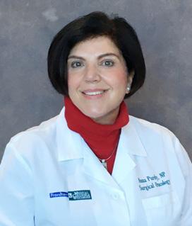
NP Anna Purdy
Selected by Oncology Nursing Certification Corporation
Anna Purdy, NP, MSN, ACONP, CBCN, from the Division of Surgical Oncology has been selected by the Oncology Nursing Certification Corporation to write items for future certification exams.
Partnering for Impact: How Advancing a Healthier Wisconsin is Using its Public Mission & Mandate to Spur Innovation & Discovery at MCW & Beyond
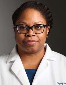
Ugwuji N. Maduekwe, MD, MMSc, MPH Associate Professor, Division of Surgical Oncology; Deputy Director, Advancing a Healthier Wisconsin Endowment
The data is familiar to us all: In national health rankings, Wisconsin ranks 33rd in health outcomes, and our residents face some of the most significant disparities in health outcomes in the nation.
As surgeons, we see the impact not only in data, but in front of us in the clinic and in the operating room. Every day we work to make the best outcomes a reality for our patients by guiding treatment and recovery, and by advancing research toward new methods to diagnose, treat, and prevent disease and injury.
At MCW, this work benefits from a unique resource: The Advancing a Healthier Wisconsin (AHW) Endowment. A statewide philanthropy based in the MCW School of Medicine, AHW has become a model of a catalyst for change.
Since issuing its first funding awards in 2004, AHW has grown into Wisconsin’s leading philanthropy dedicated solely to health, investing more than $311 million in more than 517 projects that are bringing innovation, resources, and positive health impacts to the people of Wisconsin.
AHW resides at MCW and, in a bid to improve the health of the state, provides scaffolding for research, education, and community engagement which has bolstered MCW’s position as a leading medical institution.
History & Purpose: A Public Mission & Mandate
AHW’s roots have always been in health. Following the conversion of Blue Cross Blue Shield United of Wisconsin from a nonprofit organization to a stock insurance corporation, the Insurance Commissioner of the State of Wisconsin required that the proceeds of this conversion be gifted to the state’s two medical schools on behalf of the people of Wisconsin.
The restricted gift came with a 40-page order that guides AHW’s purpose, operations, allocation of funds, and how those funds can be used by projects it invests in. It also shapes AHW’s public reporting and oversight requirements, as well as the requirement that AHW’s funds be available in perpetuity for the purpose of improving the public’s health.
Today, AHW, and its peer program at the UW Madison School of Medicine and Public Health, are counted among
the several hundred “health conversion” foundations in existence across the U.S. The funds entrusted to the MCW School of Medicine to create AHW place it in a unique position – AHW is one of the only health conversion funds to steward public dollars from within a private institution.
This placement gives AHW an extraordinary responsibility to steward this gift in alignment with its mandates, harnessing the capacity afforded by the funds to academic rigor and community partnerships to create an impact on health outcomes statewide.
Seeding Innovation: Improving Health & Advancing Health Equity
Today, AHW is delivering on its public stewardship mission by supporting communities in every region of the state address health challenges and helping to launch biomedical research and health workforce development programs that are building a foundation toward a healthier future.
AHW has invested in Wisconsin communities, deploying more than $92 million into community-academic partnerships, with 71% of those funds impacting counties with the poorest health outcomes.
Within MCW, AHW has become a force for transformational change, investing more than $218 million into MCW-led research and education innovations to date. AHW investments have led to the creation of 11 MCW degree programs, including the curricula at MCW’s regional campuses and the MCW School of Pharmacy. In biomedical and population health research, AHW has become a launching pad for early-stage studies to advance toward extramural funding and AHW’s investigator-led investments have produced a 200% return on investment.
Within the Department of Surgery, more than $8.8 million in AHW funding has supported work across the department with additional millions in related projects supported in neurosurgery, orthopedic surgery, and more. Together, these investments in Department faculty have led to research advancements in areas such as epigenetics, cardiovascular disease, trauma, cancer, while also supporting investments into the research infrastructure such as the launch of the state’s first cancer precision medicine simulation unit.
Partnering with AHW: Creating Change for Wisconsin
Each year, AHW launches an open call for proposals across biomedical and population health research, workforce development, and systems change. Each investment must link to AHW’s public stewardship mission, leading to
a benefit for the people of Wisconsin.
AHW’s role at MCW is propelling promising work and ideas forward for a healthier Wisconsin today, and for generations to come. This is aligned with the mission of the Department of Surgery, and we believe there are areas for improved collaboration.
In my role as Deputy Director of AHW, I would be happy to discuss these potential opportunities for collaboration with any interested members of the Department of Surgery. We invest in people, careers, communities, and science. How can we invest in you?
Stay connected by subscribing to the AHW e-newsletter at ahwendowment.org, and by following AHW on Facebook, Twitter, and LinkedIn. AHW staff are available to meet regularly with any interested investigators, teams, or departments to explore how your ideas can align with AHW funding opportunities.
FOR ADDITIONAL INFORMATION on this topic, visit mcw. edu/surgery or contact Dr. Ugwuji N. Maduekwe at umaduekwe@mcw.edu.
Alumnus E. Christopher Ellison, MD, FACS, MAMSE, named ACS President-Elect
The Medical College of Wisconsin Department of Surgery would like to congratulate E. Christopher Ellison, MD, FACS for becoming the American College of Surgeons President-Elect. Dr. Ellison is an alumnus of the Medical College of Wisconsin and received his MD in 1976, and he was recently awarded the MCW Distinguished Service Award.
Society of Teaching Scholars 2021 Award Recipients and New Members
Lessons Learned from Lombardi
Reproduced from July 2021 F&MCW Better Together – Periop Pulse newsletter
Paul Linsky, MD Assistant Professor, Division of Cardiothoracic Surgery; Associate Program Director, Cardiothoracic Surgery Fellowship
Growing up in Alabama, football was everything. We lived, breathed, ate and worshipped football. So naturally, as a child, all I wanted to do was play football. In all, I spent 11 years of my life playing football, all the way through college. During that time, I was around many coaches. At the top of my list was my high school head coach, Coach Buddy Anderson. Coach Anderson wasn’t just a good coach – he was a great coach. Not only was he inducted into the National Football Hall of Fame, he is the all-time winningest coach in Alabama history on any level (including Bear Bryant and Nick Saban). The first time I met Coach Anderson, he was driving us to the high school for a work out. The first thing he told us was that from now on, we would need to live on “Lombardi time”. Someone asked what that meant and he replied, “If you’re fifteen minutes early, you’re on time. If you’re on time, you’re late.”
Coach Anderson taught me and countless other young men many valuables lessons. Some lessons came directly. Coach Anderson would quote many great coaches all the time, especially Vince Lombardi. One quote from Coach Lombardi that really resonated with me then, and even more now, is:
“The achievements of an organization are the results of the combined efforts of each individual.”
The great coaches, like Coach Lombardi and Coach Anderson, all know that sports are metaphors for life. Everyone has their favorites. However, football is the sport that is most true to life. Why Football? It takes scores of people in varied roles to make a championship team. Also, despite requiring everyone, typically, only a few get credit for what they do.
When I am between cases, I like to listen and watch what our team does. I am constantly impressed with how many hard-working team members we have to do what we do every day. Patients will never know what it takes to get them into, through and out of a successful operation. This has been even truer during the current pandemic. We as the surgeons get the credit; however, we are only one part of the team that would fail without everyone else. Every day, we change the lives of the patients that roll into and out of the OR. No role is inconsequential. We win as a team and lose as a team. We have a great team here and we win a lot more than we lose. We are a team that I am proud to be a part of every day.
As Coach Lombardi pointed out many years ago, our achievements are because of each member of our team. None of us have done it alone and we need each other to keep delivering the excellent care that we give. Each day when you come to work, I want you to remember two key things. First, you and what you do are important, no matter your role. Second, we run on “Lombardi time.”
Thank you all for that you do.
*Dr. Paul Linsky was a Division 1 college offensive lineman — lucky for us, he traded his helmet for a scrub cap!
AAS December Member Spotlight:
Kyle Van Arendonk, MD, PhD
Kyle Van Arendonk, MD, PhD, Assistant Professor in the Division of Pediatric Surgery, Department of Surgery, is featured in the December’s Member Spotlight for the Association for Academic Surgery.
Scan the QR code to read more!
Illustration of the Vince Lombardi Trophy
Integrating Palliative Care into Surgical Education and the Care of Surgical Patients
Erin A. Strong, MD, MBA, MPH Department of Surgery Chief Resident
In the modern era of minimally invasive surgery, there are few remaining indications for a true laparotomy – damage control trauma laparotomy, open ruptured abdominal aortic aneurysm repair, transplant abdominal organ procurement, and cytoreductive surgery (CRS) and hyperthermic intraperitoneal chemotherapy (HIPEC) for peritoneal cancers. I think that’s what drew me to CRS-HIPEC cases, marathon operations of meticulous, methodical resection of all visible disease to allow the intraperitoneal chemotherapy to treat the remaining microscopic disease.
As a junior resident, I enjoyed the small portions of the case assigned to me, starting from stapling the small bowel resections and graduating to the peritonectomy. As a research resident, I spent time evaluating institutional outcomes for CRS-HIPEC, learning to appreciate the risks of a large open abdominal surgery relative to the cancer outcomes. As a senior resident, however, I have come to appreciate some of the nuances of patient selection for CRS-HIPEC, prompting me to think more about those patients with disease too extensive for a curative oncologic resection but who suffered debilitating symptoms directly related to their disease process. From these questions of how surgeons can better help treat these patients, I developed an interest in palliative surgery – surgery to alleviate symptoms from serious illness when a surgical cure is not available – and decided to pursue an additional year of Hospice & Palliative Medicine (HPM) fellowship.
One of the interesting aspects of HPM fellowship interviews has been the opportunity to learn about different models for the integration of palliative care for surgical patients. Few institutions have HPM fellowship-trained surgeons on their faculty, which makes sense as there are still fewer than 100 surgeons with HPM board certification in the US. The integration of palliative care specialists in the care of surgical patients is often related to the presence (or absence) of a surgeon champion who normalizes palliative care consultations at their institution. This can be, however, specific to their division or area of clinical expertise.
On the other end of the spectrum, some institutions have established formal referral patterns around established mandates. The Center for Medicare and Medicaid Services (CMS), for example, has specified that all patients
undergoing evaluation for left ventricular assist devices (LVADs) should have a palliative care specialist as a member of their interdisciplinary team. Some institutions have developed similar care pathways for patients being evaluated for extracorporeal membrane oxygenation (ECMO) or for patients in surgical ICUs that meet designated trigger criteria. In the outpatient setting, clinical relationships can be fostered by the co-location of clinical staff. When the palliative care team has office space in the same area as their surgeon and other sub-specialty colleagues with clinic scheduled for the same days, patients can benefit from the feasibility of seeing multiple teams in one visit.
As a chief resident, I am excited to see the potential growth in integrating palliative care into our surgical resident education and the care of our surgical patients. Dr. Rachel Morris, assistant professor in the Division of Trauma and Acute Care Surgery is pursuing work applying palliative care principles or intentional communication for older adult patients who present with traumatic brain injuries (TBI). Dr. Lee Ann Lau, an MCW medical student alumna and general surgeon, was the first surgeon to complete MCW’s HPM fellowship program and is currently an assistant professor in the Division of Geriatric and Palliative Medicine. After listening to a Behind the Knife podcast episode with Dr. Gretchen Schwarze, a vascular surgeon and medical ethicist at UW Madison, about the “Best Case Worst Case” scenario planning framework she developed for discussing the prospect of major surgery with older adult patients, I have incorporated an exercise in scenario planning with the medical students on my service each month.
Overall, the concept of surgical palliative care is gaining prominence. The “father of palliative care in North America,” Dr. Balfour M. Mount was a Canadian urologic surgeon and his legacy inspired the adoption of a Palliative Care Task Force through the American College of Surgeons in the 1990s, and more recently in 2020, the formation of the Surgical Palliative Care Society. I anticipate a trend in the coming years of more surgical trainees pursuing fellowship training and board certification in HPM, and as a result, more generalized training and education allowing surgeons to develop primary palliative care skills. Ultimately, the goal is to provide optimal patient care for patients with advanced diseases, even when their underlying illnesses cannot be cured. This will be another opportunity for MCW Surgery to lead the way.
FOR ADDITIONAL INFORMATION on this topic, visit mcw. edu/surgery or contact Dr. Strong at estrong@mcw.edu.
Splenic Artery Embolization vs Observation: A Randomized Controlled Trial
Thomas Carver, MD Associate Professor, Division of Trauma & Acute Care Surgery
*Dr. Carter and his team were recently awarded a $50,000 grant from the Department of Surgery’s “We Care Fund” to support this trial.
For over 30 years, the non-operative management (NOM) of splenic injury has been the standard of care in most trauma patients. As surgeons’ comfort level with this technique has increased, we have become more accustomed to observing hemodynamically stable patients even if they have severe splenic injuries (American Association for Surgery of Trauma ([AAST] grades 3-5). At MCW we certainly see our fair share of splenic trauma. In a 7-year period, we had 415 patients with splenic injuries and had a NOM success rate of 5.5%. Even in patients with higher grade injuries, our success rate is 11%, which is comparable to the other major trauma centers in the country. We recently reviewed our splenic NOM outcomes to determine if we could identify patients with a higher rate of NOM failure and one of the most striking findings was the association of a contrast blush on CT scan and failure (odds ratio 4.04 p = 0.006). A “blush” is the presence of IV contrast extravasation appearing as a localized or diffuse high-density region on a CT scan and, in the setting of splenic trauma, is felt to represent active bleeding within the spleen.1 While this association has been identified before in the literature, there is still some controversy about what to do if a splenic blush is identified.
The instinctual response to the presence of a blush is to ask Interventional Radiology to perform a splenic artery embolization (SAE). In fact, this is the tactic employed by most of the trauma centers in the country; however, the data on the use of embolization is based on non-randomized trials and the results are mixed at best. In a recent multi-center observational trial, the splenectomy rate in patients with a blush was 19.5% (our rate is 16.9%).2 While generally considered an indication for SAE3, there are studies that show no improvement in outcomes whether splenic blushes are embolized or observed4,5 and this finding was noted in a Western Trauma Association study as well (6.8% vs 7.6% splenectomy rate, respectively).2,6 Interestingly, there are proponents of SAE who cite a higher NOM success rate7 and even advocate for performing SAE on all high grade injuries (AAST grade 3-5). Those against routine SAE show evidence of no change in NOM success
and they also note the incidence of complications following SAE (access site, splenic infarct, splenic cyst, splenic abscess) can occur in up to 40% of patients.3 While some institutions use SAE for every high-grade splenic injury, regardless of the presence of a blush, at our own institution we have traditionally not used SAE for any splenic injury, blush or no blush. The findings of our recent review, however, created an incentive within the division to reconsider this stance. This combination of opinions and unclear evidence within the literature created a perfect environment for the development of a randomized controlled trial.
As we laid the groundwork for this project, we met with members of the Radiology Department including Drs. Parag Patel (Interventional Radiology), Amer Rasheed (IR resident), Parag Tolat (Body Imaging), and Adam Zorn (Body Imaging) as this project is completely dependent on our radiology colleagues helping us identify these injuries and perform SAE on our patients at any time of the night. There was considerable interest in this study within Radiology and, in a truly collaborative fashion, we developed a sound research protocol that we feel is both pragmatic and feasible.
Our study will randomize patients who meet criteria for NOM of their splenic injury to SAE or our standard of care (observation). The goal of this study is to determine if SAE improves NOM success but there are several other aims including: (1) does the SAE technique matter, (2) does SAE impact length of stay, (3) what happens to patients who have a blush on CT scan but not at the time of their angiography, (4) what is the rate of complications following either observation or SAE, and (5) is there an effect on mortality. As a top-tier trauma center, we are constantly striving to perform high quality research studies that contribute to the scientific literature and, ultimately, influence trauma care across the country. With the support of the MCW Department of Surgery and the We Care Fund for Medical Innovation and Research, we feel that this study has the potential to end the debate on the role of SAE in severe splenic injury. If our study shows that SAE is beneficial in splenic NOM, then centers such as ours should be convinced to employ this technique in the care of their patients. If SAE fails to improve outcomes, then trauma centers across the country will have to rethink their use of embolization. Either way, this study will have a meaningful impact on trauma patients and on the field of trauma as a whole.
FOR ADDITIONAL INFORMATION on this topic, visit mcw. edu/surgery or contact Dr. Thomas Carver at tcarver@ mcw.edu.
REFERENCES
1. Alarhayem AQ, Myers JG, Dent D et al. “Blush at first sight”: significance of computed tomographic and angiographic discrepancy in patients with blunt abdominal trauma. Am J Surg 2015;210:1104-10; discussion 1110-1.
2. Zarzaur BL, Dunn JA, Leininger B et al. Natural history of splenic vascular abnormalities after blunt injury: A Western Trauma Association multicenter trial. J Trauma Acute Care Surg 2017;83:999-1005.
3. Stassen NA, Bhullar I, Cheng JD et al. Selective nonoperative management of blunt splenic injury: an Eastern Association for the Surgery of Trauma practice management guideline. J Trauma Acute Care Surg 2012;73:S294-300.
4. Post R, Engel D, Pham J, Barrios C. Computed Tomography Blush and Splenic Injury: Does It Always Require Angioembolization? Am Surg 2013;73:1089-1092.
5. Burlew CC, Kornblith LZ, Moore EE, Johnson JL, Biffl WL. Blunt trauma induced splenic blushes are not created equal. World J Emerg Surg 2012;7:8.
6. Zarzaur BL, Kozar R, Myers JG et al. The splenic injury outcomes trial: An American Association for the Surgery of Trauma multi-institutional study. J Trauma Acute Care Surg 2015;79:335-42.
7. Haan JM, Bochicchio GV, Kramer N, Scalea TM. Nonoperative management of blunt splenic injury: a 5-year experience. J Trauma 2005;58:492-8.
Thank
you to the Fund Committee!
To support the We Care Fund, please visit www.mcwsupport.mcw.edu/wecarefund or scan the QR code below.
When Common Symptoms Lead Us to an Uncommon Clinical Finding: A Case Report of a Rare Diagnosis in Children
Abstract
Elyan Ruiz-Solano, MD Clinical Instructor, Division of Cardiothoracic Surgery
Michael E. Mitchell, MD Professor and Chief, Division of Congenital Heart Surgery
We present a case of a rare diagnosis of respiratory symptoms in a young child. A 2-year-old infant brought to the Emergency Department (ED) of his local hospital with cough and shortness of breath for a month. Amidst the current health pandemic, the patient was initially screened for COVID-19 infection, which was negative.
Case Description
A 2-year-old male, previously healthy, was taken to the Emergency Department (ED) with difficulty breathing and cough for a month, that was worse when laying on his side. The patient was assessed in the context of a potential COVID-19 infection. The result was negative and he was discharged home. He continued to have symptoms, now associated with a lack of appetite and lethargy. He returned to the ED where a chest X-ray (CXR) was obtained, showing a widened mediastinal space with nodular densities reminiscent of teeth or corn kernels (Fig.1). Due to the radiological findings, the patient had a contrast computed tomography (CT) of the chest.
The CT images confirmed the presence of a large mixed density mass within the anterior mediastinum of approximately 10.2 x 7.7 x 4.99 cm, demonstrating coarse calcium attenuation, as well as fat, fluid and soft tissue density, strongly suggestive of teratoma. The lesion was noted to compress and displace the superior vena cava (SVC), the
Kyle J. Van Arendonk, MD, PhD Assistant Professor, Division of Pediatric Surgery
Ali Y. Mejaddam, MD General Surgery Specialist
Fig. 2. Contrast chest CT scan showing a large mediastinal mass densely adherent to the posterior surface of the sternum (A). The lesion also occupied anterior portion of the right pleural space (B) and demonstrated displacement of the ascending aorta and MPA as well as mild compression of the RA and SVC (C and D).
ascending aorta and the pulmonary trunk. Additionally, the mass exerted mild mass effect on the superior aspect of the right atrium (RA) and superiorly displaced the thymus gland (Fig.2). Subsequently, the patient was admitted and cardiothoracic (CT) surgery was consulted.
On examination, the patient was interactive and well appearing with no edema of the head or neck, with subtle elevation of the right chest, associated with mild shortness of breath and noisy breathing without distress. There were clear breath sounds through the entire posterior chest and normal cardiac exam. However, there was transient bradycardia when laying in a right lateral decubitus position without hemodynamic compromise. Also, the anterior chest breath sounds were difficult to assess.
Echocardiogram showed the mediastinal mass adjacent to the ascending aorta and the right ventricular outflow tract (RVOT)/main pulmonary artery (MPA) without evidence of narrowing or turbulent flow. There was no evidence of obstruction in the innominate vein or SVC, but there was trivial turbulent flow into the proximal right pulmonary artery (RPA) with a peak gradient of 10mmHg. The biventricular function was preserved and there was a small pericardial effusion (Fig.3).
Blood tests showed microcytic-hypochromic anemia and thrombocytosis. Human chorionic gonadotropin
Fig. 1. Antero-posterior (A) and lateral (B) preoperative CXR. Note the teeth-like radio-opacities (red arrow).
Fig. 3. Transthoracic echocardiogram (TTE) demonstrating mild dynamic compression of RV in the parasternal long axis view (A) as well as mild RVOT compression and small pericardial effusion (solid red arrow) in the parasternal short axis view (B). Continuous wave doppler of the RPA in the parasternal short axis showing turbulent flow without significant gradient across. LV: left ventricle. RV: right ventricle. LVOT: left ventricular outflow tract. LA: left atrium. Orange circle: mediastinal mass. RVOT: right ventricular outflow tract. PV: pulmonary valve. MPA: main pulmonary artery.
(HCG) was undetected; beta subunit of HCG, alpha fetoprotein (AFP) and lactate dehydrogenase (LDH) were normal; however, his consumptive coagulopathy panel was abnormal, showing high fibrinogen, D-dimer, PT, PTT and TT values for the reference range.
The patient underwent surgical resection through a median sternotomy. The mass itself was densely adherent to the sternum, the pleura and the pericardium, surrounding the SVC and in close proximity to both phrenic nerves. This was circumferentially dissected from the nearby structures, successfully preserving both phrenic nerves while taking the mediastinal pleura attached to it. The thymus gland was excised with the mass, preserving the upper pole of the right lobe. The specimen was a large, heterogeneous mass with cystic and calcification components; measuring, approximately, 11 x 7 x 6 cm (Fig.4). Patient had an uneventful postoperative course and was discharged home 48 hours after surgery.
Discussion
Teratomas are extragonadal germ cell tumors (GCTs) that typically arise in midline locations. Their specific sites vary with age; being the anterior mediastinum, retroperi-
toneum and pineal and suprasellar regions most common in adults; while sacrococcygeal and intracranial GCT most frequently seen in infants and young children.1
Mediastinal teratomas (MT) are more common in adults than children with a 5:1 ratio2 and, must be differentiated from other tumors that can occur or invade the mediastinum, such as thymoma, lymphoma malignant tumors or mediastinal cysts.3
The diagnosis of MT is suspected with a wide mediastinum on CXR. Calcifications are described in 26% and the presence of well-formed teeth or bone are highly suspicious of the diagnosis.4 The use of CT or magnetic resonance imaging (MRI) is helpful in localizing the lesion and establishing the spatial relationship with the surrounded structures, as well as characterize the different density components suggestive of fat, sebaceous material or cystic elements.5,6 The treatment of mediastinal teratomas is surgical excision and this is curative in the vast majority of the cases.7,8 The surgical approach depends upon localization, but it can be done through median sternotomy, posterolateral thoracotomy or video assisted thoracic surgery (VATS).4
Our case reminds us how the current COVID-19 pandemic has shaped the screening process in the ED. In children presenting with respiratory symptoms, a plain CXR should be considered. This can aid in the diagnosis of other serious processes amenable to immediate treatment. Clinical care should not be delayed on patients with abnormal mediastinal silhouette. These patients should be referred to a specialized center in a timely manner to receive appropriate medical assistance.
FOR ADDITIONAL INFORMATION on this topic, visit mcw. edu/surgery or contact Dr. Ruiz-Solano at eruizsolano@ mcw.edu.
REFERENCES ON PAGE 19
Fig. 4. Surgical specimen. Note the lesion occupying all the anterior mediastinum (A). Anterior and posterior characteristics are shown in pictures B and C.
Suicide Prevention Initiatives in the Trauma Center Setting
Andrew T. Schramm, PhD
Assistant Professor, Division of Trauma & Acute Care Surgery
As a leading cause of death in the United States, suicide is a substantial yet preventable public health issue that warrants our attention.1 The opportunity to prevent suicide extends to every member of the care team--not just mental health professionals. Although trauma centers are uniquely positioned to play a pivotal role in suicide prevention, this is an understudied topic, with limited development of evidence-based prevention initiatives. Two types of trauma patients may especially be at risk for suicide: (1) patients treated for self-inflicted injuries sustained in a suicide attempt and (2) patients whose mechanism of injury is unrelated to suicidality (e.g., motor-vehicle collision) who now face considerable stressors and changes in functioning that may foster hopelessness. Regardless of the reason trauma patients are under our care, we have an obligation to assess their risk for suicide and intervene. In collaboration with the Suicide Prevention Division of MCW’s Comprehensive Injury Center, we are laying the groundwork to develop an evidence-based suicide prevention framework for our trauma center. Several projects are underway, and more are planned.
In the Preventing Suicide After Traumatic Injury (P-SAT) project, funded by the MCW Research Affairs Committee, we are identifying risk factors unique to survivors of a suicide attempt who are treated at our trauma center and go on to die from a future attempt. This will include conducting psychological autopsies--a protocol established by the American Association of Suicidology2 — with families of trauma patients who have died from suicide. This will make possible the development of a risk profile unique to this population--a critical step toward developing suicide prevention initiatives targeting these risk factors.
Our second project, Reducing Access to Lethal Means (REALM) Study is funded by the Comprehensive Injury Center (CIC). The purpose of the study is to train psychology and psychiatry providers in means reduction counseling, a best practice for preventing firearm suicides, and evaluate patient behavior and attitudes following this intervention. Since half of all Wisconsin residents who die by suicide died from a self-inflicted firearm injury, preventing firearm suicides is a statewide priority.3 This project, in collaboration with co-PI Dr. Julie Owen in the Department of Emergency Medicine and Department of Psychiatry and Behavioral Medicine, includes the provision of gun locks and instruction on safe storage of firearms. Finally, in collaboration with the Eastern Association for the Surgery of Trauma (EAST) and the American Foundation for Suicide
Jacey M. Kant Clinical Research Assistant
Sara A. Kohlbeck, MPH
Director, Division of Suicide Prevention, Comprehensive Injury Center
Prevention (AFSP), we are leading the development of a best practice guideline for suicide prevention that will be disseminated to trauma centers nationally.4
Thanks to the generous support of these funders and Department of Surgery and CIC leadership, MCW is positioned to be a national leader in the development and implementation of suicide prevention initiatives in trauma centers. These efforts are closely aligned with trauma centers’ notable focus on injury prevention and stand to save lives from self-inflicted injuries.5
FOR ADDITIONAL INFORMATION on this topic, visit mcw.edu/surgery or contact Dr. Andrew Schramm at aschramm@mcw.edu.
REFERENCES
1. World Health Organization. Public Health Action for the Prevention of Suicide: A Framework. 2012.
2. Conner KR, Beautrais AL, Brent DA, Conwell Y, Phillips MR, Schneider B. The next generation of psychological autopsy studies: Part I. Interview content. *Suicide and LifeThreatening Behavior.* 2011;41(6):594-613.
3. Prevent Suicide Wisconsin. Suicide in Wisconsin: Impact and Response. September 2020. https://www.dhs.wisconsin. gov/publications/p02657.pdf
4. Eastern Association for the Surgery Of Trauma. Practice Guidelines Under Development: *Best Practices for Suicide Screening and Prevention Among Adults with Traumatic Injury*. https://bit.ly/3mTlIHM
5. American College of Surgeons Committee on Trauma. *Resources for the Optimal care of the Injured Patient.* Chicago, IL. 2014.
Mental Health Resources
MCWAH Helpline 414-955-4798
Resident Crisis Line Weekdays 8a-5p: 414-314-5562; After hours 414-805-6700
Employee Assistance Program www.workhealthlife.com
National Suicide Prevention Lifeline (available 24/7) 800-273-TALK (8255) or text CONNECT to 741741 Milwaukee County Adult Crisis Line 414-257-7222
Children’s Mobile Crisis Team 414-257-7621
Thoughts at the Scrub Sink: “Chef”
Reproduced from December 2021 F&MCW Better Together –Periop Pulse newsletter
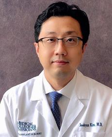
Joohyun Kim, MD, PhD
Associate Professor, Division of Transplant Surgery
During my trip to New Orleans in 2019, I visited the National World War II Museum. In a photo in the section of Normandy landings, the Theater Commander Dwight D. Eisenhower is shown talking to a paratrooper who is standing at attention, a few hours before the invasion of France. The scene reminded me of a story I heard from my grandfather when I was a little boy. My grandfather became a chef at the U.S. Embassy in South Korea before the Korean War. One day, he prepared dinner for the visit of the President of the United States. After dinner, President Eisenhower sought him out to express his gratitude. I can envision the sense of stress and pride my grandfather felt, similar to that of the paratrooper in that photo from D-Day. My grandfather died a long time before I became a surgeon, and I can only imagine his days as a chef. However, when I watched a documentary about a chef’s life, I saw similarities between the lives of chefs and surgeons.
The path to becoming a chef involves many steps, just like a surgeon’s path. Do you like cooking? Do you want to open a legendary restaurant? Do you simply need a well-paying job? Your motivation doesn’t matter. Learning to wash dishes at the sink is how you start. When you have the chance, you watch how your master chef performs. You are mesmerized at first, but then you think you can do it. You follow the recipe, but your dishes turn out inedible. Confident when you started, now you
feel like you are not as talented as you initially thought. You need to learn the basics first. Mise En Place instills discipline in kitchens – everything you need should be in place before you start. It takes time to get it. It takes too long to learn anything. Some quit, some stay, and some find another kitchen. Time passes, and you end up making the same style of dishes again and again. Perfection is the goal for every dish you serve, but you know it is never perfect. How can you make your dishes perfect? You find out who has the best veggies and meats in the market. You were taught and believe that the best quality ingredient is the key to success. You ask yourself, if your dish is only perfect on a lucky day, whether you can truly call it perfect. One night, you watch a story about helping hungry children around the world. You realize that your skills are useless for feeding them. You now question what you once cherished. One day, you hear about a robotic chef, and you are told that machine learning is the future. You think your master chef was lucky to have retired before this began to happen. You still want to create a great menu of your own before everything is taken over by artificial intelligence. Some believe that your job is an art; others say it is a business. Regardless, you are there to serve a stranger in front of you who will taste your minor masterpiece. As such, in my humble opinion, there are parallels between chefs and surgeons. My grandfather may or may not agree.
MCW’s Outstanding Medical Student Teachers
MCW’s Outstanding Medical Student Teachers for 2020-2021: M3 Surgery Clerkship
FACULTY
G. Hossein Almassi, MD
Professor, Division of Cardiothoracic Surgery
Paul Linsky, MD
Assistant Professor, Division of Cardiothoracic Surgery
Thomas Sato, MD Professor, Division of Pediatric Surgery
RESIDENTS
Colleen Flanagan, MD
Katherine Flynn-O’Brien, MD Kyla Fredrickson, MD
MCW’s Outstanding Medical Student Teachers for 2020-2021: M4 Surgical Rotations
FACULTY
Tom Carver, MD
Associate Professor, Division of Trauma & Acute Care Surgery
Christopher Davis, MD, MPH
Assistant Professor, Division of Trauma & Acute Care Surgery
Anu Elegbede, MD
Assistant Professor, Division of Trauma & Acute Care Surgery
Rana Higgins, MD
Associate Professor, Division of MinimallyInvasive Gastrointesintal Surgery
Dean Klinger, MD Professor, Division of Community Surgery
Amanda Kong, MD Professor, Division of Surgical Oncology; Chief, Section of Breast Surgery
Kate Lak, MD
Assistant Professor, Division of MinimallyInvasive Gastrointesintal Surgery
Travis Webb, MD Professor, Division of Trauma & Acute Care Surgery
RESIDENTS
Eric Anderson, MD
Paul Dyrud, MD
Christina Georgeades, MD
John Marquart, MD
Grant Renewal to Continue Work to Improve Vascular Function in People with Sickle Cell Disease
Kirkwood A. Pritchard, Jr., PhD Professor, Division of Congenital Heart Surgery
Sickle Cell Disease Impairs Vascular Function by Inducing a Destructive Cycle.
The destructive cycle starts with myeloperoxidase (MPO) released from activated neutrophils and other myeloid cells (Figure 1). MPO generates toxic oxidants that injure and kill the cells that makeup blood vessels. Dead and dying cells release high mobility group box-1 (HMGB1) from the cell’s nucleus which plays an essential role in maintaining DNA structure and regulating gene expression. However, when cells die, they passively release HMGB1 outside the cell, turning it into a potent inflammatory molecule that increases inflammation and recruitment of neutrophils to the vessel wall. After the neutrophils arrive, they bind to the endothelium lining the vessel wall and crawl into the vessel wall, where they become activated and cause even greater inflammation and tissue injury. Although all cells make HMGB1, even blood vessel
cells, when released from dead and dying cells, HMGB1 impairs the blood vessel’s ability to vasodilate, mediate blood flow, or repel activated neutrophils, damaged sickled RBC and activated platelets from the vessel wall. In this way, the MPO-HMGB1 destructive cycle increases vascular adhesion of neutrophils, RBC, and platelets. Our studies suggest the MPO-HMGB1 destructive cycle is a novel inflammatory pathway that sickle cell disease uses to injure the vessel wall and increase the risk of acute crises.
Current Therapies for Sickle Cell Disease
Hydroxyurea, L-glutamine, oxbryta, and crizanlizumabtmca are currently the only therapies approved by the FDA for treating SCD. Hydroxyurea and L-glutamine reduce sickle crisis and vasocongestion. Hydroxyurea is a chemotherapeutic agent that reduces leukocyte counts and leukocyte-dependent inflammation. It also increases fetal Hb and decreases Hb polymerization. Finally, hydroxyurea even releases nitric oxide. Oral L-glutamine provides the energy that RBC use to metabolize oxidized lipids and reduce glutathione disulfide, thereby minimizing sickling and vasocongestion. Oxbryta inhibits hemoglobin S polymerization. Crizanlizumab-tmca binds to P-selectin on activated endothelial cells that line the blood vessel inner wall and circulating platelets to prevent adhesion of sickled red cells to the vessel wall. While all of these therapies help reduce vascular inflammation and RBC injury, sickling, and sticking to the vessel wall, none of these treatments target the mechanisms by which sickle cell disease induces vasculopathy as defined above in the destructive cycle (Figure 1).
My laboratory designed and developed N-acetyllysyltyrosylcysteine amide (KYC), a first-in-class, multimodal, systems pharmacology agent for treating sickle cell disease. Unlike the FDAapproved drugs listed above, KYC inhibits the mechanisms
Figure 1. Vasculopathy in Sickle Cell Disease is caused by a Destructive Cycle.
in the destructive cycle. KYC inhibits MPO’s ability to generate toxic oxidants that kill cells. Interestingly, MPO oxidizes KYC into a new novel anti-inflammatory agent that inactivates HMGB1 and activates other proteins in the cell that increases antioxidant enzyme expression to protect the vessel wall from oxidative damage and cell death. KYC effectively improves vascular function, reducing RBC vasocongestion in the lungs and brains of sickle cell mice, thereby decreasing the risk of acute crisis and stroke. Bench to Bedside
Besides being awarded a new grant to continue my NIH research program in sickle cell disease at the Medical College of Wisconsin, MCW, myself and several past lab members were awarded a patent for KYC. Because KYC is a system pharmacology agent, it is highly effective for treating sickle cell disease, multiple sclerosis, stroke, traumatic brain injury, and bronchopulmonary dysplasia. With such potential for treating many dysregulated innate immune diseases, I started a company five years ago called ReNeuroGen LLC to develop KYC. Last year, ReNeuroGen LLC completed a phase I SBIR proposal to obtain proofof-concept data showing that KYC reduces vasocongestion in the lungs and brains of sickle mice. Last September, ReNeuroGen LLC submitted a preclinical phase II SBIR application to develop KYC further for treating people with sickle cell disease. This time is exciting for me, members of my lab, and my collaborators because we can continue our NIH-funded academic research program in sickle cell disease and watch the company move KYC from bench to bedside in real-time.
REFERENCES FROM PAGE 15
When Common Symptoms Lead Us to an Uncommon Cinical Finding: A Case Report of a Rare Diagnosis in Children
1. Wick MR, Perlman EJ, Orazi A, Muller-Hermlink HK. Germ cell tumours of the mediastinum. In: Tumours of the lung, pleura, thymus and heart; Travis WD, Brambilla E, Muller-Hermelink HK, Harris CC (Eds).
2. Agarwal R, Srinivas R, Saxena AK. Trichoptysis due to an intrapulmonary teratoma. Respir Care 2007; 52: 1779-81
3. Bohle A, Studer UE, Sonntag RW, Scheidegger JR. Primary or secondary extragonadal germ cell tumors? J Urol 1986; 135: 939.
4. Extragonadal cell tumors involving the mediastinum and retroperitoneum. Kantoff PW. Up to date. Sept 2021.
6. Dulmet EM, Macchiarini P, Suc B, Verley JM. Germ cell tumors of the mediastinum. A 30-year experience. Cancer 1993; 72: 1894
7. Takeda S, Miyoshi S, Ohta M, et al. Primary germ cell tumors in the mediastinum: a 50year experience at a single Japanese institution. Cancer 2003; 97: 367.
8. Moran CA, Suster S, Przygodzki RM, Koss MN. Primary germ cell tumors of the mediastinum: Mediastinal seminomas – a clinicopathologic and immunohistochemical study of 120 cases. Cancer 1997; 80: 691.
FOR ADDITIONAL INFORMATION on this topic, visit mcw. edu/surgery or contact Dr. Kirkwood A. Pritchard Jr., at kpritch@mcw.edu.
To refer a patient or request a transfer/consultation, please use the references below:
Stefano Schena, MD, PhD, will join the Department of Surgery faculty in February as Associate Professor of Surgery from Johns Hopkins University where he was Assistant Professor of Surgery in the Division of Cardiac Surgery. Prior to joining Johns Hopkins, Dr. Schena served as Assistant Professor of Surgery and Cardiothoracic Surgery at Washington University. He received his medical degree from the University of Bari Medical School in Italy, where he also obtained a postgraduate doctoral degree in transplant biotechnologies. Dr. Schena completed a cardiovascular surgery
residency at the University of Bari and a general surgery residency at the University of Illinois at Chicago. He completed his fellowship in cardiothoracic surgery at Washington University/Barnes-Jewish Hospital in St. Louis. Dr. Schena will primarily provide clinical care of patients on the Cardiothoracic Surgery service at the Zablocki VA Medical Center while also providing clinical care at Froedtert Hospital.
Dr. Thomas Sato to Retire June 30. Thank you for an amazing career at MCW!
Dr. Thomas Sato has announced his plan to retire, effective June 30, 2022. Dr. Sato’s contributions as an academic surgeon and leader of Children’s Specialty Group are immeasurable. Dr. Sato has mentored countless fellows, residents, and medical students while teaching all of us how to overcome adversity with collegiality, confidence and determination. Dr. Sato’s legacy will contribute to the great tradition of surgery at CW and throughout our department.
Meena Bedi, MD
Radiation Oncology
Michael B. Dwinell, PhD
Microbiology and Immunology
Beth Erickson Wittmann, MD
Radiation Oncology
Juan Felix, MD Pathology
James W. Findling, MD
Medicine, Endocrinology, Metabolism and Clinical Nutrition
Jennifer Geurts, MS, CGC
Genomic Sciences and Precision Medicine Center
Mary Beth Graham, MD Medicine, Infectious Diseases
Jaime S. Green, MD Medicine, Infectious Diseases
James B. Gosset, MD Medicine, Cardiovascular Medicine
Michael O. Griffin Jr., MD, PhD
Radiology, Diagnostic Radiology
William A. Hall, MD
Radiation Oncology
Robert A. Hieb, MD, RVT
Radiology, Vascular and Interventional Radiology
SECONDARY FACULTY BY SPECIALTY
Eric J. Hohenwalter, MD
Radiology, Vascular and Interventional Radiology
Bryon D. Johnson, PhD
Microbiology and Immunology
Mandana Kamgar, MD, PhD Medicine, Hematology and Oncology
John A. LoGiudice, MD
Plastic Surgery
Veronica Loy, DO Medicine, Gastroenterology
Peter J. Mason, MD, MPH, RPVI Medicine, Cardiovascular Medicine
Stacy D. O’Connor, MD, MPH, MMSc
Radiology, Diagnostic Radiology
Jong-In Park, PhD
Biochemistry
Parag J. Patel, MD, MS
Radiology, Vascular and Interventional Radiology
Hershel Raff, PhD
Medicine, Endocrinology, Metabolism and Clinical Nutrition
Honey Reddi, PhD
Pathology/Genomic Sciences and Precision Medicine Center
William S. Rilling, MD
Radiology, Vascular and Interventional Radiology; Vice Chair, Clinical Operations
Evan K. Robinson, DO
Emergency Medicine
Ehab R. Saad, MD
Medicine, Nephrology
Joyce Sanchez, MD Medicine, Infectious Disease
Jennifer J. Schiller, PhD, D(AHBI) Versiti Blood Center of Wisconsin
Pippa M. Simpson, PhD
Pediatrics, Quantitative Health Sciences
Parag P. Tolat, MD
Radiology, Diagnostic Radiology
J. Njeri Wainaina, MD
Medicine, General Internal Medicine
Sarah B. White, MD, MS
Radiology, Vascular and Interventional Radiology
Stefano Schena, MD, PhD
THE MEDICAL COLLEGE OF WISCONSIN DEPARTMENT OF SURGERY
FACULTY BY SPECIALTY
Bariatric & Minimally Invasive Gastrointestinal Surgery
Matthew I. Goldblatt, MD
Jon C. Gould, MD, MBA
Rana M. Higgins, MD
Andrew S. Kastenmeier, MD
Tammy L. Kindel, MD, PhD
Kathleen Lak, MD
Philip N. Redlich, MD, PhD
Cardiac Surgery
G. Hossein Almassi, MD
Nilto C. De Oliveira, MD
Lucian A. Durham III, MD, PhD
Viktor Hraska, MD, PhD
David L. Joyce, MD, MBA
Lyle D. Joyce, MD, PhD
Takushi Kohmoto, MD, PhD, MBA*
R. Eric Lilly, MD*
James E. Mace MD
Michael E. Mitchell, MD*
Paul J. Pearson, MD, PhD
Elyan Ruiz Solano MD
Stefano Schena, MD, PhD (2/22)
H. Adam Ubert, MD
Ronald K. Woods, MD, PhD*
Colorectal Surgery
Jed F. Calata, MD
Kirk A. Ludwig, MD
Mary F. Otterson, MD, MS
Carrie Y. Peterson, MD, MS*
Timothy J. Ridolfi, MD, MS
Community Surgery
Robert J. Brodish, MD
T. Clark Gamblin, MD, MS, MBA
Dean E. Klinger, MD
Kaizad Machhi, MD
Kevin V. Moss, MD
Eric A. Soneson, MD
Mark A. Timm, MD
Pediatric General & Thoracic Surgery
John J. Aiken, MD*
Casey M. Calkins, MD*
Brian T. Craig, MD
John C. Densmore, MD*
Katherine T. Flynn-O’Brien, MD, MPH
Pediatric General & Thoracic Surgery, continued
David M. Gourlay, MD*
Tammy L. Kindel, MD, PhD
Dave R. Lal, MD, MPH*
Keith T. Oldham, MD*
Jose H. Salazar Osuna, MD, PhD
Thomas T. Sato, MD*
Jack G. Schneider, MD*
Kyle Van Arendonk, MD, PhD
Amy J. Wagner, MD*
Research Faculty
Mohammed Aldakkak, MD
John E. Baker, PhD
Young-In Chi, PhD
Mats Hidestrand, PhD
Christian J. Kastrup, PhD
Gwen Lomberk, PhD
Angela J. Mathison, PhD
Aoy T. Mitchell, PhD
Mochamad Muska
Nataliansyah, MD, MPH, PhD
Kirkwood Pritchard, Jr., PhD
Raul A. Urrutia, MD
Surgical Oncology–Breast Surgery
Adrienne N. Cobb, MD (9/22)
Chandler S. Cortina, MD*
Amanda L. Kong, MD, MS*
Caitlin R. Patten, MD*
Tina W.F. Yen, MD, MS
Surgical Oncology–Endocrine Surgery
Sophie Dream, MD*
Douglas B. Evans, MD*
Jennifer L. Rabaglia, MD, MSc
Tracy S. Wang, MD, MPH*
Tina W.F. Yen, MD, MS
Surgical Oncology–Hepatobiliary & Pancreas Surgery
Kathleen K. Christians, MD
Callisia N. Clarke, MD, MS
Douglas B. Evans, MD*
T. Clark Gamblin, MD, MS, MBA
Karen E. Kersting, PhD, LCP
Susan Tsai, MD, MHS
Surgical Oncology–Regional Therapies
Callisia N. Clarke, MD, MS
T. Clark Gamblin, MD, MS, MBA
Anai N. Kothari, MD, MS
Ugwuji N. Maduekwe, MD, MMSc
Thoracic Surgery
Mario G. Gasparri, MD*
David W. Johnstone, MD*
Paul L. Linsky, MD*
Transplant Surgery
Francisco A. Durazo, MD
Calvin M. Eriksen, MD
Johnny C. Hong, MD
Christopher P. Johnson, MD
Joohyun Kim, MD, PhD
Priyal Patel, MD
Terra R. Pearson, MD
Jenessa S. Price, PhD
Allan M. Roza, MD
Motaz A. Selim, MBBCh, MSC, MD
Stephanie Zanowski, PhD
Michael A. Zimmerman, MD
Trauma/ACS
Marshall A. Beckman, MD, MA*
Nathan A. Carlson, MD
Thomas Carver, MD
Christopher S. Davis, MD, MPH
Marc A. de Moya, MD
Terri A. deRoon-Cassini, PhD
Christopher Dodgion, MD, MSPH, MBA
Anuoluwapo F. Elegbede, MsC, MD
Timothy J. Geier, PhD, LP
Daniel N. Holena, MD, MCSE
Katie Iverson, MD, MPH
Christina Megal, DNP, APNP, FNP-C, CWON-AP, CFCN
David J. Milia, MD*
Rachel S. Morris, MD
Patrick B. Murphy, MD, MSc, MPH
Todd A. Neideen, MD
Jacob R. Peschman, MD
Andrew T. Schramm, PhD
Libby Schroeder, MD
Lewis B. Somberg, MD, MSS*
Trauma/ACS , continued
Colleen Trevino, MSN, FNP, PhD
Travis P. Webb, MD, MHPE
Vascular & Endovascular Surgery
Shahriar Alizadegan, MD*
Kellie R. Brown, MD*
Joseph P. Hart, MD, RVT, RVPI
Nathan W. Kugler, MD*
Brian D. Lewis, MD
Mona S. Li, MD*
Michael J. Malinowski, MD*
Neel Mansukhani, MD
Peter J. Rossi, MD*
Abby Rothstein, MD*
Affiliated Institution
Program Directors
Gary T. Sweet Jr., MD
Aspirus Wausau Hospital
James Rydlewicz, MD
Aurora–Grafton
Nicholas Meyer, MD
Columbia St. Mary’s Hospital
Joseph C. Battista, MD
St. Joseph’s Hospital
John G. Touzious, MD
Waukesha Memorial Hospital
Chief Advance Practice
Providers
Stephen W. Robischon, PA-C
Ambulatory Chief
Cynthia L. Schulzetenberg, PA-C
Inpatient Chief
Chief Surgical Residents (2021-2022)
Christina Bence, MD
Kelly Boyle, MD
Bonnie Chow, MD
Emma Gibson, MD
Andrew Goelz, MD
Katherine Hu, MD
Zoe Lake, MD
Matthew Madion Jr., MD
Erin Strong, MD MPH
Administrative Chief
Surgical Residents (2021-2022)
Christina Bence, MD
Matthew Madion Jr., MD
Erin Strong, MD, MBA, MPH
Department of Surgery
8701 Watertown Plank Road
Milwaukee, WI 53226
Department of Surgery
Dedicated to Clinical Care, Research, and Education
• Cardiothoracic Surgery
• Colorectal Surgery
• Community Surgery
• Congenital Heart Surgery
• Surgical Education
• Minimally Invasive & Gastrointestinal Surgery
• Pediatric Surgery
• Research
• Surgical Oncology
• Transplant Surgery
• Trauma/ACS
• Vascular & Endovascular Surgery
Leading the Way is published three times yearly by The Medical College of Wisconsin
Leading the Way is written for physicians for medical education purposes only. It does not provide a complete overview of the topics covered and should not replace the independent judgment of a physician about the appropriateness or risks of a procedure for a given patient.