NANO- AND MICRO-SCALE 3D BIOPRINTING:
USING A LIGHT-BASED TECHNOLOGY FOR REGENERATIVE MEDICINE By Liliana Caldero As Dr. Chen begins his presentation in the standing room only session, eager attendees of ICALEO 2018 sit forward in their seats, taking notes as he explains how light is innovating our ability to print on the nano- and micro-scale. From 3D printed robotic microfish to 3D printed liver-like gel-nanoparticles, Dr. Shaochen Chen has been involved in headline-making research projects for many years. As a professor and vice chair of the Department of NanoEngineering and affiliated professor of the Department of Bioengineering at the University of California, San Diego, Chen leads a team of researchers in developing nano- and micro-scale 3D bioprinting techniques. When he looks out at the faces of the audience, he’s reminded of himself early in his career. While working as a faculty member at the University of Texas at Austin in 2001, he took an interest in 3D printing applications. Specifically, he wondered how the highly customizable nature of 3D printing could be utilized to its greatest potential. He concluded that there is nothing quite as custom-made as a human, and so followed his expedition into nano/ micro-manufacturing for regenerative medicine. Catching up with him after his presentation, I sat down with him to learn about part of his unique line of work - using light to print functional tissues.
So, What is 3D Bioprinting? 3D bioprinting refers to the use of three-dimensional printing methods that produce 3D tissues that are biomimetic, meaning they closely mimic the physiology and function of real organs. In essence, a scientist can print living tissue that looks just like a human liver or heart when placed under a microscope. Such tissues are printed using bio-inks derived from hydrogels, cells and growth factors, often utilizing adult human induced Pluripotent Stem Cells (iPSC). Thanks to the Nobel winning research of Shinya Yamanaka and Sir John Gurdon, skin cells can be taken from an adult human and ‘reprogrammed’ into iPSCs, which can then be used to make other types of cells that are harder to come by, such as liver cells or heart cells.
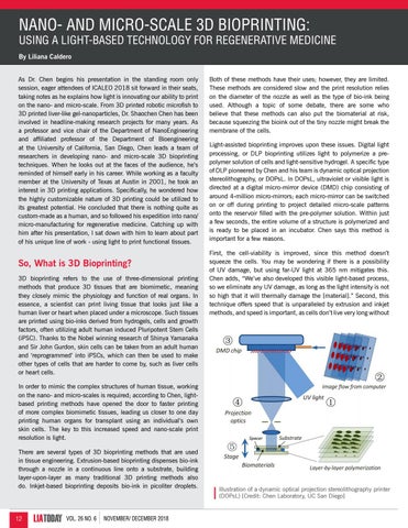
Both of these methods have their uses; however, they are limited. These methods are considered slow and the print resolution relies on the diameter of the nozzle as well as the type of bio-ink being used. Although a topic of some debate, there are some who believe that these methods can also put the biomaterial at risk, because squeezing the bioink out of the tiny nozzle might break the membrane of the cells. Light-assisted bioprinting improves upon these issues. Digital light processing, or DLP bioprinting utilizes light to polymerize a prepolymer solution of cells and light-sensitive hydrogel. A specific type of DLP pioneered by Chen and his team is dynamic optical projection stereolithography, or DOPsL. In DOPsL, ultraviolet or visible light is directed at a digital micro-mirror device (DMD) chip consisting of around 4-million micro-mirrors; each micro-mirror can be switched on or off during printing to project detailed micro-scale patterns onto the reservoir filled with the pre-polymer solution. Within just a few seconds, the entire volume of a structure is polymerized and is ready to be placed in an incubator. Chen says this method is important for a few reasons. First, the cell-viability is improved, since this method doesn’t squeeze the cells. You may be wondering if there is a possibility of UV damage, but using far-UV light at 365 nm mitigates this. Chen adds, “We’ve also developed this visible light-based process, so we eliminate any UV damage, as long as the light intensity is not so high that it will thermally damage the [material].” Second, this technique offers speed that is unparalleled by extrusion and inkjet methods, and speed is important, as cells don’t live very long without
In order to mimic the complex structures of human tissue, working on the nano- and micro-scales is required; according to Chen, lightbased printing methods have opened the door to faster printing of more complex biomimetic tissues, leading us closer to one day printing human organs for transplant using an individual’s own skin cells. The key to this increased speed and nano-scale print resolution is light. There are several types of 3D bioprinting methods that are used in tissue engineering. Extrusion-based bioprinting dispenses bio-ink through a nozzle in a continuous line onto a substrate, building layer-upon-layer as many traditional 3D printing methods also do. Inkjet-based bioprinting deposits bio-ink in picoliter droplets.
12
LIATODAY
VOL. 26 NO. 6
NOVEMBER/ DECEMBER 2018
Illustration of a dynamic optical projection stereolithography printer (DOPsL) [Credit: Chen Laboratory, UC San Diego]