Atlas anatomie člověka


Hlava & krk, vnitřní orgány, neuroanatomie
Head & Neck, Internal Organs, Neuroanatomy
Miloš Grim
Ondřej Naňka
Ivan Helekal
S hlubokou úctou a vděčností myslíme na ty, kteří svým velkorysým darem umožňují, aby anatomie byla místem, kde zemřelí učí živé.
With deep respect and gratitude we think of those whose generous gift makes anatomy the place where the dead teach the living.
Miloš Grim, Ondřej Naňka, Ivan Helekal

Upozornění pro čtenáře a uživatele této knihy Všechna práva vyhrazena. Žádná část této tištěné či elektronické knihy nesmí být reprodukována a šířena v papírové, elektronické či jiné podobě bez předchozího písemného souhlasu nakladatele. Neoprávněné užití této knihy bude trestně stíháno
Notice to readers
All rights reserved. No part of this printed or electronic publication may be reproduced, stored and sold in any form without the prior written permission of the Publisher. Unauthorized use of this book will be prosecuted
Hlava & krk, vnitřní orgány, neuroanatomie ● Head & Neck, Internal Organs, Neuroanatomy
Autoři ● Authors:
prof. MUDr. Miloš Grim, DrSc. doc. MUDr. Ondřej Naňka, Ph.D. ak. mal. Ivan Helekal
Ilustrace ● Illustrations: ak. mal. Ivan Helekal, Mgr. Jan Kacvinský, archiv Anatomického ústavu 1. LF UK v Praze, zdroje rtg, CT a MRI snímků – podrobněji viz str. XXI–XXII. Ilustraci na str. XVII (fotografii nástropní malby zachycující pitvu) poskytlo Muzeum fotografie a moderních obrazových médií, o. p. s., v Jindřichově Hradci. ● ak. mal. Ivan Helekal, Mgr. Jan Kacvinský, The Archive of Institute of Anatomy, 1st Faculty of Medicine, Charles University Prague, sources of X-ray, CT and MRI images see page XXI–XXII. Photo of fresco illustration of dissection published by courtesy of the Museum of photography and modern visual media in Jindřichův Hradec.
Ilustrace na obálce ● Cover Illustration: ak. mal. Ivan Helekal
Recenze ● Review: prof. MUDr. Svatopluk Adámek, CSc. prof. MUDr. Jaromír Mašata, CSc. prof. MUDr. Jan Betka, DrSc. prof. MUDr. Pavel Pafko, DrSc. MUDr. Bohumila Brůhová prof. MUDr. Jan Plzák, Ph.D. prof. MUDr. Rastislav Druga, DrSc. prof. MUDr. Josef Veselka, CSc.
Konzultace terminologie v anglickém jazyce ● Consultations on English terminology prof. MUDr. David Sedmera, DSc.
Konzultace terminologie v latině ● Consultations on Latin in Terminologia Anatomica
PhDr. Dana Svobodová
Texty na stranách VI, XI–XX, 382 přeložil doc. MUDr. Petr Valášek, Ph.D. ● Texts on pages VI, XI–XX, 382 were translated by doc. MUDr. Petr Valášek, Ph.D.
Vydání odborné knihy schválila Vědecká redakce nakladatelství Grada Publishing, a.s.
Nakladatelství akceptovalo přání autorů týkající se pravopisu.
© Grada Publishing, a.s., 2017
Cover Design © Grada Publishing, a.s., 2017
Vydala Grada Publishing, a.s.
U Průhonu 22, Praha 7
jako svou 6703. publikaci
Sazba a zlom ak. mal. Ivan Helekal, Jan Šístek
Počet stran 432
1. vydání, Praha 2017
Vytiskla tiskárna FINIDR s.r.o., Český Těšín
Při přípravě publikace bylo věnováno velké úsilí a pozornost správnosti informací v ní obsažených. Vydavatel ani autoři však nenesou zodpovědnost za chyby nebo důsledky vyplývající z použití informací obsažených v knize, nepřebírají jakoukoli odpovědnost za případné následky použití této publikace a ani z toho pro ně nevyplývají žádné právní důsledky.
Every effort has been taken to confirm the accuracy of the information presented. Neither the Publisher nor the authors can be held responsible for errors or for any consequences from the use of the information contained herein, and make no warranty, expressed or implied, with respect to the contents of the publication.
ISBN 978-80-271-0904-3 (pdf)
ISBN 978-80-247-4156-7 (print)
Památce zemřelých, kteří darovali své tělo pro vzdělávání posluchačů lékařství v anatomii, je věnována plastika Olbrama Zoubka nazvaná Thysia. Byla instalována v Anatomickém ústavu 1. lékařské fakulty University Karlovy v Praze v roce 1998. Její výraz a název tlumočí myšlenku obětování. Připojený nápis vyjadřuje úctu a vděčnost dárcům, kteří umožňují, aby anatomie byla místem, kde zemřelí učí živé. Ani na začátku 21. století nelze tento přístup rovnocenně nahradit. Bez něho by nebyl možný ani vznik tohoto atlasu, který je proto věnován památce našich dárců.
Olbram Zoubek dedicated his sculpture „Thysia“, installed in the Institute of Anatomy of the First Faculty of Medicine, Charles University in Prague in 1998, to the memory of those who donated their bodies for the Anatomy teaching programme. Its name and expression represent the idea of sacrifice –the ultimate offering. The inscription expresses the respect and gratitude to the donors, who made it possible for anatomy to become the place where „the dead teach the living“. Even at the beginning of the 21st century, this gift is irreplaceable; we thus dedicate this atlas to the memory of our donors.

V době, kdy jsme připravovali druhý díl anatomického atlasu, zemřel prof. MUDr. Radomír Čihák, DrSc. (30. 5. 1928 – 9. 6. 2016), který byl v letech 1970–1990 přednostou anatomického ústavu. Do povědomí několika generací studentů a lékařů se zapsal trojdílnou učebnicí anatomie, která postupně vycházela v nakladatelství Grada Publishing a nyní je k dispozici již ve třetím vydání. Vědecká práce prof. Čiháka byla zaměřena na anatomii a embryologii pohybového systému. Významně přispěl k poznání zákonitosti stavby a vývoje tohoto systému. Velmi široký byl okruh jeho dalších aktivit, ale vždy považoval za svou hlavní povinnost přispět k rozvoji anatomie jako vědního oboru a jako předmětu studia lékařství. Jako učitel byl velmi obětavý a nám, mladším spolupracovníkům, se hodně věnoval. Hlásíme se k jeho odkazu, a jeho památce si proto dovolujeme věnovat tuto knihu.
Professor Radomír Čihák, MD. (30. 5. 1928 – 9. 6. 2016) died during the preparation of the second part of the anatomical atlas. He was the head of the Institute of Anatomy between the years 1970 to 1990 and raised several generations of students and doctors by his three volume anatomy textbook published by Grada Publishing now in its third edition. Scientific work of prof. Čihák was focused on the anatomy and embryology of the locomotor system. He significantly contributed to the understanding of the structural principles and embryonic development of this system. He had a wide circle of other activities, but he always regarded his priority to contribute to the development of anatomy as a science and as a subject in medical studies. He was a very selfless teacher and supportive of us – younger colleagues. We appreciate and carry on his legacy and therefore we dedicate this book to his memory.
 Prof. MUDr. Radomír Čihák, DrSc., foto z roku 2008
Obsah VII–X Contents
Předmluva a poděkování XI–XII Preface and acknowledgments
Historie studia anatomie a anatomické pitvy v Praze (1348–1937)
XIII–XX History of study of anatomy and anatomical dissection in Prague (1348–1937)
Zdroje anatomických ilustrací XXI–XXII Sources of the anatomical illustrations
Použité zkratky 1 Abbreviations
Termíny určující v anatomii směry a roviny 2 Term of anatomy for directions and planes
HLAVA A KRK 3–81 HEAD & NECK
Lebka, spojení na lebce, lebka novorozence
Svaly hlavy a krku
Topografie hlavy a krku – cévy, nervy, lymfatické uzliny hlavy a krku, štítná žláza
Nos, nosní dutina, vedlejší dutiny nosní, dutina ústní, patro, jazyk, slinné žlázy, mandle a hltan
4–34
35–45
46–59
Bones and joints of the skull, neonatal skull
Muscles of the head & neck
Topography of the head & neck, neurovasculature and lymphatics of the skull and neck, thyroid & parathyroid glands
60–65
Nose & the nasal cavity, paranasal air sinuses, oral cavity & salivary glands, tongue, tonsils & pharynx
Zuby 66–69 Teeth
Hltan, prostory okolo hltanu, cévy, inervace 70–76 Pharynx & parapharyngeal spaces, neurovasculature of the pharynx
Hrtan, chrupavky, svaly, inervace, členění
Larynx, its cartilages & muscles, innervation, levels of the larynx
Plíce, průdušnice, průdušky, plicní segmenty, plicní tepny a žíly, lymfatický odtok
Pohrudnice, pohrudniční dutiny, projekce plic a pleury na povrch hrudníku
Brzlík, mezihrudí, v. azygos, jícen, aorta
Srdce, povrch, dutiny, chlopně, auskultační body, MRI, RTG, arterie, CT koronarografie, vény a lymfatické uzliny srdce, převodní systém a inervace
84–93
94–95
Lungs, trachea & bronchi, bronchopulmonary segments, pulmonary vessels & lymphatics
Pleura, pleural cavities & projection of the lungs & pleural cavities on the body surface
96–105 Thymus, mediastinum, azygos vein, oesophagus, aorta
106–125
Heart: its surfaces & chambers, valves, auscultation points, Xray, MRI, arteries, CT coronarography, veins & lymphatics of the heart, cardiac conduction system & innervation of the heart
Trávicí systém, poloha břišních orgánů 128 Gastrointestinal tract, situs viscerum abdominis
Břišní dutina, peritoneální závěsy 129–130 Peritoneal cavity, greater & lesser sac, mesenteries
Břišní část jícnu, žaludek, krevní a lymfatické cévy, inervace, závěsy, RTG, gastroskopie, nadledviny
Duodenum, pankreas, játra, žlučník a žlučové cesty, slezina, jejich cévní, lymfatické a nervové zásobení
Tenké a tlusté střevo, jejich cévy a lymfatická drenáž, CT angiografie, RTG endoskopie
131–136 Abdominal oesophagus, stomach, vascular supply & lymphatic drainage, innervation, suprarenal glands
137–153 Duodenum, pancreas, liver, gallbladder & the biliary tree, spleen, neurovasculature & lymphatics
154–169 Small and large intestine, neurovasculature & lymphatics, CT angiography, Xray, endoscopy
Ledviny, nadledviny, kalichy, uretery, cévy, CT, angiografie, inervace
Zadní stěna břišní, pobřišnice, retroperitoneum, nervové kmeny v břišní dutině
170–177 Kidneys, suprarenal glands, ureter, neurovasculature & lymphatics, CT, angiography
178–181 Posterior abdominal wall, peritoneum, retroperitoneal space, neurovasculature of the abdomen
Mozkový kmen a IV. komora, mikroskopické řezy
248–255 Brainstem and the fourth ventricle, microscopic sections
Mozeček 256–259 Cerebellum
Mezimozek 260–263 Diencephalon
Koncový mozek, mozková kůra, gyrifikace, funkční korové oblasti
264–269 Telencephalon, cerebral cortex, cerebral gyri, functional cortical areas
Basální ganglia 270–271 Basal nuclei
Archicortex a paleocortex 272–275 Archicortex and paleocortex
Nervové dráhy, traktografie 276–280 Pathways, tractography
Mozkové komory, mozkomíšní mok 281–282 Ventricles of the brain, cerebrospinal fluid
Frontální a transversální řezy mozkem v porovnání s MRI
283–295 Frontal and transversal sections of the brain in comparison with MRI
Obaly mozku, cévy mozku, angiografie 296–309 Meninges, brain vessels, angiography
Topografie mozku ve vztahu ke strukturám hlavy a krku, frontální a transversální řezy ve srovnání s MRI
310–329 Topography of the brain related to the structures of the head and neck, frontal and transversal sections in comparison with the MRI
SMYSLOVÉ ORGÁNY 331–363
ZRAKOVÉ ÚSTROJÍ 332–349
Víčka a spojivka, slzný aparát 332–333 Eyelids, conjunctiva, lacrimal apparatus
Oční koule a její vrstvy 334 Eyeball and its layers
Čočka, duhovka, řasnaté těleso, cévnatka, komory
Cévy oka, vrstvy sítnice, oční pozadí, optická koherentní tomografie, oftalmoskopie
335–336 Lens, iris, ciliary body, choroid, chambers of the eyeball
337–341 Blood vessels of the eyeball, layers of the retina, optic fundus, optical coherence tomography (OCT), ophthalmoscopy
Okohybné svaly 342 Extraocular muscles
Topografie očnice, CT řezy 343–345 Topography of the orbit, CT sections
Cévy a nervy očnice 346–349 Neurovasculature of the orbit
Boltec, zevní zvukovod, bubínek, ossicula auditus, otoskopie
Cavum tympani, tuba auditiva, kanálky v os petrosum, CT řezy
Vnitřní ucho – labyrinthus osseus et membranaceus a jeho inervace
350–353 Auricle, external acoustic meatus, tympanic membrane, auditory ossicles, otoscopy
354–359 Tympanic cavity, auditory tube, canals of the petrous bone
360–363 The inner ear, osseous and membranous labyrinth, innervation
Latinskoanglickočeský slovník obecných anatomických termínů
364–377 LatinEnglishCzech dictionary of general terms of anatomy
Literatura 378 References
Rejstřík 379 Index
Souhrn 403 Summary
Jsme rádi, že můžeme odevzdat do tisku druhý díl Atlasu anatomie člověka. Jeho příprava nám trvala tři roky, během nichž jsme se současně věnovali svým vědeckým a pedagogickým povinnostem, ale do atlasu jsme vložili vše, čeho jsme schopni. Druhý díl atlasu dokumentuje anatomii hlavy a krku, orgánů hrudní dutiny, břicha a pánve a dále centrálního nervového systému a smyslových orgánů. Vzhledem ke komplexnosti stavby těchto krajin a orgánových systému je druhý díl obsáhlejší než prvý.
Anatomii jednotlivých systémů dokumentují tři typy vyobrazení. Především to jsou klasické anatomické ilustrace atlasového typu. Jejich zdrojem jsou rekonstruované starší ilustrace z archivu Anatomického ústavu a nově připravené ilustrace našich vědeckých ilustrátorů. Další část atlasu tvoří didaktické kresby ve formě přehledných schémat. Nabízejí základní informace o dané struktuře. Jejich autory jsou rovněž naši ilustrátoři. Třetí typ dokumentace předkládá anatomické struktury v obrazech širokého spektra klinických zobrazovacích postupů (RTG, CT, MRI, angiografie, sonografie, 3D rekonstrukce CT, OCT a endoskopie). Jejich zařazením se atlas obrací k potřebám klinické anatomie jako zdroji informací pro interpretaci nálezů získaných zobrazovacími postupy. Tuto dokumentaci nám poskytli naši kolegové z klinických pracovišť a patří jim náš dík. Jejich jména a původ všech ilustraci je uveden v kapitole Zdroje anatomických ilustrací.
Anatomický ústav 1. LF UK má pro vytváření učebních pomůcek a vědeckých ilustrací potřebné zázemí. Je to především Výtvarný kabinet, jehož pracovníci I. Helekal a J. Kacvinský připravili obrazovou dokumentaci. Dalším faktorem, který umožnil přípravu anatomického atlasu, je rozsáhlá kolekce anatomických preparátů. Vznikaly v návaznosti na pravidelně probíhající kursy anatomické pitvy pro studenty lékařství a sloužily jako podklad některých ilustrací. V řadě případů bylo také užitečné inspirovat se ilustracemi v současných i minulých atlasech anatomie člověka.
We are happy the second volume of the Atlas of Human Anatomy is finally ready for print. Its completion took us three years, alongside with our research and teaching, and we have put the best of our expertise in it.
The second volume documents the anatomy of the head and neck, organs of the chest, abdomen and pelvis as well as the central nervous system and sensory organs. Given the complexity of these organs and regions, this volume is larger than the first one.
The anatomy of the systems is documented with three types of figures. First of all they are classical anatomical atlas illustrations. Their source were older reconstructed illustrations from the archive of our Anatomy Institute as well as new illustrations done by our research illustrators. Another part of the atlas are didactic scheme drawings for easier comprehension. They offer basic information about the given structure and were prepared by our illustrators as well. The third type shows anatomical structures using images from a wide spectrum of clinical imaging modalities (Xray, CT/CAT, MRI, angiography, ultrasonography, 3D reconstruction of CT, OCT, endoscopy). These bring us to clinical anatomy and serve as a source information for the interpretation of imaging. They were provided by our clinical colleagues. Their names and institutions are listed in section Sources of the anatomical illustrations.
The Institue of Anatomy of the First Faculty of Medicine, Charles University has excellent conditions for production of teaching materials and research illustrations. I. Helekal and J. Kacvinský made all the illustrations in our Art department. Further support for the production of this atlas was an extensive collection of anatomical preparations. These were prepared as part of regular courses of anatomical dissections for medical students and they were used as models for some illustrations. At times we also took inspiration from present and past atlases of human anatomy.
Anatomické útvary jsou popsány podle mezinárodní Terminologia Anatomica (1998). Ke každému vyobrazení je připojen také vysvětlující text v latině, češtině a v angličtině. U ilustrací jsou uvedeny iniciály autora kresby. Seznam zkratek a slovník anatomických termínů se opakují z prvého dílu. V úvodu je krátký přehled historie anatomie v Praze. Věcný rejstřík je samostatný pro každý díl.
Děkujeme recensentům za připomínky k popisu i k výběru dokumentace. Vzhledem k velkému počtu ilustrací z oblasti klinické anatomie jsme rádi, že recensi jednotlivých kapitol provedli převážně kliničtí pracovníci. Jednotlivých krajin se ujali následující kolegové: prof. MUDr. Svatopluk Adámek, CSc. –břicho; prof. MUDr. Jan Betka, DrSc. – hlava a krk; MUDr. Bohumila Brůhová – oko; prof. MUDr. Rastislav Druga, DrSc. – neuroanatomie; prof. MUDr. Jaromír Mašata, CSc. – pánev; prof. MUDr. Pavel Pafko, DrSc. – hrudník; prof. MUDr. Jan Plzák, Ph.D. – ucho; prof. MUDr. Josef Veselka, CSc. – srdce.
Za pečlivou revisi latiny v anatomickém názvosloví děkujeme D. Svobodové. Anglické texty v zápatí revidoval D. Sedmera. Úvod, kapitolu o historii anatomie v Praze a souhrn přeložil P. Valášek. Vážíme si této spolupráce, byla pro nás velmi důležitá.
Realizace atlasu by nebyla možná bez podpory nakladatelství Grada Publishing. Náš dík patří J. Šístkovi za obtížnou sazbu, O. Kopalové za redakci textu a M. Lomíčkovi za iniciativu, díky které tento atlas vznikl.
Atlas je určen posluchačům lékařských fakult a lékařům všech oborů i studentům přírodních věd a nelékařským pracovníkům ve zdravotnictví. V prvých semestrech na lékařských fakultách je nezbytnou pomůckou pro studium anatomie. Věříme, že atlas nalezne adekvátní uplatnění.
Anatomical structures are named using the international Terminologia Anatomica (1998). Each illustration contains an explanatory figure legend in Latin, Czech and English and the initials of the author of the drawing. The list of abbreviations and dictionary of anatomical terms are the same as in the first volume. The introduction includes a short overview of history of anatomy in Prague. The list of terms is specific to each volume.
We thank our reviewers for their comments and corrections as well as choice of documentation. We are glad that all the chapters were reviewed mainly by clinical doctors, especially with respect to the large number of illustrations from clinical medicine. Our colleagues have reviewed these respective regions: Prof. MUDr. Svatopluk Adámek, CSc. – the abdomen; Prof. MUDr. Jan Betka, DrSc. – the head and the neck, MUDr. Bohumila Brůhová – the eye; Prof. MUDr. Rastislav Druga, DrSc. – neuroanatomy; Prof. MUDr. Jaromír Mašata, CSc. – the pelvis; Prof. MUDr. Pavel Pafko, DrSc. – the chest; Prof. MUDr. Jan Plzák, Ph.D. – the ear; Prof. MUDr. Josef Veselka, CSc. – the heart.
We are grateful to D. Svobodová for careful correction of Latin. D. Sedmera reviewed the English texts in the legends and P. Valášek translated the introduction, summary and chapter on history of anatomy in Prague.
We highly value their common effort along with the support of Grada Publishing which was essential for the production of this atlas. Our big thanks goes to J. Šístek for his great job on very specific print layout, to O. Kopalová for text editing and to M. Lomíček for his initial effort without which this atlas would not have been produced.
The atlas is intended for students of medicine, doctors of all specialities, students of natural sciences as well as for staff in healthcare. It is an essential tool for the study of anatomy in the first semesters at medical faculties. We believe the atlas will find adequate use.
M. Grim, O. Naňka, I. Helekal Praha, srpen 2017Výuka lékařství na pražské universitě byla zahájena pravděpodobně hned po jejím založení v r. 1348. Prvé doklady o zkouškách mediků jsou z r. 1353 a vlastní medická kolej byla otevřena v r. 1380. Přednášky vycházely ze spisů Galéna, Hippokrata a Avicenny a novějším vzorem byly poznatky získané na universitách v Padově a v Boloni. V Boloni provedl prvou dokumentovanou pitvu Mondino dei Liuzzi (1270–1326) v r. 1315. Získané poznatky uveřejnil v rukopise Anathomia Mundini (1316), díky které je považován za nejvýznamnějšího anatoma před Vesaliem. Studium podle Mondiniho je v Praze doloženo Albíkem z Uničova, který byl osobním lékařem Václava IV.
Rozbouřená situace v Čechách a náboženské války však způsobily, že lékařská fakulta ukončila v r. 1419 svou činnost. Z university se zachovala pouze artistická fakulta, kde byla nepravidelně přednášena také lékařská problematika. Graduovaní lékaři, kteří působili v Praze během následujících dvou století, museli získat své vzdělání v cizině. Přesto našla Vesaliova anatomie v Praze odezvu poměrně brzy. V r. 1574 citoval Vesalia Matouš Philomates Dačický v rukopise O vyvýšení a vysokém důstojenství lidského pokolení a na artistické fakultě přednášel anatomii podle Vesalia Adam Huber (1545–1613), který k tomu používal obrazové tabule.
Obnovit studium medicíny se dlouho nedařilo. Největší úsilí pro jeho oživení vynaložili dva lékaři a profesoři pražské artistické fakulty Adam Huber (1545–1613) a Adam Zalužanský ze Zalužan (1555–1613). Huber vypisoval na artistické fakultě přednášky s lékařskou tematikou, aby upoutal zájem studentů. Adam Zalužanský ze Zalužan chtěl veřejnou pitvou obrátit pozornost k potřebě obnovit v Praze studium lékařství. Inicioval proto pozvání Jana Jessenia do Prahy a jeho pitvu uvedl projevem ke shromážděným divákům. Pitvu prezentoval jako součást vědecké práce, pro kterou je třeba vysoký stupeň vzdělání. Historickými souvislostmi dokládal morální odůvodnění pitvy a její soulad s náboženskými požadavky. Zdůraznil prospěch, který pitva přináší pro léčbu pacientů,
The teaching of medicine at University of Prague started soon after its establishment in 1348. The first evidence of medical students’ examinations date from 1353 and the medical college itself was opened in 1380. The lectures were based on the works of Galen, Hippocrates and Avicenna and a more recent model was knowledge obtained at universities in Padua and Bologna. Mondino dei Liuzzi (1270–1326) performed the first documented dissection in Bologna in 1315. He published the findings in Anathomia Mundini manuscript (1316), which made him the most prominent anatomist before Vesalius. Teaching based on Mondini’s publication at University of Prague was documented by Albík from Uničov, a personal physician of Wenceslas IV.
Bohemia’s turbulent circumstances and religious wars led to closing of the medical faculty in 1419. Only the Faculty of Liberal Arts remained as part of the university, where medicine was read on an irregular basis. Physicians with academic degree who worked in Prague during the following two centuries had to obtain their training abroad. Nevertheless, Vesalius’ anatomy was recognised in Prague relatively soon after. In 1574, Matthew Philomates Dačický cited Vesalius in his manuscript O vyvýšení a vysokém důstojenství lidského pokolení (On the elevation and great dignity of the human race). And Adam Huber (1545–1613) taught anatomy at the Faculty of Liberal Arts using Vesalius’s plates.
It took considerable time and energy to restore the study of medicine. The greatest effort to revive it came from two Doctors and Professors of the Prague Faculty of Liberal Arts Adam Huber (1545–1613) and Adam Zalužanský of Zalužany (1555–1613). Huber held lectures on medical topics at the Faculty of Liberal Arts to attract the interest of students. Adam Zalužanský of Zalužany wanted to turn attention to the necessity of restoring the study of medicine in Prague by performing a public dissection. Therefore, he invited Jan Jessenius to Prague and introduced
a vyslovil názor, že anatomie je základem veškerého lékařství. Text projevu ještě v r. 1600 vydal pod názvem Řeč pro anatomii a obnovu veškerého lékařského studia ve slavném království českém (obr. 1). Jessenius provedl pitvu v r. 1600 v Rečkově koleji a popsal ji v publikaci Anatomia Pragensis Johannis Jessenii, která vyšla ve Wittenbergu v r. 1601 (obr. 2). V knize je 283 stran věnováno popisu pitvy a vypitvaných orgánů z pohledu tehdejších anatomických znalostí. Text byl publikován bez doprovodných ilustrací. Obsahuje však portrét autora.

Úspěšná nebyla Huberova ani Zalužanského aktivita, mimo jiné také pro nedostatek finančních prostředků. Výuku na lékařské fakultě se podařilo obnovit až tři roky po bělohorské bitvě, kdy v r. 1623 převzal universitu mocný a bohatý jezuitský řád. Anatomie
his dissection to the audience. The dissection was presented as a part of scientific work that required a high level of education. He documented the moral justification of the dissection and its consistency with demands of the church by historical background. He highlighted the benefits that dissection provided for the treatment of patients and was of the opinion that anatomy is the basis of all medicine. He published the text of his speech in 1600 as a Speechfortheanatomyand restoration of all medical studies in the famous Bohemian Kingdom (fig. 1). Jessenius conducted dissection in 1600 in Rečka’s college and described it in Anatomia Pragensis Johannis Jessenii, published in Wittenberg in 1601 (fig. 2). The book has 283 pages describing the dissection and the dissected organs based on anatomical knowledge of those days. The text was published without
Obr. 1. Titulní strana dobové rukopisné kopie Zalužanského Řeči pro anatomii , která byla vydána v Praze roku 1600byla vyučována v nemocnici u milosrdných bratří a od r. 1688 také v Karolinu, kde bylo zřízeno theatrum anatomicum. Pitvy ovšem nebyly časté. Podle záznamů v archivu university z r. 1712 se za posledních 22 let konaly jen tři. To je zčásti dáno i tím, že ke studiu lékařství se v polovině 18. století ročně zapisovalo obvykle jen 10 studentů.

Prvá ilustrovaná anatomická příručka Somatotomia anthropologica (obr. 3) byla v Praze vydána v r. 1686. Napsal ji pražský doktor filosofie a medicíny Sebastian Christian Zeidler a Zeidlern (1620?–1689). Je založena na veřejné pitvě pro mediky, kterou autor provedl spolu se svým synem v klášteře milosrdných bratří v r. 1685. Byla určena pro prostředí pražské katolické university
supporting illustrations. However, it contains a portrait of the author.
However, neither Huber’s nor Zalužansky’s activities were successful, also due to the lack of financial resources. Teaching, at the medical faculty was restored only in 1623, three years after the Battle of Bílá Hora, when the university was taken over by the powerful and rich Society of Jesus. Anatomy was taught in the hospital of Brothers Hospitallers (Špitální řád milosrdných bratří) Na Františku and from 1688 also in Carolinum, where theatrum anatomicum was established. However, dissections were infrequent, with only three dissections in the past 22 years according to the university archive records from 1712. This was partly due to the
Obr. 2. První a poslední strana Jesseniovy knihy o pražské pitvě, vydané ve Wittenbergu roku 1601a zřejmě dlouho patřila k používaným učebnicím. Opírala se o starší anatomické texty, ale nepřinášela nové anatomické poznatky. Byla však doprovázena anatomickými ilustracemi od neznámého autora.
V dalším období byly v Praze jako učební texty a anatomické ilustrace používány publikace dobových autorů psané latinsky a později německy. Dvě velké učebnice napsal lékař a právník Jan František Löw z Erlsfeldu (1645–1725). Byl opakovaně děkanem pražské lékařské fakulty a je to zřejmě on, kdo je zobrazen na monumentální fresce v budově bývalé jezuitské koleje v Jindřichově Hradci, která znázorňuje pitvu (obr. 4). Freska byla objevena teprve nedávno při rekonstrukci budovy. Není přesně datovaná, ale předpokládá se, že vznikla v první třetině 18. století.
Pro studium lékařství bylo významné zavedení anatomické pitvy jako pravidelné součásti výuky lékařství. Pravidelné demonstrační pitvy v Praze
fact that in the mid of 18th century there were usually only ten students of medicine per year.
The first illustrated anatomical manual Somatotomia anthropologica (fig. 3) was published in Prague in 1686. It was written by Sebastian Christian Zeidler von Zeidlern (1620?–1689). He gained Doctor of Philosophy and Medicine in Prague. It was based on public dissection for medics and it was performed with his son in the monastery of Brothers Hospitallers in 1685. It was intended for the Prague Catholic University and was probably used for a long time afterwards. It referenced older anatomical texts, however, it did not bring any new anatomical discoveries, on the other hand, it was accompanied by anatomical illustrations by an unknown author.
In the following period anatomical texts and illustrations used in Prague were written in Latin and later in German. Doctor and lawyer Jan
 Obr. 3. Vyobrazení pitvy na titulní straně Zeidlerovy publikace Somatotopia anthropologica, vydané v Praze roku 1686
zavedl anatom a chirurg Josef Tadeáš Klinkoš (1734–1778), ale pitvy prováděné studenty se staly integrální součástí studia až v roce 1803, kdy byla zásluhou Josefa Rottenbergera (1760–1834) zřízena malá pitevna v Karolinu. V r. 1830 byly anatomii přiděleny zásluhou profesora Ilga v Karolinu nové prostory, a mohl tak být zřízen velký pitevní sál se dvanácti stoly.
Johann Georg Ilg (1771–1836) vydal v Praze v r. 1811 velmi obsáhlou učebnici systematické anatomie (Grundlagen der Zergliederungskunde des Menchenkörpers). Ilg byl anatom, který se věnoval také výzkumu. Mezi jeho žáky byl také J. E. Purkyně.
V 19. století byl nejvýznamnější učebnicí anatomie spis Josefa Hyrtla (1810–1894) Lehrbuch der Anatomie des Menschen (mit Rücksicht auf physiologischeBegründungundpraktischeAnwendung) . Deskriptivní popis makroskopické anatomie lidského těla dosáhl v této publikaci svého vrcholu. K prvému vydání došlo v Praze v r. 1846, v době, kdy Hyrtl působil na pražské anatomii.
V historii pražské anatomické pitvy a anatomické ilustrace zaujímá významné místo Pitevní atlas (obr. 5) a příručka Základowé pitwy, které napsal
František Löw von Erlsfeld (1645–1725) wrote two extensive textbooks. Several times he was reelected as the dean of Prague Medical Faculty and most likely it is he who is depicted on a monumental fresco showing a dissection (fig. 4) in the building of former Jesuit College in Jindřichův Hradec. The fresco was discovered only recently during renovation of the building. It is not dated, but it is believed to be from the first third of the 18th century.

The introduction of anatomical dissection as a regular part of teaching medicine in Prague was a significant moment, brought by an anatomist and a surgeon Josef Tadeáš Klinkoš (1734–1778). Student based dissections became an integral part of their study only in 1803, thanks to Josef Rottenberger (1760–1834), who established a small dissection theatre in Carolinum. Professor Ilg secured new rooms for anatomy in Carolinum and thus a new dissection hall with twelve tables opened in 1830.
Johann Georg Ilg (1771–1836) published a largely comprehensive textbook of systematic anatomy (Grundlagen der Zergliederungskunde des Menchenkörpers) in Prague in 1811. Ilg was an anatomist
v
Hradci (autorská práva k fotografii nástropní malby poskytlo Muzeum fotografie a moderních obrazových médií, o. p. s., v Jindřichově Hradci)
Obr. 4. Malba zachycující pitvu na stěně studovny budově bývalé jezuitské koleje v JindřichověWáclav Staněk (1804–1871). Oba svazky byly vydány v r. 1840. Anatomické obrazy nakreslil František Bělopotocký (1819–1878). Jsou to kolorované pohledy na orgány a jednotlivé krajiny, průřezy orgánů a obrysové kresby provedené na celkem deseti tabulích. W. Staněk se zapsal těmito publikacemi do historie také jako tvůrce českého anatomické názvosloví z období národního obrození. Některé jeho termíny se ujaly a jsou používány dodnes.
V letech 1864–1877 byla v Praze postavena pro anatomický ústav nová budova. O projekt a stavbu se zasloužil anatom Karl Toldt (1840–1920), který působil 7 let v Praze. Když byla v r. 1882 pražská universita rozdělena na českou a německou větev, připadla nová budova anatomického ústavu německé fakultě. Určující byla národnost, ke které patřil tehdejší přednosta. Pro české teoretické ústavy včetně anatomie byla proto postavena nová budova v Kateřinské ulici. Dnes v ní sídlí děkanát 1. lékařské fakulty. Na českou lékařskou fakultu se zapsalo 410 studentů, na německé fakultě zůstalo něco přes 200 studentů.
Nejvýznamnějším představitelem anatomie na české lékařské fakultě byl Jan Janošík (1856–1927). Vedle pedagogických povinností se plně věnoval
and a researcher and J. E. Purkyně was amongst his students.
The most important textbook of anatomy in the 19th century was Lehrbuch der Anatomie des Menschen (mit Rücksicht auf physiologische Begründung und praktische Anwendung) by Josef Hyrtl (1810–1894). Detailed description of macroscopic anatomy of human body achieved its peak in this publication. The first edition was published in Prague in 1846, while Hyrtl worked at the Prague anatomy.
Dissection atlas (fig. 5) and manual Základowé pitwy written by Wáclav Staněk (1804–1871) held an important place in the history of Prague anatomical dissection and anatomical illustrations. The two volumes were published in 1840. Anatomical illustrations were drawn by František Bělopotocký (1819–1878). He depicted views of organs, their crosssections and various regions on ten coloured plates. With this publication W. Staněk marked the history also as the author of the Czech anatomical terminology dating back to the period of Czech National Revival. Some of his terms are still used today.
 Obr. 5. Titulní strana Pitevního atlasu Wáclava Staňka, vydaného v Praze roku 1840
vědecké práci, a je proto považován za zakladatele české vědecky zaměřené anatomie. Pro studium anatomie připravil Anatomický atlas (ku studiu a praktické potřebě) . Byl vydán ve dvou částech (1900 a 1904) a byl připraven na základě původních preparátů. Obsahuje 532 zčásti kolorovaných vyobrazení, která nakreslil Josef Rejsek (1860–1932), technický a výtvarný spolupracovník prof. Janošíka. Janošík je také autorem učebnice histologie a mikroskopické anatomie (1892) a dlouho používané učebnice anatomie člověka (1897, 1912, 1920).
Po Janošíkovi stál v čele anatomického ústavu Karel Weigner (1874–1937), vynikající universitní učitel a společensky všestranně aktivní muž. Celé generace lékařů studovaly z jeho pětidílné učebnice topografické anatomie, která je velmi rozsáhlá a bohatě ilustrovaná. Členění textu podle anatomických krajin vychází vstříc klinickým potřebám.
Celá řada významných anatomů působila také na oddělené německé lékařské fakultě. Patřili k nim Carl Rabl (1853–1917), Hanson K. Corning (1861–1951), Rudolf Fick (1866–1939) a Otto Grosser (1873–1951).
Na přelomu 19. a 20. století vyčerpala deskriptivní makroskopická anatomie člověka své možnosti a pozornost se obrátila ke srovnávací anatomii, k mikroskopické anatomii a embr yologii. Zřetelně tento posun formuloval prof. Janošík, který v úvodu učebnice histologie a mikroskopické anatomie napsal: „Pouhé popsání přestalo již býti vědou a všude do popředí se tlačí otázka proč.“
Nové přístupy k anatomické deskripci si v následujícím období vyžádal rozvoj klinických zobrazovacích metod a mikrochirurgických a endoskopických technik. Základní anatomické poznatky však zůstávají nedílnou součástí vzdělání budoucího lékaře, i když některé z nich pocházejí již z doby Vesaliovy anatomie.
Miloš Grim
New building for the institute of anatomy was built in Prague between 1864–1877. The project and construction were initiated by an anatomist Karl Toldt (1840–1920), who was working in Prague for 7 years. When Prague University was divided into the Czech and German branches in 1882, the new building became part of the German faculty. The determining factor was the nationality of the head of the institute. A new building in Kateřinská street was built for the Czech theoretical institutes including anatomy. Today it is home to the deanery of the First Faculty of Medicine. The Czech medical faculty had 410 students, while the German faculty just over 200 students.
The most important representative of anatomy at the Czech medical faculty was Jan Janošík (1856–1927). He is considered to be the founder of Czech scientific anatomy, because apart from teaching he was also devoted to scientific work. He produced Anatomickýatlas(kustudiuapraktické potřebě) – Anatomical atlas (for study and for practical purposes) which was published in two parts (1900 and 1904) and was based on original preparations. It contains 532 partly coloured illustrations by Josef Rejsek (1860–1932), technical and artistic coworker to Professor Janošík. Janošík is also the author of textbooks on histology and microscopic anatomy (1892) and human anatomy (1897, 1912, 1920) that were used for long time.
Following Janošík, the head of the anatomy institute became Karel Weigner (1874–1937), an excellent university teacher and socially active man. Generations of doctors have studied from his fivevolume textbook of topographic anatomy which is richly illustrated. The division according to the anatomical regions met clinical needs.
A number of significant anatomists worked also at the separate German medical faculty. These included Carl Rabl (1853–1917), Hanson K. Corning (1861–1951), Rudolf Fick (1866–1939) and Otto Grosser (1873–1951).
At the turn of the 19th century the descriptive macroanatomy of man exhausted its possibilities and the attention was turned to comparative anatomy, microscopic anatomy and embryology.
This shift was clearly formulated by Professor Janošík in the preface of the textbook on histology and microscopic anatomy where he wrote: “Pure description ceased to be science and the question why is omnipresent.”
The development of clinical imaging methods and microsurgical and endoscopic techniques in the subsequent period demanded new approaches to the anatomical description. Basic anatomical findings however remain an integral part of the education of future physicians, even if some of them originate from times of Vesalius.
Miloš GrimNové ilustrace
• New illustrations
Ivan
254a, b, c, 257a, b, 259c, 261b, 262a, b, 263a, b, c, 264a, b, d, e, 265a, b, 266a, b, 267a, b, 268a, 269a, b, 270a, b, c, 271a, b, 275a, b, 276a, b, c, d, 277a, c, d, 281a, b, c, d, 282a, b, c, d, 294, 296a, 297b, 298a, b, c, 299a, 300a, b, c, 302, 304a, b, c, d, 306a, b, 307a, b, 308, 309, 333b, 334a, 335a, c, 336a, b, 337, 338a, 342a, b, c, d, 343a, 346a, b, 347a, b, 350a, b, c, d, 353b, c, 355a, 356a, b, 358, 360a, b, 361a, b, c.
Jan Kacvinský: 41a, b, 71a, 81d, 92b, 113a, 122a, b, c, 123b, 125b, 138b, 168a, b, 169a, b, 187d, 199d, 203d, 217a, 218a, 225a, b, 229a, b, c, 241b, 246a, b, c, d, 261a, c, d, 272b, 335b, 336c, d, e, 338b, c, 339a, 340a, b, 343b, 351b, c, d, 352c, 355b, c, 362b, c, 363a, b, c.
Ilustrace z archivu Anatomického ústavu (AAU) restaurované Ivanem Helekalem • Illustrations from the Archive of Institute of Anatomy restored by Ivan Helekal
Autor originálu Stanislav Macháček • Originally painted by Stanislav Macháček: 8, 10, 12, 13, 15a, b, 17a, b, c, 18a, b, c, 19a, b, c, 20a, b, c, d, e, f, 21a, c, 22a, b, c, 23a, b, c, d, 24b, c, 25b, c, 26a, b, c, d, 27a, b, 28b, 30b, c, 33a, 37b, 39b, 40a, 54, 55a, b, 56a, c, 57a, b, 58a, b, 59a, 60a, b, c, d, 61b, 62a, c, 63a, b, c, 64a, b, d, 65a, b, 66a, 67a, b, c, 68a, b, 69a, b, c,
c, 102b, 106a, b, 107a, b, 124, 128, 130a, b, 131a, b,
b,
b, 296b, 297a, 310, 312, 314, 316, 318, 320, 322, 324, 326, 328, 332a, b, c, 333a, c, d, 348a, b, 349a, b, 351a, 352a, b, d, 356c, 357a, b.
Ilustrace pocházející z Topografické anatomie K. Weignera, autor originálu Emil Illing • Illustrations from Topographic anatomy of K. Weigner, originally painted by Emil Illing: 15, 59b, 61a, c, 62b, 75, 79a, b, 97a, 98, 137b, 153a, b, 161a, 164a, 170c, 199a, 208b, 209a, 238, 241a, 248, 256a, b, 260, 264c, 268b, 272a, 273, 274, 283a, 284, 286, 288, 290, 292.
Zdroje snímků z vyšetřovacích a zobrazovacích metod • Sources of pictures from diagnostic and imaging methods
III. chirurgická klinika, 1. lékařská fakulta, Universita Karlova a Fakultní nemocnice v Motole • 3rd Department of Surgery, First Faculty of Medicine, Charles University and University Hospital Motol: MUDr. P. Libánský, Ph.D., MUDr. J. Pastor – 134b, 151b, c, 164b, c.
Anatomický ústav 1. LF UK • Institute of Anatomy, First Faculty of Medicine, Charles University: prof. MUDr. P. Petrovický, DrSc., a MUDr. V. Němcová, CSc. – 242c, 253a, b, c, 255a, b, c, 258b, 283, 285, 287, 289, 291. Archiv – 132a, b, 152a, b, 166a, 177b, c, 194a, 213b, c, 230a, 232b.
Affidea Praha – soukromé zdravotnické zařízení • Affidea Prague – private medical centrum: Bc. J. Pečený – 90a, 103b, 104b, 105c, 162a, b, c, 163a, 173a, 174b, c, 175a, b, c, 226b, 227b, 301a, 303a, 305a.
Gynekologicko-porodnická klinika, 1. lékařská fakulta, Universita Karlova a Všeobecná fakultní nemocnice v Praze • Department of Obstetrics and Gynaecology, First Faculty of Medicine, Charles University and General University Hospital in Prague: prof. MUDr. P. Calda, CSc. – 221a, b, c; prof. MUDr. D. Cibula, CSc. – 210b, 211b; MUDr. P. Hubka, Ph.D. – 186b, c; prof. MUDr. J. Mašata, CSc. – 212b, d, 215b, 218b, c.
Klinika hepatogastroenterologie, IKEM • Department of hepatogastroenterology, Institute of clinical and experimental medicine: MUDr. S. Fraňková – 132c, 133c, d, 144b, 148a, b, 155c, d, 166b, c, 187b, c.
Klinika otorinolaryngologie a chirurgie hlavy a krku, 1. lékařská fakulta, Universita Karlova a Fakultní nemocnice v Motole • Department of Otorhinolaryngology, Head and Neck Surgery, First Faculty of Medicine, Charles University and University Hospital Motol: prof. MUDr. J. Plzák, Ph.D. – 81a, b, 353a.
Neurochirurgická a neuroonkologická klinika, 1. lékařská fakulta, Universita Karlova a Ústřední vojenská nemocnice – Vojenská fakultní nemocnice Praha • Department of Neurosurgery and Neurooncology, First Faculty of Medicine, Charles University and Military University Hospital Prague: doc. MUDr. D. Netuka, Ph.D. – 277b, 278, 279, 280.
Neurochirurgická klinika Masarykovy nemocnice v Ústí nad Labem • Department of Neurosurgery, Masaryk’s hospital, Ústí nad Labem: MUDr. R. Bartoš, Ph.D. – 313.
Oční klinika, 1. lékařská fakulta, Universita Karlova a Všeobecná fakultní nemocnice v Praze • Department of Ophthalmology, First Faculty of Medicine, Charles University and General University Hospital in Prague: prof. MUDr. J. Heissigerová, Ph.D. – 334b, 339b, c, 340c, 341a, b, c, d.
Radiodiagnostická klinika, 1. lékařská fakulta, Universita Karlova a Všeobecná fakultní nemocnice v Praze • Department of Radiology of the First Faculty of Medicine and General University Hospital: MUDr. J. Beneš, Ph.D. – 7b, 9, 11, 25e, 29c, 30a, 48, 49, 84b, 86c, 95a, b, 97b, c, 101a, b, c, 115a, b, 118a, b, c, d, 120a, b, 139a, b, 153e, 159a, b, 167a, b, c, d, 170d, 173b, 185, 191b, 192b, 201a, b, 217b, 237b, 299b, 306c, 354a, b, c, d, 359a, b, c; MUDr. M. Mašek, Ph.D. – 113b, c, 114a, b, c, d, 117a, b, c, d, 147b, 152c, 176a, b, c, d, 196a, 215a, 223a, b, 247a, b, c, d, 283b, 293, 295, 301b, 303b, 305b, 311, 315, 317, 319, 321, 323, 325, 327, 329, 344a, b, c, d, e, 345.
Radiodiagnostické oddělení Nemocnice Na Homolce • Department of Radiology, Na Homolce Hospital: prof. MUDr. J. Vymazal, DrSc., MUDr. A. Šnajdrová – 72a, b, 73a, b.
Radiodiagnostické oddělení Thomayerovy nemocnice v Praze-Krči • Department of Radiology of the Thomayer’s Hospital, Prague-Krč: MUDr. J. Votrubová, CSc. – 129b, 140b, 157b, 177a.
Urologická klinika, 1. lékařská fakulta, Universita Karlova a Všeobecná fakultní nemocnice v Praze • Department of Urology, First Faculty of Medicine, Charles University and General University Hospital in Prague: prof. MUDr. T. Hanuš, CSc. – 195b.
Ústav histologie a embryologie, 1. lékařská fakulta, Universita Karlova • Institute of Histology and Embryology, First Faculty of Medicine, Charles University: doc. MUDr. T. Kučera, Ph.D. – 362a.
Zubní ordinace • Private dental clinic: MUDr. et MDDr. J. Šedý, Ph.D. – 66b.
a. – arteria, aa. – arteriae
ant. – anterior
dx. – dexter
dist. – distalis
dors. – dorsalis
ext. – externus
ggl. – ganglion
gl. – glandula
inf. – inferior
lat. – lateralis
lig. – ligamentum, ligg. – ligamenta
m. – musculus, mm. – musculi
med. – medialis
n. – nervus, nn. – nervi
nc. – nucleus, ncc. – nuclei
nodi – nodi lymphatici
post. – posterior
proc. – processus
prof. – profundus
prox. – proximalis
r. – ramus, rr. – rami
sin. – sinister
sup. – superior
superf. – superficialis
tr. tractus
v. – vena, vv. – venae
ventr. – ventralis
anterior, anterius – anterior, situated in front of, or in the forward part of an organ – přední posterior, posterius – posterior, situated in back of, or in the back part of an organ – zadní ventrālis, e – ventral, pertaining to the belly – břišní dorsālis, e – dorsal, pertaining to the back – hřbetní, zádový superior, superius – superior, situated above – horní inferior, inferius – inferior, situated bellow – dolní crāniālis, e – cranial, pertaining to the superior end of the body – situovaný směrem k hlavě, horní caudālis, e – caudal, pertaining to the inferior end of the body – situovaný směrem k „ocasu”, dolní mediālis, e – medial, situated nearer to the median plane – situovaný blíže středové linii laterālis, e – lateral, denoting the position farther from the median plane – boční, postranní mediānus, a, um – intermediate – ležící ve střední čáře sagittālis, e – sagittal, parallel to the median plane of the body – šípový frontālis, e – coronal, pertaining to the forehead – rovnoběžný s čelem (frōns) trānsversālis, e – transversal, placed crosswise – příčný palmāris, e – palmar, pertaining to the palm of the hand – dlaňový plantāris, e – plantar, pertaining to the sole of the foot – chodidlový proximālis, e – proximal, closer to any point of reference (e. g. to the trunk) – situovaný na končetině blíže k trupu distālis, e – distal, farther from any point of reference – situovaný na končetině dále od trupu dexter, tra, trum – right(side) – pravý (pravostranný) sinister, tra, trum – left(side) – levý (levostranný)
Ilustrace vždy znázorňují pravou stranu, neníli uvedeno jinak. • The illustrations always show the right side, unless indicated otherwise.

os parietale
os sphenoidale
os lacrimale
os nasale vomer maxilla
os parietale
os occipitale os temporale
Ossa cranii, kosti lebky
a pohled zpředu
b pohled zboku
os frontale
Bones of the skull
a frontal view

b lateral view
os temporale
os zygomaticum
os ethmoidale
os ethmoidale, concha nasalis media
concha nasalis inferior
mandibula
os frontale
os sphenoidale
os ethmoidale
os lacrimale
os nasale
os zygomaticum
maxilla mandibula
os sphenoidale
os temporale
praemaxilla
maxilla
os palatinum
os zygomaticum
vomer
condylus occipitalis
foramen magnum

os parietale
os occipitale
b c
os zygomaticum
os sphenoidale
squama ossis temporalis
os temporale
os parietale
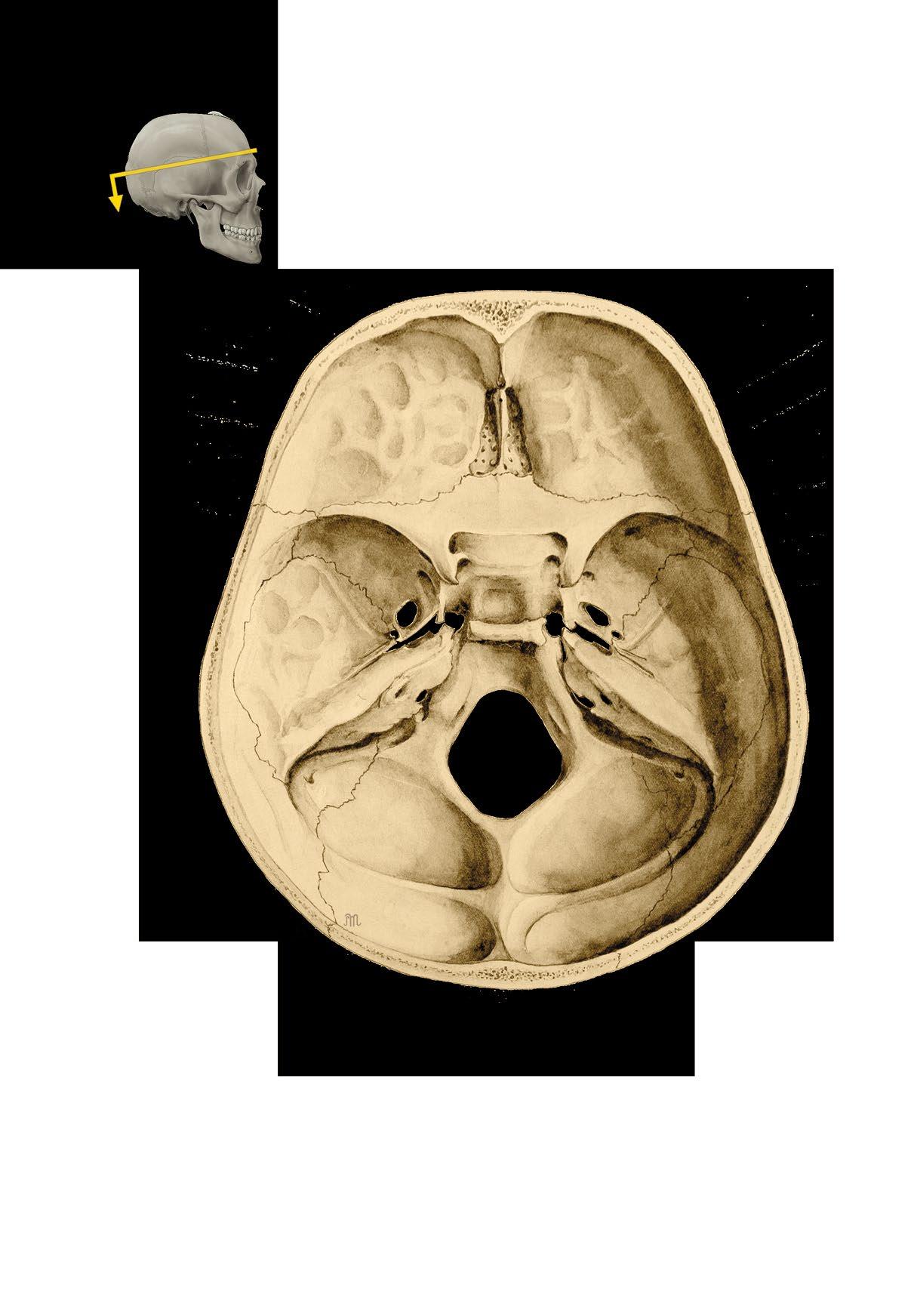
Basis cranii externa et interna
a zevní plocha base lební
b vnitřní plocha base lební
c členění vnitřní plochy base
os nasale
os frontale
os ethmoidale fossa cranii anterior fossa cranii media
os occipitale
fossa cranii posterior
External and internal base of the skull
a external surface
b internal surface
c cranial fossae
os parietale
os frontale
sutura coronalis
sutura sagittalis
os occipitale
sutura coronalis
sulci arteriae meningeae mediae
squama ossis temporalis
sutura lambdoidea
impressiones granulationum arachnoideae
os frontale
squama ossis temporalis
os parietale
lamina externa
lamina interna diploe
sutura lambdoidea
Calvaria, klenba lebeční

a zevní plocha
b vnitřní plocha
sulcus sinus sagittalis superioris
os occipitale
Calva, vault of the skull
a external surface
b internal surface
Hlava
os parietale
os temporale
os sphenoidale
os palatinum
maxilla
mandibula
sella turcica sinus sphenoidalis
os frontale
dorsum sellae
clivus
arcus anterior atlantis
dens axis
a Sagitální řez lebkou
b CT lebky v sagitální rovině
os nasale lamina perpendicularis ossis ethmoidalis/ septum nasi
concha nasalis inferior vomer
praemaxilla
sinus frontalis
praemaxilla
pharynx
mandibula os hyoideum
a Sagittal section of the skull
a krk – lebka Head and Neck – Skull 7

os frontale
arcus superciliaris
os nasale
canalis opticus
ala major
foramen
zygomaticofaciale
foramen infraorbitale
apertura piriformis
spina nasalis anterior
Cranium, lebka, pohled zpředu
protuberantia mentalis
sutura coronalis
Skull, frontal view

glabella
foramen frontale
ala major ossis sphenoidalis
os temporale
fissura orbitalis superior
fissura orbitalis inferior
arcus zygomaticus
os zygomaticum
maxilla
mandibula
foramen mentale
ala minor ossis sphenoidalis
fissura orbitalis superior cellulae ethmoidales
concha nasalis media
concha nasalis inferior angulus mandibulae
sutura sagittalis
Cranium, RTG lebky, zadopřední projekce, chlapec, 15 let
sutura lambdoidea
sinus frontalis
lamina cribrosa ossis ethmoidalis
septum nasi
os zygomaticum
sinus maxillaris
mandibula
Skull, X-ray, posterioanterior view, boy, 15 years

linea temporalis superior
os parietale
sutura squamosa
sutura coronalis
sutura sphenoparietalis
sutura sphenofrontalis
sutura lambdoidea
os occipitale
foramen mastoideum
squama ossis temporalis
meatus acusticus externus processus mastoideus processus styloideus
os zygomaticum
mandibula
Cranium, lebka, pohled zboku
Skull, lateral view

ala major ossis sphenoidalis
glabella
os lacrimale os nasale sulcus
nasolacrimalis
spina nasalis anterior
maxilla
foramen mentale
dorsum sellae
sulcus arteriae meningeae mediae
sutura coronalis sinus sphenoidalis
crista galli
sinus frontalis os nasale
sinus maxillaris spina nasalis anterior
dens incisivus
cellulae mastoideae
processus condylaris arcus atlantis anterior dens axis canalis mandibulae processus mastoideus
fossa hypophysialis
Cranium, RTG lebky, boční projekce, chlapec, 15 let
Skull, X-ray, lateral view, boy, 15 years
 sutura lambdoidea
processus palatinus maxillae
lamina horizontalis ossis palatini
foramen palatinum majus
choanae
foramen ovale
foramen spinosum
tuberculum pharyngeum
canalis caroticus
processus styloideus
fossa jugularis
condylus occipitalis
foramen magnum
crista occipitalis externa
Basis cranii externa, zevní plocha spodiny lebeční
foramen incisivum
foramen infraorbitale

arcus zygomaticus
lamina lateralis processus pterygoidei
foramen lacerum
fossa mandibularis
porus acusticus externus
foramen stylomastoideum
sulcus arteriae occipitalis
foramen mastoideum
canalis condylaris
planum nuchale
protuberantia occipitalis externa
External surface of the base of the skull
impressiones gyrorum
canalis opticus
foramen rotundum
ala major
foramen ovale
foramen lacerum
foramen spinosum
apertura interna canalis carotici
meatus acusticus internus
foramen jugulare
crista galli
canalis nervi hypoglossi
foramen magnum
protuberantia occipitalis interna fossa cerebralis
lamina cribrosa
pars orbitalis ossis frontalis ala minor
sulcus praechiasmaticus processus clinoideus anterior
fossa hypophysialis processus clinoideus posterior
dorsum sellae
clivus
Basis cranii interna, vnitřní plocha spodiny lebeční
margo superior partis petrosae
sulcus sinus sigmoidei
sulcus sinus transversi
fossa cerebellaris
Internal surface of the base of the skull
linea nuchae inferior
crista occipitalis externa
protuberantia occipitalis externa
linea nuchae suprema
linea nuchae superior canalis condylaris
planum nuchale
processus jugularis
incisura jugularis
condylus occipitalis
tuberculum pharyngeum
sutura lambdoidea
fossa cerebralis
fossa cerebellaris
Os occipitale, kost týlní
canalis nervi hypoglossi
a zevní plocha, pohled zezadu zdola
b vnitřní plocha
processus jugularis
processus intrajugularis
foramen magnum
sulcus sinus sagittalis superioris
protuberantia occipitalis interna
sulcus sinus transversi
sulcus sinus sigmoidei
processus jugularis
canalis condylaris
foramen magnum condylus occipitalis

Occipital bone
a external surface, inferoposterior view
b internal surface
foramen parietale
angulus occipitalis
linea temporalis superior
linea temporalis inferior
angulus mastoideus
margo squamosus
angulus frontalis
margo frontalis
angulus sphenoidalis
sulcus sinus sagittalis superioris
margo sagittalis
margo frontalis
Os parietale, kost temenní
a zevní plocha
b vnitřní plocha
sulci arteriae meningeae mediae
Parietal bone

a external surface
b internal surface
margo occipitalis
arcus superciliaris
squama frontalis
tuber frontale
incisura supraorbitalis
glabella
sulcus sinus sagittalis superioris
pars nasalis
facies temporalis
processus zygomaticus
margo supraorbitalis foramen frontale (incisura frontalis)
crista frontalis
apertura sinus frontalis
incisura supraorbitalis
fossa glandulae lacrimalis
pars nasalis
processus zygomaticus
pars orbitalis
fovea trochlearis
margo supraorbitalis
facies orbitalis
Os frontale, kost čelní
a pohled zpředu
b pohled zezadu
c pohled zdola
pars nasalis
Frontal bone

a anterior view
c posterior view
b inferior view
facies cerebralis alae majoris
foramen rotundum
foramen ovale
foramen spinosum
sulcus praechiasmaticus canalis opticus
ala minor
fossa hypophysialis
sulcus caroticus
fissura orbitalis superior
dorsum sellae
facies temporalis alae majoris
sinus sphenoidalis
spina ossis sphenoidalis
lamina lateralis processus pterygoidei
lamina medialis processus pterygoidei
ala major
facies cerebralis alae majoris
sulcus caroticus
canalis pterygoideus
Os sphenoidale, kost klínová
a pohled shora
b pohled zpředu
c pohled zezadu
ala minor canalis opticus
corpus ossis sphenoidalis et crista sphenoidalis
facies orbitalis alae majoris
fissura orbitalis superior
facies maxillaris alae majoris
foramen rotundum
canalis pterygoideus
incisura pterygoidea
hamulus pterygoideus
ala minor
canalis opticus
rostrum sphenoidale
fossa pterygoidea

Sphenoidal bone
a superior view
b anterior view
c posterior view
fissura orbitalis superior
foramen rotundum
foramen ovale
spina ossis sphenoidalis
lamina lateralis processus pterygoidei
lamina medialis processus pterygoidei
sulcus arteriae temporalis mediae
incisura parietalis
spina suprameatalis
processus mastoideus os tympanicum
processus styloideus
squama temporalis processus zygomaticus tuberculum articulare

fossa mandibularis
meatus acusticus externus
eminentia arcuata
sulcus sinus petrosi superioris
margo superior porus et meatus acusticus internus
apex
canalis musculotubarius
apertura interna canalis carotici
apertura externa canalis carotici
apertura canaliculi cochleae
fossa jugularis
canaliculus tympanicus
foramen
stylomastoideum
Os temporale, kost spánková
a pohled z laterální strany
b pohled z mediální strany
c pohled zdola
apertura canalis vestibuli
processus intrajugularis
processus zygomaticus
fossa mandibularis
porus acusticus externus
processus styloideus
incisura mastoidea
sulcus arteriae occipitalis
sulcus sinus sigmoidei
processus styloideus incisura jugularis
Temporal bone
a lateral view
b medial view
c inferior view
crista supramastoidea
fissura squamomastoidea
foramen mastoideum
spina suprameatalis
fissura tympanomastoidea
chorda tympani ve fissura petrotympanica
sulcus arteriae temporalis mediae
squama temporalis

tuberculum articulare
area nervi facialis crista transversa
area vestibularis superior
fissura tympanosquamosa
fissura petrosquamosa
canalis caroticus
canalis musculotubarius
canalis semicircularis anterior et lateralis
area vestibularis inferior
area cochleae et tractus spiralis foraminosus malleus
foramen singulare foramen jugulare corpus
art. incudomallearis incus crus breve
caput
collum
processus anterior
processus lateralis
Os temporale et ossicula auditus, kost spánková
a sluchové kůstky
a os temporale, pohled z laterální strany
b meatus acusticus internus z ventromediálního
pohledu
c malleus, incus, stapes
manubrium
crus longum
crus posterius
basis
crus anterius caput art. incudostapedialis
Temporal bone and auditory ossicles
a temporal bone, lateral view
b internal acustic meatus, ventromedial view
c malleus, incus, stapes
crista galli
cellulae ethmoidales anteriores
crista galli
cellulae ethmoidales anteriores
lamina orbitalis
lamina orbitalis
lamina cribrosa
cellulae ethmoidales posteriores
lamina perpendicularis
crista galli
lamina cribrosa
lamina orbitalis
concha nasalis media
lamina perpendicularis
processus ethmoidalis
processus lacrimalis
lamina perpendicularis
concha nasalis media
processus maxillaris
processus frontalis
cornu majus
corpus
cornu minus
processus temporalis
facies lateralis
foramen zygomaticofaciale
a Os ethmoidale, kost čichová, pohled shora
b Os ethmoidale, kost čichová, pohled zpředu
c Os ethmoidale, kost čichová, pohled zboku
d Concha nasalis inferior, dolní skořepa nosní, pohled z mediální strany
e Os hyoideum, jazylka, pohled zpředu
f Os zygomaticum, kost jařmová, pohled zpředu
facies orbitalis
foramen zygomaticoorbitale
a Ethmoid bone, superior view
b Ethmoid bone, anterior view
c Ethmoid bone, lateral view
processus maxillaris
d Inferior nasal concha, medial view
e Hyoid bone, anterior view
f Zygomatic bone, anterior view
