PRINCIPLES OF BIOPSYCHOLOGY
SIMON GREEN
A volume in the series Principles of Psychology
Series Editors
Michael W. Eysenck
Simon Green
Nicky Hayes
HOVE AND NEW YORK
Copyright © 1994 by Psychology Press Ltd, a member of the Taylor & Francis Group
All rights reserved. No part of this book may be reproduced in any form, by photostat, microform, retrieval system, or any other means without the prior written permission of the publisher.
Reprinted 1995,1997,1999,2001 and2004
Psychology Press Ltd
27 Church Road, Hove, East Sussex, BN3 2FA
29 West 35th Street, New York, NY 10001
www. psypress.co.uk
British Library Cataloguing in Publication Data
A catalogue record for this book is available from the British Library
ISBN 0-86377-281-l (hbk)
ISBN 0-86377-282-X (pbk)
ISSN 0965-9706
Cartoons by Sanz
Subject index compiled by Sue Ramsey
Cover design by Stuart Walden and Joyce Chester
Printed and bound in the UK by TJ International Ltd, Padstow, Cornwall
This publication has been produced with paper manufactured to strict environmental standards and with pulp derived from sustainable forests.
1. What is biopsychology? 1
Reductionism 1
Use of animals in biopsychology 3
Summary 7
2. The nervous system 9
The neuron 9
The synapse 15
Neurotransmitters 18
Neurotransmitters and drugs 21
Neurotransmitter pathways 24
Methods of investigation 27
Summary 31
3. Organisation of the nervous system 33
The peripheral nervous system 33
The central nervous system 35
The neocortex 41
Summary 51
4. Language and hemisphere function 54
Early studies-Broca and Wernicke 55
Later studies-speech, reading, and writing 57
Sperry and the split-brain 59
Summary 65
5. Hemisphere function 67
Studies with the normal brain 67
Left handedness 73
Why left handedness, or, rather, why right handedness? 74
Origins of language asymmetry in the brain 75
Left handedness and the vulnerable male- Geschwind' s hypothesis 76
Sex and the brain 77
Emotion and the right hemisphere 79
Summary 81
6. Learning and memory 83
Anterograde amnesia in humans 84
Korsakoff's psychosis 87
Alzheimer's disease-the neurochemistry of memory 90
Acetylcholine, memory, and brain grafts 92
The hippocampus and memory in rats 93
Retrograde amnesia 94
The biochemistry of memory 96
Summary 100
7. Stress, anxiety, and emotion 102
The hypothalamic-pituitaryadrenal axis 103
The autonomic nervous system 106
Stress and arousal 108
Individual differences in response to stressors-animal studies 109
Individual differences in response to stressors-human studies 110
Coping with stress 114
Anxiety 116
Emotion-central and peripheral mechanisms 117
Summary 127
8. Sleep, arousal, and biological rhythms 130
The concept of arousal 130
Psychophysiology and peripheral arousal systems 131
The reticular formation and central arousal 133
Biological rhythms 134
When rhythms go wrong-jet lag and shift work 137
Sleep 138
Summary 147
9. Motivation 149
An example of motivated behaviour-hunger 150
Thirst and drinking 159
Neurotransmitter pathways, feeding, and drinking 162
Non-homeostatic drives 163
Summary 164
10. Sensory systems 166
Somatic senses 166
Pain perception 168
Taste and smell 172
Hearing 174
Proprioception and the vestibular system 177
Summary 179
11. Vision 181
The eye, the retina, and the optic pathways 182
Receptive fields and the perception ofshape 185
Feature detection-innate or acquired? 188
Colour vision 189
Blindsight and alternative visual pathways 191
Summary 192
References 194
Glossary 198
Author Index 212
Subject Index 213
What is biopsychology?
The term biopsychology is just one of many names given to that area of science which studies the relationship between biology and behaviour. Others include psychobiology, biological psychology, and physiological psychology. Biology refers to all the physiological systems we find find in the body- muscles, the blood supply, glands, the nervous system etc. In practice, the study of biology and behaviour concentrates on the brain and the nervous system, although we will be covering the role of glands and hormones in, for instance, emotion and stress.
Researchers from many backgrounds work in biopsychology. They may be neurologists, neurophysiologists, neuroanatomists, pharmacologists, biochemists, or psychologists. Although psychologists cannot usually compete on a specialist level with, for instance, a biochemist interested in the synapse, psychology plays a vital role in the study of brain and behaviour. The biochemist or neuroanatomist often assumes that brain structure and biochemistry are complicated, whereas behaviour is simple. The psychologist, on the other hand, knows that even apparently simple behaviour, such as the rat exploring a maze or a human falling asleep, is in fact as complicated as the underlying physiology. So a complete description and explanation of the links between brain and behaviour involves an analysis of behaviour by psychologists that is just as sophisticated as the analysis of brain structure and biochemistry.
The biopsychologist therefore has to become familiar with the terminology and findings from the basic brain sciences mentioned earlier. This has led to the accusation by other psychologists that biopsychologists support a reductionist approach to the explanation of behaviour.
Reductionism
Psychology is the scientific study of behaviour. It can range from the study of large crowds, through the dynamics of small group interactions, to the study of the individual. Social psychology is more concerned with groups, crowds, and relationships between individuals. Cognitive psychology
WHAT IS BIOPSYCHOLOGY?
studies processes such as memory and attention from an individual perspective. Biopsychology studies the physiological correlates of behaviour i.e. those changes in physiological systems that occur whenever behaviour changes. It has concentrated on individual psychological processes such as memory, attention, emotion, and motivation, and attempts to demonstrate how, for instance, learning a particular task is correlated with a particular change in activity in the brain.
A reductionist approach assumes that a more fundamental level of description and explanation is always better and in some senses "truer" than description and explanation at a higher level. For psychology this would mean that a description of changes in brain activity during learning would represent a "better" explanation than a description of the psychological processes involved in learning a particular task.
The false assumption in this approach is that the behaviour of complex systems can be understood by analysing the behaviour of the separate components which make up those systems; the whole is just the sum of the parts. A clearer example would be the study of groups in social psychology. The reductionist approach would be that when you have described and understood the behaviour of individuals, you can describe and understand the behaviour of the groups those individuals are part of. This is obviously untrue. Social psychology has repeatedly shown that the behaviour of groups is not predictable from a knowledge of the individuals making up the group. Interactions between the separate individuals (components) produce unpredictable consequences at the group (systems) level, and these consequences can only be understood by studying the group.
These unpredictable consequences at the systems level are sometimes referred to as emergent properties. Individual components have their own characteristics, but when they combine, whether they are separate people forming a group, or individual brain cells (neurons) linked together to form a circuit, the system they make up has emergent properties that could not have been predicted from the behaviour of the individual components. To describe fully what is going on, we need to study the individual and the group, the single neuron and the brain circuit. Whatever level is chosen-crowd, group, individual, brain circuit, single neuron-there is no one level of explanation that has priority. Psychology is not reducible to atomic physics. The movement of atoms in brain cells in individuals during a football riot is no more or less interesting than the dynamics of the football crowd itself (although probably less). It is just that the full description and explanation of the behaviour involves studies at every level of explanation; the level chosen will reflect the background and interests of the researcher. Biopsychologists are not reductionists; they are
attracted to the links between biology and behaviour in the same way that social psychologists are attracted to the interactions between individuals. The two types of psychologist may seem to have little in common, but both are engaged in the same task of studying and explaining behaviour.
A lot of the research undertaken by biopsychologists involves the use of non-human animals. As monkeys and rats are assumed by many to be simpler in terms of behaviour and biology than humans, this has reinforced the view of biopsychologists as reductionists. However the use of animals is dictated by the methods of biopsychology, not by any commitment to reductionism. These methods (see Chapter 2), such as systematically damaging or electrically stimulating parts of the brain, cannot be used with human subjects, so if the biopsychologist believes they are essential to the study of brain function, then he or she has to use animals.
Use of animals in biopsychology
This is a controversial area with several issues to be considered. These can be divided into the practical and the ethical.
Practical issues
As mentioned earlier, biopsychologists use animals because their methods of study cannot be used with humans. The critical point at issue is whether the study of the animal brain can tell us anything about the human brain. If not, much of the justification for using animals disappears; one could argue that the study of brain and behaviour in any species is always of interest ("comparative biopsychology"), but many researchers would be unhappy if these sometimes stressful procedures had no bearing at all on human behaviour and experience.
Much of the detailed evidence used to support links between animal and human biopsychology is referred to in later chapters. At this stage I would just like to make a couple of points. First, the mammalian brain (which would include rats, cats, dogs, monkeys, apes, and humans) is built on similar lines in all species studied. Every part of our brain can also be identified in the rat brain, although evolutionary progress means that our brain is more highly developed in terms of size and connections. The units of the nervous system (the nerve cell, or neuron) are the same in all species, and work in the same way (see Chapter 2). In fact the neurons found in the most primitive invertebrate floating around in the sea also operate in the same way. Another way of putting this is that although evolution of the nervous system has produced the highly sophisticated human brain, at the level of its basic units evolution has been highly conservative.
So the animal subjects used by biopsychologists have brains similar in outline structure to our own. This would be irrelevant if the behaviours produced were totally different, and in some ways, of course, they are. Rats and monkeys do not talk a great deal, they do not take psychology examinations, they do not stand for Parliament or create civilisations. However, they do have excellent memories, they do become fearful, angry, and stressed, they can solve problems, attend selectively to some stimuli, become hungry, thirsty, or curious. In other words, if we take the major categories of human behaviour as cognitive (memory, attention, perception etc), affective (or emotional e.g. fear and anger), and motivational (behaviour that is aroused and directed, such as feeding, drinking, exploring), then examples can be found in all mammalian species.
So, with certain major exceptions such as language and self-awareness, we can identify comparable behaviours in animals and humans, produced by brains of comparable structure. To justify fully biopsychological work with animals, we would need to use detailed experimental findings to show that, for instance, memory and hunger in the rat and monkey are controlled by similar brain mechanisms as those involved in human memory and hunger. We shall see later how far we can go with these parallels, but, certainly at a general level, the proposition that research with animals tells us something about the human brain and behaviour is justifiable.
Ethical issues
Animals used in any sort of research (such as medical, pharmacological, psychological, or for testing cosmetics) are subjected to certain forms of stress. Nowadays they are bred for the specific purpose, either by the Institution involved or, more usually, by specialised and licenced breeding establishments. They are housed in cages which, however large, cannot compare with life in the wild; of course, they have never known life in the wild and life in the cage is all that they have experienced. They would certainly not find it as stressful as would an animal caught in the wild and brought into the laboratory. But no-one can argue that a caged existence is ideal.
Beyond these basic housing conditions, the types of stress to which the animal is exposed depend on the nature of the research project. I am concentrating on biopsychology, and it is, incidentally, worth bearing in mind that the number of animals used in psychology departments is a tiny fraction of the total; this is not to minimise the issues, but to give some perspective. Banning the use of animals in cosmetics testing would be a greater contribution to animal welfare than would banning their use by psychologists.
In the United Kingdom all animal research is supervised by the Home Office. Under the 1986 Animals (Scientific Procedures) Act, procedures that involve significant amounts of stress have to be part of a research programme which is itself assessed in terms of its objectives. To justify the use of animals and stressful procedures, the research has to be rigorously designed, and the potential results have to represent a significant contribution to our knowledge of medicine, pharmacology, biopsychology, or psychology. This eliminates research performed simply for its own sake or because it is a particular fancy of the experimenter.
It also means that high levels of chronic (long-lasting) stress are rarely approved; nowadays it is not possible to use some procedures that were popular in the 1970s (such as the use of inescapable footshock in the study of learned helplessness) because regulations are now much tighter.
Another consequence is that all researchers now consider their research plans in terms of their potential application to human life and behaviour. There has to be some point to the research beyond any intrinsic interest, and in biopsychology in particular the eventual aim is often to do with the biological problems underpinning human behavioural disorders. A clearer understanding of the biology of, for instance, anxiety, may contribute to the development of both physical (e.g. drugs) and psychological (e.g. hypnosis) therapies. There will be examples of this type of biopsychological research throughout this book.
The Home Office is involved in the licensing of individual researchers and their research projects where stressful procedures are used. Nonstressful procedures do not require Home Office approval, and so psychologists may use animals in studies of, for instance, simple learning of rats in Skinner boxes and mazes (i.e. spontaneous behaviours not involving food or water deprivation, or other stressful conditions or stimuli), and may do this without seeking official approval.
After licensing of researcher and project, the laboratory is visited regularly during the course of the project by a Home Office Inspector and by a designated veterinary surgeon, to ensure that the conditions of the licence are not being exceeded, and that the general state of the laboratory and the animals is entirely satisfactory. The Inspector has the power to withdraw both personal and project licences if he or she sees fit.
I have dealt at some length with the regulation of research involving animals in the UK, because many people imagine that researchers may do anything they want. This is certainly not the case. However there are still many people who would be against animal research regardless of how well that research was regulated. They believe that the exploitation and sacrifice of other species is morally and ethically unjustifiable, however beneficial such research may be for the human species. At the extreme it
is impossible to argue against this position, as it is a case of one moral position versus another, and it becomes an individual choice as to which you support.
However the less extreme position is open to debate. This position regards animal research as unethical because it has no relevance and is actually of no benefit to humankind. That is, the physiological and behavioural differences between animals and us are too great for results to be extrapolated from animals to people. In the previous section on practical issues I have given some background to this issue, pointing out that animals and humans are on the same evolutionary scale and have many features in common; suficient to expect some similarities in, for instance, brain structure and behaviour.
All drugs used in medicine and psychiatry undergo tests in animals to screen for possible toxic (poisonous) and other unwanted side-effects. Animal tests are also used to try and identify potential new medical and psychiatric treatments (as we shall see later in relation to schizophrenia). Critics say that these are simply unnecessary, as results from animals have no bearing on the human situation. One example they use is that of the drug thalidomide, whose ability to affect development of the foetus (the baby growing in the uterus) was not picked up in animal testing and resulted in the birth of many deformed babies.
However, one example of an unwanted drug effect that does not show up in animal tests does not automatically invalidate all animal tests. There are many examples of drugs in clinical use which have been developed using animals, and which could not have been developed without themanaesthetics, anti-cancer and anti-AIDS treatments, anti-epilepsy, antianxiety, and anti-depressant agents. If we can use as an argument "the end justifies the means" then, if the "end" is the alleviation of human suffering, animal research is justified. Of course another counter-argument is that if human society was radically changed so that the so-called self-inflicted conditions such as the commonest forms of lung cancer, anxiety, or depression, were eliminated, then the need for animal testing would largely disappear. Everyone should support this objective. Unfortunately society shows no particular tendency to change itself in these directions, and the need for physical treatments is as strong as ever.
I shall close with a specific example. One of the major social problems of the 1990s and onwards will be the increasing numbers, relative to the whole population, of old people. A proportion of these will suffer from senile dementia (dementia meaning a breakdown in normal mental function), the commonest form of which is Alzheimer's disease. In its severest form (and it is a progressive disease) Alzheimer's produces severe memory loss, confusion, loss of the sense of self-identity, and double
incontinence (loss of control of bowels and bladder). Caring for sufferers is usually a family affair, 24 hours a day every day of the year, watching your husband, or wife, father, or mother, degenerating in front of you. There is as yet no cure for Alzheimer's disease. It is not linked to immoral behaviour or physical self-abuse of any sort, so lifestyle changes do not prevent it. We know from the brain changes seen at post-mortem that there is a particular pattern of damage to brain circuits, and we know from animal studies that these circuits are also involved in memory in rats and monkeys. Damage to these circuits in animals produces memory loss, and this model of Alzheimer's is being used to develop possible treatments. These include new drugs to restore brain function, and the use of "grafts" of brain cells to replace cells that have died. These latter techniques have already been used in the treatment of Parkinson's disease (see Chapter 2), a progressive movement disorder which is also caused by damage to a specific brain circuit. Animal research is also revealing the biochemical abnormalities responsible for producing the brain damage. There are encouraging signs that a treatment for Alzheimer's disease may emerge, which will have depended absolutely on the use of animal testing for its development. If successful it would have a dramatic impact, improving the quality of life for both those with Alzheimer's and those who would otherwise sacrifice their lives in caring for them. Is this potential benefit sufficient to justify the use of animals in psychological research? I would say it is. Those who disagree have every right to do so, but must also have the responsibility of explaining to those in the front line-patients and their carers-why attempts to alleviate their suffering should be stopped.

Summary: What is biopsychology?
• Biopsychologists, also known as psychobiologists, study the links between behaviour and the physiological systems of the body. In practice they concentrate on the brain and the nervous system.
• Biopsychology is often accused of reductionism; the assumption that behaviour can only be explained by studying the activity of neurons in the nervous system. However, this approach makes the false assumption that activity in a system is simply the sum of the behaviour of the individual parts of the system. In fact systems show emergent properties, which cannot be predicted from the study of individual components. A full explanation of behaviour requires the study of all levels of behaviour, from the social and cultural to the biological. Most biopsychologists are not reductionists, but simply choose to work at the level of biological processes.
e Much of the work of biopsychologists involves animal experimentation. Objections to the use of animals can be practical or ethical. The practical objection is that animals are too different from humans to justify the use of evidence from animal work. However the brains of all mammals, especially those of primates, are comparable in detailed structure to the human brain, and mammals show the same categories of behaviour as we do-cognitive, emotional, and motivational. Although detailed brain organisation may differ, it is possible on a general level to justify the use of evidence from animal studies in talking about the human brain.
e Animal experimentation in the United Kingdom is closely supervised by the Home Office, and there is no longer the opportunity for casual exploitation of animals in research. All projects have to be assessed in terms of their actual or potential usefulness. Equally, animals can still suffer distress and pain, which is then weighed against the benefits for society. The proposition that animals should never be used in research is a moral position that cannot be argued with. The less extreme position is that they should only be used where there are no alternatives and where there are likely to be significant benefits from the research. If animal research leads to effective treatments for devastating conditions such as Alzheimer's disease, would this justify its continuation?
The nervous system.
The neuron
Although the biopsychologist has to deal with many systems found in the body, most of the work involves the brain and the rest of the nervous system. We shall meet some of the other systems later on, in particular covering the role of hormones in stress and emotion. However, at this stage we need to deal with the basic properties of nervous tissue. This will necessarily involve terms and processes that will be unfamiliar to many of you; but remember that the interest of the biopsychologist is in the links between brain and behaviour, and that an awareness of the basic principles of nervous system function is necessary as a background to the more interesting work presented in later chapters. The body is made up of billions of cells. Every one has a particular function; muscle cells contract, cells that form glands secrete chemicals, red blood cells carry oxygen around the body. In order to carry out their
Dendrites
Nucleus, containing the chromosomes
Cell body (soma)
Cytoplasm
The neuron. Nerve impulses are triggered on the dendrites, and travel across the soma, down the axon, and finally reach the axon terminals.
particular functions, cells have become specialised in various ways. The structure of muscle cells has been modified to allow them to contract, and the particular chemistry of glandular cells allows them to manufacture and secrete chemicals. The nervous system is made up of billions of cells called neurons, which have become specialised to conduct electrical impulses.
To enable them to do this, neurons have become modified in various ways. First of all they are characteristically elongated. The cell body (the soma) extends on one side into a long branching axon and on the other into a number of shorter dendrites. Axons and dendrites together are the neuronal processes. It is important to remember that the soma and the neuronal processes are all part of the neuron, i.e. together they make up a single cell.
The nervous system extends from the neurons connecting the muscles of the toes to the spinal cord, to the billions of neurons packed into the cerebral cortex of the brain. So neurons come in many shapes and sizes, although all follow the basic pattern just described. The length of neuronal processes can vary from several feet down to less than a millimetre, while the extent of axonal branching can be minimal (i.e. a single axon) or "tree-like". Despite these variations in shape, all neurons possess the modification vital to their ability to transmit electrical impulses: this concerns the cell membrane, the outer "skin" of the cell. This membrane is semi-permeable, which means it allows various electrically-charged particles or ions to pass through from inside the neuron to outside and vice versa. Usually the concentration of these ions on either side of the membrane is different, and this leads to an electrical difference, or gradient, across the membrane. This is known as the resting potential, and is measured at -70 millivolts (thousandths of a volt) from inside to outside. This stable condition of the membrane can be altered by stimuli of various types. If the stimulus shifts the electrical potential across the membrane towards zero, i.e. a balance between the ion concentrations inside the neuron and outside, then it may stimulate an action potential at that point on the membrane.
As the potential decreases from -70 to about -50 millivolts, there is a sudden and massive increase in the permeability of the cell membrane, which allows sodium and chloride ions to rush into the neuron and potassium ions to filter out. This exchange of ions produces a rapid change in the membrane potential from -50 millivolts to about +40 millivolts; this takes around half a millisecond (a millisecond is a thousandth of a second), and is called an action potential (see the graph opposite). The -50 millivolt threshold is critical for an action potential to occur; once it is reached, the action potential begins and the course of events is unstoppable, whereas
+100mV
Resting state
Action Refractory potential state
Resting state
The action potential. Note that for a brief period after an action potential, the membrane cannot support another. This is the refractory period.
msec
if the threshold is not reached nothing happens-the action potential is an ali-or-nothing event.
After peaking at +40 millivolts the action potential dies away and the membrane returns to its resting state of -70 millivolts. However the action potential has not disappeared. It occurs at a particular point on the membrane, but the structure of the cell membrane, besides allowing these explosive electrical changes to happen, also allows them to be carried (or propagated) along the membrane from the point at which they were first stimulated, i.e. the action potential travels along the neuron. The shift in membrane potential from -70 to +40 millivolts represents, in electrical terms, a depolarisation, and, again in electrical terms, the movement of the action potential along the neuron is represented by a wave of depolarisation passing along the cell membrane. We can record action potentials by placing a thin wire electrode close to or actually inside the neuron; this electrode is connected to a highly sensitive voltameter on which we can see the momentary blips of electrical activity that represent the passing waves of depolarisation, i.e. the action potentials. At a given point on the neuronal cell membrane the biochemical changes underlying the action potential take about half a millisecond. After restabilisation, the mem-
THE NERVOUS SYSTEM 11
brane is then unresponsive for a brief period; during this refractory period this point on the membrane cannot support another action potential, and so there is a minimum interval between action potentials of around two to four milliseconds. If we are recording action potentials, or nerve impulses as we shall now refer to them, from a particular point on the membrane, then the maximum rate or frequency at which they can arrive is between 250 and 500 every second.
Of course this is a maximum rate, and only rarely encountered under normal conditions. As every nerve impulse is electrically identical to every other impulse, the only features of nerve impulse conduction that can vary are the frequency at any one time and the patterning of trains of impulses over time. If we consider more than one neuron then we can also look for variations in the information coded by nerve impulses occuring in different regions of the nervous system. This is discussed shortly, but before moving on, we should look at another type of electrical conduction used by the nervous system.
As we shall see, nerve impulses are usually triggered on the dendrites of a neuron and then, following the processes just outlined, rapidly conducted along the neuronal membrane from dendrite to soma to axon, and then along the length of the axon to the axon terminal, the physical end-point of the neuron. However, a high proportion of neurons in the nervous system have a particular type of axon specialised for extremely fast conduction of nerve impulses. During the growth of the nervous system, both before and after birth (prenatal and postnatal), these axons become covered in a fatty sheath known as myelin; in fact the whole sheath is a single glial cell which grows around the neuron during development. (There are many different types of glial cell in the mature nervous system, and many more glial cells than neurons, but discussion of their various other functions is beyond the scope of this book.)
The myelin sheath covering the axon is not continuous, but is broken by regular gaps where the axonal membrane is exposed (see the diagram opposite). These are the nodes oJRanvier. Using a sophisticated extension of the electro-chemical processes outlined earlier, action potentials can jump from node to node along the axon, travelling much faster than when conduction is continuous along the axon. This means of transmission is known as saltatory conduction (after the Latin for "leaping") and produces conduction velocities of up to 100 metres per second. This compares with speeds in unmyelinated axons of as little as two or three metres per second. Myelinated axons are only found in vertebrates, and represent a critical stage in the evolution of the nervous system. Faster conduction of nerve impulses means faster processing of more information, and leads eventually to a more sophisticated and complex brain. An additional feature of
Dendrites
Cell body
Track of nerve impulse
Myelin sheath Nodes of Ranvier
myelination in the human brain is that most of it takes place after birth, and is not complete until well into the teenage years. This has implications for what we can expect of the infant brain; mature functioning does not occur for many years.
Once or twice I have mentioned information processing. For many years now the processes carried out by the brain have been seen by many as analogous to the computations of a sophisticated computer. I for one still believe that the computer to match even the rat brain is some decades, perhaps even centuries away. However the concept of information processing which dominates cognitive psychology has generated a great deal of interest in brain functions. At this stage it is possible to make a couple of points relevant to the discussion.
Action potentials are ali-or-nothing events with identical electrical characteristics. However they represent the only way that the nervous system can encode information. Nerve impulses in the optic nerve convey information that represents what we see; activity in motor nerve pathways to the muscles represents commands that result in movement; impulses in neurons of the cerebral cortex represent memory and thought. Everything that the brain does is reflected in nerve impulses, or, more accurately, is nerve impulses. Individual impulses are identical. Different types of information are encoded as bursts of nerve impulses varying in frequency, patterning over time, and location within the nervous system.
Myelinated neuron and saltatory conduction.
THE NERVOUS SYSTEM 13
Axon
Another important aspect of this phenomenon is that the brain can only respond to, or be aware of, stimuli and events around us if they can be converted into nerve impulses. We possess a range of sensory receptors whose job it is to convert stimulus energy into nerve impulses. So pain receptors are special cells that react to painful stimuli, such as burns or blows, by triggering nerve impulses in the axons that connect them to the central nervous system. The visual receptors at the back of the eye convert "light", or electromagnetic radiation, into nerve impulses in the optic nerve running to the visual cortex of the brain. Auditory receptors in the inner ear convert pressure changes in the air around us into nerve impulses in the auditory nerve, which are eventually interpreted as sounds. The world we are aware of is therefore defined by the range of sensory receptors we possess; we can be conscious of colours, sounds, tastes, smells, heat and cold, touch, pressure, and pain, only because we have receptors to convert all of these different types of stimuli into nerve impulses that the brain can interpret. Other animals can be aware of stimuli unavailable to us because they have a different set of receptors responding to different stimuli; bats are sensitive to ultrasonic sounds, on the whole birds have better vision, and dogs have better hearing than we do. The sensory receptors an animal possesses define the world it lives in.
The opposite also applies; we cannot be aware of stimuli for which we have no receptors. A case in point is the brain itself. Although it is the centre for the analysis of all the sensory input coming into the body, it has no sensory receptors itself. Therefore, for instance, it can suffer physical injury without apparent pain as it has no specialised pain receptors, and there are many instances of grotesque damage occurring with the person being virtually unaware of the extent of the injury (e.g. Phineas Gage, p.SO). In case you are wondering about headaches, these are usually caused by increased pressure of fluid in the blood vessels supplying the brain; this is picked up by specialised sensory pressure receptors in the walls of the blood vessels, and messages (nerve impulses) are passed to the brain centres responsible for the analysis of pain information.
The lack of pain receptors in brain tissue means that elec+rical stimulation using thin wire electrodes implanted into various parts of the brain can be done in the conscious human patient, using only a local anaesthetic for the area of scalp through which the electrode passes (skin everywhere contains pain receptors). So the brain can be stimulated, and the patient asked for their reaction. Appropriate stimulation in the visual areas leads to a report of visual sensation-flashes of light; in auditory areas, to reports of sounds being heard; in other areas to feelings of fear and foreboding or of pleasure and happiness. By mimicking the brain's electrical code in the right areas we mimic the function of those areas (these
studies, incidentally, would only be done before major brain surgery, when the surgeon wants to be sure that no damage will occur to critical functions).
These points have been rather laboured as they can be hard to grasp. The richness of human behaviour and experience depends ultimately on a relatively simple electrochemical process. The complexity comes from the interaction of the 15 to 20 billion (15x109-20x10 9 ) neurons that make up the nervous system.
The synapse
Nerve impulses usually begin on the dendrites of a neuron and then travel along the cell membrane until they reach the end of the axon-the axon terminal. If neurons were physically connected to one another, the impulse would simply continue on to the next neuron and so on. The nervous system would be a huge electrical circuit and, as neurons, by virtue of their electrical properties, automatically conduct impulses once they have been stimulated, activity would be rapidly and haphazardly transmitted throughout the nervous system. This is prevented by the existence of tiny gaps between neurons, which are called synapses, and usually occur between the axon terminals of one neuron and a dendrite of the following neuron; some variations will be described later.
The synapse (or synaptic gap or cleft) is measured in angstroms (10 billionths, or 10-10, of a metre) and can only be seen under the electron microscope. However it does represent a real physical barrier for the nerve impulse, as electrical activity cannot normally jump gaps.
Incidentally, this is a convenient time to point out that the space outside neurons is not empty. Much of it is taken up by the glial cells mentioned earlier, and the rest is filled with extracellular fluid, containing chemicals and ions, and critical for normal brain function. The synaptic gap, as an extracellular ("outside the neuron") space, contains extracellular fluid.
For the nerve impulse and the information it represents to survive, it has to cross the synapse, and if we see electrical conduction as the first basic principle of neuronal transmission, then conduction across the synapse involves the second basic principle of chemical transmission. This is a complicated process involving tiny packets of chemicals stored within the presynaptic ("before the synapse") axon terminal, as can be seen in the diagram overleaf. The chemicals are collectively referred to as synaptic neurotransmitters, and the spherical storage units as vesicles. Each vesicle contains only a few hundred molecules of the neurotransmitter.
When a nerve impulse reaches the end of the axon, i.e. the presynaptic terminal, it causes a number of these vesicles to move to the neuronal cell
Direction of impulse flow
Vesicles containing neurotransmitter
Pre-synaptic receptors
Pre-synaptic membrane
Post-synaptic membrane
Post-synaptic receptors
Pre-synaptic terminal
•Synaptic gap or cleft
Post-synaptic terminal
The synapse. Nerve impulses cause vesicles to merge with the pre-synaptic membrane and release neurotransmitter into the synaptic gap. They can then combine with the post-synaptic receptors.
membrane of the presynaptic terminal. Because of their particular structure the vesicles "merge" with the membrane, and in doing so release their contents into the synaptic gap. The molecules of neurotransmitter diffuse or drift over to the neuronal cell membrane of the dendrite of the following neuron; the postsynaptic ("after the synapse") membrane.
On the postsynaptic membrane are receptors. These are molecules with a structure which matches that of the neurotransmitter, in the same way as the structure of a lock matches that of the key that fits it. The molecules of neurotransmitter combine with the receptors for a brief period, and during this short-lived combination the permeability of the postsynaptic membrane is altered. If you recall, permeability changes are the foundation of the action potential or nerve impulse, and the neurotransmitter-receptor combination can push the electrical state of the membrane towards the all-or-none triggering of an action potential.
However the combination of a single neurotransmitter molecule with a single postsynaptic receptor is insufficient to shift membrane permeability to the critical threshold, and an action potential would not occur. What is needed is for the combination of a large number of neurotransmitter molecules with a large number of receptors to happen simultaneously, in which case the additive effect of all the individual permeability changes would be enough to trigger an action potential in the postsynaptic membrane, and for a nerve impulse to begin its journey along the postsynaptic
16 PRINCIPLES OF BIOPSYCHOLOGY
neuron. The release of neurotransmitter molecules from their vesicles in the presynaptic terminal is proportional to the number of nerve impulses arriving at the presynaptic terminal. It therefore follows that for an action potential to be triggered, sufficient nerve impulses must arrive at the presynaptic terminal in a short space of time to release enough neurotransmitter to combine with enough postsynaptic receptors to produce the threshold permeability change. Another way of putting this is to say that enough neurotransmitter must be released to depolarise the postsynaptic membrane, remembering that depolarisation is the technical term for the membrane permeability changes underlying action potentials.
If nerve impulses arrive at the presynaptic terminal only occasionally, the postsynaptic membrane will not be depolarised and the information coded by the impulses will be lost. The additive effect of impulses arriving in a short period of time is known as temporal summation. Alternatively, two or more axons may terminate close to the same patch of postsynaptic dendritic membrane; the combined effect of neurotransmitter released from these terminals may be sufficient to depolarise the postsynaptic membrane and to trigger an action potential in the postsynaptic neuron. This phenomenon is known as spatial summation. I mentioned earlier that the combination of even a single neurotransmitter molecule with a single postsynaptic receptor can shift membrane permeability towards the depolarisation threshold without actually reaching it. This electrical change, making an action potential more likely, is called an excitatory postsynaptic potential, or EPSP. There are also combinations of neurotransmitters with receptors which shift membrane permeability away from the depolarisation threshold, making an action potential less likely. This type of electrical change is known as an inhibitory postsynaptic potential, or IPSP. Although I have discussed excitatory interactions between neurons, i.e. how nerve impulses are conducted from neuron to neuron, it is important to remember that inhibitory effects, whereby one neuron can prevent another from firing by triggering IPSPs, are of equal importance in nervous system function. An example would be the hypothesis that some forms of epilepsy (a condition that involves uncontrollable electrical discharges in the brain) are caused by some problem with inhibitory circuits. Normal brain function depends on a balance between excitatory and inhibitory influences. The occurrence of a nerve impulse in the postsynaptic neuron depends on the additive effect of many impulses in presynaptic neurons, whether the addition is temporal or spatial. Therefore the frequency of postsynaptic nerve impulses is much smaller than that of presynaptic impulses. This can act as a simple filtering mechanism. If nerve impulses stimulated, say, by the presence of dust particles on the skin, travel along a sensory neuron towards the spinal cord and brain, they will not usually be frequent THE NERVOUS SYSTEM 17
enough to carry across the first synapse they meet in the spinal cord, i.e. they and the information they represent will be lost. This prevents the brain being clogged up with low-level information. In the brain itself the situation is more complicated. I have talked about synapses and pre- and postsynaptic neurons as though the arrangement of the nervous system was strictly linear, with neurons lined up in a long chain. This I have done for ease of presentation. In fact the complexity of synaptic connections almost defies the imagination. It has been estimated that on average each neuron makes of the order of a thousand synaptic contacts, and there are around 15-20 billion neurons. Axons are usually many-branched. Some of these branches may backtrack and synapse on neurons further down the brain, or even on the dendrites of the same neuron as a form of self-regulatory feedback (remembering all the time that the synapses may be excitatory or inhibitory). Each neuron will receive synaptic inputs from thousands of other neurons, some excitatory and some inhibitory, and the nerve impulse activity we might record from it is the summed total of all these inputs. Information processing in the brain is based on two elementary codes, electrical and chemical, but the organisation of neurons into what we know as a brain produces a complexity beyond the imagination of even computer programmers.
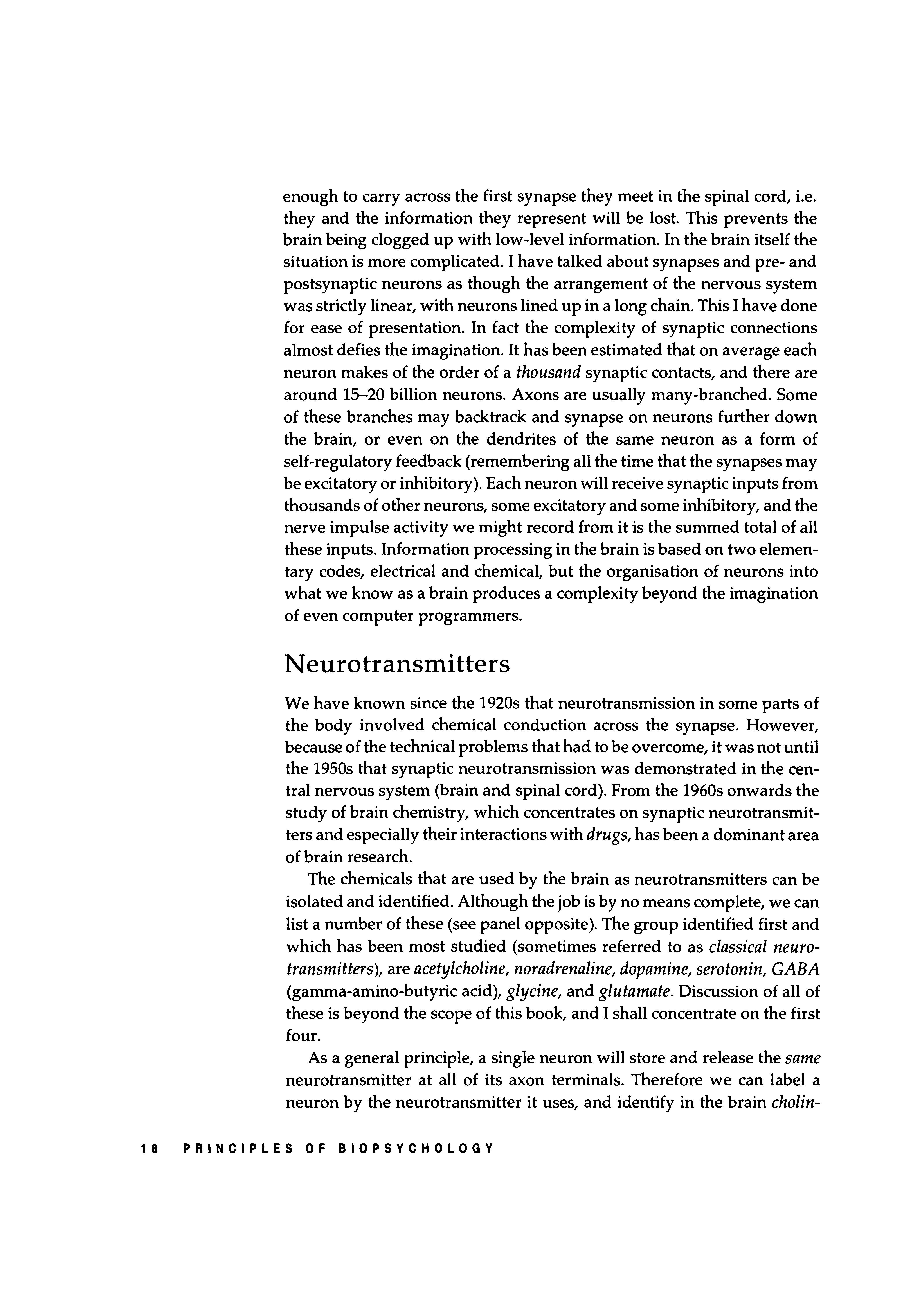
Neurotransmitters
We have known since the 1920s that neurotransmission in some parts of the body involved chemical conduction across the synapse. However, because of the technical problems that had to be overcome, it was not until the 1950s that synaptic neurotransmission was demonstrated in the central nervous system (brain and spinal cord). From the 1960s onwards the study of brain chemistry, which concentrates on synaptic neurotransmitters and especially their interactions with drugs, has been a dominant area of brain research.
The chemicals that are used by the brain as neurotransmitters can be isolated and identified. Although the job is by no means complete, we can list a number of these (see panel opposite). The group identified first and which has been most studied (sometimes referred to as classical neurotransmitters}, are acetylcholine, noradrenaline, dopamine, serotonin, GABA (gamma-amino-butyric acid), glycine, and glutamate. Discussion of all of these is beyond the scope of this book, and I shall concentrate on the first four.
As a general principle, a single neuron will store and release the same neurotransmitter at all of its axon terminals. Therefore we can label a neuron by the neurotransmitter it uses, and identify in the brain cholin-
Classical Neurotransmitters
Acetylcholine
Noradrenaline
Dopamine
Serotonin (5-hydroxytryptamine)
Amino Acid Neurotransmitters
Glycine
Glutamate
GABA (Gamma-amino-butyric-acid)
Possible Neurotransmitters/Neuromodulators
Enkephalin
Substance P
Vasopressin
Cholecystokinin
Monoamines
Neuromodulators are found within neurons, but may function to regulate the release of classical neurotransmitters such as acetylcholine and dopamine, rather than act as transmitters in their own right.
ergic, noradrenergic, dopaminergic and serotonergic neurons, using, respectively, acetylcholine, noradrenaline, dopamine, and serotonin as their neurotransmitters. Each of these chemicals has its own particular molecular structure. As their role is to be released and to combine with a postsynaptic receptor, the receptors themselves must possess a molecular structure to match the particular neurotransmitter. So we have specific cholinergic, noradrenergic, dopaminergic and serotonergic receptors. As neurotransmitters are constantly being released, there has to be a continuous supply to refill the synaptic vesicles. The manufacture of these chemicals takes place inside the neuron (intraneuronally), and I can describe it best by taking a specific example. Dopamine is synthesised ("made") from tyrosine, a fairly simple amino acid manufactured in the liver or found in the food we eat; the 20 or 30 amino acids are the building blocks of the body's proteins, among other things, and are essential to normal functioning. Tyrosine is converted by the action of tyrosine hydroxylase to dihydroxyphenylalanine, understandably referred to as DOPA. Tyrosine hydroxylase is an enzyme, one of a large group of substances whose role in the body is to enable chemical reactions to occur without themselves undergoing change. Another enzyme, aromatic amino acid decarboxylase, converts DOPA to dopamine. The newly synthesised dopamine is then packaged up in vesicles ready for use.
Synaptic neurotransmitters. The first two categories are well established. The third group of "possibles" could be extended by another 1D-15 candidates. THE NERVOUS SYSTEM 19
Synthesis of synaptic neurotransmitters. These events take place within the neuron. The rate of neurotransmitter release and breakdown at the synapse is also important, as this provides feedback regulation of synthesis; more active neurons need to synthesise more rapidly.
Dopamine neuron
neuron neuron
Noradrenaline neuron
Acetylcholine neuron
Acetylcholine neuron
Serotonin neuron
Choline + Acetyl-coenzyme A E Acetylcholine neuron F Tryptophan5-Hydroxytryptophan
5-Hydroxytryptamine (5-HT, Serotonin)
5-Hydroxytryptamine (5-HT,
The various steps are catalysed by enzymes:
A = Phenylalanine hydroxylase
B = Tyrosine hydroxylase
C = Dopa decarboxylase
D = Dopamine beta-hydroxylase
E = Choline acetyltransferase
F = Tryptophan hydroxylase
G = Aromatic amino acid decarboxylase
20 PRINCIPLES OF BIOPSYCHOLOGY
The diagram on the left outlines the synthesis of dopamine, noradrenaline, acetylcholine and serotonin. Notice that dopamine and noradrenaline share a common pathway up to dopamine itself; this is converted to noradrenaline by the action of the enzyme dopamine-beta-hydroxylase (DBH). So the only difference between a dopaminergic and a noradrenergic neuron is the presence in the latter of DBH.
It is also important to consider the fate of neurotransmitters after release. When they have combined with their receptor, molecules of dopamine, for instance, move away from the immediate area of the postsynaptic membrane. There a large proportion is broken down and inactivated by the action of the enzyme monoamine oxidase. This action is important in the regulation of synaptic activity; if the neurotransmitter molecules were not removed, stimulation of the postsynaptic receptors would be continuous and the synapse could not function properly. Each of the neurotransmitters is broken down in this way; dopamine, noradrenaline and serotonin via the action of monoamine oxidase, and acetylcholine by acetylcholine esterase. As we shall see later, drugs that interfere with the breakdown enzymes can prevent the removal of neurotransmitter molecules and so increase synaptic activation.
So synaptic transmission is a dynamic process. Neurotransmitter release is continuous, as neurons are always active even if activity is sometimes minimal. Stores of neurotransmitters are constantly being depleted and replenished, with the brain's supply of, for instance, acetylcholine, being totally replaced over a period of a few days. The neuron is a chemical factory working 24-hour shifts.
Neurotransmitters and drugs
Since the beginnings of human civilisations people have used drugs for both their medical and psychological effects. Originally they were derived from plants. Mescaline (from peyote, a form of cactus), psilocybin (from the "sacred" mushroom), opium and heroin (from the poppy), are still valued by some for their power to induce hallucinations (they are from the class of drugs called hallucinogenics). Nowadays we have a huge range of natural and artificially-synthesised drugs which can affect various aspects of behaviour and experience. The panel overleaf contains a list of the major categories of drugs used in the treatment of clinical psychiatric conditions. Virtually all of these drugs were introduced before much, if anything, was known about brain chemistry and synaptic action. They were introduced because they appeared to work.
Our knowledge of neurotransmitters and receptors has now reached a level at which we can interpret the behavioural effects of drugs in terms
Antidepressants
Drugs Used to Treat Clinical Behavioural Abnormalities
Monoamine oxidase inhibitors (MAOis) e.g. Tranylcypromine (Parnate)
Tricyclics e.g. Imipramine (Tofranil)
Serotonin re-uptake inhibitors e.g. Fluoxetine (Prozac)
Mode of Action: Antidepressants increase the activity of the monoamines (noradrenaline, serotonin, and dopamine) in the brain. MAOis inhibit the enzyme monoamine oxidase, which normally breaks down monoamines after their action at the synapse. Tricyclics block the re-uptake of monoamines into the presynaptic terminal after synaptic action, allowing the neurotransmitters to remain active at the postsynaptic receptors. Drugs like Prozac act similarly, but only on serotonin synapses.
Anxiety-reducing drugs (Anxiolytics)
Benzodiazepines (BZs) e.g. Chlordiazepoxide (Librium)
Serotonin-active drugs e.g. Buspirone (Buspar)
Mode of Action: BZs act at a specific benzodiazepine receptor in the brain. This increases GABA transmission which in turn reduces activity in other neurotransmitter pathways, notably serotonin. New anxiolytics such as Buspirone affect serotonin synapses directly, although their exact mechanism of action is unknown.
Anti-psychotics (Neuroleptics)
Phenothiazines e.g. Chlorpromazine (Largactyl)
Butyrophenones e.g. Haloperidol (Haldol)
Thioxanthines e.g. Flupenthixol (Depixol)
Mode of Action: These drugs have a broad range of effects on a number of neurotransmitter systems. It does seem that their effectiveness against schizophrenia is proportional to their ability to block dopamine synapses and reduce activity in dopamine pathways.
Anti-mania (to treat manic-depression)
Lithium (Priadel)
Mode of Action: This drug does not have a specific action on a particular neurotransmitter system. It seems to have a more general stabilising effect on the neuronal cell membrane rather than acting at the synapse.
Hypnotics (Sleep-inducing)
Benzodiazepines e.g. Nitrazepam (Mogadon)
Mode of Action: BZs act on the brain benzodiazepine receptor. This increases activity in GABA inhibitory pathways, which in turn affects other neurotransmitters. Hypnotic BZs must eventually activate sleep mechanisms, probably the serotonin systems in the brain stem (see Chapter 8).
of their actions at the synapse; drugs are themselves chemicals, and it is logical that they should affect the basic chemical processes of the brain. Obviously some of these effects are too complicated for this text, but one or two are relatively simple. As an example we can look at antidepressants. One large group of antidepressant drugs are called monoamine oxidase inhibitors. As their name implies, they inhibit, or block, the action of the enzyme mentioned earlier, monoamine oxidase (MAO). You will recall that the function of MAO is to inactivate the neurotransmitters dopamine, noradrenaline, and serotonin after their combination with synaptic receptors. If MAO is blocked, the neurotransmitters are not inactivated and are available to re-combine with and re-stimulate the receptors, i.e. synaptic activation is increased or potentiated. We may then conclude that the antidepressant effectiveness of these drugs may be related to their ability to increase transmission at dopaminergic, noradrenergic, and serotonergic synapses.
Many drugs can be defined in terms of their effects on the brain's neurotransmitters. In general, drugs that increase or potentiate the actions of a neurotransmitter are known as agonists, while drugs that reduce it are known as antagonists or blockers. Agonists and antagonists can act directly or indirectly. Direct-acting drugs have a chemical structure that is very similar to the neurotransmitter, so that they can combine with the same synaptic receptors; agonist drugs stimulate the receptor, while antagonist drugs render the receptor unavailable to the neurotransmitter without having any action themselves. Heroin is a direct-acting agonist at the opiate receptor found in the brain (see p.l71), and chlorpromazine is a direct-acting antagonist at dopaminergic receptors (see next section).
Indirect-acting agonists and antagonists alter neurotransmitter function by interfering with mechanisms other than the postsynaptic receptor. The monoamine oxidase inhibitors described earlier are indirect agonists at dopaminergic, noradrenergic, and serotonergic synapses. Similarly, amphetamine is an indirect agonist at dopaminergic and noradrenergic synapses, but works by increasing neurotransmitter release from the presynaptic terminal. Indirect antagonists are less common. An example is the drug para-chloro-phenylalanine (PCPA), which inhibits one of the enzymes critical for the synthesis of serotonin. This greatly reduces the production of serotonin, and eventually leads to a drastic reduction in brain serotonin activity as synthesis fails to keep pace with synaptic release.
So we can usually link the behavioural effects of drugs to changes in the functioning of brain neurotransmitters, and the next section covers some more examples of this. However it should be pointed out that we all (well, most of us) have experience of psychoactive (meaning that they
influence psychological processes) substances, even if we have never taken a prescribed drug or a drug of abuse such as heroin or cocaine. Alcohol, cigarettes, and coffee all contain psychoactive chemicals-alcohol itself, nicotine, and caffeine-which can affect vigilance, attention, perception, and memory, and in some cases motivation and personality. If we define drugs as chemical compounds with selective biological activity on the cells of the body, and psychoactive drugs as those which act on brain cells, i.e. neurons, then alcohol, cigarettes and coffee are drugs, or contain drugs. In addition they can all produce a state of physiological and psychological dependence, which means that withdrawal after a long period of indulgence can produce a withdrawal syndrome, characterised by tremor, headaches, nausea, irritability, and anxiety. So they could be classified with the legally defined drugs of abuse such as heroin and cocaine. However their use is tolerated by society (at least up to a point), and indeed they are a part of normal social behaviour. Therefore the definition of "drug" cannot be based purely on the pharmacological and psychological effects of compounds, but has to take into account social values.
Neurotransmitter pathways
The scientific study of drugs and behaviour is called psychopharmacology, and has grown rapidly in parallel with the developing interest in brain chemistry. An important concept for the psychopharmacologist is the neurotransmitter pathway, a particular way of looking at the organisation of the brain.
The neuron has a soma, or cell body, and an elongated axon. So the brain is made up of billions of cell bodies and billions of axons, and to enable the brain to carry out its functions these are distributed in various ways. A proportion (around 30%) of these are arranged as pathways. Pathways consist of a tight clustering of neuronal cell bodies (often called a nucleus) whose axons travel together from the cell bodies towards their target structures. Obviously the axons spread out as they approach their destinations, but overall the cell bodies and axons form an easily identifiable pathway travelling through the brain. Some of these pathways originate deep in the brain and travel up and towards regions of the forebrain (see next chapter); these are known as ascending pathways. What has intrigued the psychopharmacologist is that these pathways have a particular chemical organisation as well.
Many of the brain's pathways can be seen under the microscope and have been known since the late nineteenth and early twentieth centuries. It was only with the advances in brain chemistry over the last 20 years that
Another random document with no related content on Scribd:
If the second copy is also defective, you may demand a refund in writing without further opportunities to fix the problem.
1.F.4. Except for the limited right of replacement or refund set forth in paragraph 1.F.3, this work is provided to you ‘AS-IS’, WITH NO OTHER WARRANTIES OF ANY KIND, EXPRESS OR IMPLIED, INCLUDING BUT NOT LIMITED TO WARRANTIES OF MERCHANTABILITY OR FITNESS FOR ANY PURPOSE.
1.F.5. Some states do not allow disclaimers of certain implied warranties or the exclusion or limitation of certain types of damages. If any disclaimer or limitation set forth in this agreement violates the law of the state applicable to this agreement, the agreement shall be interpreted to make the maximum disclaimer or limitation permitted by the applicable state law. The invalidity or unenforceability of any provision of this agreement shall not void the remaining provisions.
1.F.6. INDEMNITY - You agree to indemnify and hold the Foundation, the trademark owner, any agent or employee of the Foundation, anyone providing copies of Project Gutenberg™ electronic works in accordance with this agreement, and any volunteers associated with the production, promotion and distribution of Project Gutenberg™ electronic works, harmless from all liability, costs and expenses, including legal fees, that arise directly or indirectly from any of the following which you do or cause to occur: (a) distribution of this or any Project Gutenberg™ work, (b) alteration, modification, or additions or deletions to any Project Gutenberg™ work, and (c) any Defect you cause.
Section 2. Information about the Mission of
Project Gutenberg™ is synonymous with the free distribution of electronic works in formats readable by the widest variety of computers including obsolete, old, middle-aged and new computers. It exists because of the efforts of hundreds of volunteers and donations from people in all walks of life.
Volunteers and financial support to provide volunteers with the assistance they need are critical to reaching Project Gutenberg™’s goals and ensuring that the Project Gutenberg™ collection will remain freely available for generations to come. In 2001, the Project Gutenberg Literary Archive Foundation was created to provide a secure and permanent future for Project Gutenberg™ and future generations. To learn more about the Project Gutenberg Literary Archive Foundation and how your efforts and donations can help, see Sections 3 and 4 and the Foundation information page at www.gutenberg.org.
Section 3. Information about the Project