MANAGEMENT OF COPD
Goals of treatment
• Symptomatic relief
• Prevention of complications
• Removal of risk-factors
• Treatment of complications & Ac Exacerbations
• Reduce the rate of decline in lung function
• Prevent morbidity and mortality
• Rehabilitation of patient
MANAGEMENT OF COPD Steps of therapy
I (Mild) Short acting BDs
II (Moderate) Regular BD (one / more)
III (Severe)
- Bronchodilators
- Inhaled corticosteroids
- Rx of complications
Tobacco cessation and pulmonary rehabilitation are important at all stages
Guidelines on Smoking Cessation
The “5A” Strategy for Physicians
1. ASK about tobacco use
2. ASSESS the status and severity of use
3. ADVISE to stop
4. ASSIST in smoking cessation
5. ARRANGE follow-up programme
Bronchodilators
1. Anticholinergics
Tiotropium - Long acting
Ipratropium - Short acting
2. Beta-agonists
Long acting – Maintenance (Salmeterol, Formoterol)
Short acting – Rescue (Salbutamol)
3. Combinations (1+2)
4. Oral: Theophyllines
PDE4 inhibitors (Roflumilast)
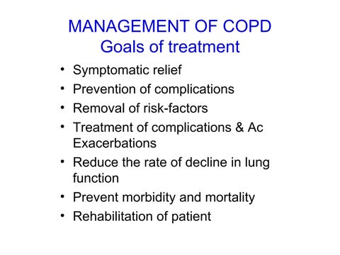
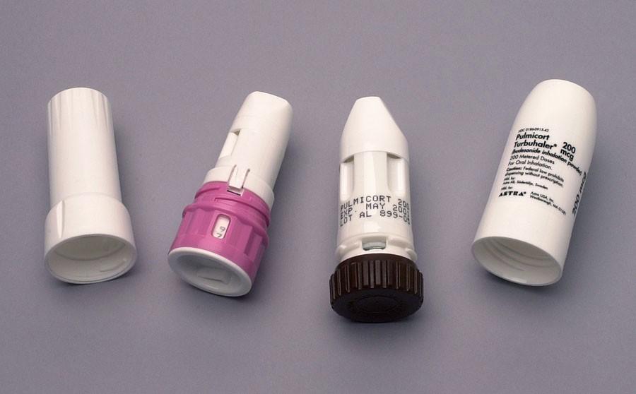
Inhalational Treatment
Preferred route for both controller and reliever therapy
Advantages: Local effect, immediate response Minimal dosage, few side effects
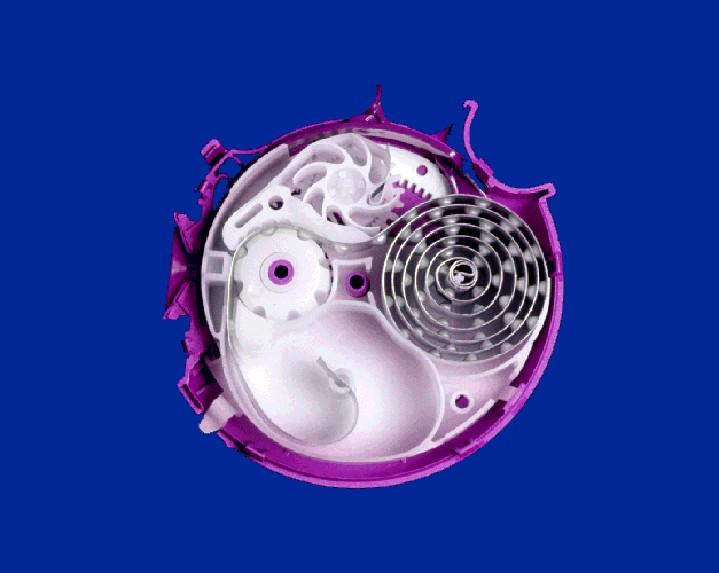
Available as : Dry powder (DPIs), Metered dose liquid inhalers MDIs);
Nebulizers
Devices: Spacers (to increase drug delivery)
Side effects of inhalation drugs
Local side effects: throat irritation, voice change, thrush (candida infection), vocal cord dysphonia
Systemic side effects of drugs: Rare may be growth retardation in young children cataracts, other steroid effects
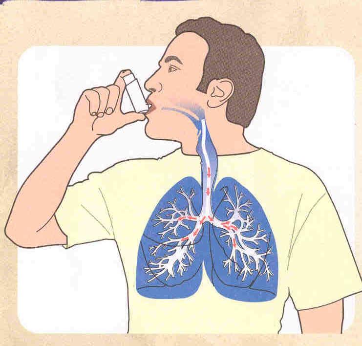
Metered Dose Inhalers
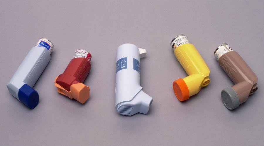
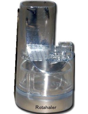
Dry powder inhalers
Anticholinergics
1. Cause effective bronchodilatation
2. Reduce rate & severity of acute exacerbations
3. Improve quality of life
4. Long acting
5. Side effects: Dryness, blurred vision, urinary retention (if BPH)
Corticosteroids
1. Oral/parenteral for acute exacerbations
2. Inhaled for moderate to severe COPD
• Improve lung function
• Reduce exacerbations
• Improve symptoms & Q.O.L.
• Reduce airway reactivity
Side effects:
• Loss of bone mineral density
• Increased skin bruising
Complications of COPD
1. Acute exacerbations
Severe airway obstruction
Acute change in baseline lung function
Marked exercise tolerance
Nocturnal hypoxemia
2.
Pulmonary hypertension and Chronic cor pulmonale
3.
Respiratory failure
Symptoms of COPD Exacerbation
• Increase in cough
• Chest pain
• Increase in breathlessness
• Increase in sputum volume and change in its colour (to green, yellow, blood streaked)
• Fever
• Increased tiredness
• Increase in oxygen requirement (for those on long-term oxygen therapy)
Management of Acute Exacerbations
1. Increase the dose and/or frequency of current bronchodilator therapy
2. Add new bronchodilators
3. Bronchodilator nebulization
4. Parenteral theophyllines
5. Systemic glucocorticoids
6. Antibiotics for infections
7. Maintenance of oxygenation
8. NIV or Assisted Ventilation for refractory respiratory failure (Hypoxaemia and/ or hypercapnia)
• Hypoxemia common in hospitalized pts.
• Small increase in FiO2 - good response
However, this can worsen hypercapnia due to:
– Release of hypoxic vasoconstriction
Increased dead-space
– Loss of hypoxic respiratory drive
• Domicilliary long term-term oxygen therapy for COPD with chronic respiratory failure
Supplemental Oxygen
Assisted Respir Supports
• Non-invasive ventilation (NIV) in case there is failure to respond to supportive therapy and controlled oxygen supplementation
Initiate as early as possible
RR > 24 and hypercapnia with acidosis (pH <7.35) are the classic indications
No benefit in milder exacerbations
• Intubation and Mechanical ventilation if NIV is contraindicated, has failed, or is not tolerated
Chronic Cor Pulmonale
Definition: Alterations in the structure and/or function of the right ventricle secondary to diseases of the lung, chest wall or lung vasculature – (which are not secondary to the diseases of the left heart or congenital heart diseases).
Manifests with features of pulmonary hypertension and right heart overload/ failure: Generalized anasarca, congested liver, ascites, cyanosis, loud P-2, cardiomegaly (rt.)
Diagnosis: H/O COPD
CXR, ECG, ECHO
Treatment of cor pulmonale
1. Long term oxygen therapy
2. Removal of fluid retention –diuretics
3. Maintenance of CO2 levels
4. Digoxin, if arterial fibrillation
5. Vasodilators - may be hazardous (Lower systemic and pulm. BP)
6. Treatment of COPD
Other complications
1. Rupture of blebs/bullae: Pneumothorax, pneumomediastinum, subcutaneous emphysema
2. Polycythemia (due to chronic hypoxemia)
3. Increased coagulation problems In situ thrombosis
Pulmonary thromboembolism
5. Hyperuricemia (and occasionally gout)
6. Systemic manifestations
Systemic manifestations of COPD
1. General Wasting, weight loss, Nutritional anomalies, anemia
2. Musculoskeletal
Skeletal muscle dysfunction, Osteoporosis
Reduced exercise tolerance, performance
3.Cardiovascular Ischemic heart disease
Cardiac failure, Stroke
4. Endocrinal Diabetes, Metabolic syndrome
Dysfunction of pituitary, thyroid, gonads and adrenals
5. Neuropsychiatric Depression
Disordered sleep
Anxiety Cognitive function decline
Long term Maintenance and Prophylaxis Treatment
• Keep off smoking
• Bronchodilators
• Inhaled corticosteroids
•
Use/avoidance of other drugs (e.g. antibiotics, mucolytics ,sedatives)
•
Prophylactic vaccination (influenza)
•
Pulmonary rehabilitation (multidisciplinary supports and management)
Pulmonary Rehabilitation
• Structured, multi-disciplinary programme tailored to one’s needs to improve quality of life, lung function and reduce breathlessness Components
• Exercise training
• Nutritional counseling
• Education on lung disease or condition and how to manage it
• Energy-conserving techniques
• Breathing strategies
• Psychological counseling and/or group support
Pulmonary Eosinophilic Disorders
Normal E counts: Differential: ≤5%, AEC: ≤0.5×10⁹/l
Eosinophilia:
•Mild: AEC 0.5-1.5×10⁹/l
•Moderate: AEC 1.5-5.0×10⁹/l
• Severe: AEC >5.0×10⁹/l
•Hyper Eosinophilic Syndrome (HES): AEC: >1.5×10⁹/l lasting for 6 months
Lack of evidence for known causes of eosinophilia
Signs and symptoms of organ
involvement/dysfunction
Pulm eosinophilic disorders: Classification
A. Primary pulmonary eosinophilia
Predominant involving lung.
Acute eosinophilic pneumonia
Chronic eosinophilic pneumonia
Systemic disease with lung disease
Churg-Strauss syndrome
Idiopathic hypereosinophilic syndrome
B. Lung disorders with associated eosinophilia
1. Interstitial lung disease, Sarcoidosis, Langerhans cell histiocytosis, Connective tissue disease
2. Asthma
3. Bronchiolitis obliterans-organizing pneumonia
4. Neoplasms:, Hematological malignancies, Solid organ tumors
C. Secondary pulmonary eosinophilia
1. Infections: Parasitic infestations
Transient passage (Löffler’s syndrome)
Ancylostoma, ascaris, strongyloides
paragonimiasis, echinococcosis
Trichinella, Visceral larva migrans
Disseminated strongyloidiasis Schistosomiasis
•Fungal infections: Coccidiomycosis, Histoplasmosis
•Other infections: Tuberculosis, Brucellosis
3. Tropical pulmonary eosinophilia
4. Allergic bronchopulmonary aspergillosis
5. Hypersensitivity pneumonia
6. Drugs, toxins and radiation
Drugs causing eosinophilic lung disease
1. Antimicrobials: Para-amino salicyclic acid, Nitrofurantoin
Penicillin, Tetracycline, Streptomycin, Isoniazid
Sulfonamide,Tetracycline, Minocycline,Dapsone + pyrimethamine
2. Antineoplastic and immunosuppressives
Bleomycin, Methotrexate, Melphalan, Gold salts
Azathioprine, Penicillamine, Beclomethasone
3. Nonsteroidal anti-inflammatory drugs (NSAIDs):
Aspirin, Naproxen, Piroxicam, Nimesulide, Phenylbutazone
4. Cardiovascular and antidiabetics:
Amiodarone, Hydralazine, Thiazides, Clofibrate, Sulfonylureas
5. Miscellaneous: Carbamazepine, Phenytoin, Dantrolene, Methylphenidate, Imipramine, Cocaine or heroin exposure
Iodinated contrast media, L-tryptophan.
Churg Strauss Syndrome
Now known as Allergic Granulomatosis with Angiitis
Include i) asthma, ii) paranasal sinusitis,
iii) monoarthropathy or polyarthropathy,
iv) migratory or transient pulmonary infiltrates,
v) peripheral blood eosinophilia greater than 10%, and
vi) extravascular eosinophils in a blood vessel on a biopsy specimen.
D/D: Wegener’s granulomatosis, polyarteritis nodosa, tuberculosis, fungal infections, allergic bronchopulmonary aspergillosis,
Tmt.: Corticosteroids and cytotoxic medications
Tropical Pulmonary Eosinophilia
• Immunological hyper-responsiveness to human filarial parasite- W. bancrofti & Brugia malayi
• Transmitted through mosquito bites
• Symptoms: Cough, breathlessness, wheeze, usually nocturnal symptoms. Systemic organ involvement.
• Diagnosis: Absolute eosinophil count – more than 3000/cmm; demonstration of parasites
• Chest X-ray: Reticulo-nodular shadow
• Elevated serum IgE and anti-falarial antibodies
• Tmt: Diethylcarbamazine (6mg/kg per day for 3 weeks)
Hypersensitivity Pneumonias
• Type 3 immunological response to sensitizing antigens (Cf. type 1 for asthma)
• Presentation delayed 4-6 hrs or more after exposure
• Symptoms: Cough, fever, breathlessness, malaise etc
• Types: Farmer’s lung, Byssinosis, Baggasosis
Psittacosis, Pigeon breeder lung, Grain lung, Air-conditioner lung, compost lung etc
Diagnosis: History of exposure-symptom relationship
CXR; Non-specific. Eosinophilia, Antibodies
Tmt: Removal of offending antigens
Symptomatic and anti-inflammatory treatment