Clinical Problem Solving in Orthodontics and Paediatric Dentistry 3rd Edition by Richard Welbury, Monty Duggal, Marie TheÌ■reÌ€se Hosey 0198565836 9780198565833
MontyS. Duggal Professor ofChildDentalHealth LeedsDentalInstitute
MarieThérèseHosey Professor ofPaediatricDentistry King’sCollegeLondon
Great Clarendon Street, Oxford OX2 6DPUnited Kingdom
Oxford University Press is a department of the University of Oxford. It furthers the University’s objective of excellence in research, scholarship, and education by publishing worldwide. Oxford is a registered trade mark of Oxford University Press in the UKand in certain other countries
The moral rights of the authors have been asserted
First Edition copyright 1997
Second Edition copyright 2001
Third Edition copyright 2005
Fourth Edition copyright 2012
Impression: 1
All rights reserved. No part of this publication may be reproduced, stored in a retrieval system, or transmitted, in any form or by any means, without the prior permission in writing of Oxford University Press, or as expressly permitted by law, by licence or under terms agreed with the appropriate reprographics rights organization. Enquiries concerning reproduction outside the scope of the above should be sent to the Rights Department, Oxford University Press, at the address above
You must not circulate this work in any other form and you must impose this same condition on any acquirer
British Library Cataloguing in Publication Data
Data available
Library of Congress Cataloging in Publication
Library of Congress Control Number: 2012939874
ISBN 978-0-19-957491-9
Printed and bound by Bell & Bain Ltd, Glasgow
Oxford University Press makes no representation, express or implied, that the drug dosages in this book are correct. Readers must therefore always check the
product information and clinical procedures with the most up-to-date published product information and data sheets provided by the manufacturers and the most recent codes of conduct and safety regulations. The authors and the publishers do not accept responsibility or legal liability for any errors in the text or for the misuse or misapplication of material in this work. Except where otherwise stated, drug dosages and recommendations are for the nonpregnant adult who is not breastfeeding.
Links to third party websites are provided by Oxford in good faith and for information only. Oxford disclaims any responsibillty for the materials contained in any third party website referenced in this work.
Professor of Special Care Dentistry University of Dublin
Professor of Paediatric Dentistry
King’s College London
Professor of Paediatric Dentistry
University of Sheffield
Department of Child Dental Health
King’s College London
Professor of Paediatric Dentistry University of Leeds
Department of Child Dental Health
University of Newcastle upon Tyne
Professor of Paediatric Dentistry
University of Glasgow
Department of Restorative Dentistry
University of Newcastle upon Tyne
Preface to the first edition
The child and adolescent deserves the best of care in all the different disciplines of dentistry. I was delighted to be given the opportunity of trying to draw together all the different aspects of paediatric dentistry and am most grateful to my colleagues for agreeing to contribute the various chapters which make up the book. We have tried to cover as much ground as possible within the obvious publishing restrictions and the finished product will inevitably reflect the editor’s perceptions of where current needs and deficiencies exist. The sudden increase in both erosive tooth surface loss and cosmetic awareness in our younger patients made their inclusion in Chapter 8 important, where previously they have not achieved such prominence in paediatric texts. Similarly, periodontal disease (Chapter 10), oral pathology and oral surgery (Chapter 14), and disability (Chapter 16) are as deserving of detailed inclusion in a ‘paediatric’ as much as in any ‘general’ text.
The book was written with undergraduate dental students in mind, but we hope it will also be useful to those engaged in postgraduate studies and to general dental practitioners. Exhaustive references are deliberately not given but suggested ‘further reading’ lists are included to help expedite Further enquiry and learning.
I hope we have shown in Paediatric dentistry that the early years of life are the time to get it right for the child and adolescent and there is no reason why our young patients should be denied correct and appropriate care.
R. R. W. Newcastle upon Tyne April 1996
Preface to the second edition
I am delighted to be given the opportunity to edit the second edition of this popular textbook and am grateful to my colleagues for their continuing contribution. The reviews of the first edition indentified the need for a chapter on the treatment of caries in the preschool child and I am grateful to Stephen Fayle for undertaking this task.
There have been small modifications and updates to most chapters, which should keep the reader abreast with current theory and practice. Greater use has also been made of ‘Key points’ for revision purposes.
I hope the second edition will continue to help both undergraduates, postgraduates, and general dental practitioners in their practice of paediatric dentistry.
R. R. W.
Newcastle upon Tyne
January 2001
Preface to the third edition
I was very pleased when my younger colleagues Marie Thérèse Hosey and Monty Duggal accepted my offer to join me in editing this third edition. Our book has now sold four and a half thousand copies since its launch in 1997 and it is essential that we maintain a contemporary outlook and publish changes in techniques and philosophies as soon as they have an evidence base.
Since 2001 and the second edition, there have been a significant number of changes of authorship, as well as a change of chapters for some existing authors.
Gerry Winter died in December 2002. He was a wise colleague and friend who was a mentor to many of us. I continue to miss his expertise and availability for consultation, by post or telephone, which he freely gave even after his retirement.
John Murray, Andrew Rugg-Gunn, and Linda Shaw have now retired from clinical practice. I am indebted to them all for their support, both in my own personal career and in the production of out textbook. I am grateful to them for allowing the new chapter authors to use their texts and figures.
The restorative section of the book has been remodelled. The endodontics chapter in the previous editions has now been incorporated into either chapters 8 or 12, and there are separate chapters relating to the operative care of the primary and the permanent dentitions. Without the help and friendship of Jim Page the original ‘Operative care of dental caries’ chapter would not have been possible. I am grateful to Jim for allowing us to continue to use his original illustrations from that chapter.
Although designed for the undergraduate we hope the new edition will continue to be used by undergraduate, postgraduate, and general dental practitioner alike, and that their practice of paediatric dentistry will be both fulfilling and enjoyable.
R. W
Glasgow January 2005
R.
Preface to thefourth edition
It is difficult to believe that 17 years have passed since work began on the first edition. Our Portuguese edition was marketed in 2007 and this has broadened our market significantly in Brazil.
This edition contains some new contributors, Liege Lourenço-Matharu, Lucy Burbridge, Jenny Harris, and Toby Gillgrass, and their enthusiasm and insight have been invaluable. We are grateful to the comments from reviewers and OUPstaff which have resulted in revision of a number of chapters, and so we hope that the end product will be as well received as previous editions.
The excitement of a new edition is tinged with sadness at the loss of Nigel Carter at such a young age. Nigel always gave very generously of his time to paediatric colleagues. On a happier note, Peter Gordon is now enjoying a wellearned retirement and we would all like to thank him for his contribution to the three previous editions.
In looking at the author list for the fourth edition we are struck by the number of contributors who have gained promotion to professorial positions since they originally contributed to Paediatric Dentistry. We must be doing something right!
Fig. 11.7, Reproduced from Heasman, P.A., Factitious Ginival Ulceration: A Manifestation of Munchausen’s Syndrome, Journal of Periodontology, 1994. With permission of the American Academy of Periodontology;
Fig. 11.11, reproduced by kind permission of Mr N.E. Carter;
Figs 11.12, 11.18, 11.19, reproduced by kind permission of Professor I.L. Chapple, Birmingham, UK;
Fig. 11.13, reproduced by kind permission of Mr D.G. Smith, Consultant in Restorative Dentistry, Newcastle upon Tyne;
Figs 12.55, 12.56, reproduced from Andreasen and Andreasen, Textbook and colour atlas of traumatic injuries to the teeth (3rd edition) with permission from John Wiley & Sons;
Fig. 14.1, R. Evans and W. Shaw (1987). Reproduced with kind permission of the Editor of the European Journal of Orthodontics; Figs 14.7, 14.35, courtesy of Mr T.G. Bennett;
Fig. 14.10, courtesy of Professor J.H. Nunn; Fig. 14.40, courtesy of Mr I.B. Buchanan;
Figs 15.4, 15.6, 15.12, reproduced by kind permission of Informa Healthcare;
Fig. 15.7, by kind permission of the Journal of Dentistry for Children;
Fig. 15.13, by kind permission of Professor C. Scully;
Fig. 15.27, image kindly supplied by Straumann Ltd;
Figs 16.5, 16.6, 16.8, courtesy of Dr Linda Shaw;
Figs 16.10, 16.11, 16.12, courtesy of Dr Alex Keightley;
Fig. 16.13 courtesy of Dr Triona Fahey;
Fig. 17.21, courtesy of Shine, www.shinecharity.org.uk;
Fig. 17.22, courtesy of HSEDental Services, Dun Laoghaire, Ireland;
Fig. 17.23, courtesy of Dr Bitte Ahlborg, Mun-H-Center, Sweden;
Fig. 18.2 data used with permission of Department for Children, Schools and Families 2010;
Figs 18.3, 18.4, 18.7, by kind permission of COPDEND;
This chapter describes, in general terms, the prenatal development and postnatal growth of the craniofacial skeleton, and the occlusal development of the primary and permanent dentitions.
1.2 Prenatal development
Understanding of embryological development is essential for the dental practitioner who may frequently face patients with the common craniofacial anomalies such as cleft lip and/or palate. For routine care, an understanding of their development and aetiology will bring insight to their likely presenting signs and symptoms.
This section will include a brief summary of the development of the face including the neural crest and pharyngeal arches. It is not the intention of this summary to be in any way a complete or thorough description but simply to describe some of the key cells/interactions and structures.
1.2.1Neuralcrest
Neural crest cells are derived from the neural fold, and are highly migratory and specialized cells capable of predetermined differentiation. The differentiation occurs after their migration and is essential for the normal development of face and teeth (Fig. 1.1).
1.2.2Branchialarches
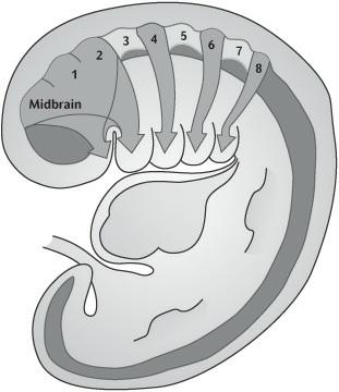
By week 4 the primitive mouth or stomatodeum is bordered laterally and from the developing heart inferiorly by the pharyngeal or branchial arches (Fig. 1.2). These are six bilateral cylindrical thickenings (although the fifth and sixth are small) which form in the pharyngeal wall and into which the neural crest cells migrate. They are separated externally by the branchial grooves and internally by the pharyngeal pouches. The first groove and pouches are involved in the formation of the auditory apparatus and the Eustachian tube.
Each arch has a derived cartilage rod, muscular, nervous, and vascular component. The first two arches and their associated components are central to the development of the facial structures.
This period is also characterized by the development of the organs for hearing, sight, and smell, namely the otic, optic, and nasal placodes.
1.2.3Facialdevelopment
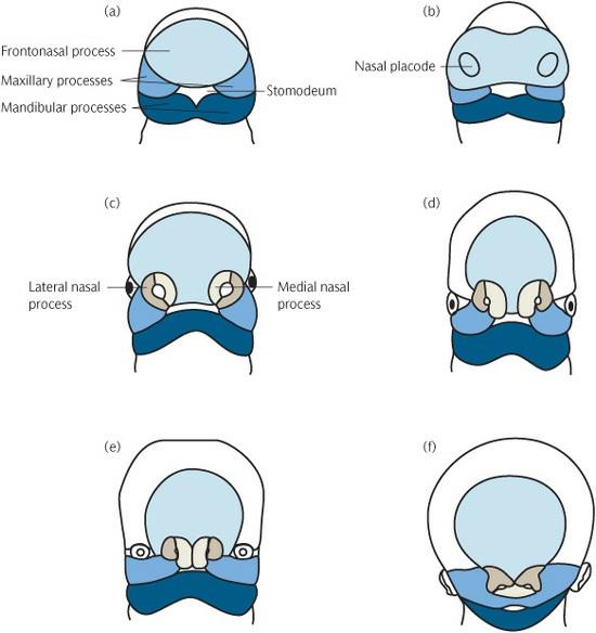
By the end of week 4, thickenings start to develop in the frontal process. The medial and lateral frontonasal processes develop from these, together with the nasal placodes.
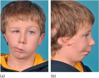
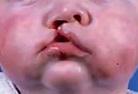
Figure1.1A child with the hemifacial microsomia part of the oculo-auricularvertebral spectrum. The unilateral inhibition of neural crest migration and bronchial arch development results in (a) marked asymmetry and (b) ear defects.
The maxillary process develops from the first pharyngeal arch and grows forward to meet the medial and nasal processes, from which it is separated by distinct grooves at week 7 (Fig. 1.3). Its eventual fusion with them creates the upper lip and, from the two medial nasal processes, the incisor teeth and the primary palate. Where this fusion is disturbed a cleft of the lip may form (Fig. 1.4).
The lower lip is formed by fusion of the mandibular process from the first arch.
By week 8 the odontogenic epithelium, which will differentiate into toothforming cells, can be determined on the inferior border of the maxillary process, the lateral aspect of the medial process, and the superior border of the mandibular processes.
1.2.4Secondarypalate
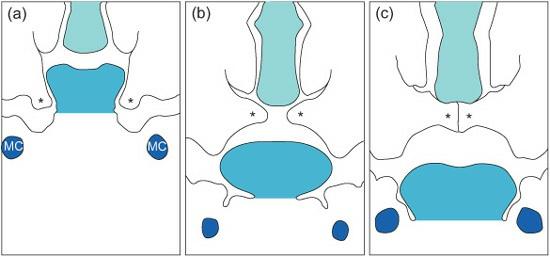
Development of the secondary palate starts around week 7. It is formed from
three processes: the nasal septum develops in the midline from the frontonasal process and the two palatine shelves develop from the maxillary processes. At this stage the palatal shelves are directed downwards on either side of the tongue. Between weeks 7 and 8 they elevate to meet the primary palate and nasal septum, to which they fuse (Fig. 1.5).
The trigger for this elevation is still unclear, although high concentrations of glycosaminoglycans which attract water and increase turgidity in the shelves, contractile fibroblasts, and the position of the tongue have all been implicated. Once in contact, the epithelial covering of the shelves must disappear to allow the fusion. Various methods including cell death (‘apoptosis’) and cell transformation have been suggested as methods by which this epithelial covering is lost.
Figure1.3Diagrammatic representation of early facial development from 4 to 10 weeks i.u. (a) 4th week i.u. (b) 28 days i.u. (c) 32 days i.u. (d) 35 days i.u. (e) 48 days i.u. (f) 10 weeks i.u. (Reproduced from Mitchell, An Introduction to Orthodontics, 2007, with permission of Oxford University Press.)
Figure1.4Failure of fusion resulting in cleft lip and primary palate. If the shelve fusion fails, this is likely to result in clefts of the palate. The extent of these clefts varies clinically from submucous clefts affecting the bony structure of the palate and underlying muscular attachment and clefts of the
soft palate which may or may not have significant effects on speech to those including the hard palate producing communication between the nasal and oral cavities (Fig. 1.6).
There appear to be distinctive differences between clefts of the palate and those of the lip and palate within different geographical and sexual distributions. This also suggests different disruptive mechanisms and timings as lip closure occurs earlier in development than palatal fusion. However, as clefts of the palate alone and lip with palate can occur in certain families, it suggests the distinction may not be complete.
Figure1.5Diagrammatic representation of palatal shelf elevation and subsequent fusion. (a) During week 7 in utero the palatal shelves begin to develop and lie on either side of the tongue. (b) During week 8 in utero the palatine shelves elevate rapidly owing to the internal shelf-elevating force and developmental changes in the face. (c) During week 9 in utero the shelves fuse with each other, the primary palate, and the nasal septum. MC, Meckel’s cartilage; asterisks, palatal shelves. (Reproduced from Mitchell, An Introduction to Orthodontics, 2007, with permission of Oxford University Press.)
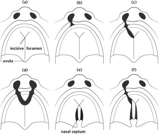
Figure1.6Diagrammatic representations of some of the different types of clefts of the lip and palate: (a) Normal; (b) unilateral cleft lip; (c) unilateral cleft lip and anterior palate; (d) bilateral cleft lip and anterior alveolus; (e) cleft of posterior palate (hard and soft); (f) unilateral cleft of the lip and anterior and posterior palate. (Reproduced from Johnson and Moore, Anatomy for Dental Students, 1997, with permission of Oxford University Press.)
1.3 Postnatal craniofacial growth
There is a great deal of individual variation in the process of postnatal growth and in the final form of the craniofacial structures. This section presents a simplified and rather idealized account of bone growth in general and as part of craniofacial growth. Occlusal development is then described, before going on to discuss the effect of individual variation in producing departures from this idealized pattern.
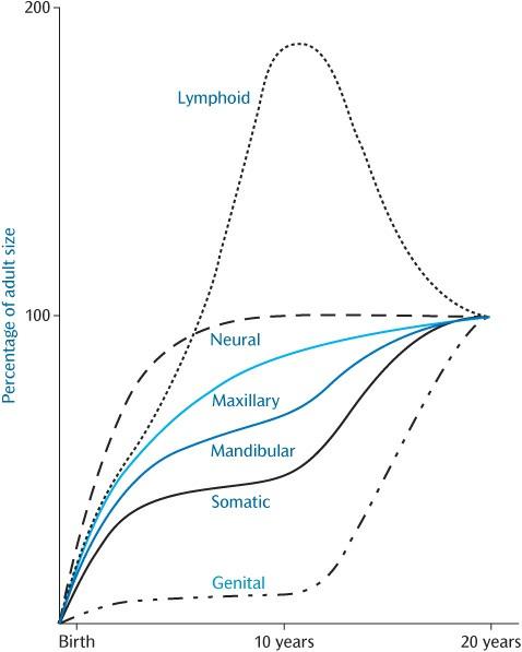
An individual’s stature can be charted on standard growth charts during growth. This will present an overall view of the process of growth and will help to detect instances where growth is not proceeding in the usual manner. However, it disguises the fact that the various tissues of the body grow at different rates at different ages (Fig. 1.7).
In order to maintain harmonious facial growth, bone growth must synchronize with that of other tissues. For example, growth of the calvarium is linked to growth of the brain. The cranial vault initially grows much more rapidly than the facial bones in order to keep pace with the developing brain, 90%of which is complete by 5 years of age.
Figure1.7Postnatal growth patterns for neural, lymphoid, somatic, and genital tissues shown as percentages of the total increase. The patterns for the maxilla and mandible are shown in blue. (This was originally redrawn from Proffit, W.R. (2000). Contemporary Orthodontics, 3rd edn, Mosby.)
1.3.1Assessmentofpostnatalcraniofacialgrowth
One way of assessing the changes that take place during craniofacial growth is to superimpose tracings of two lateral skull radiographs taken of the same
person at different ages. The two radiographs can be compared, as shown in Fig. 1.8, and the changes that have taken place during growth can be examined. A potential difficulty with this approach is that the various bones of the skull grow at different rates at different ages, and there is no single central point about which growth occurs in a radial fashion, i.e. there is no valid fixed radiographic landmark on which to superimpose the films. One convention is to superimpose the tracings of the radiographs on the outline of the sella turcica, using the line from the sella to the frontonasal suture to orientate the films. If this method of superimposition is used, it appears that the cranium expands in a more or less radial fashion to accommodate the brain and the facial skeleton then grows downwards and forwards, away from the cranial base.
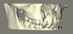
Another difficulty is that radiographs only produce a two-dimensional representation of what is a three-dimensional structure. Newer methods of radiological assessment using computed tomography or lower-radiation-dose cone-beam computed tomography (Fig. 1.9) are capable of producing threedimensional volumetric images of the facial skeleton.
Soft tissue growth and facial changes are also important for understanding the effects of underlying bony changes. Although computed tomography will capture the soft tissue, it does not accurately depict the colour and texture and, particularly in the case of conventional computed tomography, produces a significantly higher radiation dose than plain radiographs.
Figure1.8Superimpositions on the cranial base showing overall downward and forward direction of facial growth: solid line, 8 years of age; broken line, 18 years of age. (Reproduced from Mitchell, An Introduction to Orthodontics, 2007, with permission of Oxford University Press.)
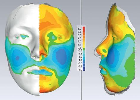
Non-invasive laser scanning or stereophotogrammetry is capable of producing photo-realistic topography of the facial soft tissues without exposure to radiation. Sequential capture is being used for longitudinal growth studies where colour mapping can help illustrate areas of maximum growth (Fig. 1.10) over a substantial time period.
1.3.2Bonegrowth
Mineralized bone is formed through a process known as ‘ossification’. This occurs in two ways, intramembranousossificationand endochondral ossification. Intramembranous ossification occurs by membrane activity and is seen in the bones of the calvarium, the facial bones, and the mandible. Endochondralossificationoccurs by replacement of a cartilage framework. Classically, endochondral ossification is described in long bones but it also occurs in the craniofacial region, most notably in the cranial base.
Figure1.9Cone-beam computed tomography of a child with a cleft of the alveolus, manipulated to produce a three-dimensional image.
Growth in bones formed by endochondral ossification occurs at growth centres known as epiphyseal plates in long bones and synchondroses in the cranial base. These are primary growth centres within which the chondroblasts are aligned and clear zones of cell division, hypertrophy, and calcification occur. The most notable of the three synchondroses within the cranial base is the spheno-occipital synchondrosis. The condylar cartilage has a different histological appearance to that of the epiphyseal plates and synchondroses. Although capable of producing bone, its stimuli appear more reactionary to growth around it rather than the primary growth sites which react to both internal and external stimuli.
The apparent growth of the facial bones is a function of remodellingand displacementor translation. Remodelling results in an alteration in the size and shape of bones by deposition and resorption of material on the external and internal surfaces of the bone and suture systems. It is a function of the ‘periosteum’ or ‘osteogenic membrane’. Deposition and resorption go hand in hand; one seldom occurs without the other. Deposition of bone on one aspect of a cortical plate of bone is accompanied by resorption on the other aspect. Displacement or translation occurs when one bone is moved relative to another, primarily due to another area of growth; for example, the maxilla is translated downwards and forwards by growth of the spheno-occipital synchondroses and nasal septum. Such translation will be accompanied by a degree of remodelling.
The suture systems form bone when subjected to traction. In the case of the calvarial bones, the suture systems form new bone and enable the bones to stay in contact with each other when the expansion of the growing brain would otherwise move them apart.
Figure1.10Colour mapping after sequential laser scanning showing facial growth in a forward direction (red) and a negative direction (blue) in the AP plane. (Courtesy of Professor Steve Richmond.)
The suture systems allow the bones to respond to growth in neighbouring soft tissue. The suture systems lying between the maxilla and the cranial base allow the downward and forward translation of the maxilla in response to the growth of the soft tissues of the face. It is not proliferation of the vascular connective tissue in the sutures that pushes the bones apart; the whole arrangement of the connective tissue in a suture seems to be designed to enable the suture to respond to a tensile force.
1.3.3Softtissuegrowth
The effects of bony growth can be masked or accentuated by the overlying soft tissues. Notably, this is shown intra-orally in the positions of the dental arches in the so-called neutral zone between the effects of the tongue, lips, and cheeks. The soft tissues are also responsible for dento-alveolar compensation where the position of the teeth attempts to compensate for skeletal jaw discrepancies.
The growth of the soft tissues, particularly the nose and the length and thickness of the lips, has a profound effect on the appearance of the face. Soft tissue growth shows sexual dimorphism, with changes occurring later and for longer in boys. Changes in the the nose continue into adulthood.
1.3.4Mechanismsofgrowth
The mechanisms controlling the process of facial growth are not completely understood. In the post-genomic era it is becoming apparent that genetically encoded factors have a major effect on craniofacial growth; after all, children tend to resemble their parents in facial appearance. This may be particularly noted in class III parents and those with a class II division 2 malocclusion. The alternative school of thought is that the growth is only loosely under genetic control; rather, the final shape is under the control of its soft tissue environment. This is known as the ‘functional matrix theory’. This is best shown in the cranial vault where the bone growth is reactionary to neural expansion. However, it fails to explain mid-facial growth through the synchondroses.
Therefore it is likely that both come into play, The genetically encoded factors can be affected by factors outwith the DNA ‘epigenetic’ that are able to switch them off or on. If this is the case, it should be theoretically possible to influence them (e.g. the use of functional appliances to encourage mandibular growth). At present, however, although a positive response is possible, it appears extremely variable and unpredictable between indviduals.
1.3.5Cranialgrowth
At birth, the cranium is some 60–65%of its adult longitudinal dimensions, and this increases to about 90%by the age of 5 years. The calvarial bones are carried away from each other by the expanding brain and respond by forming new bone in the sutures that separate the bones of the vault of the skull (Fig. 1.11). The six fontanelles that are present at birth reduce in size. The largest (the anterior fontanelle) closes at about 1 year of age and the last to close (the posterolateral fontanelle) closes at about 18 months. The calvarial bones undergo a process of remodelling, with areas of bone deposition and resorption altering the contour of the bones as the volume of the brain cavity increases.
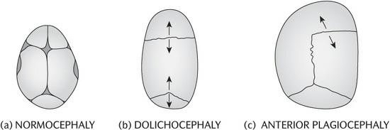
Early fusion of the cranial sutures, or ‘craniostenosis’, results in compensatory growth from the other sutures. This can result in unusual head shapes, and may produce detrimental effects on brain growth and development as it may be accompanied by increased intracranial pressure. The most common craniostenosis involves the sagittal suture. Compensatory growth results in a head shape that is increased in anteroposterior direction and narrow laterally ‘dolichocephaly’. If the suture fusion is asymmetric, the deformation is also asymmetric ‘plagiocephaly’ (Fig. 1.12).
The cranial base also grows to accommodate the changes in the size and shape of the brain, but the process is different to that seen in the calvarial
bones. There is considerable lateral growth of the cranial base as the cerebral hemispheres expand, but less increase in the anteroposterior dimension. No sutures are present to allow for expansion of the deeper compartments of the cranial base, a process that takes place by surface deposition and extensive remodelling. In addition, the three synchondroses, (spheno-occipital, intersphenoid, and spheno-ethmoidal) in the mid-ventral floor of the cranial base allow for increases in the anteroposterior dimension by endochondral ossification. Growth in the spheno-occipital synchondrosis does not cease until about the age of 15 years in boys, rather earlier in girls, and it closes fully at about the age of 20. The spheno-occipital synchondrosis has a significant influence on the growth of the facial region as the condylar fossa is posterior to it, but the anterior cranial base, and therefore the nasomaxillary complex to which it is attached by a suture system, sits anteriorly. As a consequence, as it grows it has an effect on how the maxilla and mandible relate to each other (Fig. 1.13).
The pattern and the timing of growth in the cranial base is intermediate between the neural type of growth that characterizes the growth of the calvarial bones and the musculoskeletal pattern of growth exhibited by the facial skeleton.
The shape of the cranial fossae is much more complex than the relatively smooth form of the bones of the vault of the skull. Surface deposition and subsequent remodelling occurs, with the final size and shape of the compartments being determined by the size of the lobes of the brain forming the partitions which separate the cranial fossae.
1.3.6Nasomaxillarygrowth
The nasomaxillary region, which makes up the middle third of the face, is a complex area comprising a number of bones joined to each other and to the anterior cranial base by a suture system. The nasomaxillary complex grows downwards and forwards relative to the cranial base. This is accomplished through cranial base growth and deposition within the suture system as it is carried forward; there is also deposition in the region of the tuberosity, lengthening the alveolus in this region. The anterior surface is remodelled not by apposition, as might be expected in a bone that is growing forward, but by resorption.
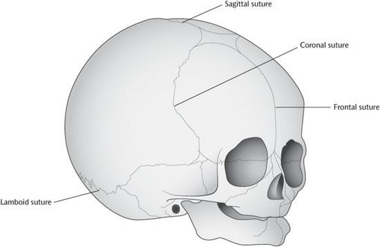
Figure1.11The skull at birth showing the sagittal, coronal, frontal, and lamboid sutures. (Reproduced from Johnson and Moore, Anatomy for Dental Students, 1997, with permission of Oxford University Press.)
Figure1.12Skull morphology: (a) normal; (b), (c) abnormal due to early fusion of cranial sutures.
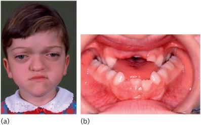
Failure of the cranial base to lengthen, as seen in achondroplasia and a number of other syndromes (Fig. 1.13), results in characteristic faces with lack of mid-face prominence.
Unlike the growth of the cranium, which occurs in conjunction with the growth of the brain, the nasomaxillary complex grows fastest at about the time of the pubertal growth spurt, in conjunction with the general growth of the musculoskeletal system.
As bone is deposited on the external aspect of the maxilla in the region of
the tuberosity, and vertically with the development of the alveolus through tooth eruption, it is also resorbed from the internal aspect of the bone in this area, thereby enlarging the maxillary sinus. As the bone is translated downwards, the nasal cavities and the maxillary sinus expand by a process of bone resorption at the floor of the nose and the sinus, together with bone deposition on the palatal aspect of the maxilla.
1.3.7Mandibular growth
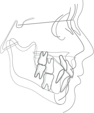
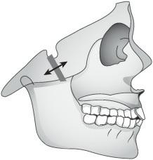
Growth of the mandible, like that of the maxilla, is coordinated with the pattern of general musculoskeletal growth, growing at its fastest rate at about the time of the pubertal growth spurt. Growth of the mandible has to be coordinated with the downward and forward growth of the maxilla. This task is made more complicated by the fact that the mandibular condyles articulate in the glenoid fossa, which lies behind the spheno-occipital synchondrosis, while the maxilla lies in front of it; therefore, growth of the mandible has to keep pace not just with the translation of the maxilla, but also with growth in the cranial base (see Fig. 1.14).
Figure1.13(a) A facial photograph of a child with Crouzon’s syndrome with typical lack of mid-facial growth resulting in (b) a significant class III incisor relationship. Coronal synostosis results in a short wide head (brachycephaly), and early fusion of the sutures surrounding the eye sockets results in shallow orbits and bulging eyes.
Figure1.14Anteroposterior growth at the spheno-occipital synchondrosis affects the anteroposterior relationship of the jaws. (Reproduced from Mitchell, An Introduction to Orthodontics, 2007, with permission of Oxford University Press.)
Taking the anterior cranial fossa as a stable reference area, it appears that the mandible, like the maxilla, grows downwards and forwards. As is the case with the maxilla, this downward and forward growth is not achieved by deposition of bone on the anterior aspect of the mandible, but by translation of the bone, accompanied by growth in the region of the ramus and the mandibular condyle. Bone is deposited on the posterior aspects of the ramus and the coronoid processes and resorbed from the anterior aspect of the ramus. At the same time the condylar cartilage contributes to growth of the mandibular condyle, although its growth appears more reactionary than a primary growth cartilage. That is, it is not proliferation of the condylar cartilage that pushes the mandible downwards and forwards, but the condyle essentially ‘fills in’ as the mandible is translated.
As the ramus of the mandible grows upwards and backwards, its anterior aspect undergoes resorption and becomes remodelled into the body of the mandible. This process involves resorption on the lateral aspect of the bone and deposition on the lingual aspect, which forms new bone in correct alignment with the body of the mandible and helps maintain an appropriate intercondylar width. Growth of the mandibular ramus and condyle has to keep pace with changes in the position of the maxilla, in both vertical and horizontal directions, and with growth in the middle cranial fossa. Until puberty the mandible will grow at approximately 1–2mm per year, but after puberty this may double. It is easy for a small discrepancy to arise, for example, in the amount of vertical growth of the mandibular ramus, resulting in a rotation of the body of the mandible and a corresponding tilt of the occlusal plane. These
rotations have been demonstrated using implant studies. The rotations may be partially masked by resorption but are capable of a significant effect on the vertical dimension of the face and on skeletal relationships.
1.3.8Normalvariation
There is always variation between individuals. Variation in the pattern of facial growth is only to be expected, and there are a number of compensatory mechanisms which operate to minimize the impact of such variation. Variation in the position or size of one structure is often compensated by corresponding change in another. The process of growth is constantly creating imbalances, as related structures grow and develop at different rates, but the overall direction of growth is towards some position of overall balance or harmony.
Anteroposterior discrepancies can arise during facial growth because of the position of a bone, or an imbalance in the sizes of bones, or a mixture of both. A class II skeletal pattern can be caused by insufficient growth of the ramus of the mandible in a backward direction; alternatively, a class II skeletal pattern can be the result of a backward tilt of the middle cranial fossa. This change in angulation results in the maxilla having a more anterior position, relative to the glenoid fossa, than would otherwise have been the case. The normal-sized mandible, occluding in the glenoid fossa, now has a class II relationship with the normal-sized maxilla. In a similar way, a converse alteration in the pattern of growth excessive backward growth of the ramus of the mandible or a more vertical tilt of the middle cranial fossa can produce a class III skeletal relationship, with the accompanying dental malocclusion.
Vertical growth in the nasomaxillary region has to be combined with vertical growth of the mandibular ramus. Maxillary growth that is not matched by mandibular growth will result in mandibular rotation. If there is an excess of vertical maxillary growth that is not matched by vertical growth of the ramus, the effect will be to produce a downward and backward rotation of the mandible. This downward and backward rotation will, in turn, produce an anteroposterior discrepancy, with a tendency towards a class II relationship.
Horizontal and vertical discrepancies tend to be accompanied by dentoalveolar compensations. In the case of a class III skeletal pattern, the upper incisor teeth are frequently proclined and the lower incisor teeth retroclined, as illustrated in Fig. 1.15.
These compensations are almost certainly brought about by muscular activity in the soft tissue integument affecting the position of the teeth, and they minimize what might otherwise have been a large reversed incisor overjet. In the case of class II skeletal patterns, the dento-alveolar
compensation can take two forms. If the lips function in front of the upper incisor teeth, these teeth are generally retroclined, with the effect that the incisor overjet is virtually normal. However, if the lower lip functions behind the upper incisors, these teeth are usually proclined and the lower incisors are retroclined to make way for the lip. This results in an increased incisor overjet (Fig. 1.16).
Downward and backward mandibular rotations tend to be accompanied by a vertical drifting of the premolar, canine, and incisor teeth to compensate for an arrangement that would otherwise have produced an anterior open bite. This vertical drifting should be distinguished from over-eruption, which would produce a lengthening of the clinical crowns of the teeth.
1.3.9Modificationofthepatternoffacialgrowth
Attempts to modify the pattern of facial growth have met with a certain amount of success. Orthodontic appliances have been designed which hold the mandible postured downwards and forwards. This moves the condyle out of the glenoid fossa and encourages upward and backward growth of the mandibular ramus. At the same time, the stretched muscles of mastication exert an upward and backward force (through the appliance) to the maxilla, which tends to inhibit its downward and forward growth. The effect on the growing face is to help correct a developing class II skeletal pattern. If this pattern is being caused not so much by underdevelopment of the mandible (which seems to be the most common cause) but by excessive forward growth of the maxilla, an appliance may be used simply to exert an upward and backward force on the maxilla without involving the mandible, thus helping to influence the course of facial growth.
These appliances apply forces to the developing maxilla and mandible via the teeth the appliances are attached to the teeth and work partly by inducing a dento-alveolar compensation for the underlying skeletal discrepancy. This process is sometimes referred to as ‘orthodontic camouflage’. There is likely to be some restraint of the downward and forward growth of the maxilla, but the appliances seem to have only a minimal effect on the eventual size of the mandible. The so-called myofunctional appliances, which derive their impetus from the muscles of mastication, are used most often to help correct class II malocclusions. The appliances work by maintaining a forward and downward posturing of the mandible. However, while it is possible to use myofunctional–functional appliances to correct a developing class III malocclusion, they are less often used in this context as it is difficult to obtain the necessary backward posturing of the mandible. In